Embed Size (px)
Citation preview
J Nippon Med Sch 2016; 83 (3) 133
―Case Reports―
Pancreatic Metastasis from Gastrointestinal Stromal Tumor of the Stomach:
A Case Report
Nobutoshi Hagiwara1, Takeshi Matsutani1, Tsutomu Nomura1, Itsuo Fujita1,
Yoshikazu Kanazawa1, Junji Ueda1, Hiroki Arai1, Daisuke Kakinuma1,
Hitoshi Kanno1, Zenya Naito2 and Eiji Uchida1
1Department of Gastrointestinal and Hepato-Biliary-Pancreatic Surgery, Nippon Medical School, Graduate School of Medicine, Tokyo, Japan2Department of Pathology and Integrative Oncological Pathology, Nippon Medical School, Graduate School of Medicine, Tokyo, Japan
We report the first documented case of pancreatic metastasis from a gastrointestinal stromal tumor of
the stomach. A 42-year-old Japanese man presented with severe abdominal discomfort. Computed to-
mography of the abdomen showed a huge heterogeneous mass consisting of cystic and solid compo-
nents in the left upper abdomen. 18F-Fluorodeoxyglucose positron-emission tomography revealed high
tracer uptake in the abdominal mass. After total gastrectomy with lymphnodectomy was performed, a
hard mass was palpated in the pancreatic tail. The pancreatic tumor was also resected under the thera-
peutic strategy. Histological examinations of the resected gastric and pancreatic specimens revealed that
both tumors consisted of uniform spindle cells with a fascicular growth pattern and were immunohisto-
chemically positive for CD34 and CD117/KIT. Gene sequencing analysis of DNA from each tumor re-
vealed an identical deletion of 21 nucleotides in exon 11 of the gene KIT. On the basis of these results,
we concluded that the pancreatic tumor was a metastatic tumor from the gastrointestinal stromal tumor
of the stomach. (J Nippon Med Sch 2016; 83: 133―138)
Key words: gastrointestinal stromal tumor, stomach, pancreatic metastasis, gene sequencing analysis
Introduction
Gastrointestinal stromal tumors (GISTs) are the most
common mesenchymal tumors of the gastrointestinal
tract but are rarely malignant tumors of the digestive
tract, accounting for 0.1% to 3.0% of all gastrointestinal
neoplasms1―5. Gastric GISTs metastisize most often to the
liver and peritoneum but rarely to the pancreas, but a
solitary pancreatic metastasis is extremely difficult to dif-
ferentiate from a primary pancreatic GIST. To our knowl-
edge, surgical resection of a solitary pancreatic metastasis
from a GIST has not previously been reported. We de-
scribe the first documented case of pancreatic metastasis
from a GIST of the stomach.
Report of a Case
A 42-year-old Japanese man was admitted to our hospital
because of severe abdominal discomfort. His medical and
family histories were unremarkable. On physical exami-
nation, a huge, elastic, hard mass was palpable in the left
hypochondriac region. The results of laboratory tests
were within normal ranges.
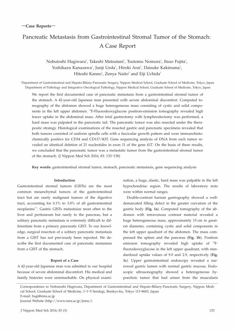
Double-contrast barium gastrography showed a well-
demarcated filling defect in the greater curvature of the
gastric body (Fig. 1a). Computed tomography of the ab-
domen with intravenous contrast material revealed a
huge heterogeneous mass, approximately 15 cm in great-
est diameter, containing cystic and solid components in
the left upper quadrant of the abdomen. The mass com-
pressed the spleen and the pancreas (Fig. 1b). Positron
emission tomography revealed high uptake of 18F-
fluorodeoxyglucose in the left upper quadrant, with stan-
dardized uptake values of 9.0 and 2.9, respectively (Fig.
1c). Upper gastrointestinal endoscopy revealed a nar-
rowed gastric lumen with normal gastric mucosa. Endo-
scopic ultrasonography showed a heterogeneous hy-
poechoic tumor that had arisen from the muscularis
Correspondence to Nobutoshi Hagiwara, Department of Gastrointestinal and Hepato-Biliary-Pancreatic Surgery, Nippon Medi-
cal School, Graduate School of Medicine, 1―1―5 Sendagi, Bunkyo-ku, Tokyo 113―8603, Japan
E-mail: [email protected]
Journal Website (http://www.nms.ac.jp/jnms/)
N. Hagiwara, et al
134 J Nippon Med Sch 2016; 83 (3)
Fig. 1 Gastrography showed a large filling defect on the posterior wall of the gastric body (black
arrows) (a). A preoperative, postcontrast, computed tomographic scan of the abdomen
shows a large heterogeneous lesion occupying most of the left upper quadrant of the ab-
dominal cavity (b). An 18F-fluorodeoxyglucose-positron emission tomographic image
shows considerable 18F-fluorodeoxyglucose accumulation in the large heterogeneous le-
sion at the same level as shown by the computed tomographic scan (c).
Fig. 2 Macroscopic findings of the resected specimen. A
huge submucosal tumor developed on the posteri-
or wall of the stomach as an extrinsic tumor. A sol-
id tumor arising in the pancreatic tail was unex-
posed on the surface of the pancreas.
propria in the body of the stomach. We strongly sus-
pected that the tumor was a primary GIST of the stom-
ach.
To eliminate the severe symptoms of abdominal full-
ness, tumor resection was indicated as an emergency on-
cological treatment. On intraoperative abdominal explo-
ration, the tumor was found to have arisen from the pos-
terior wall of the stomach, with no apparent invasion of
adjacent organs. Total gastrectomy with lymphnodec-
tomy was performed without tumor rupture.
After the huge gastric tumor was removed, a hard,
solid mass was palpated in the tail of the pancreas and
suspected to be a malignant tumor. Distal pancreatec-
tomy with splenectomy was therefore performed. Macro-
scopic examination of the resected specimen showed a
large, irregular gastric mass, measuring 19 × 23 cm and
extending mainly from the greater curvature to the poste-
rior gastric wall of the stomach (Fig. 2). The surface of
the pancreatic tumor was covered by grossly normal
pancreatic parenchyma.
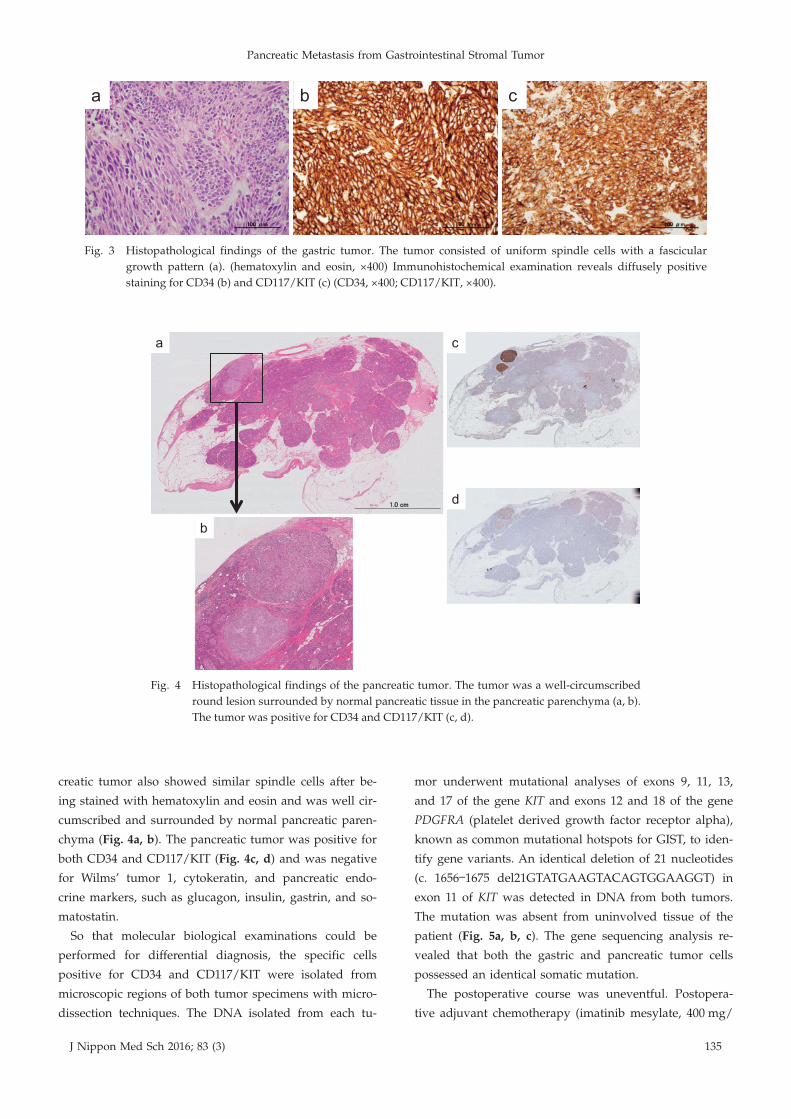
Pathological examination of the resected specimens
stained with hematoxylin and eosin revealed that the
gastric tumor consisted of uniform spindle cells with a
fascicular growth pattern (Fig. 3a). The gastric tumor had
a high mitotic index (greater than 5 mitoses per 50 high-
power fields) and a high proliferation index (Ki-67/MIB-
1 > 10%). Immunohistochemical examinations revealed
that the tumor cells were positive for CD34 and CD117/
KIT (Fig. 3b, c) and negative for α-smooth muscle actin
and S-100 protein. These pathological features were con-
sistent with those of the GIST of the stomach. The pan-
Pancreatic Metastasis from Gastrointestinal Stromal Tumor
J Nippon Med Sch 2016; 83 (3) 135
Fig. 3 Histopathological findings of the gastric tumor. The tumor consisted of uniform spindle cells with a fascicular
growth pattern (a). (hematoxylin and eosin, ×400) Immunohistochemical examination reveals diffusely positive
staining for CD34 (b) and CD117/KIT (c) (CD34, ×400; CD117/KIT, ×400).
Fig. 4 Histopathological findings of the pancreatic tumor. The tumor was a well-circumscribed
round lesion surrounded by normal pancreatic tissue in the pancreatic parenchyma (a, b).
The tumor was positive for CD34 and CD117/KIT (c, d).
creatic tumor also showed similar spindle cells after be-
ing stained with hematoxylin and eosin and was well cir-
cumscribed and surrounded by normal pancreatic paren-
chyma (Fig. 4a, b). The pancreatic tumor was positive for
both CD34 and CD117/KIT (Fig. 4c, d) and was negative
for Wilms’ tumor 1, cytokeratin, and pancreatic endo-
crine markers, such as glucagon, insulin, gastrin, and so-
matostatin.
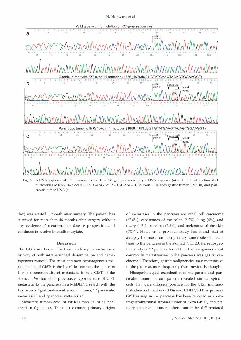
So that molecular biological examinations could be
performed for differential diagnosis, the specific cells
positive for CD34 and CD117/KIT were isolated from
microscopic regions of both tumor specimens with micro-
dissection techniques. The DNA isolated from each tu-
mor underwent mutational analyses of exons 9, 11, 13,
and 17 of the gene KIT and exons 12 and 18 of the gene
PDGFRA (platelet derived growth factor receptor alpha),
known as common mutational hotspots for GIST, to iden-
tify gene variants. An identical deletion of 21 nucleotides
(c. 1656―1675 del21GTATGAAGTACAGTGGAAGGT) in
exon 11 of KIT was detected in DNA from both tumors.
The mutation was absent from uninvolved tissue of the
patient (Fig. 5a, b, c). The gene sequencing analysis re-
vealed that both the gastric and pancreatic tumor cells
possessed an identical somatic mutation.
The postoperative course was uneventful. Postopera-
tive adjuvant chemotherapy (imatinib mesylate, 400 mg/
N. Hagiwara, et al
136 J Nippon Med Sch 2016; 83 (3)
Fig. 5 A DNA sequence of chromosome in exon 11 of KIT gene shows wild type DNA sequence (a) and identical deletion of 21
nucleotides (c.1656―1675 del21 GTATGAAGTACAGTGGAAGGT) in exon 11 in both gastric tumor DNA (b) and pan-
creatic tumor DNA (c).
KIT
KIT
KIT
day) was started 1 month after surgery. The patient has
survived for more than 48 months after surgery without
any evidence of recurrence or disease progression and
continues to receive imatinib mesylate.
Discussion
The GISTs are known for their tendency to metastasize
by way of both intraperitoneal dissemination and hema-
togenous routes6,7. The most common hematogenous me-
tastatic site of GISTs is the liver8. In contrast, the pancreas
is not a common site of metastasis from a GIST of the
stomach. We found no previously reported case of GIST
metastatic to the pancreas in a MEDLINE search with the
key words “gastrointestinal stromal tumor,” “pancreatic
metastasis,” and “pancreas metastasis.”
Metastatic tumors account for less than 2% of all pan-
creatic malignancies. The most common primary origins
of metastases to the pancreas are renal cell carcinoma
(62.6%); carcinomas of the colon (6.2%), lung (4%), and
ovary (4.7%); sarcoma (7.2%); and melanoma of the skin
(4%)9,10. However, a previous study has found that at
autopsy the most common primary tumor site of metas-
tases to the pancreas is the stomach11. In 2014 a retrospec-
tive study of 22 patients found that the malignancy most
commonly metastasizing to the pancreas was gastric car-
cinoma12. Therefore, gastric malignancies may metastasize
to the pancreas more frequently than previously thought.
Histopathological examination of the gastric and pan-
creatic tumors in our patient revealed similar spindle
cells that were diffusely positive for the GIST immuno-
histochemical markers CD34 and CD117/KIT. A primary
GIST arising in the pancreas has been reported as an ex-
tragastrointestinal stromal tumor or extra-GIST13, and pri-
mary pancreatic tumors often cannot be differentiated
Pancreatic Metastasis from Gastrointestinal Stromal Tumor
J Nippon Med Sch 2016; 83 (3) 137
from metastatic tumors with currently available histopa-
thological methods. We analyzed DNA sequences to in-
vestigate the genetic characteristics of both tumors and
found an identical mutational pattern in exon 11 of KIT
in DNA from both the gastric tumor and the pancreatic
tumor. The molecular pathogenesis of GIST has generally
been considered to result from expansion of a cell clone
that has acquired an activating mutation in the KIT
proto-oncogene14. A recent investigation of genetic muta-
tions in primary and metastatic tumor cells has found
that paired samples of primary colorectal cancer and me-
tastases show consistent mutation patterns upon the mu-
tational analysis of the gene KRAS (Kirsten rat sarcoma
viral oncogene homolog)15. These findings support the
notion that the primary GIST tumor and the metastatic
pancreatic tumor in our patient had identical origins. On
the basis of a comprehensive analysis of these clinical
and biological factors, we concluded that the pancreatic
tumor was a metastatic tumor from the GIST of the
stomach.
Local recurrence and distant metastases have been re-
ported to develop in more than 30% of patients with
high-risk GIST despite apparently complete resection16.
Most recurrences or metastases occurring after resection
with a R0 margin of a high-risk GIST appear within 3
years after primary surgery. Adjuvant therapy with
imatinib mesylate, a specific molecular inhibitor, is bene-
ficial after the primary GIST is resected. Three years of
adjuvant treatment with imatinib mesylate has been
shown to improve recurrence-free survival and overall
survival in patients with high-risk GISTs17. However, tu-
mor sensitivity to imatinib has been reported to differ ac-
cording to the location of the mutation18. The GISTs with
exon 11 mutations of KIT are more than 10 times as sen-
sitive to imatinib than are GISTs with mutations in other
exons19. Identification of the causative mutation before
starting imatinib treatment is thus considered clinically
and economically beneficial in patients with GIST20. In
our patient, both the gastric and pancreatic GISTs had
mutations of exon 11 in the KIT gene. The tumors were
therefore expected to be sensitive to imatinib treatment.
The patient has shown no sign of recurrence or disease
progression for more than 4 years after surgery and con-
tinues to receive adjuvant treatment with imatinib.
In conclusion, we have reported an extremely rare case
of gastric GIST with synchronous pancreatic metastasis
that successfully responded to multimodality therapy.
Our findings may be helpful for determining a multimo-
dality treatment strategy and for developing new treat-
ment options for a case of GIST with such unusual me-
tastases.
Conflict of Interest: The authors have no conflicts of interest
to declare.
References1.Miettinen M, Sarlomo-Rikala M, Lasota J: Gastrointestinal
stromal tumors: recent advances in understanding of their
biology. Hum Pathol 1999; 30: 1213―1220.
2.Demetri GD: Targeting the molecular pathophysiology of
gastrointestinal stromal tumors with imatinib. Mecha-
nisms, successes, and challenges to rational drug develop-
ment. Hematol Oncol Clin North Am 2002; 16: 1115―1124.
3.Chou FF, Eng HL, Sheen-Chen SM: Smooth muscle tu-
mors of the gastrointestinal tract: analysis of prognostic
factors. Surgery 1996; 119: 171―177.
4.Lewis JJ, Brennan MF: Soft tissue sarcomas. Curr Probl
Surg 1996; 33: 817―872.
5.Trent JC, Benjamin RS: New developments in gastrointes-
tinal stromal tumor. Curr Opin Oncol 2006; 18: 386―395.
6.Corless CL, Fletcher JA, Heinrich MC: Biology of gastro-
intestinal stromal tumors. J Clin Oncol 2004; 22: 3813―3825.
7.Miettinen M, Lasota J: Gastrointestinal stromal tumors:
review on morphology, molecular pathology, prognosis,
and differential diagnosis. Arch Pathol Lab Med 2006;
130: 1466―1478.
8.DeMatteo RP, Lewis JJ, Leung D, Mudan SS, Woodruff
JM, Brennan MF: Two hundred gastrointestinal stromal
tumors: recurrence patterns and prognostic factors for
survival. Ann Surg 2000; 231: 51―58.
9.Z’Graggen K, Fernandez-del Castillo C, Rattner DW, Si-
gala H, Warshaw AL: Metastases to the pancreas and
their surgical extirpation. Arch Surg 1998; 133: 413―417;
discussion 418―419.
10.Tuech JJ, Pessaux P, Chautard D, Rouge C, Binelli C, Ber-
gamaschi R, Arnaud JP: Results of duodenopancreatec-
tomy for solitary pancreatic metastasis from renal cell car-
cinoma. J Hepatobiliary Pancreat Surg 1999; 6: 396―398.
11.Nakamura E, Shimizu M, Itoh T, Manabe T: Secondary
tumors of the pancreas: clinicopathological study of 103
autopsy cases of Japanese patients. Pathol Int 2001; 51:
686―690.
12.Song SW, Cheng JF, Liu N, Zhao TH: Diagnosis and treat-
ment of pancreatic metastases in 22 patients: a retrospec-
tive study. World J Surg Oncol 2014; 12: 299.
13.Kim HH, Koh YS, Park EK, Seoung JS, Hur YH, Kim JC,
Cho CK, Kim HJ: Primary extragastrointestinal stromal
tumor arising in the pancreas: report of a case. Surg To-
day 2012; 42: 386―390.
14.Allander SV, Nupponen NN, Ringner M, Hostetter G,
Maher GW, Goldberger N, Chen Y, Carpten J, Elkahloun
AG, Meltzer PS: Gastrointestinal stromal tumors with KIT
mutations exhibit a remarkably homogeneous gene ex-
pression profile. Cancer Res 2001; 61: 8624―8628.
15.Paliogiannis P, Cossu A, Tanda F, Palmieri G, Palomba G:
Mutational concordance between primary and metastatic
colorectal adenocarcinoma. Oncol Lett 2014; 8: 1422―1426.
16.Krajinovic K, Germer CT, Agaimy A, Wünsch PH, Isbert
C: Outcome after resection of one hundred gastrointesti-
nal stromal tumors. Dig Surg 2010; 27: 313―319.
17.Eisenberg BL: The SSG XVIII/AIO trial: results change
N. Hagiwara, et al
138 J Nippon Med Sch 2016; 83 (3)
the current adjuvant treatment recommendations for gas-
trointestinal stromal tumors. Am J Clin Oncol 2013; 36:
89―90.
18.Heinrich MC, Corless CL, Demetri GD, Blanke CD, von
Mehren M, Joensuu H, McGreevey LS, Chen CJ, Van den
Abbeele AD, Druker BJ, Kiese B, Eisenberg B, Roberts PJ,
Singer S, Fletcher CD, Silberman S, Dimitrijevic S,
Fletcher JA: Kinase mutations and imatinib response in
patients with metastatic gastrointestinal stromal tumor. J
Clin Oncol 2003; 21: 4342―4349.
19.Lee JH, Kim Y, Choi JW, Kim YS: Correlation of imatinib
resistance with the mutational status of KIT and PDGFRA
genes in gastrointestinal stromal tumors: a meta-analysis.
J Gastrointestin Liver Dis 2013; 22: 413―418.
20.Blay JY, Casali PG, Dei Tos AP, Le Cesne A, Reichardt P:
Management of gastrointestinal stromal tumour: current
practices and visions for the future. Oncology 2015; 89: 1―13.
(Received,
(Accepted,
October
February
25, 2015)
3, 2016)