Embed Size (px)
Citation preview
1
Pathologic grading for predicting metastasis in phaeochromocytoma and 1
paraganglioma 2
3
Noriko Kimura1, Ryoichi Takayanagi
2, Nae Takizawa
3, Eiji Itagaki
4, Takayuki Katabami
5, Narihiko 4
Kakoi6, Hiromi Rakugi
7, Yukihiro Ikeda
8, Akiyo Tanabe
9, Takeshi Nigawara
10, Sadayoshi Ito
11, 5
Itaru Kimura12
and Mitsuhide Naruse13
6
The Phaeochromocytoma Study Group in Japan 7
8
1Department of Clinical Research, Pathology Division, National Hospital Organization, Hakodate 9
Hospital, Hakodate, Japan 10
2Department of Medicine and Bioregulatory Science, Graduate School of Medical Sciences, 11
Kyushu University, Fukuoka, Japan 12
3Department of Urology & Andrology, Kansai Medical University, Osaka, Japan 13
4Third Department of Internal Medicine (Diabetes, Endocrine and Metabolism), Kyorin University 14
School of Medicine, Tokyo, Japan 15
5Department of Medicine, Metabolism and Endocrinology, St. Marianna University, School of 16
Medicine, Kawasaki, Japan 17
6 Department of Urology, Miyagi Cancer Center, Natori, Japan 18
Page 1 of 45 Accepted Preprint first posted on 12 February 2014 as Manuscript ERC-13-0494
Copyright © 2014 by the Society for Endocrinology.
2
7Department of Geriatric Medicine & Nephrology, Osaka University Graduate School of Medicine, 19
Osaka, Japan 20
8Department of Medicine, Division of Nephrology, Hypertension and Endocrinology, Nihon 21
University Itabashi Hospital, Tokyo, Japan 22
9Department of Endocrinology and Metabolism, Tokyo Women’s Medical University, Tokyo, 23
Japan 24
10Department of Endocrinology and Metabolism, Hirosaki University, School of Medicine, Japan 25
11Department of Medicine, Division of Nephrology, Endocrinology and Vascular Medicine, Tohoku 26
University School of Medicine, Sendai, Japan 27
12Council Member Examination Committee of Social Insurance, Ministry of Health, Labour and 28
Welfare, Japan, Tokyo, Japan 29
13Department of Endocrinology, Metabolism, and Hypertension, National Hospital Organization 30
Kyoto Medical Center, Kyoto, Japan 31
32
Corresponding author: postal cord 041-8512, 18-16 Kawahara Hakodate Hokkaido Japan 33
Department of Clinical Research, Pathology Division, National Hospital Organization, Hakodate 34
Hospital, Hakodate, Japan, Telephone: 81-138-51-6281, Fax: 81-138-30-1020, 35
E-mail: [email protected] 36
Page 2 of 45
3
Short title: Pathologic grading in paragangliomas 37
Key words: phaeochromocytoma, paraganglioma, histopathologic diagnosis, succinate 38
dehydrogenase gene subunit B, immunohistochemistry, metastasis, survival 39
40
Page 3 of 45
4
Abstract 41
Phaeochromocytomas and paragangliomas are rare catecholamine-producing tumours. 42
Although 10 to 30% of these tumours metastasise, histopathologic criteria to discriminate 43
malignant from benign tumours have not been established; therefore, reliable histopathologic 44
markers predicting metastasis are urgently required. One hundred sixty-three tumours, including 45
40 metastatic tumours, collected by the Phaeochromocytoma Study Group in Japan (PHEO-J) 46
were analysed using a system we have called GAPP (grading system for adrenal 47
phaeochromocytoma and paraganglioma ). The tumours were scored based on GAPP criteria as 48
follows: histologic pattern, cellularity, comedo-type necrosis, capsular/vascular invasion, Ki67 49
labelling index, and catecholamine type. All tumours were scored from 0 to 10 points, and were 50
graded as one of three types: well differentiated (0-2 points), moderately differentiated (3-6), and 51
poorly differentiated (7-10). GAPP scores of the non-metastatic and metastatic groups were 2.08 52
± 0.17 and 5.33 ± 0.43, (mean ± SE, P<0.001) respectively. There was a significant negative 53
correlation between the GAPP score and the duration until metastasis (r=−0.438, P<0.01). The 54
mean number of years until metastasis after the initial operation was 5.5 ± 2.6 years. The study 55
included 111 well-differentiated, 35 moderately differentiated, and 17 poorly differentiated types. 56
The 5-year survival of these groups was 100%, 66.8%, and 22.4%, respectively. In addition, 57
negative immunoreactivity for succinate dehydrogenase gene subunit B (SDHB) was observed 58
Page 4 of 45
5
in 13 (8%) moderarely or poorly differentiated tumours, and 10 of the 13 (77%) had metastases. 59
Our data indicate that a combination of GAPP classification and SDHB immunohistochemistry 60
might be useful for the prediction of metastasis in these tumours. 61
Page 5 of 45
6
Introduction 62
Phaeochromocytomas (PHEO) of the adrenal gland and sympathetic paragangliomas (PGL) are 63
catecholamine-producing tumours. Although 10–30% of these tumours metastasise, 64
histopathologic criteria that discriminate malignant from benign tumours have not been 65
established and only the presence of metastasis is considered evidence of malignancy in the 66
current WHO definition (Thompson LDR et al. 2004). Although most cases of PHEO/PGL are 67
surgically curable, malignant PHEO/PGL are intractable diseases that require an early diagnosis 68
and effective treatment. We organised a task force group for PHEO/PGL in Japan (PHEO-J) that 69
was composed of endocrinologists, urologists, endocrine surgeons, radiologists, molecular 70
biologists, and pathologists and was supported by the Ministry of Health, Labour, and Welfare. 71
The aim of the study was to survey PHEO/PGL in Japan. Based on the results of PHEO-J in 72
2012, the estimated total number of patients with PHEO/PGL was 2,920, including 320 patients 73
with metastasis. The proportion of patients with malignant, multiple, extra-adrenal, and familial 74
PHEO/PGL was 11%, 12.7%, 17.3%, and 10%, respectively (Naruse 2011). Among patients with 75
malignant PHEO/PGL, 36.8% were initially diagnosed as benign and 59.6% showed absence of 76
metastasis at initial operation (Naruse 2011). The results indicated the difficulties associated with 77
differential diagnosis of malignant from benign disease in the absence of distant metastases, and 78
strongly emphasized the need to establish reliable histopathologic criteria for predicting 79
Page 6 of 45
7
metastasis. 80
Many studies have attempted to discriminate benign and malignant PHEO/PGL. Of these, some 81
molecular biomarkers such as the Ki67 labelling index (Nagura et al, 1999, Elder et al. 2003), 82
loss of cell adhesion molecules such as CD44, and human telomerase reverse transcriptase 83
expression (van der Harst et al. 2000, Elder et al. 2003, August et al.2004) have been proposed 84
as useful markers for the detection of high-grade malignancy. However, these markers failed to 85
detect low- and moderate-grade malignant PHEO/PGL. Although the presence of only one 86
indicator does not allow a definite diagnosis of malignancy in PHEO/PGL, certain pathologic 87
features, such as size and site (O'Riordain et al. 1996), local extension, angioinvasion, mitotic 88
index/proliferative activity using Ki67 labeling index (Ki67 LI), irregular zellballen pattern, and 89
presence of confluent (comedo-type) tumour necrosis (Zelinka et al. 2011) have been used in 90
previous studies (Linnoila RI et al. 1990, Kimura N & Sasano N, 1990, Unger P et al. 1991). Such 91
features have been combined in a scoring system named the Phaeochromocytoma of the 92
Adrenal gland Scaled Score (PASS) for use in diagnosis (Thompson 2003). PASS was the first 93
scoring system for diagnosis on adrenal PHEO. However, PASS reproducibility and clinical 94
significance have not been established (Wu et al. 2009), probably because the PASS 95
classification contains too many histologic parameters that cover classical features of general 96
malignancy rather than focusing on specific PHEO/PGL features. In general, for endocrine 97
Page 7 of 45
8
tumours the biological behaviour of the tumours usually reflects the differentiation of 98
hormone-producing functions. Cell maturation and cell proliferation in endocrine tumours usually 99
oppose each other; poorly differentiated tumours grow rapidly and cause poor prognosis 100
whereas well-differentiated tumours grow slowly and metastasise later. The distinction between 101
poorly differentiated tumours and well-differentiated tumours is important for the determination of 102
a patient’s prognosis (Solcia et al. 2000). Kimura et al. (2005) presented a grading system for 103
PHEO/PGL based on the concept that norepinephrine-producing tumours are less differentiated 104
than epinephrine-producing tumours. However, this study was a report from a single institute, 105
and a multicenter study is required. Here, we examined the materials gathered from a nationwide 106
survey by PHEO-J using the previously described grading system, which we have named GAPP 107
(Grading of Adrenal Phaeochromocytoma and Paraganglioma ), to determine whether GAPP 108
has clinical applications. 109
Recently, there has been rapid progress in the molecular analysis of PHEO/PGL and 110
approximately 16 genes responsible for PHEO/PGL have been discovered: NF1, VHL, RET, 111
SDHC, SDHD, SDHB, SDHAF2, SDHA, TMEM127, MAX, IHD1, KIF2, HRAS, HIF2, PHD2 and 112
FH (Gimenez-Roqueplo 2013, King et al. 2013, Castro-Vega et al.2014). Of these, mutations in 113
the succinate dehydrogenase genes (SDHA, SDHB, SDHC, and SDHD) are responsible for a 114
large percentage of hereditary PHEO/PGL syndrome (HPPS) cases, and it has been reported 115
Page 8 of 45
9
that an extra-adrenal site, recurrence, and malignancy are strongly associated with the SDHB 116
mutation (Gimenez-Roqueplo et al. 2003, Neumann et al. 2004). In the present study, SDHB 117
gene analysis was not available because of ethical considerations; however, 118
immunohistochemical studies of SDHB were carried out using central pathologic analysis. We 119
attempted to clarify the features of metastatic and non-metastatic tumours using GAPP 120
classification and SDHB immunohistochemistry to facilitate the differential diagnosis of malignant 121
from benign disease in PHEO/PGL. 122
Materials and Methods 123
Patients and tissues 124
A total of 994 patients from 178 institutes registered on-line. For ethical reasons, the registration 125
was limited to adult patients. All data on patients were sent from each institute via a registration 126
form. There were two types of forms: clinical registration and data for pathologic analysis. Briefly, 127
a clinical registration form was composed of date of informed consent, patient birthday, year, age, 128
sex, present state (alive or dead) were entered at the first registration. If there is no problem for 129
registration, then second step for registration started. Clinical information was composed of 1. 130
year and day of initial diagnosis, 2. family history, 3. clinical diagnosis at present: adrenal, 131
extra-adrenal or unknown and also benign or malignant or unknown in each tumors, 4. location 132
of tumors, 5. number of tumors: single or multiple or unknown, 6. metastasis: absence, presence, 133
Page 9 of 45
10
or unknown. This survey research was carried out three times with 6 month intervals. The last 134
time of follow up was the time of the last registration. Metastasis was confirmed by imaging (CT, 135
MRI, or MIBG scan), catecholamine analysis, and operation if possible. 136
For pathologic analysis, patient clinical information: such as 1. clinical diagnosis at the time of 137
registration, 2. age and sex, 3. year of initial operation, 4. location of tumors, 5. tumor size, 6. 138
number of tumors, 7. metastasis: absence, or presence (lymph node, bone, lung, liver, others, 139
including what place and how many), 8. year and date of metastasis, 9. catecholamine data: 140
plasma/urine, adrenalin, noradrenalin and dopamine were supplied. 141
Among these registrations, tumour specimens from 163 patients were voluntarily submitted for 142
pathologic analysis, including 123 without metastasis and 40 with metastasis. The mean age of 143
the patients was 50.7 ± 15.5 years (range: 21–80 years). The duration of follow up ranged from 1 144
to 33 years with mean follow up duration of 6.61 ± 0.74 (P<0.01, Grubbs-Smirnov examination). 145
Twelve patients died of multiple metastases. The locations of the primary tumours were as 146
follows: 127 in the adrenal glands, including 13 that occurred bilaterally, and 36 in extra-adrenal 147
regions (28 in the retroperitoneum and 8 in the urinary bladder). 148
The surgically removed tumours were fixed in 10% formalin and embedded in paraffin. Each 149
institute prepared 10 unstained glass slides with 3-µm thick tissue sections of the representative 150
tumour tissues. These slides were sent to the National Hospital Organisation Hakodate Hospital 151
Page 10 of 45
11
for central pathologic analysis. All pathologic materials were associated with patients’ clinical 152
data, photographs of gross and cut findings of the tumours, and original pathologic diagnoses 153
from each institute to provide information such as vascular or capsular invasion or histologic 154
variation that might be lacking in the section sent to Hakodate Hospital due to sampling errors. 155
Pathologic analysis 156
All sections were subjected to haematoxylin and eosin staining, elastica-Masson trichrome 157
staining to confirm vascular invasion, and immunostaining for chromogranin A to confirm 158
PHEO/PGL, Ki67 for proliferating cells, S100 protein for sustentacular cells, D2-40 to confirm 159
lymph vessel invasion, and SDHB for HPPS. Immunostaining for phenylethanolamine 160
N-methyltransferase (PNMT) was performed in cases of epinephrine-producing extra-adrenal 161
paraganglioma. Immunohistochemical procedures were performed using an automated 162
immunostainer (Benchmark, Ventana, Tucson, AZ, USA) according to the manufacturer’s 163
protocol. The primary antibodies and final dilutions were as follows: chromogranin A (monoclonal, 164
1:200; Dako, Carpinteria, CA, USA), Ki67 (MIB1, monoclonal, prediluted; Novocastra, 165
Burlingame, CA, USA), S100 (polyclonal, prediluted; Novocastra), D2-40 (monoclonal, 166
prediluted; Nichirei Bioscience, Tokyo, Japan), SDHB (FL-280, polyclonal, 1: 50, Santa Cruz 167
Biotechnology Inc., Texas, USA and HPA002868, polyclonal, 1:50, Sigma-Aldrich, Inc., MO, 168
USA), and PNMT (polyclonal, 1:500, Bioclone, Australia). The appropriate positive and negative 169
Page 11 of 45
12
controls were included in parallel. 170
For pathologic analysis, we scored histologic features based on the GAPP classification, which 171
consisted of the following parameters: histologic pattern, cellularity, presence or absence of 172
comedo-type necrosis, vascular or capsular invasion, Ki67 labelling index (%), and 173
catecholamine types produced by the tumours. More specifically, the histologic pattern was 174
classified into three types: zellballen, irregular zellballen, and pseudorosette. The zellballen 175
pattern is a specific pattern of the paraganglion system composed of nests of 176
catecholamine-producing cells, sustentacular cells, and capillaries surrounding these cells. The 177
irregular zellballen pattern was a mixture of small and large irregular tumour cell nests in which 178
the size of the larger nests was at least 10 times that of the smaller nests. The pseudorosette 179
pattern was characterised by centrally located, delicate vessels surrounded by tumour cells with 180
cytoplasmic processes around them and corresponded to the pseudopapillary pattern in 181
intrathoracic paravertebral paraganglioma described by Lack (Lack EE, 2007). Irregular 182
zellballen pattern or pseudorosette pattern is counted even if only focal. It is not necessary for 183
these patterns to be diffuse. A diffuse growth pattern was not included in the score because, in 184
the authors’ experience, it is most common in adrenergic pheochromocytomas and usually 185
innocent. 186
The scores of the histologic patterns were 0, 1, and 1 for the zellballen, irregular zellballen, and 187
Page 12 of 45
13
pseudorosette patterns, respectively. If the pseudorosette pattern was observed even focally, it 188
was counted as a score of 1. Cell number within a square (cellularity) was counted under 189
high-power magnification (×400) using a 10-mm micrometer (Nikon S-6, Ver.YS1; Nikon, Tokyo, 190
Japan) on an eyepiece. Two fields of the highest cellularity were used for counting to assess 191
cellularity. Scores of 0, 1, or 2 were assigned for number of tumour nuclei less than 150, 192
150–250, or more than 250, respectively. The presence of comedo-type necrosis scored 2 points. 193
Comedo-type necrosis was typically centrally located necrosis of a highly cellular nest. 194
PHEO/PGL occasionally showed coagulation necrosis or scar formation in the tumour due to a 195
sudden drop in blood pressure; such degenerative changes were not counted as comedo-type 196
necrosis. For Ki67 labelling index, two highest labeled areas (hot fields) were taken for 197
photographs at x200 magnification and were counted using a digital image analyser (Lumina 198
Vision, Mitani Corp., Tokyo) and scored as 0, 1, and 2 for less than 1%, 1-3%, and more than 3%, 199
respectively. Number of cells counted was different case by case depend on cellularity, however, 200
usually it was from 500 to 2000. Representative images are shown in Figure 1. Catecholamine 201
type was provided by the clinical data. If the plasma or urinary epinephrine levels were 202
abnormally high with or without elevated norepinephrine levels, the type was determined to be 203
epinephrine and scored 0 points. If the norepinephrine levels were high in the absence of 204
elevated epinephrine levels, with or without elevated dopamine levels, the type was determined 205
Page 13 of 45
14
to be norepinephrine and scored 1 point. The non-functioning type scored 0 points. Scores of 206
these parameters (0 to 2 points for each) were summed for a total number of points with a 207
maximum of 10 (Table 1). The individual GAPP point was given based on histological analysis of 208
the tumors. Based on our experience in the field, 2 points were given to findings suggestive of 209
malignancy, 1 point was given to the findings suggestive of possible malignancy, and 0 points 210
were given to the findings suggestive of rare malignancy. The total points were then classified 211
into three differentiation types: well differentiated (WD, 0-2), moderately differentiated (MD, 3-6), 212
and poorly differentiated (PD, 7-10). This grading system is summarised in Table 2. 213
For assessment of the pathologic grading, sections were thoroughly examined twice, at the initial 214
analysis and 6 months after the first observation, to avoid observational errors. 215
Statistical analysis 216
The ztTEST was applied for comparison of tumour size and GAPP score between metastatic and 217
non-metastatic tumours. The correlation between the GAPP score and duration between the 218
initial operation and the time of initial metastasis was analysed using Pearson’s simple linear 219
regression and correlation. The correlation between the GAPP score and cellularity, GAPP score 220
and KI67 LI, and Ki67 LI and cellularity was analysed using Pearson’s simple linear regression 221
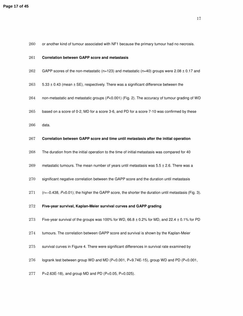
and correlation. Survival of the WD, MD, and PD types was compared using the Kaplan-Meier 222
method and logrank significance test. All the individual parameters of GAPP and the 223
Page 14 of 45
15
correspoinding P values in predicting metastasis were examined by multivariate analysis by 224
Pearson’s simple linear regression among 6 groups. Results were considered significant for a P 225
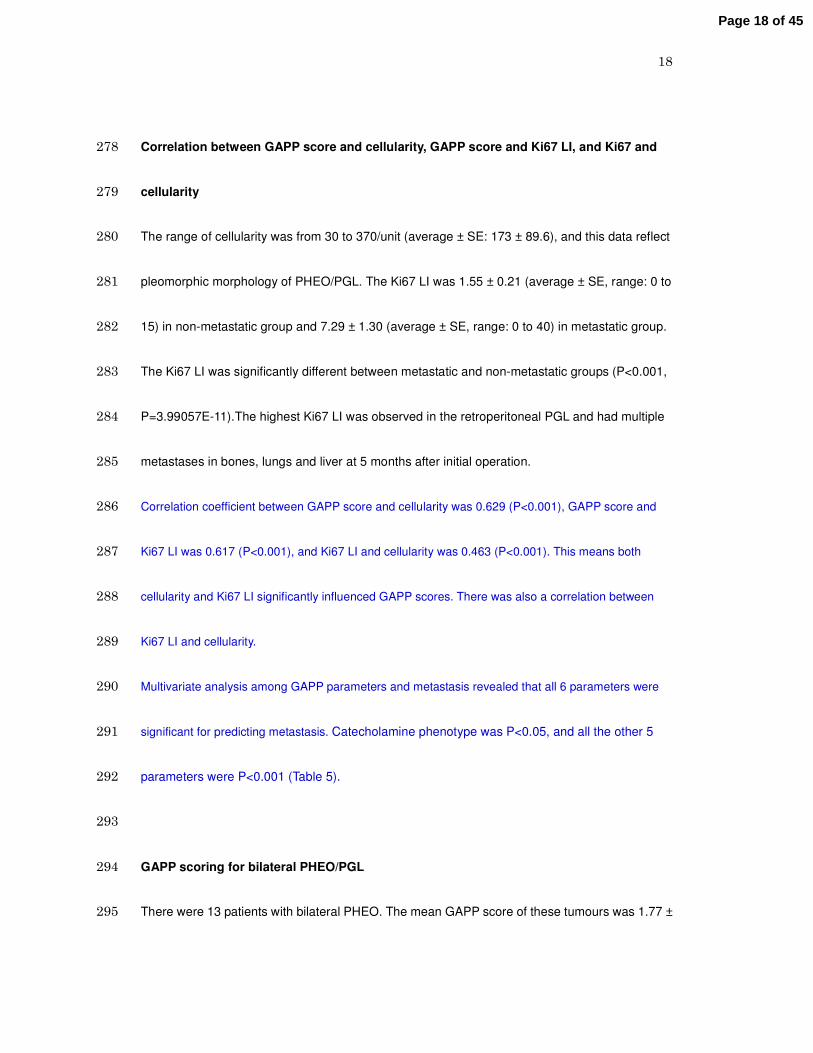
value less than 0.05. Statistical analyses were performed using StatMate IV software (Takahashi 226
Y, Atms, Tokyo, 2009). 227
Ethics 228
The study was approved by the institutional ethics committee of the Kyoto Medical Centre, 229
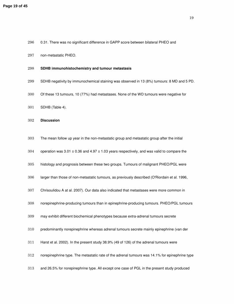
National Hospital Organisation (responsible for the PHEO-J study) and the Hakodate Hospital, 230
National Hospital Organisation (responsible for the histopathologic analysis). 231
All tissue specimens and clinical information were collected after the material was anonymised at 232
each institute. 233
Results 234
Clinical features 235
The mean follow up year after the initial operation was 3.01 ± 0.36 in the non-metastatic group 236
(range: 0-20 years) and 4.97 ± 1.03 in the metastatic group (range: 0-33 years). The mean 237
tumour size was 5.1 ± 0.3 cm in the non-metastatic group (range: 1.1–20.0 cm) and 8.7 ± 0.7 cm 238
in the metastatic group (range 3.0–16.5 cm). The metastatic tumors were larger than the 239
non-metastatic tumors (P<0.001). Catecholamine types produced by the tumours were as 240
follows: epinephrine type in 78 cases, norepinephrine type in 79 cases, and non-functioning type 241
Page 15 of 45
16
in 6 cases. Metastases were observed in 11 (14.1.0%) tumours of the epinephrine type 242
compared with 29 (36.7%) tumours of the norepinephrine type, thus metastases were 243
predominant in norepinephrine-type tumours. There were 6 non-functioning type tumours and 244
one of these (16.7%) metastasised. Locations of the metastatic tumours were the adrenal gland 245
in 24 cases (60%) and extra-adrenal regions in 16 (40%). The malignancy rate was 19.0% (24 of 246
126) in the adrenal gland and 43.2% (16 of 37) in extra-adrenal regions (Table 3). 247
Histology 248
All tumours were scored from 0 to 10 points and graded accordingly as WD, MD, and PD types. 249
There were 111 WD, 36 MD, and 16 PD tumours. Metastasis was observed in 40 tumours: 4 WD, 250
21 MD, and 15 PD. The rate of metastasis in each group was 3.6% for WD, 60% for MD, and 251
88.2% for PD. Although most metastatic tumours were PD and MD, four of the WD tumours also 252
metastasised. Of these four WD tumours, three PHEOs had remarkable invasion into the 253
capsular or central vein of the adrenal gland, and one of these three cases had fat infiltration 254
adjacent to the tumour as well as vascular invasion. The remaining PHEO was associated with 255
NF1, and the patient had multiple bone metastases 4 years after the initial operation for adrenal 256
PHEO. The submitted metastatic bone tumour specimen was not satisfactory for pathologic 257
examination due to its tiny tissue with necrosis. Although levels of urinary catecholamine 258
metabolites were slightly elevated, it was uncertain whether the metastatic tumours were PHEO 259
Page 16 of 45
17
or another kind of tumour associated with NF1 because the primary tumour had no necrosis. 260
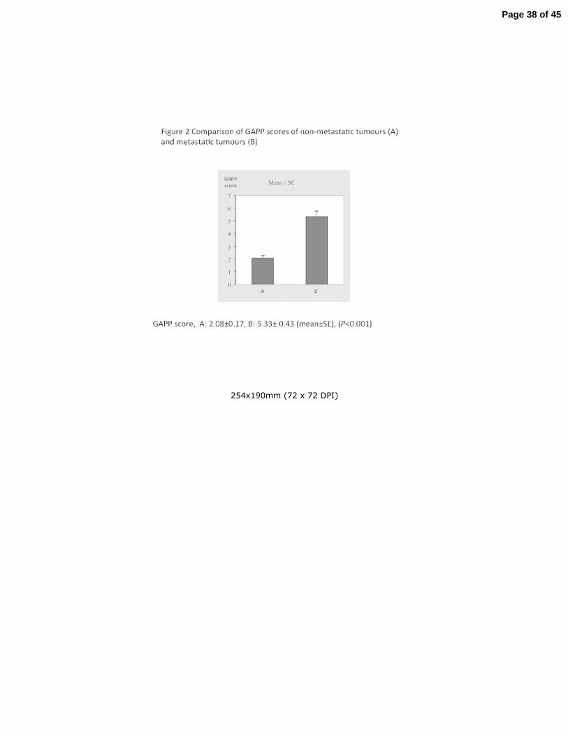
Correlation between GAPP score and metastasis 261
GAPP scores of the non-metastatic (n=123) and metastatic (n=40) groups were 2.08 ± 0.17 and 262
5.33 ± 0.43 (mean ± SE), respectively. There was a significant difference between the 263
non-metastatic and metastatic groups (P<0.001) (Fig. 2). The accuracy of tumour grading of WD 264
based on a score of 0-2, MD for a score 3-6, and PD for a score 7-10 was confirmed by these 265
data. 266
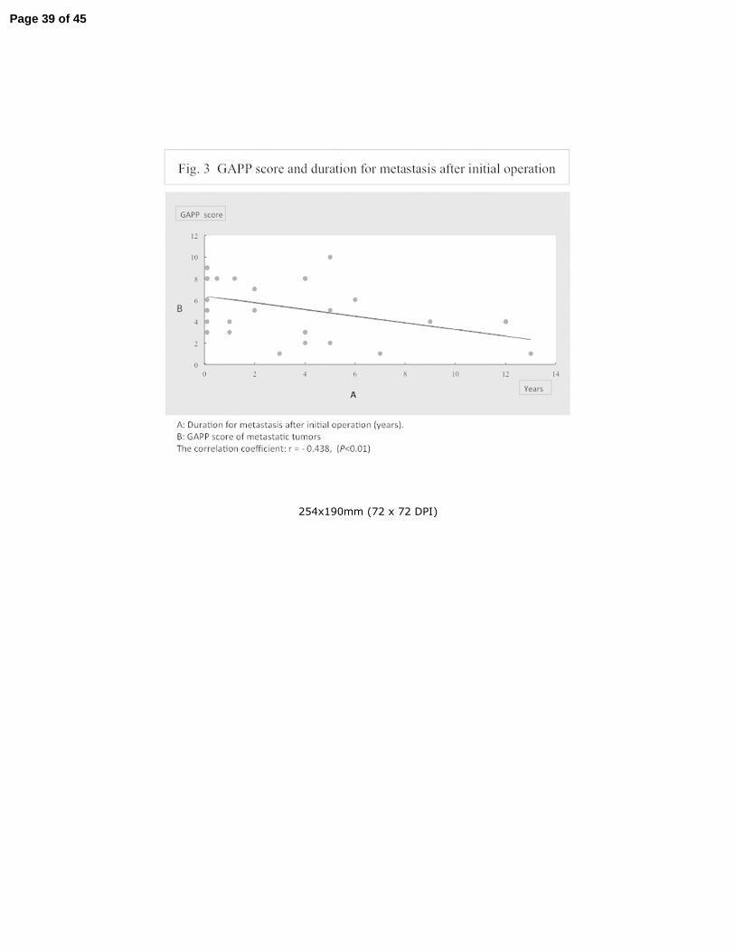
Correlation between GAPP score and time until metastasis after the initial operation 267
The duration from the initial operation to the time of initial metastasis was compared for 40 268
metastatic tumours. The mean number of years until metastasis was 5.5 ± 2.6. There was a 269
significant negative correlation between the GAPP score and the duration until metastasis 270
(r=−0.438, P<0.01); the higher the GAPP score, the shorter the duration until metastasis (Fig. 3). 271
Five-year survival, Kaplan-Meier survival curves and GAPP grading 272
Five-year survival of the groups was 100% for WD, 66.8 ± 0.2% for MD, and 22.4 ± 0.1% for PD 273
tumours. The correlation between GAPP score and survival is shown by the Kaplan-Meier 274
survival curves in Figure 4. There were significant differences in survival rate examined by 275
logrank test between group WD and MD (P<0.001, P=9.74E-15), group WD and PD (P<0.001, 276
P=2.63E-18), and group MD and PD (P<0.05, P=0.025). 277
Page 17 of 45
18
Correlation between GAPP score and cellularity, GAPP score and Ki67 LI, and Ki67 and 278
cellularity 279
The range of cellularity was from 30 to 370/unit (average ± SE: 173 ± 89.6), and this data reflect 280
pleomorphic morphology of PHEO/PGL. The Ki67 LI was 1.55 ± 0.21 (average ± SE, range: 0 to 281
15) in non-metastatic group and 7.29 ± 1.30 (average ± SE, range: 0 to 40) in metastatic group. 282
The Ki67 LI was significantly different between metastatic and non-metastatic groups (P<0.001, 283
P=3.99057E-11).The highest Ki67 LI was observed in the retroperitoneal PGL and had multiple 284
metastases in bones, lungs and liver at 5 months after initial operation. 285
Correlation coefficient between GAPP score and cellularity was 0.629 (P<0.001), GAPP score and 286
Ki67 LI was 0.617 (P<0.001), and Ki67 LI and cellularity was 0.463 (P<0.001). This means both 287
cellularity and Ki67 LI significantly influenced GAPP scores. There was also a correlation between 288
Ki67 LI and cellularity. 289
Multivariate analysis among GAPP parameters and metastasis revealed that all 6 parameters were 290
significant for predicting metastasis. Catecholamine phenotype was P<0.05, and all the other 5 291
parameters were P<0.001 (Table 5). 292
293
GAPP scoring for bilateral PHEO/PGL 294
There were 13 patients with bilateral PHEO. The mean GAPP score of these tumours was 1.77 ± 295
Page 18 of 45
19
0.31. There was no significant difference in GAPP score between bilateral PHEO and 296
non-metastatic PHEO. 297
SDHB immunohistochemistry and tumour metastasis 298
SDHB negativity by immunochemical staining was observed in 13 (8%) tumours: 8 MD and 5 PD. 299
Of these 13 tumours, 10 (77%) had metastases. None of the WD tumours were negative for 300
SDHB (Table 4). 301
Discussion 302
The mean follow up year in the non-metastatic group and metastatic group after the initial 303
operation was 3.01 ± 0.36 and 4.97 ± 1.03 years respectively, and was valid to compare the 304
histology and prognosis between these two groups. Tumours of malignant PHEO/PGL were 305
larger than those of non-metastatic tumours, as previously described (O'Riordain et al. 1996, 306
Chrisoulidou A at al. 2007). Our data also indicated that metastases were more common in 307
norepinephrine-producing tumours than in epinephrine-producing tumours. PHEO/PGL tumours 308
may exhibit different biochemical phenotypes because extra-adrenal tumours secrete 309
predominantly norepinephrine whereas adrenal tumours secrete mainly epinephrine (van der 310
Harst et al. 2002). In the present study 38.9% (49 of 126) of the adrenal tumours were 311
norepinephrine type. The metastatic rate of the adrenal tumours was 14.1% for epinephrine type 312
and 26.5% for norepinephrine type. All except one case of PGL in the present study produced 313
Page 19 of 45
20
norepinephrine. Norepinephrine-producing tumours lack PNMT (the enzyme that converts 314
norepinephrine to epinephrine), and are considered to be less differentiated than 315
epinephrine-producing tumours based on catecholamine synthesis. Dopamine hypersecretion 316
was considered a feature of immaturity and a marker for malignant PCC/PGL (van der Harst E et 317
al. 2002). Eisenhofer et al. (2012) reported that the plasma level of methoxytyramine, the 318
O-methylated metabolite of dopamine, is 4.7-fold higher in patients with metastases than in 319
those without, suggesting its use as a potential biomarker. Our study also included six 320
non-functioning type tumours with one (16.7%) case of metastasis. The mechanisms underlying 321
non-functioning PHEO/PGL are not fully understood; however, a deficiency of catecholamine 322
synthesising enzymes was reported (Kimura N et al. 1992). In the present study the metastatic 323
rate in the non-functioning type of tumour was similar to that of epinephrine-producing tumours. 324
GAPP is composed of six parameters those have been previously examined the significance for 325
differentiating between benign and malignant pheochromocytomas. Ki67 which is a marker for 326
proliferating cells has been especially interested and concentrically examined previously 327
(Nagura et al, 1999, Elder et al. 2003). Ki67 LI range is very wide in PHEO/PGL and it is 328
generally accepted that PHEO/PGL with high Ki67 LI highly metastasize and have malignant 329
course. However, it has been well known that PHEO/PGL with no or very low Ki67 LI sometimes 330
metastasize. That was the reason why Ki67 LI was not been accepted as a single indicator for 331
Page 20 of 45
21
malignant PHEO/PGL. The present study revealed that Ki67 LI in metastatic group is 332
significantly higher than that in non-metastatic group. However, the range of Ki67 LI was broad 333
and there was overlapped area in both groups. Thus Ki67 LI should be retained one of the 334
parameters. The present study first revealed that cellularity is very important parameter more 335
than Ki67 LI. 336
Histological classification using GAPP revealed that non-metastatic tumours could be 337
distinguished from metastatic tumours with few exceptions. More than 80% of PD and 60% of 338
MD tumours metastasised, compared with fewer than 4% of WD tumours. Thus, WD tumours 339
might be considered virtually benign, whereas MD and PD tumours are likely to be malignant. 340
However, if vascular/capsular invasion is evident, even WD tumours may metastasise. Thus all 341
PHEO/PGLs should be treated as malignant tumours among which WD tumours are low grade, 342
MD tumours are intermediate grade, and PD tumours are highly malignant. Furthermore, 343
Kaplan-Meier survival curves showed that patients with WD tumours had 100% survival whereas 344
patients with MD and PD tumours showed progressively worse survival. This indicates that if WD 345
tumours do metastasise they grow slowly and the patients have a long survival time after surgery. 346
Thus, it is very important to distinguish WD tumours from MD and PD tumours, and patients with 347
MD or PD tumours should be carefully followed for a long time. For assessment of GAPP score, 348
we observed twice with some intervals, and occasionally there were little difference between first 349
Page 21 of 45
22
and second look grading. If there were little differences in GAPP scores, the grading was not 350
influenced because there were ranges in grading: score 3-6 are MD, and score 7-10 are PD. 351
Such small differences of GAPP score by second look were not beyond the range of grading. 352
Since Gimenez-Roqueplo et al. (2003) first reported that an extra-adrenal site, recurrence, and 353
malignancy were strongly associated with SDHB mutations and suggested that the presence of 354
SDHB mutants should be considered a high-risk factor for malignancy or recurrence, 355
genotype-phenotype correlations in patients with SDHB-associated PHEO/PGL have been 356
closely studied in cases of malignant PHEO/PGL (Timmers et al. 2007). A malignant PGL was 357
documented in 37.5% of SDHB carriers, 3.1% of SDHD carriers, and none of the SDHC mutation 358
carriers (Burnichon et al. 2009). van Nederveen et al. (2009) reported loss of SDHB protein 359
immunoreactivity in tumours with HPPS with a sensitivity of 100% and a specificity of 84%. 360
Therefore, by routinely performing SDHB immunohistochemistry the malignant potential of 361
PCC/PGL associated with HPPS could be assessed with a high degree of reliability. In the 362
present study, SDHB-negative immunoreactivity was observed in 13 (8%) tumours, all of which 363
were MD and PD types, and 10 of the 13 (77%) had metastases. These data confirm previous 364
reports that the SDHB mutation is a high-risk factor for malignancy or recurrence 365
(Gimenez-Roqueplo et al. 2003, Neumann et al. 2004). Although immunohistochemistry is a 366
useful tool, it is also necessary to confirm gene mutations to avoid false negative or false positive 367
Page 22 of 45
23
results because assessment of immunoreactivity of SDHB is sometimes difficult especially in 368
cases of weak immunoreactivity (Nederveen et al. 2009). 369
Regarding bilateral PCC/PGL associated with multiple endocrine neoplasia type 2 (MEN2) and 370
von Hippel-Lindau disease (VHL), the GAPP score was low, and there were no significant 371
differences between bilateral and non-metastatic PCC/PGL. 372
In conclusion, we demonstrated the ability of GAPP classification to differentiate low-grade 373
malignancies from moderate to high-grade malignancies with different rates of metastasis. 374
Combined use of GAPP and SDHB immunohistochemistry might be useful for the prediction of 375
tumour metastasis and patient prognosis. The concordance rate and reproducibility of diagnosis 376
by GAPP should be validated in further studies. 377
Declaration of interest 378
There are no conflicts of interest. 379
Funding 380
This work was supported by a Health Labour Sciences Research Grant for Research on 381
Measures for Intractable Diseases from the Ministry of Health, Labour, and Welfare in Japan. 382
Author contribution statement 383
Noriko Kimura performed histologic analysis using GAPP and SDHB immunohistochemistry and 384
wrote the manuscript. 385
Page 23 of 45
24
Ryoichi Takayanagi, Nae Takizawa, Eiji Itagaki, Takayuki Katabami, Narihiko Kakoi, Hiromi 386
Rakugi, Yukihiro Ikeda, Akiyo Tanabe, Takeshi Nigawara and Sadayoshi Ito submitted materials 387
and clinical data for histopathologic analysis. 388
Itaru Kimura performed statistical analysis.
389
Mitsuhide Naruse organised the project and reviewed the manuscript. 390
Acknowledgements 391
We acknowledge all the members of the PHEO-J who submitted materials for central 392
histopathologic analyses listed in the Appendix. 393
We thank Mr Kazuhiro Terashima and Mr Takashi Moriyama for dedication and cooperation 394
regarding the preparation of pathologic materials, and Ms Keiko Umegaki for official matters of 395
registration. 396
Appendix 397
Kazuwa Nakao (Department of Medicine and Clinical Science, Kyoto University Graduate School 398
of Medicine), Toru Harabayashi (Department of Urology, National Hospital Organization 399
Hokkaido Cancer Center), Akiyoshi Katagiri (Department of Urology, Niigata Prefectural Central 400
Hospital), Michiyo Ishii (Department of Medicine, Otsu Municipal Hospital), Toru Kitazawa 401
(Division of Diabetes and Endocrinology, Japanese Red Cross Medical Center), Hidekazu 402
Yamamoto (Department of Urology, Fukui Saiseikai Hospital), Toshiyuki Nakamura (Department 403
Page 24 of 45
25
of Urology, Tatebayashi Health and Welfare Hospital), Shizuka Kaneko (Division of 404
Diabetes/Endocrinology/Lifestyle-Related Disease, Takatsuki Red Cross Hospital), Koji Shiraishi 405
(Department of Urology, Yamaguchi University), Kazurou Yoshida (Department of Cardiovascular 406
Medicine, Nagasaki Kawatana Medical Center, National Hospital Organization), Koizumi Shigeki 407
(Department of Internal Medicine, Kin-ikyo Chuo Hospital), Daisuke Kukidome (Department of 408
Metabolic Medicine, Kumamoto University), Mitsuhiro Narita (Department of Urology, Shiga 409
University), Masanori Matsukawa (Department of Urology, Takikawa City General Hospital), 410
Takanobu Yoshimoto (Department of Clinical and Molecular Endocrinology, Tokyo Medical and 411
Dental University), Tsuguka Shiwa (Department of Molecular and Internal Medicine, Hiroshima 412
University), Teppei Nishii (Respiratory Disease Center, Yokohama City University Medical 413
Center), Takeshi Takakuwa (Department of Internal Medicine, Takaoka Municipal Hospital), 414
Takashi Yoshida (Department of Urology, Saiseikai Izuo Hospital), Masafumi Kogire (Department 415
of Surgery, Kishiwada City Hospital), Hiroshi Tanaka (Department of Endocrinology and 416
Metabolism, Chukyo Hospital), Masahiro Yamamoto (Department of Internal Medicine, Shimane 417
University), Kenji Yamashiro (Division of Diabetes and Endocrinology, Department of Internal 418
Medicine, Jikei University), Hiroshi Oimatsu (Division of Cardiology, Hakodate Goryokaku 419
Hospital), Tokuhiko Kiyono (Department of Surgery, Hamamatsu Red Cross Hospital), Takanobu 420
Wakasugi (Department of Endocrinology and Metabolism, Fukui Prefectural Hospital).421
Page 25 of 45
26
References 422
1. August C, August K, Schroeder S, Bahn H, Hinze R, Baba HA, Kersting C, Buerger H 2004 423
CGH and CD 44/MIB-1 immunohistochemistry are helpful to distinguish metastasized from 424
nonmetastasized sporadic pheochromocytomas. Modern Pathology 17 1119-28. 425
2. Burnichon N, Rohmer V, Amar L, Herman P, Leboulleux S, Darrouzet V, Niccoli P, Gaillard D, 426
Chabrier G, Chabolle F, et al. 2009 The succinate dehydrogenase genetic testing in a large 427
prospective series of patients with paragangliomas. Journal of Clinical Endocrinology and 428
Metabolism 94 2817-27. 429
3. Castro-Vega LJ, Buffet A, De Cubas AA, Cascón A, Menara M, Khalifa E, Amar L, Azriel S, 430
Bourdeau I, Chabre O, et al. 2014 Germline mutations in FH confer predisposition to 431
malignant pheochromocytomas and paragangliomas. Hum Mol Genet. Jan 10. [Epub ahead 432
of print] 433
4. Chrisoulidou A, Kaltsas G, Ilias I, Grossman AB. 2007 The diagnosis and management of 434
malignant phaeochromocytoma and paraganglioma. Endocrine Related Cancer 14 569-85. 435
5. Elder EE, Xu D, Höög A, Enberg U, Hou M, Pisa P, Gruber A, Larsson C, Bäckdahl M 2003 436
KI-67 and hTERT expression can aid in the distinction between malignant and benign 437
pheochromocytoma and paraganglioma. Moden Pathology 16 246–255. 438
Page 26 of 45
27
6. Eisenhofer G, Lenders JWM, Siegert G, Bornstein SR, Friberg P, Milosevic D, Mannelli M, 439
Linehan WM, Adams K, Timmers HJ, Pacak K 2012 Plasma methoxytyramine: A novel 440
biomarker of metastatic pheochromocytoma and paraganglioma in relation to established 441
risk factors of tumor size, location and SDHB mutation status. European Journal of Cancer. 442
48 1739–1749. 443
7. Gimenez-Roqueplo AP, Favier J, Rustin P, Rieubland C, Crespin M, Nau V, Khau Van Kien P, 444
Corvol P, Plouin PF, Jeunemaitre X; COMETE Network. 2003 Mutations in the SDHB gene 445
are associated with extra-adrenal and/or malignant phaeochromocytomas. Cancer Research 446
63 5615-21. 447
8. Gimenez-Roqueplo AP, Tischler AS. 2012 Pheochromocytoma and Paraganglioma: 448
Progress. Endocrine Pathology 23 1–3. 449
9. Kimura N, Sasano N 1990 A comparative study between malignant and benign 450
pheochromocytoma using morphometry, cytophotometry, and immunohistochemistry. In 451
Endocrine Pathology Update, vol I, pp99-118. Eds Lechago J & Kameya T. New York: Field & 452
Wood Medical Publishers, Inc. 453
10. Kimura N, Miura Y, Nagatsu I, Nagura H 1992 Catecholamine synthesizing enzymes in 70 454
cases of functioning and non-functioning phaeochromocytoma and extra-adrenal 455
paraganglioma. Virchow Archiv A Pathological Anatomy 421 25-32. 456
Page 27 of 45
28
11. Kimura N, Watanabe T, Noshiro T, Shizawa S, Miura Y 2005 Histological grading of adrenal 457
and extra-adrenal pheochromocytomas and relationship to prognosis: a clinicopathological 458
analysis of 116 adrenal pheochromocytomas and 30 extra-adrenal sympathetic 459
paragangliomas including 38 malignant tumors. Endocrine Pathology 16 23-32. 460
12. King KS, Pacak K 2013 Familial pheochromocytomas and paragangliomas. 461
Molecular Cell Endocrinology Aug 7. pii: S0303-7207(13)00327-4. doi: 462
10.1016/j.mce.2013.07.032. [Epub ahead of print] 463
13. Lack EE 2007 Extraadrenal paraganglia, paragangliomas, and other features of 464
sympathoadrenal paragangnliomas. In Tumours of the adrenal glands and extraadrenal 465
paraganglia. AFIP atlas of tumor pathology. Fourth series Fascicle 8. pp 283-322. 466
Washington DC: AFIP & ARP Press. 467
14. Linnoila RI, Keiser HR, Steinberg SM, Lack EE 1990 Histopathology of benign versus 468
malignant sympathoadrenal paragangliomas: clinicopathologic study of 120 cases including 469
unusual histologic features. Human Pathology 21 1168-1180. 470
15. Nagura S, Katoh R, Kawaoi A, Kobayashi M, Obara T, Omata K.1999 Immunohistochemical 471
estimations of growth activity to predict biological behavior of pheochromocytomas.Modern 472
Pathology 12 1107-11. 473
16. Naruse M, PHEO-J Study Group. 2011 Nationwid survey and PHEO network for the study of 474
Page 28 of 45
29
pheochromocytoma/paraganglioma in Japan (PHEO-J). Endocrine Review 32 (3) Meeting 475
Abstracts: P2-631. 476
17. Neumann HP, Pawlu C, Peczkowska M, Bausch B, McWhinney SR, Muresan M, Buchta M, 477
Franke G, Klisch J, Bley TA, Hoegerle S, Boedeker CC, Opocher G, Schipper J, Januszewicz 478
A, Eng C; European-American Paraganglioma Study Group. 2004 Distinct clinical features of 479
paraganglioma syndromes associated with SDHB and SDHD gene mutations. Journal of 480
American Medical Association 292 943-51. 481
18. O'Riordain DS, Young WF Jr, Grant CS, Carney JA, van Heerden JA. 1996 Clinical spectrum 482
and outcome of functional extraadrenal paraganglioma. World J Surg 20 916-21. 483
19. Solcia E, Kloppel G, Sobin LH, Capella C, DeLellis RA, Heitz PU, Horvath E, Kovacs K, Lack 484
E, Lloyd RV et al. 2000 Histological typing of endocrine tumours. WHO international 485
histological classification of tumours. Second edition pp1-5, Berlin: Springer. 486
20. Thompson LD 2002 Pheochromocytoma of the Adrenal gland Scaled Score (PASS) to 487
separate benign from malignant neoplasms: a clinicopathologic and immunophenotypic 488
study of 100 cases. American Journal of Surgical Pathology 26 551-66. 489
21. Thompson LDR, Young Jr WF, Kawashima A, Komminoth P, Tischler AS 2004 Malignant 490
adrenal pheochromocytoma. In World Health Organization Classification of Tumours 491
Pathology & Genetics Tumours of Endocrine Organs. pp147-150. Eds DeLellis RA, Lloyd RV, 492
Page 29 of 45
30
Heitz PU, Eng C. Lyon: IARC. 493
22. Timmers HJ, Kozupa A, Eisenhofer G, Raygada M, Adams KT, Solis D, Lenders JW, Pacak 494
K 2007 Clinical presentations, biochemical phenotypes, and genotype-phenotype 495
correlations in patients with succinate dehydrogenase subunit B-associated 496
pheochromocytomas and paragangliomas. Journal of Clinical Endocrinology and 497
Metabolism 92 779-86. 498
23. Unger P, Hoffman K, Pertsemlidis D, Thung S, Wolfe D, Kaneko M 1991 S100 499
protein-positive sustentacular cells in malignant and locally aggressive adrenal 500
pheochromocytomas. Archives of Pathology and Laboratory Medicine 115 484-487. 501
24. van der Harst E, Bruining HA, Jaap Bonjer H, van der Ham F, Dinjens WN, Lamberts SW, de 502
Herder WW, Koper JW, Stijnen T, Proye C, et al. 2000 Proliferative index in 503
phaeochromocytomas: does it predict the occurrence of metastases? Journal of Pathology 504
191 175-180. 505
25. van der Harst E, de Herder WW, de Krijger RR, Bruining HA, Bonjer HJ, Lamberts SW, van 506
den Meiracker AH, Stijnen TH, Boomsma F. 2002 The value of plasma markers for the 507
clinical behaviour of phaeochromocytomas. European Journal of Endocrinology 147 85-94. 508
Page 30 of 45
31
26. van Nederveen FH, Gaal J, Favier J, Korpershoek E, Oldenburg RA, de Bruyn EM, Sleddens 509
HF, Derkx P, Rivière J, Dannenberg H, et al. 2009 An immunohistochemical procedure to 510
detect patients with paraganglioma and phaeochromocytoma with germline SDHB, SDHC, 511
or SDHD gene mutations: a retrospective and prospective analysis. Lancet Oncology 10 512
764-71. 513
27. Wu D, Tischler AS, Lloyd RV, DeLellis RA, de Krijger R, van Nederveen F, Nosé V. 2009 514
Observer variation in the application of the Pheochromocytoma of the Adrenal Gland Scaled 515
Score. American Journal of Surgical Pathology 33 599-608. 516
28. Zelinka T, Musil Z, Dušková J, Burton D, Merino MJ, Milosevic D, Widimský J Jr, Pacak K. 517
2011 Metastatic pheochromocytoma: does the size and age matter? European Journal of 518
Clinical Investigation 41 1121-8. 519
520
Page 31 of 45
32
Figure Legends 521
Figure 1. Representative images of histologic features. 522
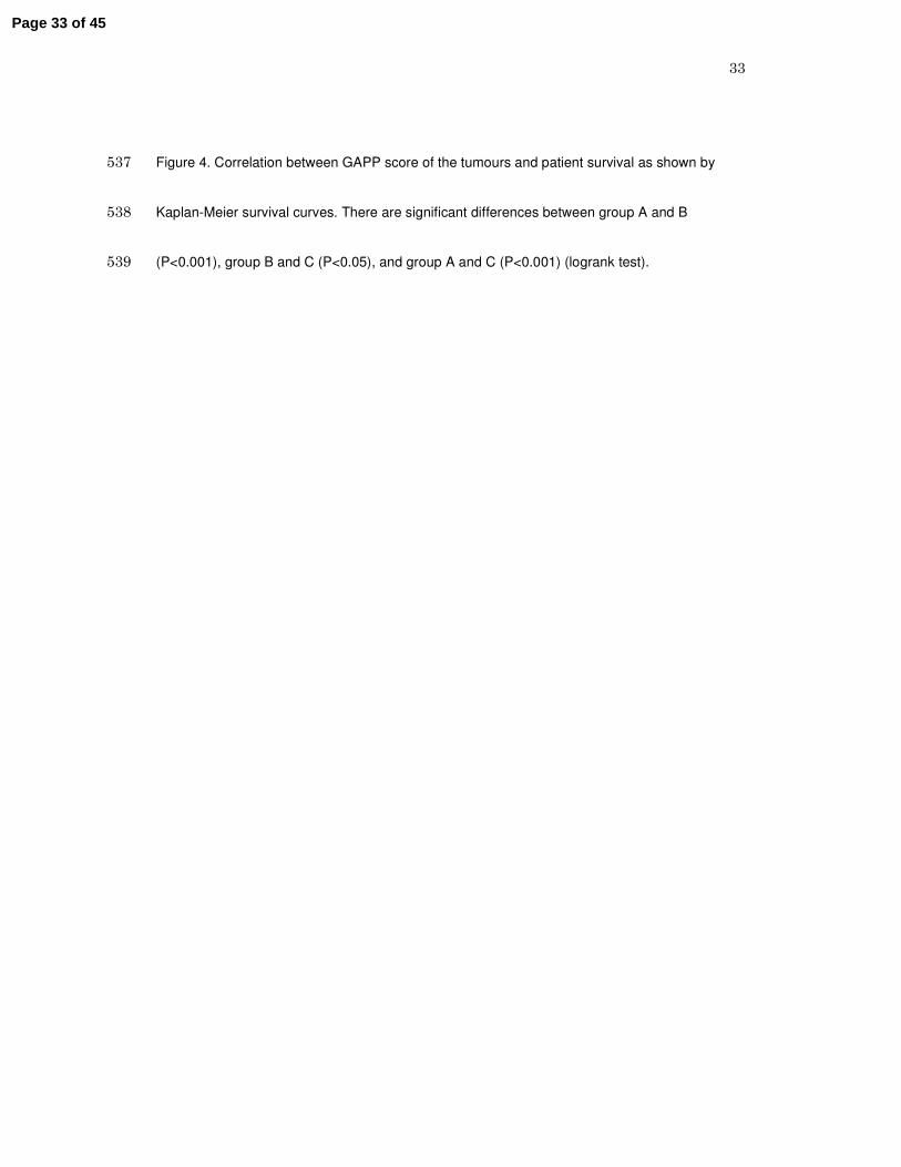
Figure 1-1. Histological pattern. A: regular zellballen pattern, B: regular zellballen 523
pattern, C: large irregular zellballen pattern, D: pseudorosette pattern (×100) 524
Figure 1-2. Cellularity. A grid in the circle on the left corner shows the area used to 525
count cellularity. These images were taken under the same magnification (×400). 526
Figure 1-3. Comedo type necrosis. Arrows indicate focus of coagulation necrosis 527
(×200). 528
Figure 1-4. Ki67 labelling index (×200). 529
Figure 2. Comparison of GAPP scores between non-metastatic (A) and metastatic (B) 530
PHEO/PGL groups. GAPP scores were 2.08 ± 0.17 in the non-metastatic group, and 5.33 ± 0.43 531
in the metastatic group (mean ± SE). There was a significant difference between these two 532
groups (P<0.001). 533
Figure 3. GAPP scores of metastatic tumours and duration between the initial operation and the 534
time of metastasis. Tumours with high GAPP scores metastasised sooner than those with low 535
scores (correlation coefficient: r=−0.438, P<0.01). 536
Page 32 of 45
33
Figure 4. Correlation between GAPP score of the tumours and patient survival as shown by 537
Kaplan-Meier survival curves. There are significant differences between group A and B 538
(P<0.001), group B and C (P<0.05), and group A and C (P<0.001) (logrank test). 539
Page 33 of 45
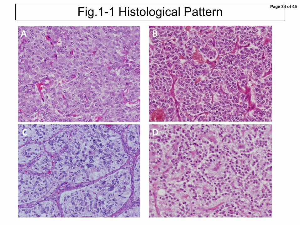
Fig.1-1 Histological Pattern A B
C D
Page 34 of 45
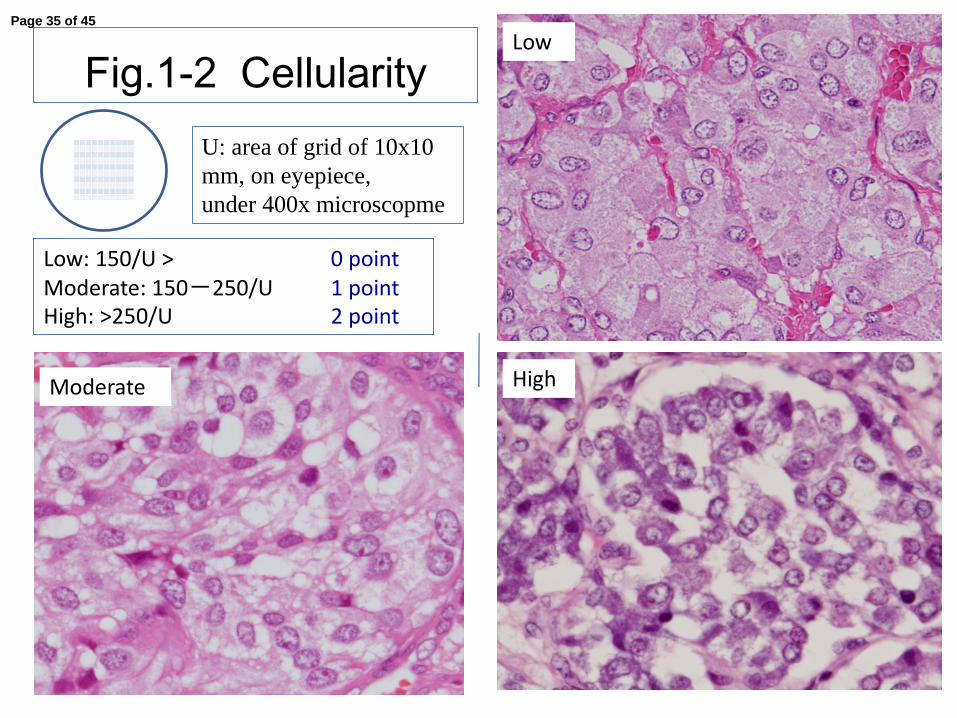
Fig.1-2 Cellularity
High
Low: 150/U > 0 point Moderate: 150-250/U 1 point High: >250/U 2 point
Low
Moderate
U: area of grid of 10x10 mm, on eyepiece, under 400x microscopme
Page 35 of 45
Fig. 1-3 Comedo type necrosis
Page 36 of 45
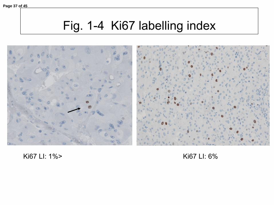
Fig. 1-4 Ki67 labelling index
Ki67 LI: 1%> Ki67 LI: 6%
Page 37 of 45
254x190mm (72 x 72 DPI)
Page 38 of 45
254x190mm (72 x 72 DPI)
Page 39 of 45

Page 40 of 45
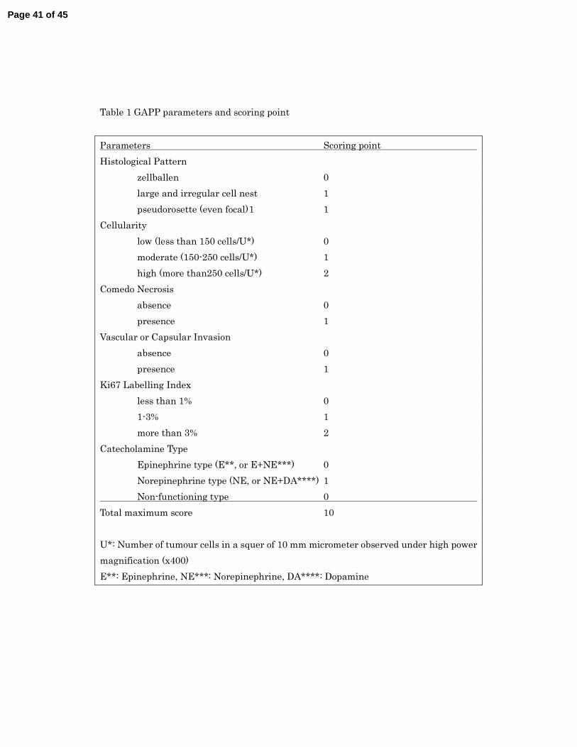
Table 1 GAPP parameters and scoring point
Parameters Scoring point
Histological Pattern
zellballen 0
large and irregular cell nest 1
pseudorosette (even focal) 1 1
Cellularity
low (less than 150 cells/U*) 0
moderate (150-250 cells/U*) 1
high (more than250 cells/U*) 2
Comedo Necrosis
absence 0
presence 1
Vascular or Capsular Invasion
absence 0
presence 1
Ki67 Labelling Index
less than 1% 0
1-3% 1
more than 3% 2
Catecholamine Type
Epinephrine type (E**, or E+NE***) 0
Norepinephrine type (NE, or NE+DA****) 1
Non-functioning type 0
Total maximum score 10
U*: Number of tumour cells in a squer of 10 mm micrometer observed under high power
magnification (x400)
E**: Epinephrine, NE***: Norepinephrine, DA****: Dopamine
Page 41 of 45
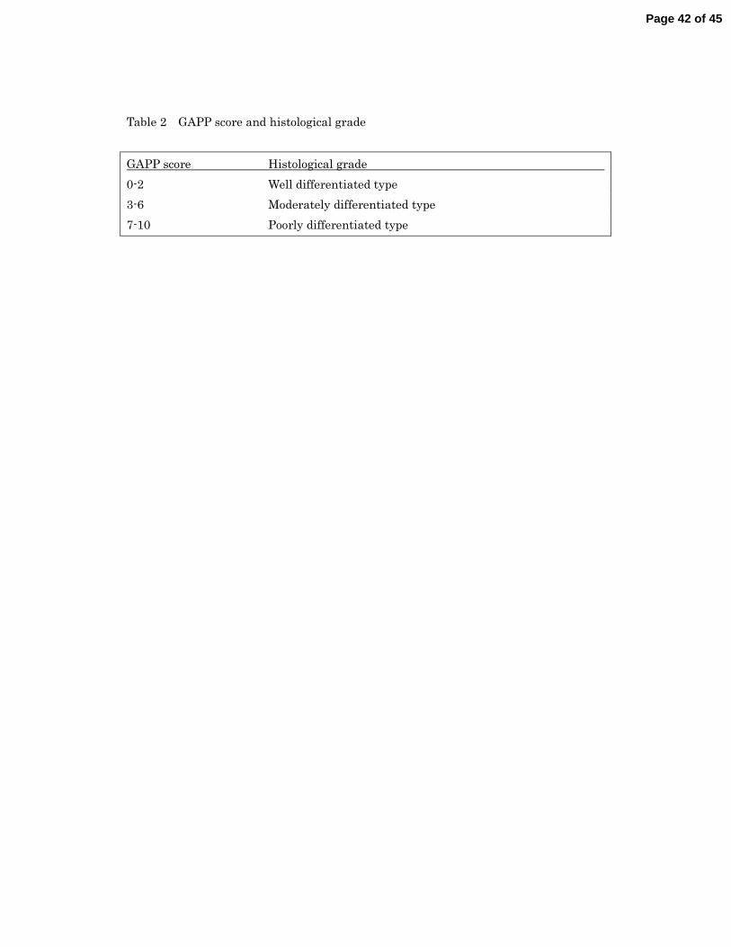
Table 2 GAPP score and histological grade
GAPP score Histological grade
0-2 Well differentiated type
3-6 Moderately differentiated type
7-10 Poorly differentiated type
Page 42 of 45
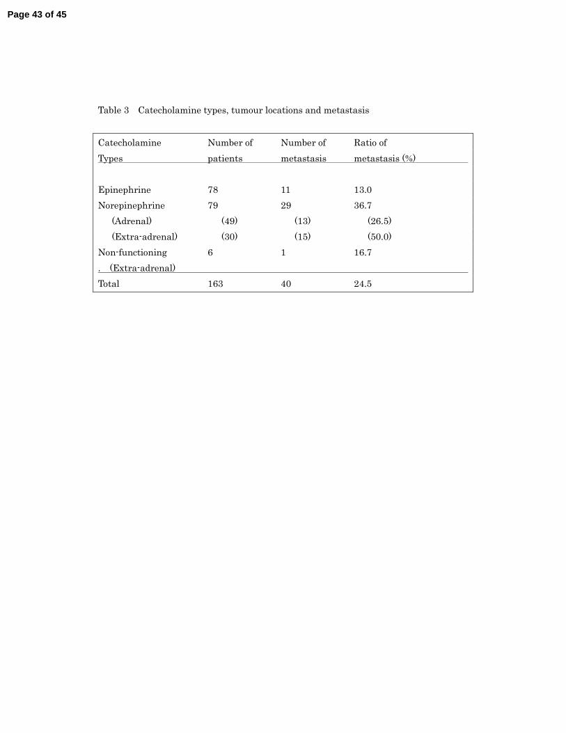
Table 3 Catecholamine types, tumour locations and metastasis
Catecholamine Number of Number of Ratio of
Types patients metastasis metastasis (%)
Epinephrine 78 11 13.0
Norepinephrine 79 29 36.7
(Adrenal) (49) (13) (26.5)
(Extra-adrenal) (30) (15) (50.0)
Non-functioning 6 1 16.7
. (Extra-adrenal)
Total 163 40 24.5
Page 43 of 45
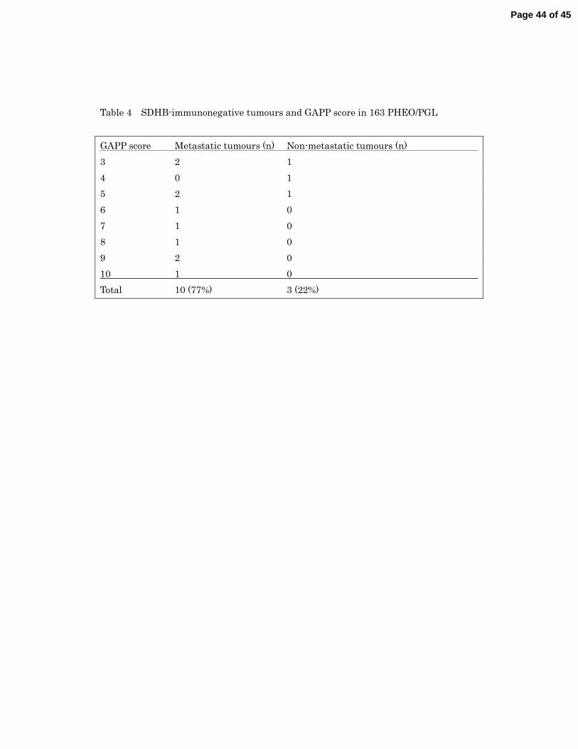
Table 4 SDHB-immunonegative tumours and GAPP score in 163 PHEO/PGL
GAPP score Metastatic tumours (n) Non-metastatic tumours (n)
3 2 1
4 0 1
5 2 1
6 1 0
7 1 0
8 1 0
9 2 0
10 1 0
Total 10 (77%) 3 (22%)
Page 44 of 45
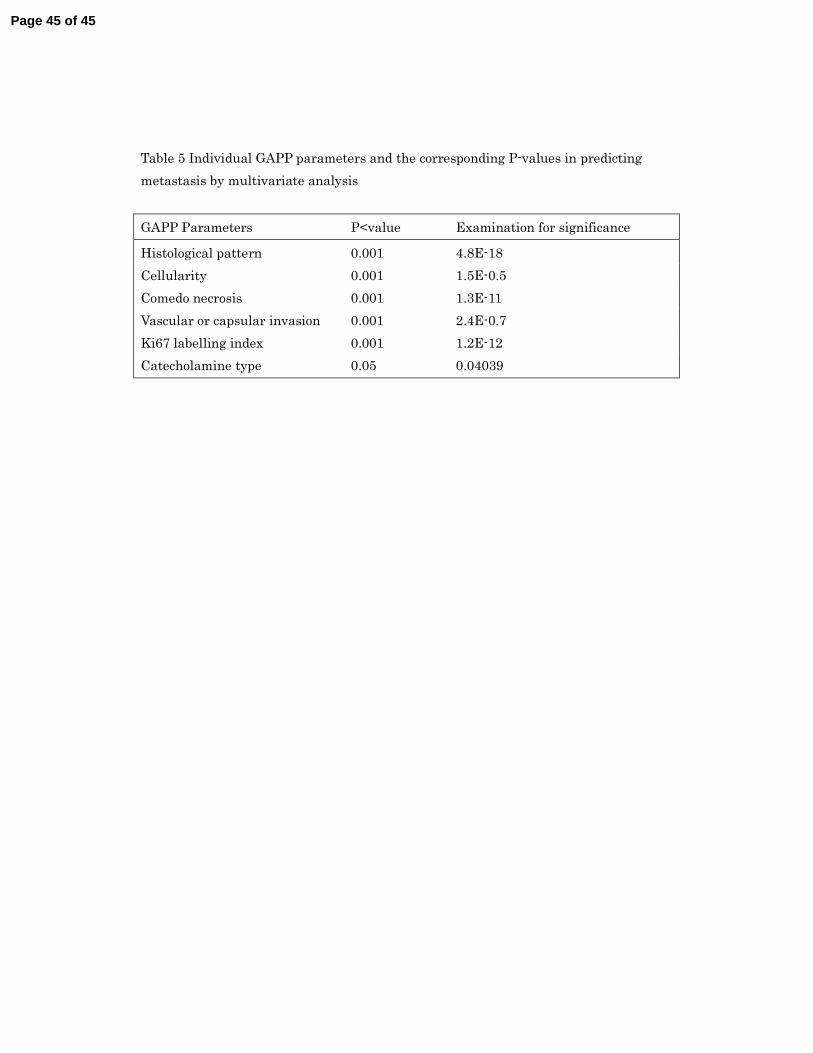
Table 5 Individual GAPP parameters and the corresponding P-values in predicting
metastasis by multivariate analysis
GAPP Parameters P<value Examination for significance
Histological pattern 0.001 4.8E-18
Cellularity 0.001 1.5E-0.5
Comedo necrosis 0.001 1.3E-11
Vascular or capsular invasion 0.001 2.4E-0.7
Ki67 labelling index 0.001 1.2E-12
Catecholamine type 0.05 0.04039
Page 45 of 45