Embed Size (px)
Citation preview
PEER-REVIEWED ARTICLE bioresources.com
Hastuti et al. (2019). “Nanocellulose & fermentation,” BioResources 14(3), 6936-6957. 6936
Nanocellulose from Oil Palm Biomass to Enhance Microbial Fermentation of Butanol for Bioenergy Applications
Novitri Hastuti,a,b Rizki Fitria Darmayanti,c,d Safrina Dyah Hardiningtyas,e,f
Kyohei Kanomata,a Kenji Sonomoto,c Masahiro Goto,e and Takuya Kitaoka a,*
Nanocellulose made by 2,2,6,6-tetramethylpiperidine-1-oxyl (TEMPO)-catalyzed oxidation, described as TEMPO-oxidized cellulose nanofibers (TOCNs), has a high density of negative charges on its surface. Its use in microbial fermentation systems is expected to benefit microbial process stability. In particular, microbial stability is strongly required in acetone–butanol–ethanol (ABE) fermentation associated with the solvent-extraction process of butanol production. Here, TOCNs derived from oil palm empty fruit bunches pulp were added to extractive ABE fermentation media containing glucose as a main source, which can be potentially obtained from biomass by saccharification. Then, microbial fermentation was carried out using free or immobilized bacterial cells, to produce butanol from glucose. The presence of TOCNs induced higher total butanol production in broth by improving the growth environment of Clostridium saccharoperbutylacetonicum N1-4, which was used as the butanol-producing strain. Microscopic analysis revealed that the spider-web-like TOCN network helped to entrap bacterial cells in alginate beads, by ionic crosslinking of TOCNs and alginates via Ca2+ ions, to increase stability of bacterial cells in the composite gel beads. The addition of TOCNs to fermentation media had significant positive effects on the total butanol yield.
Keywords: Nanocellulose; Fermentation; Biobutanol; Alginate; Cell immobilization; Oil palm empty fruit
bunch
Contact information: a: Department Agro-Environmental Sciences, Graduate School of Bioresource and Bioenvironmental Sciences, Faculty of Agriculture, Kyushu University, 744 Motooka, Nishi-ku, Fukuoka
819-0395, Japan; b: Forest Products Research and Development Center, Research Development and
Innovation Agency, Ministry of Environment and Forestry, Jalan Gunung Batu 5, Bogor, West Java 16610,
Indonesia; c: Department of Innovative Science and Technology for Bio-industry, Graduate School of
Bioresource and Bioenvironmental Sciences, Faculty of Agriculture, Kyushu University, 744 Motooka,
Nishi-ku, Fukuoka 819-0395, Japan; d: Department of Chemical Engineering, Faculty of Engineering,
University of Jember, Jalan Kalimantan, Kampus Tegal Boto, Jember, East Java 68121, Indonesia; e:
Department of Applied Chemistry, Graduate School of Engineering, Faculty of Engineering, Kyushu
University, 744 Motooka, Nishi-ku, Fukuoka 819-0395, Japan; f: Department of Aquatic Products
Technology, Faculty of Fisheries and Marine Sciences, Bogor Agricultural University, Jalan Raya
Dramaga, Bogor, West Java 16680, Indonesia; * Corresponding author: [email protected]
INTRODUCTION
Cellulose is the key material for the forthcoming sustainable society because it is
renewable and the most abundant biomass on Earth (Sadeghifar et al. 2017).
Nanocellulose, a crystalline bundle of cellulose macromolecules with nanometer-order
width, has recently emerged as a promising nanomaterial for various practical applications
PEER-REVIEWED ARTICLE bioresources.com
Hastuti et al. (2019). “Nanocellulose & fermentation,” BioResources 14(3), 6936-6957. 6937
because of its physicochemical properties such as high aspect ratio, elasticity, transparency,
thermal stability, chemical durability, and biodegradability (Fukuzumi et al. 2009, 2010;
de Mesquita et al. 2010). While woody biomass is the major cellulose resource, non-woody
biomass, such as oil palm empty fruit bunches (OPEFB), a large-scale byproduct of oil
palm plantations in Southeast Asia, is another attractive raw material for production of
nanocellulose because it contains 60% cellulose (Azrina et al. 2017). Various types of
nanocellulose are produced from cellulosic raw materials by physical disintegration,
chemical/enzymatic treatment, or combined processing (Jonoobi et al. 2011;
Nechyporchuk et al. 2016; Chen et al. 2017; Hastuti et al. 2018). Nanocellulose produced
by 2,2,6,6-tetramethylpiperidine-1-oxyl (TEMPO)-catalyzed oxidation, namely “TEMPO-
oxidized cellulose nanofibers” (TOCNs), have attracted considerable attention since they
require low energy for production and afford the narrowest reported nanofibers (Isogai et
al. 2011). TOCNs bear anionic carboxylate groups on their surfaces as the result of
TEMPO-mediated selective oxidation of surface-exposed primary alcohols to carboxylates
(Saito and Isogai 2004). These carboxylates induce a zeta potential as low as –70 mV on
the solid surfaces, resulting in high dispersibility of TOCNs in water by electrical repulsion
(Okita et al. 2010), and high dispersant effects for various suspended solid materials (Li et
al. 2015).
Although nanocellulose has been extensively studied for developing reinforcing
agents in composite plastics and films (Goetz et al. 2009; Yan et al. 2017), enhancers in
emulsion systems (Kalashnikova et al. 2012; Hu et al. 2015), functional hydrogels for
environmental remediation (Jin et al. 2015; Dwivedi et al. 2017), and carriers for metal
catalysts and enzyme immobilization (Azetsu et al. 2013; Sulaiman et al. 2014; Uddin et
al. 2017), its application in microbial systems remains limited. Yu et al. (2016) reported
the effects of the size of nanocellulose on microalgal flocculation and lipid metabolism;
cellulose nanofibrils effectively induced microalgal flocculation via a mechanical
interaction based on geometric properties such as nanocellulose morphology and hydrogen
bonding. Sun et al. (2014, 2015) observed aggregation of Pseudomonas fluorescens and
Escherichia coli K12 in the presence of nanocellulose; they found that bacterial
aggregation and adhesion to solid surfaces were significantly affected by the surrounding
solution chemistry. The electrostatic interaction promoted by the charged cellulose
nanocrystals could give rise to clustering, phase separation, and rapid aggregation of
negatively charged bacteria (Larsen et al. 2009; Sun et al. 2012, 2014). Clustering and
phase separation of bacteria are very important for product recovery in microbial biofuel
production.
Practical applications of microbial systems in the production of biofuels from
renewable resources are attracting much attention for environmental and economic reasons
(Zheng et al. 2009). Biobutanol produced by microbial fermentation is of significant
interest as demand for butanol as an industrial intermediate is rapidly increasing, and it can
be used directly in gasoline engines without any modification and/or substitution (Xue et
al. 2017). Butanol is the most attractive biofuel alternative to ethanol because it has
numerous desirable properties, including lower vapor pressure, higher calorific value, less
corrosive properties, and a non-hygroscopic nature (Dürre 2007). Butanol can be produced
from renewable resources through acetone–butanol–ethanol (ABE) fermentation (Lee et
al. 1995) using Clostridia as a high-performance butanol-producing strain (Tashiro et al.
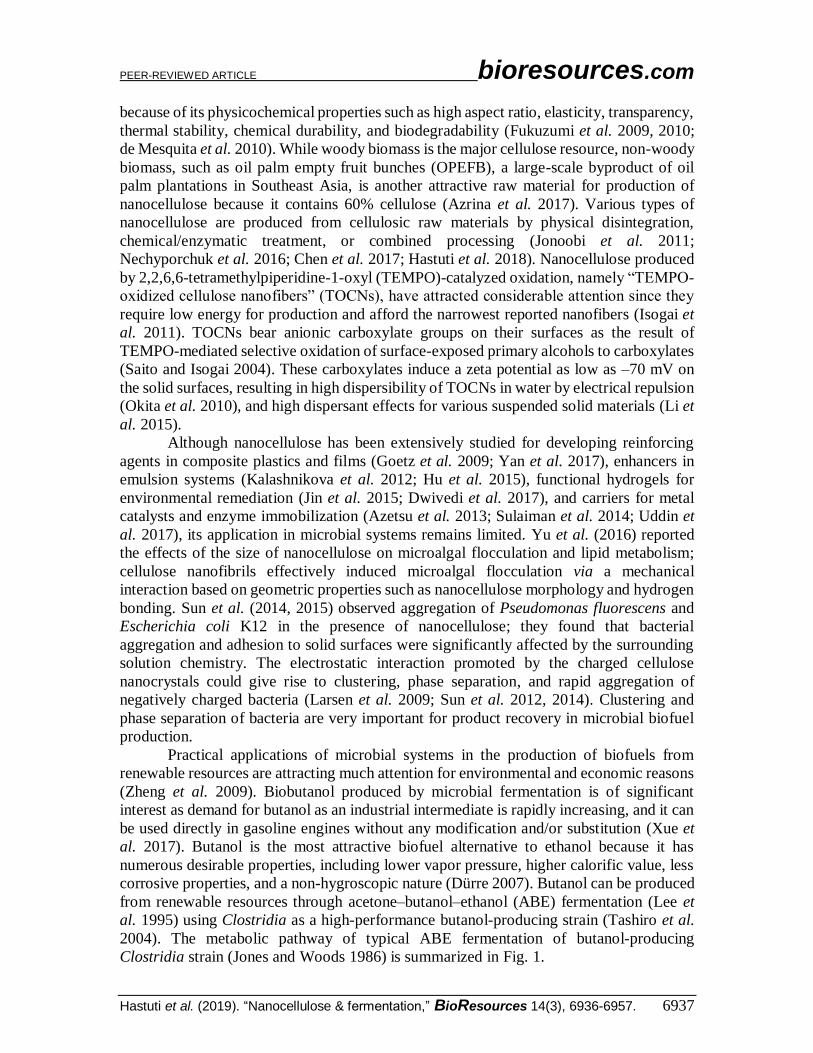
2004). The metabolic pathway of typical ABE fermentation of butanol-producing
Clostridia strain (Jones and Woods 1986) is summarized in Fig. 1.
PEER-REVIEWED ARTICLE bioresources.com
Hastuti et al. (2019). “Nanocellulose & fermentation,” BioResources 14(3), 6936-6957. 6938
Fig. 1. Metabolic pathway of acetone–butanol–ethanol (ABE) fermentation in butanol-producing Clostridia strain. Several enzymes involved in ABE fermentation are described in italics. These abbreviations are: AK, acetate kinase; PTA, phosphotransacetylase; CoAT, CoA transferase; PTB, phosphotransbutyrylase; BK, butyrate kinase; BADH, butyraldehyde dehydrogenase; BDH, butanol dehydrogenase.
However, microbial butanol production has suffered from severe product inhibition
in fermentation processes and high cost of product recovery, resulting in low butanol
productivity. Studies have been conducted to overcome these obstacles by integrating a
butanol separation step, such as cell immobilization, extractive fermentation,
pervaporation, or perstraction (Barton and Daugulis 1992; Qureshi and Maddox 1995,
2005). Among these, extractive fermentation (liquid-liquid extraction) has great potential
to increase the product titer if one can determine the most appropriate solvent for butanol
selectivity that is compatible with the cells producing the butanol (Qureshi and Maddox
1995; Ishizaki et al. 1999). In extractive fermentation, the broth is in contact with an
extracting solvent; therefore, some inhibitory products become dissolved in the solvent,
resulting in reduction of inhibitory effects on the culture (Roffler et al. 1987). However,
the efficiency of this method depends on the affinity of solutes for the extraction solvent
and the mixing ratio of the phases (de Jesus et al. 2019). Extractive fermentation may result
in inactivation of cells due to the extensive exposure of cells to extraction solvents and
product toxicity (Ishii et al. 1985). Therefore, advanced technology to immobilize cells has
been investigated to overcome such problems by using silica gel, pumice, and Ca-alginate
as microbial carriers and has been applied in microbial fermentation (Napoli et al. 2010;
Pereira et al. 2014). However, to the best of our knowledge, no study has reported an
effective strategy using nanocellulose to improve microbial stability to enhance butanol
productivity.
PEER-REVIEWED ARTICLE bioresources.com
Hastuti et al. (2019). “Nanocellulose & fermentation,” BioResources 14(3), 6936-6957. 6939
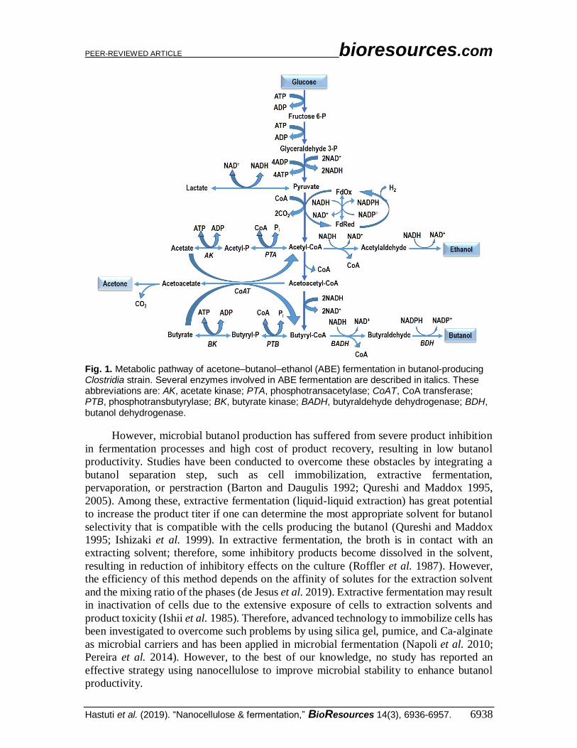
Fig. 2. Overview of use of 2,2,6,6-tetramethylpiperidine-1-oxyl (TEMPO)-oxidized cellulose nanofibers (TOCNs) in microbial biobutanol production.
Herein, microbial stability in biobutanol production was addressed by using in-
broth TOCNs, which were produced from OPEFB waste. The characteristics of raw pulp
OPEFB and the resultant TOCNs are described in Table S1 and Figs. S1–S4 in the
Appendix. TOCNs have surface anionic carboxylate groups with high density (up to 1.5
mmol g–1) and aspect ratio more than 40. The characteristics of TOCNs from company
made from wood are described in Fig. S5; these can promote electrostatic repulsion of
negatively charged bacteria producing biobutanol, which is important in microbial system
stability by preventing bacterial aggregation and then enhancing microbial dispersibility
(Fukuzumi et al. 2009; Sun et al. 2012). The strategy is illustrated in Fig. 2. The addition
of TOCNs improved microbial stability and increased butanol production. The addition of
TOCNs in extractive fermentation of biobutanol significantly improved cell growth and
the total butanol concentration after 96-h fermentation. This new application of
nanocellulose obtained from the low-cost agricultural residue OPEFB in bioalcohol
production is expected to expand the potential of natural nanomaterials from biomass.
EXPERIMENTAL Materials Microorganism inoculation
Clostridium saccharoperbutylacetonicum N1-4 ATCC 13564 was used in this
study. One milliliter of spore suspension of C. saccharoperbutylacetonicum was
transferred from sand stock and refreshed in 9 mL of fresh potato glucose medium (10%
v/v) (Ishizaki et al. 1999). The spore suspension was heat-shocked in a water bath at 100
°C for 1 min, and then refreshed at 30 °C for 24 h anaerobically using an Anaeropack
(Mitsubishi Gas Chemical, Co., Inc., Tokyo, Japan). The refreshed culture broth was
inoculated into tryptone–yeast extract–acetate (TYA) medium (Tashiro et al. 2004). After
PEER-REVIEWED ARTICLE bioresources.com
Hastuti et al. (2019). “Nanocellulose & fermentation,” BioResources 14(3), 6936-6957. 6940
inoculation, the culture broth was incubated anaerobically at 30 °C for 15 h using an
Anaeropack, and then used as a seed culture.
Methods Extractive fed-batch fermentation with free cells
Fed-batch extractive fermentation with free cells was performed in a 25-mL portion
of TYA broth and a 50-mL portion of extractant, which was composed of a 1:1 (v/v)
mixture of oleyl alcohol and tributyrin; the volume ratio of extractant (Ve) to broth (Vb) was
thus 2.0 (Ve/Vb = 2.0). In brief, seed culture (2.5 mL) was inoculated into a 25-mL portion
of TYA medium, containing glucose (2.5 mL of 50 g/L solution), calcium carbonate (2.5
mL of 3 g/L solution), and TOCNs (5.0 mL of 0.5% w/v aqueous dispersion, 25 mg of
TOCNs in dry weight; see the Supporting Information for preparation of TOCNs) in a 500-
mL Erlenmeyer flask closed with a rubber seal, as an aqueous phase. The flask was then
sparged with nitrogen gas for 10 min to obtain anaerobic conditions. The fed-batch cultures
were grown at 30 °C in a shaker (100 rpm) and by feeding 1 g of glucose powder at 24, 48,
and 72 h after seeding and maintained the anaerobic condition by sparging with N2 after
glucose feeding.
Extractive fed-batch fermentation with immobilized cells
A solution of sodium alginate (4% w/v) was prepared in boiling water and
autoclaved at 115 °C for 15 min. Precultured cells (2.5 mL) and TOCNs (0.5% w/v, 25 mg
dry weight) were added into sterilized saline water (20 mL, 0.85% NaCl). The resultant
suspension was mixed with an equal volume of 4% sodium alginate solution (final
concentration of sodium alginate, 2%). The mixture was dropped into a 3% CaCl2 solution
using a syringe with continuous stirring to form alginate beads containing cells and
TOCNs. The resultant beads were recovered by filtration. Cell-containing alginate beads
without TOCNs were also prepared as a control. The diameters of beads were
approximately 5 mm. Extractive fed-batch fermentation with immobilized cells was
performed as described above (extractive fed-batch fermentation with free cells).
Analytical methods
Cell density was determined by measuring the optical density of the suspension at
562 nm (OD562) using a UV/vis spectrophotometer (Bio-Spec, Shimadzu, Kyoto, Japan)
after diluting the samples. The dry cell weight (DCW) was calculated as previously
reported (Yoshida et al. 2012),
DCW = 0.301 × OD562 × D – 0.0008 (1)
where D is the dilution ratio.
The total butanol concentration [BuOH]Total was defined as the total amount of
butanol produced in all the phases per broth volume (g/L-broth), and calculated as follows
(Darmayanti et al. 2018),
[BuOH]Total = [BuOH]b Vb + [BuOH]c Vc + [BuOH]e Ve) / Vb (2)
PEER-REVIEWED ARTICLE bioresources.com
Hastuti et al. (2019). “Nanocellulose & fermentation,” BioResources 14(3), 6936-6957. 6941
where [BuOH]b and Vb are the butanol concentration (g/L) in the broth and the volume (L)
of the broth, [BuOH]c and Vc are the butanol concentration in the cell beads and the volume
of the beads, and [BuOH]e and Ve are the butanol concentration in the extractant and the
volume of the extractant, respectively. Vc the volume of alginate beads, is 1.5 mL for every
measurement because six beads were crushed in aqueous sodium citrate solution in a total
volume of 1.5 mL.
The concentration of glucose was measured in the supernatant liquid obtained by
centrifugation of broth by using a high-performance liquid chromatograph (US-HPLC-
1210, JASCO, Tokyo, Japan) equipped with a refractive index detector and SH-1011
column (Shodex, Tokyo, Japan). Aqueous H2SO4 (0.05 mM) was used as the mobile phase
(1.0 mL/min, 50°C), using an injection volume of 20 μL. The concentration of butanol was
measured in supernatant obtained by centrifugation of both extractant and broth using a
gas chromatograph (6890A, Agilent Technologies, Palo Alto, CA, USA) equipped with a
flame ionization detector and a 15-m capillary column (INNOWAX 19095N-121, Agilent
Technologies) in previously reported conditions (Tashiro et al. 2004).
The distribution coefficient (Kd) of butanol between the extractant (oil phase) and
broth (aqueous phase) was calculated using Eq. 3.
Kd = [BuOH]e / [BuOH]b (3)
The treatments of free TOCNs and the presence of TOCNs were evaluated with analysis
of variance (ANOVA) reported as p-values. Findings with p-values less than 0.05
suggested that differences were statistically significant.
Microscopic analysis
Samples for scanning electron microscopy (SEM) analysis were prepared as
follows. Samples of free cells were prepared by collecting a 1.5-mL portion of broth, which
was centrifuged at 120 rpm for 20 min to obtain cells as the precipitate. For immobilized
cells, several alginate beads were collected from the broth and cut into smaller pieces. A
500-µL portion of broth from free cells or the cut beads containing immobilized cells were
fixed by 2% formaldehyde (300 µL) and phosphate buffer (1 mL) at pH 5.5 and 4 °C,
overnight. Half of the fixed cells were collected and washed with deionized water and
centrifuged at 120 rpm for 10 min.
The precipitate was stained with 1% OsO4 solution for 4 h and washed with
deionized water. The specimen was washed successively with 50, 70, 80, and 99.5%
ethanol, each for 5 min. The specimens were freeze-dried and observed using an SU-8000
apparatus (Hitachi, Tokyo, Japan) at the Center of Advanced Instrumental Analysis,
Kyushu University.
The cells were observed using a confocal laser scanning microscope (LSM 700,
Carl Zeiss AG, Oberkochen, Germany). A 500-µL portion of culture medium containing
free cells was collected in a 1.5-mL plastic tube and washed with phosphate buffer at pH
5.5 by centrifugation.
The precipitate was stained with 4',6-diamidino-2-phenylindole (DAPI), and
washed again with phosphate buffer by centrifugation. The cells in alginate beads were
treated in the same manner after destroying the beads by treatment with 500 µL 0.2 M
sodium citrate for 2 h. A 20-µL portion of prepared cells in phosphate buffer was put on
an observation glass and observed under a 405-nm laser.
PEER-REVIEWED ARTICLE bioresources.com
Hastuti et al. (2019). “Nanocellulose & fermentation,” BioResources 14(3), 6936-6957. 6942
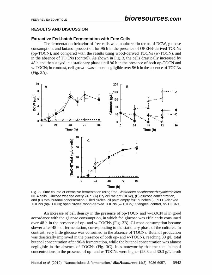
RESULTS AND DISCUSSION Extractive Fed-batch Fermentation with Free Cells
The fermentation behavior of free cells was monitored in terms of DCW, glucose
consumption, and butanol production for 96 h in the presence of OPEFB-derived TOCNs
(op-TOCN), and compared with the results using wood-derived TOCNs (w-TOCN), and
in the absence of TOCNs (control). As shown in Fig. 3, the cells drastically increased by
48 h and then stayed in a stationary phase until 96 h in the presence of both op-TOCN and
w-TOCN; in contrast, cell growth was almost negligible over 96 h in the absence of TOCNs
(Fig. 3A).
Fig. 3. Time course of extractive fermentation using free Clostridium saccharoperbutylacetonicum N1-4 cells. Glucose was fed every 24 h. (A) Dry cell weight (DCW), (B) glucose concentration, and (C) total butanol concentration. Filled circles: oil palm empty fruit bunches (OPEFB)-derived TOCNs (op-TOCN); open circles: wood-derived TOCNs (w-TOCN); triangles: control, no TOCNs.
An increase of cell density in the presence of op-TOCN and w-TOCN is in good
accordance with the glucose consumption, in which fed glucose was efficiently consumed
over 48 h in the presence of op- and w-TOCNs (Fig. 3B). Glucose consumption became
slower after 48 h of fermentation, corresponding to the stationary phase of the cultures. In
contrast, very little glucose was consumed in the absence of TOCNs. Butanol production
was drastically improved in the presence of both op- and w-TOCNs, reaching 30 g/L total
butanol concentration after 96-h fermentation, while the butanol concentration was almost
negligible in the absence of TOCNs (Fig. 3C). It is noteworthy that the total butanol
concentrations in the presence of op- and w-TOCNs were higher (28.8 and 30.3 g/L-broth
PEER-REVIEWED ARTICLE bioresources.com
Hastuti et al. (2019). “Nanocellulose & fermentation,” BioResources 14(3), 6936-6957. 6943
after 96 h, respectively) than that reported in previous work (24.2 g/L-broth) (Darmayanti
et al. 2018) in which extractive fermentation was carried out by the free cell method using
a large ratio of extractant to broth (Ve/Vb = 5.0) and C. saccharoperbutylacetonicum N1-4
was used as butanol-producing strain. The distribution coefficients of butanol (Kd) in the
present TOCN systems were also higher (3.98 for op-TOCN and 4.97 for w-TOCN) than
in the previous work (Kd = 3.14).
The time course of cell growth is in good accordance with the general features of
the metabolic pathway of butanol production by C. saccharoperbutylacetonicum N1-4,
which consists of two phases, acidogenesis and solventogenesis (Jones and Woods 1986).
The growth behavior in the first 48 h corresponds to acidogenesis, during which cells
rapidly grow while producing acetic acid and butyric acid. Solventogenesis occurs in the
stationary phase, during which the cells reassimilate the previously excreted acids to form
acetone, butanol, and ethanol (Shinto et al. 2007).
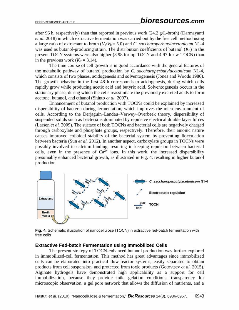
Enhancement of butanol production with TOCNs could be explained by increased
dispersibility of bacteria during fermentation, which improves the microenvironment of
cells. According to the Derjaguin–Landau–Verwey–Overbeek theory, dispersibility of
suspended solids such as bacteria is dominated by repulsive electrical double layer forces
(Larsen et al. 2009). The surface of both TOCNs and bacterial cells are negatively charged
through carboxylate and phosphate groups, respectively. Therefore, their anionic nature
causes improved colloidal stability of the bacterial system by preventing flocculation
between bacteria (Sun et al. 2012). In another aspect, carboxylate groups in TOCNs were
possibly involved in calcium binding, resulting in keeping repulsion between bacterial
cells, even in the presence of Ca2+ ions. In this work, the increased dispersibility
presumably enhanced bacterial growth, as illustrated in Fig. 4, resulting in higher butanol
production.
Fig. 4. Schematic illustration of nanocellulose (TOCN) in extractive fed-batch fermentation with free cells
Extractive Fed-batch Fermentation using Immobilized Cells
The present strategy of TOCN-enhanced butanol production was further explored
in immobilized-cell fermentation. This method has great advantages since immobilized
cells can be elaborated into practical flow-reactor systems, easily separated to obtain
products from cell suspension, and protected from toxic products (Gotovtsev et al. 2015).
Alginate hydrogels have demonstrated high applicability as a support for cell
immobilization, because they provide mild gelation conditions, transparency for
microscopic observation, a gel pore network that allows the diffusion of nutrients, and a
PEER-REVIEWED ARTICLE bioresources.com
Hastuti et al. (2019). “Nanocellulose & fermentation,” BioResources 14(3), 6936-6957. 6944
gentle environment for the entrapped materials (Smidsrod and Skjak-Braek 1990;
Andersen et al. 2015).
Cell immobilization in alginate beads was applied to the present extractive
fermentation for biobutanol production by introducing bacterial cells and TOCNs together
into alginate beads. Fermentation was conducted using cells immobilized with op- and w-
TOCNs, and cells immobilized without TOCNs served as a control. The time courses of
DCW, glucose consumption and butanol production for immobilized-cell fermentation are
shown in Fig. 5. Interestingly, the DCW with op-TOCN continued to increase for 72 h, and
remained at a high level up to 96 h. However, DCW with w-TOCN and in controls
decreased after 48 h of fermentation (Fig. 5A).
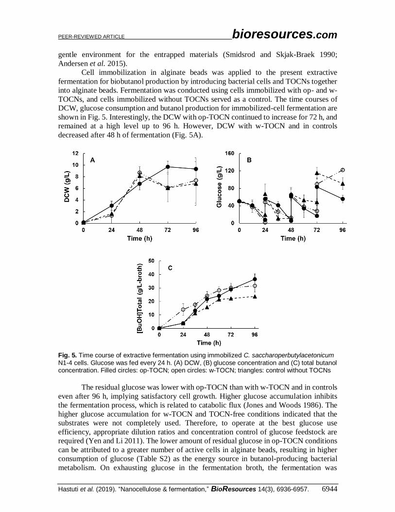
Fig. 5. Time course of extractive fermentation using immobilized C. saccharoperbutylacetonicum N1-4 cells. Glucose was fed every 24 h. (A) DCW, (B) glucose concentration and (C) total butanol concentration. Filled circles: op-TOCN; open circles: w-TOCN; triangles: control without TOCNs
The residual glucose was lower with op-TOCN than with w-TOCN and in controls
even after 96 h, implying satisfactory cell growth. Higher glucose accumulation inhibits
the fermentation process, which is related to catabolic flux (Jones and Woods 1986). The
higher glucose accumulation for w-TOCN and TOCN-free conditions indicated that the
substrates were not completely used. Therefore, to operate at the best glucose use
efficiency, appropriate dilution ratios and concentration control of glucose feedstock are
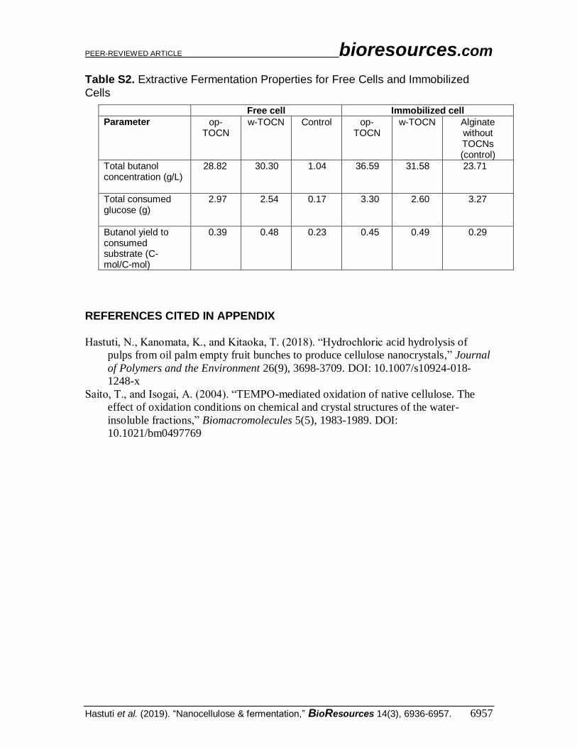
required (Yen and Li 2011). The lower amount of residual glucose in op-TOCN conditions
can be attributed to a greater number of active cells in alginate beads, resulting in higher
consumption of glucose (Table S2) as the energy source in butanol-producing bacterial
metabolism. On exhausting glucose in the fermentation broth, the fermentation was
PEER-REVIEWED ARTICLE bioresources.com
Hastuti et al. (2019). “Nanocellulose & fermentation,” BioResources 14(3), 6936-6957. 6945
immediately stopped (Zhao et al. 2019). Besides, solvent extraction generally enhances
glucose consumption, and the solvent production (butanol) in the presence of op-TOCNs
appears to be the highest among the conditions tested (Figs. 5A–C) (Ishizaki et al. 1999).
The total butanol concentrations in the immobilized-cell treatments were 36.59 g/L-
broth for op-TOCN, 31.58 g/L-broth for w-TOCN, and 23.71 g/L-broth for controls
(TOCN-free) (Fig. 5C). These results were comparable with, but higher than, those for the
free-cell method, which ranged from 29 to 30 g/L-broth in the presence of TOCNs. The
slight increase of total butanol concentration after 96-h fermentation indicated that
immobilization contributed to the extractive fermentation by altering cell density (in the
alginate beads), but did not alter the metabolic pathway (Yen and Li 2011). It is noteworthy
that the total butanol production in the presence of TOCNs (Ve/Vb = 2.0) was higher than
that reported for an immobilization method using a large ratio of extractant, Ve/Vb = 5 (30.9
g/L-broth) (Darmayanti et al. 2018).
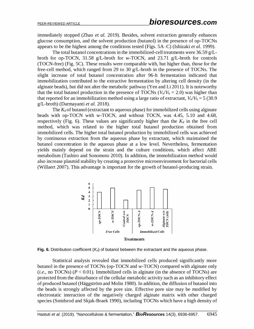
The Kd of butanol (extractant to aqueous phase) for immobilized cells using alginate
beads with op-TOCN with w-TOCN, and without TOCN, was 4.45, 5.10 and 4.68,
respectively (Fig. 6). These values are significantly higher than the Kd in the free cell
method, which was related to the higher total butanol production obtained from
immobilized cells. The higher total butanol production by immobilized cells was achieved
by continuous extraction from the aqueous phase by extractant, which maintained the
butanol concentration in the aqueous phase at a low level. Nevertheless, fermentation
yields mainly depend on the strain and the culture conditions, which affect ABE
metabolism (Tashiro and Sonomoto 2010). In addition, the immobilization method would
also increase plasmid stability by creating a protective microenvironment for bacterial cells
(Willaert 2007). This advantage is important for the growth of butanol-producing strain.
Fig. 6. Distribution coefficient (Kd) of butanol between the extractant and the aqueous phase.
Statistical analysis revealed that immobilized cells produced significantly more
butanol in the presence of TOCNs (op-TOCN and w-TOCN) compared with alginate only
(i.e., no TOCNs) (P < 0.01). Immobilized cells in alginate (in the absence of TOCNs) are
protected from the disturbance of the cellular metabolic activity such as an inhibitory effect
of produced butanol (Häggström and Molin 1980). In addition, the diffusion of butanol into
the beads is strongly affected by the pore size. Effective pore size may be modified by
electrostatic interaction of the negatively charged alginate matrix with other charged
species (Smidsrod and Skjak-Braek 1990), including TOCNs which have a high density of
PEER-REVIEWED ARTICLE bioresources.com
Hastuti et al. (2019). “Nanocellulose & fermentation,” BioResources 14(3), 6936-6957. 6946
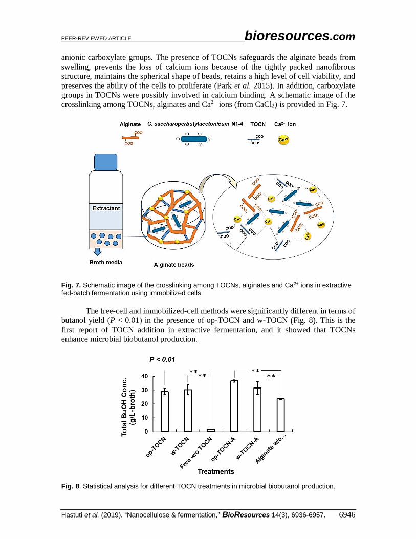
anionic carboxylate groups. The presence of TOCNs safeguards the alginate beads from
swelling, prevents the loss of calcium ions because of the tightly packed nanofibrous
structure, maintains the spherical shape of beads, retains a high level of cell viability, and
preserves the ability of the cells to proliferate (Park et al. 2015). In addition, carboxylate
groups in TOCNs were possibly involved in calcium binding. A schematic image of the
crosslinking among TOCNs, alginates and Ca2+ ions (from CaCl2) is provided in Fig. 7.
Fig. 7. Schematic image of the crosslinking among TOCNs, alginates and Ca2+ ions in extractive fed-batch fermentation using immobilized cells
The free-cell and immobilized-cell methods were significantly different in terms of
butanol yield (P < 0.01) in the presence of op-TOCN and w-TOCN (Fig. 8). This is the
first report of TOCN addition in extractive fermentation, and it showed that TOCNs
enhance microbial biobutanol production.
Fig. 8. Statistical analysis for different TOCN treatments in microbial biobutanol production.
PEER-REVIEWED ARTICLE bioresources.com
Hastuti et al. (2019). “Nanocellulose & fermentation,” BioResources 14(3), 6936-6957. 6947
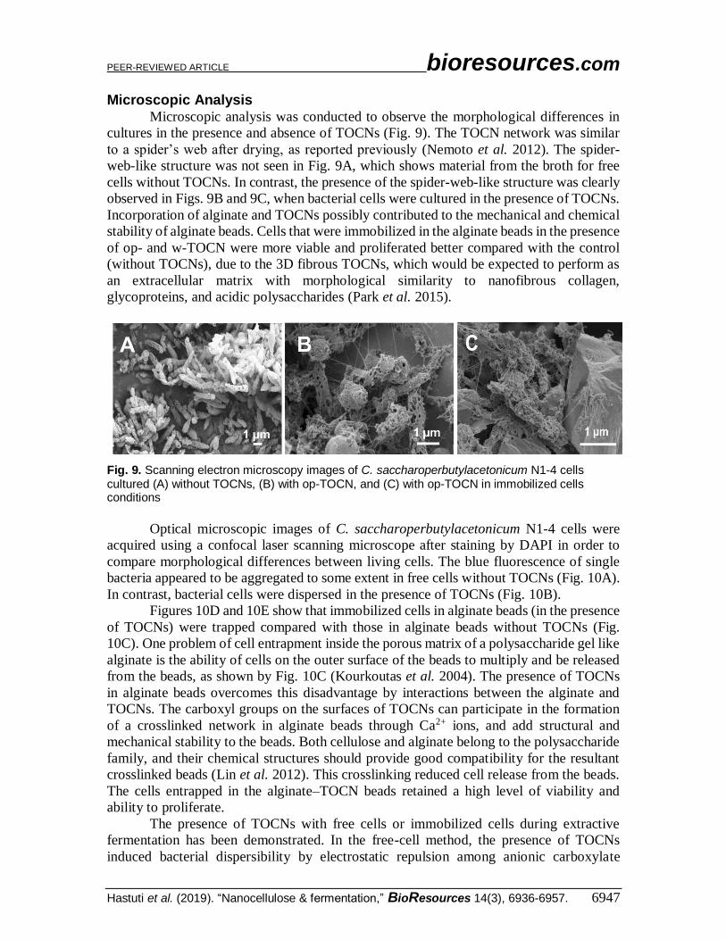
Microscopic Analysis Microscopic analysis was conducted to observe the morphological differences in
cultures in the presence and absence of TOCNs (Fig. 9). The TOCN network was similar
to a spider’s web after drying, as reported previously (Nemoto et al. 2012). The spider-
web-like structure was not seen in Fig. 9A, which shows material from the broth for free
cells without TOCNs. In contrast, the presence of the spider-web-like structure was clearly
observed in Figs. 9B and 9C, when bacterial cells were cultured in the presence of TOCNs.
Incorporation of alginate and TOCNs possibly contributed to the mechanical and chemical
stability of alginate beads. Cells that were immobilized in the alginate beads in the presence
of op- and w-TOCN were more viable and proliferated better compared with the control
(without TOCNs), due to the 3D fibrous TOCNs, which would be expected to perform as
an extracellular matrix with morphological similarity to nanofibrous collagen,
glycoproteins, and acidic polysaccharides (Park et al. 2015).
Fig. 9. Scanning electron microscopy images of C. saccharoperbutylacetonicum N1-4 cells cultured (A) without TOCNs, (B) with op-TOCN, and (C) with op-TOCN in immobilized cells conditions
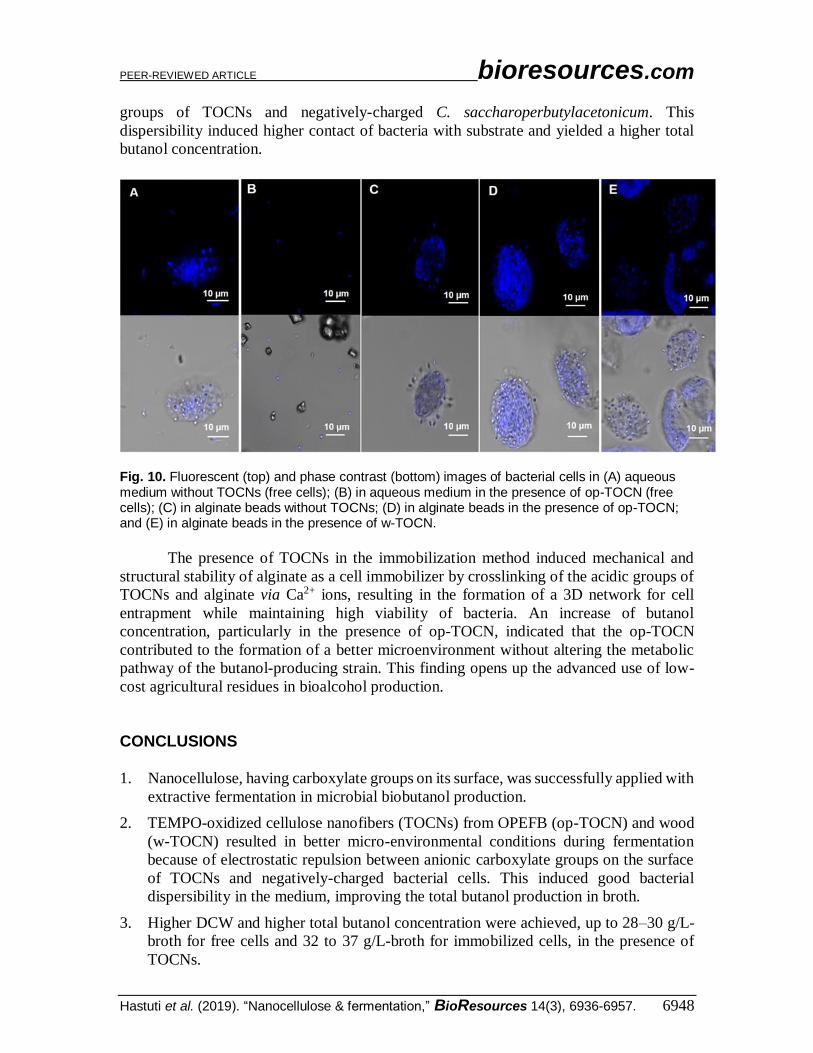
Optical microscopic images of C. saccharoperbutylacetonicum N1-4 cells were
acquired using a confocal laser scanning microscope after staining by DAPI in order to
compare morphological differences between living cells. The blue fluorescence of single
bacteria appeared to be aggregated to some extent in free cells without TOCNs (Fig. 10A).
In contrast, bacterial cells were dispersed in the presence of TOCNs (Fig. 10B).
Figures 10D and 10E show that immobilized cells in alginate beads (in the presence
of TOCNs) were trapped compared with those in alginate beads without TOCNs (Fig.
10C). One problem of cell entrapment inside the porous matrix of a polysaccharide gel like
alginate is the ability of cells on the outer surface of the beads to multiply and be released
from the beads, as shown by Fig. 10C (Kourkoutas et al. 2004). The presence of TOCNs
in alginate beads overcomes this disadvantage by interactions between the alginate and
TOCNs. The carboxyl groups on the surfaces of TOCNs can participate in the formation
of a crosslinked network in alginate beads through Ca2+ ions, and add structural and
mechanical stability to the beads. Both cellulose and alginate belong to the polysaccharide
family, and their chemical structures should provide good compatibility for the resultant
crosslinked beads (Lin et al. 2012). This crosslinking reduced cell release from the beads.
The cells entrapped in the alginate–TOCN beads retained a high level of viability and
ability to proliferate.
The presence of TOCNs with free cells or immobilized cells during extractive
fermentation has been demonstrated. In the free-cell method, the presence of TOCNs
induced bacterial dispersibility by electrostatic repulsion among anionic carboxylate
PEER-REVIEWED ARTICLE bioresources.com
Hastuti et al. (2019). “Nanocellulose & fermentation,” BioResources 14(3), 6936-6957. 6948
groups of TOCNs and negatively-charged C. saccharoperbutylacetonicum. This
dispersibility induced higher contact of bacteria with substrate and yielded a higher total
butanol concentration.
Fig. 10. Fluorescent (top) and phase contrast (bottom) images of bacterial cells in (A) aqueous medium without TOCNs (free cells); (B) in aqueous medium in the presence of op-TOCN (free cells); (C) in alginate beads without TOCNs; (D) in alginate beads in the presence of op-TOCN; and (E) in alginate beads in the presence of w-TOCN.
The presence of TOCNs in the immobilization method induced mechanical and
structural stability of alginate as a cell immobilizer by crosslinking of the acidic groups of
TOCNs and alginate via Ca2+ ions, resulting in the formation of a 3D network for cell
entrapment while maintaining high viability of bacteria. An increase of butanol
concentration, particularly in the presence of op-TOCN, indicated that the op-TOCN
contributed to the formation of a better microenvironment without altering the metabolic
pathway of the butanol-producing strain. This finding opens up the advanced use of low-
cost agricultural residues in bioalcohol production.
CONCLUSIONS
1. Nanocellulose, having carboxylate groups on its surface, was successfully applied with
extractive fermentation in microbial biobutanol production.
2. TEMPO-oxidized cellulose nanofibers (TOCNs) from OPEFB (op-TOCN) and wood
(w-TOCN) resulted in better micro-environmental conditions during fermentation
because of electrostatic repulsion between anionic carboxylate groups on the surface
of TOCNs and negatively-charged bacterial cells. This induced good bacterial
dispersibility in the medium, improving the total butanol production in broth.
3. Higher DCW and higher total butanol concentration were achieved, up to 28–30 g/L-
broth for free cells and 32 to 37 g/L-broth for immobilized cells, in the presence of
TOCNs.

PEER-REVIEWED ARTICLE bioresources.com
Hastuti et al. (2019). “Nanocellulose & fermentation,” BioResources 14(3), 6936-6957. 6949
4. Microscopic analysis revealed that the presence of TOCNs improved physical cell
entrapment when alginate beads were used as an immobilizer. This finding opens up
the advanced use of nanomaterials derived from low-value agricultural residues in
wider applications in microbial systems.
ACKNOWLEDGEMENTS
This research was supported by the Advanced Low Carbon Technology Research
and Development Program from Japan Science and Technology Agency (T.K.) and by a
Research Fellowship for Young Scientists from Japan Society for the Promotion of Science
(K.K.). The authors are grateful to Dr. Yumi Fukunaga at the Ultramicroscopy Research
Center, Kyushu University, for providing technical assistance in sample preparation for
SEM analysis. N.H. is grateful to the Ministry of Education and Culture, Republic of
Indonesia, for partial financial support through an Unggulan Scholarship. N.H is also
grateful to Dian Anggraini, from Forest Products Research and Development Center for
providing the picture of oil palm empty fruit bunch fibers.
REFERENCES CITED Andersen, T., Auk-Emblem, P., and Dornish, M. (2015). “3D cell culture in alginate
hydrogels,” Microarrays 4(2), 133-161. DOI: 10.3390/microarrays4020133
Azetsu, A., Koga, H., Yuan, L., and Kitaoka, T. (2013). “Direct synthesis of gold
nanocatalysts on TEMPO- oxidized pulp paper containing aldehyde groups,”
BioResources 8(3), 3706-3717. DOI: 10.15376/biores.8.3.3706-3717
Azrina, Z. A. Z., Beg, M. D. H., Rosli, M. Y., Ramli, R., Junadi, N., and Alam, A. K. M.
M. (2017). “Spherical nanocrystalline cellulose (NCC) from oil palm empty fruit
bunch pulp via ultrasound assisted hydrolysis,” Carbohydrate Polymers 162, 115-
120. DOI: 10.1016/j.carbpol.2017.01.035.
Barton, W. E., and Daugulis, A. (1992). “Evaluation of solvents for extractive butanol
fermentation with Clostridium acetobutylicum and the use of poly(propylene glycol)
1200,” Applied Microbiology and Biotechnology 36(5), 632-639. DOI:
10.1007/BF00183241
Chen, Y. W., Lee, H. V., and Hamid, S. B. A. (2017). “Facile production of
nanostructured cellulose from Elaeis guineensis empty fruit bunch via one pot
oxidative-hydrolysis isolation approach,” Carbohydrate Polymers 157, 1511-1524.
DOI: 10.1016/j.carbpol.2016.11.030.
Darmayanti, R. F., Tashiro, Y., Noguchi, T., Gao, M., Sakai, K., and Sonomoto, K.
(2018). “Novel biobutanol fermentation at a large extractant volume ratio using
immobilized Clostridium saccharoperbutylacetonicum N1-4,” Journal of Bioscience
and Bioengineering 126(6), 750-757. DOI: 10.1016/j.jbiosc.2018.06.006
de Jesus, S. S., Ferreira, G. F., Wolf Maciel, M. R., and Maciel Filho, R. (2019).
“Biodiesel purification by column chromatography and liquid-liquid extraction using
green solvents,” Fuel 235, 1123-1130. DOI: 10.1016/j.fuel.2018.08.107
de Mesquita, J., Donnici, C., and Pereira, F. (2010). “Biobased nanocomposites from
layer-by-layer assembly of cellulose nanowhiskers with chitosan,”
PEER-REVIEWED ARTICLE bioresources.com
Hastuti et al. (2019). “Nanocellulose & fermentation,” BioResources 14(3), 6936-6957. 6950
Biomacromolecules 11(2), 473-480. DOI: 10.1021/bm9011985
Dürre, P. (2007). “Biobutanol: An attractive biofuel,” Biotechnology Journal 2(12),
1525-1534. DOI: 10.1002/biot.200700168
Dwivedi, A. D., Sanandiya, N. D., Singh, J. P., Husnain, S. M., Chae, K. H., Hwang, D.
S., and Chang, Y. S. (2017). “Tuning and characterizing nanocellulose interface for
enhanced removal of dual-sorbate (AsVand CrVI) from water matrices,” ACS
Sustainable Chemistry and Engineering 5(1), 518-528. DOI:
10.1021/acssuschemeng.6b01874
Fukuzumi, H., Saito, T., Iwata, T., Kumamoto, Y., and Isogai, A. (2009). “Transparent
and high gas barrier films of cellulose nanofibers prepared by TEMPO-mediated
oxidation,” Biomacromolecules 10, 162-165. DOI: 10.1021/bm801065u
Fukuzumi, H., Saito, T., Okita, Y., and Isogai, A. (2010). “Thermal stabilization of
TEMPO-oxidized cellulose,” Polymer Degradation and Stability 95(9), 1502-1508.
DOI: 10.1016/j.polymdegradstab.2010.06.015
Goetz, L., Mathew, A., Oksman, K., Gatenholm, P., and Ragauskas, A. J. (2009). “A
novel nanocomposite film prepared from crosslinked cellulosic whiskers,”
Carbohydrate Polymers 75(1), 85-89. DOI: 10.1016/j.carbpol.2008.06.017
Gotovtsev, P. M., Yuzbasheva, E. Y., Gorin, K. V., Butylin, V. V., Badranova, G. U.,
Perkovskaya, N. I., Mostova, E. B., Namsaraev, Z. B., Rudneva, N. I., Komova, A.
V., et al. (2015). “Immobilization of microbial cells for biotechnological production:
Modern solutions and promising technologies,” Applied Biochemistry and
Microbiology 51(8), 792-803. DOI: 10.1134/S0003683815080025
Häggström, L., and Molin, N. (1980). “Calcium alginate immobilized cells of clostridium
acetobutylicum for solvent production,” Biotechnology Letters 2(5), 241-246. DOI:
10.1007/BF01209440
Hastuti, N., Kanomata, K., and Kitaoka, T. (2018). “Hydrochloric acid hydrolysis of
pulps from oil palm empty fruit bunches to produce cellulose nanocrystals,” J. Polym.
Environ. 26(9), 3698-3709. DOI: 10.1007/s10924-018-1248-x
Hu, Z., Ballinger, S., Pelton, R., and Cranston, E. D. (2015). “Surfactant-enhanced
cellulose nanocrystal Pickering emulsions,” Journal of Colloid and Interface Science
439, 139-148. DOI: 10.1016/j.jcis.2014.10.034
Ishii, S., Taya, M., and Kobayashi, T. (1985). “Production of butanol by Clostridium
acetobutylicum in extractive fermentation system,” Journal of Chemical Engineering
of Japan 18(2), 125-130. DOI: 10.1252/jcej.18.125
Ishizaki, A., Michiwaki, S., Crabbe, E., Kobayashi, G., Sonomoto, K., and Yoshino, S.
(1999). “Extractive acetone-butanol-ethanol fermentation using methylated crude
palm oil as extractant in batch culture of Clostridium saccaharoperbutylacetonicum
N1-4 (ATCC 13564),” Journal of Bioscience and Bioengineering 87(3), 352-356.
DOI: 10.1016/S1389-1723(99)80044-9
Isogai, A., Saito, T., and Fukuzumi, H. (2011). “TEMPO-oxidized cellulose nanofibers,”
Nanoscale 3(1), 71-85. DOI: 10.1039/c0nr00583e
Jin, L., Sun, Q., Xu, Q., and Xu, Y. (2015). “Adsorptive removal of anionic dyes from
aqueous solutions using microgel based on nanocellulose and polyvinylamine,”
Bioresource Technology 197, 348-355. DOI: 10.1016/j.biortech.2015.08.093
Jones, D. T., and Woods, D. R. (1986). “Acetone-butanol fermentation revisited,”
Microbiological Reviews 50(4), 484-524. DOI: 3540574
Jonoobi, M., Khazaeian, A., Tahir, P. M., Azry, S. S., and Oksman, K. (2011).
PEER-REVIEWED ARTICLE bioresources.com
Hastuti et al. (2019). “Nanocellulose & fermentation,” BioResources 14(3), 6936-6957. 6951
“Characteristics of cellulose nanofibers isolated from rubberwood and empty fruit
bunches of oil palm using chemo-mechanical process,” Cellulose 18(4), 1085-1095.
DOI: 10.1007/s10570-011-9546-7.
Kalashnikova, I., Bizot, H., Cathala, B., and Capron, I. (2012). “Modulation of cellulose
nanocrystals amphiphilic properties to stabilize oil/water interface,”
Biomacromolecules 13(1), 267-275. DOI: 10.1021/bm201599j.
Kourkoutas, Y., Bekatorou, A., Banat, I. M., Marchant, R., and Koutinas, A. A. (2004).
“Immobilization technologies and support materials suitable in alcohol beverages
production: A review,” Food Microbiology 21, 377-397. DOI:
10.1016/j.fm.2003.10.005
Larsen, M. U., Seward, M., Tripathi, A., and Shapley, N. C. (2009). “Biocompatible
nanoparticles trigger rapid bacteria clustering,” Biotechnology Progress 25(4), 1094-
1102. DOI: 10.1002/btpr.179
Lee, T. M., Ishizaki, A., Yoshino, S., and Furukawa, K. (1995). “Production of acetone,
butanol, and ethanol from palm oil waste by Clostridium saccharoperbutylacetonicum
N1-4,” Biotechnology Letters 17(6), 649-654. DOI: 10.1007/BF00129394
Li, Y., Zhu, H., Shen, F., Wan, J., Lacey, S., Fang, Z., Dai, H., and Hu, L. (2015).
“Nanocellulose as green dispersant for two-dimensional energy materials,” Nano
Energy 13, 346-354. DOI: 10.1016/j.nanoen.2015.02.015
Lin, N., Bruzzese, C., and Dufresne, A. (2012). “TEMPO-oxidized nanocellulose
participating as crosslinking aid for alginate-based sponges,” ACS Applied Materials
and Interfaces 4(9), 4948-4959. DOI: 10.1021/am301325r
Napoli, F., Olivieri, G., Russo, M. E., Marzocchella, A., and Salatino, P. (2010).
“Butanol production by Clostridium acetobutylicum in a continuous packed bed
reactor,” Journal of Industrial Microbiology & Biotechnology 37(6), 603-608. DOI:
10.1007/s10295-010-0707-8
Nechyporchuk, O., Belgacem, M. N., and Bras, J. (2016). “Production of cellulose
nanofibrils: A review of recent advances,” Industrial Crops and Products 93, 2-25.
DOI: 10.1016/j.indcrop.2016.02.016
Nemoto, J., Soyama, T., Saito, T., and Isogai, A. (2012). “Nanoporous networks prepared
by simple air drying of aqueous TEMPO-oxidized cellulose nanofibril dispersions,”
Biomacromolecules 13(3), 943-946. DOI: 10.1021/bm300041k
Okita, Y., Saito, T., and Isogai, A. (2010). “Entire surface oxidation of various cellulose
microfibrils by TEMPO-mediated oxidation,” Biomacromolecules 11, 1696-1700.
DOI: 10.1021/bm100214b
Park, M., Lee, D., and Hyun, J. (2015). “Nanocellulose-alginate hydrogel for cell
encapsulation,” Carbohydrate Polymers 116, 223-228. DOI:
10.1016/j.carbpol.2014.07.059
Pereira, A. P., Mendes-Ferreira, A., Oliveira, J. M., Estevinho, L. M., and Mendes-Faia,
A. (2014). “Effect of Saccharomyces cerevisiae cells immobilisation on mead
production,” LWT - Food Science and Technology 56(1), 21-30. DOI:
10.1016/j.lwt.2013.11.005
Qureshi, N., and Maddox, I. S. (2005). “Reduction in butanol inhibition by perstraction:
Utilization of concentrated lactose/whey permeate by Clostridium acetobutylicum to
enhance butanol fermentation economics,” Food and Bioproducts Processing,
83(C1), 43-52. DOI: 10.1205/fbp.04163
Qureshi, Nasibbudin, and Maddox, I. S. (1995). “Continuous production of acetone-
PEER-REVIEWED ARTICLE bioresources.com
Hastuti et al. (2019). “Nanocellulose & fermentation,” BioResources 14(3), 6936-6957. 6952
butanol-ethanol using immobilized cells of Clostridium acetobutylicum and
integration with product removal by liquid-liquid extraction,” Journal of
Fermentation and Bioengineering 80(2), 185-189. DOI: 10.1016/0922-
338X(95)93217-8
Roffler, S., Blanch, H., and Wilkey, C. (1987). “In-situ recovery of butanol during
fermentation Part 1: Batch extractive fermentation,” Bioprocess Engineering 2, 1-12.
DOI: 10.1007/BF00369221
Sadeghifar, H., Venditti, R., Jur, J., Gorga, R. E., and Pawlak, J. J. (2017). “Cellulose-
lignin biodegradable and flexible UV protection film,” ACS Sustainable Chemistry
and Engineering 5(1), 625-631. DOI: 10.1021/acssuschemeng.6b02003
Saito, T., and Isogai, A. (2004). “TEMPO-mediated oxidation of native cellulose. The
effect of oxidation conditions on chemical and crystal structures of the water-
insoluble fractions,” Biomacromolecules 5(5), 1983-1989. DOI: 10.1021/bm0497769
Shinto, H., Tashiro, Y., Yamashita, M., Kobayashi, G., Sekiguchi, T., Hanai, T., Kuriya,
Y., Okamoto, M., Sonomoto, K. (2007). “Kinetic modeling and sensitivity analysis of
acetone-butanol-ethanol production,” Journal of Biotechnology 131(1), 45-56. DOI:
10.1016/j.jbiotec.2007.05.005
Smidsrod, O., and Skjak-Braek, G. (1990). “Alginate as immobilization matrix for cells,”
Trends in Biotechnology 8, 71-78. DOI: 10.1016/0167-7799(90)90139-O
Sulaiman, S., Mokhtar, M. N., Naim, M. N., Baharuddin, A. S., and Sulaiman, A. (2014).
“A Review: Potential usage of cellulose nanofibers (CNF) for enzyme immobilization
via covalent interactions,” Applied Biochemistry and Biotechnology 175(4), 1817-
1842. DOI: 10.1007/s12010-014-1417-x
Sun, X., Danumah, C., Liu, Y., and Boluk, Y. (2012). “Flocculation of bacteria by
depletion interactions due to rod-shaped cellulose nanocrystals,” Chemical
Engineering Journal 198-199, 476-481.DOI: 10.1016/j.cej.2012.05.114
Sun, X., Lu, Q., Boluk, Y., and Liu, Y. (2014). “The impact of cellulose nanocrystals on
the aggregation and initial adhesion of Pseudomonas fluorescens bacteria,” Soft
Matter 10(44), 8923-8931. DOI: 10.1039/C4SM00946K
Sun, X., Shao, Y., Boluk, Y., and Liu, Y. (2015). “The impact of cellulose nanocrystals
on the aggregation and initial adhesion to a solid surface of Escherichia coli K12:
Role of solution chemistry,” Colloids and Surfaces B: Biointerfaces 136, 570-576.
DOI: 10.1016/j.colsurfb.2015.09.042
Tashiro, Y, and Sonomoto, K. (2010). “Advances in butanol production by clostridia,” in:
Current Research, Technology and Education Topics in Applied Microbiology and
Microbial Technology (2nd Ed.,) A. Mendez-Villaz (ed.), Formatex Research Center,
Badajoz, Spain, pp. 1383-1394.
Tashiro, Yukihiro, Takeda, K., Kobayashi, G., Sonomoto, K., Ishizaki, A., and Yoshino,
S. (2004). “High butanol production by Clostridium saccharoperbutylacetonicum N1-
4 in fed-batch culture with pH-Stat continuous butyric acid and glucose feeding
method,” Journal of Bioscience and Bioengineering 98(4), 263-268. DOI:
10.1016/S1389-1723(04)00279-8
Uddin, K. M. A., Orelma, H., Mohammadi, P., Borghei, M., Laine, J., Linder, M., and
Rojas, O. J. (2017). “Retention of lysozyme activity by physical immobilization in
nanocellulose aerogels and antibacterial effects,” Cellulose 24(7), 2837-2848. DOI:
10.1007/s10570-017-1311-0
Willaert, R. G. (2007). “Cell immobilization and its applications in biotechnology:
PEER-REVIEWED ARTICLE bioresources.com
Hastuti et al. (2019). “Nanocellulose & fermentation,” BioResources 14(3), 6936-6957. 6953
Trends and future prospects,” in: Fermentation Microbiology and Biotechnology (2nd
Ed.), E. El-Mansi, C. Bryce, A. Demain, and A. Allman (eds.), Taylor & Francis,
Boca Raton, FL, USA, pp. 313-368.
Xue, C., Zhao, J., Chen, L., Yang, S. T., and Bai, F. (2017). “Recent advances and state-
of-the-art strategies in strain and process engineering for biobutanol production by
Clostridium acetobutylicum,” Biotechnology Advances 35(2), 310-322. DOI:
10.1016/j.biotechadv.2017.01.007
Yan, Y., Wang, K., Wang, Z., Gindl-Altmutter, W., Zhang, S., and Li, J. (2017).
“Fabrication of homogeneous and enhanced soybean protein isolate-based composite
films via incorporating TEMPO oxidized nanofibrillated cellulose stablized nano-
ZnO hybrid,” Cellulose 24(11), 4807-4819. DOI: 10.1007/s10570-017-1469-5
Yen, H. W., and Li, R. J. (2011). “The effects of dilution rate and glucose concentration
on continuous acetone-butanol-ethanol fermentation by Clostridium acetobutylicum
immobilized on bricks,” Journal of Chemical Technology and Biotechnology 86(11),
1399-1404. DOI: 10.1002/jctb.2640
Yoshida, T., Tashiro, Y., and Sonomoto, K. (2012). “Novel high butanol production from
lactic acid and pentose by Clostridium saccharoperbutylacetonicum,” Journal of
Bioscience and Bioengineering 114(5), 526-530. DOI: 10.1016/j.jbiosc.2012.06.001
Yu, S. Il, Min, S. K., and Shin, H. S. (2016). “Nanocellulose size regulates microalgal
flocculation and lipid metabolism,” Scientific Reports 6, 1-9. DOI:
10.1038/srep35684
Zhao, T., Yasuda, K., Tashiro, Y., Darmayanti, R.F., Sakai, K., Sonomoto, K. (2019).
“Semi-hydrolysate of paper pulp without pretreatment enables a consolidated
fermentation system with in situ product recovery for the production of
butanol,” Bioresource Technology 278, 57-65. DOI: 10.1016/j.biortech.2019.01.043
Zheng, Y. N., Li, L. Z., Xian, M., Ma, Y. J., Yang, J. M., Xu, X., and He, D. Z. (2009).
“Problems with the microbial production of butanol, ” Journal of Industrial Micro-
biology and Biotechnology 36(9), 1127-1138. DOI: 10.1007/s10295-009-0609-9
Article submitted: May 28, 2019; Peer review completed: July 4, 2019; Revised version
received and accepted: July 7, 2019; Published: July 11, 2019.
DOI: 10.15376/biores.14.3.6936-6957
PEER-REVIEWED ARTICLE bioresources.com
Hastuti et al. (2019). “Nanocellulose & fermentation,” BioResources 14(3), 6936-6957. 6954
APPENDIX SUPPLEMENTARY INFORMATION
Materials Bleached kraft pulp of oil palm empty fruit bunches (OPEFB) was kindly supplied
by the Biomaterial Research Institute, Indonesian Institute of Sciences (Bogor, Indonesia).
Softwood-derived 2,2,6,6-tetramethylpiperidine 1-oxyl (TEMPO)-oxidized cellulose
nanofiber was kindly supplied by Nippon Paper Industries Co., Ltd. (Tokyo, Japan).
TEMPO, sodium bromide, sodium hypochlorite, and sodium borohydride were purchased
from Sigma-Aldrich (Tokyo, Japan) and used without further purification. The water used
in this study was purified with an Arium Ultrapure Water System (Sartorius Co., Ltd.,
Tokyo, Japan).
Preparation of TEMPO-oxidized Cellulose Nanofibers (TOCNs) from OPEFB The bleached kraft pulp of OPEFB was soaked in a 0.01 M HCl solution for 30 min
for demineralization. TOCN was prepared using a TEMPO/NaBr/NaClO system. In brief,
a 2.5-g portion of demineralized OPEFB pulp (about 85% cellulose content) was suspended
in water (250 mL) containing TEMPO (16 mg/g-cellulose) and NaBr (100 mg/g-cellulose).
Oxidation was initiated by adding 2 M NaClO aq. (20 mmol/g-cellulose), and the pH of
the suspension was maintained at 10 by adding 0.5 M aqueous NaOH with a pH titrator
(Mitsubishi Chemical Analytech, Yamato, Japan) during the reaction. After 2 h, the
oxidation was quenched by adding ethanol (2 mL), followed by the addition of NaBH4
(100 mg/g-cellulose), and the resultant mixture was stirred for 1 h. The obtained suspension
was thoroughly washed using deionized water and then centrifuged at 4000 × g for 10 min
(five times) and sonicated using an ultrasonic homogenizer US-300E (Nihonseiki Ltd.,
Tokyo, Japan) at the maximum level for 5 min. Remaining floating fibers were removed
by further centrifugation at 12000 × g for 10 min. Obtained TOCN was kept at 4°C until
further use, and named TOCN-OPEFB (op-TOCN).
Characterization of TEMPO-oxidized Cellulose Nanofibers (TOCNs) Elemental analysis was performed with an Organic Micro Analyzer CHN
CORDER MT-6 (Yanaco Ltd., Tokyo, Japan). The surface morphology of OPEFB pulp
and TOCN-OPEFB were observed using a scanning electron microscope (SEM SU-3500,
Hitachi Ltd., Tokyo, Japan) at the Center of Advanced Instrumental Analysis, Kyushu
University. Samples were mounted on carbon tape; the machine was operated at an
acceleration voltage 15 kV and the vacuum was set at 30 Pa. The length and width of
TOCN-OPEFB were observed by transmission electron microscopy (TEM) (JEM 2100-
HC, JEOL Ltd., Tokyo, Japan) operated at an acceleration voltage of 120 kV at the
Ultramicroscopy Research Center, Kyushu University. The size was measured using
Image-J software version 1.51s. X-ray diffraction, Fourier-transform infrared spectroscopy
and thermogravimetric analysis of TOCN were conducted as described in our previous
work (Hastuti et al. 2018). The carboxylate content of TEMPO-oxidized cellulose
nanofibers was determined by conductometric titration (Saito and Isogai,2004).
TOCNs from the company were characterized for TEM analysis and carboxylate
content only.
PEER-REVIEWED ARTICLE bioresources.com
Hastuti et al. (2019). “Nanocellulose & fermentation,” BioResources 14(3), 6936-6957. 6955
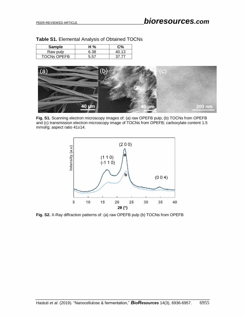
Table S1. Elemental Analysis of Obtained TOCNs
Sample H % C%
Raw pulp 6.38 40.13
TOCNs OPEFB 5.57 37.77
Fig. S1. Scanning electron microscopy images of: (a) raw OPEFB pulp; (b) TOCNs from OPEFB and (c) transmission electron microscopy image of TOCNs from OPEFB; carboxylate content 1.5 mmol/g; aspect ratio 41±14.
Fig. S2. X-Ray diffraction patterns of: (a) raw OPEFB pulp (b) TOCNs from OPEFB
2θ (°)
PEER-REVIEWED ARTICLE bioresources.com
Hastuti et al. (2019). “Nanocellulose & fermentation,” BioResources 14(3), 6936-6957. 6956
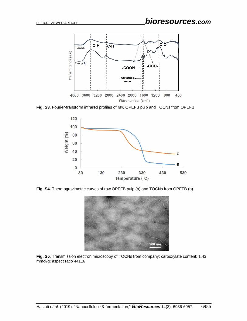
Fig. S3. Fourier-transform infrared profiles of raw OPEFB pulp and TOCNs from OPEFB
Fig. S4. Thermogravimetric curves of raw OPEFB pulp (a) and TOCNs from OPEFB (b)
Fig. S5. Transmission electron microscopy of TOCNs from company; carboxylate content: 1.43 mmol/g; aspect ratio 44±16
PEER-REVIEWED ARTICLE bioresources.com
Hastuti et al. (2019). “Nanocellulose & fermentation,” BioResources 14(3), 6936-6957. 6957
Table S2. Extractive Fermentation Properties for Free Cells and Immobilized
Cells
Free cell Immobilized cell
Parameter op-TOCN
w-TOCN Control op-TOCN
w-TOCN Alginate without TOCNs (control)
Total butanol concentration (g/L)
28.82 30.30 1.04 36.59 31.58 23.71
Total consumed glucose (g)
2.97 2.54 0.17 3.30 2.60 3.27
Butanol yield to consumed substrate (C-mol/C-mol)
0.39 0.48 0.23 0.45 0.49 0.29
REFERENCES CITED IN APPENDIX Hastuti, N., Kanomata, K., and Kitaoka, T. (2018). “Hydrochloric acid hydrolysis of
pulps from oil palm empty fruit bunches to produce cellulose nanocrystals,” Journal
of Polymers and the Environment 26(9), 3698-3709. DOI: 10.1007/s10924-018-
1248-x
Saito, T., and Isogai, A. (2004). “TEMPO-mediated oxidation of native cellulose. The
effect of oxidation conditions on chemical and crystal structures of the water-
insoluble fractions,” Biomacromolecules 5(5), 1983-1989. DOI:
10.1021/bm0497769


![New Development of Surface-Coated Polylactic … · 2019. 8. 3. · These nanofillers include silicates [5], nanocellulose crystals [6], metal-based nanoparticles [7] or carbon-based](https://img.pdfslide.tips/doc/110x75/6072911a5418f93ea7196530/new-development-of-surface-coated-polylactic-2019-8-3-these-nanoillers-include.jpg)


![Presentación de PowerPoint€¦ · Extraction of cellulose and preparation of nanocellulose from sisal fibers. Cellulose, 15(1), 149-159 [3] Parkinson, J., & Gordon, R. (1999). Beyond](https://img.pdfslide.tips/doc/110x75/5ec146897f1c2706171eedd8/presentacin-de-powerpoint-extraction-of-cellulose-and-preparation-of-nanocellulose.jpg)