Embed Size (px)
Citation preview

PENGLIHATAN PENGLIHATAN ( ( VISION VISION VISION VISION VISION VISION VISION VISION ))
YETTY MACHRINAYETTY MACHRINA
MILAHAYATI DAULAYMILAHAYATI DAULAYMILAHAYATI DAULAYMILAHAYATI DAULAY
DEPARTEMEN FISIOLOGIDEPARTEMEN FISIOLOGI
FAKULTAS KEDOKTERAN FAKULTAS KEDOKTERAN
UNIVERSITAS SUMATERA UTARAUNIVERSITAS SUMATERA UTARA
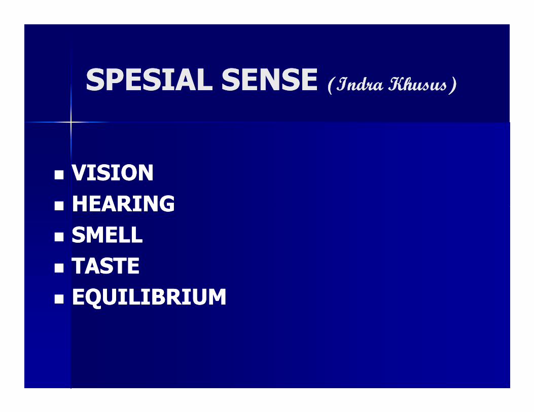
SPESIAL SENSE SPESIAL SENSE (Indra Khusus)(Indra Khusus)
�� VISIONVISION
�� HEARINGHEARING�� HEARINGHEARING
�� SMELLSMELL
�� TASTETASTE
�� EQUILIBRIUMEQUILIBRIUM

�Mata sebagai sumber informasi
awal
� Apa-apa yang kita lihat dapat
terekam sebagai satu memori
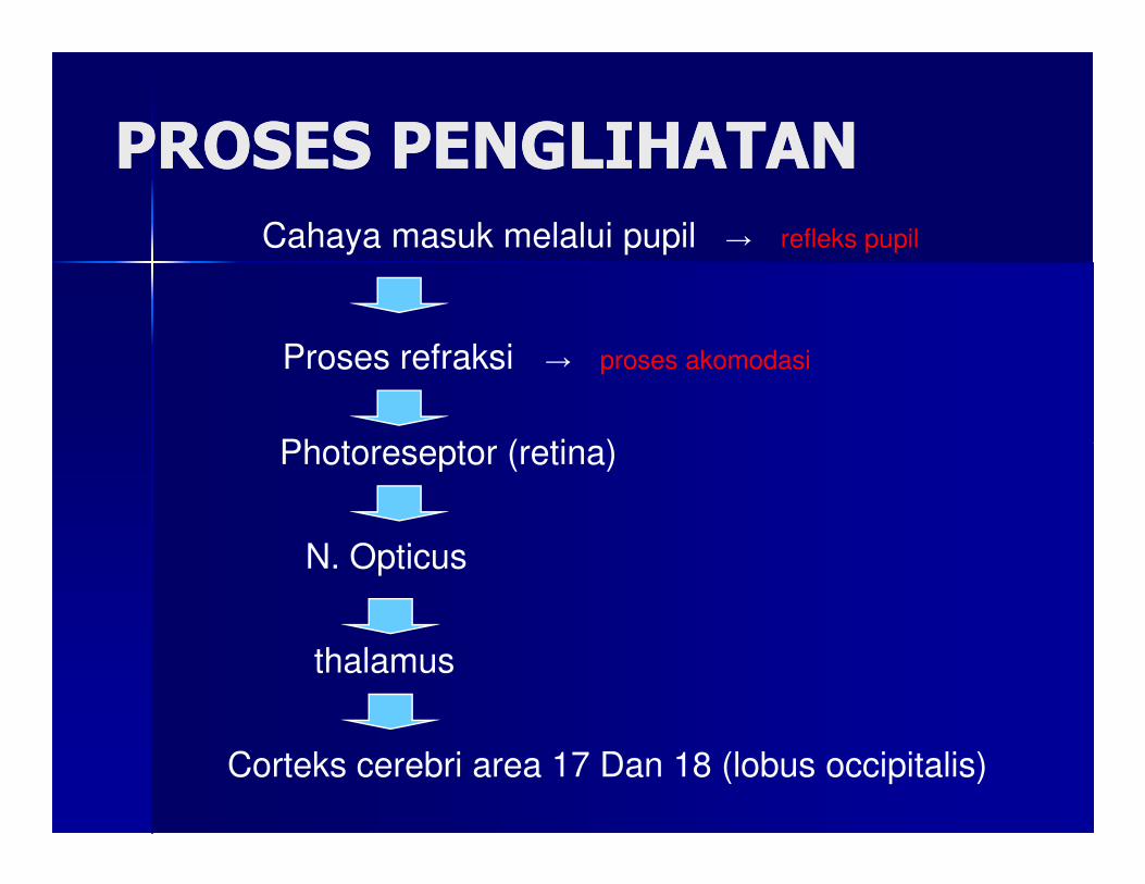
PROSES PENGLIHATANPROSES PENGLIHATAN
Cahaya masuk melalui pupil → refleks pupil
Proses refraksi → proses akomodasi
Photoreseptor (retina)Photoreseptor (retina)
N. Opticus
thalamus
Corteks cerebri area 17 Dan 18 (lobus occipitalis)
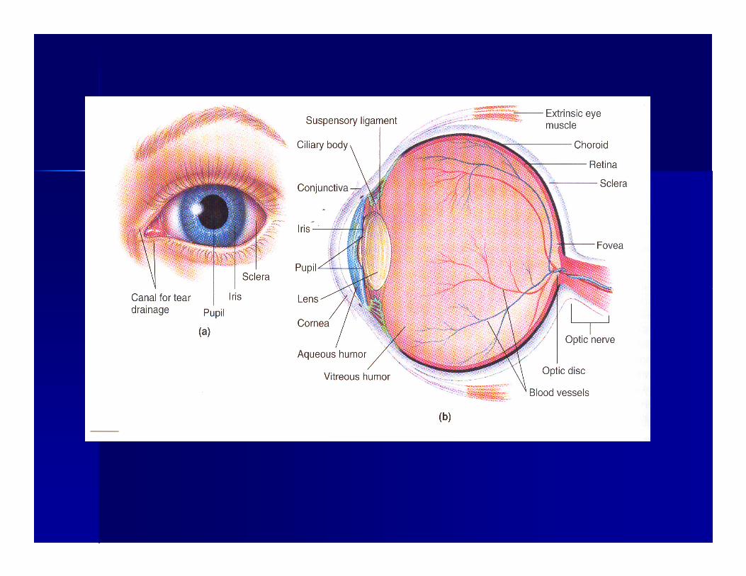
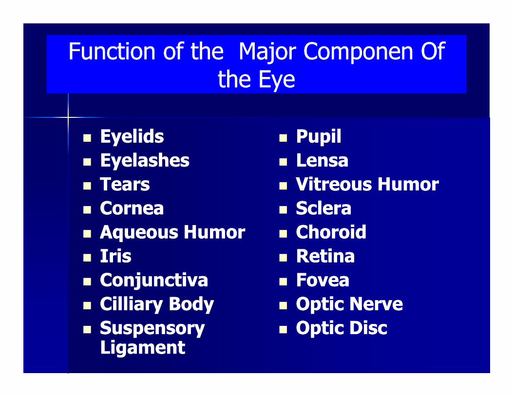
Function of the Major Componen Of Function of the Major Componen Of the Eyethe Eye
�� EyelidsEyelids
�� EyelashesEyelashes
�� TearsTears
CorneaCornea
�� PupilPupil
�� LensaLensa
�� Vitreous HumorVitreous Humor
ScleraSclera�� CorneaCornea
�� Aqueous HumorAqueous Humor
�� IrisIris
�� ConjunctivaConjunctiva
�� Cilliary BodyCilliary Body
�� Suspensory Suspensory LigamentLigament
�� ScleraSclera
�� ChoroidChoroid
�� RetinaRetina
�� FoveaFovea
�� Optic NerveOptic Nerve
�� Optic DiscOptic Disc
TEARSTEARS
�� Tears are produced Tears are produced continously by lacrimal continously by lacrimal glandgland
�� Lacrimal gland lying at the Lacrimal gland lying at the upper lateral corner of upper lateral corner of upper lateral corner of upper lateral corner of under the eyelidsunder the eyelids
�� Lacrimal gland innervate by Lacrimal gland innervate by ANS (parasympatis)ANS (parasympatis)
�� Tears thTears thrrough out each ough out each time eyelids closetime eyelids close
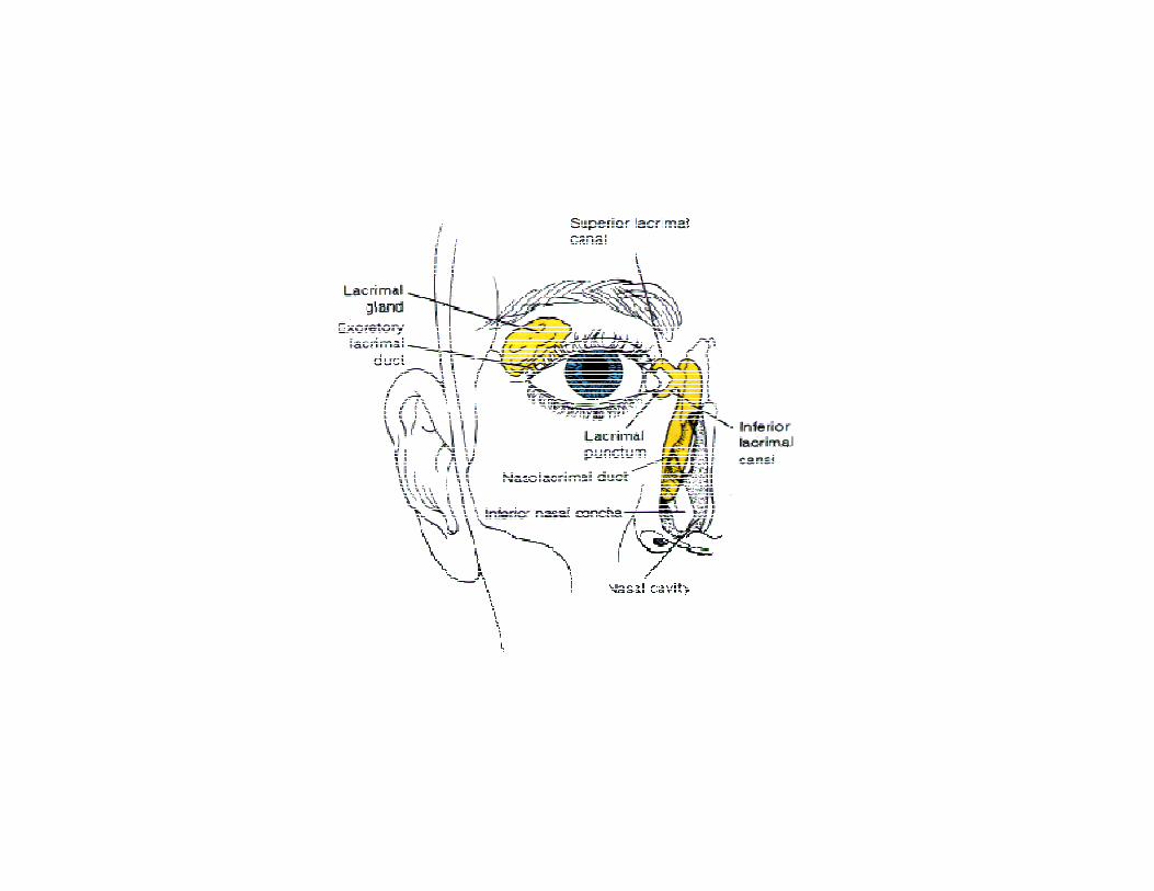
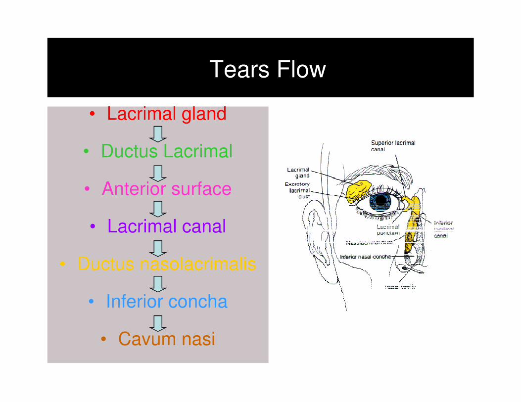
Tears Flow
• Lacrimal gland
• Ductus Lacrimal
• Anterior surface
• Lacrimal canal
• Ductus nasolacrimalis
• Inferior concha
• Cavum nasi
Tears Function Tears Function
�� ProtectionProtection
�� Keep cornea wetKeep cornea wet�� Keep cornea wetKeep cornea wet
�� Protect the eye Protect the eye from infectionfrom infection
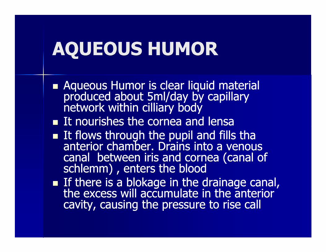
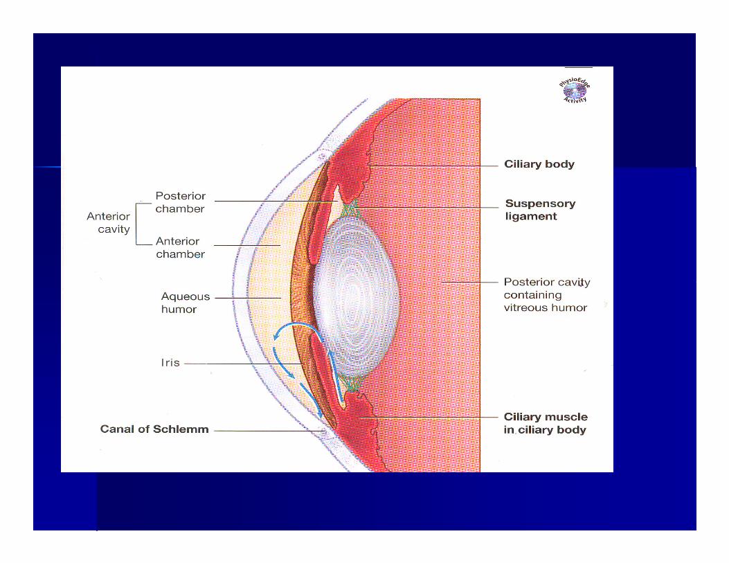
AQUEOUS HUMORAQUEOUS HUMOR
�� Aqueous Humor is clear liquid material Aqueous Humor is clear liquid material produced about 5ml/day by capillary produced about 5ml/day by capillary network witnetwork withinhin cilliary bodycilliary body
�� It nourishes the cornea and lensaIt nourishes the cornea and lensa�� It nourishes the cornea and lensaIt nourishes the cornea and lensa�� It flows through the pupil and fills tha It flows through the pupil and fills tha anterior chamber. Drains into a venous anterior chamber. Drains into a venous canal between iris and cornea (canal of canal between iris and cornea (canal of schlemm) , enters the bloodschlemm) , enters the blood
�� If there is a blokage in the drainage canal, If there is a blokage in the drainage canal, the excess will accumulate in the anterior the excess will accumulate in the anterior cavity, causing the pressure to rise call cavity, causing the pressure to rise call
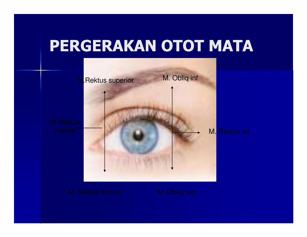
PERGERAKAN OTOT MATAPERGERAKAN OTOT MATA
M.Rektus
M.Rektus superior M. Obliq inf
M.Rektus
medial
M. Rektus inferior M. Obliq sup
M. Rektus lat
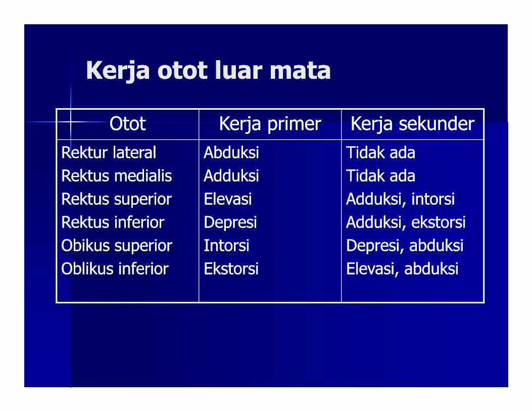
Kerja otot luar mataKerja otot luar mata
OtotOtot Kerja primerKerja primer Kerja sekunderKerja sekunder
Rektur lateralRektur lateral
Rektus medialisRektus medialis
Rektus superiorRektus superior
AbduksiAbduksi
AdduksiAdduksi
ElevasiElevasi
Tidak adaTidak ada
Tidak adaTidak ada
Adduksi, intorsiAdduksi, intorsiRektus superiorRektus superior
Rektus inferiorRektus inferior
Obikus superiorObikus superior
Oblikus inferiorOblikus inferior
ElevasiElevasi
DepresiDepresi
IntorsiIntorsi
EkstorsiEkstorsi
Adduksi, intorsiAdduksi, intorsi
Adduksi, ekstorsiAdduksi, ekstorsi
Depresi, abduksiDepresi, abduksi
Elevasi, abduksiElevasi, abduksi
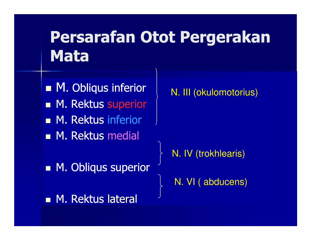
Persarafan Otot Pergerakan Persarafan Otot Pergerakan MataMata
�� MM. Obliqus inferior . Obliqus inferior
�� M. Rektus M. Rektus superiorsuperior
�� M. Rektus M. Rektus inferiorinferior
N. III (okulomotorius)
�� M. Rektus M. Rektus inferiorinferior
�� M. Rektus M. Rektus medialmedial
�� M. Obliqus superiorM. Obliqus superior
�� M. Rektus M. Rektus laterallateral
N. IV (trokhlearis)
N. VI ( abducens)
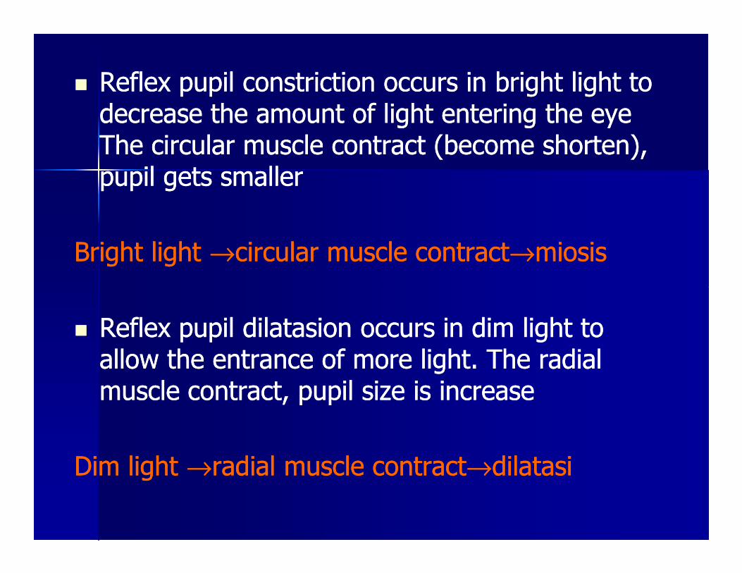
�� Reflex pupil constriction occurs in bright light to Reflex pupil constriction occurs in bright light to decrease the amount of light entering the eye decrease the amount of light entering the eye The circular muscle contract (become shorten), The circular muscle contract (become shorten), pupil gets smallerpupil gets smaller
Bright light Bright light →→circular muscle contractcircular muscle contract→→miosis miosis
�� Reflex pupil dilatasion occurs in dim light to Reflex pupil dilatasion occurs in dim light to allow the entrance of more light. The radial allow the entrance of more light. The radial muscle contract, pupil size is increasemuscle contract, pupil size is increase
Dim light Dim light →→radial muscle contractradial muscle contract→→dilatasidilatasi
PUPIL REFLEXPUPIL REFLEX
�� Pupil is controled by Pupil is controled by irisiris
�� Iris contains 2 set of smooth muscle Iris contains 2 set of smooth muscle network :network :network :network :
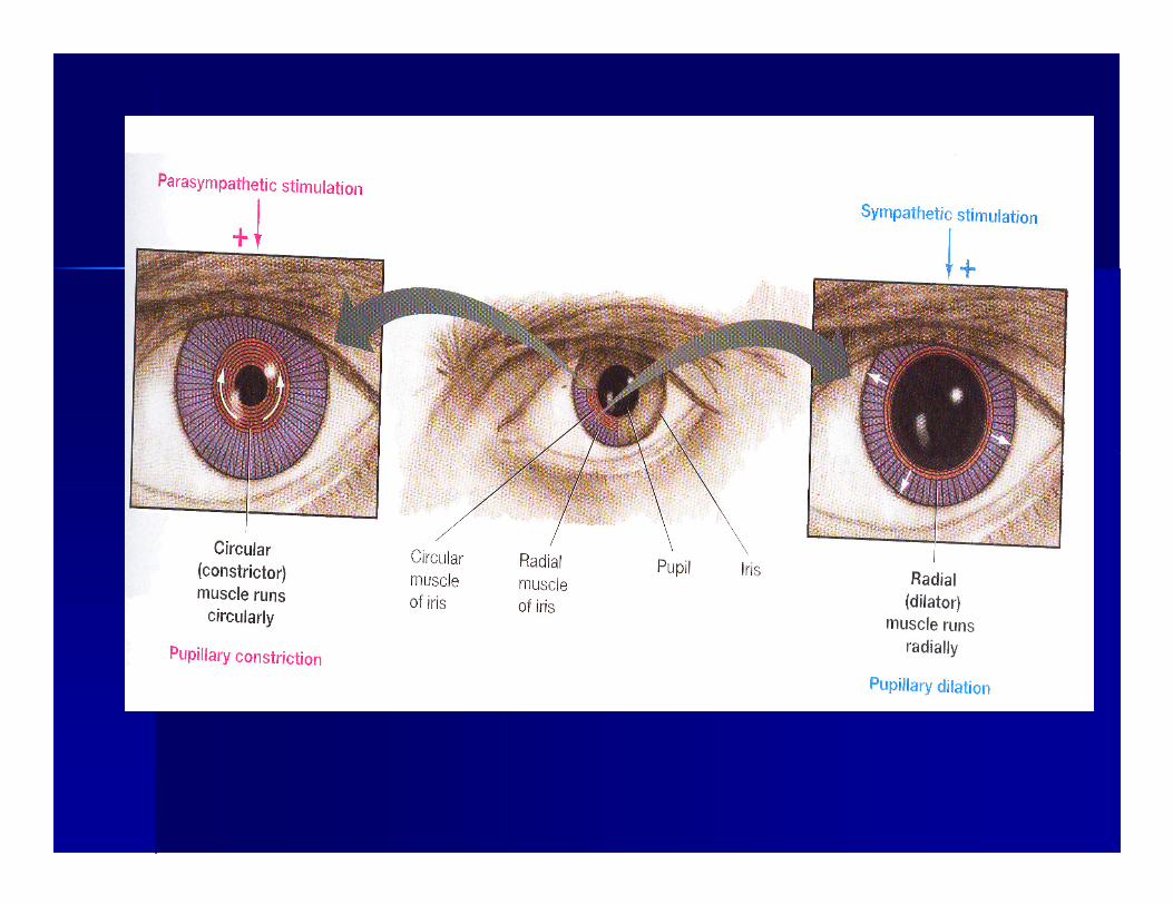
1.1. Circular muscle (ringlike fashion Circular muscle (ringlike fashion within the iris)within the iris)
2.2. Radial muscle (like bicycle spokes)Radial muscle (like bicycle spokes)
Iris muscle is controlled by autonomic nervous Iris muscle is controlled by autonomic nervous system.system.Parasympatis innervate the circular muscleParasympatis innervate the circular muscleSympatis innervate the radial muscleSympatis innervate the radial muscle
If light directed into the pupil, pupil will constrict If light directed into the pupil, pupil will constrict If light directed into the pupil, pupil will constrict If light directed into the pupil, pupil will constrict and pupil the also other eye (consensual light and pupil the also other eye (consensual light reflex). It because optic nerve fibers bring reflex). It because optic nerve fibers bring impuls to the optic nerve near the lateral impuls to the optic nerve near the lateral genuculate nucleus genuculate nucleus →→ midbrainmidbrain →→ ipsilateral ipsilateral EdingerEdinger--Westphal nucleus and contralateral Westphal nucleus and contralateral EdingerEdinger--westphal, cilliary gangglion N.III, westphal, cilliary gangglion N.III, sebagian dari ganglion ke ciliary bodysebagian dari ganglion ke ciliary body
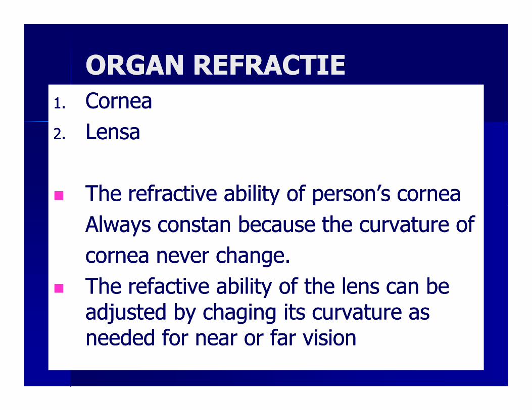
ORGAN REFRACTIEORGAN REFRACTIE
1.1. CorneaCornea
2.2. LensaLensa
�� The refractive ability of person’s corneaThe refractive ability of person’s corneaThe refractive ability of person’s corneaThe refractive ability of person’s cornea
Always constan because the curvature ofAlways constan because the curvature of
cornea never change. cornea never change.
�� The refactive ability of the lens can be The refactive ability of the lens can be adjusted by chaging its curvature as adjusted by chaging its curvature as needed for near or far visionneeded for near or far vision
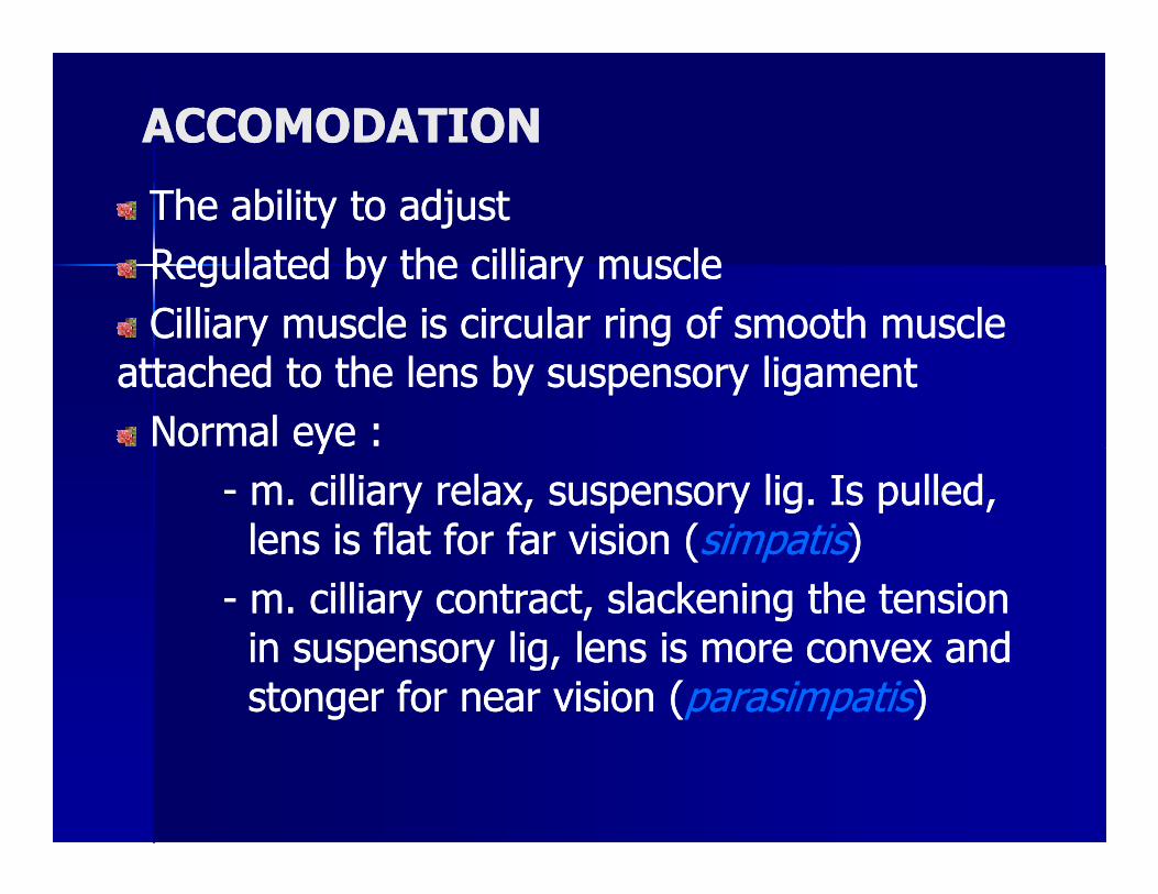
ACCOMODATIONACCOMODATION
The ability to adjustThe ability to adjust
Regulated by the cilliary muscleRegulated by the cilliary muscle
Cilliary muscle is circular ring of smooth muscle Cilliary muscle is circular ring of smooth muscle attached to the lens by suspensory ligamentattached to the lens by suspensory ligament
Normal eye :Normal eye :Normal eye :Normal eye :
-- m. cilliary relax, suspensory lig. Is pulled, m. cilliary relax, suspensory lig. Is pulled, lens is flat for far vision (lens is flat for far vision (simpatissimpatis))
-- m. cilliary contract, slackening the tension m. cilliary contract, slackening the tension in suspensory lig, lens is more convex and in suspensory lig, lens is more convex and stonger for near vision (stonger for near vision (parasimpatisparasimpatis))
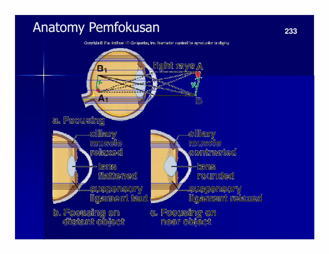
Anatomy PemfokusanAnatomy Pemfokusan 233
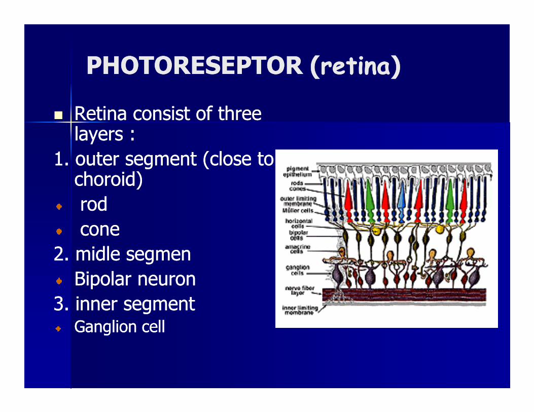
PHOTORESEPTOR (PHOTORESEPTOR (retinaretina))
�� Retina consist of three Retina consist of three layers :layers :
1. outer segment (close to 1. outer segment (close to choroid)choroid)
rodrodrodrod
conecone
2. 2. midlemidle segmensegmen
Bipolar neuronBipolar neuron
3. inner segment3. inner segmentGanglion cellGanglion cell
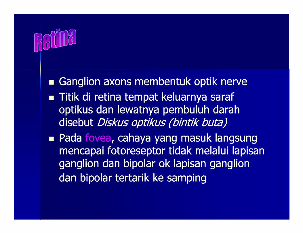
�� Ganglion axons Ganglion axons membentuk optik nervemembentuk optik nerve
�� Titik di retina tempat keluarnya saraf Titik di retina tempat keluarnya saraf optikus dan lewatnya pembuluh darah optikus dan lewatnya pembuluh darah disebut disebut Diskus optikus (bintik buta)Diskus optikus (bintik buta)disebut disebut Diskus optikus (bintik buta)Diskus optikus (bintik buta)
�� PadaPada foveafovea, cahaya yang masuk langsung , cahaya yang masuk langsung mencapai fotoreseptor tidak melalui lapisan mencapai fotoreseptor tidak melalui lapisan ganglion dan bipolar ok lapisan ganglion ganglion dan bipolar ok lapisan ganglion
dan bipolar tertarik ke sampingdan bipolar tertarik ke samping
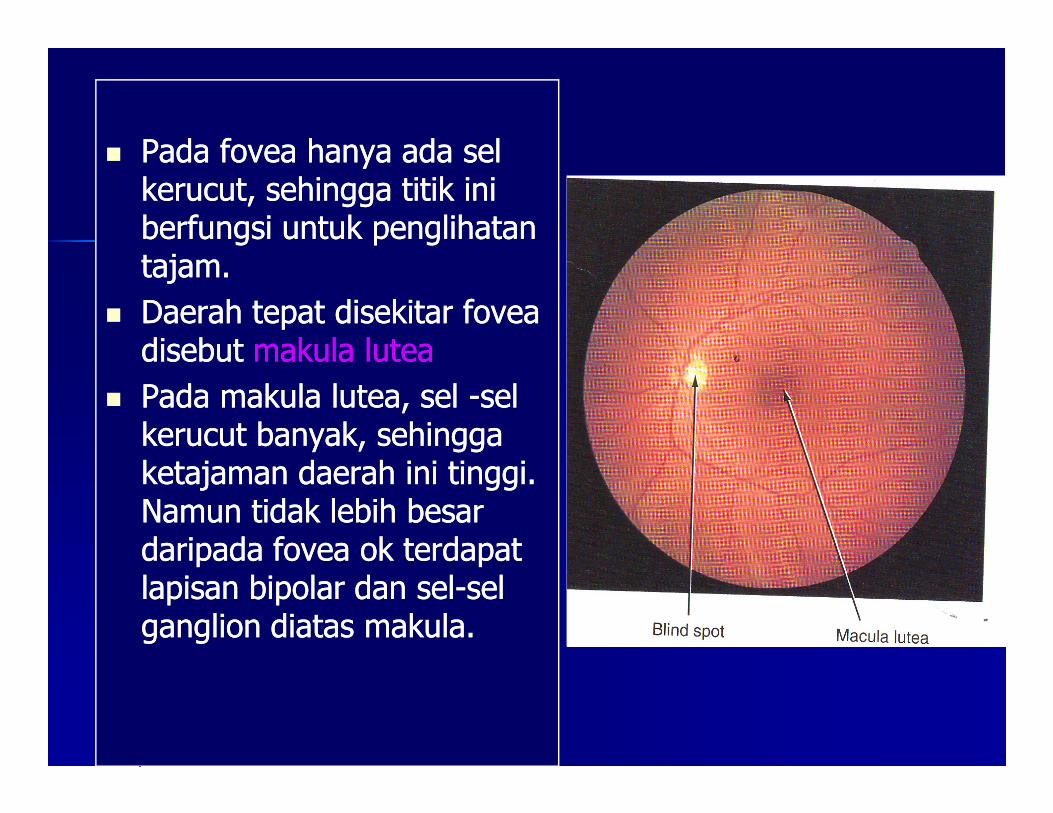
�� Pada fovea hanya ada sel Pada fovea hanya ada sel kerucut, sehingga titik ini kerucut, sehingga titik ini berfungsi untuk penglihatan berfungsi untuk penglihatan tajam.tajam.
�� Daerah tepat disekitar fovea Daerah tepat disekitar fovea disebut disebut makula luteamakula lutea
�� Pada makula lutea, sel Pada makula lutea, sel --sel sel �� Pada makula lutea, sel Pada makula lutea, sel --sel sel kerucut banyak, sehingga kerucut banyak, sehingga ketajaman daerah ini tinggi. ketajaman daerah ini tinggi. Namun tidak lebih besar Namun tidak lebih besar daripada fovea ok terdapat daripada fovea ok terdapat lapisan bipolar dan sellapisan bipolar dan sel--sel sel ganglion diatas makula.ganglion diatas makula.
OUTER SEGMEN OUTER SEGMEN
�� Banyak mengandung fotopigmenBanyak mengandung fotopigmen
�� Terdiri atas protein enzimatik yaitu opsin yang Terdiri atas protein enzimatik yaitu opsin yang berikatan dengan retinen (derivat vitamin A, berikatan dengan retinen (derivat vitamin A, red).red).red).red).
�� Retinen = 1 di sel batang Retinen = 1 di sel batang
3 di masing3 di masing--masing sel kerucut masing sel kerucut (sel merah,hijau, biru)(sel merah,hijau, biru)
FOR COLOR VISIONFOR COLOR VISION
Photoreceptor Activity in the DarkPhotoreceptor Activity in the Dark
�� The plasma membrane chemically channel of The plasma membrane chemically channel of photoreceptor respond to internal second photoreceptor respond to internal second massenger, massenger, cGMP. cGMP. BindBindiing cGMP + Nang cGMP + Na++
channels keeps them open.channels keeps them open.
�� When the is absence(stimulation), concentration When the is absence(stimulation), concentration of cGMP higher of cGMP higher →→ NaNa++ channel open.channel open.of cGMP higher of cGMP higher →→ NaNa++ channel open.channel open.
�� Passive resultant inward NaPassive resultant inward Na++leak depolarize the leak depolarize the photoreseptor photoreseptor →→ spread to synapitic terminal spread to synapitic terminal (where the photoreceptor’s neurotransmitter is (where the photoreceptor’s neurotransmitter is trored) trored) →→ synaptic terminal voltage gated synaptic terminal voltage gated channels open channels open →→ CaCa2+2+ entry entry →→trigger release of trigger release of neurotransmitterneurotransmitter from the synapticfrom the synaptic

� Inhibitory neurotransmitter that srecreted
by amacrine cells are gamma-
aminobutyric acid, glycine, dopamine, aminobutyric acid, glycine, dopamine,
acetylcholine, dolamine
�� Photoreceptor inhibited of their adequate Photoreceptor inhibited of their adequate stimulus (hyperpolarized by light).stimulus (hyperpolarized by light).
�� Excited absence of stimulation (depolarized Excited absence of stimulation (depolarized by darkness)by darkness)
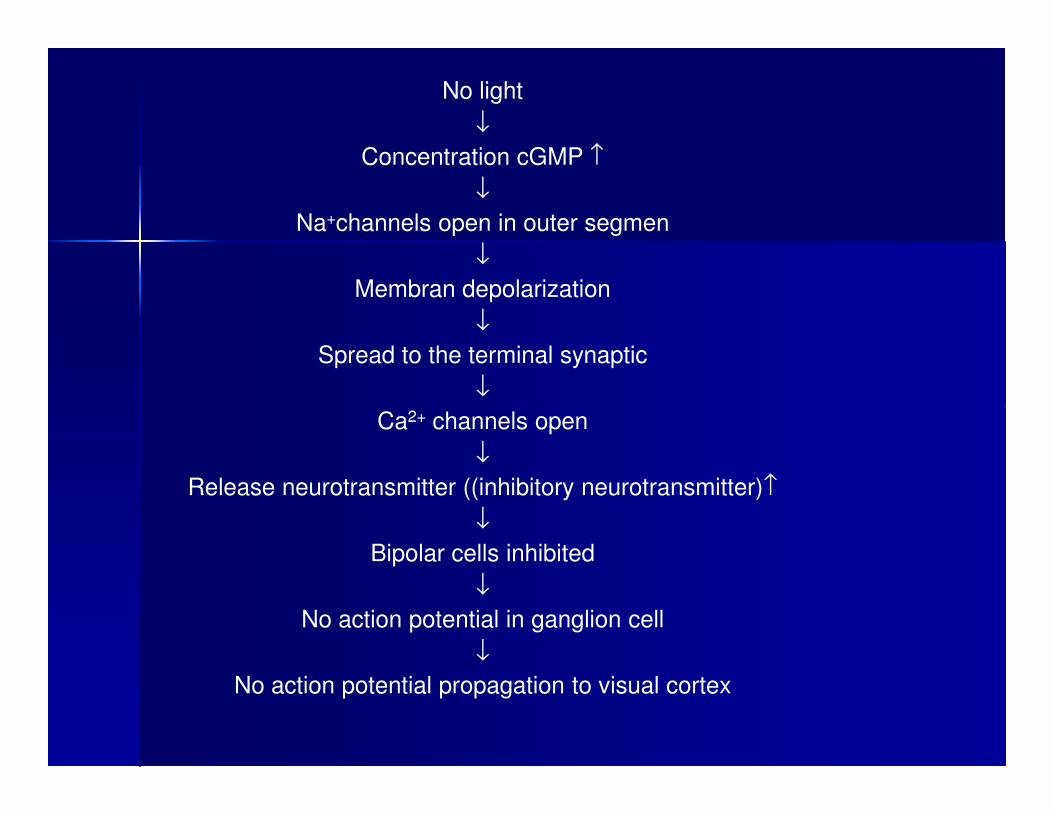
No light
↓
Concentration cGMP ↑
↓
Na+channels open in outer segmen
↓
Membran depolarization
↓
Spread to the terminal synaptic
↓
Ca2+ channels open
↓
Release neurotransmitter ((inhibitory neurotransmitter)↑
↓
Bipolar cells inhibited
↓
No action potential in ganglion cell
↓
No action potential propagation to visual cortex
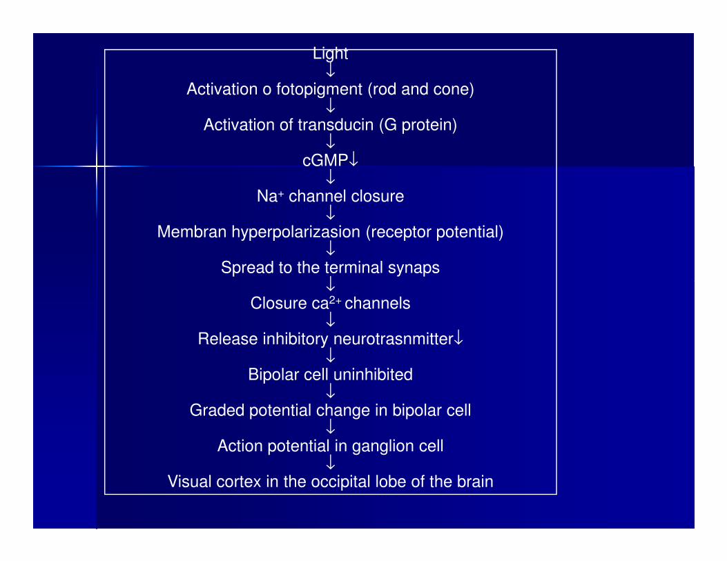
Light↓
Activation o fotopigment (rod and cone)↓
Activation of transducin (G protein)↓
cGMP↓↓
Na+ channel closure↓
Membran hyperpolarizasion (receptor potential)↓
Spread to the terminal synaps↓↓
Closure ca2+ channels↓
Release inhibitory neurotrasnmitter↓↓
Bipolar cell uninhibited↓
Graded potential change in bipolar cell↓
Action potential in ganglion cell↓
Visual cortex in the occipital lobe of the brain
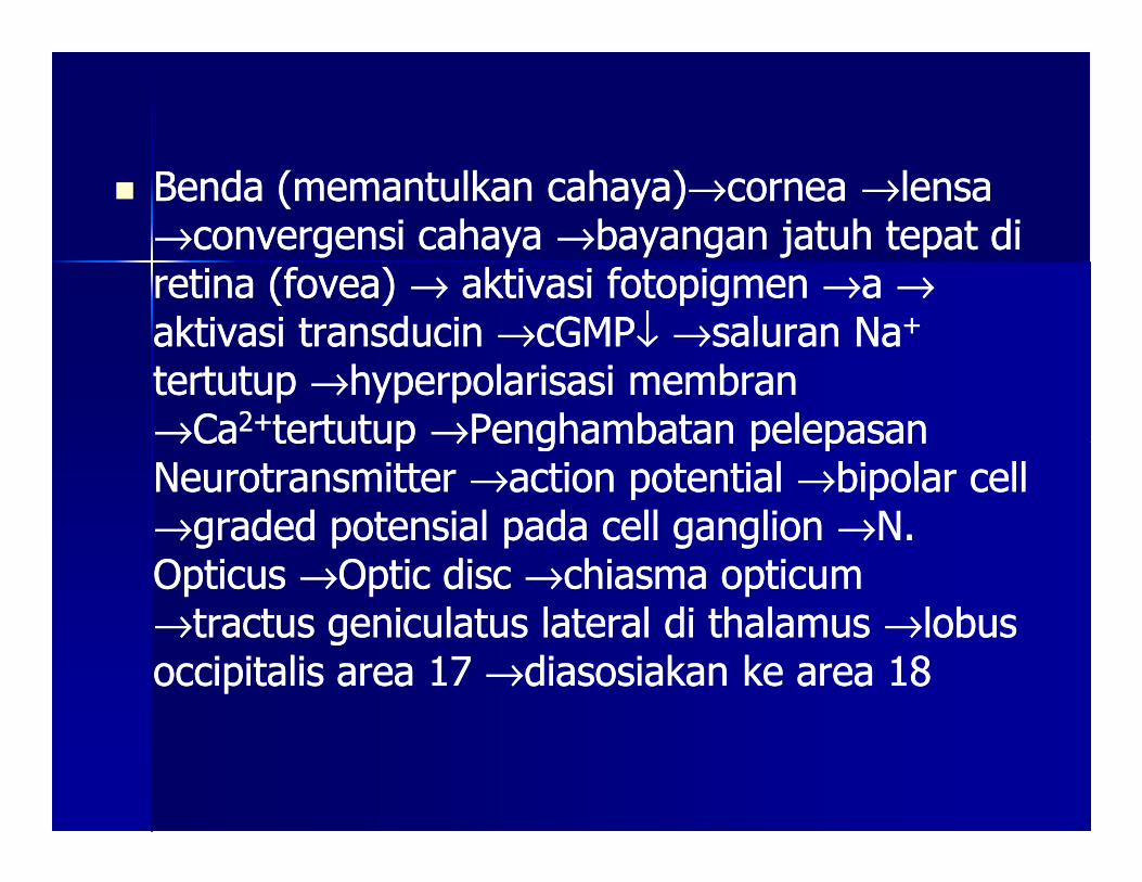
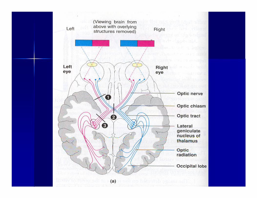
�� Benda (memantulkan cahaya)Benda (memantulkan cahaya)→→cornea cornea →→lensa lensa →→convergensi cahaya convergensi cahaya →→bayangan jatuh tepat di bayangan jatuh tepat di retina (fovea) retina (fovea) →→ aktivasi fotopigmen aktivasi fotopigmen →→a a →→aktivasi transducin aktivasi transducin →→cGMPcGMP↓↓ →→saluran Nasaluran Na++
tertutup tertutup →→hyperpolarisasi membran hyperpolarisasi membran →→CaCa2+2+tertutup tertutup →→Penghambatan pelepasan Penghambatan pelepasan →→CaCa tertutup tertutup →→Penghambatan pelepasan Penghambatan pelepasan Neurotransmitter Neurotransmitter →→action potential action potential →→bipolar cell bipolar cell →→graded potensial pada cell ganglion graded potensial pada cell ganglion →→N. N. Opticus Opticus →→Optic disc Optic disc →→chiasma opticum chiasma opticum →→tractus geniculatus lateral di thalamus tractus geniculatus lateral di thalamus →→lobus lobus occipitalis area 17 occipitalis area 17 →→diasosiakan ke area 18diasosiakan ke area 18
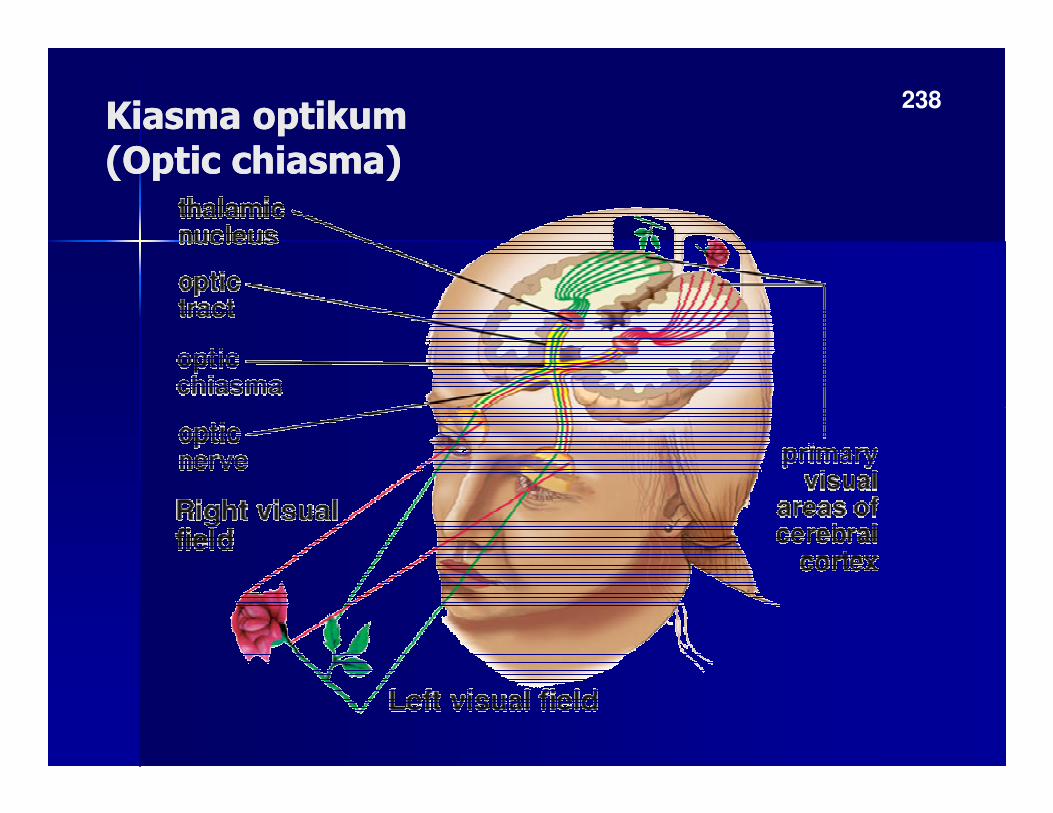
Kiasma optikumKiasma optikum(Optic chiasma)(Optic chiasma)
238
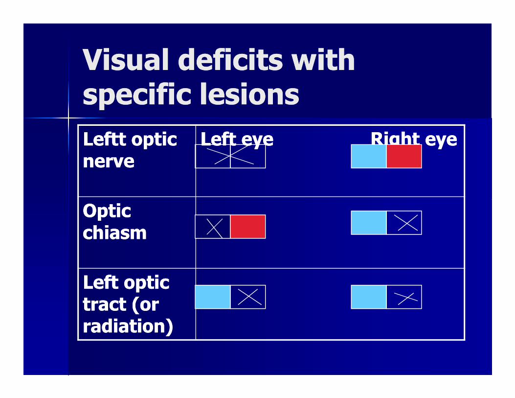
Visual deficits with Visual deficits with specific lesionsspecific lesions
Leftt optic Leftt optic nervenerve
Left eyeLeft eye Right eyeRight eye
Optic Optic Optic Optic chiasmchiasm
Left optic Left optic tract (or tract (or radiation)radiation)
VISUAL ADAPTATIONVISUAL ADAPTATION
�� DARK ADAPTATIONDARK ADAPTATION
-- Break down of photopigment during Break down of photopigment during exposure of sunlight fremendously exposure of sunlight fremendously exposure of sunlight fremendously exposure of sunlight fremendously decreases photoreceptor sensitivitydecreases photoreceptor sensitivity
--As a result, the sensitivity of our eyes As a result, the sensitivity of our eyes gradually increase gradually increase →→ u can see in the u can see in the darkdark
�� LIGHT ADAPTATIONLIGHT ADAPTATION
As some of photopigment are rapidly As some of photopigment are rapidly brokendown by intense light, the brokendown by intense light, the brokendown by intense light, the brokendown by intense light, the sensitivsensitivIIty of the eye decreases and ty of the eye decreases and normal contras can once again be normal contras can once again be detected.detected.
CCOOLLOORR VISIONVISION
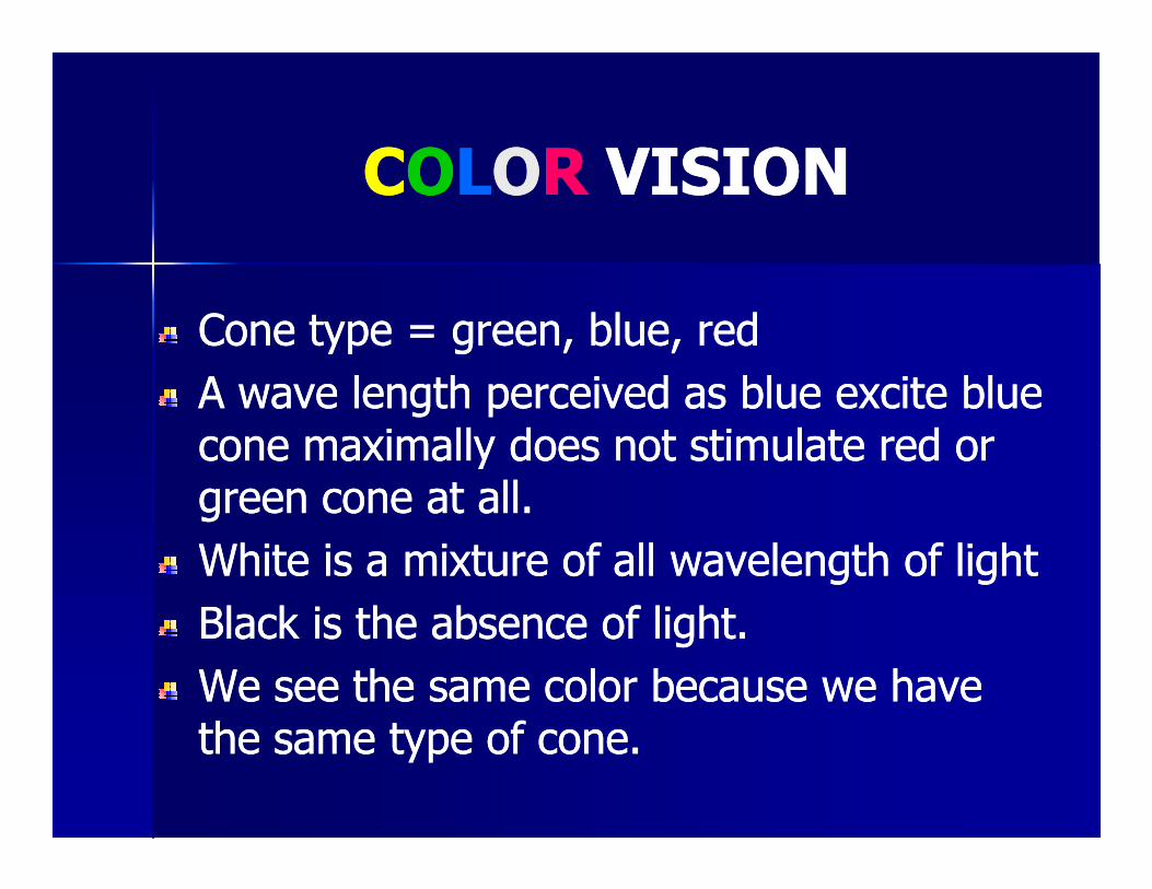
Cone type = green, blue, redCone type = green, blue, red
A wave length perceived as blue excite blue A wave length perceived as blue excite blue cone maximally does not stimulate red or cone maximally does not stimulate red or cone maximally does not stimulate red or cone maximally does not stimulate red or green cone at all.green cone at all.
White is a mixture of all wavelength of lightWhite is a mixture of all wavelength of light
Black is the absence of light.Black is the absence of light.
We see the same color because we have We see the same color because we have the same type of cone.the same type of cone.
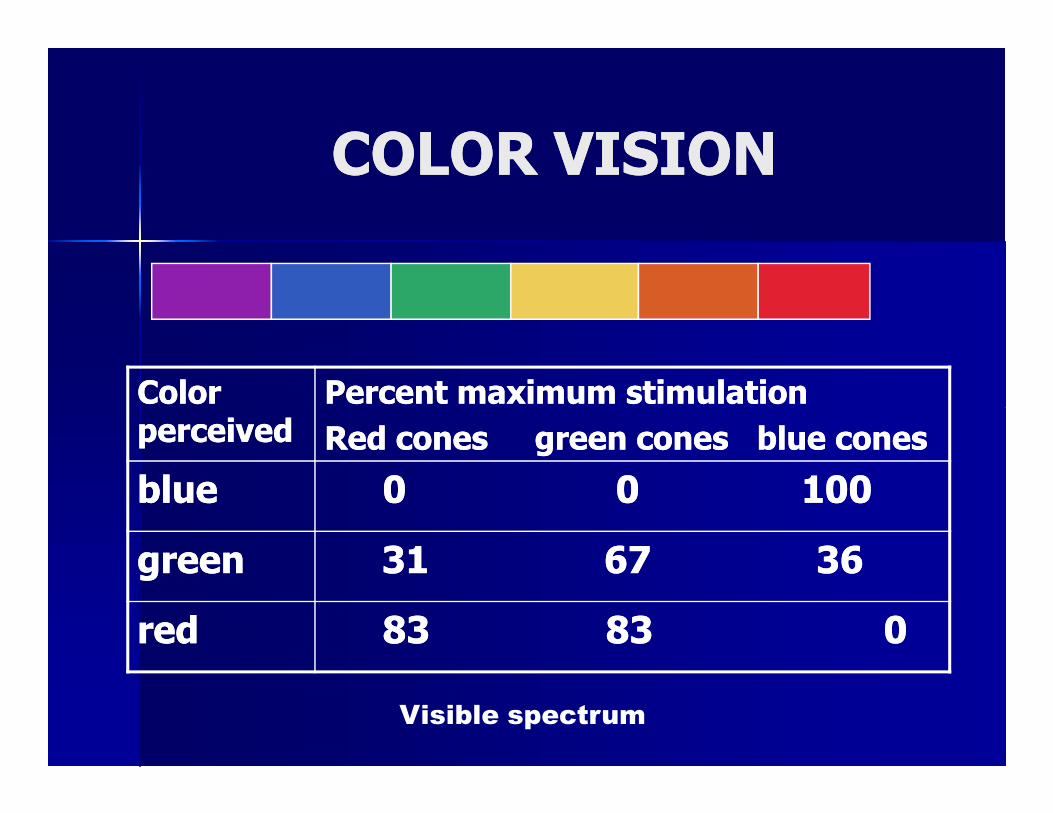
COLOR VISIONCOLOR VISION
Color Color Percent maximum stimulationPercent maximum stimulation
Visible spectrum
Color Color perceivedperceived
Percent maximum stimulationPercent maximum stimulation
Red cones green cones blue conesRed cones green cones blue cones
blueblue 00 0 0 100100
greengreen 3131 67 3667 36
redred 83 83 083 83 0
Visual fieldVisual field
�� Visual field is field of view that can be Visual field is field of view that can be seen without moving the headseen without moving the head


![sss.16. mata trauma okuli.ppt [Read-Only] - ocw.usu.ac.idocw.usu.ac.id/course/download/1110000121-special... · Gejala: proptosis mendadak,visus menurun ,rasa sakit yang hebat](https://img.pdfslide.tips/doc/110x75/5b54fb227f8b9a27658d8e69/sss16-mata-trauma-okulippt-read-only-ocwusuacidocwusuacidcoursedownload1110000121-special.jpg)


![sss.3. biokimia biokimia jaringan mata.ppt [Read-Only]ocw.usu.ac.id/course/download/1110000121-special-senses-system/s… · JALUR METABOLIK PADA JARINGAN MATA 1. Glikolisis 2. HMP](https://img.pdfslide.tips/doc/110x75/5a84bbba7f8b9ad30c8bd33b/sss3-biokimia-biokimia-jaringan-matappt-read-onlyocwusuacidcoursedownload1110000121-special-senses-systemsjalur.jpg)