Embed Size (px)
Citation preview
ONLINE FIRST
This is a provisional PDF only. Copyedited and fully formatted version will be made available soon.
ISSN: 0017-0011
e-ISSN: 2543-6767
Perinatal outcomes of the antenatally diagnosed omphalocelecases: a single tertiary center experience
Authors: Cigdem Akcabay, Fatma Islek, Erol Arslan, Masum Kayapinar, CansunDemir, Cuneyt Evruke, Selim Buyukkurt, Mete Sucu, Umran Kucukgoz Gulec
DOI: 10.5603/GP.a2021.0071
Article type: Research paper
Submitted: 2020-11-18
Accepted: 2021-02-26
Published online: 2021-04-21
This article has been peer reviewed and published immediately upon acceptance.It is an open access article, which means that it can be downloaded, printed, and distributed freely,
provided the work is properly cited.Articles in "Ginekologia Polska" are listed in PubMed.
Powered by TCPDF (www.tcpdf.org)
ORIGINAL PAPER / OBSTETRICS
Perinatal outcomes of the antenatally diagnosed omphalocele cases: a single tertiary
center experience
Cigdem Akcabay, Fatma Islek, Erol Arslan, Masum Kayapinar, Cansun Demir, Cuneyt
Evruke, Selim Buyukkurt, Mete Sucu, Umran Kucukgoz Gulec
Department of Obstetrics and Gynecology, Çukurova University, School of Medicine, Adana,
Turkey
Corresponding author:
Umran Kucukgoz Gulec
Department of Obstetrics and Gynecology, Çukurova University, School of Medicine, 01330
Saricam, Adana Turkey
phone: 90 322 3386060-3195-3196, fax: 90 322 3386527
e-mail: [email protected]
Short title: Perinatal outcomes of the omphalocele
ABSTRACT
Objectives: To evaluate the perinatal outcomes of antenatally diagnosed omphalocele cases.
Material and methods: This was a retrospective study conducted between July 2014 and
February 2020 at the prenatal diagnosis center of a university clinic. Gestational week of
diagnosis, associated anomalies, karyotype analysis results, complications during pregnancy,
termination/delivery characteristics, and postnatal results were evaluated.
Results: The analysis was performed on 58 patients. The median diagnosis time was 14.5
weeks of gestation. Thirty-three cases (57%) were defined in the first trimester. 33 (57%) of
58 patients had one or more concomitant anomalies, while 25 patients (43%) had isolated
omphalocele. The most common associated anomaly was a cardiac anomaly which was
observed in 17 fetuses (30% of all omphalocele cases). Karyotype analysis was performed in
forty-five patients (41 in the prenatal period, 4 in the postnatal period). A normal karyotype
was detected in 27 cases (60%). Trisomy 18 was the most common chromosomal anomaly (n
= 15, 33.3%). Thirty of 58 patients (52%) requested termination of pregnancy (TOP) in the
early pregnancy period. Thirteen of the cases died in-utero (22%). Fifteen pregnancies
resulted in live births (26%), of those eight were lost in the first year of life (six of them had
additional anomalies, while two of them had isolated omphalocele but the omphalocele pouch
was containing the liver in those two babies).
Conclusions: Most of the cases with an omphalocele can be diagnosed in the first trimester.
Cardiac anomalies were the most common associated anomalies, while trisomy 18 is the most
common chromosomal anomaly. Thus, earlier and effective counseling can be made about the
prognosis of pregnancy.
Key words: exomphalos; perinatal outcomes; omphalocele
Introduction
Omphalocele (exomphalos) is one of the most common anterior abdominal wall defect
and its frequency is reported to be 1 in 4000–7000 live births [1]. It is defined as herniation of
intra-abdominal organs covered with peritoneal membrane and umbilical cord due to midline
defect of the anterior abdominal wall. The peritoneal membrane consists of the wharton gel
between the inner layer of the peritoneum and the outer layer of the amnion [2]. Abdominal
wall herniation is considered physiological before the 12th gestational week. For this reason,
the diagnosis of omphalocele is made more during the first trimester screening [3, 4]
especially during the nuchal translucency (NT) measurement as part of first trimester
screening [5]. This is the main reason for the high misdiagnosis rates in the first trimester.
Detailed ultrasonographic examination including fetal echocardiography and karyotype
analysis should be requested in terms of searching for the concomitant anomalies that have an
increased rate in omphalocele cases. The frequency of chromosomal anomalies, especially
trisomy 18, was higher in omphalocele cases. Trisomy 18 is present in 80% of cases if other
anomalies accompanied to omphalocele, whereas the rate is 54% in the omphalocele cases
accompanied with only increased NT [6]. Since omphalocele may be associated by many
structural anomalies (Pentalogy of Cantrell, Beckwith-Wiedeman syndrom, bladder
exstrophy, imperforate anus, spina bifida complex/OEIS complex, neural tube defects,
diaphragmatic herniation, single gene disorders and many other syndromes) targeting detailed
ultrasonography in the second trimester and if necessary cyto-genetic analysis such as
comparative genetic hybridization should be performed even if it is thought to be isolated in
the first trimester [7]. Cardiac anomalies especially secundum atrial septal defect (ASD) and
muscular ventricular septal defect (VSD) are the most common concomitant structural
anomalies in omphalocele cases. The most common extracardiac anomalies are in the
genitourinary system (Pyelectasis, Hydronephrosis, Cloacal exstrophy, Multicystic kidney)
and gastrointestinal system (Beckwith-Wiedeman syndrome). Pulmonary hypoplasia is also
common, especially if the defect is large [8, 9]. Therefore, detailed cardiac evaluation is
essential in these cases.
The prognosis of the omphalocele is usually severe in non-isolated cases.
Omphalocele sac can include liver, stomach and intestinal. There is a high termination rate
and in-utero mortality rate for omphalocele because of the associated structural and
chromosomal anomalies. Chromosomal anomaly and/or related structural anomaly incidence
rate can be high as 88% for omphalocele. Therefore, live birth rate is reported to be 25–40%
[1, 10]. But prognosis is better in isolated omphalocele cases. The most difficult question is to
reveal whether the case is really isolated omphalocele.
In this study, we aimed to evaluate the prenatal results of patients with omphalocele
diagnosed and followed up in our clinic.
MATERIAL AND METHODS
This retrospective study was conducted between July 2014 and February 2020 at the
Maternal-Fetal Unit of a University Hospital. This study was approved by the Faculty of
Medicine Ethics Committee. This center is the tertiary referral center for the perinatal
diagnosis. Women admitted for routine ultrasound examination or referred to our hospital for
a suspected fetal anomaly underwent detailed fetal anomaly ultrasonography scan. For those
detected with omphalocele and continued pregnancy, a detailed ultrasonographic evaluation
including fetal cardiography was performed to determine associated anomalies by using
VolusonE6 (GE Medical Systems, Zipf, Austria) with a transabdominal 4–8-MHz probe. The
data were scanned retrospectively from the viewpoint recording system and the neonatal
registry system. Maternal age, gravidity, parity, gestational age at diagnosis, associated
structural malformations, whether invasive diagnostic procedures were performed or not, the
results of karyotype analysis and fetal echocardiography results were determined. Each
patient was evaluated in a council of clinical geneticists, pediatric surgeons and
perinatologists. Each family was informed in detail about the current situation by the council.
TOP were performed if the family requested the termination and the fetus was evaluated
postmortem if the family approved this examination. The ratio of TOP was determined.
During the follow-up, whether intrauterine growth restriction (IUGR), intrauterine fetal death,
preterm labor were evaluated. IUGR was defined as fetal ultrasonographic measurements
being below 10% percentile according to gestational age. Small gestational age (SGA), IUGR
distinction was not made. Doppler studies were evaluated after the diagnosis of IUGR.
Delivery before completing the 37th gestational weeks were defined as preterm delivery.
Gestational age at delivery, route of delivery, low APGAR score (10th minute ≤ 5), birth
weight, size and content of defect and postnatal information were recorded by using the labor
unit registry and neonatal intensive care (NICU) registry. Neonatal care was performed by the
neonatologist and the pediatric surgery team in same hospital.
Statistically analysis was performed by using SPSS® (SPSS Inc., Chicago, IL, USA,
version 20). Descriptive analysis was performed for this study. Continuous data was presented
as mean±SD, median (min-max.). Categorical data was presented as n (%).
RESULTS
Seventy-four patients with omphalocele were detected during the study period. Sixteen
of them were excluded from the study because follow-up data were not available. Analyzes
were performed on 58 cases. Mean maternal age was 29.0 ± 6.7, primigravidity was present in
18 (31%) cases. The median of diagnosis time was 14.5 weeks of gestation. Thirty-three cases
(57%) were defined in the first trimester. Twenty-five cases did not have any associated
anomalies (43%), 33 of total cases (57%) had one or more associated anomalies. Thirty of 58
cases (52%) performed termination of pregnancy (TOP) in the early pregnancy period.
Thirteen of the cases (22%) died in-utero. Fifteen pregnancies (26%) resulted in live births.
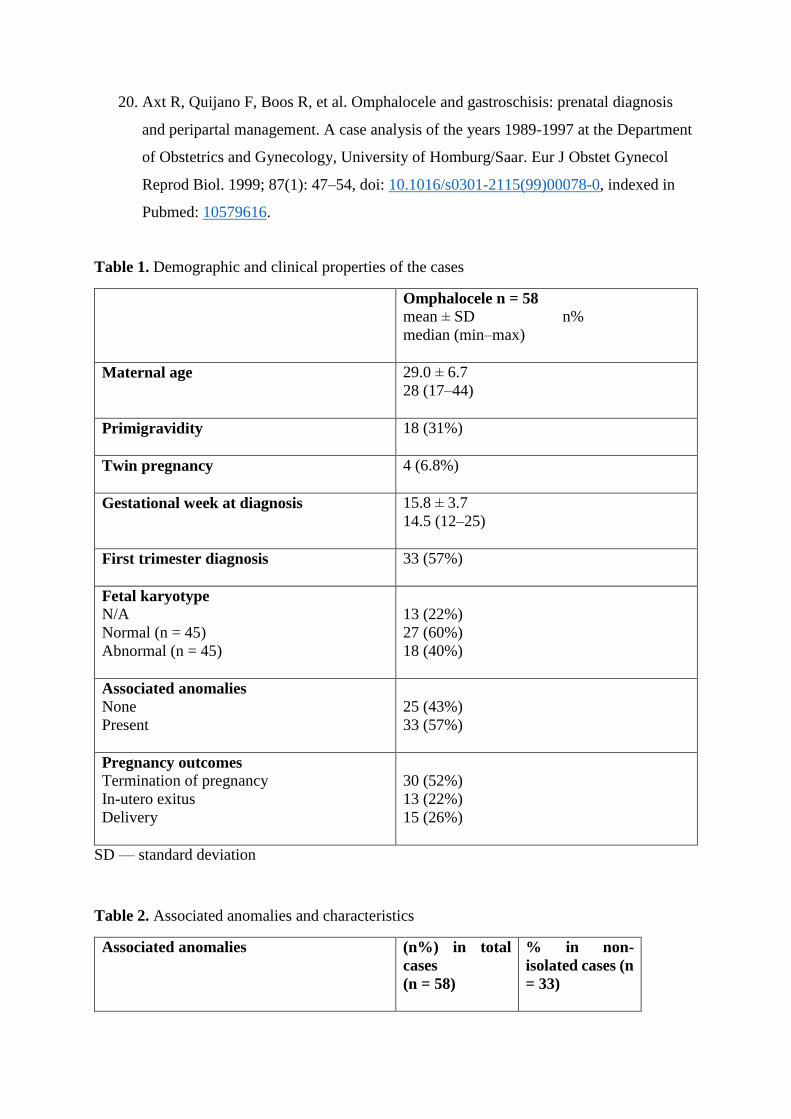
Clinical and demographic variables and results were presented in Table 1.
The most common associated anomaly is cardiac anomaly and it was observed in 17
fetuses. Among the cardiac anomalies, atrioventricular septal defect (AVSD) was the most
common anomaly. Central nervous system (CNS) anomalies were observed in 15 cases.
Urogenital system anomalies were the third most common anomalies with eight cases. Other
system anomalies most frequently belong to the skeletal system (short femur length and short
humerus length) and were seen in six cases. Thirteen cases had multiple anomalies associated
omphalocele. Associated anomalies and their properties are shown in Table 2.
Karyotype analysis was performed in 41 cases in the pre-natal period and in four cases
in the post-natal period (78%). Normal karyotype was detected in 27 cases (60%). Trisomy 18
was the most common karyotype anomaly with 15 cases (83%). Trisomy 13 was seen in two
cases and Turner (45, X0) was determined in one case. Results of the karyotype analysis were
presented as Table 3.
Preterm delivery was determined in seven cases (46%). IUGR was determined in
seven cases (25%). Fifteen live births were performed. Mean gestational week at the delivery
was 34.3 week. One-third of the cases have been delivered by the vaginal route. Omphalocele
alone was not considered a cesarean indication, and the decision for cesarean was determined
according to general obstetric indications. 10th minute low APGAR score (≤ 5) was present in
the 3 cases. Eight of them were lost in the first year (six of them had additional anomalies,
two cases were isolated omphalocele, but liver was also present in the pouch). There were no
additional major organ anomalies or karyotype abnormalities of seven babies who were born
alive and continued their lives, and the youngest was seven months old and the eldest was
three years old. Obstetrics and neonatal outcomes of the cases were presented in the Table 4.
DISCUSSION
In our series of 58 cases, we evaluated obstetric and neonatal outcomes in cases with
omphalocele. Due to the liberal use of ultrasonography in the antenatal period and increased
evaluation experience and knowledge, omphalocele diagnostic accuracy is close to 100% [1].
In the intrauterine period, the differential diagnosis of omphalocele and gastroschisis can be
made almost 100%. Gastroschisis was not ever misdiagnosed as omphalocele in our series.
The sensitivity for omphalocele diagnosis in the first trimester is reported to be 90% [10, 11].
In our study, 33 of the 58 cases (57%) were diagnosed omphalocele correctly in the first
trimester. The diagnosis of omphalocele in the first trimester is very important in terms of
detecting structural and chromosomal anomalies and enabling earlier decisions about the
pregnancy. In omphalocele cases, live birth rates are as low as 25–40% because elective TOP
rates and in-utero exitus rates are high [10]. In our series, the TOP rate was high as 52% (n =
30) and live birth rate was 25% (n = 15). According to a study conducted in 11 countries in
Europe, the live birth rate for omphalocele (n = 137) was given as 41%, fetal death rate was
22% and TOP rate was 37% [12].
The prognosis of omphalocele depends on concomitant structural and/or chromosomal
anomalies [1, 2, 6–10]. Structural anomalies were associated in 57% of our cases while
chromosomal anomalies were found in 40%. In another series of 90 cases, 69% of central
omphalocele cases had chromosomal anomaly, while in epigastric omphalocele, this rate was
12% [13]. They concluded that the types of the omphalocele may be different entities but as a
result, 22% of cases live and omphalocele has poor prognosis irrespective of the types. Thus
in a study evaluating 79 isolated omphalocele cases diagnosed in the first trimester and
without structural and chromosomal anomalies, live birth rate was 68% and the mortality rate
was 33% [10]. In another series of 67 cases, the rate of chromosomal anomaly was reported as
39% like our results [14]. In another study evaluating 98 cases diagnosed in the first trimester,
it was found that 45.9% of the cases were associated by major structural anomalies and 53.8%
had chromosomal anomalies [6]. Fleurke-Rozema H et al. [15], reported that 141 cases with
omphalocele 83% had additional anomalies of which 57% had a chromosomal anomaly.
Similarly, in the presence of increased NT (> 3.5mm), chromosomal anomaly was detected in
40.8% of cases with omphalocele [16]. A normal NT is therefore a reassuring sign, but the
residual risk of aneuploidy may still be as high as 28% [17]. In another series, the rate of
related structural anomaly was reported as 78.7% (26/33), and the rate of chromosomal
anomaly was 27.6% (8/29) [18]. The most common accompanying chromosomal anomaly is
trisomy 18 [1, 2, 8]. Trisomy 18 was the most common karyotype anomaly (15/18) in our
series. The most common associated structural anomaly in our series was cardiac anomalies
(17/58). It is stated that the most frequently observed structural anomaly is cardiac [9, 19].
However, CNS anomalies were most frequently accompanied in another case series [20]. The
general recommendation is that omphalocele cases detected in the first trimester and thought
to be isolated should definitely be evaluated in the second trimester. Because it may be
accompanied by syndromic structural conditions like Pentalogy of Cantrel; bladder exstrophy,
imperforate anus, spina bifida complex, Beckwith–Wiedemann syndrome (2). Isolated
omphaloceles are related to Beckwith–Wiedemann syndrome with a 10–20% probability,
appropriate prenatal cytogenetic testing should be discussed with patients [7]. The fact that
the negative results of molecular genetic tests do not exclude this diagnosis.
This study has some limitations due to its retrospective structure. In addition, the lack
of molecular cytogenetic methods is another limitation. But we have a good number of cases
for a single center. Follow-up data is also a positive aspect in this study, because newborn
care and surgery of cases are performed in the same hospital.
CONCLUSIONS
In conclusion, it is important to diagnose these cases in the first trimester. Intense
efforts should be made to recognize concomitant structural and chromosomal anomalies
because they determine the prognosis. If structural and/or chromosomal anomalies are
present, the prognosis is poor. Genetic counseling should also be recommended in cases
considered to be isolated.
Conflict of interest
The authors report no conflicts of interest.
REFERENCES
1. Tassin M, Benachi A. Diagnosis of abdominal wall defects in the first trimester. Curr
Opin Obstet Gynecol. 2014; 26(2): 104–109, doi: 10.1097/GCO.0000000000000053,
indexed in Pubmed: 24504173.
2. Prefumo F, Izzi C. Fetal abdominal wall defects. Best Pract Res Clin Obstet Gynaecol.
2014; 28(3): 391–402, doi: 10.1016/j.bpobgyn.2013.10.003, indexed in Pubmed:
24342556.
3. Syngelaki A, Chelemen T, Dagklis T, et al. Challenges in the diagnosis of fetal non-
chromosomal abnormalities at 11-13 weeks. Prenat Diagn. 2011; 31(1): 90–102, doi:
10.1002/pd.2642, indexed in Pubmed: 21210483.
4. Fleurke-Rozema H, van de Kamp K, Bakker M, et al. Prevalence, timing of diagnosis
and pregnancy outcome of abdominal wall defects after the introduction of a national
prenatal screening program. Prenat Diagn. 2017; 37(4): 383–388, doi:
10.1002/pd.5023, indexed in Pubmed: 28219116.
5. Syngelaki A, Guerra L, Ceccacci I, et al. Impact of holoprosencephaly, exomphalos,
megacystis and increased nuchal translucency on first-trimester screening for
chromosomal abnormalities. Ultrasound Obstet Gynecol. 2017; 50(1): 45–48, doi:
10.1002/uog.17286, indexed in Pubmed: 27558969.
6. Khalil A, Arnaoutoglou C, Pacilli M, et al. Outcome of fetal exomphalos diagnosed at
11-14 weeks of gestation. Ultrasound Obstet Gynecol. 2012; 39(4): 401–406, doi:
10.1002/uog.10048, indexed in Pubmed: 21793081.
7. Schindewolf E, Moldenhauer JS. Genetic counseling for fetal gastrointestinal
anomalies. Curr Opin Obstet Gynecol. 2020; 32(2): 134–139, doi:
10.1097/GCO.0000000000000613, indexed in Pubmed: 32039977.
8. Christison-Lagay ER, Kelleher CM, Langer JC. Neonatal abdominal wall defects.
Semin Fetal Neonatal Med. 2011; 16(3): 164–172, doi: 10.1016/j.siny.2011.02.003,
indexed in Pubmed: 21474399.
9. Gibbin C, Touch S, Broth RE, et al. Abdominal wall defects and congenital heart
disease. Ultrasound Obstet Gynecol. 2003; 21(4): 334–337, doi: 10.1002/uog.93,
indexed in Pubmed: 12704739.
10. Tassin M, Descriaud C, Elie C, et al. Omphalocele in the first trimester: prediction of
perinatal outcome. Prenat Diagn. 2013; 33(5): 497–501, doi: 10.1002/pd.4102,
indexed in Pubmed: 23529817.
11. Rossi AC, Prefumo F. Accuracy of ultrasonography at 11-14 weeks of gestation for
detection of fetal structural anomalies: a systematic review. Obstet Gynecol. 2013;
122(6): 1160–1167, doi: 10.1097/AOG.0000000000000015, indexed in Pubmed:
24201688.
12. Barisic I, Clementi M, Häusler M, et al. Euroscan Study Group. Evaluation of prenatal
ultrasound diagnosis of fetal abdominal wall defects by 19 European registries.
Ultrasound Obstet Gynecol. 2001; 18(4): 309–316, doi: 10.1046/j.0960-
7692.2001.00534.x, indexed in Pubmed: 11778988.
13. Brantberg A, Blaas HGK, Haugen SE, et al. Characteristics and outcome of 90 cases
of fetal omphalocele. Ultrasound Obstet Gynecol. 2005; 26(5): 527–537, doi:
10.1002/uog.1978, indexed in Pubmed: 16184512.
14. Fratelli N, Papageorghiou AT, Bhide A, et al. Outcome of antenatally diagnosed
abdominal wall defects. Ultrasound Obstet Gynecol. 2007; 30(3): 266–270, doi:
10.1002/uog.4086, indexed in Pubmed: 17674424.
15. Fleurke-Rozema H, van de Kamp K, Bakker M, et al. Prevalence, timing of diagnosis
and pregnancy outcome of abdominal wall defects after the introduction of a national
prenatal screening program. Prenat Diagn. 2017; 37(4): 383–388, doi:
10.1002/pd.5023, indexed in Pubmed: 28219116.
16. Syngelaki A, Guerra L, Ceccacci I, et al. Impact of holoprosencephaly, exomphalos,
megacystis and increased nuchal translucency on first-trimester screening for
chromosomal abnormalities. Ultrasound Obstet Gynecol. 2017; 50(1): 45–48, doi:
10.1002/uog.17286, indexed in Pubmed: 27558969.
17. Iacovella C, Contro E, Ghi T, et al. The effect of the contents of exomphalos and
nuchal translucency at 11-14 weeks on the likelihood of associated chromosomal
abnormality. Prenat Diagn. 2012; 32(11): 1066–1070, doi: 10.1002/pd.3959, indexed
in Pubmed: 22961234.
18. Hidaka N, Murata M, Yumoto Y, et al. Characteristics and perinatal course of
prenatally diagnosed fetal abdominal wall defects managed in a tertiary center in
Japan. J Obstet Gynaecol Res. 2009; 35(1): 40–47, doi: 10.1111/j.1447-
0756.2008.00871.x, indexed in Pubmed: 19215546.
19. Aktoz F, Ozyuncu O, Tanacan A, et al. Gestational Outcomes of Pregnancies with
Prenatally Detected Gastroschisis and Omphalocele. Fetal Pediatr Pathol. 2019; 38(4):
282–289, doi: 10.1080/15513815.2019.1585501, indexed in Pubmed: 30892123.
20. Axt R, Quijano F, Boos R, et al. Omphalocele and gastroschisis: prenatal diagnosis
and peripartal management. A case analysis of the years 1989-1997 at the Department
of Obstetrics and Gynecology, University of Homburg/Saar. Eur J Obstet Gynecol
Reprod Biol. 1999; 87(1): 47–54, doi: 10.1016/s0301-2115(99)00078-0, indexed in
Pubmed: 10579616.
Table 1. Demographic and clinical properties of the cases
Omphalocele n = 58
mean ± SD n%
median (min–max)
Maternal age
29.0 ± 6.7
28 (17–44)
Primigravidity
18 (31%)
Twin pregnancy
4 (6.8%)
Gestational week at diagnosis
15.8 ± 3.7
14.5 (12–25)
First trimester diagnosis
33 (57%)
Fetal karyotype
N/A
Normal (n = 45)
Abnormal (n = 45)
13 (22%)
27 (60%)
18 (40%)
Associated anomalies
None
Present
25 (43%)
33 (57%)
Pregnancy outcomes
Termination of pregnancy
In-utero exitus
Delivery
30 (52%)
13 (22%)
15 (26%)
SD — standard deviation
Table 2. Associated anomalies and characteristics
Associated anomalies
(n%) in total
cases
(n = 58)
% in non-
isolated cases (n
= 33)
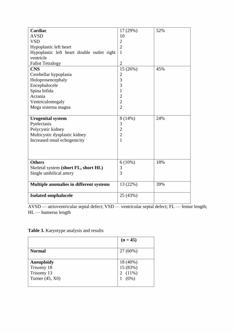
Cardiac
AVSD
VSD
Hypoplastic left heart
Hypoplastic left heart double outlet right
ventricle
Fallot Tetralogy
17 (29%)
10
2
2
1
2
52%
CNS
Cerebellar hypoplasia
Holoprosencephaly
Encephalocele
Spina bifida
Acrania
Ventriculomegaly
Mega sisterna magna
15 (26%)
2
3
3
1
2
2
2
45%
Urogenital system
Pyelectasis
Polycystic kidney
Multicystic dysplastic kidney
Increased renal echogenicity
8 (14%)
3
2
2
1
24%
Others
Skeletal system (short FL, short HL)
Single umbilical artery
6 (10%)
3
3
18%
Multiple anomalies in different systems
13 (22%) 39%
Isolated omphalocele
25 (43%)
AVSD — atrioventricular septal defect; VSD — ventricular septal defect; FL — femur length;
HL — humerus length
Table 3. Karyotype analysis and results
(n = 45)
Normal
27 (60%)
Aneuploidy
Trisomy 18
Trisomy 13
Turner (45, X0)
18 (40%)
15 (83%)
2 (11%)
1 (6%)
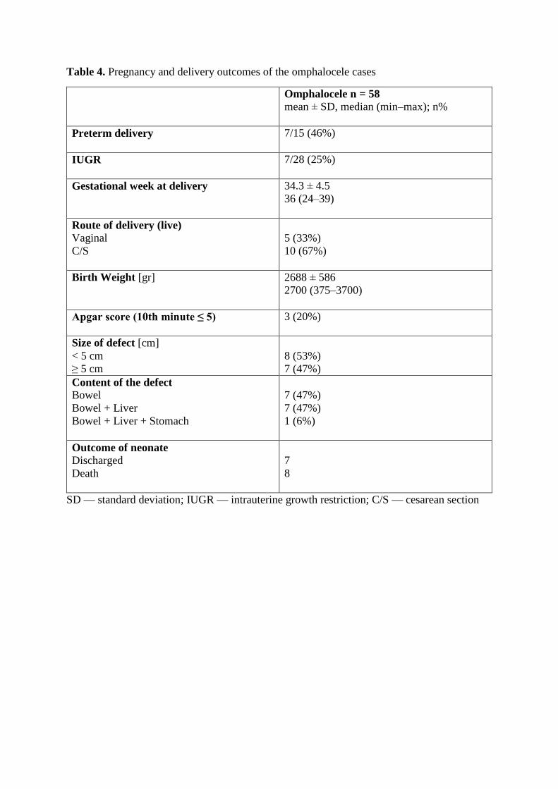
Table 4. Pregnancy and delivery outcomes of the omphalocele cases
Omphalocele n = 58
mean ± SD, median (min–max); n%
Preterm delivery
7/15 (46%)
IUGR
7/28 (25%)
Gestational week at delivery
34.3 ± 4.5
36 (24–39)
Route of delivery (live)
Vaginal
C/S
5 (33%)
10 (67%)
Birth Weight [gr] 2688 ± 586
2700 (375–3700)
Apgar score (10th minute ≤ 5)
3 (20%)
Size of defect [cm]
< 5 cm
≥ 5 cm
8 (53%)
7 (47%)
Content of the defect
Bowel
Bowel + Liver
Bowel + Liver + Stomach
7 (47%)
7 (47%)
1 (6%)
Outcome of neonate
Discharged
Death
7
8
SD — standard deviation; IUGR — intrauterine growth restriction; C/S — cesarean section