Embed Size (px)
Citation preview

JPET # 259341
1
Pharmacological evaluation of dotinurad, a selective urate reabsorption
inhibitor
Tetsuya Taniguchi, Naoki Ashizawa, Koji Matsumoto, Ryo Saito, Keisuke Motoki, Miku
Sakai, Noriko Chikamatsu, Chiharu Hagihara, Masamichi Hashiba, and Takashi
Iwanaga
Research Laboratories 2, FUJI YAKUHIN CO., LTD., 636-1 Iida-Shinden, Nishi Ward, Saitama City,
Saitama, 331-0068, JAPAN (T.T., N.A., K.M., K.M., M.S., N.C., C.H., T.I.); and Research Center,
MOCHIDA PHARMACEUTICAL CO., LTD., 722 Uenohara, Jimba, Gotemba City, Shizuoka,
412-8524, JAPAN (R.S., M.H.)
This article has not been copyedited and formatted. The final version may differ from this version.JPET Fast Forward. Published on August 1, 2019 as DOI: 10.1124/jpet.119.259341
at ASPE
T Journals on February 17, 2021
jpet.aspetjournals.orgD
ownloaded from

JPET # 259341
2
Running Title Page:
Dotinurad selectively inhibits urate reabsorption transporter
Corresponding author:
T. Taniguchi. Research Laboratories 2, FUJI YAKUHIN CO., LTD., 636-1 Iida-Shinden,
Nishi Ward, Saitama City, Saitama, 331-0068, JAPAN
Tel: +81-48-620-1611. Fax: +81-48-620-1617. E-mail address: [email protected]
Number of text page: 51
Number of tables: 4 (plus 3 supplemental table)
Number of figures: 6
Number of references: 40
Number of words: 8256
Abbreviations:
ABCG2, ATP-binding cassette sub-family G member 2; Dotinurad,
(3,5-Dichloro-4-hydroxyphenyl)(1,1-dioxo1,2-dihydro-3H-1λ6-1,3-benzothiazol-3-yl)methan
one; FEUA, fractional excretion of urate; HPLC, high performance liquid chromatography;
LC-MS/MS, liquid chromatography-tandem mass spectrometry; OAT1, organic anion
This article has not been copyedited and formatted. The final version may differ from this version.JPET Fast Forward. Published on August 1, 2019 as DOI: 10.1124/jpet.119.259341
at ASPE
T Journals on February 17, 2021
jpet.aspetjournals.orgD
ownloaded from

JPET # 259341
3
transporter 1; SURI, selective urate reabsorption inhibitor; URAT1, urate transporter 1
Section Assignment:
Metabolism, Transport, and Pharmacogenomics
This article has not been copyedited and formatted. The final version may differ from this version.JPET Fast Forward. Published on August 1, 2019 as DOI: 10.1124/jpet.119.259341
at ASPE
T Journals on February 17, 2021
jpet.aspetjournals.orgD
ownloaded from

JPET # 259341
4
Abstract
The effect of dotinurad was compared to those of commercially available uricosuric
agents, namely benzbromarone, lesinurad, and probenecid. Its effect on urate secretion
transporters was evaluated using probe substrates for respective transporters. Dotinurad,
benzbromarone, lesinurad, and probenecid inhibited URAT1 with IC50 values of 0.0372, 0.190,
30.0, and 165 μmol/l, respectively. Dotinurad weakly inhibited ABCG2, OAT1, and OAT3
with IC50 values of 4.16, 4.08, and 1.32 μmol/l, respectively, indicating higher selectivity for
URAT1. The hypouricemic effects of dotinurad and benzbromarone were evaluated in Cebus
monkeys. Dotinurad, at doses of 1–30 mg/kg, concomitantly decreased plasma urate levels
and increased fractional excretion of urate (FEUA) in a dose-dependent manner. On the
contrary, benzbromarone, at a dose of 30 mg/kg, showed a modest effect on plasma urate
levels. The inhibitory effect of dotinurad on urate secretion transporters was evaluated in
Sprague-Dawley rats, with sulfasalazine and adefovir as probe substrates of ABCG2 and
OAT1, respectively. Drugs including febuxostat as a reference ABCG2 inhibitor, were
administered orally before sulfasalazine or adefovir administration. Dotinurad had no effect
on urate secretion transporters in vivo, whereas benzbromarone, lesinurad, probenecid, and
febuxostat increased the plasma concentrations of probe substrates. These results suggested
dotinurad is characterized as a selective urate reabsorption inhibitor (SURI), which is defined
as a potent URAT1 inhibitor with minimal effect on urate secretion transporters including
This article has not been copyedited and formatted. The final version may differ from this version.JPET Fast Forward. Published on August 1, 2019 as DOI: 10.1124/jpet.119.259341
at ASPE
T Journals on February 17, 2021
jpet.aspetjournals.orgD
ownloaded from

JPET # 259341
5
ABCG2 and OAT1/3, because of its high efficacy in decreasing plasma urate levels compared
to that of other uricosuric agents.
Significance statement
Our study on the inhibitory effects of dotinurad on urate transport showed that dotinurad
had higher selectivity for URAT1 vs. ABCG2 and OAT1/3 than other uricosuric agents. In
Cebus monkeys, dotinurad concomitantly decreased plasma urate levels and increased
fractional excretion of urate (FEUA) in a dose-dependent manner. To determine the inhibitory
effect of dotinurad on urate secretion transporters, we performed studies in rats using
sulfasalazine and adefovir as probe substrates of ABCG2 and OAT1, respectively. Dotinurad
had no effect on these urate secretion transporters in vivo, whereas the other uricosuric agents
increased the plasma concentrations of the probe substrates. These results suggested dotinurad
as a potent and selective urate reabsorption inhibitor (SURI), is characterized by increased
efficacy with decreasing plasma urate levels compared to the other uricosuric agents studied.
This article has not been copyedited and formatted. The final version may differ from this version.JPET Fast Forward. Published on August 1, 2019 as DOI: 10.1124/jpet.119.259341
at ASPE
T Journals on February 17, 2021
jpet.aspetjournals.orgD
ownloaded from

JPET # 259341
6
Introduction
Gout is a form of acute arthritis induced by deposition of monosodium urate crystals in the
joints. Environmental (food and drink) and genetic (Lesch-Nyhan syndrome and familial
juvenile hyperuricemic nephropathy) factors have been reported to play a central role in the
etiology of gout (Kuo et al., 2015). Hyperuricemia, a common pathogenetic factor in the
development of gout, is typically defined by a serum urate concentration of >6.8 or 7.0 mg/dl
(Neogi, 2011; Terkeltaub et al., 2010). Hyperuricemia has been reported as a risk factor for
the onset and development of chronic renal diseases, and a predictive factor for metabolic
syndrome (Iseki et al., 2004; Obermayr et al., 2008; Yu et al., 2016).
Urate-lowering therapies with either type of urate control drug - urate production inhibitors
or uricosuric agents - are indicated for patients with hyperuricemia. Benzbromarone, a
commercially available uricosuric agent, effectively lowers serum urate levels. However,
because of its rare but severe idiosyncratic hepatotoxic adverse effects, it is not approved in
several European Union countries and the United States (Lee et al., 2008). In 2015, lesinurad,
another uricosuric agent, was approved for use in the United States. However, it has been
approved as 200 mg tablets in combination with urate production inhibitors because of its
modest effect and increased serum creatinine at higher doses. There is a need for a novel
uricosuric agent because 85% of patients develop hyperuricemia due to insufficient urate
excretion (Nakamura et al., 2003); thus, the use of a uricosuric agent is recommended by the
This article has not been copyedited and formatted. The final version may differ from this version.JPET Fast Forward. Published on August 1, 2019 as DOI: 10.1124/jpet.119.259341
at ASPE
T Journals on February 17, 2021
jpet.aspetjournals.orgD
ownloaded from
JPET # 259341
7
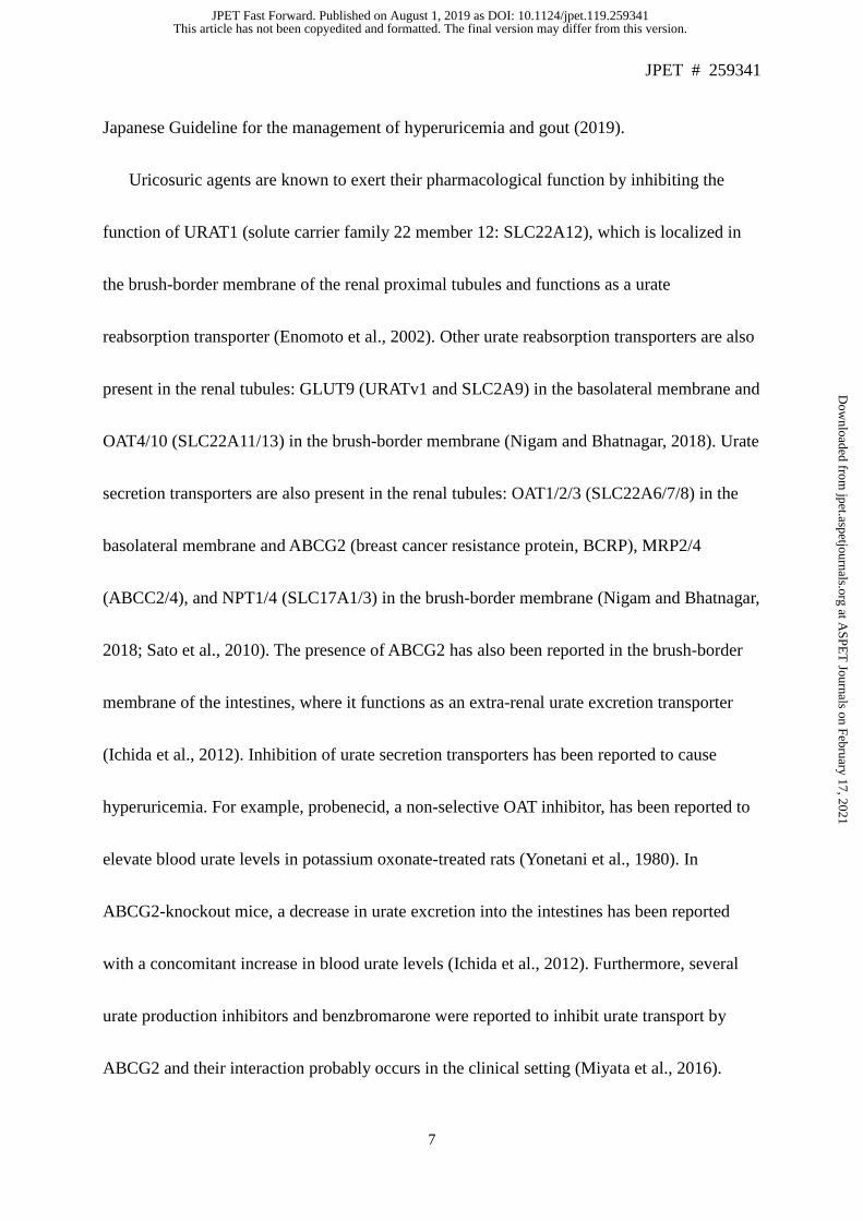
Japanese Guideline for the management of hyperuricemia and gout (2019).
Uricosuric agents are known to exert their pharmacological function by inhibiting the
function of URAT1 (solute carrier family 22 member 12: SLC22A12), which is localized in
the brush-border membrane of the renal proximal tubules and functions as a urate
reabsorption transporter (Enomoto et al., 2002). Other urate reabsorption transporters are also
present in the renal tubules: GLUT9 (URATv1 and SLC2A9) in the basolateral membrane and
OAT4/10 (SLC22A11/13) in the brush-border membrane (Nigam and Bhatnagar, 2018). Urate
secretion transporters are also present in the renal tubules: OAT1/2/3 (SLC22A6/7/8) in the
basolateral membrane and ABCG2 (breast cancer resistance protein, BCRP), MRP2/4
(ABCC2/4), and NPT1/4 (SLC17A1/3) in the brush-border membrane (Nigam and Bhatnagar,
2018; Sato et al., 2010). The presence of ABCG2 has also been reported in the brush-border
membrane of the intestines, where it functions as an extra-renal urate excretion transporter
(Ichida et al., 2012). Inhibition of urate secretion transporters has been reported to cause
hyperuricemia. For example, probenecid, a non-selective OAT inhibitor, has been reported to
elevate blood urate levels in potassium oxonate-treated rats (Yonetani et al., 1980). In
ABCG2-knockout mice, a decrease in urate excretion into the intestines has been reported
with a concomitant increase in blood urate levels (Ichida et al., 2012). Furthermore, several
urate production inhibitors and benzbromarone were reported to inhibit urate transport by
ABCG2 and their interaction probably occurs in the clinical setting (Miyata et al., 2016).
This article has not been copyedited and formatted. The final version may differ from this version.JPET Fast Forward. Published on August 1, 2019 as DOI: 10.1124/jpet.119.259341
at ASPE
T Journals on February 17, 2021
jpet.aspetjournals.orgD
ownloaded from

JPET # 259341
8
These data suggest that selective urate reabsorption inhibitors (SURIs), which are defined as
potent URAT1inhibitors that do not affect urate secretion transporters including ABCG2 and
OAT1/3, exhibit more potent hypouricemic effects than non-selective urate reabsorption
inhibitor.
In the present study, we first compared the inhibitory effect of dotinurad, a novel agent that
exhibits uricosuric effect in rodents, on urate transport in cells overexpressing URAT1,
ABCG2, and OAT1/3 with those of benzbromarone, lesinurad, and probenecid. We then
investigated the hypouricemic effects of dotinurad and benzbromarone in Cebus monkeys.
Finally, the inhibitory effects of these drugs on urate secretion transporters were evaluated in
Sprague-Dawley rats, using sulfasalazine and adefovir as probe substrates of ABCG2 and
OAT1, respectively. Considering our findings, we discuss the potency of dotinurad as a
uricosuric agent and its mechanism of action.
Materials and methods
Drugs and materials
Dotinurad (also called as FYU-981, Fig. 1) was synthesized by Fuji Yakuhin Co., Ltd.
(Saitama, Japan). Benzbromarone was purchased from Wako Pure Chemical Industries, Ltd.
(Osaka, Japan). Lesinurad was purchased from Selleck Chemicals, LLC. (Houston, TX, USA).
Probenecid and febuxostat were purchased from Tokyo Chemical Industry Co., Ltd. (Tokyo,
This article has not been copyedited and formatted. The final version may differ from this version.JPET Fast Forward. Published on August 1, 2019 as DOI: 10.1124/jpet.119.259341
at ASPE
T Journals on February 17, 2021
jpet.aspetjournals.orgD
ownloaded from

JPET # 259341
9
Japan). Adefovir was purchased from LKT Laboratories, Inc. (St. Paul, MN, USA).
Sulfasalazine was purchased from Sigma-Aldrich Co., LLC. (St. Louis, MO, USA).
[14C]-urate was synthesized by Moravek, Inc. (Brea, CA, USA). The MicroBeta2 liquid
scintillation counter was purchased from PerkinElmer, Inc. (Waltham, MA, USA). The other
drugs that were used in the present study are commercially available.
Determination of xanthine oxidase activity
Xanthine oxidase activity was measured with a spectrophotometer U-3000 (Hitachi
High-Technologies Corporation, Tokyo, Japan) at 295 nm by following the absorbance
change for initial 1 min. Assay mixture with 100 μmol/l xanthine dissolved in 0.1 mol/l NaOH
(1% final) and drugs dissolved in dimethyl sulfoxide (DMSO, 1% final) in 100 mmol/l
pyrophosphate buffer, pH 8.5 containing 0.2 mmol/l EDTA was pre-incubated for 5 min, and
reactions were started by adding xanthine oxidase (from bovine milk) to the mixture at the
final concentration of 3.2 mU/ml under the aerobic conditions at 25°C. Assay was performed
in triplicate.
Study of [14C]-Urate uptake in URAT1-overexpressing MDCKII cells or OAT1- and
OAT3-overexpressing HEK293 cells
Cells were seeded into 96-well tissue culture plates at a density of 1 × 10⁵ cells/well and
This article has not been copyedited and formatted. The final version may differ from this version.JPET Fast Forward. Published on August 1, 2019 as DOI: 10.1124/jpet.119.259341
at ASPE
T Journals on February 17, 2021
jpet.aspetjournals.orgD
ownloaded from

JPET # 259341
10
cultured at 37 ± 1 °C and a 5% CO2 atmosphere in Dulbecco's Modified Eagle's Medium with
4.5 g/l glucose for 24 h. Before initiating the experiment, culture medium was removed and
cells were washed twice in 100 µl of Hank's balanced salt solution (HBSS). Uptake
experiments were performed at 37 ± 1 °C in 50 µl of HBSS containing 20 μmol/l [14C]-urate
and test articles dissolved in DMSO. Vehicle control contained 1% DMSO instead of test
articles. URAT1-overexpressing MDCKII cells and OAT1-overexpressing HEK293 cells were
washed after 10 min of incubation, whereas OAT3-overexpressing HEK293 cells were
washed after 5 min of incubation, in 100 µL of ice-cold HBSS, followed by cell lysis with
50 µl of 0.1 mol/l NaOH. Transporter activity was determined by liquid scintillation counting
of an aliquot from each well. Assay was performed in triplicate. The amount of translocated
[14C]-urate was determined for each well in cpm. Relative urate transport activity was
reported as a percentage of the control and was calculated using the following equation:
Relative transport of urate (% of control) = (A−B)/(C−D) × 100, where A represent as the
amount of translocated [14C]-urate in the presence of test article in transfected cells; B
represent as the amount of translocated [14C]-urate in the presence of test article in mock cells;
C represent as the amount of translocated [14C]-urate in the presence of 1% DMSO in
transfected cells; and D represent as the amount of translocated [14C]-urate in the presence of
1% DMSO in mock cells.
This article has not been copyedited and formatted. The final version may differ from this version.JPET Fast Forward. Published on August 1, 2019 as DOI: 10.1124/jpet.119.259341
at ASPE
T Journals on February 17, 2021
jpet.aspetjournals.orgD
ownloaded from

JPET # 259341
11
Study of [14C]-urate uptake by membrane vesicles obtained from
ABCG2-overexpressing HEK293 cells
Membrane vesicle preparations from ABCG2-overexpressing cells (SOLVO Biotechnology,
Hungary) in the presence of 70 μmol/l urate containing 20 μmol/l [14C]-urate and 4 mmol/l
ATP or AMP to distinguish between transporter-mediated uptake and passive diffusion into
the vesicles. In 96-well plates, 50 μl of membrane vesicle suspended in 75 μmol/l urate and 30
μmol/l [14C]-urate containing ice-cold transport buffer (250 mmol/l sucrose, 10 mmol/l MgCl2,
10 mmol/l Tris-HCl, pH 7.4) were added. Test articles dissolved in DMSO, 0.75 μl, were
added to the membrane vesicle mixture. Vehicle control contained 1% DMSO instead of test
article. The mixtures were pre-incubated for 15 min at 37 ± 1°C. Reactions were initiated by
adding 25 μl of pre-warmed 12 mmol/l MgATP (or 12 mmol/l AMP as a background control)
in transport buffer. After 3 min, reactions were quenched by adding 200 μl of ice-cold
washing buffer (250 mmol/l sucrose, 100 mmol/l NaCl, 10 mmol/l Tris-HCl, pH 7.4) and
immediately filtered using glass fiber filters. The filters were washed 5 times with 200 μl of
ice-cold washing buffer and air-dried. The amount of substrate inside the filtrated vesicles was
determined by liquid scintillation counting. Assay was performed in triplicate. Relative urate
transport activity was reported as a percentage of the control and was calculated using the
following equation: Relative transport of urate (% of control) = (A−B)/(C−D) × 100, where A
represents the amount of translocated [14C]-urate in the presence of test article and ATP; B
This article has not been copyedited and formatted. The final version may differ from this version.JPET Fast Forward. Published on August 1, 2019 as DOI: 10.1124/jpet.119.259341
at ASPE
T Journals on February 17, 2021
jpet.aspetjournals.orgD
ownloaded from

JPET # 259341
12
represents the amount of translocated [14C]-urate in the presence of test article and AMP; C
represents the amount of translocated [14C]-urate in the presence of 1% DMSO and ATP; and
D represents the amount of translocated [14C]-urate in the presence of 1% DMSO and AMP.
Animals and housing
For the evaluation of hypouricemic effects, 5–10 year-old male Cebus monkeys (Cebus
paella, weight range 2.85–3.70 kg)) bred in Shin Nippon Biomedical Laboratories, Ltd.
(SNBL, Tokyo, Japan) were used for pharmacological studies. The monkeys were housed
individually in stainless steel cages in an air-conditioned animal room under a 12-/12-h
light/dark (LD) cycle at 26 ± 3 °C and a relative humidity of 55 ± 20%. Animals were fed a
pellet diet (New World Primate Diet 5040, Purina Mills, LCC) and fresh apples daily, except
one day before drug administration. Water was provided ad libitum throughout the study. The
experimental procedures were performed in accordance with the Animal Care and Utilization
Guidelines of SNBL.
For the evaluation of inhibitory effects on urate secretion transporters, 7-week-old male
Sprague-Dawley rats (weight range 180–220 g) bred in Japan SLC, Inc. (Shizuoka, Japan)
were used. The rats were housed in wire-mesh cages in an air-conditioned animal room with a
12-/12-h LD cycle at a temperature of 22 ± 4 °C and a relative humidity of 60 ± 20%.
Animals that did not develop abnormalities after a 1-week acclimatization period were
This article has not been copyedited and formatted. The final version may differ from this version.JPET Fast Forward. Published on August 1, 2019 as DOI: 10.1124/jpet.119.259341
at ASPE
T Journals on February 17, 2021
jpet.aspetjournals.orgD
ownloaded from

JPET # 259341
13
selected for the study. Rats were fed a CE-2 pellet diet (Crea Japan Inc., Tokyo, Japan) and
tap water via automatic stainless steel nozzles ad libitum throughout the study. Study
protocols were designed and refined taking animal reduction into consideration and were
approved by the Animal Care and Utilization Committee of Fuji Yakuhin Research
Laboratories.
Study of the hypouricemic effects of dotinurad and benzbromarone in Cebus monkeys
Five Cebus monkeys that were fasted for 18 h before drug administration orally received 1,
5, and 30 mg/kg of dotinurad, 30 mg/kg of benzbromarone, and 0.5% methylcellulose (MC)
as control, respectively. Blood samples (about 1 ml) obtained from the saphenous vein at
before and 2, 4, 8, and 24 h after drug administration using heparinized needle were kept on
ice. Plasma was obtained from the blood samples by centrifugation at 3,000 rpm for 10 min at
4 °C. Urine samples were collected 0–4, 4–8, and 8–24 h after drug administration. Urate and
creatinine levels in the samples were measured by a U-3000 spectrophotometer using an Iatro
LQ UAII (Mitsubishi Chemical Medience, Corp., Tokyo, Japan) and L-type Wako Creatinine
F (Wako Pure Chemical Industries, Ltd. Osaka, Japan). Each treatment was administered in
13-day intervals to wash out drugs, and treatment and sample collection were performed as
crossover experiments. FEUA was calculated as the ratio of urate clearance to creatinine
clearance. Urinary urate excretion was calculated as urinary urate concentration × urine
This article has not been copyedited and formatted. The final version may differ from this version.JPET Fast Forward. Published on August 1, 2019 as DOI: 10.1124/jpet.119.259341
at ASPE
T Journals on February 17, 2021
jpet.aspetjournals.orgD
ownloaded from

JPET # 259341
14
volume.
Determination of drug pharmacokinetics in Cebus monkeys
Plasma drug concentration was measured using the same samples described in the
previous section. Plasma samples were deproteinated with thrice volume of methanol and
centrifuged at 3,000 rpm for 10 min at 4 °C. Drug concentrations were measured using
high-performance liquid chromatography (HPLC) using the Alliance 2695 HPLC system
(Waters Corporation. Milford, MA, USA). The concentration of 6-hydroxybenzbromarone, a
major benzbromarone metabolite, was also measured in the plasma obtained from
benzbromarone-treated animals.
Study of concomitant sulfasalazine and hypouricemic agents treatment in
Sprague-Dawley rats
Sprague-Dawley rats that were fasted for 18 h before drug administration orally received
20 mg/kg of febuxostat, 50 mg/kg of benzbromarone, 1.3 mg/kg of dotinurad, and 0.5% MC
as control, respectively (n = 4, a total of 16 rats were used). Drug dosages used in the present
study were calculated based on their clinically maximal doses detailed in supplemental Table
1. Thirty minutes after the administration of these drugs, sulfasalazine suspended in 0.5% MC
was orally administered at a dose of 20 mg/kg. For drug bioavailability (F) calculations, 5
This article has not been copyedited and formatted. The final version may differ from this version.JPET Fast Forward. Published on August 1, 2019 as DOI: 10.1124/jpet.119.259341
at ASPE
T Journals on February 17, 2021
jpet.aspetjournals.orgD
ownloaded from

JPET # 259341
15
mg/kg of sulfasalazine dissolved in 50 mmol/l tris-saline was intravenously administered via
the tail vein. Blood samples (about 200 μl) were obtained from the jugular vein at 0.083 (i.v.
only), 0.25, 0.5, 1, 2, 4, 8, and 12 h after sulfasalazine administration using heparinized needle
and kept on ice. Plasma was obtained from the blood samples by centrifugation at 3,000 rpm
for 10 min at 4°C. Plasma samples were deproteinated and sulfasalazine concentration was
measured as previously described by using liquid chromatography-tandem mass spectrometry
(LC-MS/MS) on an Agilent 1100 Series HPLC Value System (Agilent Technologies, Inc.
Santa Clara, CA, USA), API3000 (AB SCIEX, LCC. Framingham, MA, USA) (Miyata et al.,
2016). F were calculated using the following equation: F (%) = (AUC0-inf value of oral
administration / 20) / (AUC0-inf value of intravenous administration / 5) ×100.
Study of concomitant adefovir and uricosuric agents treatment in Sprague-Dawley rats
Sprague-Dawley rats that were fasted for 18 h before drug administration orally received
100 mg/kg of probenecid, 67 mg/kg of lesinurad, 50 mg/kg of benzbromarone, 1.3 mg/kg of
dotinurad, and 0.5% MC as control (n = 6, a total of 30 rats were used). Thirty minutes after
drug administration, 3 mg/kg of adefovir dissolved in saline was intravenously administered
via the tail vein to all animals. Blood samples (about 200 μl) were obtained from the jugular
vein at 0.083, 0.25, 0.5, 1, 2, and 4 h after adefovir administration using heparinized needle
and kept on ice. Plasma was obtained from blood samples by centrifugation at 3,000 rpm for
This article has not been copyedited and formatted. The final version may differ from this version.JPET Fast Forward. Published on August 1, 2019 as DOI: 10.1124/jpet.119.259341
at ASPE
T Journals on February 17, 2021
jpet.aspetjournals.orgD
ownloaded from

JPET # 259341
16
10 min at 4°C. Adefovir was detected in these samples as previously described (Jullien, 2003).
Briefly, plasma was derivatized by incubation at 80 °C in 0.34% chloroacetaldehyde solution
for 50 min and detected via the excitation and emission spectra at 236 and 420 nm
wavelengths, respectively, using the Alliance 2695 HPLC system (Waters Corporation.
Milford, MA, USA).
Statistical analysis
In the study of xanthine oxidase activity, IC50 values were calculated using probit method.
In cell-based urate uptake studies, the concentration–response curves were analyzed using the
GraphPad Prism 7.03 software (GraphPad Software, CA, USA) and a 4-parameter logistic
equation was applied to calculate the IC50 values. In the studies of Cebus monkeys and
Sprague-Dawley rats, the mean and standard deviation (SD) were calculated, and differences
from the control group were analyzed using the Dunnett’s multiple comparison test (at
significance levels of 5% and 1%). In the study of Cebus monkeys, AUC0–24h was calculated
using the trapezoidal rule in Microsoft Excel 2010 (Microsoft Corporation, DC, USA) and
unbound fraction of maximum plasma concentration was calculated using Fu in supplemental
Table 3. In the study of Sprague-Dawley rats, pharmacokinetic parameters were analyzed
using the Phoenix WinNonlin 6.4 software.
This article has not been copyedited and formatted. The final version may differ from this version.JPET Fast Forward. Published on August 1, 2019 as DOI: 10.1124/jpet.119.259341
at ASPE
T Journals on February 17, 2021
jpet.aspetjournals.orgD
ownloaded from
JPET # 259341
17
Results
Effects of uricosuric agents on xanthine oxidase activity
Benzbromarone inhibited xanthine oxidase activity with an IC50 value of 15.4 μmol/l.
Dotinurad, lesinurad, and probenecid did not have any effect up to 100, 300, and 1000 μmol/l,
respectively.
Inhibitory effect of uricosuric agents on urate uptake by URAT1-overexpressing
MDCKII cells
Dotinurad, benzbromarone, lesinurad, and probenecid inhibited urate transport by
URAT1-overexpressing MDCKII cells in a concentration-dependent manner at respective
concentrations of 0.003–3, 0.003–3, 0.3–300, and 10–1000 μmol/l (Fig. 2) and respective IC50
values of 0.0372, 0.190, 30.0, and 165 μmol/l (Table 1a).
Inhibitory effect of uricosuric agents on urate uptake by ABCG2-, OAT1-, and
OAT3-overexpressing HEK293 cells
Dotinurad inhibited urate transport in a concentration-dependent manner in ABCG2-,
OAT1-, and OAT3-overexpressing HEK293 cells at respective concentrations of 0.3–300,
0.1–100, and 0.03–30 μmol/l and respective IC50 values of 4.16, 4.08, and 1.32 μmol/l. The
ratios of IC50 values of ABCG2-, OAT1-, and OAT3-overexpressing cells to that of
This article has not been copyedited and formatted. The final version may differ from this version.JPET Fast Forward. Published on August 1, 2019 as DOI: 10.1124/jpet.119.259341
at ASPE
T Journals on February 17, 2021
jpet.aspetjournals.orgD
ownloaded from

JPET # 259341
18
URAT1-overexpressing cells were 112, 110, and 35.5, respectively (Tables 1a and 1b).
Benzbromarone inhibited urate transport in ABCG2-, OAT1-, and OAT3-overexpressing cells
with respective IC50 values of 0.289, 3.14, and 0.967 μmol/l and respective IC50 value ratios
to that of URAT1-overexpressing cells were 1.52, 16.5, and 5.09. Lesinurad inhibited urate
transport with respective IC50 values of 26.4, 6.99, and 1.07 μmol/l and respective IC50 value
ratios to that of URAT1-overexpressing cells were 0.880, 0.233, and 0.0357. Probenecid
inhibited urate transport with respective IC50 values of 433, 10.9, and 2.37 μmol/l and
respective IC50 value ratios to that of URAT1-overexpressing cells were 2.62, 0.0661, and
0.0144.
Hypouricemic effects of dotinurad and benzbromarone in Cebus monkeys
Effects of dotinurad and benzbromarone on plasma urate levels, FEUA, and urinary urate
excretion were assessed in Cebus monkeys. Dotinurad dose-dependently lowered the plasma
urate levels with its maximum effect at 8 h (Fig. 3A). Changes in plasma urate level between
0 and 8 h (ΔPUA) were lower than that of control by 0.28, 0.97 (p < 0.05), and 1.79 mg/dl (p <
0.01), at doses of 1, 5, and 30 mg/kg, respectively. Furthermore, dotinurad dose-dependently
increased FEUA (Fig. 3B). The 0–4 h FEUA increased by 180%, at a dose of 30 mg/kg
compared to the control (p < 0.01). On the contrary, the effect of benzbromarone at a dose of
30 mg/kg was modest; the ΔPUA was lower than that of control by 0.46 mg/dl and the 0–4 h
This article has not been copyedited and formatted. The final version may differ from this version.JPET Fast Forward. Published on August 1, 2019 as DOI: 10.1124/jpet.119.259341
at ASPE
T Journals on February 17, 2021
jpet.aspetjournals.orgD
ownloaded from

JPET # 259341
19
FEUA increased by 30% compared to the control. Figure 3C details the urinary excretion
amount of urate in Cebus monkeys 0–8 h after dotinurad and benzbromarone administration.
Dotinurad dose-dependently increased the urinary urate excretion to 16.2, 22.8, and 25.3 mg
at doses of 1, 5, and 30 mg/kg, respectively, compared with the control, which was 13.7 mg.
On the contrary, benzbromarone increased the urinary urate excretion to 19.4 mg at a dose of
30 mg/kg. Results of creatinine clearance and urate clearance were shown in supplemental
Table 2.
Pharmacokinetic parameters of dotinurad and benzbromarone in Cebus monkeys
The plasma concentration of dotinurad increased in a dose-dependent manner with Cmax
and AUC0–24h values of 107 μg/ml and 780 μg・h/ml, respectively, at a dose of 30 mg/kg
(Table 2). On the contrary, Cmax and AUC0–24h values of benzbromarone were 20.9 μg/ml and
95.2 μg・h/ml, respectively, at a dose of 30 mg/kg. In the plasma obtained from
benzbromarone-treated animals, Cmax and AUC0–24h value of 6-hydroxybenzbromarone were
12.7 μg/ml and 80.9 μg・h/ml, respectively. Unbound fraction of maximum plasma
concentration of dotinurad, at doses of 1, 5, 30 mg/kg, and benzbromarone, at a dose of 30
mg/kg, were 0.0144, 0.0800, 0.750 μg/ml and 0.564 μg/ml, respectively.
Effect of hypouricemic agents on the plasma concentration of sulfasalazine in
This article has not been copyedited and formatted. The final version may differ from this version.JPET Fast Forward. Published on August 1, 2019 as DOI: 10.1124/jpet.119.259341
at ASPE
T Journals on February 17, 2021
jpet.aspetjournals.orgD
ownloaded from
JPET # 259341
20
Sprague-Dawley rats
Effects of febuxostat, benzbromarone, and dotinurad on the plasma sulfasalazine
concentration were assessed in Sprague-Dawley rats. Febuxostat and benzbromarone
significantly increased the plasma sulfasalazine concentration (Fig. 4). Febuxostat increased
the AUC0-inf value to 5012 ng・h/ml (p < 0.01) from a control value of 713 ng・h/ml and F to
14.1% from a control value of 2.0%. Benzbromarone also increased the AUC0-inf value to
2888 ng・h/ml (p < 0.01) and F to 8.1%. Dotinurad did not have any effect on the
pharmacokinetic parameters of sulfasalazine (Table 3).
Effect of uricosuric agents on the plasma concentration of adefovir in Sprague-Dawley
rats
Effects of probenecid, benzbromarone, lesinurad, and dotinurad on the plasma
concentration of adefovir were assessed in Sprague-Dawley rats. Probenecid and lesinurad
significantly increased the plasma adefovir concentration (Fig. 5). Probenecid increased
plasma concentration of adefovir at 0.083–0.5 h (p < 0.01) and 1 h (p < 0.05). Lesinurad also
increased plasma concentration of adefovir at 0.083 h (p < 0.01), 0.25 h (p < 0.05) and 0.5 h
(p < 0.01). On the contrary, benzbromarone and dotinurad did not have any effect on the
plasma concentration of adefovir.
This article has not been copyedited and formatted. The final version may differ from this version.JPET Fast Forward. Published on August 1, 2019 as DOI: 10.1124/jpet.119.259341
at ASPE
T Journals on February 17, 2021
jpet.aspetjournals.orgD
ownloaded from

JPET # 259341
21
Discussion
The kidneys play an important role in urate elimination. After filtration through the
glomerulus, urate undergoes complex bidirectional steps of reabsorption and secretion in the
proximal tubules, resulting in a net urinary excretion of <10% of the filtered amount (Chonko
and Grantham, 1981). Because renal handling of urate is reabsorption-dominant, the
reabsorption process is the target of uricosuric agents. In the renal tubules, several transporters
are involved in urate reabsorption. Particularly, URAT1 in the brush-border membrane and
GLUT9 in the basolateral membrane of renal tubules are the main urate reabsorption
transporters. Aberrant function of these transporters caused by single nucleotide
polymorphisms (SNPs) results in insufficient urate reabsorption, increased urinary urate
excretion, and remarkable hypouricemia (<1 mg/dl; Kikuchi et al., 2000; Dinour et al., 2010).
Uricosuric agents are known to exert their pharmacological effect via URAT1 inhibition, by
which almost maximum effect is obtained without affecting other urate reabsorption
transporters. On the contrary, urate secretion transporters are also present in the renal tubules:
OAT1/2/3 are localized in the basolateral membrane, whereas ABCG2, MRP2/4, and NPT1/4
are localized in the brush-border membrane (Nigam and Bhatnagar, 2018; Sato et al., 2010).
SNPs in ABCG2 result in a higher risk of gout caused by dysfunctional urate excretion
(extra-renal excretion) from the intestines (Matsuo et al., 2009; Miyata et al., 2016).
Furthermore, gene association studies performed in a patient cohort with chronic kidney
This article has not been copyedited and formatted. The final version may differ from this version.JPET Fast Forward. Published on August 1, 2019 as DOI: 10.1124/jpet.119.259341
at ASPE
T Journals on February 17, 2021
jpet.aspetjournals.orgD
ownloaded from

JPET # 259341
22
disease (CKD) showed that SNPs in ABCG2, MRP4, and OAT3 are associated with increased
serum urate levels, although the association of SNPs in ABCG2 with serum levels was modest
in the non-CKD cohort (Bhatnagar et al., 2016). In humans, the contribution of other urate
secretion transporters on serum urate level has not been reported. Our findings suggested that
SURIs had superior hypouricemic effects to those of non-selective urate reabsorption
inhibitors.
Our previous study showed that in rats treated with topitoxostat, a urate production
inhibitor, dotinurad decreases plasma urate levels and increases FEUA in a dose-dependent
manner without inhibiting xanthine oxidase or uricase (Taniguchi et al., 2016; Matsumoto et
al. 2011). In studies in pyrazinamide-treated rats and brush-border membrane vesicles,
dotinurad inhibits urate uptake at the brush-border membrane of the renal tubules with
pyrazinecarboxylic acid as an exchange substrate (Taniguchi et al., 2017). It was postulated
that this effect is mediated by urate transporters; however, the detailed underlying
mechanisms are unknown. Based on these findings, we next evaluated the inhibitory effect of
dotinurad on urate uptake by MDCKII or HEK293 cells overexpressing transporters.
Dotinurad inhibited urate transport mediated by URAT1 with an IC50 value of 0.0372 μmol/l.
The effect of dotinurad was approximately 5.11, 806, and 4440 times more potent than that of
benzbromarone, lesinurad, and probenecid, respectively, because the IC50 value of these
compounds was 0.190, 30.0, and 165 μmol/l, respectively (Table 1a). Dotinurad modestly
This article has not been copyedited and formatted. The final version may differ from this version.JPET Fast Forward. Published on August 1, 2019 as DOI: 10.1124/jpet.119.259341
at ASPE
T Journals on February 17, 2021
jpet.aspetjournals.orgD
ownloaded from

JPET # 259341
23
inhibited urate transport by ABCG2 and OAT1/3 with IC50 values of 4.16, 4.08, and 1.32
μmol/l, which were 112, 110, and 35.5 times higher than its IC50 values for URAT1,
respectively (Tables 1a and 1b). By contrast, the IC50 ratios of benzbromarone, lesinurad, and
probenecid for ABCG2 and OAT1/3 to URAT1 were 1.52–16.5, 0.0357–0.880, and 0.0144–
2.62 for, respectively. These findings showed that dotinurad inhibited URAT1 more
selectively than the other uricosuric agents. Several lines of evidence showed the inhibitory
effect of benzbromarone, lesinurad, and probenecid on urate transport by URAT1, ABCG2,
OAT1, and OAT3 (Miner et al., 2016; Miyata H et al., 2016; Ichida et al., 2003), although the
inhibitory effects of benzbromarone and probenecid on urate transport by OAT3 are not
reported. Our data were consistent with these reports, except for the URAT1 inhibitory effect
of lesinurad and probenecid (the IC50 values are 3.53 and 13.23 μmol/l, respectively). We
could not evaluate the inhibitory effect of uricosuric agents on MRP4, because urate transport
by MRP4-overexpressing cells was not reported. These data also suggested that dotinurad did
not affect ABCG2 and OAT1/3 in clinical use because its unbound fraction in plasma
appeared to be extremely low (Supplemental Table 3; FDA, 2018; PMDA, 2019).
Subsequently, we compared the hypouricemic efficacy of dotinurad with that of
benzbromarone in Cebus monkeys. Dotinurad at doses of 1, 5, and 30 mg/kg decreased
plasma urate levels and increased FEUA in a dose-dependent manner, and its effect at 1 mg/kg
was approximately equal to that of 30 mg/kg benzbromarone (Fig. 3A and 3B). However, the
This article has not been copyedited and formatted. The final version may differ from this version.JPET Fast Forward. Published on August 1, 2019 as DOI: 10.1124/jpet.119.259341
at ASPE
T Journals on February 17, 2021
jpet.aspetjournals.orgD
ownloaded from

JPET # 259341
24
stringency of URAT1 inhibition (the IC50 values of dotinurad and benzbromarone were 0.0372
and 0.190 μmol/l, respectively) did not sufficiently account for the difference in their
hypouricemic effects. We hypothesized that the pharmacokinetic profiles of these drugs
possibly contribute to their hypouricemic effects. Indeed, dotinurad had higher Cmax and
AUC0–24h values than those of benzbromarone (Table 2). The unbound fraction of maximal
plasma concentration of dotinurad at doses of 1, 5, 30 mg/kg, and that of benzbromarone at a
dose of 30 mg/kg, which are considered to correlate with their concentration at the renal
proximal tubules, were calculated to be 0.0144, 0.0800, 0.750, and 0.564 μg/ml, respectively
(Table 2). Because these values are 1.1-, 6.0-, 56- and 7.2-times higher than the IC50 values of
URAT1, respectively, an equal hypouricemic effect was expected to be obtained by 5 mg/kg
of dotinurad and 30 mg/kg of benzbromarone (Table 2 and Supplemental Table 3).
Considering that 6-hydroxybenzbromarone, a major metabolite of benzbromarone, also
inhibited URAT1 and contributed to the hypouricemic effect in addition to benzbromarone
itself (Shin et al., 2011), another factor was assumed to explain the difference in hypouricemic
effect. Thus, we speculated that property as SURIs, defined as potent URAT1 inhibitors with
minimal effect on urate secretion transporters, ABCG2 and OAT1/3, generated the difference
of hypouricemic effect apart from non-selective inhibitor.
In order to estimate the effect of uricosuric agents on urate secretion in vivo accurately, we
evaluated whether dotinurad and other uricosuric agents interact with ABCG2 and OAT1
This article has not been copyedited and formatted. The final version may differ from this version.JPET Fast Forward. Published on August 1, 2019 as DOI: 10.1124/jpet.119.259341
at ASPE
T Journals on February 17, 2021
jpet.aspetjournals.orgD
ownloaded from

JPET # 259341
25
using their probe substrates in Sprague-Dawley rats. Sulfasalazine is known as a typical
ABCG2 substrate with low absorption because it is excreted on the intestinal brush-border
membrane (Dahan and Amidon, 2009). In our rat study using sulfasalazine as a probe
substrate, benzbromarone and the positive control agent febuxostat markedly increased the
plasma concentration and F of sulfasalazine (Fig. 4 and Table 3), these increases were
assumed to be caused by inhibition of intestinal ABCG2, which appeared to be important for
urate excretion (Ichida et al., 2012). Adefovir, a well-known OAT1 substrate, is excreted in
urine (as adefovir itself), and its fraction of drug elimination in urine (fe) is approximately
90% in humans (Cundy et al., 1995). In our rat study using adefovir as a probe substrate,
lesinurad and probenecid increased the plasma concentration of adefovir (Fig. 5); this increase
was assumed to be caused by inhibition of renal OAT1, which mediates urate secretion (Wu et
al., 2017). Because these drugs showed more potent inhibition on OAT3 than on OAT1,
further experiments should be conducted using a probe substrate for OAT3. In our rat studies,
dotinurad did not affect the plasma concentration of probe substrates, indicating that dotinurad
hardly affected urate secretion transporter in vivo.
We must consider that the probe substrates did not move exactly in the same manner as
urate. It can be predicted by evaluating the inhibitory effect of a compound on urate and probe
substrates. For example, another ABCG2 inhibitor, rifampicin, inhibits both urate and
sulfasalazine transport by ABCG2 at a similar concentration (Hosomi et al., 2012; Kosa et al.,
This article has not been copyedited and formatted. The final version may differ from this version.JPET Fast Forward. Published on August 1, 2019 as DOI: 10.1124/jpet.119.259341
at ASPE
T Journals on February 17, 2021
jpet.aspetjournals.orgD
ownloaded from

JPET # 259341
26
2018). In terms of OAT1, probenecid inhibits adefovir transport with an inhibition constant
(Ki) of 18.6 μmol/l (Maeda et al., 2014), suggesting a similarity to our result. These reports
indicated that sulfasalazine and adefovir were transported in the same manner as urate.
Furthermore, species differences between rats and humans have to be taken into account. The
AUC0–48h of sulfasalazine was three-fold higher for functional ABCG2 SNPs, and a similar
effect was observed in ABCG2-knockout mice (Gotanda et al., 2015; Miyata et al., 2016). The
fe of adefovir in rodents is approximately 80%, which is nearly the same as that for humans
(Naesens et al., 1996). Therefore, we concluded that the data obtained from our rat studies
could predict urate handling in humans.
Studies on Cebus monkeys showed that the hypouricemic effect of UR-1102, a selective
inhibitor of URAT1 vs. OAT1/3, showed a higher estimated Emax of urinary urate excretion
than that shown by benzbromarone (Ahn et al., 2016). In this report, authors discussed about
URAT1 selectivity, defined as potent URAT1 inhibition without effect on OAT1/3, is
important for producing potent hypouricemic effect. In fact, UR-1102, in a dose-dependent
manner, lowered serum urate in humans at doses (0.25–2 mg) much lower than that of
benzbromarone (Jun et al., 2017).
Figure 6 shows a scheme of urate pool reduction by SURIs or non-selective inhibitors.
SURIs effectively reduce urate pool by increasing renal excretion of urate. On the contrary,
non-selective inhibitors modestly reduce urate pool because of their inhibitory effect on urate
This article has not been copyedited and formatted. The final version may differ from this version.JPET Fast Forward. Published on August 1, 2019 as DOI: 10.1124/jpet.119.259341
at ASPE
T Journals on February 17, 2021
jpet.aspetjournals.orgD
ownloaded from

JPET # 259341
27
secretion transporters. For non-selective inhibitors to reduce urate pool at a similar level as
SURIs, they require more URAT1 inhibition at higher doses, which results in excessively
elevated renal excretion of urate and pose a risk factor for urolithiasis (Yu and Gutman, 1967).
In the present study, benzbromarone did not exert an apparent effect on plasma urate levels in
Cebus monkeys (Fig. 3A), although it increased urinary urate excretion more potently than 1
mg/kg of dotinurad (Fig. 3C). The phenomenon appeared to be mediated by inhibition of
urate secretion transporters, especially ABCG2 in the intestine. These findings suggested that
non-selective inhibitors can only reduce urate pool. Furthermore, our findings proved that
SURIs were not associated with a risk of drug-drug interaction under clinical settings.
In conclusion, we showed that dotinurad, a SURI, could be an effective therapeutic option
for the treatment of hyperuricemia because of its more effective hypouricemic action than that
of commercially available uricosuric agents. Furthermore, dotinurad could be a useful agent
in studies on urate transporters because of its ability to selectively and potently inhibit
URAT1.
Acknowledgments
We appreciate to Mr. Jo Tanaka, Department of Drug Safety Research Laboratories, Shin
Nippon Biomedical Laboratories Ltd. and Dr. Erika Csonka and Dr. Daniel Gliesche, SOLVO
Biotechnology for their assistance of pharmacological evaluation. We appreciated to Mr.
This article has not been copyedited and formatted. The final version may differ from this version.JPET Fast Forward. Published on August 1, 2019 as DOI: 10.1124/jpet.119.259341
at ASPE
T Journals on February 17, 2021
jpet.aspetjournals.orgD
ownloaded from

JPET # 259341
28
Kazuma Sekine for his assistance of pharmacokinetic evaluation. We appreciated to Ms.
Kazumi Miyamoto for her assistance of pharmacological evaluation.
Authorship Contributions
Participated in research design: Tetsuya Taniguchi, Naoki Ashizawa, Koji Matsumoto,
Keisuke Motoki, and Takashi Iwanaga
Conducted experiments: Tetsuya Taniguchi, Keisuke Motoki, Miku Sakai, Noriko Chikamatsu,
and Chiharu Hagihara
Performed data analysis: Tetsuya Taniguchi, Naoki Ashizawa, Koji Matsumoto, and Keisuke
Motoki
Wrote or contributed to the writing of the manuscript: Tetsuya Taniguchi, Naoki Ashizawa,
Koji Matsumoto, Ryo Saito, Keisuke Motoki, Masamichi Hashiba, and Takashi Iwanaga
This article has not been copyedited and formatted. The final version may differ from this version.JPET Fast Forward. Published on August 1, 2019 as DOI: 10.1124/jpet.119.259341
at ASPE
T Journals on February 17, 2021
jpet.aspetjournals.orgD
ownloaded from

JPET # 259341
29
References
Ahn SO, Ohtomo S, Kiyokawa J, Nakagawa T, Yamane M, Lee KJ, Kim KH, Kim BH,
Tanaka J, Kawabe Y, and Horiba N (2016) Stronger uricosuric effects of the novel selective
URAT1 Inhibitor UR-1102 lowered plasma urate in tufted capuchin monkeys to a greater
extent than benzbromarone. J Pharmacol Exp Ther 357: 157–166.
Bhatnagar V, Richard EL, Wu W, Nievergelt CM, Lipkowitz MS, Jeff J, Maihofer AX, and
Nigam SK (2016) Analysis of ABCG2 and other urate transporters in uric acid homeostasis
in chronic kidney disease: potential role of remote sensing and signaling. Clin Kidney J 9:
444–453.
Chonko AM, and Grantham JJ (1981) Disorders of urate metabolism and excretion, in The
Kidney, 2nd ed. (Brenner BM, and Rector FC eds) pp 1023–1055, WB Saunders,
Philadelphia.
Cundy KC, Barditch-Crovo P, Walker RE, Collier AC, Ebeling D, Toole J, and Jaffe HS
(1995) Clinical pharmacokinetics of adefovir in human immunodeficiency virus type
1-infected patients. Antimicrob Agents Chemother 39: 2401–2405.
Dahan A, and Amidon GL (2009) Small intestinal efflux mediated by MRP2 and BCRP shifts
sulfasalazine intestinal permeability from high to low, enabling its colonic targeting. Am J
Physiol Gastrointest Liver Physiol 297: G371–377.
Dinour D, Gray NK, Campbell S, Shu X, Sawyer L, Richardson W, Rechavi G, Amariglio N,
This article has not been copyedited and formatted. The final version may differ from this version.JPET Fast Forward. Published on August 1, 2019 as DOI: 10.1124/jpet.119.259341
at ASPE
T Journals on February 17, 2021
jpet.aspetjournals.orgD
ownloaded from

JPET # 259341
30
Ganon L, Sela BA, Bahat H, Goldman M, Weissgarten J, Millar MR, Wright AF, and
Holtzman EJ (2010) Homozygous SLC2A9 mutations cause severe renal hypouricemia. J
Am Soc Nephrol 21: 64–72.
Enomoto A, Kimura H, Chairoungdua A, Shigeta Y, Jutabha P, Cha SH, Hosoyamada M,
Takeda M, Sekine T, Igarashi T, Matsuo H, Kikuchi Y, Oda T, Ichida K, Hosoya T,
Shimokata K, Niwa T, Kanai Y, and Endou H (2002) Molecular identification of a renal
urate anion exchanger that regulates blood urate levels. Nature 417: 447–452.
Gotanda K, Tokumoto T, Hirota T, Fukae M, and Ieiri I (2015) Sulfasalazine disposition in a
subject with 376C>T (nonsense mutation) and 421C>A variants in the ABCG2 gene. Br J
Clin Pharmacol 80: 1236–1237.
Hosomi A, Nakanishi T, Fujita T, and Tamai I (2012) Extra-renal elimination of uric acid via
intestinal efflux transporter BCRP/ABCG2. PLoS One 7: e30456.
Ichida K, Hosoyamada M, Kimura H, Takeda M, Utsunomiya Y, Hosoya T, and Endou H
(2003) Urate transport via human PAH transporter hOAT1 and its gene structure. Kidney
Int 63: 143–155.
Ichida K, Matsuo H, Takada T, Nakayama A, Murakami K, Shimizu T, Yamanashi Y, Kasuga
H, Nakashima H, Nakamura T, Takada Y, Kawamura Y, Inoue H, Okada C, Utsumi Y,
Ikebuchi Y, Ito K, Nakamura M, Shinohara Y, Hosoyamada M, Sakurai Y, Shinomiya N,
Hosoya T, and Suzuki H (2012) Decreased extrarenal urate excretion is a common cause of
This article has not been copyedited and formatted. The final version may differ from this version.JPET Fast Forward. Published on August 1, 2019 as DOI: 10.1124/jpet.119.259341
at ASPE
T Journals on February 17, 2021
jpet.aspetjournals.orgD
ownloaded from

JPET # 259341
31
hyperuricemia. Nat Commun 3: 764.
Iseki K, Ikemiya Y, Inoue T, Iseki C, Kinjo K, and Takishita S (2004) Significance of
hyperuricemia as a risk factor for developing ESRD in a screened cohort. Am J Kidney Dis
44: 642–650.
Japanese society of gout and nucleic acid metabolism (2019): Guideline for the management
of hyperuricemia and gout, 3rd ed. (in Japanese) (Hisatome I ed) pp 110–113, Shindan to
Chiryo-sha, Tokyo, Japan.
Jullien V, Tréluyer JM, Pons G, and Rey E (2003) Determination of tenofovir in human
plasma by high-performance liquid chromatography with spectrofluorimetric detection. J
Chromatogr B Analyt Technol Biomed Life Sci 785: 377–381.
Jun JB, Lee H, Suh CH, Lee CK, Kim DW, Choe JY, Lee SH, Kim SH, Hong SJ, Bang SY,
Choi SJ, Park YB, Onohara M, Choi J, Song JS, and Park W (2017) A series of
double-blind, placebo-controlled, randomized, multicenter, phase 2 studies to evaluate the
efficacy, safety, and dose–response relationship of orally administered URC102, a novel
URAT1 inhibitor, in Korean patients with gout. American college of rheumatology annual
meeting. Abstract number 2074.
Kikuchi Y, Koga H, Yasutomo Y, Kawabata Y, Shimizu E, Naruse M, Kiyama S, Nonoguchi
H, Tomita K, Sasatomi Y, and Takebayashi S (2000) Patients with renal hypouricemia with
exercise-induced acute renal failure and chronic renal dysfunction. Clin Nephrol 53: 467–
This article has not been copyedited and formatted. The final version may differ from this version.JPET Fast Forward. Published on August 1, 2019 as DOI: 10.1124/jpet.119.259341
at ASPE
T Journals on February 17, 2021
jpet.aspetjournals.orgD
ownloaded from

JPET # 259341
32
472.
Kosa RE, Lazzaro S, Bi YA, Tierney B, Gates D, Modi S, Costales C, Rodrigues AD,
Tremaine LM, and Varma MV (2018) Simultaneous assessment of transporter-mediated
drug–drug interactions using a probe drug cocktail in Cynomolgus monkey. Drug Metab
Dispos 46: 1179–1189.
Kuo CF, Grainge MJ, Zhang W, and Doherty M (2015) Global epidemiology of gout:
prevalence, incidence and risk factors. Nat Rev Rheumatol 11: 649–662.
Lee MH, Graham GG, Williams KM, and Day RO (2008) A benefit-risk assessment of
benzbromarone in the treatment of gout. Was its withdrawal from the market in the best
interest of patients? Drug Saf 31: 643–665.
Maeda K, Tian Y, Fujita T, Ikeda Y, Kumagai Y, Kondo T, Tanabe K, Nakayama H, Horita S,
Kusuhara H, and Sugiyama Y (2014) Inhibitory effects of p-aminohippurate and probenecid
on the renal clearance of adefovir and benzylpenicillin as probe drugs for organic anion
transporter (OAT) 1 and OAT3 in humans. Eur J Pharm Sci 59: 94–103.
Matsumoto K, Okamoto K, Ashizawa N, Nishino T (2011) FYX-051: a novel and potent
hybrid-type inhibitor of xanthine oxidoreductase. J Pharmacol Exp Ther 336: 95–103.
Matsuo H, Takada T, Ichida K, Nakamura T, Nakayama A, Ikebuchi Y, Ito K, Kusanagi Y,
Chiba T, Tadokoro S, Takada Y, Oikawa Y, Inoue H, Suzuki K, Okada R, Nishiyama J,
Domoto H, Watanabe S, Fujita M, Morimoto Y, Naito M, Nishio K, Hishida A, Wakai K,
This article has not been copyedited and formatted. The final version may differ from this version.JPET Fast Forward. Published on August 1, 2019 as DOI: 10.1124/jpet.119.259341
at ASPE
T Journals on February 17, 2021
jpet.aspetjournals.orgD
ownloaded from

JPET # 259341
33
Asai Y, Niwa K, Kamakura K, Nonoyama S, Sakurai Y, Hosoya T, Kanai Y, Suzuki H,
Hamajima N, and Shinomiya N (2009) Common defects of ABCG2, a high-capacity urate
exporter, cause gout: a function-based genetic analysis in a Japanese population. Sci Transl
Med 1: 5–11.
Miner JN, Tan PK, Hyndman D, Liu S, Iverson C, Nanavati P, Hagerty DT, Manhard K, Shen
Z, Girardet JL, Yeh LT, Terkeltaub R, and Quart B (2016) Lesinurad, a novel, oral
compound for gout, acts to decrease serum uric acid through inhibition of urate
transporters in the kidney. Arthritis Res Ther 18: 214.
Miyata H, Takada T, Toyoda Y, Matsuo H, Ichida K, and Suzuki H (2016) Identification of
febuxostat as a new strong ABCG2 inhibitor: potential applications and risks in clinical
situations. Front Pharmacol 7: 518.
Naesens L, Balzarini J, Bischofberger N, and De Clercq E (1996) Antiretroviral activity and
pharmacokinetics in mice of oral
bis(pivaloyloxymethyl)-9-(2-phosphonylmethoxyethyl)adenine, the
bis(pivaloyloxymethyl) ester prodrug of 9-(2-phosphonylmethoxyethyl)adenine.
Antimicrob Agents Chemother 40: 22–28.
Nakamura T (2003) Pathogenesis and pathophysiology of hyperuricemia (in Japanese), in
Nakamura T, editor. Treatment of hyperuricemia and gout. Medical Review, Osaka, Japan.
21–39.
This article has not been copyedited and formatted. The final version may differ from this version.JPET Fast Forward. Published on August 1, 2019 as DOI: 10.1124/jpet.119.259341
at ASPE
T Journals on February 17, 2021
jpet.aspetjournals.orgD
ownloaded from

JPET # 259341
34
Neogi T (2011) Clinical practice. Gout. N Engl J Med 364: 443–452.
Nigam SK, and Bhatnagar V (2018) The systems biology of uric acid transporters: the role of
remote sensing and signaling. Curr Opin Nephrol Hypertens 27: 305–313.
Obermayr RP, Temml C, Gutjahr G, Knechtelsdorfer M, Oberbauer R, and Klauser-Braun R
(2008) Elevated uric acid increases the risk for kidney disease. J Am Soc Nephrol 19:
2407–2413.
Pharmaceuticals and medical devices agency (PMDA) (2019): Guideline on drug interaction
for drug development and appropriate provision of information.
Sato M, Mamada H, Anzai N, Shirasaka Y, Nakanishi T, and Tamai I (2010) Renal secretion
of uric acid by organic anion transporter 2 (OAT2/SLC22A7) in human. Biol Pharm Bull
33: 498–503.
Shin HJ, Takeda M, Enomoto A, Fujimura M, Miyazaki H, Anzai N, and Endou H (2011)
Interactions of urate transporter URAT1 in human kidney with uricosuric drugs.
Nephrology (Carlton) 16: 152–162.
Taniguchi T, Ashizawa N, Matsumoto K, Iwanaga T, and Saitoh K (2016) Uricosuric agents
decrease the plasma urate level in rats by concomitant treatment with topiroxostat, a novel
xanthine oxidoreductase inhibitor. J Pharm Pharmacol 68: 76–83.
Taniguchi T, Ashizawa N, Matsumoto K, and Iwanaga T (2017) Enhancement of
pharmacological effects of uricosuric agents by concomitant treatment with pyrazinamide
This article has not been copyedited and formatted. The final version may differ from this version.JPET Fast Forward. Published on August 1, 2019 as DOI: 10.1124/jpet.119.259341
at ASPE
T Journals on February 17, 2021
jpet.aspetjournals.orgD
ownloaded from

JPET # 259341
35
in rats. Naunyn Schmiedebergs Arch Pharmacol 390: 253–260.
Terkeltaub R (2010) Update on gout: new therapeutic strategies and options. Nat Rev
Rheumatol 6: 30–38.
USA food and drug administration (FDA) (2017): Guidance for industry. In vitro metabolism
and transporter mediated drug–drug interaction studies.
Wu W, Bush KT, and Nigam SK (2017) Key role for the organic anion transporters, OAT1
and OAT3, in the in vivo handling of uremic toxins and solutes. Sci Rep 7: 4939.
Yonetani Y, Ishii M, and Iwaki K (1980) Hyperuricemia induced by some antihypertensives
and uricosuric drugs in oxonate-treated rats. Jpn J Pharmacol 30: 829–840.
Yu T, and Gutman AB (1967) Uric acid nephrolithiasis in gout. Predisposing factors. Ann
Intern Med 67: 1133–1148.
Yu TY, Jee JH, Bae JC, Jin SM, Baek JH, Lee MK, and Kim JH (2016) Serum uric acid: A
strong and independent predictor of metabolic syndrome after adjusting for body
composition. Metabolism 65: 432–440.
This article has not been copyedited and formatted. The final version may differ from this version.JPET Fast Forward. Published on August 1, 2019 as DOI: 10.1124/jpet.119.259341
at ASPE
T Journals on February 17, 2021
jpet.aspetjournals.orgD
ownloaded from

JPET # 259341
36
Footnotes:
Thesis information:
A part of Cebus monkey studies in the work was previously presented at the 86 th annual
meeting of the Japanese pharmacological society (abstract number. P2-143).
To receive reprint requests:
Tetsuya Taniguchi. Research Laboratories 2, FUJI YAKUHIN CO., LTD., 636-1
Iida-Shinden, Nishi Ward, Saitama City, Saitama, 331-0068, JAPAN,
This article has not been copyedited and formatted. The final version may differ from this version.JPET Fast Forward. Published on August 1, 2019 as DOI: 10.1124/jpet.119.259341
at ASPE
T Journals on February 17, 2021
jpet.aspetjournals.orgD
ownloaded from

37
Figure Legends:
Fig. 1 Chemical structures of dotinurad and commercially available uricosuric agents.
A: Dotinurad, B: Benzbromarone, C: Lesinurad, D: Probenecid
Fig. 2 Concentration-response curve of dotinurad and commercially available uricosuric
agents on urate transport by URAT1-overexpressing MDCKII cells.
Each value is the mean ± SD of urate transport relative to the control (n = 3) and described by
GraphPad Prism 7.03 software (GraphPad Software, CA, USA).
Fig. 3 Effects of dotinurad and benzbromarone on plasma urate level, FEUA, and urinary urate
excretion in Cebus monkeys.
FEUA: fractional excretion of urate; MC: methylcellulose.
Data are represented as change in plasma urate level (A), FEUA at 0–4, 4–8, and 8–24 h after
dosing (B) and the amount of urinary urate excretion for 8 h after dosing (C). Each value is
the means ± SD of four to five animals. *P < 0.05, **P < 0.01 for significant differences from
the control at each time point using the Dunnett’s multiple comparison test.
Fig. 4 Effect of dotinurad and other hypouricemic agents on the plasma concentration of
This article has not been copyedited and formatted. The final version may differ from this version.JPET Fast Forward. Published on August 1, 2019 as DOI: 10.1124/jpet.119.259341
at ASPE
T Journals on February 17, 2021
jpet.aspetjournals.orgD
ownloaded from

38
sulfasalazine in Sprague-Dawley rats
MC: methylcellulose.
Hypouricemic agents or their vehicle as control (0.5% MC) were orally administered to
Sprague-Dawley rats fasted for 18 h. After 30 min, sulfasalazine was orally administered.
Blood samples were collected at 0.25, 0.5, 1, 2, 4, 8, and 12 h after sulfasalazine
administration. Plasma samples were deproteinated and sulfasalazine concentrations were
measured using LC-MS/MS. Each value is the mean ± SD of three to four animals.
**P < 0.01 shows significant differences from the 0.5% MC control group after Dunnett’s
multiple comparison test.
Fig. 5 Effect of dotinurad and other uricosuric agents on the plasma concentration of adefovir
in Sprague-Dawley rats
MC: methylcellulose.
Hypouricemic agents or their vehicle as control (0.5% MC) were orally administered to
Sprague-Dawley rats fasted for 18 h. After 30 min, adefovir was intravenously administered.
Blood samples were collected at 0.083, 0.25, 0.5, 1, 2, 4, 8, and 12 h after adefovir
administration. Plasma samples were derivatized and adefovir concentrations were measured
using HPLC. Each value is the mean ± SD of six animals.
*P < 0.05 and **P < 0.01 show significant differences from the 0.5% MC control group after
This article has not been copyedited and formatted. The final version may differ from this version.JPET Fast Forward. Published on August 1, 2019 as DOI: 10.1124/jpet.119.259341
at ASPE
T Journals on February 17, 2021
jpet.aspetjournals.orgD
ownloaded from

39
Dunnett’s multiple comparison test.
Fig. 6 Comparison of the potency of selective and non-selective urate reabsorption inhibitors
in decreasing urate pool and the concomitant effects of non-selective inhibitors.
The upper figure shows the usual state of urate handling showing urate excretion from the
renal tubules and the small intestine. SURI increases urate excretion from the renal tubules
due to URAT1 inhibition, leading to a potent reduction of urate pool (A). A non-selective
inhibitor decreases urate excretion from the renal tubules and/or the small intestine thorough
inhibition of ABCG2 and/or OAT1/3. For a non-selective inhibitor to obtain the same effect at
a level similar to SURI, more potent inhibition of URAT1 and additional renal excretion are
required (B). Arrow width represents the quantity of urate handling, which is affected by
inhibition of URAT1, ABCG2, and OAT1/3.
This article has not been copyedited and formatted. The final version may differ from this version.JPET Fast Forward. Published on August 1, 2019 as DOI: 10.1124/jpet.119.259341
at ASPE
T Journals on February 17, 2021
jpet.aspetjournals.orgD
ownloaded from

40
Table 1a. IC50 values of dotinurad and commercially available uricosuric agents against urate
transport by URAT1 and urate secretion transporters
Test article
IC50 (μmol/l)
URAT1 ABCG2 OAT1 OAT3
Dotinurad
0.0372
+ 0.0065
4.16
+ 2.49
4.08
+ 1.02
1.32
+ 0.07
Benzbromarone
0.190
+ 0.029
0.289
+ 0.074
3.14
+ 1.59
0.967
+ 0.071
Lesinurad
30.0
+ 3.9
26.4
+ 4.7
6.99
+ 1.94
1.07
+ 0.27
Probenecid
165
+ 14
433
+ 218
10.9
+ 0.5
2.37
+ 0.94
IC50: half maximal inhibitory concentration
For measurement of transport activities, MDCKII-URAT1 (passage number 3–11),
HEK293-OAT1 (passage number 8), HEK293-OAT3 (passage number 5), and
HEK293-ABCG2 membrane vesicles (SOLVO Biotechnology, Hungary) were used.
IC50 was calculated using four-parameter logistic equation. Each value is the mean + SEM (n
= 2–3).
This article has not been copyedited and formatted. The final version may differ from this version.JPET Fast Forward. Published on August 1, 2019 as DOI: 10.1124/jpet.119.259341
at ASPE
T Journals on February 17, 2021
jpet.aspetjournals.orgD
ownloaded from

41
Table 1b. IC50 ratios for ABCG2 and OAT1/3 to URAT1 of dotinurad and commercially
available uricosuric agents
Test article
IC50 ratio to URAT1
URAT1 ABCG2 OAT1 OAT3
Dotinurad 1 112 110 35.5
Benzbromarone 1 1.52 16.5 5.09
Lesinurad 1 0.880 0.233 0.0357
Probenecid 1 2.62 0.0661 0.0144
Each value was calculated as the IC50 of each compound for ABCG2, OAT1, and OAT3/IC50
for URAT1.
This article has not been copyedited and formatted. The final version may differ from this version.JPET Fast Forward. Published on August 1, 2019 as DOI: 10.1124/jpet.119.259341
at ASPE
T Journals on February 17, 2021
jpet.aspetjournals.orgD
ownloaded from

42
Table 2. Pharmacokinetic parameters of dotinurad and benzbromarone in Cebus monkeys
Administered
article
Dose
(mg/kg)
Measured article
Pharmacokinetic parameters
Cmax
(μg/ml)
Fu × Cmax
(μg/ml)
Tmax
(h)
AUC0-24h
(μg・h/ml)
Dotinurad
1
Dotinurad
2.09 ± 0.78 0.0144 3.2 ± 1.1 20.3 ± 8.6
5 11.4 ± 4.8 0.0800 2.8 ± 1.1 109 ± 53
30 107 ± 22 0.750 2.0 ± 0.0 780 ± 265
Benzbromarone 30
Benzbromarone 20.9 ± 9.0 0.564 2.0 ± 0.0 95.2 ± 28.1
6-Hydroxybenzbromarone 12.7 ± 4.4 - 2.0 ± 0.0 80.9 ± 33.9
Cmax: maximum plasma concentration; Fu: unbound fraction in plasma; Fu × Cmax: unbound
maximum plasma concentration; Tmax: time to reach maximum plasma concentration; AUC0–
24h: area under the plasma concentration-time curve at 0–24 h. “-” means not calculated.
Plasma samples were deproteinated and drug concentrations were measured using HPLC.
AUC0–24h was calculated using the trapezoidal rule. Each value is the mean ± SD of five
animals.
This article has not been copyedited and formatted. The final version may differ from this version.JPET Fast Forward. Published on August 1, 2019 as DOI: 10.1124/jpet.119.259341
at ASPE
T Journals on February 17, 2021
jpet.aspetjournals.orgD
ownloaded from

43
Table 3. Pharmacokinetic parameters of sulfasalazine and effects of concomitant treatment
with hypouricemic agents in Sprague-Dawley rats
Test article
Dose
(mg/kg)
Cmax
(ng/ml)
T1/2
(h)
AUCo-t
(ng・h/ml)
AUCinf
(ng・h/ml)
CLT
(ml/h/kg)
F
(%)
0.5% MC -
422
± 182
1.49
± 0.28
665
± 94
713
± 90
28395
± 3800
2.0
Febuxostat 20
3138**
± 272
1.30
± 0.13
5004**
± 851
5012**
± 848
4067**
± 607
14.1
Benzbromarone 50
1520**
± 202
1.24
± 0.46
2858**
± 750
2888**
± 729
7294**
± 1989
8.1
Dotinurad 1.3
415
± 68
1.88
± 1.56
708
± 166
885
± 264
24126
± 7777
2.5
MC: methylcellulose; Cmax: maximum plasma concentration; T1/2: elimination half-life;
AUC0–t: area under the plasma concentration-time curve at 0–12 h; AUCinf: AUC from zero to
infinity; CLT: total clearance; F: bioavailability.
Pharmacokinetic parameters were calculated using Phoenix WinNonlin (Pharsight Corp.
Sunnyvale, CA, USA). F were calculated using the following equation: F = (AUC0-inf value of
oral administration / 20) / (AUC0-inf value of intravenous administration / 5) ×100. Values are
reported as the mean ± SD of three to four animals.
This article has not been copyedited and formatted. The final version may differ from this version.JPET Fast Forward. Published on August 1, 2019 as DOI: 10.1124/jpet.119.259341
at ASPE
T Journals on February 17, 2021
jpet.aspetjournals.orgD
ownloaded from

44
**P < 0.01 shows significant differences from the 0.5% MC control group, as analyzed by
Dunnett’s multiple comparison test.
This article has not been copyedited and formatted. The final version may differ from this version.JPET Fast Forward. Published on August 1, 2019 as DOI: 10.1124/jpet.119.259341
at ASPE
T Journals on February 17, 2021
jpet.aspetjournals.orgD
ownloaded from

45
Fig. 1
A B
C D
This article has not been copyedited and formatted. The final version may differ from this version.JPET Fast Forward. Published on August 1, 2019 as DOI: 10.1124/jpet.119.259341
at ASPE
T Journals on February 17, 2021
jpet.aspetjournals.orgD
ownloaded from

46
Fig. 2
This article has not been copyedited and formatted. The final version may differ from this version.JPET Fast Forward. Published on August 1, 2019 as DOI: 10.1124/jpet.119.259341
at ASPE
T Journals on February 17, 2021
jpet.aspetjournals.orgD
ownloaded from

47
Fig. 3
A
B
1
2
3
4
5
6
0 8 16 24
Pla
sma
ura
tele
vel
(m
g/d
l)
Time after administration (h)
0.5% MC
Dotinurad 1 mg/kg
Dotinurad 5 mg/kg
Dotinurad 30 mg/kg
Benzbromarone 30 mg/kg
****
**
0
20
40
0-4 4-8 8-24
FE
UA
(%)
Time after administration (h)
0.5% MCDotinurad 1 mg/kgDotinurad 5 mg/kgDotinurad 30 mg/kgBenzbromarone 30 mg/kg
**
This article has not been copyedited and formatted. The final version may differ from this version.JPET Fast Forward. Published on August 1, 2019 as DOI: 10.1124/jpet.119.259341
at ASPE
T Journals on February 17, 2021
jpet.aspetjournals.orgD
ownloaded from

48
C
0
10
20
30
40
Control F12981
(1)
F12981
(5)
F12981
(30)
Benz (30)
Uri
nar
y u
rate
ex
cret
ion (
mg
)
1 5 30 30 (mg/kg)
0.5% MC Dotinurad Benzbromarone
This article has not been copyedited and formatted. The final version may differ from this version.JPET Fast Forward. Published on August 1, 2019 as DOI: 10.1124/jpet.119.259341
at ASPE
T Journals on February 17, 2021
jpet.aspetjournals.orgD
ownloaded from

49
Fig. 4
0.1
1
10
100
1000
10000
0 2 4 6 8 10 12
Pla
sma s
ulf
asa
lazi
ne
con
c. (
ng/m
l)
Time (h)
0.5% MC
Febuxostat
Benzbromarone
Dotinurad
****
****
**
**
**
**
****
**
This article has not been copyedited and formatted. The final version may differ from this version.JPET Fast Forward. Published on August 1, 2019 as DOI: 10.1124/jpet.119.259341
at ASPE
T Journals on February 17, 2021
jpet.aspetjournals.orgD
ownloaded from

50
Fig. 5
0.01
0.1
1
10
0 1 2 3 4
Pla
sma a
def
ovir
con
c. (μg/m
l)
Time (h)
0.5% MC
Probenecid
Benzbromarone
Lesinurad
Dotinurad
****
****
**
**
*
This article has not been copyedited and formatted. The final version may differ from this version.JPET Fast Forward. Published on August 1, 2019 as DOI: 10.1124/jpet.119.259341
at ASPE
T Journals on February 17, 2021
jpet.aspetjournals.orgD
ownloaded from

51
Fig.6
Excretion in feces Excretion in urine
urate pool
ABCG2, OAT1/3
URAT1
Glomerular filtrationABCG2
(A) Selective urate reabsorption inhibitor (SURI)
URAT1
Renaltubule
Smallintestine
(B) Non-selective inhibitor
ABCG2, OAT1/3
URAT1
ABCG2
Blood
This article has not been copyedited and formatted. The final version may differ from this version.JPET Fast Forward. Published on August 1, 2019 as DOI: 10.1124/jpet.119.259341
at ASPE
T Journals on February 17, 2021
jpet.aspetjournals.orgD
ownloaded from