Embed Size (px)
Citation preview
Pharmacologyonline 3: 48-70 (2011) ewsletter Gulia and Choudhary
48
PEPTIC ULCER DISEASE: A REVIEW
Yuvraj Gulia*, Manjusha Choudhary
Institute of Pharmaceutical Sciences, Kurukshetra University, Kurukshetra
Summary
Peptic ulcer is defined as disruption of the mucosal integrity of stomach and/or
duodenum leading to a local defect or excavation due to active inflammation. Most common
symptom of peptic ulcer is abdominal discomfort. It occurs because of an imbalance
between aggressive factors (gastric acid and pepsin) and defensive factors (gastric mucus,
bicarbonate, prostaglandins). About 25 percent of patients with this disease have a serious
complication such as haemorrhage, perforation, or gastric outlet obstruction. Peptic ulcer
can be diagnosed either by direct visualization using an endoscope or by using contrast
radiography. Various class of drugs are used in the treatment of this disease like H2
antagonists, Proton pump inhibitors, Prostaglandin analogues, Antacids, Ulcer protectives
and Anti-H.pylori drugs. The present review summarizes the history, symptoms,
complications, types, epidemiology, pathogenesis, diagnosis and therapy of Peptic Ulcer
Disease.
Keywords: Peptic ulcer, pathogenesis, diagnosis, therapy.
*Corresponding Author
Yuvraj Gulia
Institute of Pharmaceutical Sciences,
Kurukshetra University, Kurukshetra
E-mail: [email protected]
Phone no. +91 9991410952
Pharmacologyonline 3: 48-70 (2011) ewsletter Gulia and Choudhary
49
Peptic Ulcer
Peptic ulcer is defined as disruption of the mucosal integrity of stomach and/or
duodenum leading to a local defect or excavation due to active inflammation[1]
. The word
‘peptic’ refers to pepsin a stomach enzyme that break down proteins. Peptic ulcer located in
stomach is called gastric ulcer[2]
.
History of Peptic Ulcer
The existence of gastric ulceration was acknowledged by Diocles of Carystos (350
B.C.), Celsus, and Galen (131–201 A.D.). In 1910, Schwartz first quoted the dictum “No
Acid, No Ulcer”. In 1982, Warren and Marshall provided the first insight into an important
pathogenic factor in peptic ulcer disease with the discovery of Helicobacter pylori
(H.pylori)[3]
. The etiopathogenesis of peptic ulcer has changed from Schwartz’s dictum “No
acid-No ulcer” to “No mucosal damage-No ulcer” and recently to “No Helicobacter pylori-
No ulcer”[4]
. Before 16th
century, Avicenna noted the relationship between gastric pain and
mealtimes. Later on Stahl (1728) hypothesized that some fevers and gastric inflammation are
associated with ulcerations[5]
. In 1737, Morgagni described both gastric and duodenal ulcer
at autopsy. In 1761, it was hypothesized that pain is associated with stomach ulcers. G.
Bottcher and M.Letulle (1875) and J.Cohnheim (1880) hypothesized that ulcers are caused
by bacteria and chemical factors, respectively[6]
.
John Lykoudis, a general practitioner in Greece, treated patients for peptic ulcer
disease with antibiotics, beginning in 1958, long before it was commonly recognized that
bacteria were a dominant cause for the disease.
It was a previously widely accepted misunderstanding that the use of chewing gum
resulted in gastric ulcers. The medical profession believed that this was because the action of
masticating on gum caused the over-stimulation of the production of hydrochloric acid
(HCl) in the stomach. The low acidity (pH 2) or hyperchlorhydria was then believed to
cause erosion of the stomach lining in the absence of food, thus causing the development of
the gastric ulcers[7]
. On the other hand, in the recent past, some believed that natural tree
resin extract, mastic gum, actively eliminates the H.pylori bacteria[8]
. However, multiple
subsequent studies have found no effect of using mastic gum on reducing H.pylori
levels[9,10]
.
In the year 1959, Leiber and Lefevre published a follow up study demonstrating that
antibiotics prevent the conversion of urea to ammonia in the human stomach[11]
. In 1962,
Susser and Stein proved that stress causes peptic ulcer disease[12]
. The discovery of the
compound cimetidine by researchers at the UK laboratories of Smith Kline & French in the
1970s, transformed the lives of millions of people. It was sold under the trademark Tagamet,
and was the first effective anti-ulcer drug that made a revolutionary impact on treatment. It
decreases acid secretion, thus promoting healing of ulcers[13]
.
In 1982, Warren and Marshall showed that there is a relationship between H.pylori
and Peptic Ulcer Disease (PUD). Their paper was published in June, 1984. Many reviewers
disliked the paper. In order to answer his critics, he tested on himself and consumed H.pylori
Pharmacologyonline 3: 48-70 (2011) ewsletter Gulia and Choudhary
50
and became ill. He then took antibiotics and got rid of his symptoms. In 2005, Warren and
Marshall were awarded the Nobel prize in physiology or medicine for their work on H.pylori
and PUD[14]
.
In 1990, Rauws and Tytgat described the cure of duodenal ulcer by eradication of
H.pylori using a triple-therapy regimen consisting of bismuth and 2 antibiotics. Triple
therapy, modernized to a PPI and 2 antibiotics soon became first line therapy for
eradication[15]
.
In 1992, Covocci et al sequenced the CagA gene, which encodes for a cytotoxin
associated surface protein, which correlated strongly with strains of H.pylori that caused
duodenal ulcers. It was the first description of a virulence factor for H.pylori infection
determined by molecular techniques[16]
.
In 1997, the Centers for Disease Control and Prevention, with other government
agencies, academic institutions, and industry, launched a national education campaign to
inform health care providers and consumers about the link between H.pylori and ulcers. This
campaign reinforced the news that ulcers are a curable infection, and that health can be
greatly improved and money saved by disseminating information about H.pylori[17]
. Later in
the same year, Tomb et al completely sequenced the entire 16,67,867 base pair H.pylori
genome. This assisted in identifying new virulence factors for the infectivity of H.pylori on
the molecular level[18]
.
In 2001, Chan et al showed in a randomized control trial that eradication of H.pylori
even prevents bleeding from ulcers that are caused by aspirin and other Non-steroidal anti-
inflammatory drugs (NSAID)[19]
.
Symptoms
Most common symptom of peptic ulcer is abdominal discomfort. This discomfort
comes and goes for several days of week, generally occurs 2 to 3 hrs after a meal, in the
middle of night, when stomach is empty. Other symptoms include blood loss leading to
anaemia, weight loss, poor appetite, bloating, burping, nausea and vomiting. In patients with
advanced stage emergency symptoms are sharp, sudden, persistent accompanied with
stomach pain, bloody or black stools, blood in vomit, etc.
Complications
About 25 percent of patients with peptic ulcer disease have a serious complication
such as haemorrhage, perforation, or gastric outlet obstruction. Silent ulcers and
complications are more common in older patients and in patients taking NSAID.
1. Bleeding: It is the most frequent complication, which occurs in 15-20% patients. It
accounts for 25% ulcer deaths.
2. Perforation: It occurs in 5% patients. It accounts for about 67% ulcer deaths.
3. Obstruction from edema: It occurs in 2% patients. It most often occurs due to
pyloric channel ulcers. It causes crampy abdominal pain[20,21]
.
Pharmacologyonline 3: 48-70 (2011) ewsletter Gulia and Choudhary
51
Types of Peptic Ulcer
On the basis of location, peptic ulcers are categorized as follows: Gastric Ulcer: Occurrence
of ulcer in the stomach, more commonly in older age group. Duodenal Ulcer: Occurrence of
ulcer in the duodenum. They occur commonly in younger individuals and are evenly
distributed among various socioeconomic groups. These patients have higher than normal
levels of acid secretion rates. Depending on severity, peptic ulcers are classified as: Acute
Peptic Ulcer: These ulcers involve tissues to the depth of the submucosa. They may arise in
the form of single or multiple lesions. They are found in many sites of stomach and in the
first few centimetres of duodenum. Chronic Peptic Ulcer: These ulcers penetrate through the
epithelial and muscle layers of stomach wall and may include the adjacent pancreas or liver.
In majority of cases, they occur singly in the pyloric antrum of the stomach and in the
duodenum[22]
.
Epidemiology of Peptic ulcer
Peptic ulcer used to be a rare disease before 19th
century. Acute perforations of
gastric ulcers were first reported in young girls in the beginning of 19th
century. With the
progress of 19th
century, peptic ulcer disease became frequent both in men and women[23]
.
In the West, this disease affects equally in men and women whereas, in India, men
are affected 18 times more commonly than women. In a vast, developing country like India
it is impossible to obtain exact figures of disease incidence and differences are bound to
exist between regions.
In India, peptic ulcer is more prevalent in Jammu and Kashmir, followed by
Southern India. North India comes next, and East and North East have comparatively lower
prevalence[24]
. Greater prevalence of peptic ulcer in southern India over northern India may
be attributed to the fact that rice is the staple food in this region[25,26]
.
In United States approximately 4 million people have peptic ulcers, 3,50,000 new
cases diagnosed, 1,80,000 hospitalized and 5000 people die each year as a result of peptic
ulcer. The lifetime likelihood for developing a peptic ulcer is about 10% for males and 4%
for females. Through autopsy studies and biopsy, prevalence of 6 to 14% for men and 2 to
6% for women was found. The male to female ratio for duodenal ulcers is 3:1 and for gastric
ulcers is 2:1[27]
. The mortality rate for duodenal ulcer in 1962 was 3.1 per 100,000 and for
gastric ulcer it was 3.5 per 100,000; these rates had decreased to about 1 per 100,000 each
by 1979[28,29]
. In 1998, the total mortality rate due to peptic ulcer in United States was found
to be 3.47 per 100,000[27]
. Gastric ulcer has a higher mortality rate than duodenal ulcer
because of its prevalence in older patients[30-32]
.
In young Norwegians, the annual incidence of duodenal ulcer was approximately 2
in 1000 men and 0.9 in 1000 women, while for gastric ulcer the annual incidence was
approximately 1.5 in 1000 men and 0.9 in 1000 women[33]
.
Pharmacologyonline 3: 48-70 (2011) ewsletter Gulia and Choudhary
52
In Japan, the male-to-female ratio for peptic ulcer was 2:1, but the overall gastric
ulcer rate was about 1.5 times greater than that for duodenal ulcer[34]
. In USA, the
prevalence of duodenal ulcer is 4 times that of gastric ulcer, while in Pakistan, it is 5 times
that of gastric ulcers, while in some parts of India this ratio is 32:1[35]
.
H.pylori infection is a major etiologic factor of peptic ulcer. Over 90% duodenal
ulcers and 70% gastric ulcers occur due to H.pylori infection[36,37]
. There are large
differences in the prevalence of infection among ethnic groups in different societies. White
Americans have lower infection rates than black Americans, and Australians of southern
European origin have higher infection rates than Australians of Anglo-Celtic ancestry[38]
.
In developing countries such as India, Peru and Thailand, exposure to H.pylori
occurs in childhood. Sero-surveys indicate a sero-prevalence of 22-57% in children under
the age of five, increasing to 80-100% by the age of 20 and thereafter it remains constant. In
Peru, age specific prevalence rates rise as high as 90% in persons over 60 years of age. In
Thailand, 17.5% of children 5-9 years old and 75% of individuals 30-49 years of age were
infected with H.pylori[39-41]
.
In developed countries such as US, serologic evidence of H.pylori is uncommon
before the age 10, increases to 10% in those between 18 and 30 years of age and to 50% in
those older than 60 years[42]
. The incidence of H.pylori below the age 30 is dramatically
decreasing in developed countries due to improved socioeconomic conditions[43]
.
In northern Iran, 40% children of a school were diagnosed as H.pylori positive and
results suggested that the source of drinking water may play a role in transmission of
H.pylori[44]
.
In a study by Drumm et al, a specific antibody was detected in 74% of the parents
and 82% of the siblings of H.pylori-infected children. These results suggested strong person-
to-person transmission of this infective organism, which occurs via oral-oral route or fecal-
oral route[45,46]
.
Studies from India suggest that between 75%-90% of ulcers in India heal with
antibiotic therapy aimed at H.pylori eradication[39]
. Now, PUD encompassing gastric and
duodenal ulcer is the most prevalent gastrointestinal disorder. Three out of 1000 individuals
have peptic ulcer every year and an estimated 15,000 deaths occur each year as a result of
PUD. 20% of the ulcer episodes are associated with bleeding[47,48]
.
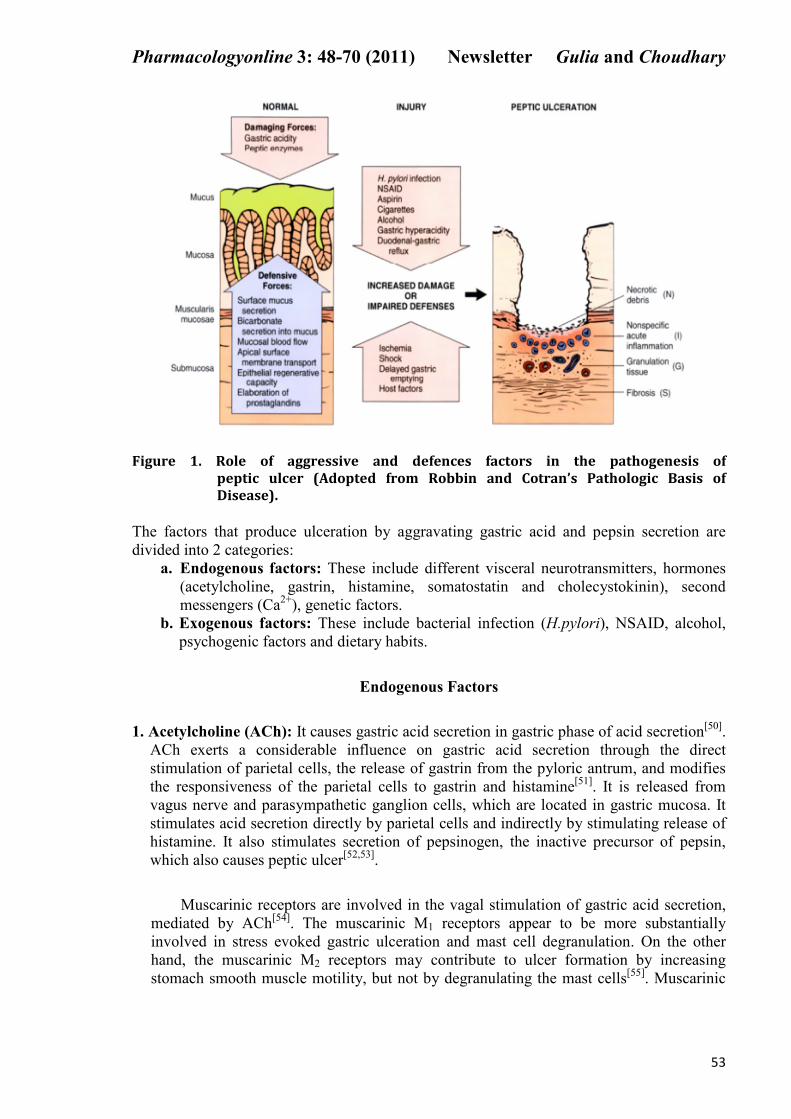
Pathogenesis of Peptic Ulcer
Peptic ulcers occur because of an imbalance between aggressive factors (gastric acid
and pepsin) and defensive factors (gastric mucus, bicarbonate, Prostaglandins). Gastric
ulceration occurs when mucosal defences fail, as when mucosal blood flow drops, gastric
emptying is delayed, or epithelial restitution is impaired[49]
(Figure 1).
Pharmacologyonline 3: 48-70 (2011) ewsletter Gulia and Choudhary
53
Figure 1. Role of aggressive and defences factors in the pathogenesis of
peptic ulcer (Adopted from Robbin and Cotran’s Pathologic Basis of
Disease).
The factors that produce ulceration by aggravating gastric acid and pepsin secretion are
divided into 2 categories:
a. Endogenous factors: These include different visceral neurotransmitters, hormones
(acetylcholine, gastrin, histamine, somatostatin and cholecystokinin), second
messengers (Ca2+
), genetic factors.
b. Exogenous factors: These include bacterial infection (H.pylori), NSAID, alcohol,
psychogenic factors and dietary habits.
Endogenous Factors
1. Acetylcholine (ACh): It causes gastric acid secretion in gastric phase of acid secretion[50]
.
ACh exerts a considerable influence on gastric acid secretion through the direct
stimulation of parietal cells, the release of gastrin from the pyloric antrum, and modifies
the responsiveness of the parietal cells to gastrin and histamine[51]
. It is released from
vagus nerve and parasympathetic ganglion cells, which are located in gastric mucosa. It
stimulates acid secretion directly by parietal cells and indirectly by stimulating release of
histamine. It also stimulates secretion of pepsinogen, the inactive precursor of pepsin,
which also causes peptic ulcer[52,53]
.
Muscarinic receptors are involved in the vagal stimulation of gastric acid secretion,
mediated by ACh[54]
. The muscarinic M1 receptors appear to be more substantially
involved in stress evoked gastric ulceration and mast cell degranulation. On the other
hand, the muscarinic M2 receptors may contribute to ulcer formation by increasing
stomach smooth muscle motility, but not by degranulating the mast cells[55]
. Muscarinic
Pharmacologyonline 3: 48-70 (2011) ewsletter Gulia and Choudhary
54
M3 receptors plays a significant role in ACh mediated gastrin secretion, which plays a
major role in gastric acid secretion[50]
.
2. Gastrin: It is a major stimulant of acid secretion. After ingestion of food, the release of
gastrin is modulated by protein content of food and accounts for acid secretory response
but as the acidity of food increases or intragastric pH decreases, the gastrin secretion
initiated by protein content diminishes[56]
. Serum gastrin concentration in peptic ulcer
patients are 120 pg/ml. Gastrenoma (Zollinger-Ellison syndrome) is a disorder which
result from the oversecretion of gastrin producing adenoma of pancreas. There is a
continuous stimulation of HCl secretion which cannot be turned off as with normal
physiological mechanism. Fasting serum gastrin concentration in this syndrome ranges
from 500 pg/ml to 7500 pg/ml. So, gastrin oversecretion is an important pathophysiologic
factor for peptic ulcer development[20]
. Stimuli that affect gastrin secretion are shown in
Table 1.
Table 1. Summary of stimuli affecting gastrin secretion. (Adopted from Ganong)
3. Histamine: It is a paracrine regulator of gastric acid secretion. It exerts its ulcerogenic
effect during abnormal physiology by acting through H2 receptors on parietal cell. It is
stored in local storage site mast cells and endocrine cells. Studies on H2 receptor
antagonist suggest histamine as the final common mediator of acid secretion as these
antagonists block both histamine stimulation but also block the stimulatory effect by
gastrin and ACh. Release mediated by increased levels of Ca2+
by gastrin in the parietal
cell.
Stimuli that increase gastrin secretion
Luminal
� Peptides and Amino acids
� Distension
Neural
� Increased vagal discharge, probably noncholinergic
Blood-borne
� Calcium
� Adrenaline
Stimuli that inhibit gastrin secretion
Luminal
� Acidic contents
� Somatostatin
Blood-borne
� Secretin, GIP, VIP, Glucagons and Calcitonin
Starvation
Pharmacologyonline 3: 48-70 (2011) ewsletter Gulia and Choudhary
55
4. Somatostatin and Cholecystokinin: Major evidence that cholecystokinin (CCK) acts as
inhibitor of gastric acid secretion is that exogenous CCK infused intravenously in a
physiological dose is capable of inhibiting gastric acid secretion. Both receptor subtypes
CCK A and CCK B are involved in inhibition and facilitation of gastrin action
respectively. On activation of CCK A receptor, somatostatin is released which act
through somatostatin receptors on gastrin G cells to inhibit gastrin secretion and CCK B
receptor stimulation by CCK causes increased release gastric acid. So, it can be assumed
that peptic ulcer patients are deficient in response of CCK A receptor activation by
endogenous CCK, resulting in deficiency of somatostatin and increased gastric acid
release[57-59]
.
5. Ca2+
as second messengers: Ca2+
play an important role in pathogenesis of gastric
ulcers. The administration of calcium both orally or intravenously, stimulates acid
secretion and increases circulating concentration of gastrin. Cytosolic free calcium
increases the effects of ACh and gastrin on stimulation of acid secretion by parietal
cells[60]
. Ca2+
also plays an important role
in the release of histamine from
enterochromaffin-like (ECL) cells, a powerful chemical mediator of gastric acid secretion,
which involves both mobilization of an intracellular calcium pool and influx of calcium
over the ECL cell membrane[61]
.
6. Genetics: Increased familial history is found in 20-50% of patients. Ulcers are also
more common in blood group O subjects and in those who do not secrete blood group
antibodies into gastric secretions[62]
.
Exogenous Factors
1. H.pylori: H.pylori is a curved or S-shaped gram negative bacterium approximately 0.5 by
3 µm in size containing four to seven sheathed flagella at one pole[63,64]
.
Mechanism of H.pylori Infection
The success of a pathogen depends on both its virulence and its pathogenicity.
Virulence is the ability to infect a host, whereas pathogenicity is the ability to cause a
disease in the host. Sufficient number of H.pylori must survive the gastric acid barrier and
colonize the enteric fluid or mucous layer. Examples of important virulence factors are
attachment mechanisms and motility in the intestinal mucous layer. Once the organism is
established in the gut, pathogenic effects on the host may be produced by one or several
means; examples are physical effects, elaboration of enzymes or toxins, and competition
with the host for nutrients.
(i) Binding to mucus and epithelial cells: H.pylori have cell wall associated lectins
which allow them to bind selectively to mucus and epithelial cells. Targets of H.pylori
lectins exist in the gastric mucus as glycoproteins and glycolipids. H.pylori appears to
bind to all of these, including sulfated (acid) mucins, L-fucose, D-galactose and sialic
acids. H.pylori lectins also attaches to red blood cells of various animal species[65]
.
Pharmacologyonline 3: 48-70 (2011) ewsletter Gulia and Choudhary
56
(ii) Tight attachment to cells: H.pylori attaches tightly to the epithelial cell, and a
characteristic structure called an ‘attachment pedestal’ forms. This attachment causes
localized cell damage characterized by enhancement of microvilli and disruption of
cytoskeletal elements of the cell[66]
. Actin polymerization also occurs below the sites
of ‘attachment pedestels’[67]
.
(iii) Elaboration of Enzymes
(a) Urease: Most common and virulence producing enzyme produced by H.pylori is
urease. This enzyme is highly active between the pH of 5 and 8. It hydrolyses urea into
ammonia. Ammonia thus generated act as a potent cellular toxin in 3 ways[68]
. Firstly, it
combines with α-ketoglutarate to form glutamine, thus depleting krebs cycle of an essential
intermediate substrate[69]
. Secondly, it interacts with hypochlorus acid to form mono-n-
chloramine, which also acts as potent cellular toxin[70]
. Thirdly, its toxic effect may also be
mediated by neutrophil generated oxygen radicals because it is inhibited by anti-neutrophil
serum[71]
.
Thus, urease enzyme plays an important role in virulence of H.pylori and its
colonization in gastric mucosa. In the course of H.pylori infection, accumulation of
phagocytic cells in the gastric mucosa occurs through two distinct mechanisms: (i)
neutrophil recruitment through Interleukin (IL) 8 production which is then released by the
gastric epithelial cells, and (ii) release by the bacterium itself of substances with
chemotactic activity able to attract phagocytes
[72,73]. These phagocytic cells ingest the
microorganism and, as in the case of other pathogens, destroy it through oxygen-dependent
and oxygen independent mechanisms. The release of free oxygen radicals by the
neutrophils might play a role in the genesis of chronic inflammation and in the
development of peptic ulcer[74]
.
(b) Phospholipases A2 and C: The epithelial cell membrane consists of a phospholipid
bilayer. Phospholipids are similar to triglycerides except that one of the terminal fatty acids
is replaced by a phosphate group. Phospholipase A2 of H.pylori removes a long-chain fatty
acid group from the second carbon. It also attacks membrane phospholipids to liberate
arachidonic acid which may then be converted to leukotriene, prostaglandin or
thromboxane. These compounds are known to cause mucus release, chemotaxis of
inflammatory cells and altered membrane permeability. Phospholipase C removes the
phosphate group from the third carbon of the phospholipids. The resultant compounds,
diacyl glyceride and particularly lysolecithin, are incapable of forming the normal
phospholipid bilayers and may form micellar structures instead, potentially affecting the
integrity of the epithelial cell membrane[75]
.
(iv) Production of Toxins: H.pylori also produces certain chemotoxins, known as
vacuolating cytotoxin (VacA) which directly act on epithelial cell surface and damage
the defence system[76]
. This toxin causes cell injury (characterized by vacuole
formation) in vitro and gastric tissue damage in vivo[49]
. VacA renders the cell
membrane permeable to urea by causing formation of transmembrane pores, suggesting
it can increase H.pylori pathogenicity by enhancing urease activity[77]
. Thus, VacA
plays an important role in pathogenesis of peptic ulcer.
Pharmacologyonline 3: 48-70 (2011) ewsletter Gulia and Choudhary
57
2. on-steroidal Antiinflammatory Drugs ( SAID): NSAID use has been associated with
development of gastric ulcers and with the major complications of ulcers i.e. gastrointestinal
bleeding, perforation and can even lead to death[78,79]
. The clinically important effects of
NSAID – the production of ulcers with an increased risk of significant complications appear
to be caused by their systemic actions[80]
. NSAID reduce tissue levels of prostaglandins,
especially PGE1, PGE2 and PGI2, by inhibiting COX-1, which is the most important
mechanism of action. By inhibition of prostaglandin synthesis, NSAID interfere with
following lines of mucosal defence[81]
:
1. Mucous cell secretion of mucin and surface active phospholipid.
Both PGE and PGF induce the secretion of polysaccharide material in the stomach
known as mucin, which acts as a protective agent against potential stomach ulceration
induced by HCl and pepsin. This implies that NSAID cause gastric ulcers by inhibiting
the secretion of this cytoprotective substance[82]
.
2. Basal bicarbonate secretion from gastric mucosa[83]
.
3. Mucosal proliferation necessary for ulcer healing[84]
.
4. Regulation of mucosal blood flow[85]
.
5. Physiological regulation of gastric acid secretion via feedback inhibition[86]
.
NSAID may also cause ulceration by generation of oxygen derived free radicals and
products of the lipooxygenase pathway[87]
. This can be explained as follows. COX inhibition
by NSAID results in diversion of arachidonic acid metabolism towards lipooxygenase
pathway, resulting in increased leukotriene synthesis. These leukotrienes can contribute to
gastrointestinal ulceration, by two mechanisms, firstly, by a reduction in prostaglandin level
and secondly, through the release of oxygen radicals mediated mucosal injury produced in
this pathway[88]
.
3. Ethanol: Ethanol damage to the gastrointestinal mucosa starts with micro-vascular
injury, namely disruption of the vascular endothelium resulting in increased vascular
permeability, oedema formation and epithelial lifting. These effects are secondary to ethanol
induced slowing or cessation of gastric mucosal flow. Alcohol causes the stomach cells to
oversecrete both acid and histamine which make the stomach linings vulnerable to ulcer
formation. Ethanol also reduces prostaglandin levels, increases the release of histamine and
influx of calcium ions. Ethanol also produces a marked contraction of the circular muscles
of fundic strip. Such a contraction can lead to mucosal compression at the site of the greatest
mechanical stress, at the crests of mucosal folds leading to necrosis and ulceration. This
reduces the secretion of bicarbonates and production of mucus and also leads to increased
neutrophil infilteration into the gastric mucosa. These neutrophils adheres to endothelial
cells, thereby blocking capillaries and induce damage to the endothelial cells through the
release of proteases, leukotriene (LTC4) and oxygen free radicals[89-91]
. These oxygen free
radicals also cause increased lipid peroxidation which causes damage to cell and cell
membranes, thereby playing a major role in pathogenesis of acute mucosal injury induced
by ethanol[92]
. Ethanol promotes oxygen radical attack on proteins at the lipophilic side
chain of amino acids[93]
. Evidence for role of oxygen free radicals in the pathogenesis of
ethanol induced mucosal injury is supported by the fact that administration of antioxidants
reduced the ethanol-induced gastric injury in rat[94]
.
Pharmacologyonline 3: 48-70 (2011) ewsletter Gulia and Choudhary
58
4. Cigarette Smoking: Continued smoking with advancing age augments the secretion of
HCl and pepsin and is also expected to modify the contents of gastric juice and pepsin
isoenzyme patterns. The increased gastric acid secretion is mediated through the stimulation
of H2-receptor by histamine released after mast cell degranulation and due to the increase of
the functional parietal cell volume or secretory capacity in smokers. Smoking causes
mucosal injury by increasing content of free oxygen radicals, PAF, pituitary vasopressin,
gastric endothelin and pituitary vasopressin. Smoking and nicotine stimulate pepsinogen
secretion also by increasing chief cell number or with an enhancement of their secretory
capacity. Long-term nicotine treatment in rats also significantly decreases total mucus neck
cell population and neck-cell mucus volume. Bile salt reflux rate and gastric bile salt
concentration are increased thereby increasing duodenogastric reflux that raises the risk of
gastric ulcer in smokers. Smoking and nicotine not only induce ulceration, but they also
potentiate ulceration caused by H.pylori, alcohol, NSAID or cold restrain stress. Smoking
also alter processes important in gastric and duodenal mucosal integrity or protection such as
mucosal bicarbonate secretion, prostaglandin content, mucosal blood flow, or epidermal
growth factor[95,96]
.
5. Diet: All foods are capable of stimulating gastric acid secretion through distention of the
stomach, but proteins are the major stimulants. Digested protein in the form of peptides,
peptones, and amino acids act primarily through the stimulation of gastrin from antral G
cells. The aromatic amino acids are the most potent of the amino acids. Amino acids
absorbed into the circulation stimulates acid secretion by directly stimulating parietal cells or
via gastrin release in humans. Caffeinated beverages (eg. tea, coffee), cola type beverages,
beer and milk are potent stimulants of gastric acid secretion. Coffee produces acid output
equal to 70 percent of peak acid output as compared to pentagastrin. 5% aqueous tea and
coffee beverages act by decreasing PGI2 synthesis. A low fiber diet, high dietary
consumption of salt and red/black peppers also causes peptic ulcer[97-99]
.
6. Psychological Factor (Stress Ulcers): There is considerable evidence that supports the
role of stressful life events in the aetiology of PUD. Stress induced ulcers are due to increase
in free radical generation apart from acid pepsin factors[100]
. Stress causes increase in gastric
motility, vagal over activity, mast cell degeneration, reduces gastric mucosal blood flow.
Stress may also produce ulceration by release of histamine with enhanced acid secretion and
reduced mucous production[99]
. Cold restrained stress induced ulcers are result of auto
digestion of gastric mucosal barrier, accumulation of HCl and generation of free radicals[101]
.
Diagnosis
Peptic ulcer can be diagnosed either by direct visualization using an endoscope or by
using contrast radiography.
Endoscopy: Endoscopy is the preferred diagnostic test in most cases because of its superior
sensitivity and specificity for significant organic disease and the ability to
obtain biopsies. It is particularly helpful in identifying lesions too small to be
detected by radiographic examination. It is recommended in patients with over
50 years of age. With modern wide-angle endoscopic television chip and
endoscopic instruments, visualization of the stomach and duodenum should be
complete in almost all patients.
Pharmacologyonline 3: 48-70 (2011) ewsletter Gulia and Choudhary
59
Radiology: A double contrast barium study is a radiological technique which can easily
detect ulcers both in the stomach and duodenum, but it is only used where
endoscopy is either technically difficult or where the patient prefers it to
endoscopy. It is indicated when endoscopy is unsuitable or not feasible or
complications such as gastric outlet obstruction is present.
Since, these endoscopic and radiographic procedures are highly expensive, attempts
have been made to develop a surrogate marker for ulcer diagnosis, such as the response to an
empirical trial with antisecretory agents or the evaluation of H.pylori status with serologic
markers[23,102]
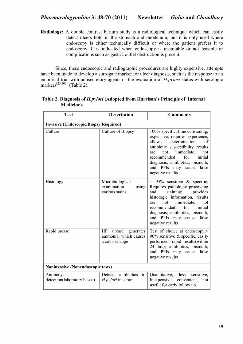
(Table 2).
Table 2. Diagnosis of H.pylori (Adopted from Harrison’s Principle of Internal
Medicine).
Test Description Comments
Invasive (Endoscopic/Biopsy Required)
Culture Culture of Biopsy 100% specific, time consuming,
expensive, requires experience,
allows determination of
antibiotic susceptibility results
are not immediate, not
recommended for initial
diagnosis; antibiotics, bismuth,
and PPIs may cause false
negative results
Histology Microbiological
examination using
various stains
> 95% sensitive & specific,
Requires pathologic processing
and staining; provides
histologic information, results
are not immediate, not
recommended for initial
diagnosis; antibiotics, bismuth,
and PPIs may cause false
negative results
Rapid urease HP urease generates
ammonia, which causes
a color change
Test of choice at endoscopy,>
90% sensitive & specific, easily
performed, rapid results(within
24 hrs); antibiotics, bismuth,
and PPIs may cause false
negative results
oninvasive ( onendoscopic tests)
Antibody
detection(laboratory based)
Detects antibodies to
H.pylori in serum
Quantitative, less sensitive,
Inexpensive, convenient, not
useful for early follow up.
Pharmacologyonline 3: 48-70 (2011) ewsletter Gulia and Choudhary
60
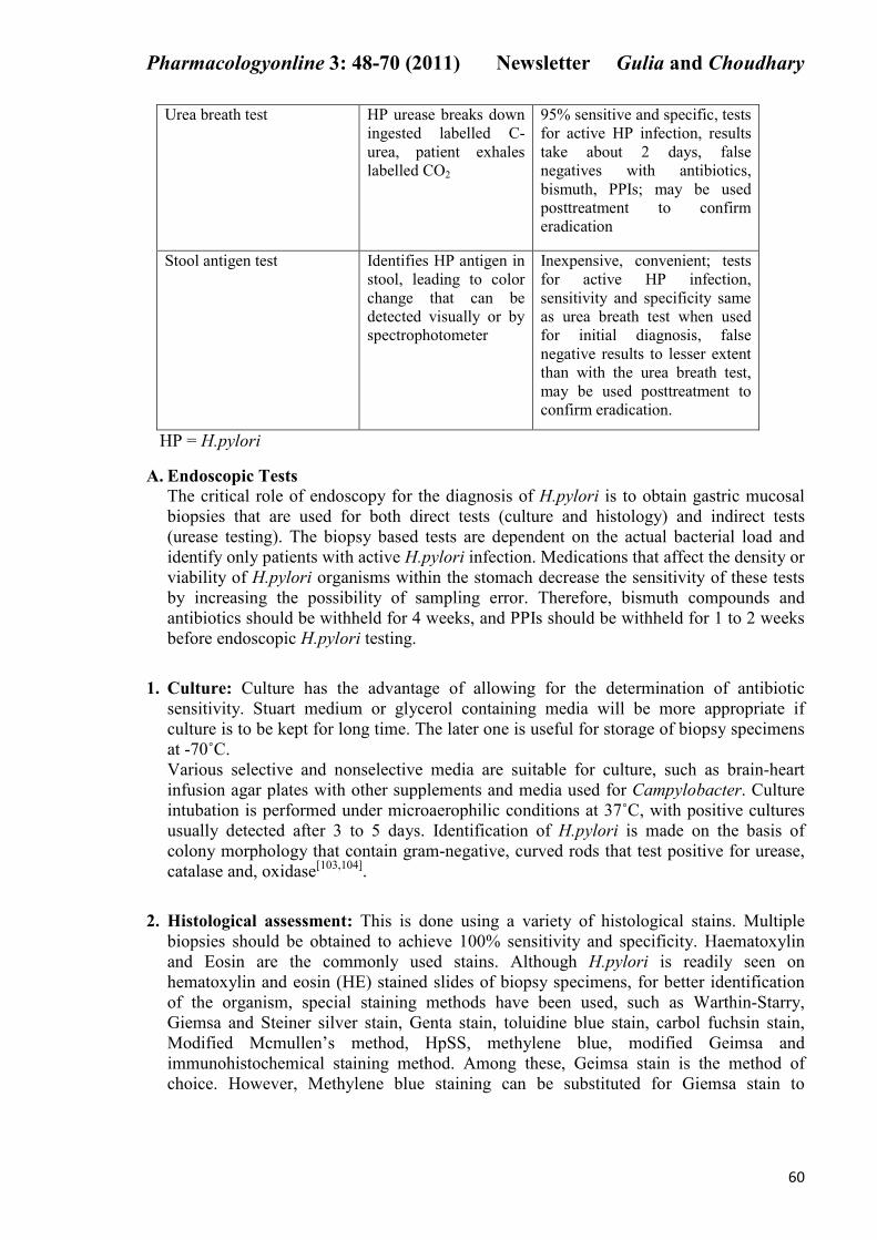
Urea breath test HP urease breaks down
ingested labelled C-
urea, patient exhales
labelled CO2
95% sensitive and specific, tests
for active HP infection, results
take about 2 days, false
negatives with antibiotics,
bismuth, PPIs; may be used
posttreatment to confirm
eradication
Stool antigen test Identifies HP antigen in
stool, leading to color
change that can be
detected visually or by
spectrophotometer
Inexpensive, convenient; tests
for active HP infection,
sensitivity and specificity same
as urea breath test when used
for initial diagnosis, false
negative results to lesser extent
than with the urea breath test,
may be used posttreatment to
confirm eradication.
HP = H.pylori
A. Endoscopic Tests
The critical role of endoscopy for the diagnosis of H.pylori is to obtain gastric mucosal
biopsies that are used for both direct tests (culture and histology) and indirect tests
(urease testing). The biopsy based tests are dependent on the actual bacterial load and
identify only patients with active H.pylori infection. Medications that affect the density or
viability of H.pylori organisms within the stomach decrease the sensitivity of these tests
by increasing the possibility of sampling error. Therefore, bismuth compounds and
antibiotics should be withheld for 4 weeks, and PPIs should be withheld for 1 to 2 weeks
before endoscopic H.pylori testing.
1. Culture: Culture has the advantage of allowing for the determination of antibiotic
sensitivity. Stuart medium or glycerol containing media will be more appropriate if
culture is to be kept for long time. The later one is useful for storage of biopsy specimens
at -70˚C.
Various selective and nonselective media are suitable for culture, such as brain-heart
infusion agar plates with other supplements and media used for Campylobacter. Culture
intubation is performed under microaerophilic conditions at 37˚C, with positive cultures
usually detected after 3 to 5 days. Identification of H.pylori is made on the basis of
colony morphology that contain gram-negative, curved rods that test positive for urease,
catalase and, oxidase[103,104]
.
2. Histological assessment: This is done using a variety of histological stains. Multiple
biopsies should be obtained to achieve 100% sensitivity and specificity. Haematoxylin
and Eosin are the commonly used stains. Although H.pylori is readily seen on
hematoxylin and eosin (HE) stained slides of biopsy specimens, for better identification
of the organism, special staining methods have been used, such as Warthin-Starry,
Giemsa and Steiner silver stain, Genta stain, toluidine blue stain, carbol fuchsin stain,
Modified Mcmullen’s method, HpSS, methylene blue, modified Geimsa and
immunohistochemical staining method. Among these, Geimsa stain is the method of
choice. However, Methylene blue staining can be substituted for Giemsa stain to
Pharmacologyonline 3: 48-70 (2011) ewsletter Gulia and Choudhary
61
visualize H.pylori. Fluorescence in situ hybridization is a complementary technique to
histology, which is used to detect the presence of H.pylori[105,106]
.
3. Rapid urease tests: The CLOtest was the first of the commercially available biopsy
urease tests designed for H.pylori detection. H.pylori urease hydrolyzes the urea
contained in the agar gel of the test packet and leads to production of ammonia, pH rise
and a color change of the phenol red indicator. The test is interpreted up to 24 hours after
insertion of the gastric biopsy sample into the well containing the agar gel. Specificity of
the CLOtest is uniformly excellent (95% to 100%), but when read at 24 hours, false
positive results may be encountered. Hpfast is another gel test, similar to the CLO test,
but it uses a different pH indicator. It is interpreted up to 24 hours. PyloriTek is a strip
test. In the presence of urease, ammonia is produced from urea impregnated into a
reaction strip. An overlying pH indicator detects the diffusion of ammonia through a
membrane. A potential advantage of this test is that interpretation may be performed only
1 hour after tissue inoculation. Simultaneous studies of the three tests have been
performed, and the results have been comparable[107,108]
.
4. Polymerase chain reaction (PCR): PCR assays, which have been shown to be sensitive
and specific, have been developed for the detection of H.pylori in gastric mucosal
biopsies. However, the diverse genetic organization of H.pylori may affect the sensitivity
of the assay. Currently, PCR assays should be restricted to the research setting for
identification of different H.pylori strains.
onendoscopic Tests
1. Antibody test: Antibody tests both under diagnose (false negative results) and over
diagnose (false positive results) H.pylori infection with some frequency. Antibody
testing offers numerous advantages: it is non-invasive, relatively inexpensive, and
overcomes some of the limitations that identify patients with active infection such as
urea breath test or stool antigen test. Ingested bismuth compounds, PPIs, or antibiotics
do not cause false negative serologic test results[109]
. In addition to laboratory antibody
tests, rapid qualitative antibody tests (in-office or near patient tests) using either serum or
finger stick whole blood are commercially available. They are inexpensive, results are
available in 5 to 15 minutes and they are simple to use[110]
.
2. Urea breath test: The urea breath test (UBT) is one of the most important non-invasive
methods for detecting H.pylori infection. The examination is simple, innocuous when 13
C
is used, easy to repeat, highly accurate, and requires a low number of precautions in order
to obtain reliable results[111]
. The diagnostic accuracy of UBT is >95%[112]
.
Patients ingest 13
C or 14
C-labelled urea. If H.pylori is present in the stomach,
urease hydrolyzes the labelled urea and releases labelled HCO3-, which is transported by
the bloodstream to the lungs and is exhaled as labelled carbon dioxide. Breath or blood is
collected, and either the radioactive 14
C isotype is detected using a scintillation counter or
mass spectroscopy or infrared spectroscopy is used to detect nonradioactive 13
C[111]
. The
performance characteristics of both the 13
C and 14
C tests are similar. False negative
results can occur in patients taking certain drugs, such as PPIs, that decrease the density
of H.pylori organisms or its metabolic activity[113]
.
Pharmacologyonline 3: 48-70 (2011) ewsletter Gulia and Choudhary
62
3. Stool antigen test: The stool antigen test is considered as a valuable non-invasive
alternative to diagnose H.pylori when UBT is not available. This test identifies H.pylori
antigens in stool. This test utilizes polyclonal anti-H.pylori capture antibody adsorbed to
microwells. Diluted stool and a peroxidase conjugated polyclonal antibody are added,
followed by substrate 1 hour later. In infected patients, enzyme-substrate binding leads to
a color change, which can be detected visually or spectrophotometrically. After
collection, stool samples can be stored at 2 to 8˚C for 3 days and at -20˚C indefinitely[114]
.
Sensitivity and specificity for the faecal antigen test is more than 90%[115]
. The sensitivity
of the stool test is decreased by the recent use of antibiotics, bismuth or PPIs[116]
.
Therapy for Peptic Ulcer
Overall treatment is aimed at relieving ulcer pain, healing the ulcer, preventing ulcer
recurrence, and reducing ulcer-related complications. Over 99% of peptic ulcers are caused
by infection with the bacterium H.pylori or by use of NSAID. The goal of therapy in
H.pylori-positive ulcer patients is to eradicate this bacterium. Successful eradication heals
ulcers and reduces risk of recurrence to less than 10% at 1 year. The goal of therapy in a
patient with NSAID induced ulcer is to heal the ulcer as rapidly as possible. Patients at high
risk of developing NSAID ulcers should be switched to a COX-2 inhibitor or receive
prophylactic drug cotherapy to reduce ulcer risk and ulcer related complications. PPIs,
H2RAs, and sucralfate are used to heal H.pylori-negative NSAID-induced ulcers.
Prophylactic cotherapy with a PPI or misoprostol is used to decrease the risk of an ulcer and
upper GI complications in patients taking nonselective NSAID[109,117]
.
on Pharmacological Treatment of PUD
� Identify and instruct patients to avoid foods that cause excess HCl secretion;
doing so improves symptoms for some individuals.
� Educate patients that avoidance of alcohol and caffeine improves symptoms and
increases healing of a pre-existing ulcer.
� Fiber rich diet can reduce the risk of developing an ulcer and can also speed the
recovery if it already exists.
� Flavonoid rich foods like apples, celery, cranberries, onions, garlic and tea may
inhibit the growth of H.pylori.
� Discontinue, reduce NSAID ingestion or switch to selective COX-2 inhibitor
therapy; this often relieves symptoms in mild cases.
� Strongly urge individuals who smoke to quit because tobacco both irritates the gut
and delays healing.
� Stress management with relaxation techniques such as yoga, or sedatives can be
used to relieve psychological influences.
Pharmacological Treatment of PUD
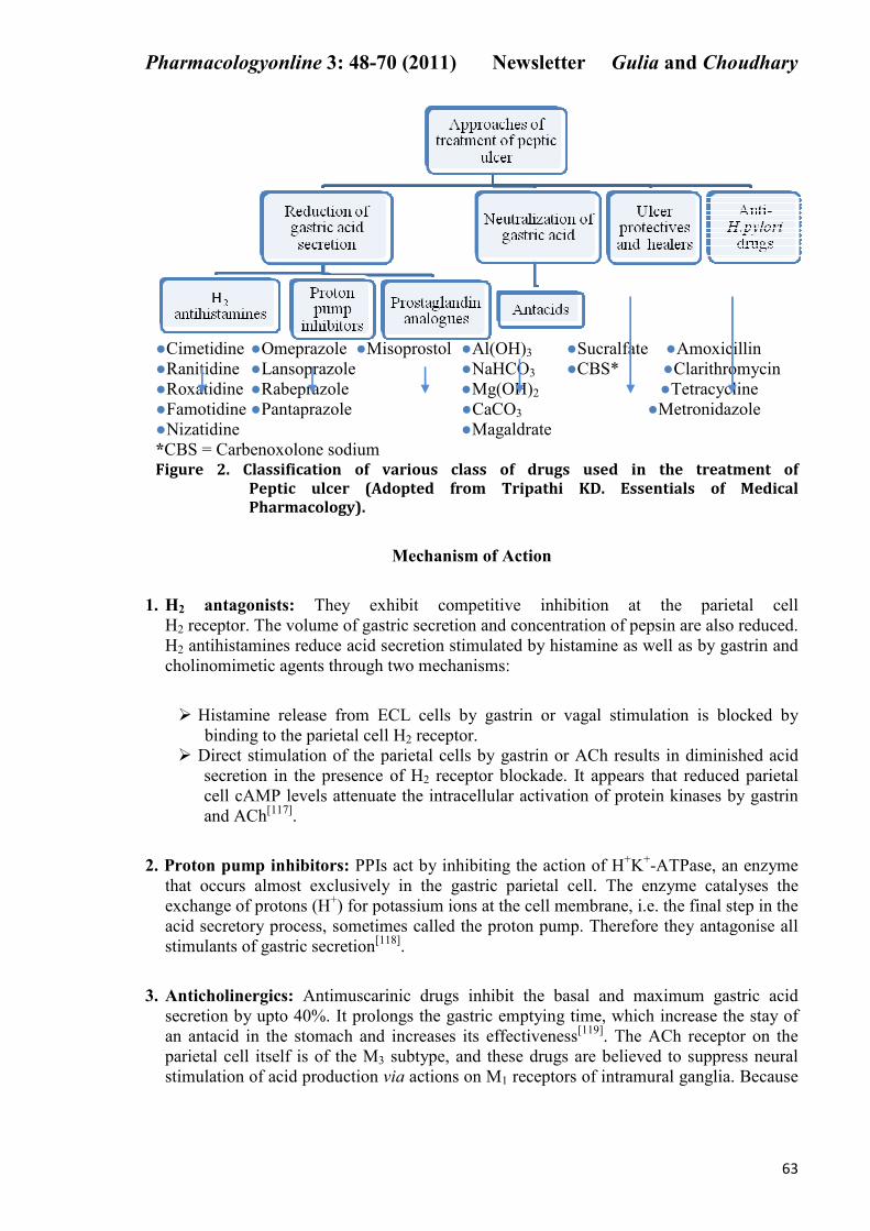
Pharmacologyonline 3: 48-70 (2011) ewsletter Gulia and Choudhary
63
●Cimetidine ●Omeprazole ●Misoprostol ●Al(OH)3 ●Sucralfate ●Amoxicillin
●Ranitidine ●Lansoprazole ●NaHCO3 ●CBS* ●Clarithromycin
●Roxatidine ●Rabeprazole ●Mg(OH)2 ●Tetracycline
●Famotidine ●Pantaprazole ●CaCO3 ●Metronidazole
●Nizatidine ●Magaldrate
*CBS = Carbenoxolone sodium
Figure 2. Classification of various class of drugs used in the treatment of
Peptic ulcer (Adopted from Tripathi KD. Essentials of Medical
Pharmacology).
Mechanism of Action
1. H2 antagonists: They exhibit competitive inhibition at the parietal cell
H2 receptor. The volume of gastric secretion and concentration of pepsin are also reduced.
H2 antihistamines reduce acid secretion stimulated by histamine as well as by gastrin and
cholinomimetic agents through two mechanisms:
� Histamine release from ECL cells by gastrin or vagal stimulation is blocked by
binding to the parietal cell H2 receptor.
� Direct stimulation of the parietal cells by gastrin or ACh results in diminished acid
secretion in the presence of H2 receptor blockade. It appears that reduced parietal
cell cAMP levels attenuate the intracellular activation of protein kinases by gastrin
and ACh[117]
.
2. Proton pump inhibitors: PPIs act by inhibiting the action of H+K
+-ATPase, an enzyme
that occurs almost exclusively in the gastric parietal cell. The enzyme catalyses the
exchange of protons (H+) for potassium ions at the cell membrane, i.e. the final step in the
acid secretory process, sometimes called the proton pump. Therefore they antagonise all
stimulants of gastric secretion[118]
.
3. Anticholinergics: Antimuscarinic drugs inhibit the basal and maximum gastric acid
secretion by upto 40%. It prolongs the gastric emptying time, which increase the stay of
an antacid in the stomach and increases its effectiveness[119]
. The ACh receptor on the
parietal cell itself is of the M3 subtype, and these drugs are believed to suppress neural
stimulation of acid production via actions on M1 receptors of intramural ganglia. Because
Pharmacologyonline 3: 48-70 (2011) ewsletter Gulia and Choudhary
64
of their relatively poor efficacy, significant and undesirable anticholinergic side effects,
and risk of blood disorders (pirenzepine), they rarely are used today[120]
.
4. Prostaglandin analogues: Misoprostol has both acid inhibitory and mucosal protective
properties. They enhance mucous bicarbonate secretion, stimulate mucosal blood flow,
and decrease mucosal cell turnover. In addition, it binds to a prostaglandin receptor on
parietal receptor, reducing histamine stimulated cAMP production and causing modest
acid inhibition[117]
.
5. Antacids: These are basic substances which neutralize gastric acid and raise pH of gastric
contents to > 4. Peptic activity is indirectly reduced if the pH rises above 4, because
pepsin is secreted as a complex with an inhibitory terminal moiety that dissociates below
pH 5; optimum peptic activity is exerted between pH 2 to 4[57]
. They also increase tone of
oesophageal sphincter and reduce the reflux of the acid and gastric contents in to the
oesophagus. Hence, they are also useful in the treatment of gastroesophageal reflux
disease (GERD)[118]
.
6. Ulcer protectives: Sucralfate is a complex sucrose salt in which hydroxyl groups have
been substituted by aluminium hydroxide and sulphate. Sucralfate may act by 2
mechanisms:
� In the gastric environment, aluminium hydroxide dissociates, leaving the polar
sulphate anion, which can bind to positively charged tissue proteins found within
the ulcer bed, and providing a physicochemical barrier impeding further tissue
injury by acid and pepsin.
� It may induce a trophic effect by binding growth factors such as EGF, enhance
prostaglandin synthesis, stimulate mucous and bicarbonate secretion, and mucosal
defence and repair[1]
.
7. Ulcer healers: Carbenoxolone sodium (CBS) is derived from liquorice, and is a synthetic
derivative of glycyrrhizinic acid. It can be used orally for the treatment of gastric ulcers
because it acts as cytoprotective and promotes healing[121]
. It acts by stimulating mucus
secretion which protects ulcer site from gastric acid. But, it has aldosterone like side
effects with fluid retention and hypokalemia, making it an undesirable therapeutic
option[122]
(Figure 2).
8. Anti H.pylori drugs: Antimicrobial agents that are clinically effective against H.pylori
are: amoxicillin, clarithromycin, tetracycline and metronidazole. However, any single
drug is relatively ineffective because resistance develops rapidly, especially to
metronidazole. Earlier, bismuth was used in the combination regimens for eradication of
H.pylori but due to poor patient acceptability is infrequently used now. In 1994, NIH
Consensus Development Conference on H.pylori concluded that ulcer patients with
H.pylori infection require treatment with antimicrobial agents in addition to antisecretory
drugs whether on first presentation with the illness or on recurrence[123]
.
(i) Triple therapy: A number of 3-drug regimens of 1 or 2 weeks duration have been
tested reporting 60-96% eradication. However, the 2 week treatment is considered
Pharmacologyonline 3: 48-70 (2011) ewsletter Gulia and Choudhary
65
more appropriate, because higher relapse rate after one week regimen indicates
incomplete eradication leading to recrudescence.
(ii) Quadruple therapy: A 4 drug regimen consisting of PPI, metronidazole,
tetracycline, and bismuth subcitrate has been reported to be highly efficacious
against metronidazole resistant strains[124]
.
References
1. Kasper DL, Fauci AS, Longo DL, Hauser SL, Jameson JL. Harrison’s Principle of
Internal Medicine. 16th
ed. Vol. II, McGraw Hill: Medical Publishing Division,
p.1746-62.
2. Jamal A, Siddiqui A, Tajuddin, Jafri MA. A review on gastric ulcer remedies used in
unani system of medicine. Nat Prod Rad 2006;5(2):153-59.
3. Warren JR, Marshall B. Unidentified curved bacilli on gastric epithelium in active
chronic gastritis. Lancet 1983;1(8336):1273-75.
4. Shankaran K, Desai HG. Medical treatment of peptic ulcer-a critical analysis. Indian J
Gastroenterol 1995;14(1):15-16.
5. Kidd M, Modlin IM. A century of Helicobacter pylori. Digestion 1998;59(1):1–15.
6. Buckley MJ, O'Morain CA. Helicobacter biology – discovery. Br Med Bull
1998;54(1):7–16.
7. Toohey M. Medicine for Nurses. London: Churchill Livingstone 1974.
8. Huwez FU, Thirlwell D, Cockayne A, Ala'Aldeen DA. Mastic gum kills Helicobacter
pylori. N Engl J Med 1998;339(26):1946.
9. Loughlin MF, Ala'Aldeen DA, Jenks PJ. Monotherapy with mastic does not eradicate
Helicobacter pylori infection from mice. J Antimicrob Chemother 2003;51(2):367–71.
10. Bebb JR, Bailey-Flitter N, Ala'Aldeen D, Atherton JC. Mastic gum has no effect on
Helicobacter pylori load in vivo. J Antimicrob Chemother 2003;52(3):522–23.
11. Lieber CS, Lefèvre A. Ammonia as a source of gastric hypoacidity in patients with
uremia. J Clin Invest 1959;38(8):1271–77.
12. Susser M, Stein Z. Civilization and peptic ulcer. Lancet 1962;1(7221):115–19.
13. Smith LH, Scharschmidt B. Medical Staff Conference: Cimetidine. West J Med
1979;131(5):417-25.
14. Marshall B. The discovery that Helicobacter pylori, a spiral bacterium, caused peptic
ulcer disease. In Barry J. Marshall. Helicobacter pioneers: firsthand accounts from the
scientists who discovered helicobacters, 1892–1982. Oxford: Blackwell 2002, p.165–
202.
15. Rauws E, Tytgat G. Cure of duodenal ulcer associated with eradication of
Helicobacter pylori. Lancet 1990;335(8700):1233–35.
16. Covacci A, Censini S, Bugnoli M, Petracca R, Burroni D, Macchia G, et al. Molecular
characterization of the 128-kDa immunodominant antigen of Helicobacter pylori
associated with cytotoxicity and duodenal ulcer. Proc Natl Acad Sci U.S.A.
1993;90(12):5791–95.
17. http://www.cdc.gov/ulcer/history.htm.
18. Tomb J, White O, Kerlavage A, Clayton RA, Sutton GG, Fleischmann RD, et al. The
complete genome sequence of the gastric pathogen Helicobacter pylori. Nature
1997;388(6642):539–47.
19. Chan FKL, Chung S, Suen B, Lee Y, Leung W, Leung VKS, et al. Preventing
recurrent upper gastrointestinal bleeding in patients with Helicobacter pylori infection
who are taking low-dose aspirin or naproxen. N Engl J Med 2001;344(13): 967–73.
Pharmacologyonline 3: 48-70 (2011) ewsletter Gulia and Choudhary
66
20. Ganong WF. Review of Medical Physiology. 21st ed. McGraw-Hill Publications;
2003. p.486-91.
21. Ramakrishnan K. Peptic ulcer disease. Am Fam Physician 2007;76(7):1005-12.
22. Pahwa R, Neeta, Kumar V, Kohli K. Clinical manifestations, causes and management
strategies of peptic ulcer disease. Int J Pharm Sci Drug Res 2010;2(2):99-106.
23. Sonnenberg A. Geographic and temporal variations in the occurrence of peptic ulcer
disease. Scand J Gastroenterol Suppl. 1985;110:11-24.
24. Das S, Deka S, Gohain K. A preclinical study on the gastric ulcer protective activity of
world’s hottest chilli, Capsicum frutescenes. J Clin Diag Res 2008;2:1024-27.
25. Das D, Dash D, Mandal T, Kishore A, Bairy KL. Protective effects of Moringa
oleifera on experimentally induced gastric ulcers in rats. Res J Pharm Biol Chem Sci
2011;2(2):50-55.
26. Tovey FI. Peptic ulcer in India and Bangladesh. Gut 1979;20(4):329-347.
27. Sandler RS, Everhart JE, Donowitz M, Adams E, Cronin A, Goodman C, et al. The
burden of selected digestive diseases in the United States. Gastroenterology
2002;122(5):1500-11.
28. Bloom BS. Cross-national changes in the effects of peptic ulcer disease. Ann Intern
Med 1991;114(7):558-62.
29. Kurata JH, Elashoff JD, Haile BM, Honda GD. A reappraisal of time trends in ulcer
disease: factors related to changes in ulcer hospitalization and mortality rates. Am J
Public Health 1983;73(9):1066-72.
30. Langman MJ. Trends in ulcer frequency. Postgrad Med J 1988;64[Suppl 1]:37-39.
31. Tilvis RS, Vuoristo M, Varis K. Changed profile of peptic ulcer disease in hospital
patients during 1969-1984 in Finland. Scand J Gastroenterol 1987;22(10):1238-44.
32. Sonnenberg A, Fritsch A. Changing mortality of peptic ulcer disease in Germany.
Gastroenterology 1983;84(6):1553-57.
33. Johnsen R, Straume B, Forde OH, Burhol PG. Changing incidence of peptic ulcer-
facts or artefacts? A cohort study from tromso. J Epidemiol Community Health
1992;46(4):433-36.
34. Watanabe Y, Kurata JH, Kawamoto K, Kawai K. Epidemiological study of peptic
ulcer disease among Japanese and Koreans in Japan. J Clin Gastroenterol
1992;15(1):68-74.
35. Javed M, Amin K, Muhammad D, Husain A, Mahmood N. Prevalence of H.pylori.
Professional Med Sep 2010;17(3):431-39.
36. Stenstrom B, Mendis A, Marshall B. Helicobacter pylori-the latest in diagnosis and
treatment. Aust Fam Physician 2008;37(8):608-12.
37. Lai LH, Sung JJ. Helicobacter pylori and benign upper digestive disease. Best Pract
Res Clin Gastroenterol 2007;21(2):261-79.
38. Lee A. The microbiology and epidemiology of Helicobacter pylori infection. Scand J
Gastroenterol Suppl 1994;201:2-6.
39. Ramakrishna BS. Helicobacter pylori infection in India: the case against eradication.
Indian J Gastroenterol 2006;25(1):25-28.
40. Helicobacter pylori and gastritis in Peruvian patients: relationship to socioeconomic
level, age, and sex. The Gastrointestinal Physiology Working Group. Am J
Gastroenterol 1990;85(7):819-23.
41. Perez-Perez GI, Taylor DN, Bodhidatta L, Wongsrichanalai J, Baze WB, Dunn BE, et
al. Seroprevalence of Helicobacter pylori infections in Thailand. J Infect Dis
1990;161(6):1237-41.
42. Pounder RE, Ng D. The prevalence of Helicobacter pylori infection in different
countries. Aliment Pharmacol Ther 1995;9(Suppl 2):33-39.
Pharmacologyonline 3: 48-70 (2011) ewsletter Gulia and Choudhary
67
43. Soll AH, Weinstein WM, Kurata J, McCarthy D. Nonsteroidal anti-inflammatory
drugs and peptic ulcer disease. Ann Intern Med 1991;114(4):307-19.
44. Mansour-Ghanaei F, Yousefi Mashhour M, Joukar F, Sedigh M, Bagher-Zadeh AH,
Jafarshad R. Prevalence of Helicobacter Pylori Infection among Children in Rasht,
Northern Iran. Middle East Journal of Digestive Diseases 2009;1(2):84-88.
45. Drumm B, Perez-Perez GI, Blaser MJ, Sherman PM. Intrafamilial clustering of
Helicobacter pylori infection. N Engl J Med 1990;322(6):359-63.
46. Thomas JE, Gibson GR, Darboe MK, Dale A, Weaver LT. Isolation of Helicobacter
pylori from human faeces. Lancet 1992;340(8829):1194-95.
47. Dharmani P, Palit G. Exploring Indian medicinal plants for antiulcer activity. Indian J
Pharmacol 2006;38(2):95-99.
48. Lindell G, Celebioglu F, Von Holstein CS, Graffner H. On the natural history of peptic
ulcer. Scand J Gastroenterol 1994;29(11):979-82.
49. Kumar V, Abbas A, Fausto N. Robbins and Cotran’s Pathologic Basis of Disease. 7th
ed. Saunders Publication, p.816-27.
50. Matsuno M, Matsui T, Iwasaki A, Arakawa Y. Role of acetylcholine and gastrin-
releasing peptide (GRP) in gastrin secretion. J Gastroenterol 1997;32(5):579-86.
51. Bunce KT, Marsh GF, Parsons ME. The effect of atropine on acid secretion stimulated
by acetylcholine, histamine and gastrin in the whole stomach of the rat. Br J
Pharmacol 1977;61:279-84.
52. Walker MC. Physiology of the digestive system. In: Slatter DH. Textbook of small
animal surgery. 3rd
ed. Saunders Publication;2002. p.522-29.
53. Venables CW. Mucus, pepsin and peptic ulcer. Gut 1986;27(3):233-38.
54. Black JW, Shankley NP. Pharmacological analysis of the inhibition by pirenzepine
and atropine of vagal-stimulated acid secretion in the isolated stomach of the mouse.
Br J Pharmacol 1986;88:291-97.
55. Ogle CW, Qiu BS, Cho CH. Nicotine and gastric ulcers in stress. J Physiol
1993;87(6):359-65.
56. Lam SK, Isenberg JI, Grossman MI, Lane WH. Gastric acid secretion is abnormally
sensitive to endogenous gastrin released after peptone test meals in duodenal ulcer
patients. J Clin Invest 1980;65(2):555-62.
57. Konturek SJ, Bilski J, Tasler J, Cieszkowski M. Role of cholecystokinin in the
inhibition of gastric acid secretion in dogs. J Physiol 1992;451:477-89.
58. Chayvialle JA, Descos F, Bernard C, Martin A, Barbe C, Partensky C. Somatostatin in
mucosa of stomach and duodenum in gastroduodenal disease. Gastroenterology
1978;75(1):13-19.
59. Harty RF, Maico DG, McGuigan JE. Antral release of gastrin and somatostatin in
duodenal ulcer and control subjects. Gut 1986;27(6):652-58.
60. Kadalmani B. Gastric ulcer protective property of calcium channel blockers in male
albino rats. Int J Pharm Biosciences 2011;2(1):629-36.
61. Sandvik AK, Brenna E, Waldum HL. Calcium mediates gastrin-induced gastric
histamine release in the rat. Am J Physiol 1993 (Gastrointest. Liver Physiol.
27);264:G51-G56.
62. Green RJ, Harris ND. Pathology and Therapeutics for pharmacists. 3rd
ed.
Pharmaceutical Press 2008, p.96-97.
63. Marshall, Warren JR. Unidentified curved bacilli in the stomach of patients with
gastritis and peptic ulceration. Lancet 1984;1(8390):1311-15.
64. Bode G, Malfertheiner P, Ditschuneit H. Pathogenetic implications of ultrastructural
findings in Campylobacter pylori related gastroduodenal disease. Scand J
Gastroenterol Suppl. 1988;142:25-29.
Pharmacologyonline 3: 48-70 (2011) ewsletter Gulia and Choudhary
68
65. Emody L, Carlsson A, Ljungh A, Wadstrom T. Mannose-resistant haemagglutination
by Campylobacter pylori. Scand J Infect Dis 1988;20(3):353-54.
66. Goodwin CS, Armstrong JA, Marshall BJ. Campylobacter pyloridis, gastritis, and
peptic ulceration. J Clin Pathol 1986;39:353-65.
67. Smoot DT, Mobley HLT, Gilliam T, Phelps P, Resau JH. Pedestal formation of
Helicobacter pylori with gastric epithelial cells in vitro may require actin
polymerization. Gastroenterology 1989;5:A127.
68. Megraud F, Neman-simha V, Brugmann D. Further evidence of the toxic effect of
ammonia produced by Helicobacter pylori urease on human epithelial cells. Infect
Immun 1992;60(5):1858-63.
69. Nelson DL, Cox MM. Lehninger-Principles of Biochemistry. 4th
ed. New York: WH
Freeman and company 2004, p.657-60.
70. Graham DY, Go MF, Evans DJ. Review Article: Urease, gastric
ammonium/ammonia, and Helicobacter pylori – the past, the present, and
recommendations for future research. Aliment Pharmacol Ther 1992;6(6):659-69.
71. Suzuki M, Miura S, Suematsu M, Fukumura D, Kurose I, Suzuki H, et al.
Helicobacter pylori-associated ammonia production enhances neutrophil-dependent
gastric mucosal cell injury. Am J Physiol 1992;263(5 Pt 1):G719-G725.
72. Mai UE, Perez-Perez GI, Allen JB, Wahl SM, Blaser MJ, Smith PD. Surface
proteins from Helicobacter pylori exhibit chemotactic activity for human leukocytes
and are present in gastric mucosa. J Exp Med 1992;175(2):517-25.
73. Nielsen H, Andersen LP. Chemotactic activity of Helicobacter pylori sonicate for
human polymorphonuclear leucocytes and monocytes. Gut 1992;33(6):738-42.
74. Salim AS. The relationship between Helicobacter pylori and oxygen-derived free
radicals in the mechanism of duodenal ulceration. Intern Med 1993;32(5):359-64.
75. Marshall BJ. Virulence and pathogenicity of Helicobacter pylori. J Gastroenterol
Hepatol 1991;6(2):121-24.
76. Leunk RD, Johnson PT, David BC. Cytotoxic activity in broth-culture filtrates of
Campylobacter pylori. J Med Microbiol 1988;26(2):93-99.
77. Tombola F, Morbiato L, Del Giudice G, Rappuoli R, Zoratti M, Papini E. The
Helicobacter pylori VacA toxin is a urea permease that promotes urea diffusion across
epithelia. J Clin Invest 2001;108(6):929-37.
78. Armstrong CP, Blower AL. Nonsteroidal anti-inflammatory drugs and life
threatening complications of peptic ulceration. Gut 1987;28(5):527-32.
79. Faulkner G, Prichard P, Somerville K, Langman MJ. Aspirin and bleeding peptic
ulcers in the elderly. Br Med J 1988;297(6659):1311-13.
80. Scheiman JM. NSAIDs, gastrointestinal injury, and cytoprotection. Gastroenterol Clin
North Am 1996;25(2):279-98.
81. Scheiman JM. Pathogenesis of gastroduodenal injury due to non-steroidal anti-
inflammatory drugs: implications for prevention and therapy. Semin Arthritis Rheum
1992;21(4):201-10.
82. Mehanna AS. NSAIDs: Chemistry and pharmacological actions. Am J Pharm Educ
2003;67(2):1-7.
83. Garner A, Flemstrom G, Heylings JR. Effects of antiinflammatory Agents and
prostaglandins on acid and bicarbonate secretions in the amphibian isolated gastric
mucosa. Gastroenterology 1979;77(3):451-57.
84. Levi S, Walport MJ, Hodgson HJF, Goodlad RA, Lee CY, Stamp G, Wright NA.
Inhibitory effect of non-steroidal anti-inflammatory drugs on mucosal cell
proliferation associated with gastric ulcer healing. Lancet 1990;336(8719):840-43.
Pharmacologyonline 3: 48-70 (2011) ewsletter Gulia and Choudhary
69
85. Guslandi M, Foppa L, Fanti L, Sorghi M. Nonsteroidal anti-inflammatory drugs and
gastric mucosal blood flow. J Clin Gastroenterol 1999;28(3):258-60.
86. Feldman M, Colturi TJ. Effect of Indomethacin on gastric acid and bicarbonate
secretion in humans. Gastroenterology 1984;87(6):1339-43.
87. Rainsford KD. Mechanisms of gastrointestinal toxicity of non-steroidal anti-
inflammatory drugs. Scand J Gastroenterol 1989;24(Suppl 163):9-16.
88. Khayyal MT, Seif-El-Nasr M, El-Ghazaly MA, Okpanyi SN, Kelber O, Weiser D.
Mechanisms involved in the gastro-protective effect of STW 5 (Iberogasts) and its
components against ulcers and rebound acidity. Phytomedicine 2006;13(Suppl 5):56–
66.
89. Bardi DAA, Sarah Khan MA, Sabri SZ, Kadir FA, Mahmood AA, Zahra AA, et al.
Anti-ulcerogenic activity of Typhonium flagelliforme aqueous leaf extract against
ethanol-induced gastric mucosal injury in rats. Scientific Research and Essays
2011;6(15):3232-39.
90. Ohya Y, Guth PH. Ethanol-induced gastric mucosal blood flow and vascular
permeability changes in the rat. Dig Dis Sci 1988;33(7):883-88.
91. Alrdahe SS, Abdulla MA, Razak SA, Kadir FA, Hassandarvish P. Gastroprotective
activity of Swietenia mahagoni seed extract on ethanol-induced gastric mucosal injury
in rats. World Acad Sci Eng Tech 2010;67:883-87.
92. Shetty R, Vijay Kumar K, Naidu MUR, Ratnakar KS. Effects of Gingko biloba extract
on ethanol-induced gastric mucosal lesions in rats. Indian J Pharmacol
2000;32(5):313-17.
93. Remmer H, Kessler W, Einsele H, Hintze TH, Toranzo GDD, Gharaibeh AM, et al.
Ethanol promotes oxygen-radical attack on proteins but not on lipids. Drug Metab Rev
1989;20(2-4):219-32.
94. Ligumsky M, Sestieri M, Okon F, Ginsburg I. Antioxidants inhibit ethanol-induced
gastric injury in the rat: role of manganese, glycine, and carotene. Scand J
Gastroenterol 1995;30(9):854-60.
95. Walker V, Taylor WH. Cigarette smoking, chronic peptic ulceration, and pepsin I
secretion. Gut 1979;20(11):971-76.
96. Maity P, Biswas K, Roy S, Banerjee RK, Bandyopadhyay U. Smoking and the
pathogenesis of gastroduodenal ulcer--recent mechanistic update. Mol Cell Biochem
2003;253(1-2):329-38.
97. Peterson WL. The influence of food, beverages and NSAIDs on gastric acid
secretion and mucosal integrity. Yale J Biol Med 1996;69:81-84.
98. Malhotra SL. A comparison of unrefined wheat and rice diets in the management of
duodenal ulcer. Postgrad Med J 1978;54(627):6-9.
99. Sen S, Chakraborty R, De B, Mazumder J. Plants and phytochemicals for peptic ulcer:
An overview. Phcog Review 2009;3(6):270-79.
100. Sairam K, Rao CV, DoraBabu M, Vijay Kumar K, Agrawal VK, Goel RK.
Antiulcerogenic effect of methanolic extract of Emblica officinalis: an experimental
study. J Ethnopharmacol 2002;82(1):1-9.
101. Thamotharan G, Sekar G, Ganesh T, Sen S, Chakraborty R, Senthil Kumar N.
Antiulcerogenic effects of Lantana camara linn. leaves on in vivo test models in rats.
Asian J Pharm Clin Res 2010;3(3):57-60.
102. Enaganti S. Peptic ulcer disease – the disease and non drug treatment. Hospital
Pharmacist 2006;13:239-43.
103. Han SW, Flamm R, Hachem CY, Kim HY, Clarridge JE, Evans DG, et al. Transport
and storage of Helicobacter pylori from gastric mucosal biopsies and clinical isolates.
Eur J Clin Microbiol Infect Dis 1995;14(4):349-52.
Pharmacologyonline 3: 48-70 (2011) ewsletter Gulia and Choudhary
70
104. Veenendaal RA, Lichtendahl-Bernards AT, Pena AS, Endtz HP, van Boven CP,
Lamers CB. Effect of transport medium and transportation time on culture of
Helicobacter pylori from gastric biopsy specimens. J Clin Pathol 1993;46(6):561-63.
105. Rotimi O, Cairns A, Gray S, Moayyedi P, Dixon MF. Histological identification of
Helicobacter pylori: comparison of staining methods. J Clin Pathol 2000;53(10):756-
59.
106. Cirak MY, Akyon Y, Megraud F. Diagnosis of Helicobacter pylori. Helicobacter
2007;12(Suppl.1):4-9.
107. Laine L, Lewin D, Naritoku W, Estrada R, Cohen H. Prospective comparison of
commercially available rapid urease tests for the diagnosis of Helicobacter pylori.
Gastrointest Endosc 1996;44(5):523-26.
108. Yousfi MM, el-Zimaity HM, Genta RM, Graham DY. Evaluation of a new reagent
strip rapid urease test for detection of Helicobacter pylori infection. Gastrointest
Endosc 1996;44(5):519-22.
109. Dipiro JT, Talbert RL, Yees GC, Matzke GR, Wells BG, Posey LM.
Pharmacotherapy: A Pathophysiologic Approach. 6th
ed. McGraw Hill Medical
Publishing Division;2005, p.629-46.
110. Sadowski D, Cohen H, Laine L, Greenberg P, Goldstein J, Mihalov M, Cutler AF.
Evaluation of the Flex-sure HP fingerstick blood test for detection of Helicobacter
pylori infection. Am J Gastroenterol 1998;93(11):2119-23.
111. Savarino V, Vigneri S, Celle G. The 13
C urea breath test in the diagnosis of
Helicobacter pylori infection. Gut 1999;45(Suppl 1):118-22.
112. Stenstrom B, Mendis A, Marshall B. Helicobacter pylori- the latest in diagnosis and
treatment. Aust Fam Physician 2008;37(8):608-12.
113. Chey WD, Chathadi KV, Montague J, Ahmed F, Murthy U. Intragastric acidification
reduces the occurrence of false-negative urea breath test results in patients taking a
proton pump inhibitor. Am J Gastroenterol 2001;96(4):1028-32.
114. Gulcan EM, Varol A, Kutlu T, Cullu F, Erkan T, Adal E, et al. Helicobacter pylori
stool antigen test. Indian J Pediatr 2005;72:675-78.
115. Gisbert JP, Pajares JM. Diagnosis of Helicobacter pylori infection by stool antigen
determination: a systematic review. Am J Gastroenterol 2001;96(10):2829-38.
116. Bravo LE, Realpe JL, Campo C, Mera R, Correa P. Effects of acid suppression and
bismuth medications on the performance of diagnostic tests for Helicobacter pylori
infection. Am J Gastroenterol 1999;94(9):2380-83.
117. Katzung BG. Basic and Clinical Pharmacology. 9th
ed. McGraw Hill Publications
2004, p.1034-43.
118. Laurence DR, Bennett PN. Clinical Pharmacology. 7th
ed. Singapore: Longman
Singapore Publishers Ltd.;1992, p.517-24.
119. Satoskar RS, Bhandarkar SD, Nirmala NR. Pharmacology and
Pharmacotherapeutics. 19th
ed. Mumbai: Popular Prakashan Ltd.;2005, p.601-25.
120. Brunton LL, Lazo JS, Parker KL. Goodman & Gilman’s The Pharmacological Basis
of Therapeutics. 11th
ed. McGraw-Hill Medical Publishing Division;2006, p.1860-84.
121. Morton IK, Hall JM. Concise dictionary of pharmacological agents: properties and
synonyms. 1st ed. Springer publications;1999, p.65.
122. Murugesh N. A concise textbook of pharmacology. 6th
ed.;2004, p.217.
123. Helicobacter pylori in peptic ulcer disease. NIH consens statement 1994;12(1):1-23.
124. Borody TJ, Andrews P, Fracchia G, Brandl S, Shortis NP, Bae H. Omeprazole
enhances efficacy of triple therapy in eradicating Helicobacter pylori infection. Gut
1995;37(4):477-81.