Embed Size (px)
Citation preview
Phase separation by ssDNA binding protein controlledvia protein−protein and protein−DNA interactionsGábor M. Haramia,1,2, Zoltán J. Kovácsa,1,2, Rita Pancsab, János Pálinkása, Veronika Barátha, Krisztián Tárnokc,András Málnási-Csizmadiad, and Mihály Kovácsa,d,2
aELTE-MTA “Momentum” Motor Enzymology Research Group, Department of Biochemistry, Eötvös Loránd University, H-1117 Budapest, Hungary;bInstitute of Enzymology, Research Centre for Natural Sciences of the Hungarian Academy of Sciences, H-1117 Budapest, Hungary; cDepartment ofPhysiology and Neurobiology, Eötvös Loránd University, H-1117 Budapest, Hungary; and dMTA-ELTE Motor Pharmacology Research Group, Department ofBiochemistry, Eötvös Loránd University, H-1117 Budapest, Hungary
Edited by Irene Chiolo, University of Southern California, and accepted by Editorial Board Member Philip C. Hanawalt September 4, 2020 (received for reviewJanuary 23, 2020)
Bacterial single-stranded (ss)DNA-binding proteins (SSB) are essen-tial for the replication and maintenance of the genome. SSBs sharea conserved ssDNA-binding domain, a less conserved intrinsicallydisordered linker (IDL), and a highly conserved C-terminal peptide(CTP) motif that mediates a wide array of protein−protein inter-actions with DNA-metabolizing proteins. Here we show that theEscherichia coli SSB protein forms liquid−liquid phase-separatedcondensates in cellular-like conditions through multifaceted inter-actions involving all structural regions of the protein. SSB, ssDNA,and SSB-interacting molecules are highly concentrated within thecondensates, whereas phase separation is overall regulated by thestoichiometry of SSB and ssDNA. Together with recent results onsubcellular SSB localization patterns, our results point to a con-served mechanism by which bacterial cells store a pool of SSBand SSB-interacting proteins. Dynamic phase separation enablesrapid mobilization of this protein pool to protect exposed ssDNAand repair genomic loci affected by DNA damage.
SSB | liquid−liquid phase separation | DNA repair | phase transition |membraneless organelle
Single-stranded (ss) DNA-binding proteins (SSB) are essential,ubiquitous, and abundant proteins that are involved in all as-
pects of genome maintenance in all kingdoms of life (1). Pro-karyotes possess a structurally well-conserved SSB protein whichforms nucleoprotein filaments with ssDNA to protect it fromreannealing and harmful chemical alterations (1, 2). In addition,SSBs recruit essential DNA metabolic enzymes to sites of actionvia protein−protein interactions (1). In Escherichia coli and mostbacteria, SSBs function as homotetramers, but dimeric SSBs arealso known (1). Regardless of quaternary structure, functional unitsof SSBs contain four oligonucleotide/oligosaccharide-binding (OB)folds that assemble into a conserved three-dimensional (3D)structure. Importantly, the vast majority of bacteria encode at leastone SSB variant in which the OB fold is followed by a “tail” regionwhich can be subdivided to the intrinsically disordered linker (IDL)and C-terminal peptide (CTP, C-terminal nine amino acids) re-gions (Fig. 1A) (3–5). The IDL of SSB is one of the few knownprotein regions with a universally conserved propensity for struc-tural disorder (6); however, its length and amino acid compositionare not conserved (3, 7). In contrast, the sequence of the CTP ishighly conserved among bacteria, and it mediates essential inter-actions with at least 18 proteins involved in nucleic acid metabolism(1, 2). Accordingly, CTP mutations weakening protein−proteininteractions are lethal (8). The CTP of E. coli SSB is also known tointeract with the OB fold, possibly within the tetramer or betweenadjacent SSB tetramers (9–14).In the past few years, many eukaryotic and viral proteins were
shown to form dynamic liquid condensates termed as membranelessorganelles that fulfill crucial functions in normal cellular physiologyas well as stress tolerance (15–17). Such condensates include, amongothers, the nucleolus, stress granules, P bodies, neuronal granules,postsynaptic densities, the heterochromatin, and super enhancers
(16). These entities form via liquid−liquid phase separation (LLPS)that is usually driven by multivalent weak protein−protein or pro-tein−nucleic acid interactions, mostly mediated by intrinsically dis-ordered regions (IDRs) of proteins (17). The proteins driving phaseseparation and their specific interaction partners are highly con-centrated in the resulting condensates, while the contents of thecondensates can also undergo dynamic exchange with the sur-roundings through the phase boundary (17, 18). Such phase-separated protein condensates provide diverse functional advan-tages for cells. For instance, they can serve as activators of reac-tions due to the high local concentrations of components, asbiomolecular filters due to their selectivity, as stress sensors due totheir responsiveness, and as reservoirs due to their ability to storemacromolecules in an inactive, condensed state (15, 19). Ac-cordingly, mutations affecting the phase separation propensity ofproteins are often linked to neurodegeneration and other patho-logical conditions (20). Whereas LLPS seems to be a heavilyexploited process in eukaryotes and viruses (21–24), up until to-day, only a few bacterial proteins were reported to undergo LLPS.These include Caulobacter crescentus Ribonuclease E (RNase E)that orchestrates RNA degradosomes (25), E. coli FtsZ that formsthe division ring during cell divisions (26), and Mycobacteriumtuberculosis Rv1747 that is a membrane transporter of cell wall
Significance
Cells must rapidly and efficiently react to DNA damage to avoidits harmful consequences. Here we report a molecular mecha-nism that gives rise to a model of how bacterial cells mobilizeDNA repair proteins for timely response to genomic stress andinitiation of DNA repair upon exposure of single-strandedDNA. We found that bacterial single-stranded DNA bindingprotein (SSB), a central player in genome metabolism, can un-dergo dynamic phase separation under physiological condi-tions. SSB condensates can store a wide array of DNA repairproteins that specifically interact with SSB. However, elevatedlevels of single-stranded DNA during genomic stress can dis-solve SSB condensates, enabling rapid mobilization of SSB andSSB-interacting proteins to sites of DNA damage.
Author contributions: G.M.H., R.P., K.T., A.M.-C., and M.K. designed research; G.M.H.,Z.J.K., R.P., J.P., V.B., and K.T. performed research; G.M.H., Z.J.K., R.P., and K.T. analyzeddata; and G.M.H., R.P., K.T., A.M.-C., and M.K. wrote the paper.
The authors declare no competing interest.
This article is a PNAS Direct Submission. I.C. is a guest editor invited by the Editorial Board.
This open access article is distributed under Creative Commons Attribution-NonCommercial-NoDerivatives License 4.0 (CC BY-NC-ND).1G.M.H. and Z.J.K. contributed equally to this work.2To whom correspondence may be addressed. Email: [email protected] or [email protected].
This article contains supporting information online at https://www.pnas.org/lookup/suppl/doi:10.1073/pnas.2000761117/-/DCSupplemental.
First published October 5, 2020.
26206–26217 | PNAS | October 20, 2020 | vol. 117 | no. 42 www.pnas.org/cgi/doi/10.1073/pnas.2000761117
Dow
nloa
ded
by g
uest
on
Nov
embe
r 30
, 202
1
biosynthesis intermediates (27). All in all, the hitherto accumu-lated observations imply that LLPS is a fundamental mechanismfor the effective spatiotemporal organization of cellular space,with emerging but hardly explored functions in bacteria.LLPS is typically driven by multivalent weak interactions, which
ensure the dynamic, liquid-like properties of the resulting con-densates. Diverse protein sequence compositional features, struc-tural modules, overall architectural properties, and modes ofprotein−protein interactions are known to favor multivalent weakinteractions and thus to promote LLPS. Disordered regions of lowsequence complexity, prion-like regions, repeated short linearmotifs and their cognate binding domains, nucleic acid-bindingdomains, and oligomerization domains can all be hallmarks ofLLPS propensity based on the properties of the hitherto describedcases (28).The modular architecture of SSB, its ability to homooligo-
merize and to interact with multiple binding partners, its weakly
conserved but compositionally biased IDL region, its conservedshort linear CTP motif, and its central integrator role in genomemaintenance processes all prompted us to raise the hypothesisthat SSB drives the formation of liquid condensates throughLLPS. Here we show that SSB is indeed capable of LLPS amongphysiological conditions, mediated via multifaceted intertetramerinteractions involving all SSB protein regions. SSB, ssDNA, andSSB-interacting partners are highly concentrated within the phase-separated droplets, whereas LLPS is overall regulated by the stoi-chiometry of SSB and ssDNA. Our bioinformatics analysis indicatesthat the LLPS-forming propensity of the SSB IDL is broadly con-served across all major phylogenetic groups of eubacteria. Togetherwith the recently observed dynamic spot-like subcellular localizationpatterns of SSB (29), our results suggest that bacterial cells store anabundant pool of SSB and SSB-interacting proteins in phase-separated condensates via a conserved mechanism. The discov-ered features enable rapid mobilization of SSB to ssDNA regions
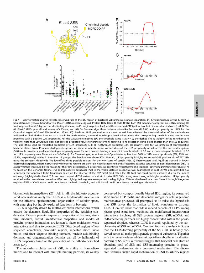
Fig. 1. Bioinformatics analysis reveals conserved role of the IDL region of bacterial SSB proteins in phase separation. (A) Crystal structure of the E. coli SSBhomotetramer (yellow) bound to two 35mer ssDNA molecules (gray) (Protein Data Bank ID code 1EYG). Each SSB monomer comprises an ssDNA-binding OBfold (oligonucleotide/oligosaccharide-binding domain), an IDL region (yellow line), and the conserved CTP (yellow box; last nine residues indicated). (B–D) The(B) PLAAC (PRD: prion-like domain), (C) PScore, and (D) CatGranule algorithms indicate prion-like features (PLAAC) and a propensity for LLPS for theC-terminal region of E. coli SSB (residues 113 to 177). Predicted LLPS propensities are shown as red lines, whereas the threshold values of the methods areindicated as black dashed lines on each graph. For each method, the residues with predicted values above the corresponding threshold value are the onespredicted with a positive LLPS propensity. For the CatGranule method (D), the threshold value is at y = 0; the dashed line is slightly shifted to enhance itsvisibility. Note that CatGranule does not provide predicted values for protein termini, resulting in its prediction curve being shorter than the SSB sequence.The algorithms used are validated predictors of LLPS propensity (74). (E) CatGranule-predicted LLPS propensity scores for SSB proteins of representativebacterial strains from 15 major phylogenetic groups of bacteria indicate broad conservation of the LLPS propensity of SSB across the bacterial kingdom.CatGranule provides a profile and a single propensity value for each protein, having a basic minimum threshold of 0.0 and a more stringent threshold of 0.5for LLPS propensity (see Materials and Methods). For Thermotogae, Aquificae, and Cyanobacteria, less than 50% of SSBs scored positively (0%, 25% and16.7%, respectively), while, in the other 12 groups, this fraction was above 50%. Overall, LLPS propensity is highly conserved (502 positive hits of 717 SSBsusing the stringent threshold). We identified three possible reasons for the low scores of certain SSBs. 1) Thermotogae and Aquificae abound in hyper-thermophilic species, wherein structurally disordered regions are generally heavily shortened and affected by adaptive sequence composition changes (75). Toassess whether this could be the reason for their low predicted LLPS propensity, we identified hyperthermophilic species [optimum growth temperature ≥ 75°C based on the Genomes Online (76) and/or BacDive databases (77)] in the whole dataset (respective SSBs highlighted in red). 2) We also identified SSBsequences that appeared to be fragments based on the absence of the CTP motif (and often the IDL too) but could not be excluded due to the lack oforthologs (highlighted in blue). 3) As we do not expect all SSB variants of a strain to drive LLPS, SSBs having an ortholog with higher predicted LLPS propensityretained in the clean dataset were identified and highlighted in green. As expected, the highlighted SSBs tend to have low scores. Cases 1 through 3 togetherexplain ∼35% of CatGranule predictions below the basic threshold, and ∼21.4% of predictions below the stringent threshold.
Harami et al. PNAS | October 20, 2020 | vol. 117 | no. 42 | 26207
BIOCH
EMISTR
Y
Dow
nloa
ded
by g
uest
on
Nov
embe
r 30
, 202
1
exposed upon DNA damage or metabolic processes to serve effi-cient repair, replication, and recombination.
ResultsSSB Forms Dynamic LLPS Condensates in Physiologically RelevantConditions In Vitro. Due to the fact that SSB shares several fea-tures with hitherto described LLPS drivers, we sought to deter-mine whether LLPS could be a yet undiscovered capability ofSSB facilitating its multifaceted roles in genome maintenance.We performed a bioinformatics analysis that revealed that E. coliSSB, in particular, its IDL region, shows high propensity forLLPS, according to multiple dedicated sequence-based predic-tion methods (30–32) (Fig. 1 B–D). This feature of SSB is uni-versally conserved among bacteria, as the majority of analyzedSSBs (72.1%) harbored by representative bacterial species from15 large phylogenetic groups show similar LLPS propensities(Datasets S1 and S2 and Fig. 1E).In line with its predicted LLPS propensity, purified E. coli SSB
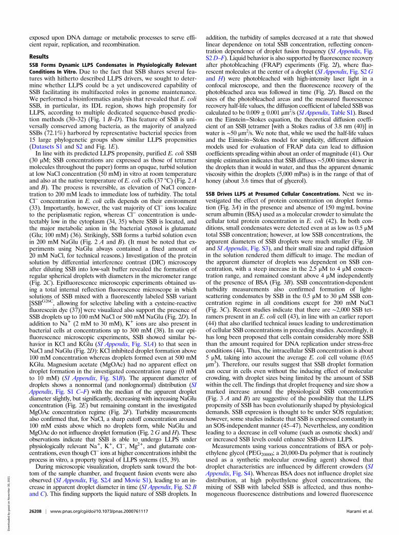
(30 μM; SSB concentrations are expressed as those of tetramermolecules throughout the paper) forms an opaque, turbid solutionat low NaCl concentration (50 mM) in vitro at room temperatureand also at the native temperature of E. coli cells (37 °C) (Fig. 2 Aand B). The process is reversible, as elevation of NaCl concen-tration to 200 mM leads to immediate loss of turbidity. The totalCl− concentration in E. coli cells depends on their environment(33). Importantly, however, the vast majority of Cl− ions localizeto the periplasmatic region, whereas Cl− concentration is unde-tectably low in the cytoplasm (34, 35) where SSB is located, andthe major metabolic anion in the bacterial cytosol is glutamate(Glu; 100 mM) (36). Strikingly, SSB forms a turbid solution evenin 200 mM NaGlu (Fig. 2 A and B). (It must be noted that ex-periments using NaGlu always contained a fixed amount of20 mM NaCl, for technical reasons.) Investigation of the proteinsolution by differential interference contrast (DIC) microscopyafter diluting SSB into low-salt buffer revealed the formation ofregular spherical droplets with diameters in the micrometer range(Fig. 2C). Epifluorescence microscopic experiments obtained us-ing a total internal reflection fluorescence microscope in whichsolutions of SSB mixed with a fluorescently labeled SSB variant[SSBG26C, allowing for selective labeling with a cysteine-reactivefluorescein dye (37)] were visualized also support the presence ofSSB droplets up to 100 mM NaCl or 500 mM NaGlu (Fig. 2D). Inaddition to Na+ (2 mM to 30 mM), K+ ions are also present inbacterial cells at concentrations up to 300 mM (38). In our epi-fluorescence microscopic experiments, SSB showed similar be-havior in KCl and KGlu (SI Appendix, Fig. S1A) to that seen inNaCl and NaGlu (Fig. 2D): KCl inhibited droplet formation above100 mM concentration whereas droplets formed even at 500 mMKGlu. Magnesium acetate (MgOAc) had no apparent effect ondroplet formation in the investigated concentration range (0 mMto 10 mM) (SI Appendix, Fig. S1B). The apparent diameter ofdroplets shows a nonnormal (and nonlognormal) distribution (SIAppendix, Fig. S1 C–F) with the median of the apparent dropletdiameter slightly, but significantly, decreasing with increasing NaGluconcentration (Fig. 2E) but remaining constant in the investigatedMgOAc concentration regime (Fig. 2F). Turbidity measurementsalso confirmed that, for NaCl, a sharp cutoff concentration around100 mM exists above which no droplets form, while NaGlu andMgOAc do not influence droplet formation (Fig. 2G andH). Theseobservations indicate that SSB is able to undergo LLPS underphysiologically relevant Na+, K+, Cl−, Mg2+, and glutamate con-centrations, even though Cl− ions at higher concentrations inhibit theprocess in vitro, a property typical of LLPS systems (15, 39).During microscopic visualization, droplets sank toward the bot-
tom of the sample chamber, and frequent fusion events were alsoobserved (SI Appendix, Fig. S2A and Movie S1), leading to an in-crease in apparent droplet diameter in time (SI Appendix, Fig. S2 Band C). This finding supports the liquid nature of SSB droplets. In
addition, the turbidity of samples decreased at a rate that showedlinear dependence on total SSB concentration, reflecting concen-tration dependence of droplet fusion frequency (SI Appendix, Fig.S2D–F). Liquid behavior is also supported by fluorescence recoveryafter photobleaching (FRAP) experiments (Fig. 2I), where fluo-rescent molecules at the center of a droplet (SI Appendix, Fig. S2 Gand H) were photobleached with high-intensity laser light in aconfocal microscope, and then the fluorescence recovery of thephotobleached area was followed in time (Fig. 2J). Based on thesizes of the photobleached areas and the measured fluorescencerecovery half-life values, the diffusion coefficient of labeled SSB wascalculated to be 0.009 ± 0.001 μm2/s (SI Appendix, Table S1). Basedon the Einstein−Stokes equation, the theoretical diffusion coeffi-cient of an SSB tetramer [with a Stokes radius of 3.8 nm (40)] inwater is ∼50 μm2/s. We note that, while we used the half-life valuesand the Einstein−Stokes model for simplicity, different diffusionmodels used for evaluation of FRAP data can lead to diffusioncoefficients spreading within about an order of magnitude (41). Oursimple estimation indicates that SSB diffuses ∼5,000 times slower inthe droplets than it would in water, and thus the apparent dynamicviscosity within the droplets (5,000 mPas) is in the range of that ofhoney (about 3.6 times that of glycerol).
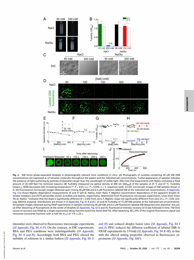
SSB Drives LLPS at Presumed Cellular Concentrations. Next we in-vestigated the effect of protein concentration on droplet forma-tion (Fig. 3A) in the presence and absence of 150 mg/mL bovineserum albumin (BSA) used as a molecular crowder to simulate thecellular total protein concentration in E. coli (42). In both con-ditions, small condensates were detected even at as low as 0.5 μMtotal SSB concentration; however, at low SSB concentrations, theapparent diameters of SSB droplets were much smaller (Fig. 3Band SI Appendix, Fig. S3), and their small size and rapid diffusionin the solution rendered them difficult to image. The median ofthe apparent diameter of droplets was dependent on SSB con-centration, with a steep increase in the 2.5 μM to 4 μM concen-tration range, and remained constant above 4 μM independentlyof the presence of BSA (Fig. 3B). SSB concentration-dependentturbidity measurements also confirmed formation of light-scattering condensates by SSB in the 0.5 μM to 30 μM SSB con-centration regime in all conditions except for 200 mM NaCl(Fig. 3C). Recent studies indicate that there are ∼2,000 SSB tet-ramers present in an E. coli cell (43), in line with an earlier report(44) that also clarified technical issues leading to underestimationof cellular SSB concentrations in preceding studies. Accordingly, ithas long been proposed that cells contain considerably more SSBthan the amount required for DNA replication under stress-freeconditions (44). Thus, the intracellular SSB concentration is about5 μM, taking into account the average E. coli cell volume (0.65μm3). Therefore, our results suggest that SSB droplet formationcan occur in cells even without the inducing effect of molecularcrowding, with droplet sizes being limited by the amount of SSBwithin the cell. The findings that droplet frequency and size show amarked increase around the physiological SSB concentration(Fig. 3 A and B) are suggestive of the possibility that the LLPSpropensity of SSB has been evolutionarily shaped by physiologicaldemands. SSB expression is thought to be under SOS regulation;however, some studies indicate that SSB is expressed constantly inan SOS-independent manner (45–47). Nevertheless, any conditionleading to a decrease in cell volume (such as osmotic shock) and/or increased SSB levels could enhance SSB-driven LLPS.Measurements using various concentrations of BSA or poly-
ethylene glycol (PEG20000; a 20,000-Da polymer that is routinelyused as a synthetic molecular crowding agent) showed thatdroplet characteristics are influenced by different crowders (SIAppendix, Fig. S4). Whereas BSA does not influence droplet sizedistribution, at high polyethylene glycol concentrations, themixing of SSB with labeled SSB is affected, and thus nonho-mogeneous fluorescence distributions and lowered fluorescence
26208 | www.pnas.org/cgi/doi/10.1073/pnas.2000761117 Harami et al.
Dow
nloa
ded
by g
uest
on
Nov
embe
r 30
, 202
1
intensities were observed in fluorescence microscopic experiments(SI Appendix, Fig. S4 A–F). On the contrary, in DIC experiments,BSA and PEG conditions were indistinguishable (SI Appendix,Fig. S4 A and B). Accordingly, both BSA and PEG altered theturbidity of solutions in a similar fashion (SI Appendix, Fig. S4 G
and H) and reduced droplet fusion rates (SI Appendix, Fig. S4 Iand J). PEG reduced the diffusion coefficient of labeled SSB inFRAP experiments by 3.9-fold (SI Appendix, Fig. S4 K–M), in linewith the altered mixing properties observed in fluorescence ex-periments (SI Appendix, Fig. S4F).
Fig. 2. SSB forms phase-separated droplets in physiologically relevant ionic conditions in vitro. (A) Photographs of cuvettes containing 30 μM SSB (SSBconcentrations are expressed as of tetramer molecules throughout the paper) and the indicated salt concentrations. Turbid appearance of samples indicatesthe presence of light-scattering by particles of diameters larger than the wavelength of visible light. (We note that experiments with NaGlu contained a fixedamount of 20 mM NaCl for technical reasons.) (B) Turbidity (measured via optical density at 600 nm [OD600]) of the samples at 25 °C and 37 °C. Turbidity(means ± SEM) decreased with increasing temperature (*: P < 0.01; n.s.: P = 0.076; n = 3, unpaired t test). (C) DIC microscopic images of SSB samples shown inA. (D) Fluorescence microscopic images obtained upon mixing 30 μM SSB and 0.3 μM fluorescein-labeled SSB at the indicated salt concentrations. SI Appendix,Fig. S1A shows MgOAc dependence measurements. (E and F) Salt (E, NaGlu, Inset: NaCl; F, MgOAc) concentration dependence of the apparent droplet di-ameter (medians and 25/75 percentiles shown as bullets and dashes, respectively), determined from fluorescence microscopic experiments. Lines show linearfits (E, NaGlu: *indicates that the slope is significantly different (P < 0.05) from zero; F, MgOAc: slope not significantly different from zero [n.s., P > 0.05; one-way ANOVA analysis]). Distributions are shown in SI Appendix, Fig. S1 B and C. (G and H) Turbidity of 15 μM SSB samples at the indicated salt concentrations.(I) Example images obtained during FRAP experiments in samples containing 30 μM SSB and 0.3 μM fluorescein-labeled SSB (bleached area diameter: 4,9 μm).(J) After bleaching of fluorophores at the center of droplets (SI Appendix, Fig. S2 G and H), fluorescence intensity recovery (in I) was followed in time. The timecourse of recovery was fitted by a single exponential decay function (solid line shows best fit). After bleaching, 68 ± 8% of the original fluorescence signal wasrecovered (recovered fraction) with a half-life (t1/2) of 175 ± 23 s.
Harami et al. PNAS | October 20, 2020 | vol. 117 | no. 42 | 26209
BIOCH
EMISTR
Y
Dow
nloa
ded
by g
uest
on
Nov
embe
r 30
, 202
1
To calculate the concentration of SSB in the droplets, we sep-arated the condensed fractions of samples of various SSB con-centrations using centrifugation, and then determined the proteinconcentration from the supernatants and back-diluted pellets(resuspended in 1 M NaCl to starting volume) (Fig. 3D). Thefraction of SSB molecules localized to the condensed phase in-creased linearly until 5 μM SSB concentration and remainedconstant at higher concentrations (up to 30 μM investigated) withan average of 95 ± 3% of SSBs located in the condensed phase inthe 5 μM to 30 μM regime (Fig. 3E). Importantly, in the 3 μM to5 μM SSB concentration regime, 65 to 95% of SSB was in con-densed form, further supporting that phase separation can occurat physiologically relevant SSB concentrations, while the size ofdroplets dynamically increased in the 2.5 μM to 4 μM concen-tration regime in microscopic experiments (Fig. 3 A and B). Next,we estimated the volume fractionation of droplets and surround-ings using 3D images reconstructed from z-stack images preparedusing a spinning disk microscope for samples containing 5 μM to30 μM total SSB concentration, as used in the centrifugation ex-periments (SSB was mixed with fluorescently labeled SSB in thespinning-disk experiments) (Fig. 3F). Based on the total SSBconcentration, the fraction of droplet volume within the sample,and the fraction of SSB molecules localized to the droplets, SSBconcentration within droplets was calculated to be 3.9 ± 0.3 mMon average, independent of total SSB concentration (Fig. 3G).Based on the Stokes radius of SSB, the water content of SSB
droplets is calculated to be around 46 ± 4 vol/vol%, which ismarkedly lower than that of the cytoplasm (∼70 vol/vol%).
Multivalent Interactions between SSB IDL Regions Are Required forLLPS, whereas Interactions between the CTP and the OB Fold EnhanceLLPS Propensity. Previous studies suggested that the IDLs of ad-jacent SSB molecules can interact with each other to enhance thecooperativity of ssDNA binding (12, 13), while the IDL was alsoproposed to interact with the OB fold (48). Moreover, the centralrole played by similar low-sequence-complexity, Gln/Gly/ProrichIDRs in previously investigated LLPS systems (49–52) as well asthe predicted LLPS propensities suggest a role for the IDL in SSBLLPS (Fig. 1 B–D). To test how the different structural modules ofSSB contribute to LLPS, we purified an SSB variant lacking theCTP (SSBdC, comprising amino acids [aa] 1 to 170) and a variantthat also lacks the IDL (SSBdIDL, aa 1 to 113) (Fig. 4A and SIAppendix, Fig. S5). Both constructs were previously shown to beable to bind ssDNA (12). Strikingly, in contrast to SSB, SSBdConly forms droplets in the presence of 150 mg/mL BSA, whereasSSBdIDL forms amorphous nonspherical aggregates even in thepresence of BSA, as assessed by DIC microscopy (Fig. 4B). Influorescence experiments, 30 μM SSBdC formed droplets with 0.3μM fluorescently labeled SSB in both the presence and absence ofBSA (Fig. 4C), whereas the formation of SSBdC (30 μM) dropletswith 0.3 μM fluorescently labeled SSBdC was only observed in thepresence of BSA (Fig. 4D). However, 0.3 μM fluorescently labeled
Fig. 3. SSB forms highly dense droplets at cellular SSB concentrations in vitro. (A) Fluorescence images of samples containing SSB at the indicated con-centrations (including 0.3 μM fluorescein-labeled SSB) in the absence and presence of BSA (150 mg/mL). (B) SSB concentration dependence of the apparentdiameter of droplets (medians and 25/75 percentiles marked by bullets and dashes, respectively) in the absence and presence of BSA (150 mg/mL). SI Ap-pendix, Fig. S3 shows diameter distributions. (C) SSB concentration dependence of turbidity in the presence of the indicated solution components. (D)Schematics of centrifugation-based concentration determination experiments. (E) Fraction of SSB in the dissolved (black) and droplet (blue) phases. Solid linesshow linear fits. Slopes were not significantly different from zero (n.s., P > 0.05; one-way ANOVA analysis). (F) Representative 3D fluorescence image obtainedin spinning disk microscopic experiments (15 μM SSB, 0.3 μM fluorescein-labeled SSB). (G) Calculated concentration of SSB in droplets. Solid line shows linearbest fit. The slope was not significantly different from zero (n.s, P = 0.804, n = 3, one-way ANOVA analysis). SI Appendix, Fig. S4 shows control experimentsrevealing the effect of molecular crowders.
26210 | www.pnas.org/cgi/doi/10.1073/pnas.2000761117 Harami et al.
Dow
nloa
ded
by g
uest
on
Nov
embe
r 30
, 202
1
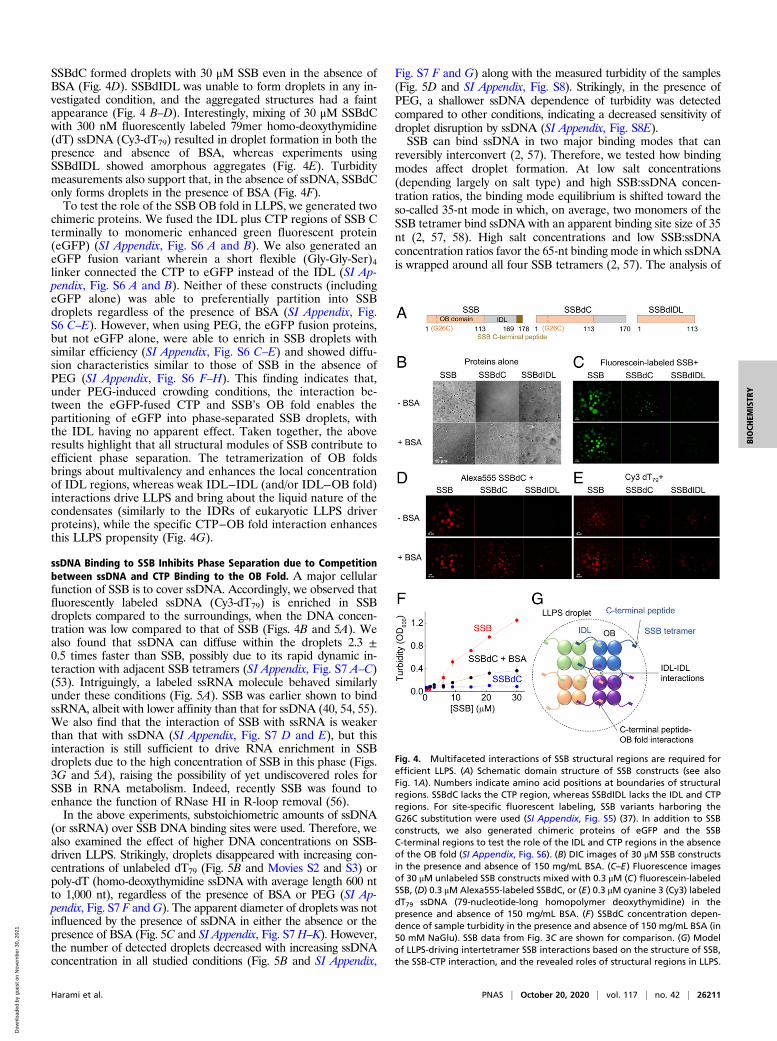
SSBdC formed droplets with 30 μM SSB even in the absence ofBSA (Fig. 4D). SSBdIDL was unable to form droplets in any in-vestigated condition, and the aggregated structures had a faintappearance (Fig. 4 B–D). Interestingly, mixing of 30 μM SSBdCwith 300 nM fluorescently labeled 79mer homo-deoxythymidine(dT) ssDNA (Cy3-dT79) resulted in droplet formation in both thepresence and absence of BSA, whereas experiments usingSSBdIDL showed amorphous aggregates (Fig. 4E). Turbiditymeasurements also support that, in the absence of ssDNA, SSBdConly forms droplets in the presence of BSA (Fig. 4F).To test the role of the SSB OB fold in LLPS, we generated two
chimeric proteins. We fused the IDL plus CTP regions of SSB Cterminally to monomeric enhanced green fluorescent protein(eGFP) (SI Appendix, Fig. S6 A and B). We also generated aneGFP fusion variant wherein a short flexible (Gly-Gly-Ser)4linker connected the CTP to eGFP instead of the IDL (SI Ap-pendix, Fig. S6 A and B). Neither of these constructs (includingeGFP alone) was able to preferentially partition into SSBdroplets regardless of the presence of BSA (SI Appendix, Fig.S6 C–E). However, when using PEG, the eGFP fusion proteins,but not eGFP alone, were able to enrich in SSB droplets withsimilar efficiency (SI Appendix, Fig. S6 C–E) and showed diffu-sion characteristics similar to those of SSB in the absence ofPEG (SI Appendix, Fig. S6 F–H). This finding indicates that,under PEG-induced crowding conditions, the interaction be-tween the eGFP-fused CTP and SSB’s OB fold enables thepartitioning of eGFP into phase-separated SSB droplets, withthe IDL having no apparent effect. Taken together, the aboveresults highlight that all structural modules of SSB contribute toefficient phase separation. The tetramerization of OB foldsbrings about multivalency and enhances the local concentrationof IDL regions, whereas weak IDL−IDL (and/or IDL−OB fold)interactions drive LLPS and bring about the liquid nature of thecondensates (similarly to the IDRs of eukaryotic LLPS driverproteins), while the specific CTP−OB fold interaction enhancesthis LLPS propensity (Fig. 4G).
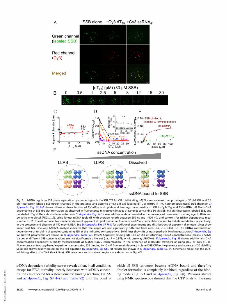
ssDNA Binding to SSB Inhibits Phase Separation due to Competitionbetween ssDNA and CTP Binding to the OB Fold. A major cellularfunction of SSB is to cover ssDNA. Accordingly, we observed thatfluorescently labeled ssDNA (Cy3-dT79) is enriched in SSBdroplets compared to the surroundings, when the DNA concen-tration was low compared to that of SSB (Figs. 4B and 5A). Wealso found that ssDNA can diffuse within the droplets 2.3 ±0.5 times faster than SSB, possibly due to its rapid dynamic in-teraction with adjacent SSB tetramers (SI Appendix, Fig. S7 A–C)(53). Intriguingly, a labeled ssRNA molecule behaved similarlyunder these conditions (Fig. 5A). SSB was earlier shown to bindssRNA, albeit with lower affinity than that for ssDNA (40, 54, 55).We also find that the interaction of SSB with ssRNA is weakerthan that with ssDNA (SI Appendix, Fig. S7 D and E), but thisinteraction is still sufficient to drive RNA enrichment in SSBdroplets due to the high concentration of SSB in this phase (Figs.3G and 5A), raising the possibility of yet undiscovered roles forSSB in RNA metabolism. Indeed, recently SSB was found toenhance the function of RNase HI in R-loop removal (56).In the above experiments, substoichiometric amounts of ssDNA
(or ssRNA) over SSB DNA binding sites were used. Therefore, wealso examined the effect of higher DNA concentrations on SSB-driven LLPS. Strikingly, droplets disappeared with increasing con-centrations of unlabeled dT79 (Fig. 5B and Movies S2 and S3) orpoly-dT (homo-deoxythymidine ssDNA with average length 600 ntto 1,000 nt), regardless of the presence of BSA or PEG (SI Ap-pendix, Fig. S7 F andG). The apparent diameter of droplets was notinfluenced by the presence of ssDNA in either the absence or thepresence of BSA (Fig. 5C and SI Appendix, Fig. S7 H–K). However,the number of detected droplets decreased with increasing ssDNAconcentration in all studied conditions (Fig. 5B and SI Appendix,
Fig. S7 F and G) along with the measured turbidity of the samples(Fig. 5D and SI Appendix, Fig. S8). Strikingly, in the presence ofPEG, a shallower ssDNA dependence of turbidity was detectedcompared to other conditions, indicating a decreased sensitivity ofdroplet disruption by ssDNA (SI Appendix, Fig. S8E).SSB can bind ssDNA in two major binding modes that can
reversibly interconvert (2, 57). Therefore, we tested how bindingmodes affect droplet formation. At low salt concentrations(depending largely on salt type) and high SSB:ssDNA concen-tration ratios, the binding mode equilibrium is shifted toward theso-called 35-nt mode in which, on average, two monomers of theSSB tetramer bind ssDNA with an apparent binding site size of 35nt (2, 57, 58). High salt concentrations and low SSB:ssDNAconcentration ratios favor the 65-nt binding mode in which ssDNAis wrapped around all four SSB tetramers (2, 57). The analysis of
Fig. 4. Multifaceted interactions of SSB structural regions are required forefficient LLPS. (A) Schematic domain structure of SSB constructs (see alsoFig. 1A). Numbers indicate amino acid positions at boundaries of structuralregions. SSBdC lacks the CTP region, whereas SSBdIDL lacks the IDL and CTPregions. For site-specific fluorescent labeling, SSB variants harboring theG26C substitution were used (SI Appendix, Fig. S5) (37). In addition to SSBconstructs, we also generated chimeric proteins of eGFP and the SSBC-terminal regions to test the role of the IDL and CTP regions in the absenceof the OB fold (SI Appendix, Fig. S6). (B) DIC images of 30 μM SSB constructsin the presence and absence of 150 mg/mL BSA. (C–E) Fluorescence imagesof 30 μM unlabeled SSB constructs mixed with 0.3 μM (C) fluorescein-labeledSSB, (D) 0.3 μMAlexa555-labeled SSBdC, or (E) 0.3 μM cyanine 3 (Cy3) labeleddT79 ssDNA (79-nucleotide-long homopolymer deoxythymidine) in thepresence and absence of 150 mg/mL BSA. (F) SSBdC concentration depen-dence of sample turbidity in the presence and absence of 150 mg/mL BSA (in50 mM NaGlu). SSB data from Fig. 3C are shown for comparison. (G) Modelof LLPS-driving intertetramer SSB interactions based on the structure of SSB,the SSB-CTP interaction, and the revealed roles of structural regions in LLPS.
Harami et al. PNAS | October 20, 2020 | vol. 117 | no. 42 | 26211
BIOCH
EMISTR
Y
Dow
nloa
ded
by g
uest
on
Nov
embe
r 30
, 202
1
ssDNA-dependent turbidity curves revealed that, in all conditions,except for PEG, turbidity linearly decreases with ssDNA concen-tration (as expected for a stoichiometric binding reaction; Fig. 5Dand SI Appendix, Fig. S8 A–D and Table S2) until the point at
which all SSB tetramers become ssDNA bound and thereforedroplet formation is completely inhibited, regardless of the bind-ing mode (Fig. 5D and SI Appendix, Fig. S8). Previous studiesusing NMR spectroscopy showed that the CTP binds to the same
Fig. 5. SsDNA regulates SSB phase separation by competing with the SSB CTP for OB fold binding. (A) Fluorescence microscopic images of 30 μM SSB, and 0.3μM fluorescein-labeled SSB (green channel) in the presence and absence of 0.1 μM Cy3-labeled dT79 or ssRNA (41 nt, nonhomopolymeric) (red channel). SIAppendix, Fig. S7 A–E shows diffusion characteristics of Cy3-dT79 in droplets and binding characteristics of SSB to Cy3-dT79 and Cy3-ssRNA. (B) The ssDNAdependence of SSB droplet formation, as observed in fluorescence microscopic images of samples containing 30 μM SSB, 0.3 μM fluorescein-labeled SSB, andunlabeled dT79 at the indicated concentrations. SI Appendix, Fig. S7F shows additional data recorded in the presence of molecular crowding agents (BSA andpolyethylene glycol [PEG20000]), using longer ssDNA (poly-dT with average length between 600 nt and 1,000 nt), and controls for ssDNA dependence mea-surements. (C) The dT79 concentration dependence of apparent droplet diameters (medians and 25/75 percentiles marked by bullets and dashes, respectively)in the presence and absence of 150 mg/mL BSA. See SI Appendix, Fig. S7 H–K for additional experiments and distributions of apparent diameters. Lines showlinear best fits. One-way ANOVA analysis indicates that the slopes are not significantly different from zero (n.s., P > 0.05). (D) The ssDNA concentrationdependence of turbidity of samples containing SSB at the indicated concentrations. Solid lines show fits using a quadratic binding equation (SI Appendix, Eq.S2; best-fit parameters are shown in SI Appendix, Table S2). (Inset) Apparent binding site size of SSB at saturating ssDNA concentrations (means ± SEM).Values at different SSB concentrations are not significantly different (n.s., P = 0.979, n =3, one-way ANOVA). SI Appendix, Fig. S8 shows additional ssDNAconcentration-dependent turbidity measurements at higher NaGlu concentration, in the presence of molecular crowders or using dT36 or poly-dT. (E)Fluorescence anisotropy-based experiments monitoring SSB binding to 15 nM fluorescein-labeled, isolated SSB CTP in the presence and absence of 50 μM dT79.Solid line shows best fit based on the Hill equation (SI Appendix, Eq. S1). Fit results are shown in SI Appendix, Table S3. (F) Schematic model for the LLPS-inhibiting effect of ssDNA (black line). SSB tetramers and structural regions are shown as in Fig. 4G.
26212 | www.pnas.org/cgi/doi/10.1073/pnas.2000761117 Harami et al.
Dow
nloa
ded
by g
uest
on
Nov
embe
r 30
, 202
1
region on the OB fold as does ssDNA (10) and it was shown thatthe IDL, and especially the CTP, influence ssDNA binding (2, 9,12, 14). In line with these observations, we found that, when SSB isbound to ssDNA, the binding of the isolated CTP peptide to SSBis significantly weaker than that for DNA-free SSB (Fig. 5E). Thisobservation, together with the LLPS-promoting effect of the CTP(Fig. 4 and SI Appendix, Fig. S6), suggests that droplet formation isregulated by the competition between the CTP and ssDNA for theOB fold (Fig. 5F).
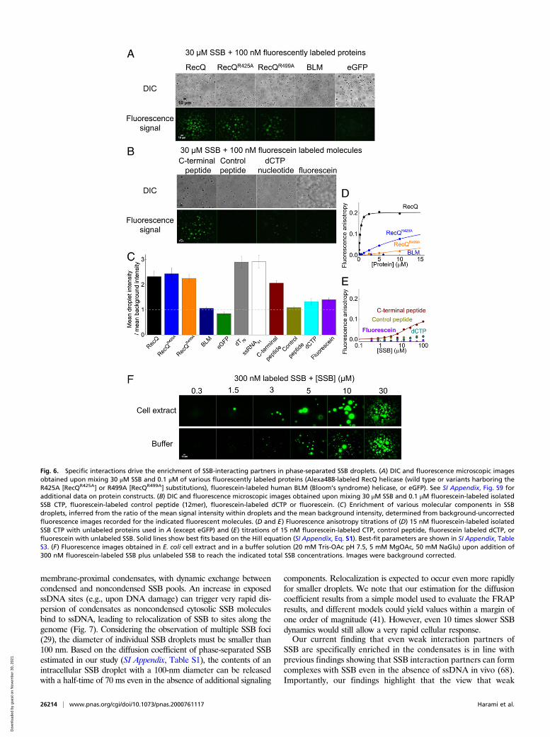
Even Weak Interaction Partners of SSB Enrich in Phase-Separated SSBCondensates. The above results demonstrate that specific interac-tions with SSB allow enrichment of interactors in SSB droplets.Thus, we tested how the interaction strength and the molecularsize of interactors influence their ability to partition into phase-separated SSB condensates (Fig. 6 A and B and SI Appendix, Fig.S9A). E. coli RecQ helicase, a 67-kDa protein that was shown tointeract with SSB dominantly via the CTP (59, 60), readily en-riches in SSB droplets (Fig. 6 A, C, and D). Interestingly, RecQpoint mutants with a markedly weakened affinity to the CTP (61)also enrich in the droplets with similar efficiency (Fig. 6 A, C, andD and SI Appendix, Table S3). In contrast, BLM helicase (a hu-man RecQ homolog) and eGFP are both unable to enrich in SSBdroplets (Fig. 6 A, C, and D and SI Appendix, Table S3), high-lighting that specific interactions with SSB are required for pro-teins to partition into SSB condensates. Probably due to the highSSB concentration within the droplets (Fig. 3G), even weak in-teractions (Kd in the range of tens of micromolars) appear to besufficient for enrichment in the SSB condensates. In line with thisproposition, the isolated SSB CTP peptide became enriched in thedroplets, but a control peptide that does not interact with SSB wasnot enriched (Fig. 6 B, C, and E and SI Appendix, Table S3). Inaddition, small molecules, such as deoxycytidine triphosphate(dCTP) or fluorescein, are slightly enriched in droplets, but not aseffectively as SSB interactors, indicating that small molecules areable to enter the separated phase, and the droplet environmenthas a slight concentrating effect (Fig. 6 B and C). As expected forinteractors, both RecQ and the isolated CTP peptide can diffusewithin the droplets, and their rate of diffusion is apparently dic-tated by their size (SI Appendix, Fig. S9 B–D).
SSB Condensates Form also in Cytosolic Extracts, Supporting theOccurrence of LLPS In Vivo. To further assess the possibility thatSSB phase separation occurs in the cellular environment, we in-vestigated condensate formation in E. coli cell extracts using epi-fluorescence microscopy (see SI Appendix, Supplementary Materialsand Methods for more specifications). Condensate formation wasnot observed upon addition of 0.3 μM fluorescein-labeled SSBalone to the extracts. However, droplets formed when unlabeledSSB was also added to at least 1.5 μM total SSB concentration(Fig. 6F). Under identical conditions, eGFP was not able to formcondensates in control experiments (SI Appendix, Fig. S10A).Moreover, SSB condensates disappeared upon the addition ofNaCl or dT79 (SI Appendix, Fig. S10 B and C) similar to what wasobserved in measurements performed in simple buffer solutions(Figs. 2D and 5B). Additional experiments suggested that the re-quirement for added SSB (at a minimum of 1.5 μM) for dropletformation originated from the presence of significant amounts ofssDNA generated during cell lysis (SI Appendix, Fig. S10D andcorresponding legend). Taken together, these measurementsclearly demonstrate that SSB-driven LLPS can indeed occur in thecytosolic molecular environment.
Bioinformatics Analysis Indicates Conservation of LLPS Propensity inHuman SSB Homologs. Besides Replication Protein A, the long-known main eukaryotic ssDNA-binding protein, recent studiesidentified human SSB homologs hSSB1/SOSB1 and hSSB2/SOSB2that are similar in size to bacterial SSBs and similarly possess
C-terminal IDL regions (62, 63). Using the bioinformatics toolsapplied in Fig. 1 B–D, we assessed whether the LLPS propensity ofbacterial SSB proteins is conserved in these human homologs. Thealgorithms indicated similar degrees of LLPS propensity for theIDL regions of both human proteins to that for E. coli SSB, indi-cating a potential role for LLPS in eukaryotic DNA metabolicprocesses (SI Appendix, Fig. S11).
DiscussionTaken together, here we show that E. coli SSB undergoes LLPSand forms viscous, liquid-state protein droplets under physio-logically relevant ionic conditions and protein concentrations, inboth the presence and absence of molecular crowders. Efficientphase separation requires all structural modules of SSB and isregulated by the specific interaction between the CTP and theOB fold as well as the stoichiometry of available SSB andssDNA. Saturation of SSB binding sites by ssDNA, independentof SSB’s DNA-binding mode, prevents LLPS, because ssDNAand the CTP compete for the same binding site on the OB folds.We also observed an LLPS inhibitory effect specifically for Cl−
ions, which were shown to be able to interact with the OB fold,likely through lysine side chains that were previously shown tointeract with phosphate groups of ssDNA (64). As the CTP in-teraction site of the OB fold is located at its ssDNA binding site,it is possible that Cl− ions compete with the CTP for binding tothe OB, as does ssDNA. In line with this proposition, in NMRstudies, increasing NaCl concentrations inhibited the OB fold–CTP interaction, whereas even 300 mM NaGlu had no effect onthe interaction (11). While other effects of Cl− cannot be ex-cluded, these results highlight the importance of the OBfold−CTP interaction in LLPS. Nevertheless, the Cl− concen-tration in the E. coli cytosol is much lower than 100 mM (34, 35),underscoring that the LLPS-inhibiting effect of Cl− is not rele-vant under cellular conditions.Given that an E. coli cell may contain around 2,000 SSB tet-
ramers and that we found the concentration of SSB to be steadyaround 3 mM within the droplets, the theoretical maximum forintracellular SSB droplet size is ∼117 nm in diameter that couldbe achieved if the entire pool of cellular SSB molecules wouldform a single droplet. Although particles of this size range can bevisualized by superresolution microscopy, the compelling in vivodemonstration of LLPS by SSB is highly challenging.Previous studies using widefield fluorescence microscopy on cells
expressing SSB proteins labeled on their C terminus with fluores-cent proteins showed the presence of SSB foci dominantly at rep-lication forks (65, 66). These experiments required the simultaneousexpression of wild-type SSB, as the fusion of fluorescent proteins tothe C terminal inhibits the essential functions of SSB. This limita-tion has been recently overcome by a novel SSB fluorescent proteintoolbox in which different fluorescent proteins are inserted into theIDL region (67). This approach revealed the same widefield fluo-rescence localization pattern for SSB as that in the aforementionedexperiments (65, 66). Nevertheless, the effect of fluorescent tags onthe phase separation properties of SSB are yet unknown. Impor-tantly, a recent study using structured illumination superresolutionmicroscopy combined with C-terminal GFP-labeled SSB showedthat, in E. coli cells, SSB not only localizes to DNA replication forksbut also forms multiple DNA-free protein foci near the inner cellmembrane (29). Membrane localization is explained by specificbinding of SSB to lipid membrane components. Upon DNA dam-age, membrane-localized SSB were shown to disperse, and, con-comitantly, SSB relocalized to sites on the bacterial chromosome,likely to those containing exposed ssDNA (29). Importantly, theability of SSB to undergo ssDNA concentration-dependent LLPS,discovered in our current study, can explain the observed localiza-tion patterns. We propose that, when only a small amount ofssDNA is exposed, the majority of cellular SSB molecules (to-gether with interacting partner proteins) will localize to (multiple)
Harami et al. PNAS | October 20, 2020 | vol. 117 | no. 42 | 26213
BIOCH
EMISTR
Y
Dow
nloa
ded
by g
uest
on
Nov
embe
r 30
, 202
1
membrane-proximal condensates, with dynamic exchange betweencondensed and noncondensed SSB pools. An increase in exposedssDNA sites (e.g., upon DNA damage) can trigger very rapid dis-persion of condensates as noncondensed cytosolic SSB moleculesbind to ssDNA, leading to relocalization of SSB to sites along thegenome (Fig. 7). Considering the observation of multiple SSB foci(29), the diameter of individual SSB droplets must be smaller than100 nm. Based on the diffusion coefficient of phase-separated SSBestimated in our study (SI Appendix, Table S1), the contents of anintracellular SSB droplet with a 100-nm diameter can be releasedwith a half-time of 70 ms even in the absence of additional signaling
components. Relocalization is expected to occur even more rapidlyfor smaller droplets. We note that our estimation for the diffusioncoefficient results from a simple model used to evaluate the FRAPresults, and different models could yield values within a margin ofone order of magnitude (41). However, even 10 times slower SSBdynamics would still allow a very rapid cellular response.Our current finding that even weak interaction partners of
SSB are specifically enriched in the condensates is in line withprevious findings showing that SSB interaction partners can formcomplexes with SSB even in the absence of ssDNA in vivo (68).Importantly, our findings highlight that the view that weak
Fig. 6. Specific interactions drive the enrichment of SSB-interacting partners in phase-separated SSB droplets. (A) DIC and fluorescence microscopic imagesobtained upon mixing 30 μM SSB and 0.1 μM of various fluorescently labeled proteins (Alexa488-labeled RecQ helicase (wild type or variants harboring theR425A [RecQR425A] or R499A [RecQR499A] substitutions), fluorescein-labeled human BLM (Bloom’s syndrome) helicase, or eGFP). See SI Appendix, Fig. S9 foradditional data on protein constructs. (B) DIC and fluorescence microscopic images obtained upon mixing 30 μM SSB and 0.1 μM fluorescein-labeled isolatedSSB CTP, fluorescein-labeled control peptide (12mer), fluorescein-labeled dCTP or fluorescein. (C) Enrichment of various molecular components in SSBdroplets, inferred from the ratio of the mean signal intensity within droplets and the mean background intensity, determined from background-uncorrectedfluorescence images recorded for the indicated fluorescent molecules. (D and E) Fluorescence anisotropy titrations of (D) 15 nM fluorescein-labeled isolatedSSB CTP with unlabeled proteins used in A (except eGFP) and (E) titrations of 15 nM fluorescein-labeled CTP, control peptide, fluorescein labeled dCTP, orfluorescein with unlabeled SSB. Solid lines show best fits based on the Hill equation (SI Appendix, Eq. S1). Best-fit parameters are shown in SI Appendix, TableS3. (F) Fluorescence images obtained in E. coli cell extract and in a buffer solution (20 mM Tris-OAc pH 7.5, 5 mM MgOAc, 50 mM NaGlu) upon addition of300 nM fluorescein-labeled SSB plus unlabeled SSB to reach the indicated total SSB concentrations. Images were background corrected.
26214 | www.pnas.org/cgi/doi/10.1073/pnas.2000761117 Harami et al.
Dow
nloa
ded
by g
uest
on
Nov
embe
r 30
, 202
1
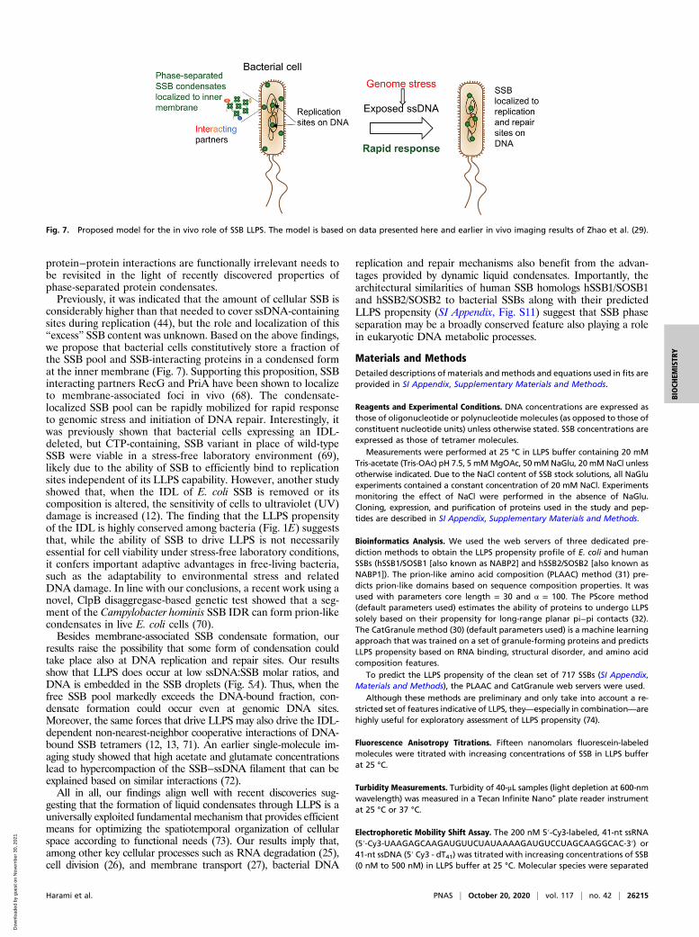
protein−protein interactions are functionally irrelevant needs tobe revisited in the light of recently discovered properties ofphase-separated protein condensates.Previously, it was indicated that the amount of cellular SSB is
considerably higher than that needed to cover ssDNA-containingsites during replication (44), but the role and localization of this“excess” SSB content was unknown. Based on the above findings,we propose that bacterial cells constitutively store a fraction ofthe SSB pool and SSB-interacting proteins in a condensed format the inner membrane (Fig. 7). Supporting this proposition, SSBinteracting partners RecG and PriA have been shown to localizeto membrane-associated foci in vivo (68). The condensate-localized SSB pool can be rapidly mobilized for rapid responseto genomic stress and initiation of DNA repair. Interestingly, itwas previously shown that bacterial cells expressing an IDL-deleted, but CTP-containing, SSB variant in place of wild-typeSSB were viable in a stress-free laboratory environment (69),likely due to the ability of SSB to efficiently bind to replicationsites independent of its LLPS capability. However, another studyshowed that, when the IDL of E. coli SSB is removed or itscomposition is altered, the sensitivity of cells to ultraviolet (UV)damage is increased (12). The finding that the LLPS propensityof the IDL is highly conserved among bacteria (Fig. 1E) suggeststhat, while the ability of SSB to drive LLPS is not necessarilyessential for cell viability under stress-free laboratory conditions,it confers important adaptive advantages in free-living bacteria,such as the adaptability to environmental stress and relatedDNA damage. In line with our conclusions, a recent work using anovel, ClpB disaggregase-based genetic test showed that a seg-ment of the Campylobacter hominis SSB IDR can form prion-likecondensates in live E. coli cells (70).Besides membrane-associated SSB condensate formation, our
results raise the possibility that some form of condensation couldtake place also at DNA replication and repair sites. Our resultsshow that LLPS does occur at low ssDNA:SSB molar ratios, andDNA is embedded in the SSB droplets (Fig. 5A). Thus, when thefree SSB pool markedly exceeds the DNA-bound fraction, con-densate formation could occur even at genomic DNA sites.Moreover, the same forces that drive LLPS may also drive the IDL-dependent non-nearest-neighbor cooperative interactions of DNA-bound SSB tetramers (12, 13, 71). An earlier single-molecule im-aging study showed that high acetate and glutamate concentrationslead to hypercompaction of the SSB−ssDNA filament that can beexplained based on similar interactions (72).All in all, our findings align well with recent discoveries sug-
gesting that the formation of liquid condensates through LLPS is auniversally exploited fundamental mechanism that provides efficientmeans for optimizing the spatiotemporal organization of cellularspace according to functional needs (73). Our results imply that,among other key cellular processes such as RNA degradation (25),cell division (26), and membrane transport (27), bacterial DNA
replication and repair mechanisms also benefit from the advan-tages provided by dynamic liquid condensates. Importantly, thearchitectural similarities of human SSB homologs hSSB1/SOSB1and hSSB2/SOSB2 to bacterial SSBs along with their predictedLLPS propensity (SI Appendix, Fig. S11) suggest that SSB phaseseparation may be a broadly conserved feature also playing a rolein eukaryotic DNA metabolic processes.
Materials and MethodsDetailed descriptions of materials and methods and equations used in fits areprovided in SI Appendix, Supplementary Materials and Methods.
Reagents and Experimental Conditions. DNA concentrations are expressed asthose of oligonucleotide or polynucleotide molecules (as opposed to those ofconstituent nucleotide units) unless otherwise stated. SSB concentrations areexpressed as those of tetramer molecules.
Measurements were performed at 25 °C in LLPS buffer containing 20 mMTris-acetate (Tris-OAc) pH 7.5, 5 mMMgOAc, 50 mMNaGlu, 20 mMNaCl unlessotherwise indicated. Due to the NaCl content of SSB stock solutions, all NaGluexperiments contained a constant concentration of 20 mM NaCl. Experimentsmonitoring the effect of NaCl were performed in the absence of NaGlu.Cloning, expression, and purification of proteins used in the study and pep-tides are described in SI Appendix, Supplementary Materials and Methods.
Bioinformatics Analysis. We used the web servers of three dedicated pre-diction methods to obtain the LLPS propensity profile of E. coli and humanSSBs (hSSB1/SOSB1 [also known as NABP2] and hSSB2/SOSB2 [also known asNABP1]). The prion-like amino acid composition (PLAAC) method (31) pre-dicts prion-like domains based on sequence composition properties. It wasused with parameters core length = 30 and α = 100. The PScore method(default parameters used) estimates the ability of proteins to undergo LLPSsolely based on their propensity for long-range planar pi−pi contacts (32).The CatGranule method (30) (default parameters used) is a machine learningapproach that was trained on a set of granule-forming proteins and predictsLLPS propensity based on RNA binding, structural disorder, and amino acidcomposition features.
To predict the LLPS propensity of the clean set of 717 SSBs (SI Appendix,Materials and Methods), the PLAAC and CatGranule web servers were used.
Although these methods are preliminary and only take into account a re-stricted set of features indicative of LLPS, they—especially in combination—arehighly useful for exploratory assessment of LLPS propensity (74).
Fluorescence Anisotropy Titrations. Fifteen nanomolars fluorescein-labeledmolecules were titrated with increasing concentrations of SSB in LLPS bufferat 25 °C.
Turbidity Measurements. Turbidity of 40-μL samples (light depletion at 600-nmwavelength) was measured in a Tecan Infinite Nano+ plate reader instrumentat 25 °C or 37 °C.
Electrophoretic Mobility Shift Assay. The 200 nM 5′-Cy3-labeled, 41-nt ssRNA(5′-Cy3-UAAGAGCAAGAUGUUCUAUAAAAGAUGUCCUAGCAAGGCAC-3′) or41-nt ssDNA (5′ Cy3 - dT41) was titrated with increasing concentrations of SSB(0 nM to 500 nM) in LLPS buffer at 25 °C. Molecular species were separated
Fig. 7. Proposed model for the in vivo role of SSB LLPS. The model is based on data presented here and earlier in vivo imaging results of Zhao et al. (29).
Harami et al. PNAS | October 20, 2020 | vol. 117 | no. 42 | 26215
BIOCH
EMISTR
Y
Dow
nloa
ded
by g
uest
on
Nov
embe
r 30
, 202
1
on a 1 wt/vol% agarose gel and detected using a Typhoon Trio+ VariableMode Imager.
Centrifugation-Based Concentration Determination. SSB in various concen-trations was incubated in LLPS buffer (20 mM Tris-OAc pH 7.5, 20 mM NaCl,50 mM NaGlu and 5 mM MgOAc) at 25 °C. Samples were centrifuged using atabletop Eppendorf centrifuge (13,400 rpm [12,000 × g], 30 min). Proteinconcentrations of the supernatant and resuspended pellet were determinedwith UV spectrophotometry.
Microscopy and Image Analysis. DIC images were captured using a ZeissAxioImager M2 upright microscope. Raw, unprocessed DIC images are shownin the paper.
Epifluorescence images were captured with a Nikon Eclipse Ti-E TIRF mi-croscope in epifluorescent mode. Raw, unprocessed images were analyzedusing the Fiji (ImageJ) software. For illustration purposes, images were back-ground corrected (SI Appendix, Supplementary Materials and Methods).Methods of determination of size distribution and fluorescent partitioning aredescribed in SI Appendix, Supplementary Materials and Methods.
FRAP experiments were performed at 23 °C using an inverted LSM800confocal microscope (Zeiss).
For evaluation of droplet volumes, microscopic images were taken on aZeiss Spinning Disk system at 24 °C. Image analysis methods are described inSI Appendix, Supplementary Materials and Methods.
Data Analysis. Mean ± SEM (SE of mean) values are reported in the paperunless otherwise specified. For apparent droplet size distributions, mean ±
SD values and medians are shown. Sample sizes (n) are given for the numberof observed particles in fluorescence microscopic experiments or the numberof ensemble in vitro measurements performed (n = 3 unless otherwisespecified).
Data Availability. All data generated or analyzed during this study are in-cluded in this published article and Datasets S1 and S2. Source code of thescript used for analyses of spinning disk microscopy data are freely availableat GitHub, https://github.com/SzegaX/VolumeOfSmearedSpheres.
ACKNOWLEDGMENTS. This work was supported by the Human FrontierScience Program (Grant RGY0072/2010 to M.K.), the “Momentum” Programof the Hungarian Academy of Sciences (Grant LP2011-006/2011 to M.K.),Grants ELTE (Eötvös Loránd University) KMOP‐4.2.1/B‐10‐2011‐0002, NKFIH(National Research, Development and Innovation Office) K-116072, NKFIH K-123989, and NKFIH ERC_HU 117680 to M.K. and Grant NKFIH FK-128133 toR.P. Both G.M.H. and R.P. are supported by the Premium Postdoctoral Pro-gram of the Hungarian Academy of Sciences (Grants PREMIUM-2017-17 toG.M.H. and PREMIUM-2017-48 to R.P.). The project was supported by theNKFIH (Grant VEKOP-2.3.3-15-2016-00007 to ELTE) grant. Z.J.K. and J.P. aresupported by the New National Excellence Program of the Ministry for In-novation and Technology (Grants ÚNKP-19-3 to Z.J.K. and ÚNKP-19-2 to J.P.).This work was completed in the ELTE Thematic Excellence Programme sup-ported by the Hungarian Ministry for Innovation and Technology. We aregrateful to Dr. Tibor Kovács for the help with DIC measurements, GáborSzegvári for the custom-written script for volume estimation of dropletsobserved in spinning disc fluorescence microscopic experiments, and AnnaBudai for the help in the cloning of expression vectors encoding GFPfusion proteins.
1. R. D. Shereda, A. G. Kozlov, T. M. Lohman, M. M. Cox, J. L. Keck, SSB as an organizer/
mobilizer of genome maintenance complexes. Crit. Rev. Biochem. Mol. Biol. 43,
289–318 (2008).2. E. Antony, T. M. Lohman, Dynamics of E. coli single stranded DNA binding (SSB)
protein-DNA complexes. Semin. Cell Dev. Biol. 86, 102–111 (2019).3. P. R. Bianco, The tale of SSB. Prog. Biophys. Mol. Biol. 127, 111–118 (2017).4. S. Raghunathan, C. S. Ricard, T. M. Lohman, G. Waksman, Crystal structure of the
homo-tetrameric DNA binding domain of Escherichia coli single-stranded DNA-
binding protein determined by multiwavelength x-ray diffraction on the selenome-
thionyl protein at 2.9-A resolution. Proc. Natl. Acad. Sci. U.S.A. 94, 6652–6657 (1997).5. S. N. Savvides et al., The C-terminal domain of full-length E. coli SSB is disordered even
when bound to DNA. Protein Sci. 13, 1942–1947 (2004).6. R. Pancsa, P. Tompa, Essential functions linked with structural disorder in organisms of
minimal genome. Biol. Direct 11, 45 (2016).7. T. Parad�zik, Ž. Fili�c, D. Vujaklija, Variations in amino acid composition in bacterial
single stranded DNA–binding proteins correlate with GC content. Period. Biol. 118,
385–397 (2017).8. E. Antony et al., Multiple C-terminal tails within a single E. coli SSB homotetramer
coordinate DNA replication and repair. J. Mol. Biol. 425, 4802–4819 (2013).9. A. G. Kozlov, M. M. Cox, T. M. Lohman, Regulation of single-stranded DNA binding by
the C termini of Escherichia coli single-stranded DNA-binding (SSB) protein. J. Biol.
Chem. 285, 17246–17252 (2010).10. D. Shishmarev et al., Intramolecular binding mode of the C-terminus of Escherichia
coli single-stranded DNA binding protein determined by nuclear magnetic resonance
spectroscopy. Nucleic Acids Res. 42, 2750–2757 (2014).11. X.-C. Su et al., Bound or free: Interaction of the C-terminal domain of Escherichia coli
single-stranded DNA-binding protein (SSB) with the tetrameric core of SSB. Bio-
chemistry 53, 1925–1934 (2014).12. A. G. Kozlov et al., Intrinsically disordered C-terminal tails of E. coli single-stranded
DNA binding protein regulate cooperative binding to single-stranded DNA. J. Mol.
Biol. 427, 763–774 (2015).13. A. G. Kozlov, M. K. Shinn, E. A. Weiland, T. M. Lohman, Glutamate promotes SSB
protein-protein interactions via intrinsically disordered regions. J. Mol. Biol. 429,
2790–2801 (2017).14. A. G. Kozlov, M. J. Jezewska, W. Bujalowski, T. M. Lohman, Binding specificity of
Escherichia coli single-stranded DNA binding protein for the chi subunit of DNA pol III
holoenzyme and PriA helicase. Biochemistry 49, 3555–3566 (2010).15. S. Alberti, A. Gladfelter, T. Mittag, Considerations and challenges in studying liquid-
liquid phase separation and biomolecular condensates. Cell 176, 419–434 (2019).16. S. F. Banani, H. O. Lee, A. A. Hyman, M. K. Rosen, Biomolecular condensates: Orga-
nizers of cellular biochemistry. Nat. Rev. Mol. Cell Biol. 18, 285–298 (2017).17. Y. Shin, C. P. Brangwynne, Liquid phase condensation in cell physiology and disease.
Science 357, eaaf4382 (2017).18. S. F. Banani et al., Compositional control of phase-separated cellular bodies. Cell 166,
651–663 (2016).19. R. Pancsa, E. Schad, A. Tantos, P. Tompa, Emergent functions of proteins in non-
stoichiometric supramolecular assemblies. Biochim. Biophys. Acta. Proteins Proteo-
mics 1867, 970–979 (2019).20. S. Alberti, D. Dormann, Liquid-liquid phase separation in disease. Annu. Rev. Genet.
53, 171–194 (2019).
21. B. S. Heinrich, Z. Maliga, D. A. Stein, A. A. Hyman, S. P. J. Whelan, Phase transitionsdrive the formation of vesicular stomatitis virus replication compartments. mBio 9,e02290-17 (2018).
22. J. Nikolic et al., Negri bodies are viral factories with properties of liquid organelles.Nat. Commun. 8, 58 (2017).
23. J. Nikolic, C. Lagaudrière-Gesbert, N. Scrima, D. Blondel, Y. Gaudin, Rabies virus fac-tories are formed by liquid-liquid phase separation. Med. Sci. 34, 203–205 (2018).
24. Y. Zhou, J. M. Su, C. E. Samuel, D. Ma, Measles virus forms inclusion bodies withproperties of liquid organelles. J. Virol. 93, e00948-19 (2019).
25. N. Al-Husini, D. T. Tomares, O. Bitar, W. S. Childers, J. M. Schrader, α-ProteobacterialRNA degradosomes assemble liquid-liquid phase-separated RNP bodies. Mol. Cell 71,1027–1039.e14 (2018).
26. B. Monterroso et al., Bacterial FtsZ protein forms phase-separated condensates withits nucleoid-associated inhibitor SlmA. EMBO Rep. 20, e45946 (2019).
27. F. Heinkel et al., Phase separation and clustering of an ABC transporter in Myco-bacterium tuberculosis. Proc. Natl. Acad. Sci. U.S.A. 116, 16326–16331 (2019).
28. T. Mittag, R. Parker, Multiple modes of protein-protein interactions promote RNPgranule assembly. J. Mol. Biol. 430, 4636–4649 (2018).
29. T. Zhao et al., Super-resolution imaging reveals changes in Escherichia coli SSB lo-calization in response to DNA damage. Genes Cells 24, 814–826 (2019).
30. B. Bolognesi et al., A concentration-dependent liquid phase separation can causetoxicity upon increased protein expression. Cell Rep. 16, 222–231 (2016).
31. A. K. Lancaster, A. Nutter-Upham, S. Lindquist, O. D. King, PLAAC: A web andcommand-line application to identify proteins with prion-like amino acid composi-tion. Bioinformatics 30, 2501–2502 (2014).
32. R. M. Vernon et al., Pi-Pi contacts are an overlooked protein feature relevant to phaseseparation. eLife 7, e31486 (2018).
33. S. G. Schultz, N. L. Wilson, W. Epstein, Cation transport in Escherichia coli. II. Intra-cellular chloride concentration. J. Gen. Physiol. 46, 159–166 (1962).
34. J. B. Stock, B. Rauch, S. Roseman, Periplasmic space in Salmonella typhimurium andEscherichia coli. J. Biol. Chem. 252, 7850–7861 (1977).
35. F. Stull, H. Hipp, R. B. Stockbridge, J. C. A. Bardwell, In vivo chloride concentrationssurge to proteotoxic levels during acid stress. Nat. Chem. Biol. 14, 1051–1058 (2018).
36. B. D. Bennett et al., Absolute metabolite concentrations and implied enzyme activesite occupancy in Escherichia coli. Nat. Chem. Biol. 5, 593–599 (2009).
37. M. S. Dillingham et al., Fluorescent single-stranded DNA binding protein as a probefor sensitive, real-time assays of helicase activity. Biophys. J. 95, 3330–3339 (2008).
38. B. Richey et al., Variability of the intracellular ionic environment of Escherichia coli.Differences between in vitro and in vivo effects of ion concentrations on protein-DNAinteractions and gene expression. J. Biol. Chem. 262, 7157–7164 (1987).
39. Y.-H. Lin, J. D. Forman-Kay, H. S. Chan, Theories for sequence-dependent phase be-haviors of biomolecular condensates. Biochemistry 57, 2499–2508 (2018).
40. R. R. Meyer, P. S. Laine, The single-stranded DNA-binding protein of Escherichia coli.Microbiol. Rev. 54, 342–380 (1990).
41. N. O. Taylor, M.-T. Wei, H. A. Stone, C. P. Brangwynne, Quantifying dynamics inphase-separated condensates using fluorescence recovery after photobleaching.Biophys. J. 117, 1285–1300 (2019).
42. I. M. Kuznetsova, K. K. Turoverov, V. N. Uversky, What macromolecular crowding cando to a protein. Int. J. Mol. Sci. 15, 23090–23140 (2014).
43. A. Schmidt et al., The quantitative and condition-dependent Escherichia coli pro-teome. Nat. Biotechnol. 34, 104–110 (2016).
26216 | www.pnas.org/cgi/doi/10.1073/pnas.2000761117 Harami et al.
Dow
nloa
ded
by g
uest
on
Nov
embe
r 30
, 202
1
44. E. V. Bobst, A. M. Bobst, F. W. Perrino, R. R. Meyer, D. C. Rein, Variability in the nucleicacid binding site size and the amount of single-stranded DNA-binding protein inEscherichia coli. FEBS Lett. 181, 133–137 (1985).
45. G. Villani, A. Pierre, B. Salles, Quantification of SSB protein in E. coli and its variationduring RECA protein induction. Biochimie 66, 471–476 (1984).
46. F. W. Perrino, D. C. Rein, A. M. Bobst, R. R. Meyer, The relative rate of synthesis andlevels of single-stranded DNA binding protein during induction of SOS repair in Es-cherichia coli. Mol. Gen. Genet. 209, 612–614 (1987).
47. J. Courcelle, A. Khodursky, B. Peter, P. O. Brown, P. C. Hanawalt, Comparative geneexpression profiles following UV exposure in wild-type and SOS-deficient Escherichiacoli. Genetics 158, 41–64 (2001).
48. P. R. Bianco et al., The IDL of E. coli SSB links ssDNA and protein binding by mediatingprotein-protein interactions. Protein Sci. 26, 227–241 (2017).
49. B. Bakthavachalu et al., RNP-granule assembly via ataxin-2 disordered domains isrequired for long-term memory and neurodegeneration. Neuron 98, 754–766.e4(2018).
50. T. Mannen, S. Yamashita, K. Tomita, N. Goshima, T. Hirose, The Sam68 nuclear body iscomposed of two RNase-sensitive substructures joined by the adaptor HNRNPL. J. CellBiol. 214, 45–59 (2016).
51. D. Milovanovic, Y. Wu, X. Bian, P. De Camilli, A liquid phase of synapsin and lipidvesicles. Science 361, 604–607 (2018).
52. B. R. Sabari et al., Coactivator condensation at super-enhancers links phase separationand gene control. Science 361, eaar3958 (2018).
53. A. G. Kozlov, T. M. Lohman, Kinetic mechanism of direct transfer of Escherichia coliSSB tetramers between single-stranded DNA molecules. Biochemistry 41,11611–11627 (2002).
54. I. J. Molineux, A. Pauli, M. L. Gefter, Physical studies of the interaction between theEscherichia coli DNA binding protein and nucleic acids. Nucleic Acids Res. 2,1821–1837 (1975).
55. L. B. Overman, W. Bujalowski, T. M. Lohman, Equilibrium binding of Escherichia colisingle-strand binding protein to single-stranded nucleic acids in the (SSB)65 bindingmode. Cation and anion effects and polynucleotide specificity. Biochemistry 27,456–471 (1988).
56. C. Wolak et al., Interaction with single-stranded DNA-binding protein localizes ribo-nuclease HI to DNA replication forks and facilitates R-loop removal. Mol. Microbiol.114, 495–509 (2020).
57. T. M. Lohman, M. E. Ferrari, Escherichia coli single-stranded DNA-binding protein:Multiple DNA-binding modes and cooperativities. Annu. Rev. Biochem. 63, 527–570(1994).
58. W. Bujalowski, T. M. Lohman, Escherichia coli single-strand binding protein formsmultiple, distinct complexes with single-stranded DNA. Biochemistry 25, 7799–7802(1986).
59. M. Mills et al., RecQ helicase triggers a binding mode change in the SSB-DNA complexto efficiently initiate DNA unwinding. Nucleic Acids Res. 45, 11878–11890 (2017).
60. R. D. Shereda, D. A. Bernstein, J. L. Keck, A central role for SSB in Escherichia coli RecQ
DNA helicase function. J. Biol. Chem. 282, 19247–19258 (2007).61. R. D. Shereda, N. J. Reiter, S. E. Butcher, J. L. Keck, Identification of the SSB binding
site on E. coli RecQ reveals a conserved surface for binding SSB’s C terminus. J. Mol.
Biol. 386, 612–625 (2009).62. N. W. Ashton, E. Bolderson, L. Cubeddu, K. J. O’Byrne, D. J. Richard, Human single-
stranded DNA binding proteins are essential for maintaining genomic stability. BMC
Mol. Biol. 14, 9 (2013).63. T. Lawson et al., A structural perspective on the regulation of human single-stranded
DNA binding protein 1 (hSSB1, OBFC2B) function in DNA repair. Comput. Struct. Bi-
otechnol. J. 17, 441–446 (2019).64. A. G. Kozlov, T. M. Lohman, Effects of monovalent anions on a temperature-
dependent heat capacity change for Escherichia coli SSB tetramer binding to single-
stranded DNA. Biochemistry 45, 5190–5205 (2006).65. R. Reyes-Lamothe, C. Possoz, O. Danilova, D. J. Sherratt, Independent positioning and
action of Escherichia coli replisomes in live cells. Cell 133, 90–102 (2008).66. L. M. Spenkelink et al., Recycling of single-stranded DNA-binding protein by the
bacterial replisome. Nucleic Acids Res. 47, 4111–4123 (2019).67. K. Dubiel et al., Development of a single-stranded DNA-binding protein fluorescent
fusion toolbox. Nucleic Acids Res. 48, 6053–6067 (2020).68. C. Yu et al., SSB binds to the RecG and PriA helicases in vivo in the absence of DNA.
Genes Cells 21, 163–184 (2016).69. U. Curth, J. Genschel, C. Urbanke, J. Greipel, In vitro and in vivo function of the
C-terminus of Escherichia coli single-stranded DNA binding protein. Nucleic Acids Res.
24, 2706–2711 (1996).70. E. Fleming, A. H. Yuan, D. M. Heller, A. Hochschild, A bacteria-based genetic assay
detects prion formation. Proc. Natl. Acad. Sci. U.S.A. 116, 4605–4610 (2019).71. A. G. Kozlov, M. K. Shinn, T. M. Lohman, Regulation of nearest-neighbor cooperative
binding of E. coli SSB protein to DNA. Biophys. J. 117, 2120–2140 (2019).72. J. C. Bell, B. Liu, S. C. Kowalczykowski, Imaging and energetics of single SSB-ssDNA
molecules reveal intramolecular condensation and insight into RecOR function. eLife
4, e08646 (2015).73. B. Mészáros et al., PhaSePro: The database of proteins driving liquid–liquid phase
separation. Nucleic Acids Res. 48, D360–D367 (2019).74. R. M. Vernon, J. D. Forman-Kay, First-generation predictors of biological protein
phase separation. Curr. Opin. Struct. Biol. 58, 88–96 (2019).75. R. Pancsa, D. Kovacs, P. Tompa, Misprediction of structural disorder in halophiles.
Molecules 24, 479 (2019).76. S. Mukherjee et al., Genomes OnLine database (GOLD) v.6: Data updates and feature
enhancements. Nucleic Acids Res. 45, D446–D456 (2017).77. L. C. Reimer et al., BacDive in 2019: Bacterial phenotypic data for high-throughput
biodiversity analysis. Nucleic Acids Res. 47, D631–D636 (2019).
Harami et al. PNAS | October 20, 2020 | vol. 117 | no. 42 | 26217
BIOCH
EMISTR
Y
Dow
nloa
ded
by g
uest
on
Nov
embe
r 30
, 202
1