Embed Size (px)
Citation preview
PIK3CA Cooperates with Other Phosphatidylinositol 3¶-Kinase
Pathway Mutations to Effect Oncogenic Transformation
Katsutoshi Oda,1,3Jennifer Okada,
1Luika Timmerman,
1Pablo Rodriguez-Viciana,
1
David Stokoe,1Keiko Shoji,
3Yuji Taketani,
3Hiroyuki Kuramoto,
4
Zachary A. Knight,2Kevan M. Shokat,
2and Frank McCormick
1
1Cancer Research Institute and 2Department of Cellular and Molecular Pharmacology, Howard Hughes Medical Institute,University of California at San Francisco, San Francisco, California; 3Department of Obstetrics andGynecology, University of Tokyo, Tokyo, Japan; and 4Department of Clinical Cytology,Kitasato University Graduate School of Medical Sciences, Kanagawa, Japan
Abstract
Mutations in genes functioning in different pathways fre-quently occur together in the same cancer, whereas mutationsin the same pathway tend to be mutually exclusive. However,the majority of colon, breast, and endometrial cancers thatpossess mutations in PIK3CA , the catalytic subunit p110A ofphosphatidylinositol 3¶-kinase (PI3K), also possess mutationsor alterations in genes upstream of PI3K such as Ras, ERBB2/ERBB3 , or PTEN. PIK3CA mutations occur almost exclusivelyin invasive tumors, whereas upstream mutations occur asfrequently in early-stage and late-stage tumors, suggestingthat PIK3CA mutation is a late-stage event that may augmentearlier activation of the PI3K pathway. Consistent with this, wefind that levels of p-AKT (Ser473) induced by mutant Ras orknockdown of PTEN were dramatically increased by additionof mutant PIK3CA. Soft agar assays revealed that anchorage-independent growth induced by mutant Ras was greatlyincreased in the presence of mutant PIK3CA. In breast, colon,and endometrial cancers in which the PI3K pathway isactivated by a combination of mutant PIK3CA and alterationsin Ras, ERBB2/3, or PTEN, signaling to downstream elementssuch as Akt was mediated exclusively by the p110A isoform,rather than a combination of different PI3K isoforms. Ourdata therefore suggest that in tumors with co-occurringmutations in multiple components of the PI3K pathway,selective inhibition of the A isoform of p110 is an attractivetherapeutic strategy, especially for late-stage tumors. [CancerRes 2008;68(19):8127–36]
Introduction
The phosphatidylinositol 3¶-kinases (PI3K) activate Akt and awide range of downstream effectors to regulate multiple cellularactivities, including cell proliferation, survival, and migration (1).Numerous activators of PI3K signaling are mutated or overex-pressed in cancer, including receptor tyrosine kinases (EGFRmutation and ERBB2 overexpression; refs. 2, 3), Ras (4, 5), PTEN(6, 7), and PIK3CA , which encodes the catalytic subunit p110a ofPI3K (8). Several types of alterations in this pathway are reported tobe mutually exclusive [e.g., N-Ras mutations and PTEN mutations in
melanoma (9) and loss of PTEN expression and PIK3CA mutationsin breast cancer (10)]. However, it has become evident that sometypes of alterations in the PI3K pathway coexist in several types oftumors. We previously reported that PIK3CA mutations frequentlycoexist with PTEN mutations in endometrial cancer (11). Saal andcolleagues (10) reported that ERBB2 overexpression associates withPIK3CA mutations in breast cancer. In addition, Parsons andcolleagues (12) showed that 27 of 37 colorectal cancers with PIK3CAmutations also possess K-ras mutations. As summarized inSupplementary Table S1, K-ras mutations in the colorectum, ERBB2overexpression in the breast, and PTEN mutations in theendometrium are as commonly observed in premalignant (nonin-vasive) tumors (38–75%, 34–50%, and 18–48%, respectively) as inmalignant tumors (43–65%, 16–29%, and 26–57%, respectively; refs.4, 13–19). On the other hand, in spite of the common prevalence inmany malignant tumors, PIK3CA mutations are rare in noninvasivedisease of these tissues; the ratio is 6% in the colorectum (14), 0% inthe breast (20), and 7% in the endometrium (19). Moreover, thecoexistence of PTEN and PIK3CA mutations was not detected in anynoninvasive disease of the endometrium (19). Thus, K-Ras, ERBB2,and PTEN alterations readily occur in noninvasive tumors, whereasPIK3CA mutations occur later, during or after invasion.
Class I PI3Ks are activated by receptor tyrosine kinases andcomprise one of several signaling activities induced by activatedRas. Their activity is required for Ras contribution to cellulartransformation (21) and is produced by four isoforms of the class Icatalytic subunit (p110a, p110h, and p110y for class IA, and p110gfor class IB; ref. 22). Although p110a is the only subunit in whichactivating mutations have been reported, the other three isoformsalso have oncogenic potential (23). Indeed, p110h is more stronglyactivated than p110a in a PTEN-null prostate cancer cell line(PC-3; ref. 24). Thus, the activity of non-PIK3CA (non-p110a)isoforms, induced by upstream signaling events, might befunctioning in early stages of tumor formation and augmentedby activating mutations in PIK3CA to potentiate total PI3Ksignaling during malignant progression. Alternatively, the majorityof PI3K oncogenic activity might always occur via PIK3CA, with amutational event in this gene enforcing signaling duration orintensity requisite for later transformation events.
In this study, we first identify patterns of naturally occurringcoexistent alterations in the PI3K pathway in endometrial and breastcancer. Second, we show that PI3K signaling is weakly activated byvarious upstream PI3K pathway alterations alone and is augmentedwhen mutant p110a is coexpressed. Third, we show that mutantp110a, in combination with mutant Ras, contributes to efficientmalignant transformation of immortalized human mammaryepithelial cells (HMEC). Lastly, we show that the p110a isoform is
Note: Supplementary data for this article are available at Cancer Research Online(http://cancerres.aacrjournals.org/).
Requests for reprints: Katsutoshi Oda, Department of Obstetrics and Gynecology,University of Tokyo, 7-3-1 Hongo Bunkyo-ku, Tokyo 113-8655, Japan. Phone: 81-3-5800-8657; Fax: 81-3-3816-2017; E-mail: [email protected].
I2008 American Association for Cancer Research.doi:10.1158/0008-5472.CAN-08-0755
www.aacrjournals.org 8127 Cancer Res 2008; 68: (19). October 1, 2008
Research Article
Research. on July 29, 2018. © 2008 American Association for Cancercancerres.aacrjournals.org Downloaded from
most responsible for PI3K pathway activation in tumors withcoexistent PIK3CA mutations and other PI3K activating alterations.
Materials and Methods
Cell lines and retroviral infection. The culture condition of cell lines is
described in Supplementary data. HMLE (HMECs with LT and hTERT) cellsand HMLE cells with pBabe-zeo-H-Ras (G12V) and pBabe-puro-H-Ras (G12V)
were a kind gift from Dr. Robert A. Weinberg. pBabe-zeo-H-Ras (G12V) clone
expressed H-Ras at a lower level than the pBabe-puro-H-Ras (G12V) clone
(7.2- versus 12-fold in comparison with endogenous level; ref. 25).To create amphotropic retroviruses, Phoenix cells were transfected with
retroviral vectors using Lipofectamine 2000 (Invitrogen): pFB-neo-p110a[wild-type (WT) and mutant], pFB-neo-GFP (green fluorescent protein), andpLXSP3-puro-K-Ras (G12V). The resulting supernatants were used to infect
PAE, U2OS, and HMLE cells. Drug selection was used to purify cell popu-
lations after infections. Cells were selected in neomycin (500–1,500 Ag/mL,
7 d) and puromycin (0.5–1.5 Ag/mL, 3 d). Retroviral vectors carrying onlydrug resistance genes were used as controls. PTEN knockdown was
achieved by infection of retrovirus expressing PTEN-specific short hairpin
RNA (shRNA; Open Biosystems) and drug selection in puromycin (0.5–
1.5 mg/mL, 3 d). We successfully introduced mutant p110a (H1047R) and
GFP with neomycin selection at 1,500 Ag/mL in HMECs, although neomycin
had been used at 200 Ag/mL for LT selection (most of the cells were GFP
positive in the neo-GFP clone; data not shown).
Endometrial tumor samples and genomic DNA. The clinicalcharacteristics and genomic DNA extraction of 66 endometrial tumors
were described previously (11, 26). All of the patients provided informed
consent for the research use of their samples, and the collection and use of
tissues for this study were approved by the appropriate institutional ethicscommittees.
Statistical analysis. The association of variables was evaluated by the
Fisher’s exact test. P < 0.05 was considered to be significant in all the tests.Quantification of protein and mRNA levels. A semiquantitative
measurement of PTEN protein levels, p-ERBB2 (Tyr1248), p-ERBB3 (Tyr1289),
total ERBB2, and total ERBB3 in breast cancer cell lines was determined
from TIFF files of Western blots using Scion Image software.5 Protein levelsacross the Westerns were normalized using the common controls within
each blot. The PTEN expression is considered ‘‘reduced’’ when the
expression level is <25% compared with the average from positive cell
lines. ERBB2 and ERBB3 are considered overexpressed when the mRNA
5 Free software available at http://www.scioncorp.com/.
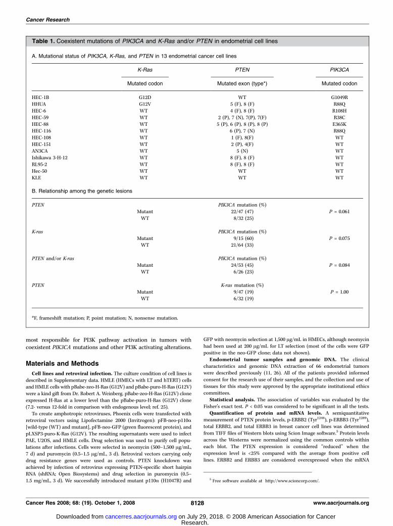
Table 1. Coexistent mutations of PIK3CA and K-Ras and/or PTEN in endometrial cell lines
A. Mutational status of PIK3CA, K-Ras, and PTEN in 13 endometrial cancer cell lines
K-Ras PTEN PIK3CA
Mutated codon Mutated exon (type*) Mutated codon
HEC-1B G12D WT G1049RHHUA G12V 5 (F), 8 (F) R88Q
HEC-6 WT 4 (F), 8 (F) R108H
HEC-59 WT 2 (P), 7 (N), 7(P), 7(F) R38C
HEC-88 WT 5 (P), 6 (P), 8 (P), 8 (P) E365KHEC-116 WT 6 (P), 7 (N) R88Q
HEC-108 WT 1 (F), 8(F) WT
HEC-151 WT 2 (P), 4(F) WTAN3CA WT 5 (N) WT
Ishikawa 3-H-12 WT 8 (F), 8 (F) WT
RL95-2 WT 8 (F), 8 (F) WT
Hec-50 WT WT WTKLE WT WT WT
B. Relationship among the genetic lesions
PTEN PIK3CA mutation (%)
Mutant 22/47 (47) P = 0.061
WT 8/32 (25)
K-ras PIK3CA mutation (%)
Mutant 9/15 (60) P = 0.075WT 21/64 (33)
PTEN and/or K-ras PIK3CA mutation (%)
Mutant 24/53 (45) P = 0.084WT 6/26 (23)
PTEN K-ras mutation (%)
Mutant 9/47 (19) P = 1.00WT 6/32 (19)
*F, frameshift mutation; P, point mutation; N, nonsense mutation.
Cancer Research
Cancer Res 2008; 68: (19). October 1, 2008 8128 www.aacrjournals.org
Research. on July 29, 2018. © 2008 American Association for Cancercancerres.aacrjournals.org Downloaded from
level in expression array hybridization is more than 4- and 15-fold,compared with that of the nontumorigenic cell line MCF10A, respectively.
‘‘Intermediate’’ ERBB3 expression corresponds to a 10- to 15-fold increase
from that in MCF10A.
Small interfering RNA transfection. Small interfering RNA (siRNA)was used to inhibit the expression of ERBB2, ERBB3, p110a, or p110h.The targeted sequences of ERBB2, p110a, and p110h siRNAs are
5¶-AAGGTGCTTGGATCTGGCGCT-3¶, 5¶-AAGGAGCCCAAGAATGCACAA-3¶,and 5¶-AAAGGGAGCGAGTGCCTTTTA-3¶, respectively. siRNA to ERBB3(siGenome) was purchased from Dharmacon RNA Technologies. Cells were
seeded at 2.0 � 105 per six-well plate 24 h before transfection and
transfected with 60 to 120 nmol/L siRNA duplexes using Lipofectamine
2000 (Invitrogen). Cells were collected 72 h after transfection and analyzedby immunoblotting. Suppression of p110a and p110h by siRNA was
confirmed by real-time PCR.
Cluster analysis. Unsupervised Cluster analysis was done on an
Affymetrix U133A–based breast tumor cell line data set (27) using Clusterand Treeview software. Average linkage clustering results using the 15,000
array elements, which exhibit the most variance across the data set, are
reported. Red squares represent high and green squares represent low
relative expression levels of ERBB2 and ERBB3.Immunoblotting. Cells were lysed as described previously (11).
Antibodies to PTEN, phospho-PTEN (Ser380), total Akt, phospho-Akt
(Ser473), total p110a, total GSK3h, phospho-GSK3h (Ser9), total S6,
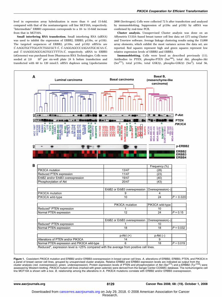
Figure 1. Coexistent PIK3CA mutation and ERBB2 and/or ERBB3 overexpression in breast cancer cell lines. A, alterations of ERBB2, ERBB3, PTEN, and PIK3CA ina panel of breast cancer cell lines, grouped by unsupervised cluster analysis. Relative ERBB2 and ERBB3 expression levels are indicated as output from thecluster analysis (red, overexpression; green, underexpression). Protein expression levels of PTEN and phosphorylation of Akt (Ser473) and p-ERBB2 (Tyr1248) wereassessed by Western blotting. PIK3CA mutant cell lines (marked with green asterisk) were derived from the Sanger Center COSMIC database. The nontumorigenic cellline MCF10A is shown with a box. B, relationship among the alterations in A. PIK3CA mutations correlate with ERBB2 and/or ERBB3 overexpression.
PIK3CA Cooperation for Efficient Transformation
www.aacrjournals.org 8129 Cancer Res 2008; 68: (19). October 1, 2008
Research. on July 29, 2018. © 2008 American Association for Cancercancerres.aacrjournals.org Downloaded from
phospho-S6 (Ser235/236), total FoxO1, phospho-FoxO1 (Thr24), phospho-
FoxO3a (Thr32) (Cell Signaling Technology), h-actin, FLAG tag (Sigma-
Aldrich), total K-Ras, total ERBB2, total ERBB3 (Santa Cruz Biotechnology),and phospho-ERBB2 (Tyr1248) (Lab Vision Corp.) were used for immuno-
blotting as recommended by the manufacturer and were detected with
enhanced chemiluminescence Western blot detection kit (Amersham
Biosciences) or Immobilon Western detection reagents (Millipore Bio-sciences).
Soft agar assays. PAE, U2OS, and HMLE cells were seeded at 1 � 103
or 4 � 103 per well in six-well dishes with a bottom layer of 0.6% agar
in DMEM and a top layer of 0.35% agar in DMEM ( for PAE and U2AOS)or mammary epithelium growth medium ( for HMLEs). Fresh medium
(0.5 mL; DMEM for PAE and U2OS, mammary epithelium growth
medium for HMLEs) was added after 1.5 wk. Colonies (>0.2 mm indiameter) were counted after 2 to 3 wk. At least two independent assays
were done in triplicate.
Results
PIK3CA mutations coexist with K-ras and PTEN mutationsin endometrial carcinomas. We screened 79 endometrialcarcinomas (66 clinical specimens and 13 cell lines) for mutationsof K-ras, PIK3CA , and PTEN . The mutational status of 13endometrial cancer cell lines is shown in Table 1A . Overall, themutation frequency in 79 endometrial cancers is 19% for K-ras , 57%for PTEN , and 37% for PIK3CA . Tumors with K-ras mutation
exhibited a tendency to carry PIK3CA mutation more frequently(9 of 15, 60%) than tumors without K-ras mutation (21 of 64, 33%),although statistical significance was not reached (P = 0.075, Fisher’sexact test). We did not find any association between PTENmutations and K-ras mutations (9 of 47 versus 6 of 32, P = 1.0; Table1B ), suggesting that mutation of these two genes occursindependently. As shown in Table 1B , 24 of 30 (80%) tumors withPIK3CA mutations possess K-ras and/or PTEN mutations.PIK3CA mutations are associated with ERBB2 and/or
ERBB3 overexpression in breast cancer cell lines. We screeneda panel of 47 breast cancer cell lines for activators of PI3Ksignaling. The mutational status of PIK3CA was determined byexamination of the COSMIC database (Sanger Center).6 ERBB2,ERBB3, PTEN, and p-Akt (Ser473) levels were assessed by expressionarray hybridization and/or Western blotting (Fig. 1A ; summarizedin Supplementary Table S2). We focused on ERBB2 and ERBB3expression levels in breast cancer because (a) the associationbetween PIK3CA mutations and ERBB2 overexpression is stillcontroversial (28); (b) the ERBB2/ERBB3 heterodimer functions asan oncogenic unit (29); and (c) ERBB3 overexpression in breastcancer is also common (30). In addition, ERBB3 contains six
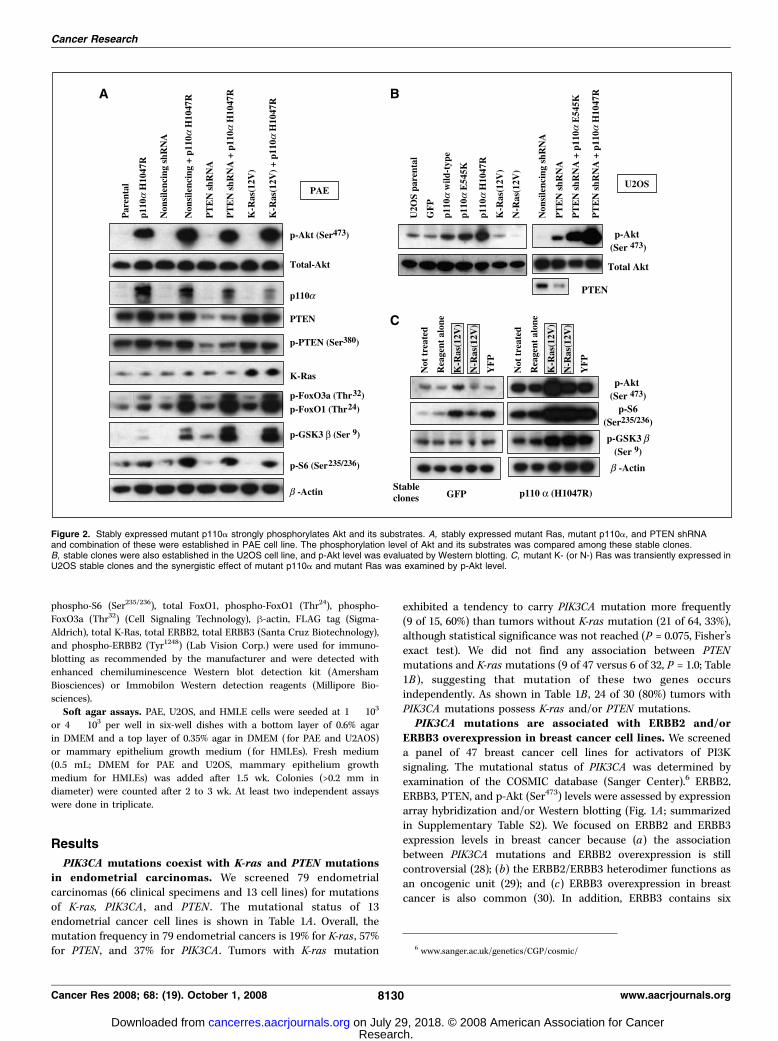
Figure 2. Stably expressed mutant p110a strongly phosphorylates Akt and its substrates. A, stably expressed mutant Ras, mutant p110a, and PTEN shRNAand combination of these were established in PAE cell line. The phosphorylation level of Akt and its substrates was compared among these stable clones.B, stable clones were also established in the U2OS cell line, and p-Akt level was evaluated by Western blotting. C, mutant K- (or N-) Ras was transiently expressed inU2OS stable clones and the synergistic effect of mutant p110a and mutant Ras was examined by p-Akt level.
6 www.sanger.ac.uk/genetics/CGP/cosmic/
Cancer Research
Cancer Res 2008; 68: (19). October 1, 2008 8130 www.aacrjournals.org
Research. on July 29, 2018. © 2008 American Association for Cancercancerres.aacrjournals.org Downloaded from
docking and activation sites for PI3K, and p-ERBB3 is shownto activate the PI3K pathway (31). Micro-RNAs miR-125a andmiR-125b, which suppress both ERBB2 and ERBB3 expression, aredown-regulated in breast cancers (32), suggesting that ERBB2/ERBB3 signaling is important for breast tumorigenesis. Relative toexpression levels found in the nontumorigenic cell line MCF10A,we found ERBB2 and/or ERBB3 overexpression in 19 of 47 (40%)cell lines; 10 with ERBB2 overexpression and 13 with ERBB3overexpression, including four (BT474, HCC202, UACC-812, andSUM225CEM) with overexpression of both molecules. PIK3CAmutations were positive in 13 (28%) cell lines (Fig. 1A ;Supplementary Table S2). Nine of 13 (69%) with PIK3CA mutationsharbor ERBB2 and/or ERBB3 overexpression (P = 0.020, Fisher’sexact test; Fig. 1B). On the other hand, reduced PTEN expression(11 of 47, 23%) was less common in cells with PIK3CA mutations(1 of 13, 8%), compared with those without PIK3CA mutations(10 of 34, 29%; P = 0.15). In addition, reduced PTEN expressionwas inversely correlated with ERBB2 and/or ERBB3 overexpression
(P = 0.032). Elevated phosphorylation of Akt (20 of 47, 43%) wassignificantly associated with PIK3CA mutations and/or reducedPTEN expression (P = 0.019). In contrast to endometrialcarcinomas, loss of PTEN expression is reported to be very rarein noninvasive breast cancer (ductal carcinoma in situ) and morecommon in advanced stage tumors (33). Thus, loss of PTEN may bea late event in breast cancer. Semiquantitative analysis of phospho-ERBB2 (Tyr1248) and phoshpho-ERBB3 (Tyr1289) levels (27) showedthat overexpression of each molecule was well associated with thelevel of its phosphorylation and p-Akt (Fig. 1A ; SupplementaryTable S3). Six of nine PIK3CA mutated tumors showed highp-ERBB2 and/or p-ERBB3 levels (Supplementary Table S3). Ourdata indicate that PIK3CA mutations frequently coexist with activeERBB2/ERBB3 signaling in breast carcinomas.Mutant p110A is more potent in phosphorylation of Akt
than mutant Ras or reduced PTEN expression. According to ourprevious report that multiple alterations in the PI3K pathway couldfurther activate PI3K signaling in endometrial cell lines (11), we
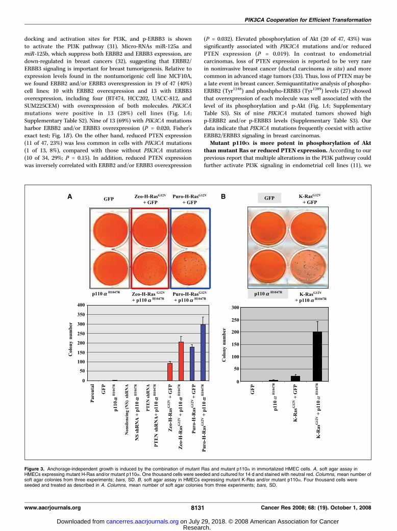
Figure 3. Anchorage-independent growth is induced by the combination of mutant Ras and mutant p110a in immortalized HMEC cells. A, soft agar assay inHMECs expressing mutant H-Ras and/or mutant p110a. One thousand cells were seeded and cultured for 14 d and stained with neutral red. Columns, mean number ofsoft agar colonies from three experiments; bars, SD. B, soft agar assay in HMECs expressing mutant K-Ras and/or mutant p110a. Four thousand cells wereseeded and treated as described in A. Columns, mean number of soft agar colonies from three experiments; bars, SD.
PIK3CA Cooperation for Efficient Transformation
www.aacrjournals.org 8131 Cancer Res 2008; 68: (19). October 1, 2008
Research. on July 29, 2018. © 2008 American Association for Cancercancerres.aacrjournals.org Downloaded from
hypothesized that the presence of the upstream PI3K activationmight cooperate with mutant p110a to increase PI3K activity, andthat the presence of mutant p110a might be important foroncogenic conversion or progression. Therefore, we introducedmutant K- (or N-) Ras (G12V), mutant p110a (H1047R), shRNAsthat targeted PTEN, and combinations of these alleles into PAE(porcine aortic endothelial) and U2OS osteosarcoma cells byretrovirus-mediated gene transfer. Both PAE cells and U2OS cellsdo not harbor any mutations in Ras, PTEN , and PIK3CA , resultingin very low basal p-Akt levels. Either PTEN knockdown or mutantRas expression effected the p-Akt at a much lower level thandid mutant p110a alone in both cells (Fig. 2A and B). Thephosphorylation level of FoxO1, FoxO3a, and GSK3h was enhancedby either mutant K-Ras or PTEN silencing when combined withmutant p110a (Fig. 2A). Transient introduction of mutant Rasincreased the p-Akt level in U2OS cells stably expressing mutantp110a (Fig. 2C). In summary, either mutant Ras or PTEN silencingalone has limited effect on the activation of the PI3K pathway, but
the effect is much more drastic when combined with mutantp110a.Anchorage-independent growth is induced by mutant
p110A in PAE and U2OS cells. We tested whether mutantp110a might induce oncogenic behavior (anchorage-independentgrowth) in the PAE and U2OS cell lines because both PAE(untransformed) and U2OS cells are unable to grow withoutattachment to a substratum (34). Stable expression of mutantp110a (H1047R) resulted in growth of colonies in soft agar in bothcells (Supplementary Fig. S1A and B), suggesting that the higherPI3K activity via mutant p110a is involved in anchorage-independent growth in these cells.Mutant Ras and p110A efficiently induced oncogenic
transformation in immortalized HMEC cells. Because alter-ations of Ras or ERBB2 are observed in noninvasive tumors of thecolorectum, breast, and endometrium, we hypothesized thatactivation of multiple Ras effectors is useful for tumor initiationand that higher PI3K activation via additional mutations in p110a
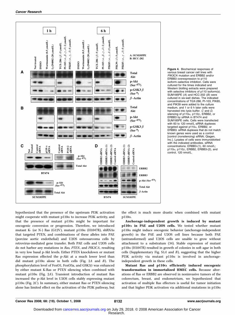
Figure 4. Biochemical responses ofvarious breast cancer cell lines withPIK3CA mutation and ERBB2 and/orERBB3 overexpression to p110isoform–selective inhibition. Cells werecultured for the times indicated andWestern blotting extracts were preparedwith selective inhibitors of p110 isoform(s).SUM185PE (A) and HCC-202 (B) werecultured in six-well dishes. The indicatedconcentrations of TGX-286, PI-103, PIK85,and PIK39 were added to the culturemedium, and 1 or 6 h later cells wereharvested into lysis buffer. C and D,silencing of p110a, p110h, ERBB2, orERBB3 by siRNA in BT474 andSUM185PE cells. Cells were transfectedwith 60 to 120 nmol/L siRNA duplexestargeted against p110a, ERBB2, orERBB3. siRNA duplexes that do not matchknown genes were used as a control[control (nonsilencing) siRNA; Qiagen,Inc.]. Lysates of cells were immunoblottedwith the indicated antibodies. siRNAconcentrations: ERBB3-(1), 60 nmol/L;p110a, p110h, ERBB2, ERBB3-(2), andcontrol, 120 nmol/L.
Cancer Research
Cancer Res 2008; 68: (19). October 1, 2008 8132 www.aacrjournals.org
Research. on July 29, 2018. © 2008 American Association for Cancercancerres.aacrjournals.org Downloaded from
is helpful for malignant progression of noninvasive tumors. Inaddition, because PIK3CA mutations without other alterations arerare in these tumor types, mutant p110a alone might beinsufficient for oncogenic transformation. Mutant H-Ras (G12V)transforms immortalized HMECs only when it is highly overex-pressed (25); we assumed that additional mutant p110a mightincrease transformation efficiency in cells expressing lower levelsof mutant Ras. To test this hypothesis, we obtained HMECspreviously immortalized with large T-antigen and TERT (HMLE),with and without various alleles of activated H-Ras (25), andintroduced activating alleles of p110a, K-Ras, or PTEN-silencingshRNAs in various combinations to determine their relative trans-formation potential (summarized in Supplementary Table S4).
Akt was more potently activated by introduction of mutantp110a than by mutant (H- or K-) Ras or PTEN shRNA (Sup-plementary Fig. S2A and B). The transformation potential ofthese stable cell lines was then assessed by soft agar assay.Mutant p110a (H1047R) alone failed to cause anchorage-independent growth in the parental HMLE (Fig. 3A). In addition,mutant p110a plus partial reduction of PTEN expression byshRNA did not cause anchorage-independent growth. However,in HMLE sublines, which constitutively express zeo-H-Ras (G12V)or puro-H-Ras (G12V), addition of mutant p110a (H1047R)doubled the colony number relative to control GFP (Fig. 3A).Unlike mutant H-Ras, introduction of mutant K-Ras (G12V) alonedid not induce anchorage-independent growth; however, the
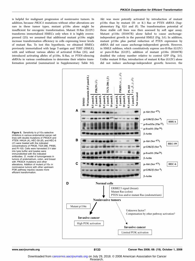
Figure 5. Sensitivity to p110a-selectiveinhibitors in various endometrial cancer celllines with double mutations of PIK3CA andPTEN . HHUA (A ), HEC-59 (B ), and HEC-6(C ) were treated with the indicatedconcentrations of PIK39, TGX-286, PIK85,and PI-103. Cells were harvested 3 h laterinto lysis buffer and lysates wereimmunoblotted with the indicatedantibodies. D, model of tumorigenesis intumors of endometrium, colon, and breastwith PIK3CA mutations and otheralterations. Addition of mutant p110a innoninvasive tumors with other upstreamPI3K pathway input(s) causes moreefficient transformation.
PIK3CA Cooperation for Efficient Transformation
www.aacrjournals.org 8133 Cancer Res 2008; 68: (19). October 1, 2008
Research. on July 29, 2018. © 2008 American Association for Cancercancerres.aacrjournals.org Downloaded from
combination of mutant p110a (H1047R) and mutant K-Rasenabled cells to grow in soft agar (Fig. 3B).p110A is the predominant form of PI3K activity in breast
cancer cells with coexistent mutant p110A and overexpressionof ERBB2 and/or ERBB3. To determine which p110 isoforms wereactivated by upstream PI3K pathway activating alterations in cellswith PIK3CA mutation, we examined the effect of p110 isoform–selective inhibitors in five breast cancer cell lines with coexistentPIK3CA mutations and ERBB2 and/or ERBB3 overexpression(SUM185PE, MDA-MB361, and T47D: ERBB3 overexpression;HCC202 and BT474: both ERBB2 and ERBB3 overexpression). Fourtypes of p110 inhibitors were tested on each cell line at 2 or5 Amol/L for 1 to 6 h. These include the selective p110h inhibitorTGX-286; the selective p110y inhibitor PIK39; PIK85, whichselectively inhibits p110a, p110g, and p110y; and the pan-selectivep110 inhibitor PI-103. The structure of these four compounds isavailable from ref. 35 and Supplementary Fig. S3. The IC50 values ofthe four compounds were listed in Supplementary Table S5. BothPIK39 (p110y inhibitor) and TGX-286 (p110h inhibitor) failed tosuppress PI3K signaling as assessed by phosphorylation of Akt(Ser473) and GSK-3h (Ser9), whereas PIK85 (p110a, p110g, and p110yinhibitor) drastically suppressed phosphorylation of these mole-cules to a level similar to that of PI-103, wortmannin, and Ly294002(pan-selective agents) in all these breast cancer cell lines (Fig. 4Aand B , and data not shown). The p110g hybridization signal is lowto undetectable in the analysis of Affymetrix gene expressionprofiling data (27) in breast cancer cell lines, indicating that little, ifany, of this mRNA is expressed in the breast, whereas p110a, p110h,and p110y are expressed at comparable and more robust levels.These data suggest that breast cancer cell lines with mutant p110aand overexpression of ERBB2 and/or ERBB3 are dependent onp110a activity, rather than other isoforms, for activation of the PI3Kpathway. Transfection with p110a-specific siRNA, but not withp110h-specific siRNA, suppresses Akt phosphorylation in BT474cells (Fig. 4C). The p-Akt level was also decreased in SUM185PEcells by p110a-specific siRNA (Fig. 4C). These data also suggestthat p110a is the major activated form in these cells.
To identify the contribution of ERBB2 and/or ERBB3 over-expression on the activity of the PI3K pathway, we used siRNAknockdown to reduce the expression of ERBB2 or ERBB3 in BT474.Attenuating the expression of these proteins partially reduces thelevel of p-Akt (Fig. 4D). ERBB3 suppression reduced the p-Akt levelin SUM185PE cells (Fig. 4D). In addition, serum starvationdecreased the p-Akt level in all five cell lines (data not shown).These data indicate that activation of PI3K signaling depends notonly on mutant p110a but also on ERBB2/ERBB3 overexpressionand/or serum-derived growth factors.p110A is predominantly active in colorectal cancer cells
with double mutations of p110A and Ras. The pattern ofcoexistent alterations in the PI3K pathway in colorectal cancer isdouble mutations of PIK3CA and K-ras . To determine whether PI3Kactivity with these coexistent mutations also relies primarily onp110a, we tested the p110 isoform–selective inhibitors in twocolorectal cancer cell lines with these double mutations (DLD1 andHCT116). The phosphorylation of Akt was sensitive to PIK85 andPI-103 but resistant to TGX-286 and PIK39 (Supplementary Fig. 4Aand B) in these two cells. These data suggest that double mutationsof K-ras and PIK3CA predominantly activate the p110a isoform incolorectal cancer.p110A is predominantly active in endometrial cancer
cells with double mutations of PIK3CA and PTEN . Next, we
analyzed five endometrial cancer cell lines (HHUA, HEC-59, HEC-6,HEC-116, and HEC-88) with double mutations of PIK3CA andPTEN (Table 1A). PIK85 and PI-103, but neither TGX-286 norPIK39, drastically decreased the level of p-Akt in all five cell lines(Fig. 5A–C ; Supplementary Fig. S5A and B). In addition, PIK85,PI-103, and wortmannin suppressed the phosphorylation of Aktsubstrates such as p-GSK3h (Ser9), p-FoxO1 (Thr24), and p-S6(Ser235/236; Fig. 5A–C ; Supplementary Fig. S5A and B). Reduction ofp110a by siRNA also suppressed Akt phosphorylation in Hec-6cells, whereas reduction of p110h by siRNA did not (SupplementaryFig. S5C). These data indicate that p110a is constitutively activatedin endometrial tumor cells with double mutations of PTEN /PIK3CA .
Discussion
These results highlight the important role of PIK3CA mutationsduring oncogenic transformation in multiple epithelial tumortypes. In this study, we addressed the meaning of multiplealterations in the PI3K pathway by analyzing (a) the patterns ofcoexistent alterations, (b) the efficiency of PI3K activation by singlealterations, (c) the oncogenic transformation by coexistentalterations, and (d) the p110 isoform selectivity. These issues arediscussed in more detail below.Coexistent PIK3CA mutations and other alterations in
endometrial, breast, and colorectal cancers. Endometrial,breast, and colorectal cancers are three major tumor types thatshow a high frequency of PIK3CA mutations (8, 11, 36). Curiously,the partners of PIK3CA mutations differ between these tissues:PTEN mutations in endometrium, ERBB2 overexpression in breast,and K-ras mutations in colorectum were reported to commonlycoexist with PIK3CA mutations (10–12). In this study, we furtheridentified double mutations of K-ras and PIK3CA in endometrialcancer and the coexistence of PIK3CA mutation and ERBB2/ERBB3signaling in breast cancer cell lines. Various patterns of coexistentalterations of PIK3CA and the genes upstream of PI3K suggest thatthe role of mutant PIK3CA might be different from the otheralterations. Ras and ERBB2/ERBB3 are involved in varioussignaling pathways, whereas PTEN also has PI3K-independentfunctions. Therefore, the co-occurring alterations in the PI3Kpathway may have two major roles: augmentation of the PI3Kpathway and activation of other pathways.
Although PIK3CA mutations in six endometrial cell linesare outside the three well-characterized mutation hotspots(E542, E545, and H1047; refs. 22, 37), all five p110a mutants(R38C, R88Q, R108H, E365K, and G1049R) significantly increasedthe phosphorylation levels of Akt, GSK3h, FoxO1, FoxO3a, and S6compared with WT p110a in the U2OS sublines (SupplementaryFig. S6). These data indicate that these rare p110a mutations causea gain of PI3K function.Enhanced activation of the PI3K pathway by additional
mutant p110A. Combined PIK3CA mutation and other alterationsin the Ras-PI3K pathway (PTEN and/or K-ras mutations inendometrium, ERBB2 and/or ERBB3 overexpression in breast,and K-ras mutation in colorectum) have two common features:First, these other alterations already exist in noninvasive tumors,whereas PIK3CA mutation is rarely observed in noninvasivetumors. Second, the combined ratio (z40%) of other alterationsis higher than that of PIK3CA mutations (20–40%) in these cancers(Table 1B ; Supplementary Table S1). These data suggest thatactivation of the PI3K pathway via any WT p110 isoform is not
Cancer Research
Cancer Res 2008; 68: (19). October 1, 2008 8134 www.aacrjournals.org
Research. on July 29, 2018. © 2008 American Association for Cancercancerres.aacrjournals.org Downloaded from
sufficiently high in tumors with single alterations, although thosetumors occasionally progress to invasive tumors without anadditional PIK3CA mutation (possibly via other unknown alter-ations or compensation from other pathways). In our experiments,sole expression of mutant Ras or knocking down PTEN was lesseffective than mutant p110a, and addition of mutant p110adrastically increased p-Akt levels in PAE, U2OS, and HMLE cells.Samuels and colleagues (38) established isogenic HCT116 andDLD1 colorectal cell lines in which either the WT or mutant allelesof PIK3CA were disrupted. Because both HCT116 and DLD1possess mutant K-Ras (G13D), their analysis compared mutantK-Ras alone versus double mutants of K-Ras and PIK3CA. ThePIK3CA mutant clones showed higher levels of p-Akt andp-FoxO1/p-FoxO3a compared with PIK3CA WT clones in both celllines. Their data implied that mutant Ras alone is not sufficient forfull activation of the PI3K pathway and that addition of theendogenous level of mutant p110a enhances the pathway,including some downstream effectors such as FoxO1/FoxO3a.Efficient oncogenic transformation by combined mutant
Ras and mutant p110A. Oncogenic transformation by mutantp110a was reported in chicken embryo fibroblasts in vitro andin vivo (37, 39). In addition, we found anchorage-independentgrowth by mutant p110a in PAE and U2OS cells (SupplementaryFig. S1A and B). However, it has not been clear whether mutantp110a alone is sufficient for malignant transformation inimmortalized human epithelial cells. In HMLEs, mutant p110afailed to induce anchorage-independent growth (Fig. 3A), suggest-ing that activation of the PI3K pathway alone is insufficient fortransformation. This is compatible with the observation thatmutant p110a frequently coexists with other alterations in the Ras-PI3K pathway. Transformation by mutant Ras is well analyzed invarious types of immortalized human epithelial cells (25, 40–42).However, low levels of mutant H-Ras alone failed to confer a fullmalignant phenotype (Fig. 3A ; refs. 25, 40) or required anotherinput in the PI3K pathway [constitutively active form of Akt (41) orSV40 small t antigen (42)]. In addition, mutant K-Ras did notinduce anchorage-independent growth in HMLE (Fig. 3B). Theseobservations suggest that endogenous levels of mutant Ras aloneare insufficient for full transformation in immortalized humanepithelial cells. However, mutant p110a significantly increased thenumber of colonies in soft agar when combined with mutant H-Rasor K-Ras. In our experiments, mutant Ras itself activated the PI3Kpathway at a weaker level, but additional mutant p110a clearlyenhanced the level of p-Akt, suggesting that the activity of the PI3Kpathway was more strengthened. Our data indicate that additionalmutant p110a plays a significant role in tumorigenicity and suggestthat the enhanced PI3K activity might be responsible for theoncogenic activity.
The role of PTEN alterations in tumorigenesis is still unclear andmay be tissue type specific. PTEN knockdown was incomplete inHMLE cells in our soft agar assay, although p-Akt was enhanced.That might be why the transformation efficiency might not beaffected by PTEN shRNA. In addition, reduced PTEN expressionin breast cancer is rare in noninvasive tumors and is insteadassociated with tumor invasion (33). However, PTEN mutationsare common in atypical hyperplasia of the endometrium, as well asK-ras mutations (19, 43), and also frequent in ovarian endometriosis(44). In mouse models, either mutant K-Ras or conditional PTENdeletion within the ovarian surface gives rise to preneoplasticovarian (endometriosis like) lesions and leads to invasive endome-trioid ovarian cancer when combined (45). These observations
indicate that PTEN alterations may have a similar effect withmutant K-Ras in tumor initiation and cooperate with mutant p110ain tumorigenesis of the endometrium and the ovary. Anotherpossibility is that PI3K-independent PTEN functions (i.e., phospha-tase-independent function) may be important in the tumorigenesis.Therefore, each mutation of K-ras, PTEN , and PIK3CA may havedifferent meanings in endometrial tumorigenesis.
The model of tumorigenesis in colorectal, breast, and endometrialtumors with coexistent PIK3CA and other alterations is summarizedin Fig. 5D . Mutant Ras (or ERBB2 overexpression, etc.) is importantfor tumor initiation, which might be achieved through multiple Raseffector pathways, including the mitogen-activated protein kinase,Ral guanine nucleotide exchange factor, and PI3K pathways (21, 46,47). However, the PI3K activation is not saturated in thesenoninvasive tumors. Once PIK3CA is mutated during the latentphase, the PI3K pathway could be activated to a greater level, whichmay result in efficient malignant transformation of noninvasivetumors. Without PIK3CA mutations, the noninvasive tumors mayneed another input (possibly PI3K pathway independent) to becometransformed or they may need a longer latent phase. It is still unclearwhether the augmented PI3K pathway activation alone is respon-sible for the transformation efficiency in PIK3CA mutated tumors.Mutant p110a may have any functions of cross talk with the otherpathways, which are already activated by the earlier alterations. Inaddition, the magnitude of PI3K pathway activation is not alwayslinked to the transformation efficiency. In PAE cells, mutant K-Rasinduced more colony numbers than mutant p110a in soft agar assay(data not shown), suggesting that the requirement for PI3K pathwayenhancement depends on the cellular context.Predominant p110A activation in tumors with coexistent
mutant p110A and other PI3K-activating alteration(s). Ourobservations with p110 isoform–selective inhibitors highlighted theimportance of p110a activity in tumors with mutant p110a andother alterations. We used PIK-85 to inhibit p110a, p110g, andp110y. p110g expression is suggested to be exclusively restricted tohematopoietic cells (48), and the expression in cancer cell lines wasalso at low to undetectable levels. PIK39 was useful to excludep110y activation, as it is a highly selective p110y inhibitor. We hadassumed that the higher PI3K activation seen with coexistentmutations in the PI3K pathway might be due to the activation ofmultiple p110 isoforms. In fact, we observed that several cancer celllines with PTEN alterations alone (without PIK3CA mutations) inbreast and endometrium showed some sensitivity to TGX-286 orPIK39 (data not shown), suggesting that loss of PTEN function isnot always associated with activation of p110a. One possibleexplanation is that other alterations without a PIK3CA mutationmay activate the PI3K pathway through multiple (or non-a) p110isoform(s), and that isoform dependency might be switched top110a once p110a is mutated. To clarify the mechanism of p110isoform selectivity, cells from invasive and noninvasive tumors thatdo not harbor PIK3CA mutations but have alterations in other PI3Kpathway elements might be studied to address this question.
Predominant p110a activation in PIK3CA mutant tumorsindicates that p110a-selective inhibition might be a goodtherapeutic target in tumors with mutant p110a. Trastuzumab(Herceptin) is a well-established targeted therapy in breast cancer(49), but our data suggest that trastuzumab might not be sufficientto suppress the PI3K pathway in tumors with double alterations ofERBB2 and PIK3CA . p110a isoform–selective inhibition may be lesstoxic than pan-selective PI3K inhibition and may be more effectivethan trastuzumab in tumors with mutant p110a.
PIK3CA Cooperation for Efficient Transformation
www.aacrjournals.org 8135 Cancer Res 2008; 68: (19). October 1, 2008
Research. on July 29, 2018. © 2008 American Association for Cancercancerres.aacrjournals.org Downloaded from
Conclusion
We confirmed that PIK3CA mutations frequently coexist withother alterations in the PI3K pathway in endometrium, breast, andcolorectum. All cell lines with coexistent mutant p110a and otheralterations showed p110a dependency and enhanced activity of thePI3K signaling pathway. Mutant p110a cooperates with mutant Rasto efficiently transform HMECs. Taken together, the high activity ofp110a by a coexistent PIK3CA mutation with other alterations canbe a critical step for malignant transformation.
Disclosure of Potential Conflicts of Interest
K. Oda: commercial research grant, Daiichi-Sankyo Pharmaceutical Co. Ltd.F. McCormick: commercial research grant and consultant, Daiichi-Sankyo
Pharmaceutical Co. Ltd.; speaker honoraria, Novartis; ownership interest, Onyx,Exelixis, and Nexgenix. The other authors disclosed no potential conflicts of interest.
Acknowledgments
Received 2/28/2008; revised 6/22/2008; accepted 7/6/2008.Grant support: Daiichi-Sankyo Pharmaceutical Co., Ltd. Japan, KAKENHI
(19599005), Japan, and the Susan G. Komen Foundation (K.M. Shokat).The costs of publication of this article were defrayed in part by the payment of page
charges. This article must therefore be hereby marked advertisement in accordancewith 18 U.S.C. Section 1734 solely to indicate this fact.
We thank Robert A. Weinberg for HMLE cells and its sublines, Masato Nishida forIshikawa 3-H-12 cell line, and Victor E. Velculescu for useful information. We thankMayumi Kitagawa, Sang-Hyun Lee, Masashi Aonuma, Osamu Tetsu, Abigail Miller,Frank Bos, Jesse Lyons, Anthony Karnezis, and Vivianne Wei Ding for helpfuldiscussions. We also thank Tetsu Yano, Toshiharu Yasugi, Shunsuke Nakagawa andTomomi Nei for support and assistance, especially for organizing clinical samplesand data.
References
1. Stokoe D, Stephens LR, Copeland T, et al. Dual role ofphosphatidylinositol-3,4,5-trisphosphate in the activa-tion of protein kinase B. Science 1997;277:567–70.
2. Lynch TJ, Bell DW, Sordella R, et al. Activatingmutations in the epidermal growth factor receptorunderlying responsiveness of non-small-cell lung cancerto gefitinib. N Engl J Med 2004;350:2129–39.
3. Slamon DJ, Godolphin W, Jones LA, et al. Studies of theHER-2/neu proto-oncogene in human breast andovarian cancer. Science 1989;244:707–12.
4. Vogelstein B, Fearon ER, Hamilton SR, et al. Geneticalterations during colorectal-tumor development.N Engl J Med 1988;319:525–32.
5. Bos JL. ras oncogenes in human cancer: a review.Cancer Res 1989;49:4682–9.
6. Li J, Yen C, Liaw D, et al. PTEN, a putative proteintyrosine phosphatase gene mutated in human brain,breast, and prostate cancer. Science 1997;275:1943–7.
7. Kong D, Suzuki A, Zou TT, et al. PTEN1 is frequentlymutated in primary endometrial carcinomas. Nat Genet1997;17:143–4.
8. Samuels Y, Wang Z, Bardelli A, et al. High frequency ofmutations of the PIK3CA gene in human cancers.Science 2004;304:554.
9. Tsao H, Goel V, Wu H, et al. Genetic interactionbetween NRAS and BRAF mutations and PTEN/MMAC1inactivation in melanoma. J Invest Dermatol 2004;122:337–41.
10. Saal LH, Holm K, Maurer M, et al. PIK3CA mutationscorrelate with hormone receptors, node metastasis, andERBB2, and are mutually exclusive with PTEN loss inhuman breast carcinoma. Cancer Res 2005;65:2554–9.
11. Oda K, Stokoe D, Taketani Y, et al. High frequency ofcoexistent mutations of PIK3CA and PTEN genes inendometrial carcinoma. Cancer Res 2005;65:10669–73.
12. Parsons DW, Wang TL, Samuels Y, et al. Colorectalcancer: mutations in a signalling pathway. Nature 2005;436:792.
13. Burmer GC, Loeb LA. Mutations in the KRAS2oncogene during progressive stages of human coloncarcinoma. Proc Natl Acad Sci U S A 1989;86:2403–7.
14. Kann L, Han J, Ahlquist D, et al. Improved markercombination for detection of de novo genetic variationand aberrant DNA in colorectal neoplasia. Clin Chem2006;52:2299–302.
15. Somerville JE, Clarke LA, Biggart JD. c-erbB-2 over-expression and histological type of in situ and invasivebreast carcinoma. J Clin Pathol 1992;45:16–20.
16. Latta EK, Tjan S, Parkes RK, et al. The role of HER2/neu overexpression/amplification in the progression ofductal carcinoma in situ to invasive carcinoma of thebreast. Mod Pathol 2002;15:1318–25.
17. Park K, Han S, Kim HJ, et al. HER2 status in pureductal carcinoma in situ and in the intraductal andinvasive components of invasive ductal carcinomadetermined by fluorescence in situ hybridization andimmunohistochemistry. Histopathology 2006;48:702–7.
18. Sun H, Enomoto T, Fujita M, et al. Mutationalanalysis of the PTEN gene in endometrial carcinomaand hyperplasia. Am J Clin Pathol 2001;115:32–8.
19. Hayes MP, Wang H, Espinal Witter R, et al. PIK3CAand PTEN mutations in uterine endometrioid carcino-ma and complex atypical hyperplasia. Clin Cancer Res2006;12:5932–5.
20. Maruyama N, Miyoshi Y, Taguchi T, et al. Clinico-pathologic analysis of breast cancers with PIK3CAmutations in Japanese women. Clin Cancer Res 2007;13:408–14.
21. Lim KH, Counter CM. Reduction in the requirementof oncogenic Ras signaling to activation of PI3K/AKTpathway during tumor maintenance. Cancer Cell 2005;8:381–92.
22. Bader AG, Kang S, Zhao L, et al. Oncogenic PI3Kderegulates transcription and translation. Nat RevCancer 2005;5:921–9.
23. Kang S, Denley A, Vanhaesebroeck B, et al. Oncogenictransformation induced by the p110h, -g, and -yisoforms of class I phosphoinositide 3-kinase. Proc NatlAcad Sci U S A 2006;103:1289–94.
24. Schwarzer R, Tondera D, Arnold W, et al. REDD1integrates hypoxia-mediated survival signaling down-stream of phosphatidylinositol 3-kinase. Oncogene 2005;24:1138–49.
25. Elenbaas B, Spirio L, Koerner F, et al. Human breastcancer cells generated by oncogenic transformation ofprimary mammary epithelial cells. Genes Dev 2001;15:50–65.
26. Minaguchi T, Yoshikawa H, Oda K, et al. PTENmutation located only outside exons 5, 6, and 7 is anindependent predictor of favorable survival in endome-trial carcinomas. Clin Cancer Res 2001;7:2636–42.
27. Neve RM, Chin K, Fridlyand J, et al. A collection ofbreast cancer cell lines for the study of functionallydistinct cancer subtypes. Cancer Cell 2006;10:515–27.
28. Buttitta F, Felicioni L, Barassi F, et al. PIK3CAmutation and histological type in breast carcinoma:high frequency of mutations in lobular carcinoma.J Pathol 2006;208:350–5.
29. Holbro T, Beerli RR, Maurer F, et al. The ErbB2/ErbB3heterodimer functions as an oncogenic unit: ErbB2requires ErbB3 to drive breast tumor cell proliferation.Proc Natl Acad Sci U S A 2003;100:8933–8.
30. Naidu R, Yadav M, Nair S, et al. Expression of c-erbB3protein in primary breast carcinomas. Br J Cancer 1998;78:1385–90.
31. Kobayashi M, Iwamatsu A, Shinohara Kanda A, et al.Activation of ErbB3–3-kinase pathway is correlated withmalignant phenotypes of adenocarcinomas. Oncogene2003;22:1294–301.
32. Scott GK, Goga A, Bhaumik D, et al. Coordinatesuppression of ERBB2 and ERBB3 by enforced expres-sion of micro-RNA miR-125a or miR-125b. J Biol Chem2007;282:1479–86.
33. Bose S, Crane A, Hibshoosh H, et al. Reducedexpression of PTEN correlates with breast cancerprogression. Hum Pathol 2002;33:405–9.
34. Kalinichenko VV, Major ML, Wang X, et al. Foxm1btranscription factor is essential for development ofhepatocellular carcinomas and is negatively regulatedby the p19ARF tumor suppressor. Genes Dev 2004;18:830–50.
35. Knight ZA, Gonzalez B, Feldman ME, et al. Apharmacological map of the PI3-K family defines a rolefor p110a in insulin signaling. Cell 2006;125:733–47.
36. Campbell IG, Russell SE, Choong DY, et al. Mutationof the PIK3CA gene in ovarian and breast cancer.Cancer Res 2004;64:7678–81.
37. Kang S, Bader AG, Vogt PK. Phosphatidylinositol 3-kinase mutations identified in human cancer areoncogenic. Proc Natl Acad Sci U S A 2005;102:802–7.
38. Samuels Y, Diaz LA, Jr., Schmidt-Kittler O, et al.Mutant PIK3CA promotes cell growth and invasion ofhuman cancer cells. Cancer Cell 2005;7:561–73.
39. Bader AG, Kang S, Vogt PK. Cancer-specific muta-tions in PIK3CA are oncogenic in vivo . Proc Natl AcadSci U S A 2006;103:1475–9.
40. Sato M, Vaughan MB, Girard L, et al. Multipleoncogenic changes (K-RAS(V12), p53 knockdown, mu-tant EGFRs, p16 bypass, telomerase) are not sufficient toconfer a full malignant phenotype on human bronchialepithelial cells. Cancer Res 2006;66:2116–28.
41. Zhao JJ, Gjoerup OV, Subramanian RR, et al. Humanmammary epithelial cell transformation through theactivation of phosphatidylinositol 3-kinase. Cancer Cell2003;3:483–95.
42. Campbell PM, Groehler AL, Lee KM, et al. K-Raspromotes growth transformation and invasion ofimmortalized human pancreatic cells by Raf andphosphatidylinositol 3-kinase signaling. Cancer Res2007;67:2098–106.
43. Levine RL, Cargile CB, Blazes MS, et al. PTENmutations and microsatellite instability in complexatypical hyperplasia, a precursor lesion to uterineendometrioid carcinoma. Cancer Res 1998;58:3254–8.
44. Sato N, Tsunoda H, Nishida M, et al. Loss ofheterozygosity on 10q23.3 and mutation of the tumorsuppressor gene PTEN in benign endometrial cyst of theovary: possible sequence progression from benignendometrial cyst to endometrioid carcinoma and clearcell carcinoma of the ovary. Cancer Res 2000;60:7052–6.
45. Dinulescu DM, Ince TA, Quade BJ, et al. Role of K-rasand Pten in the development of mouse models ofendometriosis and endometrioid ovarian cancer. NatMed 2005;11:63–70.
46. Shields JM, Pruitt K, McFall A, et al. UnderstandingRas: ‘‘it ain’t over ’til it’s over.’’ Trends Cell Biol 2000;10:147–54.
47. Rodriguez Viciana P, Tetsu O, Oda K, et al. Cancertargets in the Ras pathway. Cold Spring Harb SympQuant Biol 2005;70:461–7.
48. Li Z, Jiang H, Xie W, et al. Roles of PLC-h2 and -h3and PI3Kg in chemoattractant-mediated signal trans-duction. Science 2000;287:1046–9.
49. Hudis CA. Trastuzumab-mechanism of action anduse in clinical practice. N Engl J Med 2007;357:39–51.
Cancer Research
Cancer Res 2008; 68: (19). October 1, 2008 8136 www.aacrjournals.org
Research. on July 29, 2018. © 2008 American Association for Cancercancerres.aacrjournals.org Downloaded from
2008;68:8127-8136. Cancer Res Katsutoshi Oda, Jennifer Okada, Luika Timmerman, et al. Transformation-Kinase Pathway Mutations to Effect Oncogenic
′PIK3CA Cooperates with Other Phosphatidylinositol 3
Updated version
http://cancerres.aacrjournals.org/content/68/19/8127
Access the most recent version of this article at:
Material
Supplementary
http://cancerres.aacrjournals.org/content/suppl/2008/09/26/68.19.8127.DC1
Access the most recent supplemental material at:
Cited articles
http://cancerres.aacrjournals.org/content/68/19/8127.full#ref-list-1
This article cites 49 articles, 26 of which you can access for free at:
Citing articles
http://cancerres.aacrjournals.org/content/68/19/8127.full#related-urls
This article has been cited by 24 HighWire-hosted articles. Access the articles at:
E-mail alerts related to this article or journal.Sign up to receive free email-alerts
Subscriptions
Reprints and
To order reprints of this article or to subscribe to the journal, contact the AACR Publications
Permissions
Rightslink site. (CCC)Click on "Request Permissions" which will take you to the Copyright Clearance Center's
.http://cancerres.aacrjournals.org/content/68/19/8127To request permission to re-use all or part of this article, use this link
Research. on July 29, 2018. © 2008 American Association for Cancercancerres.aacrjournals.org Downloaded from

![Strukturelle und quantitative Identifizierung der ...elib.suub.uni-bremen.de/edocs/00104054-1.pdf · Phospholipide ist PA der Ausgangsstoff [Wolff et al. 2008]. Phosphatidylinositol](https://img.pdfslide.tips/doc/110x75/5dd079dd06d5421854455439/strukturelle-und-quantitative-identifizierung-der-elibsuubuni-phospholipide.jpg)