Embed Size (px)
Citation preview

Plasma Proteins
Immunoglobulins

Plasma Proteins
1. Blood is a very important fluid
• What is blood?
• What does blood do?

Blood - Functions
• Respiratory
– Transport O2 from lungs to tissues
– Transport CO2 from tissues to lungs
• Nutrition– Transport “food” from gut to tissues
(cells)
• Excretory– Transport waste from tissues to kidney
(urea, uric acid, water)

• Regulatory– Water Content of Tissues
• Water exchanged through vessel walls to tissue
• Body Temperature
• Protective– Antibodies, antitoxins, white blood cells
(WBC)
• Acid-base balance– pH 7.35~7.45, NaHCO3/H2CO3
• Coagulation

• Blood composition– 70 mL/kg of body weight– 5 L (average) in an adult – Suspension of cells in a carrier fluid (plasma)
• Cells - 45% by volume• Plasma - 55% by volume
• Cells– Red cells (erythrocytes):
• 5x10⁶/L– White cells (leukocytes)
• 7x10³/L– Platelets (thrombocytes)
• 3x10⁵/L

Centrifuged Blood SampleAdd anticoagulants
(heparin, potassium oxalate)

Separation of Components
Plasma = Less Dense
RBCsMore Dense
Platelets / WBCs

2. Plasma vs. serum•Plasma is the liquid, cell-free part of blood, that has been treated with anti-coagulants.
Anticoagulated
Serum is the liquid part of blood AFTER coagulation, therfore devoid of clotting factors as fibrinogen.
•serum= plasma - fibrinogen
Clotted

Components of Plasma
Blood plasma Consists of:• Water 90%• Plasma Proteins 6-8 %• Electrolytes (Na+ & Cl-) 1%
Other components: • Nutrients (e.g. Glucose and amino acids)• Hormones (e.g. Cortisol, thyroxine)• Wastes (e.g. Urea)• Blood gases (e.g. CO2, O2)
※A large number of dissolved proteins of the plasma
※includes
※simple proteins, conjugated proteins
※carry out a number of different functions.
3. Plasma proteins

Separation
1. Electrophoresis 2. Ultra-centrifuge
A/G=1.5~2.5

Plasma proteins

General characteristics of plasma proteins
1. They are synthesized in liver except immunoglobulin.
2. Almost all plasma proteins are glycoproteins.3. Many plasma proteins exhibit polymorphism s
uch as α1-antitrypsin, transferrin, haptoglobin.4. Each plasma protein has a characteristic half-li
fe in the circulation.5. Acute Phase Proteins, APP

3.1 Albumin
• Albumin (69 kDa), single polypeptide chain having 585 aa with 17 disulfide bonds, is the most abundant protein (60%) in the blood plasma. (3.5-5.0 g/dl)
• Synthesis of albumin: – Liver produced about 12g albumin per day which represent
25% of total hepatic protein synthesis and 50% of secreted protein. half-life: 20 days
– For this reason, measurement of serum albumin concentration is used to assays liver function test.

Structure
1. Shape: ellipsoid.
2. Charge: pI=4.0.
3. Domain: three major domains.
Human Serum Albumin

1. Transport: It can bind and transport many diverse molecules and serve as low-specificity transport protein, which include: – a. Metal ions: such as calcium and copper. – b. Free fatty acid: albumin binds to free fatty acid released
by adipose tissue and facilitates their transfer to other tissue.
– c. Bilirubin: this protects from the toxic side effects of unconjugated bilirubin.
– d. Bile acid: albumin carries the bile acids that are recycle from the intestine to the liver in the hepatic portal vein.
– e. Hormones: such as thyroid hormones and the steroid hormones.
Function

2. Maintain of osmotic pressure
• Colloid osmotic pressure, is a form of osmotic pressure exerted by proteins in blood plasma that usually tends to pull water into the circulatory system.– Because large plasma proteins cannot easily cross th
rough the capillary walls.• In conditions where plasma proteins are reduced,
– e.g. from being lost in the urine (proteinuria) or from malnutrition,
– there will be a reduction in osmotic pressure, leading to enhanced fluid retention in tissue spaces (edema).

Colloid osmotic pressure
Low albumin, causing edema.

Clinical aspects
1. Albumin binds different drugs and strongly affects the pharmacokinetics of these drugs. For example, sulfonamides
can cause the release of unconjugated bilirubin from albumin by competitive binding. If given to infants, sulfonamides may lead to kernicterus.
2. In cases of liver disease or starvation, albumin synthesis decreases. This lead to edema.

Clinical aspects
3. Hypoalbuminemia – lowered plasma albumin– in malnutrition, nephrotic syndrome and cirrhos
is of liver.4. Albuminuria
– albumin is excreted into urine– in nephrotic syndrome and certain inflammator
y conditions of urinary tract.5. Albumin is therapeutically useful for the tre
atment of burns and hemorrhage.

3.2 Globulins

3.2.1 α1–Antitrypsin/α1–Antiproteinase(α1–AT or AAT)
It (52 kDa) is a glycoprotein with 394 aa. It is a major constituent of α1 globulin fraction of plasma protein, normal concentration about 200mg/dl.It is a serine protease inhibitor and can combines with trypsin, elastase and other protease and inhibits them.

Clinical significance
1. Emphysema: used to represent the abnormal distension of lungs.
– About 5% is due to the deficiency of α1–AT.
– This is associated with lung infection and increase the activity of macrophage to release elastase that damage lungs tissue.
Smoking can cause oxidation of Met358 to methionine sulfoxide and inactivate α1–AT.

2. α1 –antitrypsin
deficiency liver disease due to mutant α1 –
antitrypsin accumulates and aggregates to form polymers, by unknown mechanism, cause liver damage followed by accumulation of collagen resulting
in fibrosis (cirrhosis).

3.2.2 α2 –Macroglobulin (α2 –MG)
It (720 kDa) is a glycoprotein with 4 identical subunits, a major constituent of α2 fraction.It is a panprotease inhibitor and can combine and inhibit many protease.It can bind cytokines such as PDGF and TGFβ and target them to particular cells to affect on cell growth or function.

Clinical significance
α2 -MG levels are increased in nephrotic syndrome– a condition wherein the kidneys start to leak
out some of the smaller blood proteins. Because of its size, α2 -MG is retained in the bloodstream.
• This increase has little adverse effect on the health, but is used as a diagnostic clue.
nephrotic syndromenormal

3.2.3 Hepatoglobin (Hp)
It (90 kDa) is a glycoprotein. It can bind with the free hemoglobin (extra-corpuscular Hb) in a tight noncovalent complex Hp-Hb during hemolysis. Hp-Hb(155 kDa) cannot pass through glomeruli of kidney while free Hb(65kDa) can and Hp prevent the loss of free Hb into urine.
※Low levels of plasma concentration of Hp can diagnose hemolytic anemia. t1/2 of Hp: 5 day, Hp-Hb: 90 min.

3.2.4 Ceruloplasmin(CER)
It (160 kDa) is a blue-coloured, copper-containing α2 fraction.It can carry 90% of plasma copper tightly so that copper is not readily exchangeable. It processes copper-dependent oxidase activity. Albumin carries the other 10% , which is the major supplier of copper to tissue.

Clinical significance
Low level of ceruloplasmin is associated with Wilson’s disease (hepatolenticular degeneration)
Wilson's disease is an inherited disorder in which there is too much copper in the body's tissues. The excess copper damages the liver and nervous system .
Treatment: penicillamine is the first treatment used.
This binds copper (chelation) and leads to excretion of copper in the urine.

3.2.5 Transferrin (Tf)
It (76 kDa) is a glycoprotein, part of β fraction.It can transport iron in plasma as ferric ions (Fe3+) and protect the body against the toxic effects of free iron.

The levels of certain plasma proteins change during inflammation, infection, injury, cancer etc. These proteins are “Acute Phase Proteins, APP ”
Include C-reactive protein,CRP, α1-acid glycoprotein, fibrinogen, haptoglobin ,α1-antitrypsin, albumin and transferrin.
APP are believed to play a role in the body’s response to inflammation, changes in their plasma concentrations are generally regarded as being sensitive, although non-specific, indicators if inflammation.
3.2.6 Acute Phase Proteins, APP

C-reactive protein, CRP• A major component of acute phase prote
in.• It reacts with the C polysaccharide of pn
eumococci.• Involved in the promotion of immune sys
tem through the activation of complement cascade.
• Estimation of CRP in serum is important for the evaluation of acute phase response.– CRP rises up to 50,000-fold in acute inflamm
ation, such as infection. It rises above normal
limits within 6 hours, and peaks at 48 hours. Complement: rupturing membranes of foreign cells, cell swells and bursts.
Complement activation

Immunoglobulins

※Immunoglobulin(Ig)/antibody(Ab): ※Glycoprotein molecules that are p
roduced by plasma cells in response to an immunogen and which function as antibodies, mostly associated with γ fraction.
※But γ-globulin and Ig are not synonymous. ※Ig is a functional term ※γ-globulin is physical term.
Am
ou
nt
of
pro
tein
Mobility
albumin
globulins
+-

General Functions of Immunoglobulin
1. Antigen(Ag) binding- Ig binds to a specific antigenic determinant
2. Effector functions - Complement activation
- Binding to various cells such as phagocytic cells, lymphocytes, mast cells: antibody-mediated phagocytosis or antibody-dependent cell-mediated cytotoxicity (ADCC).

Two Forms of Ig
mIg
1. Membrane Ig, mIg• It confers antigenic specificity on B cells.
2. Secreted Ig, SIgIt can circulate in the blood and serve as the effectors of humoral immunity by searching out and neutralizing antigens or marking them for elimination.
SIg

Basic Structure
1. four chains (H2L2): Y shape• two identical light chains
(L): 23 kDa • two identical heavy chain
s (H): 53-75 kDa2. Disulfide bonds and such non
covalent interactions as salt linkages, hydrogen bonds and hydrophobic bonds to form heterodimer (H-L).
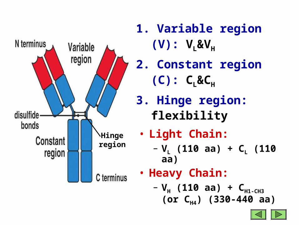
1. Variable region (V): VL&VH
2. Constant region (C): CL&CH
3. Hinge region: flexibility
Hingeregion
• Light Chain: – VL (110 aa) + CL (110 aa)
• Heavy Chain: – VH (110 aa) + CH1-CH3 (or C
H4) (330-440 aa)

Structural Regions

CH3

CH3
CH2

CH3
CH2
CH1
CH3
CH2
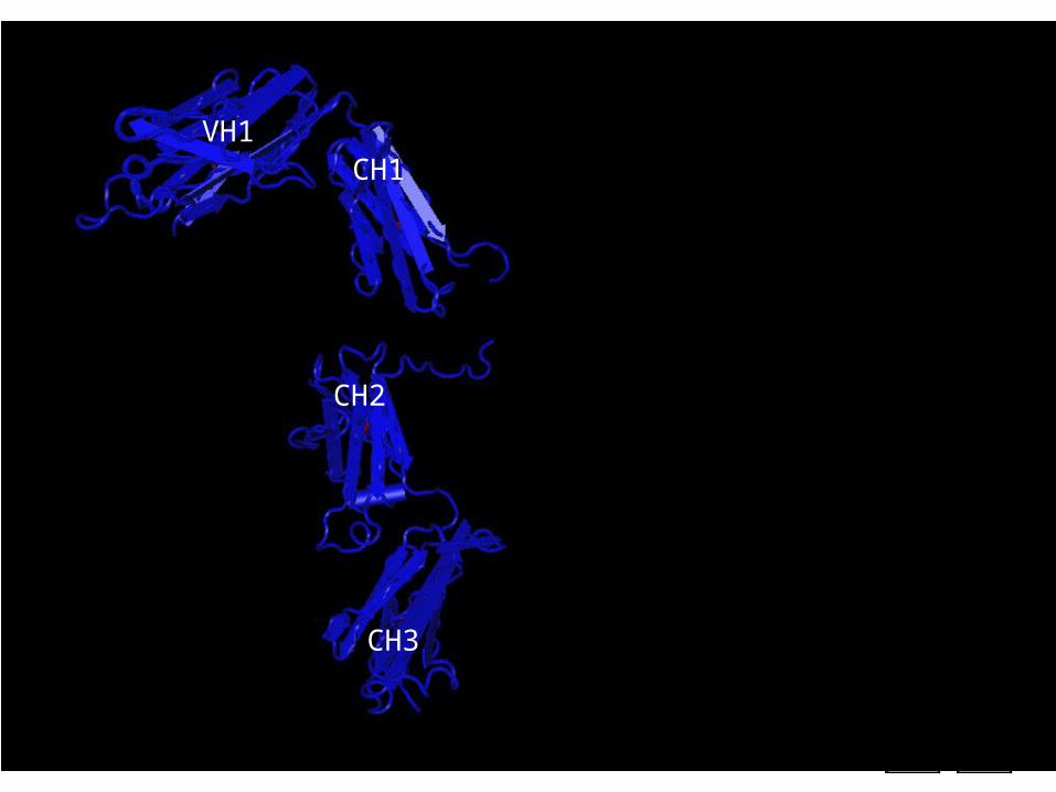
CH1VH1

CH3
CH2
CH1VH1
VL
CH3
CH2
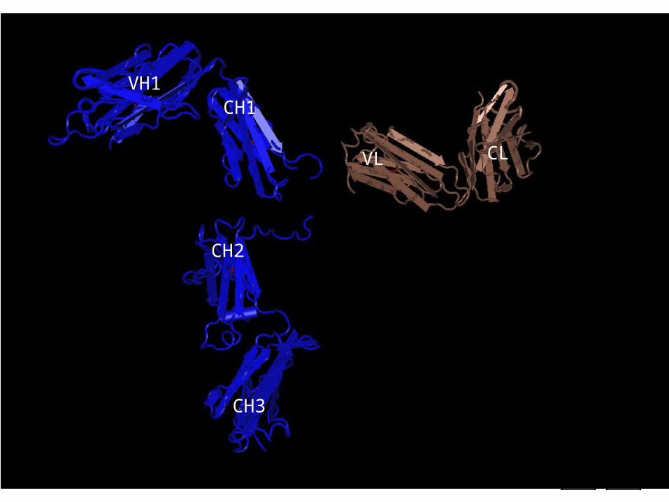
CH1VH1
CLVL

CH3
CH2
CH1VH1
CLVL

Hinge
CH3
CH2
CH1VH1
VLCL
Elbow
Enzymatic Digestion Products of Immunoglobulins

Immunoglobulin Fragments: Structure/Function Relationships
• Fab– Ag binding– Valence = 1– Specificity deter
mined by VH and VL
Papain
Fc
Fab
• Fc ( crystallizable)– Effector functions
Papain is a sulfhydryl protease from Carica
papaya latex


F(ab’)2: a single fragment composed of two Fab-like fragments, it is divalent.

Hydrolytic fragment of Immunoglobulin

Ag Binding
Complement Binding Site
Placental Transfer
Binding to Fc Receptors
• Functions of the domains on Ig

• Hypervariable region:
also called Complementarity Determining Regions (CDRs)

Antigenic determinant or epitope: The structure recognized by an antibody
Concept: Epitopes can bind in pockets or grooves or on extended surfaces in the binding site of antibodies.
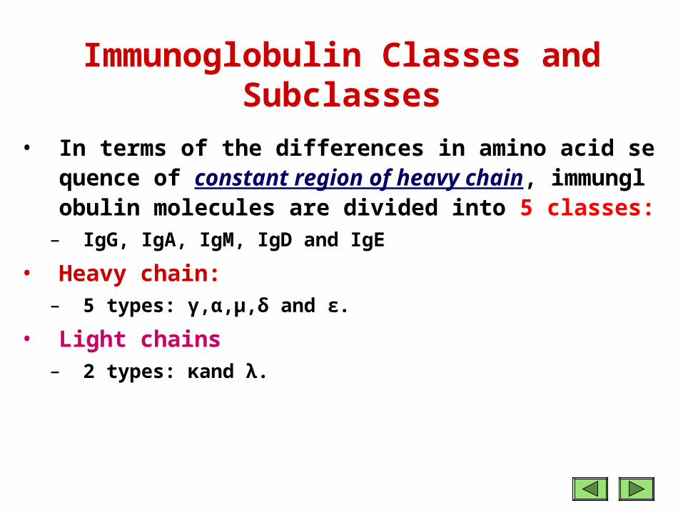
• In terms of the differences in amino acid sequence of constant region of heavy chain, immunglobulin molecules are divided into 5 classes:
– IgG, IgA, IgM, IgD and IgE• Heavy chain:
– 5 types: γ,α,μ,δ and ε.• Light chains
– 2 types: κand λ.
Immunoglobulin Classes and Subclasses

1. IgG - γ heavy chains
2. IgM - µ heavy chains
• pentamer
3. IgA - α heavy chains
• dimer
4. IgD - δ heavy chains
5. IgE - ε heavy chains
Immunoglobulin Classes of Mammals
dimer pentamermonomer


IgG
It is the most abundant class in serum, constitutes about 80% of the total serum Ig. 4 subclasses, IgG1, IgG2, IgG3, and IgG4. All IgG's are monomers. The subclasses differ in the number of disulfide bonds and length of the hinge region.
IgG1, IgG2, IgG4 IgG3

Functions of IgG
1. Major Ig in extravascular spaces.2. Placental transfer: IgG is the only class of Ig that crosses the placenta. 3. Complement activation. 4. Binding to cells - Macrophages, monocytes, PMNs (polymorphonuclear leukocyte), and some lymphocytes have Fc receptors for the Fc region of IgG.

Fc receptor
• Fc receptor: protein found on the surface of certain cells (including natural killer cells, macrophages, neutrophils, and mast cells).
• Fc receptors bind to antibodies that are attached to infected cells or invading pathogens.
• Their activity stimulates phagocytic or cytotoxic cells to destroy microbes, or infected cells by antibody-mediated phagocytosis or antibody-dependent cell-mediated cytotoxicity.
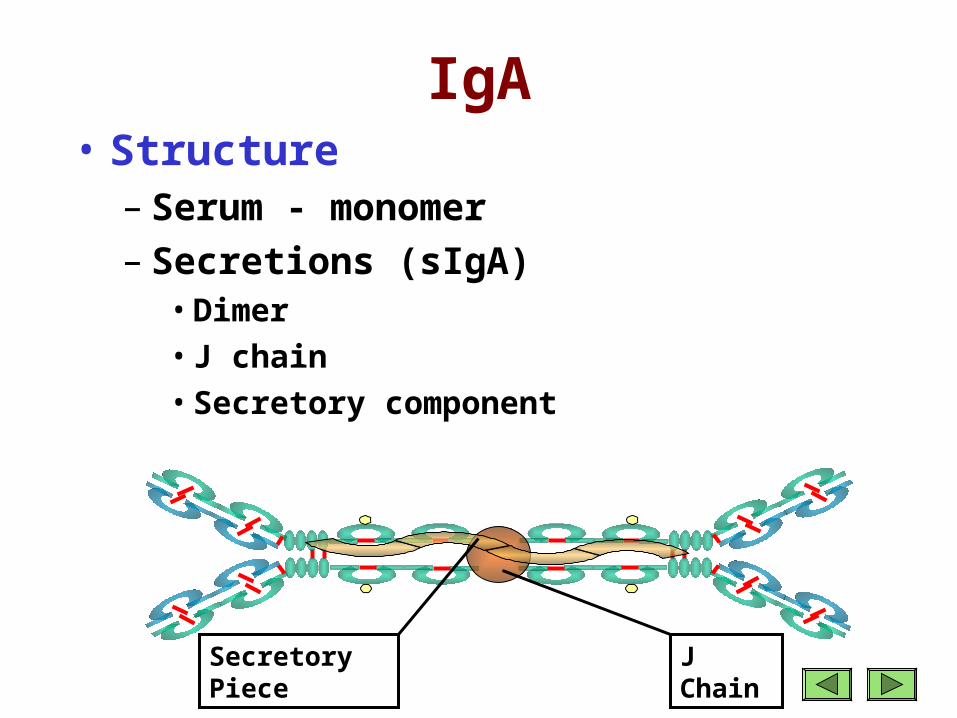
IgA• Structure
– Serum - monomer– Secretions (sIgA)
• Dimer • J chain• Secretory component
J ChainSecretory Piece

IgA• Function
– 2nd highest serum Ig– Major secretory Ig (Mucosal or Local Immunity)
• Found in the body secretions: tears, breast milk, saliva, mucus of the bronchial, genitourinary, and digestive tract
• IgA is the most predominant antibody in the colostrum, the initial secretion from the mother’ breast after a baby is born.
– Does not activate complement (unless aggregated)
– Binds to Fc receptors on some cells

IgM
Structure• The largest Ig compo
sed of 5 Y-shaped units held together by a J polypeptide chain.
1. Pentamer 2. Extra domain (CH4)3. J chain

Functions of IgM
1. 3rd highest serum Ig. 2. IgM cannot traverse blood vessels, hence it i
s restricted to the blood stream. 3. 1st Ig produced in a primary response to an
antigen and serve as first line of defense.4. a good complement activation Ig. Thus, I
gM is the most effective in leading to the lysis of microorganisms.
5. Binds to Fc receptors.

IgD
• Structure– Monomer– Tail piece
Tail Piece
IgD
• Properties– 4th highest serum Ig, its role in serum
uncertain. – B cell surface Ig.– Does not bind complement.

IgE
• Structure– Monomer– Extra domain
(CH4)
CH4

IgE
• Function– Least common serum Ig– Allergic reactions Binds to basophils and mast cells (Does
not require Ag binding)– Parasitic infections (Helminths)
• Binds to Fc receptor on eosinophils– no complement activation.

Points• Plasma vs. serum• Components and functions of Plasma• Plasma proteins
– Albumin: Function(Transport, Maintain of osmotic pressure), Clinical aspects
– Globulins• α1–Antitrypsin, α2 –Macroglobulin, Hepatoglobin (Hp),
Ceruloplasmin, Transferrin (Tf)
– Acute Phase Proteins(APP), C-reactive protein (CRP)
• Immunoglobulin(Ig)/antibody(Ab)– General Functions, Forms, Basic Structure, Enzymatic Dig
estion Products (Fab,Fc, F(ab’)2)
– Classes: IgG, IgM, IgA, IgD, IgE (Structure and Function)