Embed Size (px)
Citation preview
8/2/2019 Plosone Me
http://slidepdf.com/reader/full/plosone-me 1/11
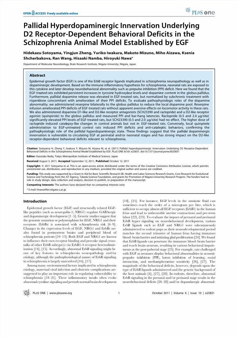
Pallidal Hyperdopaminergic Innervation UnderlyingD2 Receptor-Dependent Behavioral Deficits in theSchizophrenia Animal Model Established by EGF
Hidekazu Sotoyama, Yingjun Zheng, Yuriko Iwakura, Makoto Mizuno, Miho Aizawa, Ksenia
Shcherbakova, Ran Wang, Hisaaki Namba, Hiroyuki Nawa*Department of Molecular Neurobiology, Brain Research Institute, Niigata University, Niigata, Japan
Abstract
Epidermal growth factor (EGF) is one of the ErbB receptor ligands implicated in schizophrenia neuropathology as well as indopaminergic development. Based on the immune inflammatory hypothesis for schizophrenia, neonatal rats are exposed tothis cytokine and later develop neurobehavioral abnormality such as prepulse inhibition (PPI) deficit. Here we found that theEGF-treated rats exhibited persistent increases in tyrosine hydroxylase levels and dopamine content in the globus pallidus.Furthermore, pallidal dopamine release was elevated in EGF-treated rats, but normalized by subchronic treatment withrisperidone concomitant with amelioration of their PPI deficits. To evaluate pathophysiologic roles of the dopamineabnormality, we administered reserpine bilaterally to the globus pallidus to reduce the local dopamine pool. Reserpineinfusion ameliorated PPI deficits of EGF-treated rats without apparent aversive effects on locomotor activity in these rats.We also administered dopamine D1-like and D2-like receptor antagonists (SCH23390 and raclopride) and a D2-like receptoragonist (quinpirole) to the globus pallidus and measured PPI and bar-hang latencies. Raclopride (0.5 and 2.0 mg/site)significantly elevated PPI levels of EGF-treated rats, but SCH23390 (0.5 and 2.0 mg/site) had no effect. The higher dose of raclopride induced catalepsy-like changes in control animals but not in EGF-treated rats. Conversely, local quinpiroleadministration to EGF-untreated control rats induced PPI deficits and anti-cataleptic behaviors, confirming thepathophysiologic role of the pallidal hyperdopaminergic state. These findings suggest that the pallidal dopaminergicinnervation is vulnerable to circulating EGF at perinatal and/or neonatal stages and has strong impact on the D2-likereceptor-dependent behavioral deficits relevant to schizophrenia.
Citation: Sotoyama H, Zheng Y, Iwakura Y, Mizuno M, Aizawa M, et al. (2011) Pallidal Hyperdopaminergic Innervation Underlying D2 Receptor-DependentBehavioral Deficits in the Schizophrenia Animal Model Established by EGF. PLoS ONE 6(10): e25831. doi:10.1371/journal.pone.0025831
Editor: Kazutaka Ikeda, Tokyo Metropolitan Institute of Medical Science, Japan
Received August 3, 2011; Accepted September 12, 2011; Published October 12, 2011
Copyright: ß 2011 Sotoyama et al. This is an open-access article distributed under the terms of the Creative Commons Attribution License, which permitsunrestricted use, distribution, and reproduction in any medium, provided the original author and source are credited.
Funding: This study was supported by a Grant-in-Aid for Basic Scientific Research (B), Health and Labor Sciences Research Grants, Core Research for Evolutional
Science and Technology from the JST Agency, Takeda Science Foundation, and grants for Promotion of Niigata University Research Projects. The funders had norole in study design, data collection and analysis, decision to publish, or preparation of the manuscript.
Competing Interests: The authors have declared that no competing interests exist.
* E-mail: [email protected]
Introduction
Epidermal growth factor (EGF) and structurally related EGF-
like peptides (such as neuregulin-1; NRG1) regulate GABAergic
and dopaminergic development [1–5]. Genetic studies suggest that
the genomic mutation or polymorphism for EGF, NRG1 and their
receptors (ErbBs) is associated with schizophrenia risk [6–9].
Changes in the expression levels of EGF, NRG1 and ErbBs are
also found in postmortem brains and peripheral blood of
schizophrenia patients [10–13]. Both EGF and NRG1 are knownto influence their own receptor binding and provoke signal cross-
talks of other ErbB subtype(s) via ErbB1-4 receptor heterodimir-
ization [14], [15]. Accordingly, abnormal ErbB signaling might be
one of key features in schizophrenia neuropathology and/or
etiology, although the pathophysiological nature of ErbB signaling
in schizophrenia is largely unresolved [16], [17].
Among many environmental factors implicated in schizophrenia
etiology, maternal viral infection and obstetric complications are
suggested to play an important role in regulating vulnerability to
schizophrenia [18–21]. These inflammatory insults often evoke
abnormal cytokine signaling and perturb normal brain development
[18], [21]. For instance, EGF levels in the amniotic fluid can
sometimes reach the order of a microgram per liter, which is
sufficient to occupy almost all EGF receptors (ErbB1) in the human
fetus and lead to unfavorable uterine contractions and pre-term
labor [22], [23]. To evaluate the impact of prenatal and perinatalErbB hyper–signaling on neurobehavioral development, various
ErbB ligands such as EGF and NRG1 were subcutaneously
administered to rodent pups as their neurodevelopmental period
matches the second trimester of human fetus having immature
blood–brain barrier and initiating glial proliferation [24]. We foundthat ErbB ligands can penetrate the immature blood–brain barrier
and reach brain neurons, resulting in various behavioral impair-
ments at the post-pubertal stage [25]. For example, rats challenged
with EGF as neonates display behavioral abnormalities in acousticprepulse inhibition (PPI), latent inhibition of learning, social
interaction, and methamphetamine sensitivity [26], [27]. The
magnitude of the behavioral deficits, however, depends upon the
type of ErbB ligands administered and the genetic background of
the host animals [4], [27], [28]. In rodents, therefore, abnormal
ErbB signaling in the prenatal and/or perinatal stage results in the
neurobehavioral deficits [26–28] and/or dopaminergic abnormal-
PLoS ONE | www.plosone.org 1 October 2011 | Volume 6 | Issue 10 | e25831

8/2/2019 Plosone Me
http://slidepdf.com/reader/full/plosone-me 2/11
ities relevant to schizophrenia [4], although the neuropathologicmechanism underlying the individual deficits remains to be
clarified.
Here, we prepared the animal model for schizophrenia bysubcutaneously injecting EGF to newborn rats and studied the
mechanism for their PPI abnormality. Based upon our latestfinding on the neurotrophic interactions between EGF signaling
and dopamine [5], [29], [30], we mainly characterized dopami-
nergic neuropathology of these rats using neurochemical andanatomical approaches. Furthermore we explored the pathologic
mechanism underlying their behavioral deficits by pharmacolog-
ically manipulating local dopamine transmission.
Results
Upregulation of dopaminergic markers in the globuspallidus continuing until adulthood
At the early postnatal stage of rats, EGF is verified to reach the
brain through the immature blood-brain barrier and promote
phenotypic development of midbrain dopaminergic neurons,
leading to neurobehavioral abnormalities relevant to schizophre-
nia [26], [31]. However, there was a question of whether the
neurotrophic influences on dopaminergic neurons can continue
until the post-pubertal stage when rats develop the neurobehav-ioral abnormalities [26]. To address this question, here, wequantitated and compared protein levels of tyrosine hydroxylase
(TH; a rate-limiting enzyme for dopamine synthesis) in variousbrain regions of EGF-treated and control rats at their adult stage
using enzyme immunoassay (ELISA) [32], [33] (Fig. 1A). In the
globus pallidus, TH protein levels were significantly higher in
EGF-treated rats than in control rats ( P = 0.035). There were no
significant differences in the other brain regions in this ELISA
(Fig. 1A). In the globus pallidus, there was concomitant elevation
of tissue dopamine content ( P = 0.030) and its metabolites,
DOPAC ( P = 0.014) (Fig. 1B, C) and HVA ( P = 0.019, data not
shown). We confirmed the pallidal hyperdopaminergic state by
immunoblotting; protein levels of TH and VMAT2, but not those
of DbH (a maker for noradrenergic neurons), were significantly
elevated in EGF-treated rats at the adult stage (Fig. 1D).
To determine whether the TH increase in the globus pallidus
accompanies morphological alterations, we investigated the
neuroanatomical features of dopaminergic fibers and terminals
in EGF-treated rats (Fig. 2). In contrast to the neurochemical
alterations, the anatomical difference in TH immunoreactivity was
less marked. The density of TH-immunoreactive fibers was higherin EGF-treated rats only in the lateral area of medial and caudal
globus pallidus neighboring the striatum. In rostral globus pallidus,however, we failed to detect the difference (data not shown).
Antipsychotic effects on extracellular dopamine levels inthe globus pallidus of EGF-treated rats
Sensorimotor gating involves pallidal function and dopaminer-
gic transmission [34]. We next monitored extracellular dopaminelevels in the globus pallidus using a microdialysis technique and
estimated a link between local dopamine concentration and
sensorimotor gating (Fig. 3A). In agreement with the previous
results [26], neonatal EGF treatment reduced PPI scores at the
adult stage and an atypical antipsychotic agent ameliorated the
PPI reduction (Fig. 3B). EGF-treated animals also exhibited an
increase in pulse alone-startle amplitude compared with control
animals but not with risperidone-treated animals [F(2,33)= 3.61,
P = 0.038] (Fig. 3C). We found that basal extracellular dopamine
levels (1.5260.19 nM) in pallidal dialysates from EGF-treated rats
were significantly elevated than in those from controls
(0.8560.06 nM; P,0.001, N = 11–13) but decreased by subchro-
nic treatment with risperidone (0.6760.08 nM; P,0.001, N = 11)
[F(2,33) = 14.8, P,0.001] (Fig. 3D). Following potassium depo-
larization stimuli, extracellular dopamine levels were also elevated
in EGF-treated rats but not in risperidone-administered EGF-
treated rats [F(2,33) = 5.07, P = 0.008, N = 11–13]. The release
difference at both the basal and evoked states suggests that EGF
altered the capacity of net dopamine release but not the
excitability of dopamine terminals. Of note, the overall changes
in extracellular dopamine levels exhibited an opposite trend to that
of the changes in PPI levels in the above experiments. These
Figure 1. Effects of neonatal EGF challenge on tyrosinehydroxylase and dopamine metabolism at the adult stage.EGF or cytochrome c (control) was daily injected (s.c.) into neonatal rats
from PND2 to PND10, and rats were bred until adulthood (PND 56). (A)Levels of tyrosine hydroxylase (TH) immunoreactivity in brain tissuehomogenates were determined by ELISA and presented as a ratio of control levels (mean 6 SEM). Tissue contents of dopamine (B) and itsmetabolite DOPAC (C) were measured by HPLC. (D) Tissue lysates fromthe globus pallidus of EGF rats and controls (8 weeks old, N = 4) weresubjected to immunoblotting for antibodies directed against TH,vesicular monoamine transporter 2 (VMAT2), dopamine beta hydrox-ylase (DbH), dopamine D1 receptor (D1R), dopamine D2 receptor (D2R),and b-actin (an internal control). Immunoreactivity was measured bydensitometric analysis and its percentage ratio to that of control ratswas calculated (mean 6 SEM). Abbreviations; medial prefrontal cortex(prelimbic cortex; mPFC), striatum (STR), nucleus accumbens (NAC),cingulate cortex (Cg), globus pallidus (GP), amygdala (AMG), hypothal-amus (Hyp), subthalamic nucleus (STN), and substantia nigra (SN).*P,0.05, **P,0.01 by two-tailed t-test.doi:10.1371/journal.pone.0025831.g001
Pallidal Dopamine Increase and Behavioral Deficits
PLoS ONE | www.plosone.org 2 October 2011 | Volume 6 | Issue 10 | e25831

8/2/2019 Plosone Me
http://slidepdf.com/reader/full/plosone-me 3/11
results raise the hypothesis that enhanced pallidal dopamine
release might be associated with the PPI decrease in this model.
Local reserpine administration to the globus pallidusameliorates prepulse inhibition deficits
To confirm our hypothesis, we administered reserpine (an
inhibitor of VMAT) or vehicle to reduce local dopamine content
in synaptic vesicles and measured prepulse inhibition (Fig. 4A). A
three-way ANOVA with EGF treatment and reserpine challenge
as the between-subjects factors and prepulse intensity as thewithin-subjects factor revealed a significant interaction between
EGF treatment and reserpine challenge [ F (1,29) = 7.81, p = 0.009].
Repeated Fisher’s LSD revealed that reserpine challenge signifi-
cantly improved PPI scores in EGF rats ( P = 0.012). In contrast,
reserpine did not alter PPI scores of control rats or pulse-alone
startles of both groups of rats. Following the behavior tests, we
monitored local dopamine contents to ascertain the regional
specificity of the reserpine action (Fig. 4B). The dose of reserpine
reduced the dopamine pool in the globus pallidus of EGF rats
(P,0.001) but not in the neighboring striatum of EGF rats or in
these regions of control rats.
There was a significant difference in pulse-alone startle amplitude
between EGF-treated and control rats [ F (1,28) = 16.4, P ,0.001 for
EGF]. Our current design to evaluate reserpine action on %PPI
might be inappropriate due to the basal difference in pulse-alone
startle amplitudes of EGF and control rats [35]. We prepared
another set of animals and gave the lower intensity of tone stimuli
(110 dB for main pulses) to EGF-treated rats (Fig. 4C, D). The PPI
difference between EGF and control groups as well as the
risperidone effect still remained, even though there was no significant
difference in the amplitudes of pulse-alone startles among groups.
Figure 2. Immunohistochemical analysis of tyrosine hydroxy-lase-positive fibers and terminals in the globus pallidus.Coronal sections of the striatum containing medial and caudal globuspallidus (1.2 and 1.6 mm posterior from the bregma) were preparedfrom control and EGF-treated rats and immunostained with anti-THantibody (N = 3–4 rats per group). TH-immunoreactive fibers in medial(A, B) and caudal (C, D) globus pallidus of control (A, C) and EGF rats (B,D) are shown. A lateral area of the globus pallidus is marked with a
window, enlarged 2-fold, and presented in the top-right corner of eachpicture. ST; striatum, GP; globus pallidus, ic; internal capsule. Scale bar,500 mm.doi:10.1371/journal.pone.0025831.g002
Figure 3. Pallidal dopamine release enhanced in EGF-treated
rats and normalized with risperidone. Neonatal rats were treatedwith EGF or cytochrome c (control) as described in Figure 1. Risperidone(1 mg/kg, i.p.) was administered to some of the EGF-treated rats for 14days at the adult stage. (A) The location of dialysis probe was examinedand is shown in rat brain atlas. Six rats were excluded for incorrect probeplacement. The digit represents the distance from the bregma. (B) PPIlevels were monitored in control, EGF-treated and EGF+risperidone-treated animals following microdialysis. (C) Pulse-alone startle responsesand PPI levels were monitored in control, EGF-treated and EGF+risper-idone-treated animals following microdialysis. (D) Basal concentrations of dopamine in dialysates were monitored for 150 min, dopamine releasewas evoked by perfusion of 80 mM KCl over 60 min (solid bar), and thenmonitored over 150 min. Data represent dopamine concentrations in 30-min fractions (nM, mean 6 SEM, N = 11–13 rats per group). There was asignificant interaction between time and dopamine release[F(12,198) = 2.72, P = 0.002]. *P,0.05, ***P,0.001, compared withcontrols and ++P,0.01, compared with EGF-treated rats by Fisher’s LSD.
doi:10.1371/journal.pone.0025831.g003
Pallidal Dopamine Increase and Behavioral Deficits
PLoS ONE | www.plosone.org 3 October 2011 | Volume 6 | Issue 10 | e25831

8/2/2019 Plosone Me
http://slidepdf.com/reader/full/plosone-me 4/11
To control the potential aversive effects of reserpine injection, weassessed the exploratory motor behaviors of reserpine-injected
animals (Fig. 5A, B). We found no significant influence of reserpine
on horizontal movement. As EGF-treated rats are known to exhibit
a decrease in social interaction scores [26], we also evaluated the
effects of reserpine on social behaviors following the above
exploratory locomotor test (Fig. 5C). Two-way ANOVA for sniffing
duration revealed a significant main effect of EGF [F(1,30) = 17.8,P,0.001] but no main effect of reserpine or no interaction between
EGF and reserpine. The same statistical conclusion was drawn for
sniffing counts as well. These results indicate that reserpine-induced
pallidal dopamine reduction affected PPI levels of EGF rats, but did
not influence their motor function or deficits in social behaviors.
Effects of pallidal dopamine D2-like receptor blockadeand activation on prepulse inhibition
To test the possibility that the reserpine effects on PPI might
result from its influences on the noradrenergic or serotonergic
systems, we manipulated local dopaminergic transmission using
dopamine receptor antagonists. We bilaterally administered a
dopamine D1-like receptor antagonist, SCH23390 (0.5 mg and
2 mg per site), or a dopamine D2-like receptor antagonist,
raclopride (0.5 mg a n d 2 mg per site), to the globus pallidus
(Fig. 6A). SCH23390 failed to affect PPI scores at any dose in both
groups of rats (Fig. 6B). In contrast, raclopride had differential
effects in EGF and control rats [F(2,43) = 3.91 for EGF6raclo-
pride dose, P = 0.028] (Fig. 6C). Post-hoc testing revealed thatraclopride injection significantly ameliorated the PPI deficits of
EGF rats ( P = 0.019 and 0.043 for 0.5 mg and 2 mg raclopride,
respectively) but not affected PPI levels of control rats. Similar to
the results of the reserpine experiment, vehicle-treated control rats
failed to react to the D2-like receptor antagonist at any dose.
To address whether a hyperdopaminergic state in the globus
pallidus is sufficient to induce PPI deficits, we examined the effects
of local stimulation of D2-like receptors. The bilateral challenge of
naı ve rats with quinpirole, a D2-like receptor agonist (5 mg per
site), markedly disrupted PPI [F(1,16) = 6.50, P = 0.022] without
altering pulse-alone startle (Fig. 7). These results support our
Figure 4. Effects of pallidal reserpine infusion on prepulseinhibition. (A) Startle responses of EGF and control (CON) rats weretriggered with 120-dB tone pulse and PPI scores were measured with75-, 80-, and 85-dB prepulse stimuli 120 min after local vehicle orreserpine infusion to both hemispheres of the globus pallidus. Pulse-alone startle responses to a 120-dB tone were measured in arbitraryunits and are shown in the inset. (C) To match pulse-alone startleresponses between EGF and control rats, 110-dB and 120-dB pulseswere given to EGF and control rats, respectively, as shown in the insert.
Following PPI test in (A) and (C), dopamine content was measured inthe globus pallidus (GP) and neighboring striatum and shown in (B) and(D), respectively. Bars indicate mean 6 SEM (N= 8–9 each). *P,0.05,**P,0.01, ***P,0.001, compared with vehicle-infused control rats;++P,0.01, +++P,0.001, compared with vehicle-infused EGF rats byFisher’s LSD.doi:10.1371/journal.pone.0025831.g004
Figure 5. Influences of pallidal reserpine infusion on locomotoractivity and social interaction. EGF and control (CON) rats receivedlocal reserpine- or vehicle-infusion to both hemispheres of the GP. (A)Cannula placement was confirmed in fixed brains, and two out of 36rats were excluded from the final data analysis due to incorrect cannulaplacement. (B) Two hours after pallidal infusion, rats were placed in the
automated activity monitoring chamber for 60 min. Data representhorizontal movement (cm) for every 5 min (mean 6 SEM, N = 7–9 foreach group). (C) Following locomotor test, an unfamiliar male rat wasplaced in the same chamber. The number and duration of sniffingbehaviors of operated rats were counted for 10 min. Bars indicate mean6 SEM (N = 8–9 for each group). **p,0.01, ***p,0.001 by Fisher’s LSD,compared with vehicle-infused controls.doi:10.1371/journal.pone.0025831.g005
Pallidal Dopamine Increase and Behavioral Deficits
PLoS ONE | www.plosone.org 4 October 2011 | Volume 6 | Issue 10 | e25831

8/2/2019 Plosone Me
http://slidepdf.com/reader/full/plosone-me 5/11
argument that elevated dopamine release in the globus pallidus is
responsible for the PPI deficits.
Difference in cataleptic actions of a D2-like receptorantagonist on EGF-treated and control rats
Because dopamine D2-like receptor blockade is known to induce
cataleptic behaviors [36], we used the bar-hang immobility test to
estimate the effects of the antagonist on catalepsy scores (Fig. 8A).
Prior to drug administration, EGF-treated and control rats were
subjected to the first session of the bar-hang test. There was asignificant basal difference in bar-hang latency [F(1,43)= 19.0,
P,0.001 for EGF] (Fig. 8B). Pallidal infusion of the D2-like receptor
antagonist produced differential effects on EGF and control rats
[F(2,44) =9.07, P,0.001 for raclopride dose6EGF]. The higher
dose of raclopride significantly increased bar-hang latency in control
rats (P,0.001) but not in EGF rats. Thus, EGF rats appear to be
insensitive to the given doses of the D2-like receptor blocker in this
test. These results rule out the possibility that the observed
raclopride effects on PPI in EGF-treated rats might reflect its
cataleptic action. Moreover, this experiment revealed the lower
sensitivity of EGF-treated rats to pallidal D2-like receptor blockade.
Of note, the pallidal infusion of the D2-like receptor agonist
quinpirole to naı ve rats significantly diminished the bar-hang
latency, compared with the latency before injection [F(2,24) = 4.54,
P = 0.021] (Fig.8C). The bar-hang latency of quinpirole-infused rats
was significantly shorter than that of vehicle-infused controls
(P,0.001) and indistinguishable from that of EGF-treated rats.
All these results suggest that pallidal dopaminergic signals negatively
regulate bar-hang latency. Therefore, the shorter bar-hung latency
of EGF-treated rats supports our argument that pallidal dopamine
function was up-regulated in these animals.
Discussion
In the present investigation, we attempted to address the
question of how EGF challenge at the perinatal stage alters later
Figure 6. Effects of pallidal infusion of dopamine receptorantagonists on PPI deficits. Different doses of the D1-like receptorantagonist [SCH23390; 0 mg (vehicle), 0.5 mg or 2 mg per site] or the D2-like receptor antagonist [raclopride; 0 mg (vehicle alone), 0.5 mg or 2 mgper site] were administered to both hemispheres of the globus pallidusof EGF and control rats. (A) Cannula placement was confirmed in fixedbrains, and four out of 69 rats were excluded from the final data analysisdue to incorrect cannula placement. Rats receiving SCH23390 (B) orraclopride (C) were subjected to PPI test with 75-, 80- and 85-dBprepulse stimuli combined with a 120-dB startle tone. Pulse-alonestartle responses to a 120-dB tone were measured in arbitrary units andare shown in the inset. Bars indicate mean 6 SEM (N=8–9 for eachgroup). Data of rats receiving vehicle alone (control) were shared in (B)and (C). *P,0.05, **P,0.01, compared with vehicle-infused controls;+P,0.05, ++P,0.01, compared with vehicle-infused EGF rats by Fisher’sLSD.doi:10.1371/journal.pone.0025831.g006
Figure 7. Effects of pallidal infusion of a dopamine D2-likeagonist on PPI. A dopamine D2-like agonist, quinpirole (QUIN) (5 mgper site), or vehicle was administered to both hemispheres of the GP of naıve rats. (A) Cannula placement was confirmed in fixed brains, andone out of 18 rats was excluded from the final data analysis due toincorrect cannula placement. Fifteen minutes after quinpirole injection,pulse-alone startles (B) and PPI scores (C) were measured with 75-, 80-and 85-dB prepulse stimuli combined with a 120-dB startle tone. Barsindicate mean 6 SEM (n=8 or 9 each). *p,0.05, **p,0.01, comparedwith vehicle-infused controls by Fisher LSD.doi:10.1371/journal.pone.0025831.g007
Pallidal Dopamine Increase and Behavioral Deficits
PLoS ONE | www.plosone.org 5 October 2011 | Volume 6 | Issue 10 | e25831

8/2/2019 Plosone Me
http://slidepdf.com/reader/full/plosone-me 6/11
dopaminergic neurotransmission and dopamine-associated behav-
ioral traits. The present analyses of this model provided us with the
following information: 1) The hyperdopaminergic phenotype wasmaintained in the globus pallidus until adulthood. 2) The amounts
of pallidal dopamine release were elevated at the adult stage of
EGF-treated rats and normalized with antipsychotic treatment. 3)
The reserpine-driven reduction in pallidal dopamine pool
ameliorated the PPI deficits of EGF-treated rats. 4) The D2-like
receptor blocker also normalized PPI levels of EGF-treated rats
without affecting the catalepsy index. 5) Conversely, local
administration of the D2-like receptor agonist to naı ve rats caused
deficits in PPI and bar-hang latency similar to that seen in EGF-
treated rat. These results reveal that EGF exposure to rat pups
produces persistent neurotrophic influences on the nigropallidal
dopamine neurons and their functions. Thus, we postulate that the
behavioral deficits of this EGF model, in part, can be ascribed to
the D2 receptor-dependent hyperdopaminergic states of this basal
ganglia circuit.
Trophic actions and selectivity of epidermal growthfactor peripherally administered
EGF and other ErbB ligands (such as neuregulin-1) are
neurotrophic not only for midbrain dopaminergic neurons butalso for GABAergic neurons and glial cells [1–5], [37], [38].
Although we have been investigating the phenotypic influences of
peripheral EGF challenges on developing GABAergic and glial
cells, these effects did not persist until the adult stage [2], [26],
[31]. As such, the hyperdopaminergic influence found in the
present investigation is the sole phenotypic change that we have
detected in the adult stage of EGF-treated rats. Similar to this
study, we find long-lasting influences of neonatal neuregulin-1
challenges on the dopamine system [4]. With the given wide-
spread action of these ErbB ligands, however, we cannot rule out
the possibility that undetected changes in GABAergic or glial
phenotype or function still remain in this model [2], [5], [37], [38].
Individual dopaminergic systems of mesostriatal, mesolimbic,
and mesofrontocortical pathways appear to be differentiallyregulated by individual neurotrophic factors [5], [37–39]. Among
these neurons, a subset of dopaminergic neurons in the ventral tier
of the substantia nigra pars compacta mainly express the EGF
receptor (ErbB1), which constitute of highly branched dopami-
nergic neurons in the mesostriatal and pallidal pathways [5], [40],
while the neuregulin-1 receptors (ErbB4) are most enriched in
mesofrontocortical dopamine neurons [37]. Therefore, it is likely
that mesostriatal dopaminergic population responded to exoge-
nous EGF and contributed to the pallidal hyperdopaminergic
states of the present animal model. In contrast to the neurochem-
ical changes, the number of dopamine terminals in the globus
pallidus was less remarkable. In this context, the neurotrophic
feature of EGF on dopamine neurons remains to be fully
characterized; its effects on cell survival, phenotypic enhancement,
or terminal arborization. Our preliminary study failed to detectapparent difference in the number of dopamine neurons (data not
shown).
In contrast to the present EGF model, the mice treated with
neuregulin-1 as neonates, in which dopaminergic influences are
most pronounced in the prelimbic cortex, exhibit less marked
deficits in PPI but more severe impairments in social behaviors [4],
[41]. The difference of the behavioral traits between the two
models might reflect the distinct neurotrophic actions of EGF and
neuregulin-1 on these cell subpopulations.
Neuropathological implication of the globus pallidus forschizophrenia and its animal model
The globus pallidus is part of the indirect pathway of the basal
ganglia circuit and receives dopaminergic innervation [42], [43].The pathway initiates from the medium spiny neurons (MSN) in
the striatum carry dopamine D2 receptors [44–46] and regulates
sensorimotor gating, motor coordination, attention, learning and
antipsychotic pharmacology [47–51]. Kodsi and Swerdlow (1995)
showed that parts of globus pallidus, the ventral and caudal
pallidum, are involved in sensorimotor gating [50]. The preceding
report does not contradict the present finding although we did not
distinguish these subregions of the globus pallidus. The dopami-
nergic abnormality in the indirect pathway represents one of the
neuropathological features of this model and contributes to its
behavioral deficits.
Figure 8. Measurement of immobility in the bar-hang testfollowing pallidal infusion of the dopamine receptor antago-nist and agonist. (A) Cannula placement was confirmed in fixed
brains, and four out of 63 rats were excluded from the final data analysisdue to incorrect cannula placement. The bar-hang test consisted of 3blocks with 20-min intervals. Vehicle, raclopride (B; 0.5 and 2.0 mg persite) or quinpirole (QUIN) (C; 10.0 mg per site) was bilaterally injectedinto the globus pallidus of EGF and control (CON) rats after block #1. Ineach session we measured the latency until one of the paws of rats wasremoved from the horizontal bar (mean 6 SEM, N=8–9 rats each).*P,0.05, **P,0.01, ***P,0.001, compared with vehicle-infused controlrats; +P,0.05, ++P,0.01, +++P,0.001, compared with the value of session #1 by Fisher’s LSD. Note: The pallidal infusion of the D2-likereceptor agonist to control rats (green star) significantly reduced thelatency.doi:10.1371/journal.pone.0025831.g008
Pallidal Dopamine Increase and Behavioral Deficits
PLoS ONE | www.plosone.org 6 October 2011 | Volume 6 | Issue 10 | e25831

8/2/2019 Plosone Me
http://slidepdf.com/reader/full/plosone-me 7/11
With respect to the antipsychotic effect of the D2 receptor
antagonism in the indirect pathway, however, several controver-
sies still remain; systemic administration of haloperidol failed to
result in clear PPI improvement in EGF-treated rats [26]. In this
context, neurobehavioral consequences of D2 receptor antagonism
may significantly differ depending upon the brain regions where
the antagonist acts [50]. Although we made the best efforts to
minimize the technical artifacts and variations of cannula
implantation, we cannot fully rule out aversive influences of themicrosurgery on behavioral testing. The regional specificity of the
drug actions should warrant future independent studies. As far as
we compared the animals in control and experimental groups that
equally received the surgery, however, the anti-dopaminergic
manipulations produced the consistent results in behavioral tests.
The inhibition of dopamine D2 receptor at presynaptic sites of
MSN fibers facilitates GABA release in the globus pallidus, leading
to motor dysfunction such as catalepsy [42], [52–55]. In
accordance with this cataleptic mechanism, our intrapallidal
infusion of the D2-like receptor antagonist to control rats resulted
in an increase in the catalepsy index while the D2-like receptor
agonist conversely decreased the catalepsy index. We observed a
similar D2-like receptor-dependency of the agonist and antagonistin the PPI paradigm. These results suggest the possibility that these
behavioral deficits of EGF-treated rats involve the hyper-activationof D2-like receptors. However, the abnormalities of the present
model in social behavior and pulse-alone startle appeared to be
distinct from the pallidal dopamine neuropathology. In this
context, EGF-treated rats should have the unrevealed structural
or phenotypic alteration(s) underlying these behavioral impair-
ments. Whether these impairments also involve the dopamine
system remains to be investigated.
Of note, alterations in pallidal morphology and abnormal
activation of this brain region are often implicated in schizophre-
nia. Brain imaging studies reveal pallidal structural abnormalities
in schizophrenia patients [56–61]. Functional imaging also
suggests an activation impairment in this brain region of patients
[62], [63]. These abnormalities might have any association with
the pallidal hyperdopaminergic states found in the present EGF
model, especially if excess dopamine exerts neurotrophic and cell
mitotic activities on pallidal cells and increases the tissue volume or
function [64].
Vulnerability of midbrain dopaminergic development toneurotrophic cytokines
Schizophrenia animal models established by maternal immune
challenges or neonatal hypoxia appear to support the dopamine
hypothesis for schizophrenia although several controversies still
remain [65–67]. For example, the animal model established by
maternal challenge with bacterial lipopolysaccharide shows a long-
lasting increase in TH levels and dopamine turnover, at least, in
the nucleus accumbens [68], [69]. Perinatal asphyxia and viral
infection also result in distinct abnormalities in the dopamine
system [70–74]. Our preceding study suggests that neuregulin-1,another ErbB receptor ligand, also induce several behavioral
abnormalities relevant to schizophrenia, when neuregulin-1 is
administered to newborn mice [4]. These findings indicate the
possibility that the phenotypic and functional abnormalities in
these immune/inflammatory models might involve EGF or other
ErbB ligand(s) acting on midbrain dopamine neurons at prenatalor perinatal stages [25].
In conclusion, the developmental regulation of midbrain
dopaminergic neurons is more vulnerable against peripheral
cytokine signals than previously thought. The present results
indicate that perinatal and potentially prenatal exposure to EGF
or EGF-related cytokines may produce crucial and persistent
impact on dopaminergic innervation and function in the indirect
pathway of the basal ganglia circuit. In light of dopamine D2
receptor antagonism, which is commonly implicated in antipsy-
chotic pharmacology, the present cytokine model may help to
elucidate its antipsychotic mechanism as well as to validate the
dopamine hypothesis for schizophrenia.
Materials and MethodsEthics statement
All of the animal experiments described were approved by the
Animal Use and Care Committee guidelines of Niigata University
(Approval No18 on April 26, 2010) and performed in accordance
with the guidelines of NIH-USA. Every effort was made to
minimize the discomfort of the animals in addition to the number
of animals used in the experiments.
AnimalsMale newborn Sprague-Dawley rats (SLC Ltd., Hamamatsu,
Japan) were housed with a dam under a 12-h light/dark cycle
(lights on 8:00 a.m.) in a plastic cage (27664456205 mm). The
rats were allowed free access to food and water. After weaning
[postnatal day (PND) 20–30], rats were separated and housed with2–3 rats per cage. Each adult animal (PND 56–94) was used in
each experiment. Naı ve Sprague-Dawley rats (all male, PND 56–
70; SLC) were also used in control experiments. Recombinant
human EGF (Higeta Shouyu Co., Chiba Japan) was dissolved insaline. EGF (0.875 mg/g) was administered subcutaneously (s.c.)
each day to half of the littermates during PND 2–10 [26]. Control
littermates received an injection of the same dose of cytochrome c
(control protein) on the same schedule. The dose of EGF used in
this study did not produce any apparent growth retardation in rats
and mice [26], [28]. Some of adult rats daily received risperidone
(1 mg/kg, i.p.; Janssen Pharmaceuticals Inc) or saline for 14 days.
Enzyme-linked immunosorbent assay (ELISA)
Rats were anesthetized with halothane, and brains wereremoved and cut into 1-mm thick slices. Using published
boundaries [75], we identified and punched out each brain region
of interest. TH levels were measured using ELISA [10]. In brief,
brain tissues were homogenized in 10 volumes of homogenization
buffer [0.1 mM phenylmethanesulfonyl fluoride, 0.1 mM ben-
zethonium chloride, 1 mM benzamidine (Sigma Chemical Co., St.
Louis, MO), and 10 mg/ml aprotinin]. Brain homogenates were
centrifuged at 140006g for 20 min at 4uC, and the supernatants
were stored at 280uC until use. Protein concentrations were
determined using a Micro BCA kit (Pierce, Rockland, IL) with
bovine serum albumin (BSA) as a standard.
Tissue homogenates or striatal lysates (standards) were loaded
into ELISA plate wells coated with mouse monoclonal anti-TH
antibody (a gift from Dr. Hatanaka and Dr. Takei). Plates were
incubated with rabbit polyclonal anti-TH antibody (Chemicon,Temecula, CA) followed by incubation with anti-rabbit IgG b-
galactosidase (1:1000, American Qualex, San Clemente, CA). The
fluorescence of the enzyme products from a reaction with 4-
methylumbelliferyl-b-D-galactoside (MUG, Sigma) was measured
using a microplate reader (COLONA electric Co., Ltd., Ibaraki,
Japan).
ImmunoblottingEach brain tissue was dissected as described above and
homogenized in 200 ml lysis buffer [2% sodium dodecylsulfate
(SDS), 10 mM Tris-HCl buffer (pH 7.4), 5 mM ethylenediamine-
Pallidal Dopamine Increase and Behavioral Deficits
PLoS ONE | www.plosone.org 7 October 2011 | Volume 6 | Issue 10 | e25831
8/2/2019 Plosone Me
http://slidepdf.com/reader/full/plosone-me 8/11
N,N,N 9,N 9-tetraacetic acid (EDTA), 10 mM NaF, 2 mM Na3VO4,
0.5 mM phenylarsine oxide], and boiled at 95uC for 5 min. After
centrifugation at 12000 rpm for 20 min, the supernatant was
harvested. The supernatant was mixed with 56 sample buffer
[0.31 M Tris-HCl (pH 6.8), 10% SDS, 50% glycerol, 25% 2-
mercaptoethanol] and boiled at 95uC for 5 min. Denatured
protein samples were subjected to 7.5% SDS-polyacrylamide gel
electrophoresis and transferred to a nitrocellulose membrane
(Schleicher and Schull, Dassel, Germany) by electrophoresis. Themembrane was probed with anti-TH (1:2000, Chemicon), anti- vesicular monoamine transporter 2 (VMAT2) (1:1000, Chemicon),
anti-dopamine-beta-hydroxylase (DbH) (1:500, Chemicon), anti-
dopamine D1 receptor (1:1000, Sigma) or anti-D2 receptor
(1:1000, Chemicon) antibodies. After washing, membrane immu-
noreactivity was detected using anti-rat, anti-rabbit, or anti-mouse
immunoglobulin antibody conjugated to horseradish peroxidase
(Jackson Immunoresearch Laboratory, West Grove, PA) followed
by a chemiluminescence reaction (ECL kit, GE Health Science
Inc., Tokyo, Japan) and exposure to X-ray films. Film images
carrying a liner range of darkness of bands were subjected to film
scanning and converted to the 8-bit digital data. Densitometric
quantification of band intensity was performed with the free
software Image J (National Institutes of Health, USA).
ImmunohistochemistryRats were anesthetized with halothane, perfused transcardially
for 7 min with phosphate-buffered saline (150 mM NaCl, 0.1 Msodium phosphate; pH 7.5) followed by 4% paraformaldehyde in
phosphate-buffered saline. Brains were removed and post-fixed in
the same fixation solution for 24 h at 4uC. Fixed brains were
immersed in 30% sucrose solution for 3–5 days, and frozen in resin
(Tissue-Tek, Sakura Finetek U.S.A. Inc. Torrance, CA). Sections
(40 mm) were cut with a cryostat (CM1510, Leica, Nussloch,
Germany). After rinsing in Tris-buffered saline [TBS; 0.1 M Tris-
HCl (pH 7.4), 150 mM NaCl] containing 0.2% Triton X-100,
sections were pretreated with 6% BSA and 0.2% Triton X-100 in
TBS and then incubated with anti-TH antibody (1:1000,
Chemicon) in TBS containing 3% BSA and 0.2% Triton X-100for 48–72 h at 4uC. After rinsing in TBS/0.2% Triton X-100three times, sections were incubated with biotinylated anti-rabbit
immunoglobulin antibody (1:200, Jackson Immunoresearch Lab-
oratory). Immunoreactivity was visualized using a Vectastain Elite
ABC kit (Vector Laboratories, Burlingame, CA) using diamino-
benzidine as a substrate.
Determination of monoamine contentsEach brain region was homogenized in 0.1 M perchloric acid
containing 0.1 mM EDTA, and 100 nM isoproterenol. After
centrifugation at 12000 rpm for 20 min, the supernatants and
pellets were harvested. Concentrations of dopamine and its
metabolites, 3,4-dihydroxyphenylacetic acid (DOPAC) and homo-
vanillic acid (HVA), in supernatants were analyzed by HPLC-
electrochemistry [4]. The mobile phase containing 50 mMtrisodium citrate (pH 3.2), 25 mM NaH2PO4, 10 mM diethyl-amine, 0.03 mM EDTA, 2.5 mM 1-octane sulfonic acid sodium
salt, 6% methanol, 1% dimethylacetamide was delivered at
0.5 mL/min. Monoamines were separated on an analytical HPLC
column (CA-50DS, 4.66150 mm, Eicom, Kyoto, Japan) and
detected with a graphite electrode (WE-3G, Eicom) to which+700 mV was applied. Data analysis was performed with a data
acquisition computer (Powerchrom, Eicom). Tissue pellets were
homogenized in 0.5 N NaOH and subjected to protein determi-
nation with a Micro BCA kit (see above). Tissue monoamine
contents were normalized with protein concentrations.
Local drug administration to the globus pallidusControl and EGF-treated rats (PND 56–70) were anesthetized
with sodium pentobarbital (65 mg/kg, i.p.). After confirming
deep anesthesia, a rat was mounted on a stereotaxic apparatus
with an incisor bar set at 3.3 mm below the interaural line. The
skull was exposed and two holes were drilled for bilateral
implantation of guide cannulae (23 G stainless-steel pipes) into
the GP. The stereotaxic coordinates were 0.9 mm anterior,
63.0 mm lateral from the bregma, and 4.5 mm below the duramater. After allowing the rat at least 10 days of recovery from
surgery, a 30-G needle connected to Teflon tubing and a
Hamilton syringe was placed 2 mm below the tip of the guide
cannula. The drug (0.5 ml) was injected over a period of 30 sec,
and the needle left in place for an additional 30 sec. Rats were
placed in their home cage for 5–15 min, to allow for local
diffusion of the drug; the rats were then subjected to behavioral
tests (see below). When rats received reserpine, rats were placed
in their home cage for 120 min to deplete the local dopamine
pool. The cannula position was confirmed after the completion of
the behavioral tests (see below).
Reserpine (Daiichi Sankyo Pharmaceutical Inc., Tokyo, Japan)
was dissolved in phosphate-buffered solution (pH 4.0) containing
3 mg/mL DL-methionine and 70 mg/mL propylene glycol.
Conventional dopamine receptor ligands, SCH23390, raclopride
and quinpirole, were all obtained from Sigma and dissolved in
10% dimethyl sulfoxide (DMSO) in saline (vehicle).
MicrodialysisRats were anesthetized with sodium pentobarbital (65 mg/kg
i.p.) and mounted in a stereotaxic apparatus. The skull was
exposed and a hole was drilled for unilateral implantation of a
guide cannula (AG-8, Eicom) into the GP. The stereotaxic
coordinates were 0.9 mm anterior; 3.0 mm lateral from the
bregma, and 4.8 mm below the dura mater. After allowing the rat
at least 10 days of recovery from surgery microdialysis experiments
were performed.
The microdialysis probe (2 mm active area, A-I-8-02, Eicom)
was connected to Teflon tubing (0.65 mm o.d., 0.12 mm i.d.;
Bioanalytical Systems Inc., West Lafayette, IN). The rat was
perfused with artificial cerebrospinal fluid (pH 7.0) containing
147 mM NaCl, 2.7 mM KCl, 1.2 mM CaCl2, and 0.5 mM
MgCl2 at a flow rate of 0.7 mL/min. Dialysate was discarded to
obtain a steady state for at least 18 h after implantation of the
probe, and then dialysate samples were collected every 30 min.
The first five fractions were collected to determine basal levels of
dopamine. The perfusion medium was switched to the medium
containing a high concentration of potassium (80 mM KCl,
69.7 mM NaCl, 1.2 mM CaCl2, 0.5 mM MgCl2; pH 7) for
60 min (for two fractions). The perfusion medium was then
switched back to the original medium and five fractions were
additionally collected. In all, total 12 fractions were collected.
Dopamine in the dialysates was determined by HPLC withelectrochemical detection. The mobile phase containing 48 mM
citric acid, 24 mM sodium acetate, 10 mM NaCl, 0.5 mM
EDTA, 2.5 mM SDS, and 16% acetonitrile (pH 4.8) was
delivered at 50 mL/min. Dopamine was separated on an
analytical column (BDS Hypersil C18 16100 mm, Thermo
Fisher Scientific, Yokohama, Japan) and detected with a 3 mm
glassy carbon electrode detector (Unijet flow cell; Bioanalytical
Systems Inc.) to which +550 mV was applied. Data analysis was
performed with the analysis software (Epsilon LC; Bioanalytical
Systems Inc.). Data were not compensated with the recovery
rate.
Pallidal Dopamine Increase and Behavioral Deficits
PLoS ONE | www.plosone.org 8 October 2011 | Volume 6 | Issue 10 | e25831

8/2/2019 Plosone Me
http://slidepdf.com/reader/full/plosone-me 9/11
Confirmation of cannula positioning After local drug injection or microdialysis, rats were deeply
anesthetized with halothane and decapitated. Brains were quicklyremoved and fixed in 4% paraformaldehyde for 3 days. Fixed
brains were cut into 50-mm sections using a vibratome (Dosaka
EM Ltd., Kyoto, Japan). Each section was stained with 0.5%
cresyl violet solution. The location of a microdialysis probe or
injection needle was determined under a microscope according to
a stereotaxic atlas [75]. Animals that exhibited incorrect cannulaplacement were removed from the data analysis.
Measurement of acoustic startle and prepulse inhibitionof startle response
Acoustic startle amplitude was measured in a startle chamber
(SR-Lab Systems, San Diego Instruments, San Diego, CA) [34].
Rats were placed into a startle chamber with 70-dB background
noise. Five minutes later, the startle amplitude was recorded in a
session that included multiple trial types: (i) a 120-dB 40-ms noise
burst presented alone (pulse); (ii–iv) 40-ms 120-dB noise burst
following prepulses by 100 ms (20-ms noise burst) that were 5, 10,
and 15 dB above background noise (i.e., 75-, 80-, 85-dB prepulse,respectively); and (v) no stimulus (background noise alone). The
percentage PPI of startle responses was calculated as: 1002[(startleresponse on prepulse-pulse stimulus trials – no stimulus trials)/
(pulse-alone trials – no stimulus trials)]6100. To match the
magnitude of pulse alone startles between groups, a 120-dB noise
burst was replaced with a 110-dB noise burst in some experiments.
Analysis of locomotor activitySide effects of reserpine were estimated by monitoring
spontaneous locomotor activity under novel conditions. Reser-
pine-infused rats were placed in an open field box (45 cm
length645 cm width630 cm height, MED Associates, St. Albans,
VA, USA) under a moderate light level (200 Lx). Line crossings
and rearing counts were measured by photo-beam sensors (25 mm
intervals for horizontal axis and 150 mm for vertical axis).
Social interaction testThe index for social interaction of rats was measured according
to Futamura et al. (2003) [26]. Following the above locomotor test,
reserpine- or vehicle-infused rats were exposed to an unfamiliarmale rat that was housed in another cage, and was age, body-
weight, and gender-matched. All tests were videotaped and scored
in blind. Scoring of social interaction times and duration was
based on sniffing behaviors, defined as active chasing of the
partner, shaking the nose near the partner, and contacting the
partner with the nose.
Measurement of immobilization levels in the bar-hangtest
Immobilization levels and the cataleptic effects of drugs were
measured using a bar-hang test method [76]. In the bar-hang test,
the front paws of the rat were gently placed on a horizontal metal
bar (5 mm diameter) and placed 10 cm above ground level. The
test was terminated when the paw of animal was released from the
bar or 300 sec had passed, and the total time until the animals
removed the paw from the bar was recorded. Rats were subjectedto three blocks (separated by 20-min intervals) of three trials.
Scores at the three different time blocks (after 1-h acclimation, 20
and 40 min after drug infusion) were monitored for comparison
purposes.
Statistical analysis All data are expressed as means 6 SEM. Statistical differences
in the behavioral data were determined by analysis of variance
(ANOVA). When univariate data were obtained from two groups,
two-tailed t -test was used for comparison. Behavioral scores were
initially subjected to factorial ANOVA using neonatal EGF
treatment (two levels) and local drug infusion (two or three levels)
as between-subjects factors, and prepulse magnitude (three levels)
or test session (two levels) as a within-subjects factor. As the initialanalyses yielded a significant factorial interaction, data were
subjected to a Fisher’s LSD post-hoc test with or without repeated
measure. The interaction of a within-subjects factor with a
between-subjects factor was estimated by multivariate analysis of
variance (MANOVA). Individual statistical differences between
data points are shown in the figures. Correlations between
dopamine release and PPI were examined by Pearson’s correlation
analysis. A p value less than 0.05 was regarded as statistically
significant. Statistical analyses were performed using StatView
software (SAS Institute Inc., Cary, NC, USA). N values in
parentheses represent the number of animals used in each group.
Acknowledgments
We thank Dr. Hatanaka and Dr. Takei for their gift of the monoclonalantibody for TH.
Author Contributions
Conceived and designed the experiments: H. Nawa. Performed the
experiments: HS YZ YI MM MA KS RW. Analyzed the data: HS H.
Nawa. Contributed reagents/materials/analysis tools: H. Namba. Wrote
the paper: H. Nawa.
References
1. Woo RS, Li XM, Tao Y, Carpenter-Hyland E, Huang YZ, et al. (2007)Neuregulin-1 enhances depolarization-induced GABA release. Neuron 54:
599–610.
2. Nagano T, Namba H, Abe Y, Aoki H, Takei N, et al. (2007) In vivo
administration of epidermal growth factor and its homologue attenuatesdevelopmental maturation of functional excitatory synapses in cortical
GABAergic neurons. Eur J Neurosci 25: 380–390.
3. Fazzari P, Paternain AV, Valiente M, Pla R, Lujan R, et al. (2010) Control of
cortical GABA circuitry development by Nrg1 and ErbB4 signaling. Nature 464:
1376–1380.
4. Kato T, Abe Y, Sotoyama H, Kakita A, Kominami R, et al. (2011) Transientexposure of neonatal mice to neuregulin-1 results in hyperdopaminergic states in
adulthood: implication in neurodevelopmental hypothesis for schizophrenia.Mol Psychiatry 16: 307–320.
5. Iwakura Y, Zheng YJ, Sibilia M, Abe Y, Piao YS, et al. (2011) Qualitative and
quantitative re-evaluation of epidermal growth factor-ErbB1 action on
developing midbrain dopaminergic neurons in vivo and in vitro: target-derivedneurotrophic signaling (Part 1). J Neurochem 118: 45–56.
6. Stefansson H, Sigurdsson E, Steinthorsdottir V, Bjornsdottir S, Sigmundsson T,et al. (2001) Neuregulin 1 and susceptibility to schizophrenia. Am J Hum Genet
71: 877–892.
7. Munafo MR, Thiselton DL, Clark TG, Flint J (2006) Association of the NRG1gene and schizophrenia: a meta-analysis. Mol Psychiatry 11: 539–546.
8. Anttila S, Illi A, Kampman O, Mattila KM, Lehtimaki T, et al. (2004) Association of EGF polymorphism with schizophrenia in Finnish men.
Neuroreport 15: 1215–1218.
9. Benzel I, Bansal A, Browning BL, Galwey NW, Maycox PR, et al. (2007)
Interactions among genes in the ErbB-Neuregulin signalling network areassociated with increased susceptibility to schizophrenia. Behav Brain Funct 3: 31.
10. Futamura T, Toyooka K, Iritani S, Niizato K, Nakamura R, et al. (2002)
Abnormal expression of epidermal growth factor and its receptor in theforebrain and serum of schizophrenic patients. Mol Psychiatry 7: 673–682.
11. Shibuya M, Komi E, Wang R, Kato T, Watanabe Y, et al. (2010) Measurementand comparison of serum neuregulin 1 immunoreactivity in control subjects and
patients with schizophrenia: an influence of its genetic polymorphism. J NeuralTransm 117: 887–895.
Pallidal Dopamine Increase and Behavioral Deficits
PLoS ONE | www.plosone.org 9 October 2011 | Volume 6 | Issue 10 | e25831

8/2/2019 Plosone Me
http://slidepdf.com/reader/full/plosone-me 10/11
12. Chong VZ, Thompson M, Beltaifa S, Webster MJ, Law AJ, et al. (2008)Elevated neuregulin-1 and ErbB4 protein in the prefrontal cortex of schizophrenic patients. Schizophr Res 100: 270–280.
13. Hashimoto R, Straub RE, Weickert CS, Hyde TM, Kleinman JE, et al. (2004)Expression analysis of neuregulin-1 in the dorsolateral prefrontal cortex inschizophrenia. Mol Psychiatry 9: 299–307.
14. Zscheppang K, Korenbaum E, Bueter W, Ramadurai SM, Nielsen HC, et al.(2006) ErbB receptor dimerization, localization, and co-localization in mouselung type II epithelial cells. Pediatr Pulmonol 41: 1205–1212.
15. Yarden Y, Sliwkowski MX (2001) Untangling the ErbB signaling network. NatRev Mol Cell Biol 2: 127–137.
16. Mei L, Xiong WC (2008) Neuregulin 1 in neural development, synapticplasticity and schizophrenia. Nat Rev Neurosci 9: 437–452.
17. Harrison PJ, Law AJ (2006) Neuregulin 1 and schizophrenia: genetics, geneexpression, and neurobiology. Biol Psychiatry 60: 132–140.
18. Patterson PH (2007) Maternal effects on schizophrenia risk. Science 318:576–577.
19. Boksa P (2010) Effects of prenatal infection on brain development and behavior:a review of findings from animal models. Brain Behav Immun 24: 881–897.
20. Meyer U, Feldon J, Fatemi SH (2009) In-vivo rodent models for theexperimental investigation of prenatal immune activation effects in neurodevel-opmental brain disorders. Neurosci Biobehav Rev 33: 1061–1079.
21. Ehninger D, Sano Y, de Vries PJ, Dies K, Franz D, et al. (2010) Gestationalimmune activation and Tsc2 haploinsufficiency cooperate to disrupt fetalsurvival and may perturb social behavior in adult mice. Mol Psychiatry (doi:10.1038/mp.2010.115) in press.
22. Varner MW, Dildy GA, Hunter C, Dudley DJ, Clark SL, et al. (1996) Amnioticfluid epidermal growth factor levels in normal and abnormal pregnancies. J SocGynecol Investig 3: 17–19.
23. Hofmann GE, Abramowicz JS (1990) Epidermal growth factor (EGF)
concentrations in amniotic fluid and maternal urine during pregnancy. ActaObstet Gynecol Scand 69: 217–221.
24. Virgintino D, Maiorano E, Errede M, Vimercati A, Greco P, et al. (1998) Astroglia-microvessel relationship in the developing human telencephalon.Int J Dev Biol 42: 1165–1168.
25. Nawa H, Takei N (2006) Recent progress in animal modeling of immuneinflammatory processes in schizophrenia: implication of specific cytokines.Neurosci Res 56: 2–13.
26. Futamura T, Kakita A, Tohmi M, Sotoyama H, Takahashi H, et al. (2003)Neonatal perturbation of neurotrophic signaling results in abnormal sensori-motor gating and social interaction in adults: implication for epidermal growthfactor in cognitive development. Mol Psychiatry 8: 19–29.
27. Mizuno M, Malta RS, Jr., Nagano T, Nawa H (2004) Conditioned placepreference and locomotor sensitization after repeated administration of cocaineor methamphetamine in rats treated with epidermal growth factor during theneonatal period. Ann N Y Acad Sci 1025: 612–618.
28. Tohmi M, Tsuda N, Mizuno M, Takei N, Frankland PW, et al. (2005) Distinctinfluences of neonatal epidermal growth factor challenge on adult neurobehav-ioral traits in four mouse strains. Behav Genet 35: 615–629.
29. Iwakura Y, Wang R, Abe Y, Piao YS, Shishido Y, et al. (2011) Dopamine-dependent ectodomain shedding and release of epidermal growth factor indeveloping striatum: target-derived neurotrophic signaling (Part 2). J Neurochem.in press.
30. Tsuda N, Mizuno M, Yamanaka T, Komurasaki T, Yoshimoto M, et al. (2008)Common behavioral influences of the ErbB1 ligands transforming growth factoralpha and epiregulin administered to mouse neonates. Brain Dev 30: 533–543.
31. Namba H, Nagano T, Iwakura Y, Xiong H, Jourdi H, et al. (2006)Transforming growth factor alpha attenuates the functional expression of
AMPA receptors in cortical GABAergic neurons. Mol Cell Neurosci 31:628–641.
32. Prensa L, Gimenez-Amaya JM, Parent A, Bernacer J, Cebrian C (2009) Thenigrostriatal pathway: axonal collateralization and compartmental specificity.
J Neural Transm Suppl 73: 49–58.
33. Rommelfanger KS, Wichmann T (2010) Extrastriatal dopaminergic circuits of the Basal Ganglia. Front Neuroanat 4: 139.
34. Swerdlow NR, Geyer MA, Braff DL (2001) Neural circuit regulation of prepulseinhibition of startle in the rat: current knowledge and future challenges.Psychopharmacology (Berl) 156: 194–215.
35. Brody SA, Dulawa SC, Conquet F, Geyer MA (2004) Assessment of a prepulseinhibition deficit in a mutant mouse lacking mGlu5 receptors. Mol Psychiatry 9:35–41.
36. Ogren SO, Fuxe K (1988) D1- and D2-receptor antagonists induce catalepsy viadifferent efferent striatal pathways. Neurosci Lett 85: 333–338.
37. Abe Y, Namba H, Zheng Y, Nawa H (2009) In situ hybridization revealsdevelopmental regulation of ErbB1-4 mRNA expression in mouse midbrain:implication of ErbB receptors for dopaminergic neurons. Neuroscience 161:95–110.
38. von Bohlen und Halbach O, Unsicker K (2009) Neurotrophic support of midbrain dopaminergic neurons. In: Development and Engineering of Dopamine Neurons. New York: Springer Science. pp 73–77.
39. Solbrig M, Fallon J, Liplin I (2004) Drawings of some other features of the SN-VTA, including dopamine transporter sites, cannabinoid receptors, and differentnuclei and fiber tracts in the SN-VTA. In: Paxinos G, ed. The Rat NervousSystem Australia: Academic Press. 229 p.
40. Kornblum HI, Gall CM, Seroogy KB, Lauterborn JC (1995) A subpopulation of striatal gabaergic neurons expresses the epidermal growth factor receptor.Neuroscience 69: 1025–1029.
41. Kato T, Kasai A, Mizuno M, Fengyi L, Shintani N, et al. (2011) Phenotypiccharacterization of transgenic mice overexpressing neuregulin-1. PLoS One 5:e14185.
42. Querejeta E, Delgado A, Valdiosera R, Erlij D, Aceves J (2001) Intrapallidal D2dopamine receptors control globus pallidus neuron activity in the rat. NeurosciLett 300: 79–82.
43. Fuchs H, Hauber W (2004) Dopaminergic innervation of the rat globus palliduscharacterized by microdialysis and immunohistochemistry. Exp Brain Res 154:
66–75.44. Gerfen CR, Engber TM, Mahan LC, Susel Z, Chase TN, et al. (1990) D1 and
D2 dopamine receptor-regulated gene expression of striatonigral and striato-pallidal neurons. Science 250: 1429–1432.
45. Smith Y, Bevan MD, Shink E, Bolam JP (1998) Microcircuitry of the direct andindirect pathways of the basal ganglia. Neuroscience 86: 353–387.
46. Sano H, Yasoshima Y, Matsushita N, Kaneko T, Kohno K, et al. (2003)Conditional ablation of striatal neuronal types containing dopamine D2 receptordisturbs coordination of basal ganglia function. J Neurosci 23: 9078–9088.
47. Takahashi K, Nagai T, Kamei H, Maeda K, Matsuya T, et al. (2007) Neuralcircuits containing pallidotegmental GABAergic neurons are involved in theprepulse inhibition of the startle reflex in mice. Biol Psychiatry 62: 148–157.
48. Qu Y, Swerdlow NR, Weber M, Stouffer D, Parsons LH (2008) Quinelorane, adopamine D3/D2 receptor agonist, reduces prepulse inhibition of startle and
ventral pallidal GABA efflux: time course studies. Pharmacol Biochem Behav 90:686–690.
49. Qu Y, Saint Marie RL, Breier MR, Ko D, Stouffer D, et al. (2009) Neural basisfor a heritable phenotype: differences in the effects of apomorphine on startlegating and ventral pallidal GABA efflux in male Sprague-Dawley and Long-
Evans rats. Psychopharmacology (Berl) 207: 271–280.50. Kodsi MH, Swerdlow NR (1995) Prepulse inhibition in the rat is regulated by ventral and caudodorsal striato-pallidal circuitry. Behav Neurosci 109: 912–928.
51. Sotoyama H, Namba H, Takei N, Nawa H (2007) Neonatal exposure toepidermal growth factor induces dopamine D2-like receptor supersensitivity inadult sensorimotor gating. Psychopharmacology (Berl) 191: 783–792.
52. Hauber W, Lutz S (1999) Dopamine D1 or D2 receptor blockade in the globuspallidus produces akinesia in the rat. Behav Brain Res 106: 143–150.
53. Fuchs H, Hauber W (2004) Changes in extracellular dopamine in the rat globuspallidus induced by typical and atypical antipsychotic drugs. Neurochem Int 45:1029–1038.
54. Chiken S, Shashidharan P, Nambu A (2008) Cortically evoked long-lasting inhibition of pallidal neurons in a transgenic mouse model of dystonia. J Neurosci28: 13967–13977.
55. Bouali-Benazzouz R, Tai CH, Chetrit J, Benazzouz A (2009) Intrapallidalinjection of 6-hydroxydopamine induced changes in dopamine innervation andneuronal activity of globus pallidus. Neuroscience 164: 588–596.
56. Hokama H, Shenton ME, Nestor PG, Kikinis R, Levitt JJ, et al. (1995) Caudate,putamen, and globus pallidus volume in schizophrenia: a quantitative MRI
study. Psychiatry Res 61: 209–229.57. Gur RE, Maany V, Mozley PD, Swanson C, Bilker W, et al. (1998) SubcorticalMRI volumes in neuroleptic-naive and treated patients with schizophrenia.
Am J Psychiatry 155: 1711–1717.
58. Spinks R, Nopoulos P, Ward J, Fuller R, Magnotta VA, et al. (2005) Globuspallidus volume is related to symptom severity in neuroleptic naive patients withschizophrenia. Schizophr Res 73: 229–233.
59. Mamah D, Wang L, Barch D, de Erausquin GA, Gado M, et al. (2007)Structural analysis of the basal ganglia in schizophrenia. Schizophr Res 89:59–71.
60. Goldman AL, Pezawas L, Mattay VS, Fischl B, Verchinski BA, et al. (2008)Heritability of brain morphology related to schizophrenia: a large-scaleautomated magnetic resonance imaging segmentation study. Biol Psychiatry63: 475–483.
61. Hashimoto R, Mori T, Nemoto K, Moriguchi Y, Noguchi H, et al. (2009) Abnormal microstructures of the basal ganglia in schizophrenia revealed bydiffusion tensor imaging. World J Biol Psychiatry 10: 65–69.
62. Early TS, Reiman EM, Raichle ME, Spitznagel EL (1987) Left globus pallidusabnormality in never-medicated patients with schizophrenia. Proc Natl Acad Sci
USA 84: 561–563.63. Menon V, Anagnoson RT, Glover GH, Pfefferbaum A (2001) Functionalmagnetic resonance imaging evidence for disrupted basal ganglia function inschizophrenia. Am J Psychiatry 158: 646–649.
64. Iwakura Y, Nawa H, Sora I, Chao MV (2008) Dopamine D1 receptor-inducedsignaling through TrkB receptors in striatal neurons. J Biol Chem 283:15799–15806.
65. Goto Y, Grace AA (2007) The dopamine system and the pathophysiology of schizophrenia: a basic science perspective. Int Rev Neurobiol 78: 41–68.
66. Toda M, Abi-Dargham A (2007) Dopamine hypothesis of schizophrenia:making sense of it all. Curr Psychiatry Rep 9: 329–336.
67. Simpson EH, Kellendonk C, Kandel E (2010) A possible role for the striatum inthe pathogenesis of the cognitive symptoms of schizophrenia. Neuron 65:585–596.
68. Borrell J, Vela JM, Arevalo-Martin A, Molina-Holgado E, Guaza C (2002)Prenata immune challenge disrupts sensorimotor gating in adult rats.
Pallidal Dopamine Increase and Behavioral Deficits
PLoS ONE | www.plosone.org 10 October 2011 | Volume 6 | Issue 10 | e25831

8/2/2019 Plosone Me
http://slidepdf.com/reader/full/plosone-me 11/11
Implications for the etiopathogenesis of schizophrenia. Neuropsychopharma-cology 26: 204–215.
69. Romero E, Guaza C, Castellano B, Borrell J (2010) Ontogeny of sensorimotorgating and immune impairment induced by prenatal immune challenge in rats:implications for the etiopathology of schizophrenia. Mol Psychiatry 15: 372–383.
70. El-Khodor BF, Boksa P (1997) Long-term reciprocal changes in dopamine levelsin prefrontal cortex versus nucleus accumbens in rats born by Caesarean sectioncompared to vaginal birth. Exp Neurol 145: 118–129.
71. Wakuda T, Matsuzaki H, Suzuki K, Iwata Y, Shinmura C, et al. (2008) Perinatalasphyxia reduces dentate granule cells and exacerbates methamphetamine-induced hyperlocomotion in adulthood. PLoS One 3: e3648.
72. Winter C, Djodari-Irani A, Sohr R, Morgenstern R, Feldon J, et al. (2009)Prenatal immune activation leads to multiple changes in basal neurotransmitterlevels in the adult brain: implications for brain disorders of neurodevelopmentalorigin such as schizophrenia. Int J Neuropsychopharmacol 12: 513–524.
73. Winter C, Reutiman TJ, Folsom TD, Sohr R, Wolf RJ, et al. (2008) Dopamine
and serotonin levels following prenatal viral infection in mouse–implications for
psychiatric disorders such as schizophrenia and autism. Eur Neuropsychophar-
macol 18: 712–716.
74. Vuillermot S, Weber L, Feldon J, Meyer U (2010) A longitudinal examination of
the neurodevelopmental impact of prenatal immune activation in mice reveals
primary defects in dopaminergic development relevant to schizophrenia.
J Neurosci 30: 1270–1287.
75. Paxinos G, Watson C (1986) The Rat Brain in Stereotaxic Coordinates. San
Diego: Academic Press.
76. Marchese G, Casti P, Ruiu S, Saba P, Sanna A, et al. (2003) Haloperidol, but
not clozapine, produces dramatic catalepsy in delta9-THC-treated rats: possibleclinical implications. Br J Pharmacol 140: 520–526.
Pallidal Dopamine Increase and Behavioral Deficits
PLoS ONE | www.plosone.org 11 October 2011 | Volume 6 | Issue 10 | e25831