Embed Size (px)
Citation preview
218 http://www.jstoma.com
J Stoma 2015; 68, 2: 218-225 © 2015 Polish Dental SocietyDOI: 10.5604/00114553.1156244
R E V I E W A R T I C L E
StreszczeniePonticulus posticus jest kostnym łukiem zlokalizowanym na tylnym łuku kręgu szczytowego. Jego położenie koreluje z przebiegiem tętnicy kręgowej. Obecność tego kostnego mostu na pierwszym kręgu szyjnym została skojarzona z występowaniem zaburzeń mózgowo-naczyniowych oraz dolegliwości bólowych szyi. Anomalia ta nie może być wciąż uznawana za rzadko występującą, a wykrycie jej na zdjęciu cefalometrycznym bocznym, może mieć ogromne znaczenie dla pacjentów, u których występuje to zwyrodnienie. Informacja o obecności tej nieprawidłowości może okazać się przydatna w diagnostyce bólu głowy i szyi, objawiającego się w późniejszym wieku pacjenta.
Ponticulus posticus: radiographic findings on the posterior arch of the atlas vertebrae – review article
Ponticulus posticus: spostrzeżenia radiologiczne na tylnym łuku kręgu szczytowego. Praca przeglądowa
Velpula Nagalaxmi, Faisal Taiyebali Zardi, Mithare Sangmesh, Kondangal SrikanthDepartment of Oral Medicine and Radiology, Sri Sai College of Dental Surgery, Kothrepally, Vikarabad, India Katedra Medycyny Jamy Ustnej i Radiologii, Wyższa Szkoła Chirurgii Stomatologicznej, Kothrepally, Vikarabad, Indie Head: Dr. V. Nagalaxmi
AbstractThe ponticulus posticus is a bony arch located on the posterior arch of the atlas vertebrae. It lies in relationship with the vertebral artery. The presence of ponticulus posticus on the atlas vertebrae has been associated with cerebrovascular disorders and cervical pain. It can no longer be considered as a rare anomaly and its finding on the lateral cephalogram can be of great importance for the patients, in whom this anomaly is present, as this information can prove beneficial for the diagnosis of head and neck symptoms in later life.
IntroductionThe ponticulus posticus is an anomalous
bony arch which forms on the posterior portion of the superior articular process and the posteriolateral portion of the superior margin of the posterior arch of the atlas vertebrae. It is a bony arch that connects the retroglenoid tubercle located posterior to the superior articular fossa of the atlas with their posterior arch.1 In Latin, the ponticulus posticus means “the little posterior bridge”.2
WstępPonticulus posticus to nieprawidłowy kostny
łuk, który formuje się na tylnej części górnego wyrostka stawowego atlasu i tylno-bocznej części, górnej krawędzi tylnego łuku tego kręgu. Jest to kostny łuk łączący guzek zastawowy zlokalizowa-ny dotylnie w stosunku do górnego dołka stawo-wego kręgu szczytowego, z jego łukiem tylnym.1 Z języka łacińskiego ponticulus posticus oznacza „mały tylny most”.2 W literaturze używanych jest wiele terminów opisujących tę nieprawidłowość,
KEYWORDS: ponticulus posticus, atlas vertebrae, lateral cephalogram, arcuate foramen, cervical pain, vertebral artery, foramen-sagittale
HASŁA INDEKSOWE: ponticulus posticus, kręg szczytowy, zdjęcie cefalometryczne bocz-ne, otwór łukowaty, ból szyi, tętnica kręgowa, otwór strzałkowy

Ponticulus posticus: radiographic findings on the posterior arch ... J Stoma 2015; 68, 2
http://www.jstoma.com 219
łącznie z anomalią Kimmetes, otworem strzałko-wym, otworem łukowatym. Ten mały tylny most można wyraźnie zobaczyć na zwykłym zdjęciu radiologicznym wykonanym w projekcji bocz-nej, na przykład na rutynowym zdjęciu cefalome-trycznym, jednak niewystarczającą uwagę zwra-ca się na anomalie odcinka szyjnego kręgosłupa, przez co często ta patologia nie jest diagnozowana. Celem niniejszej pracy poglądowej jest skupienie się na znaczeniu właściwego wykorzystywania zdjęć cefalometrycznych bocznych, które są po-wszechnie wykonywane i oceniane w gabinetach stomatologicznych. Cefalogram boczny powinien stanowić bazowe narzędzie przesiewowe do wy-krywania nieprawidłowości i patologii w obrę-bie szyjnego odcinka kręgosłupa, umożliwiając w ten sposób lekarzowi dentyście oraz radiologowi bycie pierwszą osobą, odgrywającą ważną rolę w diagnostyce tej anomalii.
AnatomiaKręg szczytowy (atlas) jest pierwszym spośród
siedmiu kręgów szyjnych. Jest to kość o masie dwóch uncji, która odpowiada za wspieranie cię-żaru głowy. Kręg ten ma kształt pierścienia, nie posiada trzonu, ani wyrostka kolczystego. W bu-dowie atlasu wyróżniamy prawą i lewą część bocz-ną, wyrostki poprzeczne: prawy i lewy, które prze-bite są otworami wyrostków poprzecznych oraz dwa łuki (przedni i tylny). Na powierzchni gór-nej i dolnej obu części bocznych spoczywają po-wierzchnie stawowe górne i dolne. Powierzchnia stawowa górna pierwszego kręgu szyjnego tworzy ruchome połączenie z kłykciem kości potylicz-nej – staw szczytowo-potyliczny, a powierzch-nia stawowa dolna, wraz z powierzchnią stawo-wą górną drugiego kręgu szyjnego tworzy staw szczytowo-obrotowy. Na przedniej powierzchni łuku przedniego kręgu szczytowego znajduje się guzek przedni. Na wewnętrznej powierzchni łuku przedniego znajduje się powierzchnia stawowa dla połączenia z zębem drugiego kręgu szyjnego. Łuk tylny stanowi 2/5 części pierścienia atlasu, a śro-dek jego tylnej powierzchni oznaczony jest przez guzek tylny. Na łuku tylnym znajduje się bruzda, którą przebiega tętnica kręgowa i pierwszy nerw rdzeniowy.
Many terms have been used in literature to describe this anomaly including Kimmetes anomaly, foramen sagittale, foramen arcuate or arcuate foramen. This little posterior bridge can be seen clearly on plain films of the lateral projection such as the routine lateral cephalograms, however, inadequate attention is paid to this radiographic anomaly to identify the pathology. The objective of this review paper is to focus on the importance of using lateral cephalograms, which are taken for the evaluation of dental conditions, as a baseline screening tool for detecting anomalies and pathologies of the cervical spine region and thereby giving the opportunity to the oral physician and radiologist to be the first person to play an important role in the diagnosis of this anomaly.
Anatomy The Atlas is the first cervical vertebrae among
the seven cervical vertebrae present. It is a 2-ounce bone responsible for supporting the weight of the head. It has the shape of a ring and no body or spine. It consists of right and left lateral masses, right and left transverse process with the foramen transverse. It has two arches (anterior and posterior). Each lateral mass presents upper and lower surfaces bearing superior and inferior articular facets. The superior articular facet corresponds with the condyle of the occipital bone to form the atlanto-occipital joint and the inferior articular facet corresponds with the facets on the axis vertebra forming the atlanto-axial joint. The anterior arch of the atlas has anterior tubercle on anterior aspect and on oral facet on its posterior aspect to articulate with the dens of the 2nd cervical vertebrae. The posterior arch forms the 2/5th of the ring and its posterior aspect is marked by a median posterior tubercle. It has a groove which lodges vertebral artery and the 1st cranial nerve.
DiscussionThe ponticulus posticus is a bony bridge
which connects the retroglenoid tubercle located posterior to the superior articular fossa of the atlas with their posterior arch.1 Its importance

J Stoma 2015; 68, 2 Nagalaxmi V., Zardi F.T., Sangmesh M., Srikanth K.
220 http://www.jstoma.com
DyskusjaPonticulus posticus jest kostnym mostem łą-
czącym guzek zastawowy, zlokalizowany dotylnie w stosunku do górnego dołka stawowego atlasu, z łukiem tylnym tego kręgu.1 Istota leży w ko-relacji pomiędzy jego lokalizacją a przebiegiem tętnicy kręgowej, która opuszcza otwór wyrost-ka poprzecznego kręgu szczytowego, a następnie opiera się na jego łuku tylnym i kontynuuje swój przebieg przez otwór włóknisto-kostny, zmierza-jąc ku błonie szczytowo-potylicznej tylnej, którą przebija, by wejść do kanału kręgowego.3
Miki i wsp.1 wyróżnili trzy typy mostów kost-nych:
– Typ pełny – tworzy kompletny pierścień.– Typ niekompletny – brakuje niektórych partii
pierścienia kostnego.– Typ uwapniony – zachodzi liniowe lub amor-
ficzne wapnienie.Paraskevas i wsp.4 rozpatrują stopień zwapnie-
nia mostu kostnego począwszy od stanu, gdy łuk kostny jest niekompletny, aż do jego zupełnego skostnienia. Paraskevas na podstawie swoich ba-dań, którym została poddana trójka dzieci poniżej 10 roku życia z całkowitym skostnieniem mostu oraz cztery przypadki z częściowym skostnieniem,
lies in the relationship with the vertebral artery which leaves the foramen transversarium of the atlas vertebra and relies on its posterior arch and continues through the fibro-osseous foramen in search of the posterior atlanto-occipital membrane which it pierces to enter into the vertebral canal.3
Miki et al.1 has classified this bony bridge into three types:
– Full type – it forms a complete bony ring,– Incomplete type – some portions of the bony
ring are defective,– Calcified type – there is a linear or amorpho-
us calcification.Paraskevas et al.4 describes the calcification of
this bony bridge from an incomplete bony arch to a complete ossification of the arch. In his study, he also found that there was no clear relationship with the age of the individual as they found three cases of children aged less than 10 years with complete ossification, and four cases with partial ossification. Thus, the ossification of the arch cannot be related to the age of the patient. Schilling et al.3 also concluded in their study that ponticulus posticus is a condition independent of age. The prevalence of the radiographic anomaly is very high in European countries and has been reported to be high in
Fig. 1. Atlas vertebrae.Kręg szczytowy.
Fig. 2. Showing partial and complete ossification.Pokazuje częściowe i całkowite skostnienie.
Ponticulus posticus: radiographic findings on the posterior arch ... J Stoma 2015; 68, 2
http://www.jstoma.com 221
doszedł do wniosku, iż stopień skostnienia nie ma wyraźnego związku z wiekiem osoby, zatem kostnienie łuku nie może być wiązane z wie-kiem pacjenta. Schilling i wsp.3 w swojej pracy również wysnuli wniosek, że ponticulus posticus jest stanem niezależnym od wieku. Ta radiogra-ficznie widoczna anomalia najbardziej jest po-wszechna w krajach europejskich, a wg statystyk cierpi na nią od 5,1% do 37,8% populacji krajów Zachodu.5,6 W badaniach Lambarty i Zivanovic,7 przeprowadzonych na 60 osobach, u 22 badanych (36,3%) stwierdzono obecność kostnego łuku na tylnym łuku atlasu. W 15% spośród tych przypad-ków most miał charakter kompletnego pierścienia, a w 21,6% przypadków był częściowo uwapniony. Young i wsp.6 zaobserwowali występowanie kost-nego mostu w 15,5% przypadków na zdjęciach radiologicznych bocznych szyi. Simsek i wsp.8 stwierdzili na podstawie wszystkich zbadanych przypadków, występowanie niekompletnego mo-stu kostnego w 5,6%, a kompletnego mostu kost-nego w 3,8%. Pod względem częstości występo-wania w zależności od płci, w badaniu przepro-wadzonym przez Stubbs,2 odnotowano przewagę u kobiet, jednak Paraskevas i wsp.4 na podstawie swoich badań wywnioskowali, że występowanie
western population between 5.1% and 37.8%.5,6 In a study conducted by Lambarty and Zivanovic7 on 60 individuals, 22 cases (36.6%) had this ossification, of which 15% cases had the complete ring and 21.6% cases had partial ring ossification. Young et al.6 found a prevalence of 15.5% of the bony bridge on lateral neck radiographs. Simsek et al.8 found a prevalence of 5.6% with a partial arch and 3.8% with a complete bony arch in all the studied samples. In terms of prevalence by sex, female predominance has been reported in a study conducted by Stubbs,2 however, Paraskevas et al.4 concluded in their study that the prevalence of the complete bony arch is slightly higher in men compared with women. Sharma et al.10 in their study conducted on Indian population also concluded that male predominance was higher compared with females. Thus, there are mixed reports in literature regarding the prevalence of ponticulus posticus in terms of gender.
Ponticulus posticus has been a matter of academic interest of anatomists for years but very little attention has been paid to it clinically. Lambarty and Zivanovic7 have suggested that the vertebral artery may be compressed by this bony arch in the rotator or lateral flexion movements of
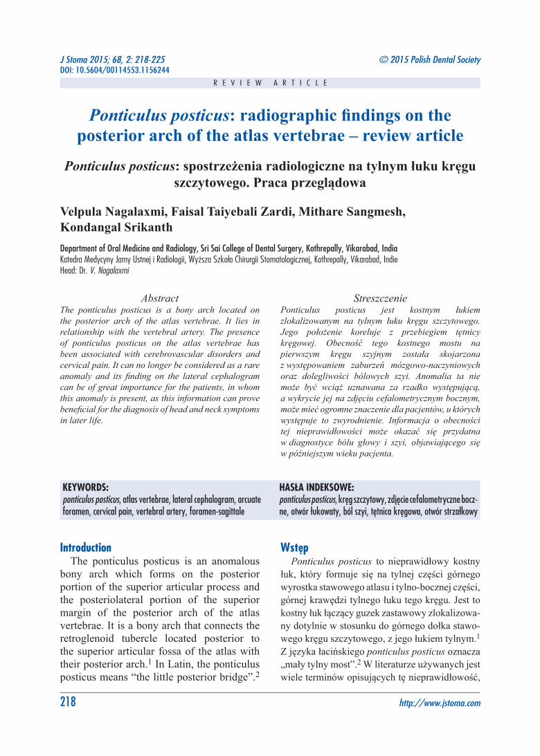
Fig. 3. Lateral cephalogram showing a complete ponticulus posticus.Zdjęcie cefalometryczne boczne uwidaczniające kompletny most kostny.
Fig. 4. Lateral cephalogram showing a partial ponticulus posticus.Zdjęcie cefalometryczne boczne uwidaczniające niekompletny most kostny.

J Stoma 2015; 68, 2 Nagalaxmi V., Zardi F.T., Sangmesh M., Srikanth K.
222 http://www.jstoma.com
kompletnego mostu kostnego jest nieco częstsze u płci męskiej, w porównaniu do płci żeńskiej. Sharma i wsp.,10 w swoich badaniach przeprowa-dzonych na populacji indyjskiej, również wykazali przewagę występowania u płci męskiej niż żeń-skiej. Podsumowując, w literaturze istnieją różne dane dotyczące częstości występowania ponticu-lus posticus w zależności od płci.
Od wielu lat ponticulus ponticus był przed-miotem zainteresowania anatomów akademic-kich, jednak zbyt mało uwagi poświęcano mu pod względem klinicznym. Lambarty i Zivanovic7 za-sugerowali, że tętnica kręgowa może być uciskana przez kostny most podczas ruchów obrotowych głowy lub ruchów zgięciowych bocznych szyi, wywołując zespół niewydolności kręgowo-pod-stawnej. Stwierdzono, że obecność tego małego kostnego łuku związana jest z występowaniem migreny bez aury.5 Jeśli jest on przymocowany do błony szczytowo-potylicznej, która łączy się z oponą twardą, może wytwarzać się niewielkie napięcie wywierane na oponę twardą, co skutkuje silnymi bólami głowy, jakie występują przy migre-nie. Liczne badania wykazały, że obecność kost-nego mostu na łuku tylnym kręgu szczytowego może wywoływać objawy zespołu niewydolności kręgowo-podstawnej, takie jak bóle i zawroty gło-wy oraz podwójne widzenie.6 Wight i wsp.7 łączą występowanie wielu procesów patologicznych z obecnością ponticulus ponticus. White i Panjabi9 zwrócili uwagę na wpływ rozciągania i skręca-nia tętnic kręgowych podczas ruchów obrotowych głowy.
Etiologa omawianego mostu kostnego do tej pory nie została wyjaśniona. Jak wspomniał Schilling,3 niektóre dotychczasowe badania suge-rowały, że być może jest to wrodzona wada roz-wojowa, spowodowana cechą genetyczną, bądź kostnienie postępujące z wiekiem lub wynik dzia-łania zewnętrznych czynników mechanicznych.
W związku z tym, że kliniczne znaczenie oma-wianej jednostki wzrasta z dnia na dzień, istnie-je ogromna potrzeba zapoznania się z cechami morfologicznymi tej nieprawidłowości. Zdjęcie cefalometryczne boczne może być radiogramem najczęściej wykorzystywanym w stomatologii klinicznej, jednak obszar szyjnego odcinka krę-
the neck causing basilar insufficiency syndrome. This little bony arch has been found to be associated with migraine without aura.5 As it is attached to the atlanto-occipital membrane, which is attached to the dura mater, this can cause small tensions exerted on the dura resulting in severe head pain as experienced in migraine. Several studies have suggested that the presence of this bony arch can cause symptoms of vertebra basilar insufficiency such as headache, vertigo and diplopia.6 Multiple pathological processes have been related to the presence of ponticulus posticus by Wight et al.7 White and Panjabi9 have reported stretching and kinking effect on the vertebral artery with head rotation.
The origin of this bony bridge is still debatable. As mentioned by Schilling,3 some previous studies have suggested that it may be due to congenital development, a genetic trait, ossification due to age, or could be a result of external mechanical factors.
As the clinical importance of this entity is growing day by day it has become quite necessary to understand the morphological features of this anomaly. The lateral cephalogram can be the most common radiograph used in clinical dentistry, however, the cervical spine area is usually omitted in cephalometric tracings. Though the cervical maturation index (CVMI) is now commonly used to interpret the growth potential of young patients, adequate attention is not paid to the morphological and radiographic anatomy of this region. Cephalograms can be studied comprehensively in a stepwise manner so that any deviations from the norm in cervical spine can be detected. Radiographic examinations of the cervical spine may reveal a pathology which can be symptomatic or asymptomatic in patients. Many abnormalities of the spine are not symptomatic until early adulthood and the oral radiologist can be the first person to detect these abnormalities in the early stages and can help in mitigating the severity of further consequences.
The ponticulus posticus has become an important anomaly of the atlas as the use of the lateral mass serves for the fixation of the atlas vertebrae in the management of atlanto-axial instability. As this

Ponticulus posticus: radiographic findings on the posterior arch ... J Stoma 2015; 68, 2
http://www.jstoma.com 223
gosłupa zwykle jest pomijany podczas analizy tego zdjęcia. Pomimo tego, że szyjny wskaźnik dojrzałości szkieletowej (CVMI) obecnie jest po-wszechnie stosowany w celu określenia potencjału wzrostowego u młodych pacjentów, nie zwraca się odpowiedniej uwagi na morfologiczną i radiogra-ficzną anatomię tego obszaru. Zdjęcia cefalome-tryczne mogą być badane szczegółowo, w sposób etapowy, a wtedy mogą zostać wykryte wszelkie odchylenia od normy w odcinku szyjnym kręgo-słupa. Badania radiologiczne kręgosłupa szyjne-go mogą ujawnić występowanie patologii, która może być objawowa lub bezobjawowa. Wiele nie-prawidłowości kręgosłupa przebiega bezobjawo-wo do czasu wkraczania pacjenta w okres doro-słości, a radiolog, bądź lekarz dentysta może być pierwszą osobą, która wykryje tę nieprawidłowość we wczesnym stadium i która może pomóc złago-dzić nasilenie konsekwencji tego stanu.
Ponticulus posticus stał się ważną anomalią kręgu szczytowego, którego boczna część wyko-rzystywana jest do ustabilizowania atlasu w po-stępowaniu terapeutycznym w przypadku niesta-bilności szczytowo-obrotowej. W związku z tym, że w regionie tym znajduje się splot żylny ze-wnątrzoponowy oraz nerw potyliczny większy, uszkodzenie tego obszaru może doprowadzić do znacznego krwawienia i neuralgii potylicznej.6 Próby umieszczenia śrub w takich obszarach mogą doprowadzić nawet do uszkodzenia tętnicy krę-gowej, prowadzącego do udaru mózgu lub zgo-nu, spowodowanego zakrzepicą, zatorowością lub rozwarstwieniem tętnicy kręgowej.
Skany tomografii komputerowej mogą mieć dużą wartość diagnostyczną, w porównaniu do zdjęcia cefalometrycznego bocznego, na którym zazwyczaj nie ma możliwości określenia, czy nie-prawidłowość występuje jednostronnie czy obu-stronnie. W badaniu przeprowadzonym przez Young Jae Cho,11 ponticulus posticus był niemal równie często wykrywany zarówno po lewej, jak i po prawej stronie, lecz nie było to jasno określo-ne w prostej rentgenografii, ponieważ na zwykłym zdjęciu radiologicznym niemożliwa do odróżnie-nia jest w tym przypadku prawa i lewa strona. Skany tomografii komputerowej pozwalają okre-ślić lateralizację, różnorodność widma kształtów
region contains the epidural venous plexus and the greater occipital nerve, injury to this region can lead to significant bleeding and occipital neuralgia.6 Attempts to place a screw in such regions can even lead to injury of the vertebral artery leading to stroke or death due to thrombosis, embolism or arterial dissection.
Computed Tomography scan may have high diagnostic value as compared with the lateral cephalogram in which it is not usually possible to determine if the anomaly is unilateral or bilateral. In a study conducted by Young Jae Cho11 the ponticulus posticus was almost equally detected on both left and right sides and this was not clearly identified in the plain radiography as plain films fail to differentiate the right and the left. CT scans can even reveal the laterality, various spectra of shape and size of the ponticulus posticus which is observed or suspected on a lateral cephalogram. A 3-D CT scan could be helpful to check the size, shape, unilaterality or bilaterality, and this would be very helpful for further treatment planning. Hence, a 3-D CT scan should be taken when a ponticulus posticus is suspected or observed on routine lateral cephalogram of patients specially in those who are about to undergo a lateral mass screw placement in the posterior arch of the atlas vertebrae.
ConclusionThe lateral cephalogram is the most common
diagnostic radiograph used in clinical dentistry. The finding of ponticulus posticus can be of utmost importance in early diagnosis of pathologies of the atlas vertebrae. It can also have clinical significance during the management of cervical spine surgical intervention. The presence of this bony arch is a serious condition which, if left untreated, may result in severe discomfort to the patient. The presence of such an anomaly can be the basis for referral by the oral physician to an orthopedist or a neurosurgeon for further evaluation. Along with the lateral cephalogram a computed tomography scan can also be used to substantiate the size and shape of the arch if required, hence early diagnosis of the pathology not only gives the patient a chance to live normal

J Stoma 2015; 68, 2 Nagalaxmi V., Zardi F.T., Sangmesh M., Srikanth K.
224 http://www.jstoma.com
i rozmiarów kostnego mostu na tylnym łuku atla-su, stwierdzonego lub podejrzewanego na podsta-wie analizy zdjęcia cefalometrycznego bocznego. Obraz trójwymiarowej tomografii komputerowej może być wykorzystany do określenia rozmiaru, kształtu, jednostronności lub dwustronności, co może być bardzo pomocne w planowaniu dalszego leczenia. W związku z powyższym, zdjęcia trój-wymiarowej tomografii komputerowej powinny być wykonywane, kiedy podejrzewa się ponticu-lus posticus, bądź dostrzega się go na rutynowym zdjęciu cefalometrycznym bocznym, szczególnie u tych pacjentów, u których w bocznej części łuku tylnego atlasu ma zostać umieszczony wszczep.
WnioskiZdjęcie cefalometryczne boczne jest radiogra-
mem diagnostycznym najczęściej wykorzystywa-nym w stomatologii klinicznej. Wykrycie ponticu-lus posticus ma największe znaczenie we wcze-snej diagnostyce tej patologii kręgu szczytowego. Może mieć to również ważne znaczenie klinicz-ne podczas interwencji chirurgicznych w obrę-bie kręgosłupa szyjnego. Obecność tego kostnego łuku jest poważnym schorzeniem, które nieleczo-ne może doprowadzić do poważnego dyskomfor-tu pacjenta. Obecność takiej anomalii może być zauważona przez lekarza dentystę, a następnie skierowana do ortopedy lub neurochirurga w celu przeprowadzenia dalszej diagnostyki. Jeżeli wy-magane jest skonkretyzowanie rozmiaru i kształtu łuku, oprócz zdjęcia cefalometrycznego bocznego mogą zostać wykonane skany tomografii kompu-terowej. Wczesne zdiagnozowanie tej patologii daje pacjentowi szansę na prowadzenie normalne-go życia, przy zmianie jego stylu. Wczesne posta-wienie diagnozy może również dostarczyć wielu przydatnych danych, do dokumentacji zmian spo-wodowanych urazami, starzeniem się, czy proce-sami zwyrodnieniowymi. Podsumowując, cefalo-gram musi być traktowany jako bazowe badanie przesiewowe obszaru kręgosłupa szyjnego, a le-karz dentysta odgrywa bardzo ważną, a w zasadzie kluczową rolę w diagnostyce tej anomalii.
life by changing their lifestyles but it can also provide useful documentation of change due to an injury, ageing or progress of a degenerative process. Lastly, the cephalogram must be looked upon as a baseline screening tool for examining the spine region, and the dentist or oral physician has a very important and even critical role to play in the diagnosis of this anomaly.

Ponticulus posticus: radiographic findings on the posterior arch ... J Stoma 2015; 68, 2
http://www.jstoma.com 225
References
1. Miki T, Oka M, Urushidani H, Hirofuji E, Tanaka S, Iwamoto S: Ponticulus posticus: its clinical significance. Acta Medica Kinki Univ 1979; 4: 427-430.
2. Stubbs DM: The arcuate foramen: variability in distribution related to race and sex. Spine 1992; 17: 1502-1504.
3. Schilling J, Schilling A, Suazo GI: Ponticulus posticus en el arco posterior del atlas, analisis de su prevalencia enpacientes asintomaticos. Int J Morphol 2010; 28: 317-322.
4. Paraskevas G, Papaziogas B, Tsonidis C, Kapetanos G: Gross morphology of the bridges over the vertebral artery groove on the atlas. Surg Radiol Anat 2005; 27: 129-136.
5. Wight S, Osborne N, Breen AC: Incidence of ponticulus posterior of the atlas in migraine and cervicogenic headache. J Manipulative Physiol Ther 1999; 22: 15-20.
6. Young JP, Young PH, Ackermann MJ, Anderson PA, Riew KD: The ponticulus posticus: implications for screw insertion into the first cervical lateralmass. J Bone Joint Surg Am 2005; 87: 2495-2498.
7. Lambarty BGH, Zivanovic S: The retroarticular
vertebral artery ring of the atlas and its significance. Acta Anatomica 1973; 85: 113-122.
8. Simsek S, Yigitkanli K, Comert A, Acar HI, Seckin H, Er U: Posterior osseous bridging of C1. J Clin Neurosci 2008; 15: 686-688.
9. White AA, Panjabi MM: Clinical biomechanics of the spine. 2nd ed. Philadelphia: Lippincott Williams & Wilkins; 1978.
10. Sharma V, Chaudhary D, Mitra R: Prevalence of ponticulus posticus in Indian orthodontic patients. Dentomaxillofac Radiol 2010; 39: 277-283.
11. Cho YJ: Radiological analysis of ponticulus posticus in Koreans. Yonsei Med J 2009: 50: 45-49.
Address: Department of Oral Medicine and Radiology, Sri Sai College of Dental Surgery, Kothrepally, Vikarabad, India 501101Tel.: 00919849442438e-mail: [email protected]
Received: 8th January 2015Accepted: 14th February 2015