Embed Size (px)
Citation preview

Possible Dopaminergic Stimulation ofLocus Coeruleus a1-AdrenoceptorsInvolved in Behavioral Activation
YAN LIN,1 DAVID QUARTERMAIN,2 ADRIAN J. DUNN,3 DAVID WEINSHENKER,4 AND ERIC A. STONE1*1Department of Psychiatry, New York University School of Medicine, New York, New York 100162Department of Neurology, New York University School of Medicine, New York, New York 10016
3Department of Psychology, University of Hawaii, Honolulu, Hawaii 968224Department of Human Genetics, Emory University School of Medicine, Atlanta, Georgia 30322
KEY WORDS locus coeruleus; a1-adrenoceptor; DA; motor activity; novelty
ABSTRACT a1-Adrenoceptors of the locus coeruleus (LC) have been implicated inbehavioral activation in novel surroundings, but the endogenous agonist that activatesthese receptors has not been established. In addition to the canonical activation of a1-receptors by norepinephrine (NE), there is evidence that dopamine (DA) may also acti-vate certain brain a1-receptors. This study examined the contribution of DA to explor-atory activity in a novel cage by determining the effect of infusion of various dopami-nergic and adrenergic drugs into the mouse LC. It was found that the D2/D3 agonist,quinpirole, which selectively blocks the release of CNS DA, produced a dose-dependentand virtually complete abolition of exploration and all movement in the novel cagetest. The quinpirole-induced inactivity was significantly attenuated by coinfusion ofDA but not by the D1 agonist, SKF38390. Furthermore, the DA attenuation of quin-pirole inactivity was blocked by coinfusion of the a1-adrenergic receptor antagonist,terazosin, but not by the D1 receptor antagonist, SCH23390. LC infusions of eitherquinpirole or terazosin also produced profound inactivity in DA-b-hydroxylase knock-out (Dbh 2/2) mice that lack NE, indicating that their behavioral effects were notdue to an alteration of the release or action of LC NE. Measurement of endogenousDA, NE, and 5HT and their metabolites in the LC during exposure to the novel cageindicated an increase in the turnover of DA and NE but not 5HT. These results indi-cate that DA is a candidate as an endogenous agonist for behaviorally activating LCa1-receptors and may play a role in the activation of this nucleus by novel surround-ings. Synapse 62:516–523, 2008. VVC 2008 Wiley-Liss, Inc.
INTRODUCTION
a1-Adrenoceptors in widely distributed regions ofthe CNS have been shown to play a significant rolein behavioral activation under a variety of conditions(Stone et al., 2007b). Pharmacological blockade ofthese receptors in the motor and piriform cortex, nu-cleus accumbens, preoptic area, lateral hypothala-mus, vermis cerebellum, locus coeruleus (LC), anddorsal raphe produces immobility in a novel testcage, whereas stimulation leads to behavioral activa-tion in the familiar home cage (Stone et al.,2004a,b). The LC appears to be a key region in thissystem in that it contains a dense concentration ofa1-receptors (Jones et al., 1985), which when blockedwith locally injected terazosin produces one of thestrongest degrees of immobility and catalepsy in thenovel cage test (7 out of 10 min immobile), and whenstimulated with catecholamines produces vigorous
exploratory behavior in familiar surroundings (Stoneet al., 2004a,b). Blockade of LC a1-receptors alsoimpairs lateral hypothalamic self-stimulation behav-ior in rats (Lin et al., 2007), suggesting a broad rolein positively motivated behaviors. Presumably, LCactivation via stimulation of local a1-receptors pro-duces increased postsynaptic a1-noradrenergic neu-rotransmission in one or more of these target areasand is responsible for behavioral activation underthese conditions.
Contract grant sponsor: NIMH; Contract grant numbers: MH45265, MH50947.
*Correspondence to: Eric A. Stone, Department of Psychiatry, MHL HN510,NYU Med Ctr, 550 First Ave, New York, NY 10016, USA.E-mail: [email protected]
Received 27 August 2007; Accepted 19 December 2007
DOI 10.1002/syn.20517
Published online in Wiley InterScience (www.interscience.wiley.com).
VVC 2008 WILEY-LISS, INC.
SYNAPSE 62:516–523 (2008)

The endogenous neurotransmitter that activatesLC a1-receptors is either norepinephrine (NE), dopa-mine (DA), or epinephrine (EPI). EPI-containingnerve terminals emanating from the paragigantocel-lularis nucleus of the ventrolateral medulla areknown to innervate the LC (Astier et al., 1990).Although we have shown that exogenous EPI injectedin the LC can stimulate behavioral activity in thehome cage, only a portion of this effect is blocked byan a1-receptor antagonist (Stone et al., 2003). Fur-thermore, preliminary experiments in our laboratoryhave failed to reveal substantial amounts of EPI inhomogenates of the LC or reliable inhibition of be-havioral activity after local inhibition of EPI biosyn-thesis in the LC by the phenylethanolamine-N-methyltransferase (PNMT) inhibitor dichloromethyl-benzylamine (Stone, Quartermain, Lin, unpublishedresults).
There is more consistent evidence that either orboth NE and DA are the endogenous agonists forthese receptors. With respect to NE, the high concen-trations of extracellular NE found in the LC origi-nates from either recurrent noradrenergic axon collat-erals (Nakamura et al., 1988) or somatodendriticrelease from LC noradrenergic neurons (Fernandez-Pastor et al., 2005; Singewald and Philippu, 1998).We and others (De Sarro et al., 1987; Stone et al.,2005) have shown that local reduction of NE releasein the LC by injection of a2-adrenoceptor agonistsproduces marked immobility in the novel cage test inmice and rats.
With respect to DA, although tract tracing and im-munohistochemical studies have been inconclusiveregarding a dopaminergic innervation of the LC(Deutch et al., 1986; Somerville et al., 2007), substan-tial amounts of this catecholamine (Dishman et al.,1997; Versteeg et al., 1976) and high concentrationsof its metabolite, dihydroxyphenylacetic acid, arefound in this nucleus (Lambas-Senas et al., 1990),although its cellular localization is still unclear.Although a microdialysis study of the cat LC foundrelatively small amounts of extracellular DA (Crochetand Sakai, 1999), a push–pull cannula study in thesame species found equal levels of extracellular DAand NE in this nucleus (Kaehler et al., 1999), whichis not unexpected given that DA is the immediateprecursor of NE. In addition, an early study showedthat activation of the DAergic cell bodies in the ven-tral tegmental area produced activation of LC neu-rons as evidenced by the accumulation of metabolitesof NE in noradrenergic projection areas (Deutchet al., 1986). Although DA possesses only 1/50 the af-finity of NE at a1-receptors (Leedham and Pennefa-ther, 1986), endogenous DA has been shown conclu-sively by electrophysiological methods to activatethese receptors in the avian preoptic area (Cornilet al., 2002) and is also known to be equally effica-
cious as NE at these receptors in cell culture (Reyet al., 2001; Zhang et al., 2004).
In addition, the arousing effect of the stimulantmodafinil, which is dependent on a1-receptor activity(Duteil et al., 1990; Stone et al., 2002), has beenfound to be potently inhibited by selective blockade ofDA release with the D2 dopaminergic agonist, quin-pirole (Wisor and Eriksson, 2005). The latter authorshave suggested that DA is an endogenous agonist ata1-receptors involved in arousal.
A contribution of DA to the activation of the LCwould have significant implications for an under-standing of the role of this nucleus in behavioral reg-ulation and motivation. Therefore, the present experi-ments were undertaken to test this hypothesis. Thiswas accomplished by examining the effect of microin-jection of various selective dopaminergic and adrener-gic agents in the vicinity of the mouse LC on novelcage-induced behavioral activity in both outbred andgenetically altered (NE-deficient) mice.
MATERIALS AND METHODSSubjects
Swiss Webster male mice (Taconic), 8–10 weeks old,female DA-b-hydroxylase knockout (Dbh 2/2) micethat completely lack NE, and female and male wild-type control mice, 4–9 months of age, of a mixedC57BL6/J and 129SvEv genetic background (Thomaset al., 1998) were subjects. The animals were housedsingly with nesting material for 5 days prior to sur-gery in standard polycarbonate mouse cages (12.5 cm3 17 cm 3 28 cm) at a room temperature of 228C 618C under a 12 h light/dark cycle (lights on 05:00 h).Food and water were available ad libitum.
Surgery
Mice were anesthetized with pentobarbital (70 mg/kg) and implanted stereotaxically with unilateral 26ga cannula guide tubes in the dorsal pons in theregion of the LC at the following coordinates (25.5mm posterior to Bregma, 60.9 mm lateral to the mid-line, 3.9 mm beneath the surface of the skull). Wehave shown in a previous study that unilateral block-ade of LC a1-adrenoceptors in the mouse is sufficientto produce maximal inactivity in the 10-min novelcage test (Stone et al., 2004a,b). All animals weregiven 10 days for recovery prior to behavioral testing.
Procedure
All experiments were performed between 1000 and1400 h. Mice were gently restrained under a doublelayer of gauze on a corkboard and a 33 ga cannulaconnected by PE tubing to a syringe pump (Razel)was inserted into the cannula guide tube (protruding1 mm below the bottom of the guide). A total of
517DOPAMINERGIC STIMULATION OF LC �1-RECEPTORS
Synapse

350 nl of solution was infused at 150 nl/min over a2.3-min period with the cannula remaining in placefor 30 s after infusion. The animals received eithervehicle (saline), the D2 dopaminergic agonist, quinpir-ole (Q), DA, the D1/D5 agonist, SKF38393 (SKF), thea1-adrenoceptor antagonist, terazosin (T), or the D1/D5 antagonist, SCH23390 (SCH), singly or in combi-nation, in doses ranging from 0.3 to 30 nmol permouse. All drugs were prepared fresh each day in sa-line. Minimum doses were determined empirically inpilot studies. Immediately following the injection, theanimal was transferred to a clean unfamiliar mousecage of identical type and dimensions as the homecage (‘‘novel home cage’’) with Bed-O-Cob bedding(0.63 mm) and was videotaped for the next 10 min.The animals received four such injections at 4 daysintervals with the first and fourth using Q at 3 nmol,and the second and third being any of the drugs sin-gly or in combination. Thus different groups of ani-mals received two different drug treatments in addi-tion to the Q3 trials. A given drug treatment was rep-resented equally on the second and third injections.
Behavioral ratings
Videotapes were rated either by a trained observer(in blind fashion) for a measure of total behavioral ac-tivity [the number of gross movements (GM) made]and time immobile (no observable movements) or by avideotracking system (Smart System, San Diego Inst)for ambulation (the number of times the animalcrossed the long and short axes of the cage). GMswere defined as any movement involving at least thehead plus forelimbs that was followed by a momen-tary pause. These include ambulations to a cage walland other discrete forward movements, rearingresponses, stretch and attend responses, turns andlarge grooming movements. Interrater agreement onthis measure has averaged above r 5 0.9 in this labo-ratory. Central a1-adrenoceptor activity has beenshown in previous studies to have a general modulat-ing effect on a variety of different active behaviors,and the number of GM made in the novel cage testhas been shown to be a function of this activity (Stoneet al., 1999, 2001, 2004a,b). All experiments were con-ducted in accordance with the National ResearchCouncil Guide for the Care and Use of LaboratoryAnimals (1996), and were approved by the New YorkUniversity School of Medicine IUCAC.
DA turnover in the vicinity of the LC
Undisturbed mice or mice exposed to a novel homecage for 20 min were sacrificed by rapid decapitation.The LC and adjacent tissue were grossly dissectedover ice as a 1.5 mm 3 1.5 mm 3 1 mm block of tissuecentering on the lateral extent of IV. ventricle from a1-mm brain section between 25 and 26 mm Bregma.
NE, 3-methoxy-4-hydroxyphenylglycol (MHPG), DA,3,4-dihydroxyphenylacetic acid (DOPAC), serotonin(5-HT), and 5-hydroxyindoleacetic acid (5-HIAA) wereassayed by high-performance liquid chromatography(HPLC) as described by Dunn (1993).
Histology
After the experiment, the implanted animals weredeeply anesthetized and injected through the cannulawith 350 nl of 2% methyl blue. Brains were fixed in10% formalin for 3 days and then sectioned for deter-mination of cannula location.
Statistical analysis
The individual data points comprised 2–3 observa-tions per animal taken under different drug condi-tions. All analyses involved either one- or two-wayANOVAs. Because of the diverse nature of drug treat-ments used there were no significant correlationsbetween responses to the different drug treatmentsacross the four trials. Therefore, each of the datapoints was treated as an independent observation. Allindividual mean comparisons were planned and wereBonferroni-corrected.
RESULTSLocalization of quinpirole effect
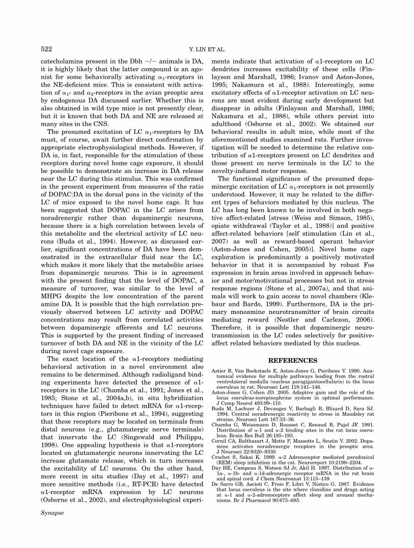
An initial group of 44 animals that were implantedat various sites in the pons and cerebellum wereinjected with Q at 3 nmol and assessed for behavioralactivity in the novel home cage test. Of these, 19(43.2%) developed immobility of greater than 7.5 minout of the 10 min test and were judged as Q3-positiveanimals. Histological analysis (Fig. 1) indicated thatof the 19, 4 were located in the 4th ventricle andwere excluded, whereas 14 of the remaining 15(93.3%) had cannulas within 0.5 mm of the border ofthe LC core, and only 9 (31.0%) of the 25 Q3-negativeanimals had cannulas this close. This difference inpercentage was highly significant by Fisher’s exacttest (P < 0.0001).
Alternately, if the group of 40 mice was divided intothose having cannulas within 0.5 mm of the LC andthose outside this distance, a similar result wasobtained, with the ‘‘within’’ group having more than athreefold higher level of immobility than the ‘‘outside’’group (within, 6.73 6 0.64 min (23); outside, 2.21 60.56 min (17), t38 5 5.24, P < 0.001).In subsequent experiments, we therefore adopted
the procedure of first screening all implanted animalswith an initial Q3 test and only using Q3-positive ani-mals for two subsequent drug challenges at 4 daysintervals (the location of the novel home cage in thelaboratory was changed for each test). Pilot experi-ments showed that the majority of animals had reli-
518 Y. LIN ET AL.
Synapse

able responses to repeated administrations of thisdrug for up to four injections at 4 days intervals. Afinal Q3 test (fourth injection) was therefore employedto determine if the initial response to the drug wasmaintained throughout the series of injections. Of atotal of 116 initially Q3-positive animals, 62.2% main-tained the immobility response to the drug on the 4thchallenge, and of these, 75.3% were found to havecannulas within 0.5 mm of the LC and were used inthe final analysis. All responses to Q3 were computedas the average of the first and fourth injection,whereas all responses to other Q doses and to allother drugs were based on responses to the second orthird injections. There was a high and significant cor-relation between immobility scores of separate groupsof mice given the same drug treatments on differenttests (r10 5 0.92, P < 0.001), which suggests thatresponses to the same treatments were stable andlargely independent of order effects.
Dose response to quinpirole and comparisonwith terazosin and SCH23390
The effect of Q at doses between 0.3 and 3 nmol onactivity level in the novel cage is shown in Figure 2.A one-way ANOVA comprising the vehicle and all Qdoses indicated that the drug produced significantdose-dependent reductions of both GM (F3,27 5 91.23,P < 0.0001) and ambulation (F3,27 5 25.98, P <0.0001) and a significant dose-dependent increase inimmobility (F3,27 5 73.57, P < 0.0001). Figure 2 alsoshows the effects of terazosin 3 nmol (T3) andSCH23390 3 nmol (SCH3). Comparison of the vehicle,Q3, T3, and SCH3 conditions by one-way ANOVAsfollowed by posthoc Bonferroni tests revealed that T3significantly reduced GM (P < 0.0001) and ambula-tion (P < 0.0001) and increased immobility (P <0.0001), and that SCH3 also reduced GM (P < 0.01)and ambulation (P < 0.005), but produced only a bor-derline increase in immobility (P < 0.06). However,the degree of inactivity produced by Q3 on all behav-ioral measures was significantly greater than thatproduced by SCH3 (all Ps < 0.001), whereas therewere no significant differences between the Q3 andT3 conditions on any measure.
Attenuation of Q3 immobility by DA butnot by SKF38393
The effect of coinfusion of DA and SKF38393 (SKF)at 10 and 30 nmol on Q3-induced inactivity is shownin Figure 3. A one-way ANOVA comprising only theDA conditions (0, 10, and 30 nmol) showed that thecatecholamine produced a dose-dependent attenuationof the reduction in GM (F2,24 5 14.98, P < 0.0001)and ambulation (F2,24 5 3.52, P < 0.05), and of theincrease in immobility (F2,24 5 16.21, P < 0.0001)caused by the D2 agonist (Fig. 3). In the absence ofQ3, DA had no significant effect on any behavior ateither dose (data not shown). The same analysis forthe SKF conditions (0, 10, and 30 nmol) failed toreveal a significant action on Q3-induced immobility.
Blockade of the attenuating effect of DAby an antagonist of a1-adrenergic but not D1
dopaminergic receptors
The effects of T and SCH (both at 3 and 10 nmol)on the attenuating effect of DA (30 nmol) of Q3-induced inactivity are shown in Figure 4. In this fig-ure, the Q3-DA30 group is the same as used in theforegoing comparison (3). A one-way ANOVA compris-ing the Q3-DA30 condition in the presence of 0, 3,and 10 nmol T showed that the a1-antagonist pro-duced a blockade of the DA reversal effect that wassignificant for GM (F2,27 5 8.63, P < 0.005) andimmobility (F2,27 5 6.86, P 5 0.005), and was of bor-derline significance for ambulation (F2,27 5 3.24,
Fig. 1. Locations of cannula tips for quinpirole-positive (A) and -negative (B) animals in initial experiment. Quinpirole was infusedat 3 nmol unilaterally. LC, locus coeruleus; BAR, Barrington’s nu-cleus. Figures adapted from Franklin and Paxinos (1997).
519DOPAMINERGIC STIMULATION OF LC �1-RECEPTORS
Synapse

P < 0.06). Analyses of linear trend components sug-gested that these effects were dose-dependent for GM(F1,27 5 11.22, P < 0.005) and for time immobile(F1,27 5 9.08, P < 0.01). The one-way ANOVA forSCH at 0, 3, and 10 nmol did not reveal a significanteffect on any measure. Neither T nor SCH at 10 nmolsignificantly altered any of the behaviors of animalstreated with only Q3 (data not shown).
Effect of quinpirole and terazosin inNE-deficient mice
Because of the smaller size and increased fragilityof the Dbh 2/2 mice, the initial screening injection
was omitted and the animals were injected only threetimes with Q3, T3, and vehicle in that order. Eight ofthe 10 WT and 6 of the 11 Dbh 2/2 mice were subse-quently found to have cannulas located within 0.5mm of the LC and completed the Q3 and T3 injec-tions. Because of the loss of cannulas, only five ofeach group completed the vehicle injection. Theresults are shown in Figure 5. A 2 3 3 (genotype 3drug) ANOVA revealed significant main effects ofdrug for each dependent measure (GM, F2,31 5 13.08,P < 0.001; ambulation, F2,31 5 13.48, P < 0.001; timeimmobile, F2,31 5 16.19, P < 0.0001), but there wasno significant effects of genotype (GM, F1,31 5 0.02,NS; ambulation, F1,31 5 2.15, NS; time immobile,
Fig. 2. Effect of various doses of quinpirole (Q), terazosin (T), and SCH23390 (SCH) or vehicle (v,saline) in the LC on behavioral activation in novel home cage test. Values are means and SEMs of 7–10 determinations for Q 0.3–3 nmol and 5–7 determinations for T3 and SCH3. *P < 0.05, **P < 0.01,***P < 0.001 vs. vehicle condition.
Fig. 3. Dose-dependent reversal of quinpirole-induced inactivity by coinfusion of DA but notSKF38393. Mice were infused with quinpirole (3 nmol) in the presence of either DA or SKF at 0, 10,and 30 nmol. DA or SKF38393 were dissolved in the Q3-containing saline solution (veh). Means andSEMs of 6–10 determinations. *P < 0.05, **P < 0.001 vs. vehicle.
Fig. 4. Blockade of DA-reversal of quinpirole-inactivity by terazosin but not by SCH23390. Micewere infused with 3 nmol quinpirole 1 30 nmol DA in the presence of either 0 (veh), 3 or 10 nmol ofterazosin or SCH23390. Terazosin or SCH23390 were dissolved in the Q3-DA30-containing saline solu-tion (veh). Means and SEMs of 6–10 determinations. *P < 0.01 vs. vehicle condition.
520 Y. LIN ET AL.
Synapse

F1,31 5 0.04, NS) or interactions of genotype and drugfor any dependent measure (GM, F2,31 5 0.17, NS;ambulation, F2,31 5 1.67, NS; time immobile, F2,31 50.16, NS).
Turnover of LC DA during exposureto novel home cage
The effects of novel home cage exposure on levelsand turnover of DA, NE, and 5HT in the vicinity ofthe LC are shown in Table I. Novel cage exposureproduced a highly significant increase in the concen-tration of DOPAC and in the ratios of DOPAC/DA,and MHPG/NE indicative of increases in the turnoverof DA and NE. The concentrations of 5HT and 5HIAAand the 5HIAA/5HT ratio were not significantlyaltered.
DISCUSSION
These results provide indirect pharmacological evi-dence that DA can stimulate the population of a1-adrenoceptors in the vicinity of the LC that functionsin behavioral activation in the novel home cage test.Quinpirole, a D2/D3 agonist that selectively reducesthe release of CNS DA (Devoto et al., 2002), produceda significant and near total dose-dependent abolitionof all movement in this test when injected within 0.5mm of this nucleus, but was much less effective atgreater distances. No loss of the righting reflex (main-tenance of an upright position) was observed as wouldhave occurred with a sedative effect. The quinpirole-induced immobility was significantly and dose-dependently reversed by the addition of DA, whereasDA given alone had no effect on nonquinpirole-treated
animals who were already maximally active. The DAreversal appeared to be acting via stimulation of a1-adrenoceptors, since (a) it was significantly blockedby the a1-antagonist, terazosin; (b) it was not blockedby the D1 antagonist, SCH23390; and (c) the D1 ago-nist, SKF38390, failed to reverse the Q-induced inac-tivity. These data therefore add to the previous directelectrophysiological evidence of the stimulation ofCNS a1-adrenoceptors by DA discussed earlier.
Since D2 receptors may be present on noradrenergicas well as dopaminergic neurons (Smiakowska andLegutko, 1992), it could be argued that the behavioraleffects of quinpirole were due to an inhibition of NErelease and a subsequent failure of NE to activate a1-receptors in the LC. This possibility, however, appearsvery unlikely because quinpirole and terazosin wereequally effective in control and Dbh 2/2 mice, whichcompletely lack NE. Combining these results suggeststhat DA is an endogenous ligand for a1-receptors inthe LC, although the source of the DA is uncertain.The DA could be coming from dopaminergic terminalsthat innervate the LC, or from the LC itself, whichproduces DA instead of NE in Dbh 2/2 mice (Thomaset al., 1998). DA release from noradrenergic neuronshas also been observed in wild-type animals. BecauseDA possesses only 1/50 the affinity of NE at a1-adreno-ceptors, the question arises as to how this catechol-amine can stimulate these receptors as suggested bythe present and previous experiments. However, a1-receptors, are now known to form heterodimers thatpossess agonist properties different from individualsubtypes (Hague et al., 2006). It is possible thereforethat DA has a greater affinity for a receptor hetero-dimer than for individual a1-receptors. Since the only
Fig. 5. Induction of inactivity in NE-deficient mice by quinpirole and terazosin. Dbh 2/2 or wildtype mice were infused with vehicle (v, saline), quinpirole or terazosin at 3 nmol. Means and SEMs of6–8 determinations for Q and T and 5 determinations for vehicle. *P < 0.05, **P < 0.01.
TABLE I. Effect of novel home cage exposure on turnover of NE, DA, and 5HT in the vicinity of the LC
Group DA DOPAC DOPAC/DA NE MHPG MHPG/NE 5HT 5HIAA 5HIAA/5HT
Control (9) 0.019 0.015 0.786 0.349 0.016 0.047 0.398 0.485 1.1090.002 0.002 0.027 0.040 0.002 0.001 0.042 0.112 0.147
Novel cage (9) 0.023 0.025 1.049 0.369 0.023 0.060 0.406 0.568 1.2810.001 0.003 0.072 0.032 0.003 0.003 0.038 0.134 0.189
t 21.76 23.35 3.43 20.40 1.92 4.40 20.13 20.47 20.72p 0.0960 0.0004 0.0034 NS 0.0720 0.0004 NS NS NS
Values are means and SEMs of groups of 9 mice. Values of amines are in ng/mg wet weight.
521DOPAMINERGIC STIMULATION OF LC �1-RECEPTORS
Synapse
catecholamine present in the Dbh 2/2 animals is DA,it is highly likely that the latter compound is an ago-nist for some behaviorally activating a1-receptors inthe NE-deficient mice. This is consistent with activa-tion of a1- and a2-receptors in the avian preoptic areaby endogenous DA discussed earlier. Whether this isalso obtained in wild type mice is not presently clear,but it is known that both DA and NE are released atmany sites in the CNS.
The presumed excitation of LC a1-receptors by DAmust, of course, await further direct confirmation byappropriate electrophysiological methods. However, ifDA is, in fact, responsible for the stimulation of thesereceptors during novel home cage exposure, it shouldbe possible to demonstrate an increase in DA releasenear the LC during this stimulus. This was confirmedin the present experiment from measures of the ratioof DOPAC:DA in the dorsal pons in the vicinity of theLC of mice exposed to the novel home cage. It hasbeen suggested that DOPAC in the LC arises fromnoradrenergic rather than dopaminergic neurons,because there is a high correlation between levels ofthis metabolite and the electrical activity of LC neu-rons (Buda et al., 1994). However, as discussed ear-lier, significant concentrations of DA have been dem-onstrated in the extracellular fluid near the LC,which makes it more likely that the metabolite arisesfrom dopaminergic neurons. This is in agreementwith the present finding that the level of DOPAC, ameasure of turnover, was similar to the level ofMHPG despite the low concentration of the parentamine DA. It is possible that the high correlation pre-viously observed between LC activity and DOPACconcentrations may result from correlated activitiesbetween dopaminergic afferents and LC neurons.This is supported by the present finding of increasedturnover of both DA and NE in the vicinity of the LCduring novel cage exposure.
The exact location of the a1-receptors mediatingbehavioral activation in a novel environment alsoremains to be determined. Although radioligand bind-ing experiments have detected the presence of a1-receptors in the LC (Chamba et al., 1991; Jones et al.,1985; Stone et al., 2004a,b), in situ hybridizationtechniques have failed to detect mRNA for a1-recep-tors in this region (Pieribone et al., 1994), suggestingthat these receptors may be located on terminals fromdistal neurons (e.g., glutamatergic nerve terminals)that innervate the LC (Singewald and Philippu,1998). One appealing hypothesis is that a1-receptorslocated on glutamatergic neurons innervating the LCincrease glutamate release, which in turn increasesthe excitability of LC neurons. On the other hand,more recent in situ studies (Day et al., 1997) andmore sensitive methods (i.e., RT-PCR) have detecteda1-receptor mRNA expression by LC neurons(Osborne et al., 2002), and electrophysiological experi-
ments indicate that activation of a1-receptors on LCdendrites increases excitability of these cells (Fin-layson and Marshall, 1986; Ivanov and Aston-Jones,1995; Nakamura et al., 1988). Interestingly, someexcitatory effects of a1-receptor activation on LC neu-rons are most evident during early development butdisappear in adults (Finlayson and Marshall, 1986;Nakamura et al., 1988), while others persist intoadulthood (Osborne et al., 2002). We obtained ourbehavioral results in adult mice, while most of theaforementioned studies examined rats. Further inves-tigation will be needed to determine the relative con-tribution of a1-receptors present on LC dendrites andthose present on nerve terminals in the LC to thenovelty-induced motor response.
The functional significance of the presumed dopa-minergic excitation of LC a1-receptors is not presentlyunderstood. However, it may be related to the differ-ent types of behaviors mediated by this nucleus. TheLC has long been known to be involved in both nega-tive affect-related [stress (Weiss and Simson, 1985),opiate withdrawal (Taylor et al., 1988)] and positiveaffect-related behaviors [self stimulation (Lin et al.,2007) as well as reward-based operant behavior(Aston-Jones and Cohen, 2005)]. Novel home cageexploration is predominantly a positively motivatedbehavior in that it is accompanied by robust Fosexpression in brain areas involved in approach behav-ior and motor/motivational processes but not in stressresponse regions (Stone et al., 2007a), and that ani-mals will work to gain access to novel chambers (Kle-baur and Bardo, 1999). Furthermore, DA is the pri-mary monoamine neurotransmitter of brain circuitsmediating reward (Nestler and Carlezon, 2006).Therefore, it is possible that dopaminergic neuro-transmission in the LC codes selectively for positive-affect related behaviors mediated by this nucleus.
REFERENCES
Astier B, Van Bockstaele E, Aston-Jones G, Pieribone V. 1990. Ana-tomical evidence for multiple pathways leading from the rostralventrolateral medulla (nucleus paragigantocellularis) to the locuscoeruleus in rat. Neurosci Lett 118:141–146.
Aston-Jones G, Cohen JD. 2005. Adaptive gain and the role of thelocus coeruleus-norepinephrine system in optimal performance.J Comp Neurol 493:99–110.
Buda M, Lachuer J, Devauges V, Barbagli B, Blizard D, Sara SJ.1994. Central noradrenergic reactivity to stress in Maudsley ratstrains. Neurosci Lett 167:33–36.
Chamba G, Weissmann D, Rousset C, Renaud B, Pujol JF. 1991.Distribution of a-1 and a-2 binding sites in the rat locus coeru-leus. Brain Res Bull 26:185–193.
Cornil CA, Balthazart J, Motte P, Massotte L, Seutin V. 2002. Dopa-mine activates noradrenergic receptors in the preoptic area.J Neurosci 22:9320–9330.
Crochet S, Sakai K. 1999. a-2 Adrenoceptor mediated paradoxical(REM) sleep inhibition in the cat. Neuroreport 10:2199–2204.
Day HE, Campeau S, Watson SJ Jr, Akil H. 1997. Distribution of a-1a-, a-1b- and a-1d-adrenergic receptor mRNA in the rat brainand spinal cord. J Chem Neuroanat 13:115–139.
De Sarro GB, Ascioti C, Froio F, Libri V, Nistico G. 1987. Evidencethat locus coeruleus is the site where clonidine and drugs actingat a-1 and a-2-adrenoceptors affect sleep and arousal mecha-nisms. Br J Pharmacol 90:675–685.
522 Y. LIN ET AL.
Synapse

Deutch A, Goldstein M, Roth R. 1986. Activation of the locus coeru-leus induced by selective stimulation of the ventral tegmentalarea. Brain Res 363:307–314.
Devoto P, Flore G, Pira L, Diana M, Gessa GL. 2002. Co-release ofnoradrenaline and dopamine in the prefrontal cortex after acutemorphine and during morphine withdrawal. Psychopharmacology160:220–224.
Dishman RK, Renner KJ, Youngstedt SD, Reigle TG, Bunnell BN,Burke KA, Yoo HS, Mougey EH, Meyerhof JL. 1997. Activitywheel running reduces escape latency and alters brain monoa-mine levels after footshock. Brain Res Bull 42:399–406.
Dunn A. 1993. Neurochemical methods for evaluating cerebral bio-genic amine responses to cytokines and their involvement in thecentral actions of interleukin-1. In: De Souza E, editor. Neurobiol-ogy of cytokines. San Diego: Academic Press. p 209–222.
Duteil J, Rambert FA, Pessonnier J, Hermant J-F, Gombert R,Assous E. 1990. Central a1-adrenergic stimulation in relation tothe behaviour stimulating effect of modafinil; studies with experi-mental animals. Eur J Pharmacol 180:49–58.
Fernandez-Pastor B, Mateo Y, Gomez-Urquijo S, Meana JJ. 2005.Characterization of noradrenaline release in the locus coeruleusof freely moving awake rats by in vivo microdialysis. Psychophar-macology 180:570–579.
Finlayson PG, Marshall KC. 1986. Locus coeruleus neurons in cul-ture have a developmentally transient a-1 adrenergic response.Brain Res 390:292–295.
Franklin KBT, Paxinos G. 1997. The mouse brain in stereotaxiccoordinates. San Diego: Academic Press.
Hague C, Lee SE, Chen ZJ, Prinster SC, Hall RA, Minneman KP.2006. Heterodimers of a1B- and a1D-adrenergic receptors form asingle functional entity. Mol Pharmacol 69:45–55.
Ivanov A, Aston-Jones G. 1995. Extranuclear dendrites of locuscoeruleus neurons: Activation by glutamate and modulation of ac-tivity by a-adrenoceptors. J Neurophysiol 74:2427–2436.
Jones LS, Gauger LL, Davis JN. 1985. Anatomy of brain a-1 adre-nergic receptors: In vitro autoradiography with [125-I]-heat.J Comp Neurol 231:190–208.
Kaehler ST, Singewald N, Philippu A. 1999. The release of catechol-amines in hypothalamus and locus coeruleus is modulated byperipheral chemoreceptors. Naunyn-Schmied Arch Pharmacol360:428–434.
Klebaur J, Bardo M. 1999. The effects of anxiolytic drugs on nov-elty-induced place preference. Behav Brain Res 101:51–57.
Lambas-Senas L, Gillon J-Y, Bouilloux J-P, Seccia M, Buda M,Renaud B. 1990. In vivo monitoring of catecholaminergic metabo-lism in the C1 region of rat medulla oblongata: A comparativestudy by voltammetry and intracerebral microdialysis. J Neuro-chem 54:2042–2049.
Leedham JA, Pennefather JN. 1986. Selectivities of some agonistsacting at a-1- and a-2-adrenoreceptors in the rat vas deferens.J Auton Pharmacol 6:39–46.
Lin Y, Cabeza de Vaca S, Carr K, Stone E. 2007. Role of a1-adreno-ceptors of the locus coeruleus in self-stimulation of the medialforebrain bundle. Neuropsychopharmacology 32:835–841.
Nakamura S, Sakaguchi T, Kimura F, Aoki F. 1988. The role of a1-adrenoceptor-mediated collateral excitation in the regulation ofthe electrical activity of locus coeruleus neurons. Neuroscience27:921–929.
Nestler EJ, Carlezon WA Jr. 2006. The mesolimbic dopamine rewardcircuit in depression. Biol Psychiatry 59:1151–1159.
Osborne PB, Vidovic M, Chieng B, Hill CE, Christie MJ. 2002.Expression of mRNA and functional a(1)-adrenoceptors that sup-press the GIRK conductance in adult rat locus coeruleus neurons.Br J Pharmacol 135:226–232.
Pieribone VA, Nicholas AP, Dagerlind A, Hokfelt T. 1994. Distribu-tion of a-1 adrenoceptors in rat brain revealed by in situ hybrid-ization experiments utilizing subtype-specific probes. J Neurosci14:4252–4268.
Rey E, Hernandez-Diaz FJ, Abreu P, Alonso R, Tabares L. 2001. Do-pamine induces intracellular Ca21 signals mediated by a1B-adre-noceptors in rat pineal cells. Eur J Pharmacol 430:9–17.
Singewald N, Philippu A. 1998. Release of neurotransmitters in thelocus coeruleus. Prog Neurobiol 56:237–267.
Smiakowska M, Legutko B. 1992. Haloperidol-induced increase inneuropeptide Y immunoreactivity in the locus coeruleus of the ratbrain. Neuroscience 47:351–355.
Somerville EM, Horwood JM, Lee MD, Kennett GA, Clifton PG.2007. 5-HT2C receptor activation inhibits appetitive and consum-matory components of feeding and increases brain c-fos immunor-eactivity in mice. Eur J Neurosci. 25:3115–3124.
Stone E, Zhang Y, Rosengarten H, Yeretsian J, Quartermain D.1999. Brain a1-adrenergic neurotransmission is necessary for be-havioral activation to environmental change in mice. Neuro-science 94:1245–1252.
Stone E, Lin Y, Itteera A, Quartermain D. 2001. Pharmacologicalevidence for the role of brain a1B-adrenergic receptors in themotor activity and spontaneous movement of mice. Neuropharma-cology 40:254–261.
Stone E, Cotecchia S, Lin Y, Quartermain D. 2002. Role of braina1B-adrenoceptors in modafinil-induced behavioral activity. Syn-apse 46:269–270.
Stone E, Grunewald G, Lin Y, Ahsan R, Rosengarten H, Kramer K,Quartermain D. 2003. Role of epinephrine stimulation of CNS a1-adrenoceptors in motor activity in mice. Synapse 49:67–76.
Stone E, Lin Y, Ahsan R, Quartermain D. 2004a. Role of locuscoeruleus a1-adrenoceptors in motor activity in rats. Synapse54:164–172.
Stone E, Lin Y, Ahsan R, Quartermain D. 2004b. Gross mapping ofa1-adrenoceptors that regulate behavioral activation in the mousebrain. Behav Brain Res 152:167–175.
Stone E, Lin Y, Ahsan M, Quartermain D. 2005. a1 and a2-adrener-gic balance in the dorsal pons and gross behavioral activity ofmice in a novel environment. Psychopharmacology 183:127–132.
Stone E, Lehmann M, Lin Y, Quartermain D. 2007a. Reducedevoked fos expression in activity-related brain regions in animalmodels of depression. Prog Neuropsychopharmacol Biol Psychiat31:1196–1207.
Stone E, Quartermain D, Lin Y, Lehmann M. 2007b. Central a1-ad-renergic system in behavioral activity and depression. BiochemPharmacol 73:1063–1075.
Taylor JR, Elsworth JD, Garcia EJ, Grant SJ, Roth RH, RedmondDE Jr. 1988. Clonidine infusions into the locus coeruleus attenu-ate behavioral and neurochemical changes associated with nalox-one-precipitated withdrawal. Psychopharmacology 96:121–134.
Thomas SA, Marck BT, Palmiter RD, Matsumoto AM. 1998. Resto-ration of norepinephrine and reversal of phenotypes in mice lack-ing dopamine b-hydroxylase. J Neurochem 70:2468–2476.
Versteeg DH, Van Der GJ, De Jong W, Palkovits M. 1976. Regionalconcentrations of noradrenaline and dopamine in rat brain. BrainRes 113:563–574.
Weiss J, Simson PG. 1985. Neurochemical basis of stress-induceddepression. Psychopharmacol Bull 21:447–457.
Wisor JP, Eriksson KS. 2005. Dopaminergic-adrenergic interactionsin the wake promoting mechanism of modafinil. Neuroscience132:1027–1034.
Zhang WP, Ming OY, Thomas SA. 2004. Potency of catecholaminesand other L-tyrosine derivatives at the cloned mouse adrenergicreceptors. Neuropharmacology 47:438–449.
523DOPAMINERGIC STIMULATION OF LC �1-RECEPTORS
Synapse