Embed Size (px)
Citation preview

European Journal of Pharmacology 667 (2011) 80–90
Contents lists available at ScienceDirect
European Journal of Pharmacology
j ourna l homepage: www.e lsev ie r.com/ locate /e jphar
Molecular and Cellular Pharmacology
Pregabalin is a potent and selective ligand for α2δ-1 and α2δ-2 calciumchannel subunits
Zheng Li a, Charles P. Taylor a, Mark Weber a, Julie Piechan a, Faith Prior a, Feng Bian a, Mei Cui a,Diane Hoffman b, Sean Donevan a,⁎a Dept. CNS Pharmacology, Pfizer Global Research & Development, 2800 Plymouth Rd., Ann Arbor, MI 48105, USAb UBC Scientific Solutions, 3530 Post Road, Southport, CT 06890, USA
⁎ Corresponding author at: Pfizer Inc, 235 East 42nd StTel.: +1 212 733 3640; fax: +1 212 309 4565.
E-mail addresses: [email protected] (Z. Li), taylor(C.P. Taylor), [email protected] (M. Weber), [email protected] (F. Prior), [email protected] ((M. Cui), [email protected] (D. Hoffman),(S. Donevan).
0014-2999/$ – see front matter © 2011 Elsevier B.V. Adoi:10.1016/j.ejphar.2011.05.054
a b s t r a c t
a r t i c l e i n f oArticle history:Received 30 November 2010Received in revised form 19 May 2011Accepted 22 May 2011Available online 1 June 2011
Keywords:PregabalinCalcium channel α2δ subunitReceptor autoradiographyAnalgesicAnticonvulsantAnxiolytic
Pregabalin, a synthetic branched chain γ-amino acid with anticonvulsant, anxiolytic, and analgesic activities,has been shown to bind with high affinity to the voltage-gated calcium channel α2δ subunit. Given the broadtherapeutic utility of pregabalin, a series of experiments was undertaken to determine the potency,selectivity, and specificity of pregabalin's receptor-binding profile at α2δ-1 and α2δ-2 subunits of voltage-gated calcium channels along with 38 widely studied receptors and channels. Receptor autoradiography wasused to assess regional-binding density of pregabalin throughout the rat spinal cord and brain. In addition, aseries of studies using in vivo electrophysiological recordings of γ-aminobutyric acid (GABA)A- and GABAB-evoked currents was undertaken to determine the interaction of pregabalin with GABAergic receptorsubtypes. Together, the results of these studies demonstrate potent and selective binding of pregabalin toα2δ-1 and α2δ-2 subunits in native and recombinant human and porcine systems. Pregabalin did not interact withany of the 38 receptors and ion channels evaluated, and a variety of central nervous system (CNS)-targetedtherapeutic drugs did not show activity at the α2δ subunits of voltage-gated calcium channels. Receptorautoradiography demonstrated extensive [3H]-pregabalin binding throughout the CNS, with high-levelbinding in the cortex, hippocampus, cerebellum, dorsal horn of the spinal cord, and amygdala. Finally,receptor-binding and electrophysiological techniques failed to show evidence of an interaction betweenpregabalin and GABAA or GABAB receptors. These studies suggest that the clinical effects of pregabalin arelikely due to direct and selective interactions with α2δ-1 and α2δ-2 subunits of voltage-gated calciumchannels.
reet, New York, NY 10017 USA.
[email protected]@gmail.com (J. Piechan),F. Bian), [email protected]@pfizer.com
ll rights reserved.
© 2011 Elsevier B.V. All rights reserved.
1. Introduction
Pregabalin ((S)-3-(aminomethyl)-5-methylhexanoic acid) is effi-cacious in treating a variety of disorders, including partial seizures(French et al., 2003), neuropathic pain (Dworkin et al., 2003),generalized anxiety disorder (Feltner et al., 2003), and fibromyalgia(Straube et al., 2010). The mechanism by which pregabalin producesits broad therapeutic efficacy is not entirely understood. Pregabalin issimilar in structure to gabapentin, and both gabapentin andpregabalin bind selectively with high affinity to the α2δ subunit ofvoltage-gated calcium channels, and reduce the release of neuro-transmitters evoked by synchronous stimulation of tissue slices from
the spinal cord and brain, presumably as a result of binding to the α2δprotein (Taylor et al., 2007). Genetically modified mice have beendeveloped that express a mutated form of the α2δ-1 subtype with asingle altered amino acid that causes a reduction in binding affinity to[3H]-gabapentin (Wang et al., 1999). These mice show significantlyreduced specific pregabalin binding in brain regions known topreferentially express the α2δ-1 protein (Bian et al., 2006), coincidingwith significantly reduced anxiolytic (Lotarski et al., 2011), anticon-vulsant (Offord et al., 2010) and anti-allodynic (Field et al., 2006)responses to pregabalin, which underscores the importance ofbinding to α2δ-1 for the mechanism of action of pregabalin.Furthermore, a study comparing different compounds structurallyrelated to pregabalin and gabapentin indicates that high-affinitybinding to the α2δ protein may be required for anticonvulsant,anxiolytic, and analgesic-like activities (Belliotti et al., 2005).
Given the broad therapeutic utility of pregabalin, along with theimportance of identifying its precise mechanism of action, the presentseries of experiments was designed to systematically evaluate thepotency, selectivity, and specificity of pregabalin at α2δ-1 and α2δ-2subunits of voltage-gated calcium channels. The potency of pregabalin

81Z. Li et al. / European Journal of Pharmacology 667 (2011) 80–90
was characterized in a series of [3H]-pregabalin saturation-bindingexperiments, while the selectivity of pregabalin was assessed at 38widely studied receptors and channels. Pregabalin's specificity (orunique mechanism of action) was inferred by measuring the affinityof several central nervous system (CNS)-targeted therapeutic agentsto the α2δ-1 and α2δ-2 subunits. Finally, the regional localization ofpregabalin binding was determined using receptor autoradiographyin rat spinal cord and brain.
In addition, a series of studies was specifically undertaken toevaluate the interaction of pregabalin with γ-aminobutyric acid(GABA)A- and GABAB-binding sites. Gabapentin was originally synthe-sized as a GABA-mimetic that could penetrate the blood–brain barrier,and while initial studies did not demonstrate an effect of gabapentin onGABA receptors, more recent studies suggested that gabapentinmay actas a subtype-selective agonist at GABAB receptors (Bertrand et al., 2001;Bertrand et al., 2003; Ng et al., 2001), although these results were notconfirmed by other investigators (Jensen et al., 2002; Lanneau et al.,2001). The present study incorporated a series of receptor radioligandbinding and electrophysiological assays to systematically address theeffects of pregabalin on GABAA and GABAB receptors.
2. Materials and methods
2.1. Animal care
Animal care and experimental procedures were conducted inaccordancewith the Guide for the Care and Use of Laboratory Animals asadopted by the National Institutes of Health. Housing facilities wereaccredited by the American Association for the Accreditation ofLaboratory Animal Care. All experimental procedures were approvedby an internal animal use committee.
2.2. [3H]-pregabalin receptor binding
2.2.1. Cell membrane preparationTissues fromnative pig or human cortexwere homogenized in 10×
(weight/volume) buffer A (10 mM HEPES, 1 mM ethylenediaminete-traacetic acid [EDTA], 1 mM EGTA, 320 mM sucrose, 0.1 M phenyl-methanesulfonyl fluoride, pH at 7.4 by potassium hydroxide) using aglass/Teflon homogenizer, and cell pellets of recombinant cellsexpressing α2δ-1 and α2δ-2 subunits were broken by sonication.Debris was removed by low-speed centrifugation for 10 min (1000×gfor brain tissues, 3000×g for cells). The supernatant was centrifugedat 45,000×g for 20 min at 4 °C to isolate cell membrane fractions. Thesupernatant was discarded and the membrane pellet was resus-pended in 10 volume buffer B (10 mM HEPES, 1 mM EDTA, 1 mMEGTA, 0.1 M phenylmethanesulfonyl fluoride, pH at 7.4 by potassiumhydroxide). The suspension was stirred on ice for 30 min and thencentrifuged at 40,000×g for 20 min at 4 °C. The membrane pellet waswashed twice in 10 volume of buffer B before it was resuspended in10 mMHEPES. The membrane fractions were stored at−80 °C beforeuse.
2.2.2. Receptor-binding assayScintillant proximity assay (SPA) using wheat germ agglutinin
beads (Amersham Pharmacia Biotech, now known as GE HealthcareBiosciences, Piscataway, NJ, USA) was used for the [3H]-pregabalin-binding assays. The binding assay was carried out using Costar 363296-well clear bottom assay plates. In each well, 10–20 μg ofmembrane protein and 0.5 mg of SPA beads were mixed in 200 μlfinal volume with gradually increasing concentrations (1–125 nM) of[3H]-pregabalin (46 Ci/mMol, synthesized by Pfizer Inc, Ann Arbor,MI, USA) for saturation-binding studies and 30 nM [3H]-pregabalin forcompetition studies. The mixture containing membrane protein, SPAbeads, and [3H]-pregabalin was mixed gently on an Eppendorf mixerfor 15 min and was then incubated at room temperature for 24 h
before being counted by a Wallac Trilux 1450 Microbeta scintillationcounter (PerkinElmer Life and Analytical Sciences, Boston, MA, USA).The non-specific binding was determined in the presence of 10 μMcold (unlabeled) gabapentin.
The ability of common CNS drugs to compete with [3H]-pregabalinbinding at α2δ-1 and α2δ-2 calcium channel subunits was evaluated.[3H]-pregabalin binding to recombinant porcine α2δ-1 and humanα2δ-2 membranes was performed in the presence of seven concen-trations (10–100 μM) of 46 CNS compounds (Table 1). Binding wasperformed in 10 mM HEPES-magnesium sulfate (MgSO4) buffer, pH7.4, using 30 nM [3H]-pregabalin.
The affinity of pregabalin at 38 widely studied receptors wasevaluated at NovaScreen Biosciences Corporation (Hanover, MD,USA). Receptor-binding assays were performed under standardconditions using their respective listed radioligand (Table 2). Threeconcentrations of pregabalin (0.01, 1, and 100 μM)were tested in eachassay and assays were run in duplicate.
2.3. [3H]-pregabalin receptor autoradiography
Male Sprague–Dawley rats (approximately 250 g body weight,Harlan Laboratories, USA) were sacrificed by decapitation. The brainsand spinal cords were rapidly removed and fresh frozen on dry ice.Fresh-frozen brains were sectioned either coronally or sagittally at20 μm on a cryostat and thaw-mounted onto gelatin double-subbedslides. The slides were dried at room temperature for at least 6 h andthen stored at −20 °C with desiccants until use. On the day of thebinding experiment, sections were thawed and air-dried for 2 h. Theywere then pre-incubated in a balanced salt buffer containing 50 mMTris–HCl (pH 7.4), 120 mM sodium chloride (NaCl), 5 mM potassiumchloride (KCl), 2 mM calcium chloride (CaCl2), and 1 mMmagnesiumchloride (MgCl2) at room temperature for 30 min. Sections were thenincubated with radioligands or radioligands plus cold gabapentin ascompetitors (diluted in the balanced salt buffer) at room temperaturefor 30 min. [3H]-labeled pregabalin (specific activity 46 Ci/nmol) wassynthesized by Pfizer Inc (Ann Arbor, MI, USA). The final concentra-tion of 30 nM [3H]-pregabalin was used in the binding studies (Hillet al., 1993). Non-specific binding was determined by 10 μM cold(unlabeled) gabapentin. After incubation, tissue sections werewashed in ice-cold balanced salt buffer four times (1 min each) andthen rinsed in de-ionized distilled water for 10 s. The sections weredried quickly by a stream of air at room temperature and placed on atritium-sensitive phosphoimager (FUJIFILM Medical Systems USA,Inc., Stamford, CT, USA) for 3 days. The digital images were thenobtained using a Fuji BAS 5000 scanner (FUJIFILM Medical SystemsUSA, Inc., Stamford, CT, USA). For data analysis, Fuji image gagesoftware (version 3.11) was used for optical density quantification. Atritium standard obtained from Perkin-Elmer Life and AnalyticalSciences (Boston, MA, USA) was used to ensure linearity of the signalrange. The P value between specific and non-specific binding wasanalyzed statistically using a t test (GraphPad Prism v. 4.02, GraphPadSoftware Inc, San Diego, CA, USA).
2.4. GABAA receptor radioligand binding
Cloned human embryonic kidney 293 cell lines stably expressinghuman recombinant GABAA α1β2γ2 and α2β2γ2 receptors wereprepared using a standard transfection and selection methodology.Briefly, human embryonic kidney 293 cells were transfected with apcDNA3.1/neomycin vector containing the α2 or α1 gene, thepcDNA3.1/hygromycin vector containing the human β2 gene, andthe pcDNA3.1/zeocin vector containing the human γ2 gene. Tripleselection was completed using neomycin at 200 μg/ml, hygromycin at100 μg/ml, and zeocin at 25 μg/ml. After selection, single colonieswere isolated using cloning cylinders and grown to scale.

Table 1Summary of [3H]-pregabalin competition binding.
Drug class Compound name Mechanism of action α2δ-1 IC50 α2δ-2 IC50
Pregabalin α2δ 0.052±0.013 μM 0.055±0.006 μMGabapentin α2δ 0.075±0.015 μM 0.118±0.018 μM
AntidepressantsMAOI Phenelzine Non-reversible MAO-A, B inhibitor N100 μM N100 μM
Tranylcypromine Non-reversible MAO-A, B inhibitor N100 μM N100 μMNDRI Bupropion NE and DA reuptake inhibitor N100 μM N100 μMSNRI Venlafaxine 5-HT and NE reuptake inhibitor N100 μM N100 μMSSRI Fluoxetine 5-HT reuptake inhibitor N100 μM N100 μM
Citalopram 5-HT reuptake inhibitor N100 μM N100 μMFluvoxamine 5-HT reuptake inhibitor N100 μM N100 μM
Tricyclics Desipramine NE reuptake inhibitor N100 μM N100 μMNortriptyline 5-HT and NE reuptake inhibitor N100 μM N100 μMProtriptyline 5-HT and NE reuptake inhibitor N100 μM N100 μMAmitriptyline 5-HT and NE reuptake inhibitor N100 μM N100 μMImipramine 5-HT and NE reuptake inhibitor N100 μM N100 μM
Antipsychotics N100 μM N100 μMTypical Haloperidol DA antagonist N100 μM N100 μM
Pimozide DA antagonist N100 μM N100 μMAtypical Clozapine DA and 5-HT antagonist N100 μM N100 μM
Olanzapine DA and 5-HT antagonist N100 μM N100 μMQuetiapine DA and 5-HT antagonist N100 μM N100 μMRisperidone DA and 5-HT antagonist N100 μM N100 μM
Anxiolytics/sedatives N100 μM N100 μMBZD Diazepam GABA-BZD agonist N100 μM N100 μM
Chlordiazepoxide GABA-BZD agonist N100 μM N100 μMAlprazolam GABA-BZD agonist N100 μM N100 μM
Non-BZD anxiolytic Buspirone Partial 5-HT1A agonist N100 μM N100 μMNon-BZD sedatives Chloral hydrate Unknown N100 μM N100 μM
Zolpidem GABA agonist N100 μM N100 μMZopiclone GABA agonist N100 μM N100 μM
Mood stabilizers Carbamazepine Na channel blocker N100 μM N100 μMValproate Na channel blocker/HDAC inhibitor N100 μM N100 μMLamotrigine Na channel blocker N100 μM N100 μMLithium Unknown N100 μM N100 μM
Stimulants Amphetamine DA and NE reuptake inhibitor and releaser N100 μM N100 μMMethylphenidate DA and NE reuptake inhibitor and releaser N100 μM N100 μM
Anticonvulsants Phenytoin Na channel blocker N100 μM N100 μMPhenobarbital GABAA PAM N100 μM N100 μMFelbamate Na channel blocker N100 μM N100 μMVigabatrin GABA transaminase inhibitor N100 μM 75 μMLevetiracetam Synaptic vesicle protein 2A ligand N100 μM N100 μM
Analgesics Morphine Opiate receptor agonist N100 μM N100 μMNaproxen Cyclooxygenase inhibitor N100 μM N100 μMIbuprofen Cyclooxygenase inhibitor N100 μM N100 μMAcetaminophen Cyclooxygenase inhibitor N100 μM N100 μM
Other GABA GABA agonist N100 μM N100 μM(S,S) reboxetine NE reuptake inhibitor N100 μM N100 μMAtomoxetine NE reuptake inhibitor N100 μM N100 μMTaurine Unknown N100 μM N100 μMTrans-(1S,3R)-ACPD mGluR agonist N100 μM 80 μMZD 7288 IH channel inhibitor N100 μM N100 μM
Abbreviations: MAOI: monoamine oxidase inhibitor; NE: norepinephrine; DA: dopamine; NDRI: norepinephrine-dopamine reuptake inhibitor; SNRI: serotonin-norepinephrinereuptake inhibitor; 5-HT: serotonin; SSRI: selective serotonin reuptake inhibitor; GABA: γ-aminobutyric acid; BZD: benzodiazepine; HDAC: histone deacetylase; PAM: positiveallosteric modulator; Trans-(1S,3R)-ACPD: trans-(1S,3R)-1-aminocyclopentane-1,3-dicarboxylic acid; mGluR: metabotropic glutamate receptor; ZD 7288: 4-ethylphenylamino-1,2-dimethyl-6-methylaminopyrimidinium chloride.
82 Z. Li et al. / European Journal of Pharmacology 667 (2011) 80–90
Membrane homogenates containing 26 μg of cells expressingGABAA α1β2γ2 or α2β2γ2 receptors, diluted in 50 mM Tris–HCL,pH 7.4, were incubated for 1 h 15 min on ice with 7.2 nM [3H]-muscimol (36.5 Ci/mMol, Net 574, PerkinElmer Life and AnalyticalSciences. Waltham, MA, USA) diluted in 50 mM Tris–HCl, pH 7.4, andincreasing concentrations of GABA (A5835; Sigma-Aldrich, St. Louis,MO, USA), pregabalin (supplied in house), and bicuculline (TocrisBioscience, Ellisville, MO, USA). Dose ranges were: 0.001–100 μM forGABA and bicuculline and 0.01–1000 μM for pregabalin. GABA andpregabalin were dissolved in water and bicuculline was dissolved indimethyl sulfoxide (final concentration of dimethyl sulfoxide in assaywas 0.8%). Total assay volume was 300 μl and buffer used was 50 mMTris–HCl, pH 7.4. Non-specific binding was determined using 100 μMGABA and was subtracted from the total binding to determine thespecific bound in dpm. Reactions were terminated by rapid filtration
onto 96-well Whatman GF/B glass fiber polyethyleneimine-coatedplates (PerkinElmer Life and Analytical Sciences, Waltham, MA, USA).Filter plates were washed three times using a Hewlett-Packard TopCount Unifilter Harvester (PerkinElmer Life and Analytical Sciences,Waltham, MA, USA) and cold 50 mM Tris–HCl, pH 7.4, and allowed todry overnight. Plates were counted by liquid scintillation spectrom-etry using a Hewlett-Packard Top Count plate reader (PerkinElmerLife and Analytical Sciences, Waltham, MA, USA).
2.5. GABAA-cultured rat cortical neuron recordings
Whole-cell voltage-clamp recordings from cultured rat corticalneurons were used to examine the effect of pregabalin on GABA-evoked currents. Cortical cells were cultured from 18-day-oldSprague–Dawley rat fetuses (Charles River Labs International Inc.,

Table 2Pregabalin activity in standardized receptor-binding assays.
Receptor Radioligand Pregabalin percentageinhibition (n=2)
0.01 μM 1 μM 100 μM
Adenosine A1 [3H]-CPX 8.5 10.1 10.5Adenosine A2 [3H]-CGS 21680 3.1 5.3 11.3α1-adrenoceptor [3H]-Prazosin 1.0 3.7 5.4α2-adrenoceptor [3H]-RX 781094 0.5 –2.4 –1.0β-adrenoceptor [3H]-DHA 2.1 –0.6 3.8Dopamine D1 [3H]-SCH 23390 4.1 2.1 0.9Dopamine D2 [3H]-Sulpiride 3.2 6.1 5.0GABAA [3H]-GABA –1.0 –1.1 –6.9GABAB [3H]-GABA+isoguvacine 6.5 –9.7 –10.1Histamine H1 [3H]-Pyrilamine –9.3 –18.7 –16.8Serotonin 5-HT1 [3H]-5-HT –11.0 –6.4 1.5Serotonin 5-HT2 [3H]-Ketanserin 8.2 8.0 7.7Muscarinic M1 [3H]-Pirenzepine 8.0 11.0 10.4Muscarinic M2 [3H]-AFDX 384 1.8 0.1 0.7Nicotinic acetylcholine [3H]-NMCI 7.9 7.9 0.5Glutamate NMDA [3H]-CGS 19755 2.1 –6.8 8.8Glutamate kainate [3H]-Kainic acid –16.6 10.5 18.7Glutamate AMPA [3H]-AMPA 5.8 –8.8 –3.0Regulatory sites
GABAA (benzodiazepine) [3H]-Flunitrazepam –3.8 –1.4 –1.8Glycine [3H]-Glycine –4.5 –4.4 –15.5Glycine [3H]-Strychnine –1.7 –6.5 –8.2PCP [3H]-TCP 10.7 –5.3 12.6Sigma [3H]-DTG –2.5 –4.6 5.8
OpioidsOpioid μ [3H]-DAMGO –4.5 –4.8 –3.6Opioid δ [3H]-DPDPEN 0.3 7.2 2.3Opioid κ [3H]-U69593 –5.2 0.4 4.0
Ion channelsCaV2.2 (type N) [125I]-ω-conotoxin –0.3 –0.5 –4.6CaV1 and CaV3 (type T&L) [3H]-Nitrendipine –3.6 –5.9 –0.6Chloride [3H]-TBOB 4.9 3.1 –0.9Potassium [125I]-Apamin 10.3 –11.8 –1.4
ProstaglandinsLeukotriene B4 [3H]-Leukotriene B4 20.3 16.3 22.6Leukotriene D4 [3H]-Leukotriene D4 –1.3 –2.8 1.4Thromboxane A2 [3H]-SQ 29548 –1.4 –1.1 5.2
Second messengersForskolin [3H]-Forskolin 0.4 1.1 –1.7Phorbol ester [3H]-PDBU –4.4 –7.7 –1.2
Uptake sitesDopamine [3H]-WIN 35428 –2.8 –1.6 –4.1Norepinephrine [3H]-Desmethylimipramine –11.5 –4.3 –5.2Serotonin [3H]-Citalopram –6.9 –5.6 –4.0
Abbreviations: CPX: 8-cyclopentyl-1,3-dipropylxanthine ; CGS 21680, 2-p-(2-carboxyethyl)phenethylamino-5′-N-ethylcarboxamidoadenosine; RX 781094: 2-(2-(1,4 benzodioxanyl))2-imidazoline HCl; DHA: dihydroalprenolol; SCH 23390: R(+)-7-chloro-8-hydroxy-3-methyl-1-phenyl-2,3,4,5-tetrahydro-1H-3-benzazepinehydrochloride; GABA: γ-aminobutyric acid; 5-HT: serotonin; AFDX 384: 5,11-dihydro-11-([(2-[2-[(dipropylamino)methyl]-1-piperidinyl)ethyl)amino]carbonyl)-6H-pyrido(2,3-b)(1,4)benzodiazepine-6-one; NMCI: N-methyl carbamylcholine iodide; NMDA:N-methyl-D-aspartic acid; CGS 19755: cis-4-phosphonomethyl-2-piperidinecarboxylicacid; AMPA: α-amino-3-hydroxy-5-methyl-4-isoxazolepropionic acid; PCP:phencyclidine; TCP: thienylcyclohexylpiperidine; DTG: 1,3-di-(2-tolyl)guanidine;DAMGO: D-Ala(2),N-MePhe(4),Gly-ol(5)]enkephalin; DPDPEN, [D-Pen(2,5)]enkephalin; U69593: (+)-(5α,7α,8β)-N-Methyl-N-[7-(1-pyrrolidinyl)-1-oxaspiro[4.5]dec-8-yl]-benzeneacetamide; TBOB: t-butylbicycloorthobenzoate; SQ 29548: [1S-[1α,2α(Z),3α,4α]]-7-[3-[[2-[(phenylamino)carbonyl]hydrazino]methyl]-7-oxabicyclo[2.2.1]hept-2-yl]-5-heptenoic acid; PDBU: phorbol 12,13-dibutyrate; WIN 35428:((−)-2-β-carbomethoxy-3-β-(4-fluorophenyl)tropane 1,5-napthalenedisulfonate.
83Z. Li et al. / European Journal of Pharmacology 667 (2011) 80–90
Portage, MI, USA) and used 1–2 weeks after plating. Recordings werecarried out at room temperature (23 °C) according to previouslydescribed techniques (Donevan and Rogawski, 1996) in a controlbathing solution containing 142 mM NaCl, 1.5 mM KCl, 0.1 mM CaCl2,10 mM HEPES, 10 mM glucose, and 20 mM sucrose (320 mOsm, pH7.4). The bathing solution also contained 200–500 nM of tetrodotoxinto block voltage-gated sodium channels. Recordings were obtainedwith an Axopatch 200 amplifier (Molecular Devices, Sunnyvale, CA,USA) using patch electrodes (2–4 MΩ) filled with an intracellular
solution containing 153 mM cesium chloride, 10 mM EGTA, 10 mMHEPES, and 4 mM MgCl2 (290 mOsm, pH 7.4). Currents were filteredat 2 kHz, digitally sampled at 1 kHz, and acquired on computer usingAxotape or pClamp7 software (Molecular Devices, Sunnyvale, CA,USA).
Cells were held at −60 mV unless otherwise noted. GABA anddrug-containing solutions were applied using a rapid perfusionsystem that consisted of a commercially available linear array system(Warner Instruments, Hamden, CT, USA)whosemovementwas underpClamp control. GABAA receptor currents were evoked using 1 μMGABA. GABA was applied for 2–4 s and separated by a 20–30 s washperiod. With this protocol, GABA-evoked currents were relativelystable for the duration of the recording period.
2.6. GABAB receptor radioligand binding
2.6.1. Materials for receptor-binding studies[3H]-CGP 54626A (specific activity=40 Ci/mmol; [S-(R*,R*)]-[3-
[[1-(3,4-dichlorophenyl)ethyl]amino]-2-hydroxypropyl](cyclohexyl-methyl)phosphinic acid), CGP 55845A ((2S)-3-[[(1S)-1-(3,4-dichlor-ophenyl)ethyl]amino-2-hydroxypropyl](phenylmethyl)phosphinicacid hydrochloride), and SCH 50911 ((2S)-(+)-5,5-dimethyl-2-morpholineacetic acid) were obtained from Tocris Cookson Ltd.(Avonmouth, Bristol, UK). Baclofen and GABA were obtained fromSigma-Aldrich RBI (Natick, MA, USA). Frozen dissected rat corticeswere obtained from ABS Inc. (Wilmington, DE, USA). Tissue culturereagents were purchased from Life Technologies Corporation (GrandIsland, NY, USA). Polypropylene microtubes were from MarshBiomedical Products, Inc. (Rochester, NY, USA). All other reagentsand chemicals were purchased from various commercial sources.
2.6.2. Membrane preparation for native or cloned GABAB receptorsRat neocortex buffy coat tissuewas prepared from frozen dissected
rat cortices obtained from ABS Inc. (Wilmington, DE, USA). Each ratcortex was immersed in 20 ml of ice-cold (4 °C) sucrose (0.32 M) andhomogenized with a Brinkman Polytron (setting 6 for 20 s). Thepreparation was centrifuged at 250×g for 10 min at 4 °C. Thesupernatant was retained and centrifuged at 18,000×g for 20 min at4 °C. The supernatant was decanted slowly until the “buffy coat” wasisolated. The pellet was resuspended in 20 ml of ice-cold distilled H2O,homogenized (setting 6 for 5 s), and then centrifuged at 7200×g for20 min at 4 °C. The centrifuge tube was covered in parafilm andshaken briefly to collect the buffy coat. On the day of the assay, thebuffy coat fraction was resuspended in 20 ml of assay buffer (50 mMTris–HCl, 2.5 mM CaCl2 at pH 7.4) per rat cortex, homogenized(setting 6 for 5 s), and then centrifuged at 34,540×g for 10 min at4 °C. This step was repeated two more times, and the pellet wasresuspended in an appropriate amount of assay buffer and stored onice until needed for the assay. The resulting tissue preparation washomogenized with a Brinkman Polytron (setting 6 for 5 s) before use.
Human GABAB1a, GABAB1b, and GABAB2 were cloned from humanbrain library. Human GABAB1a and GABAB2 DNAwere cotransfected inChinese hamster ovary cells cultured in F-12 nutrient mixture(Invitrogen, Carlsbad, CA, USA) supplemented with 10% fetal calfserum, 1% penicillin, and 1% streptomycin using Lipofectamine Plus(Invitrogen, Carlsbad, CA, USA) reagent, following the manufacturer'sprotocol with minor modifications. For Lipofectamine Plus transfec-tion, equal amounts of GABAB1a and GABAB2 DNA were diluted inserum-free medium and mixed with Plus reagent at room temper-ature for 15 min. Lipofectamine reagent was diluted and combinedwith precomplexed DNA at room temperature for another 15 min.Cells were washed and replaced with serum-free medium, and thenDNA-Plus-Lipofectamine mix was added. After incubation at 37 °C in5% CO2 for 3 h, the complex-containing medium was replaced withfresh, complete growth medium. The cells were harvested for bindingexperiments 24–48 h following transfection. Human GABAB1b and
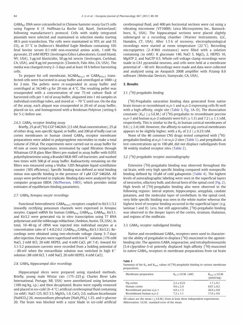
Table 3Summary of the KD and Bmax values of [3H]-pregabalin binding to various membranepreparations.
Membrane preparation KD±S.E.M. (nM) Bmax±S.E.M.(pmol/mg)
Pig cortex 2.2±0.23 1.7±0.1Human cortex 9.6±2.4 0.67±0.2Recombinant porcine α2δ-1 6.0±1.3 26.0±0.8Recombinant human α2δ-2 7.2±1.1 17.0±0.6
All values are the mean (±S.E.M.) from at least three independent experiments.Abbreviation: S.E.M.: standard error of the mean.
84 Z. Li et al. / European Journal of Pharmacology 667 (2011) 80–90
GABAB2 DNA were cotransfected in Chinese hamster ovary Gα15 cellsusing Fugene 6 (F. Hoffman-La Roche Ltd, Basel, Switzerland)following manufacturer's protocol. Cells with stably integratedplasmids were selected and maintained in selection media starting48 h post-transfection. The cells were maintained in 95% air and 5%CO2 at 37 °C in Dulbecco's Modified Eagle Medium containing 10%fetal bovine serum 0.1 mM non-essential amino acids, 1 mM Napyruvate, 25 mMHEPES (Invitrogen/Gibco Laboratories, Grand Island,NY, USA), 3 μg/ml blasticidin, 50 μg/ml zeocin (Invitrogen, Carlsbad,CA, USA), and 8 μg/ml puromycin (Clontech, Palo Alto, CA, USA). Themedia was changed every 2–3 days and at least 18 h before harvestingcells.
To prepare for cell membrane, hGABAB1a/2 or GABAB1b/2 trans-fected cells were harvested in assay buffer and centrifuged at 1000×gfor 2 min. The pellets were re-suspended in assay buffer andcentrifuged at 34,540×g for 20 min at 4 °C. The resulting pellet wasresuspended with a concentration of one 75 ml culture flask ofharvested cells per 1 ml of assay buffer, aliquoted into 1 ml samples inindividual centrifuge tubes, and stored at−70 °C until use. On the dayof the assay, each aliquot was resuspended in 20 ml of assay buffer,placed on ice, and homogenized with a Brinkman Polytron (setting 6for 5 s) before use.
2.6.3. GABAB receptor binding assayBriefly, 25 μl of [3H]-CGP 54626A (2.5 nM, final concentration), 25 μl
of either drug, non-specific ligand, or buffer, and 200 μl of buffy coat ratcortex membranes or human cloned GABAB receptor membranepreparations were added to polypropylene microtubes to make a totalvolume of 250 μl. The experiments were carried out in assay buffer for45 min at room temperature, terminated by rapid filtration throughWhatman GF/B glass fiber filters pre-soaked in assay buffer with 0.05%polyethyleneimine using a BrandelMLR-96T cell harvester, andwashedtwo times with 500 μl of assay buffer. Radioactivity remaining on thefilters was measured using a Wallac 1205 Betaplate liquid scintillationcounter (50% efficiency). Specific binding was defined as total bindingminus non-specific binding in the presence of 1 μM CGP 54626A. Allassays were performed in triplicate. Binding data were analyzed by thecomputer program EBDA (McPherson, 1985), which provides initialestimates of equilibrium-binding parameters.
2.7. GABAB Xenopus oocyte recordings
Functional heterodimeric GABAB1a/2 receptors coupled to Kir3.1/3.2inwardly rectifying potassium channels were expressed in Xenopusoocytes. Capped mRNA for human GABAB1a, GABAB1b, GABAB2, Kir3.1,and Kir3.2 were generated via in vitro transcription using T7 RNApolymerase and themMessagemMachine (Ambion, Austin, TX,USA). Intotal, 10–40 ng of cRNA was injected into individual oocytes at aconcentration ratio of 1:4:0.2:0.2 (GABAB1a:GABAB2:Kir3.1:Kir3.2). Re-cordings were obtained using two-electrode voltage clamp 3–7 daysafter injection. Oocyteswere superfusedwith lowK+ solution (176 mMNaCl, 2 mM KCl, 20 mM HEPES, and 4 mM CaCl, pH 7.4). Inward Kir3.1/3.2 potassium currents were recorded from a holding potential of−80mV when the extracellular solution was switched to high K+
solution (88 mM KCl, 1 mM NaCl, 20 mM HEPES, 4 mM CaCl).
2.8. GABAB hippocampal slice recordings
Hippocampal slices were prepared using standard methods.Briefly, young male Wistar rats (175–225 g; Charles River LabsInternational, Portage, MI, USA) were anesthetized using ketamine(100 mg/kg, i.p.) and then decapitated. Brains were rapidly removedand placed in ice-cold (0–4 °C) artificial cerebrospinal fluid containing(in mM): NaCl 125, KCl 3.5, MgSO4 1.0, CaCl2 2.0, sodium bicarbonate[NaHCO3] 26, monosodium phosphate [NaH2PO4] 1.25, and D-glucose20. The brain was blocked with a razor blade in ice-cold artificial
cerebrospinal fluid, and 400 μm horizontal sections were cut using avibrating microtome (VT1000S; Leica Microsystems Inc., Bannock-burn, IL, USA). The hippocampal sections were placed slightlysubmerged in a recording chamber (Warner Instruments, LLC,Hamden, CT, USA). After 1.5 h of recovery, electrophysiologicalrecordings were started at room temperature (22 °C). Recordingmicropipettes (2–8 MΩ resistance) were filled with a solutioncontaining (in mM): K gluconate 140, NaCl 5, MgCl2 2, HEPES 10,MgATP 2, and NaGTP 0.5. Whole-cell voltage-clamp recordings weremade in CA1 pyramidal neurons, and cells were held at a membranepotential of −60 mV. Recordings were made and data were collectedand analyzed using an Axopatch 200B amplifier with Pclamp 8.0software (Molecular Devices, Sunnyvale, CA, USA).
3. Results
3.1. [3H]-pregabalin binding
[3H]-Pregabalin saturation binding data generated from nativebrain tissues or recombinant α2δ-1 and α2δ-2 expressing cells fit wellwith a high-affinity, single site (Table 3, Fig. 1A–D). The dissociationconstants (KD) (±S.E.M.) of [3H]-pregabalin to recombinant porcineα2δ-1 and humanα2δ-2 subunits were 6.0 (±1.3) and 7.2 (±1.1)nM,respectively. This is similar to the KD in human cortical membranes of9.6 (±2.4)nM. However, the affinity to native pig cortical membranesappears to be slightly higher, with a KD of 2.2 (±0.23)nM.
None of the 46 common CNS drugs tested competed with [3H]-pregabalin binding atα2δ-1 orα2δ-2 sites (Table 1), and pregabalin, attest concentrations up to 100 μM, did not displace radioligands from38 widely studied receptor sites (Table 2).
3.2. [3H]-pregabalin receptor autoradiography
Extensive [3H]-pregabalin binding was observed throughout therat brain, with very high specific binding compared with nonspecificbinding defined by 10 μM of cold gabapentin (Table 4). The highestlevels of autoradiographic labeling were seen in the superficial layersof neocortex, olfactory bulb, and dorsal horn of the spinal cord (Fig. 2).High levels of [3H]-pregabalin binding also were observed in thefollowing regions: lateral septum, hippocampus, amygdala, caudateputamen, and the molecular layer of cerebellum. In the spinal cord,very little specific binding was seen in the white matter whereas thehighest level of receptor binding occurred in the superficial layer (e.g.laminae I and II). Less, but still appreciable, [3H]-pregabalin bindingwas observed in the deeper layers of the cortex, striatum, thalamus,and regions of the midbrain.
3.3. GABAA receptor radioligand binding
Native and recombinant GABAA receptors were used to character-ize the ability of pregabalin to displace [3H]-muscimol to the agonist-binding site. The agonists GABA, isoguvacine, and tetrahydroisoxazolo[5,4-c]pyridine-3-ol potently displaced high-affinity [3H]-muscimolto native GABAA receptors in membrane preparations from rat brain

Bou
nd (
pmol
/mg)
Bou
nd (
pmol
/mg)
Bound (pmol/mg)
B/F
(pm
ol/m
g/nM
)
Bound (pmol/mg)
B/F
(pm
ol/m
g/nM
)
Free [3H]-pregabalin (nM)
A 2.0
25
20
15
10
5
0
1.5
0.9
0.6
0.3
0.0
0.09
0.06
0.03
0.000.0 0.5 1.0 1.5 2.0 0.0 0.1 0.2 0.3 0.50.4
1.0
0.5
0.00 25 50 75 100 125
Free [3H]-pregabalin (nM)
0 25 50 75 100 125
Free [3H]-pregabalin (nM)0 25 50 75 100 125
Free [3H]-pregabalin (nM)
0.6
0.4
0.2
0.0
15
10
5
0
0 25 50 75 100 125
B
DC
Bou
nd (
pmol
/mg)
Bou
nd (
pmol
/mg)
Bound (pmol/mg)
B/F
(pm
ol/m
g/nM
) 2.5
1.5
0.5
0.00 5 10 15 20 25
Bound (pmol/mg)
B/F
(pm
ol/m
g/nM
) 2
1
00 5 10 15 20
Fig. 1. Scatchard analysis of [3H]-pregabalin binding to membranes from (A) pig cortex, (B) human cortex, (C) porcine α2δ-1 cell membranes, and (D) human α2δ-2 cell lines. Thesaturation-binding curve and the Scatchard plot (insert) were generated from a single, representative experiment. The binding assay was run in triplicate with data points generatedfrom the averaged value. The specific binding activity was expressed as pmol of pregabalin bound per milligram of membrane protein. Binding was performed in Kreb–Hepes buffer,pH 7.0.
85Z. Li et al. / European Journal of Pharmacology 667 (2011) 80–90
and to recombinant GABAA receptors frommembranes prepared fromcell lines expressing human α1, β2, and γ2 subunits and α2, β2, andγ2 subunits. In contrast, pregabalin had no effect on [3H]-muscimolbinding to native and recombinant GABAA receptors at concentrationsof up to 1 mM (Table 5).
3.4. GABAA receptor function: rat-cultured cortical neurons
In whole-cell voltage-clamp recordings from cultured neocorticalneurons, 1 μM GABA evoked an inward current response that showedminimal desensitization during the 2–5 s application period. Theagonist-induced current was evoked at intervals of 30–60 s tominimize rundown of the GABA response (Fig. 3A). Diazepamproduced a concentration-dependent enhancement of the GABA-evoked current when co-applied with GABA. Maximal potentiation ofthe GABA response was observed with 300 nM diazepam. Similarconcentration–response studies were carried out with hexobarbital.
Table 4Quantitative summary of [3H]-pregabalin binding in selected brain regions.
Brain region Specific binding(average density: PSL/mm2)a
Spinal cord (dorsal horn gray matter) 95.7±21.5Cerebral cortex 89.7±9.6Amygdala 71.7±17.6Hypothalamus 70.0±19.4Hippocampus 42.4±3.6Thalamus 32.8±3.8
Abbreviation: PSL/mm2: photon stimulated luminescence per square millimeter (density oa Average density is shown in mean±S.D.
In addition to enhancing GABA-evoked responses (Fig. 3B), hexobar-bital also directly activated GABAA receptors in the absence of GABA(data not shown), which has been seen with other barbiturates suchas pentobarbital and phenobarbital (Rho et al., 1996).
To investigate whether pregabalin produces a similar enhance-ment at GABAA receptors, the acute effects of pregabalin (100 μM) on1 μM GABA-evoked currents were tested. The traces in the top panelof Fig. 3A are from the same neuron and show that while 300 nMdiazepam produced a marked enhancement of the GABA-evokedcurrent, the GABA-evoked current in the presence of 100 μMpregabalin (right trace) was similar to control (left trace). Pregabalin(100 μM) by itself did not induce a current response when applied tocells in a similar manner (data not shown). Data from a series ofexperiments similar to that shown in Fig. 3A are summarized in thebar chart in Fig. 3B and clearly indicate that while diazepam andhexobarbital enhance GABA-evoked current responses, pregabalin iswithout effect.
Non-specific binding(average density: PSL/mm2)
n P value
5.2±0.6 2 0.026.8±1.1 4 b0.00016.7±2.6 8 b0.00017.0±3.0 8 b0.00016.6±0.3 4 b0.00017.9±1.1 8 b0.0001
f radioactivity).

A
B
Ctx Hip
Cbm
DH
WM 30 nM pregabalin
30 nM pregabalin + 10 M gabapentin
30 nM pregabalin 30 nM pregabalin+ 10 M gabapentin
ctx
LS Cpu
thDG
H
BLA
ML
Fig. 2. (A) [3H]-Pregabalin binding is abundant but not uniform in rat brain. Twenty-micrometer sagittal sections were incubated with 30 nM [3H]-pregabalin. High level ofbinding is observed in superficial layer of the cortex, hippocampus, cerebellum, and thedorsal horn of the spinal cord. (B) High level of [3H]-pregabalin binding is also observedin lateral septum, basal lateral amygdala, hypothalamus, and mammillary nucleus.Abbreviations: Ctx: cortex; Hip: hippocampus; Cbm: cerebellum; DH: dorsal horn ofthe spinal cord; WM: white matter of the spinal cord; LS: lateral septum; Cpu: caudateputamen, BLA: basolateral amygdala, th: thalamus; H: hypothalamus; ML: mediallateral mammillary nucleus.
Table 5Pregabalin does not displace [3H]-muscimol binding to recombinant and native GABAA
receptors.
Ki (nM)
Compound α1β2γ2 α2β2γ2 Rat brain
Pregabalin N1 mM N1 mM N1 mMGABA 128.2±19.1 134.7±21.3 38.8±11.7THIP 670.3±95.3 477.0±41.0 204.3±39.3Muscimol 23.0±1.67 19.1±3.2 10.0±1.2Isoguvacine 303.7±14.1 219.3±66.7 45.9±14.96Bicuculline 4246.7±1799.7 2856.7±841.6 3510.0a, N100 μM
Data are mean±S.E.M. of three separate experiments.Abbreviations: GABA: γ-aminobutyric acid; THIP: 4,5,6,7-tetrahydroisoxazolo[5,4-c]pyridine-3-ol; S.E.M.: standard error of the mean.
a Mean of two experiments.
3
2
1
0
Fra
ctio
n of
con
trol
(Idr
ug/I c
ontr
ol)
GABA100 M Pregabalin
0.3 M Diazepam
2 sec
500 pA
Pregabalin Diazepam Hexobarbital
A
B
Fig. 3. Lack of effect of pregabalin on GABAA currents in recordings from culturedcortical neurons. (A) Representative traces (from the same neuron) show that while300 nM diazepam produced a marked enhancement of the GABA-evoked current, theGABA-evoked current in the presence of 100 μM pregabalin (top right trace) wassimilar to control (top left trace). (B) Mean fraction of control (±S.E.M.) from a series ofexperiments similar to that shown in Fig. 3A. Abbreviation: GABA: γ-aminobutyric acid.
86 Z. Li et al. / European Journal of Pharmacology 667 (2011) 80–90
3.5. GABAB receptor radioligand binding
Native and recombinant GABAB receptors were used to character-ize the ability of pregabalin to displace [3H]-CGP 54626A to theagonist-binding site. As summarized in Table 6, binding of [3H]-CGP54626A was displaced by several GABAB antagonists in membranesfrom both native and recombinant GABAB receptors. This binding was
also displaced by the synthetic agonist, baclofen. In addition, theendogenous agonist, GABA, displaced [3H]-CGP 54626A binding tonative GABAB receptors in membranes from rat cortex (Ki=1.3 μM)and had lower affinity to human GABAB1a/2 (Ki=30.8 μM) andGABAB1b/2 (Ki=29.5 μM) receptors. In contrast, pregabalin had noaffinity for recombinant GABAB1a/2 and GABAB1b/2 receptors or nativeGABAB receptors, producing minimal displacement of ligand bindingat the highest concentration tested (1 mM).
3.6. GABAB receptor function: xenopus oocyte and rat hippocampal slices
Recombinant GABAB receptors have been shown to couple to G-protein gated inwardly rectifying K+ channels. Gabapentin has beenreported to act as a subtype selective agonist at GABAB receptors anddirectly activate GABAB receptors coupled to inwardly rectifying K+
channels in oocytes expressing GABAB1a/GABAB2 receptors and notGABAB1b/GABAB2 receptors, although these results were not con-firmed by others (Jensen et al., 2002). Similar experiments werecarried out with pregabalin. As shown in Fig. 4, GABA and baclofenproduced an inward current in the presence of high K+ in oocytesexpressing GABAB receptors containing GABAB1a/GABAB2 subunitsand Kir 3.1/3.4 receptors. The inward current responses to GABA andbaclofen were concentration-dependent and were blocked by the

Table 6Pregabalin does not displace [3H]-CGP 54626A binding to native and recombinantGABAB receptors.
Ki (nM)
Compound hGABAB1a/2 clonedreceptors
hGABAB1b/2 clonedreceptors
“Buffy coat” ratcortex
Pregabalin N1 mMa N1 mMa N1 mMa
GABA 30,802±3854 29,527±11,689 1326±280Baclofen 64,596±84,830 54,827±3363 2964±650CGP 54626A 3.8±0.5 5.3±1.0 4.7±0.3CGP 55845A 6.0±0.2 15.9±3.6 6.0±0.7SCH 50911 1130±177 2600±659 895±96
Data are mean±S.E.M. of 2–11 separate experiments.Abbreviations: CGP 54626A: S-(R*,R*)-[3-[[1-(3,4-dichlorophenyl)ethyl]amino]-2-hydroxypropyl](cyclohexylmethyl)phosphinic acid; GABA: γ-aminobutyric acid CGP55845A: (2S)-3-[[(1S)-1-(3,4-dichlorophenyl)ethyl]amino-2-hydroxypropyl](phenylmethyl)phosphinic acid hydrochloride; SCH 50911: (2S)-(+)-5,5-dimethyl-2-morpholine acetic acid; S.E.M.: standard error of the mean.
a IC50.
87Z. Li et al. / European Journal of Pharmacology 667 (2011) 80–90
competitive antagonist, CGP 54626A (data not shown); these data areconsistent with those previously reported by Jensen and colleagues(Jensen et al., 2002). In contrast, pregabalin at concentrations up to100 μM did not induce an inward current response in these sameoocytes.
Finally, in whole-cell recordings from CA1 neurons in hippocampalslices, held at −80 mV, baclofen induced an outward currentresponse that was presumablymediated by GABAB receptor activationof K+ channels (Fig. 5A). The baclofen-induced outward current wasblocked by the specific GABAB receptor antagonist, CGP 55845(Fig. 5A). In contrast, pregabalin, when tested at 100 and 1000 μM
Per
cent
age
of m
axim
alcu
rren
t res
pons
e
0.3 M
1 M
3 M
10 M
500 nA
60 sec
Hi K+
Hi K+
10 M
100 M
GABA
Pregabalin
500 nA
60 sec
BA
DC
Per
cent
age
ofen
hanc
emen
t
Hi K+GABA
Fig. 4. Lack of effect of pregabalin on GABAB currents in oocytes. (A) Representative traces illudependent responses of GABA and baclofen averaged over multiple experiments (average pe100 μM pregabalin. (D) Mean percentage enhancement (±S.E.M.) of GABA vs. pregabalin resp
in these same cells, failed to evoke a baclofen-like outward current(Fig. 5B).
4. Discussion
A series of experiments was undertaken to determine the potency,selectivity, and specificity of pregabalin's receptor-binding profile atα2δ-1 and α2δ-2 subunits of voltage-gated calcium channels. An SPA-binding assay was used to determine the affinity (KD) and receptordensity (Bmax) of [3H]-pregabalin binding to pig and human cerebralcortex, as well as recombinant porcine α2δ-1 and human α2δ-2subunits. [3H]-Pregabalin binds with high affinity to pig and humancortical membranes and binds with high affinity to recombinantporcine α2δ-1 cell membranes and recombinant human α2δ-2 cellmembranes with no subtype preference.
That the predominant ligand binding interaction of pregabalin iswith α2δ-1 and α2δ-2 subunits was further supported by the inabilityto measure a pregabalin effect in any of 38 standard receptor bindingassays tested. Although it is impossible to rule out potentialinteractions at unknown or yet untested receptor sites, to date α2δ-1 and α2δ-2 subtypes of voltage-gated calcium channels are the onlyknown binding sites of pregabalin.
The interaction of pregabalin with α2δ-1 and α2δ-2 subunitsappears unique among other classes of CNS drugs. This wasdemonstrated by the inability of 46 common anticonvulsant,anxiolytic, sedative, antidepressant, antipsychotic, and analgesiccompounds to displace [3H]-pregabalin binding from either subunitwith significant affinity. All of the tested compounds had inhibitoryconcentration 50% (IC50) values greater than 100 μM in both α2δbinding assays, with the exceptions of vigabatrin and trans-(1S,3R)-
1.0
0.8
0.6
0.4
0.2
0.00.01 0.1
10 µMGABA
100 µMPregabalin
1 10
[Agonist], µM
100 1000
GABA Baclofen
200
0
400
600
800
1000
strating concentration-dependent effects of GABA on GABAB currents. (B) Concentration-rcentage of maximal current response±S.E.M.). (C) Individual traces of 10 μM GABA vs.onses (n=15). Abbreviation: GABA: γ-aminobutyric acid.

40 µM 40 µMR-Baclofen R-Baclofen
+10 µMCGP 55845
100 µMPregabalin
1000 µMPregabalin
(17)
(3)
*
(3)
*
(12)
*
B
Cur
rent
(pA
)
20
0
40
60
80
40 M R-Baclofen 40 M R-Baclofen
1000 MPregabalin 10 M CGP 55845
40 M R-Baclofen +10 M CGP 55845
2 min
100 pA
A
Fig. 5. Lack of effect of pregabalin on GABAB activity in CA1 pyramidal neurons of the rat hippocampus. (A) Voltage-clamp recording from a CA1 pyramidal neuron in a rathippocampal slice in vitro. Representative trace from one experiment (one cell) holding at −60 mV. Application of GABAB agonist, R-baclofen (30 s pulse), produced a transientoutward K+ current while application of pregabalin (30 s pulse) did not elicit a current. When the cell was pre-treated for 5 min with CGP 55845A (GABAB antagonist) beforeapplication of R-baclofen, only a very small current was produced. (B) Mean current (±S.E.M.) produced by various treatments for all voltage-clamp experiments. All treatmentswere significantly different from 40 μM R-baclofen, t test, *P≤0.05. Number in parentheses indicates the number of recorded cells.
88 Z. Li et al. / European Journal of Pharmacology 667 (2011) 80–90
ACPD, which had IC50 values of 75 and 80 μM in the α2δ-2 bindingassay, respectively. Both vigabatrin and trans-(1S,3R)-ACPD sharesome structural similarities with pregabalin, which may explain theirweak activity in the competition-binding assay. Nonetheless, theconcentrations at which these compounds showed binding affinity(N2000× compared with pregabalin) are unlikely to be associatedwith α2δ activity in vivo.
In the autoradiography study, the binding of [3H]-pregabalin wasextensive throughout the CNS, with the highest level of bindingobserved in cortex, olfactory bulb, hypothalamus, amygdala, hippo-campus, cerebellum, and dorsal horn of the spinal cord. Bian et al.(2006) reported a comparable radioligand binding profile in micewith similar brain regions containing a high level of [3H]-pregabalin-binding sites. In mice with mutant α2δ-1 protein, R217A, where asingle amino acid, arginine (R) at the 217th position was replaced byalanine (A), the α2δ-1 binding affinity (KD) to [3H]-pregabalin wasreduced by at least 10-fold (Bian et al., 2006; Li et al., 2005), and [3H]-pregabalin binding was dramatically reduced (~90%) in CNS regionsknown to preferentially express the α2δ-1 protein but remained inthose areas where α2δ-2 protein is abundant, such as the cerebellum(Bian et al., 2006). Using an antibody specific to α2δ-1, it was shownthat the immunostaining ofα2δ-1 in the CNS had a similar distributionpattern (e.g. cortex, hippocampus, thalamic and hypothalamic nuclei,and the spinal cord) as that revealed by the [3H]-pregabalin binding inthe present study (Taylor and Garrido, 2008). Although the level of[3H]-pregabalin binding in the molecular layer of the cerebellum washigh, the immunostaining by an α2δ-1 specific antibody only showedlight staining in the cerebellum. These results suggest that the majorbinding sites in the CNS are α2δ-1 subunits, whereas the binding sitesrevealed by [3H]-pregabalin in the cerebellum are mostly α2δ-2subunits. The close correlation between the localization of [3H]-pregabalin-binding sites and α2δ-1 distribution in the CNS revealed
by immunostaining further demonstrates α2δ-1 as the primarybinding site in the brain for pregabalin.
Because initial evidence suggested that gabapentin might act as anagonist at GABAB receptor sites, a series of receptor-binding andelectrophysiological assays was undertaken to systematically addressthe interaction of pregabalin at GABAA and GABAB receptors.Pregabalin, up to concentrations of 1 mM, had no affinity forrecombinant or native GABAA or GABAB receptors. In whole-cellvoltage-clamp recordings from cultured neocortical neurons, diaze-pam and hexobarbital clearly enhanced GABAA-evoked currentresponses while pregabalin was without effect. Gabapentin has beenreported to act as a subtype selective agonist at GABAB receptors anddirectly activate GABAB receptors coupled to inwardly rectifying K+
channels in oocytes expressing GABAB1a/GABAB2 receptors and notGABAB1b/GABAB2 receptors (Bertrand et al., 2001; Bertrand et al.,2003; Ng et al., 2001), but these results were not confirmed by others(Jensen et al., 2002; Lanneau et al., 2001). In the present study,pregabalin, up to 100 μM, failed to induce an inward current responsein similarly prepared oocytes. Finally, in whole-cell recordings fromCA1 neurons in hippocampal slices, pregabalin at concentrations up to1 mM failed to evoke a baclofen-like outward current. Together, thesedata from radioligand receptor-binding and electrophysiologicalassays strongly support a lack of interaction between pregabalin atGABAA and GABAB receptors.
Another potential target for gabapentin and pregabalin is thehyperpolarization-activated current, Ih (Peng et al., 2011; Surges et al.,2003). This mixed cation conductance channel causes inward currentin response to hyperpolarization, and is also modulated by cytosoliccyclic nucleotides. Gabapentin has been reported to increase Ihcurrents, but to date, this action has not been shown to be relevantfor any of the pharmacological actions of gabapentin observed in vivo,and no results have been reported with pregabalin.

89Z. Li et al. / European Journal of Pharmacology 667 (2011) 80–90
Although pregabalin and gabapentin demonstrate potent andselective affinity atα2δ-1 andα2δ-2 subunits of voltage-gated calciumchannels, many studies have failed to demonstrate consistent func-tional effects on calcium currents [for review see (Taylor et al., 2007;Uchitel et al., 2010)]. The reason for these contradictory effects isunclear, especially since drug effects to reduce neurotransmitterrelease are more consistently observed (Taylor et al., 2007; Uchitelet al., 2010). Studies using recombinant voltage-gated calciumchannelsor recordings fromneuronal cell bodies rather than presynaptic endingsmay have contributed to the controversy— several of these studies failto show an effect of gabapentin on calcium influx and it has beenspeculated that this may be due to the lack of interacting proteins thatare normally found at synapses (Taylor, 2009; Uchitel et al., 2010).Indeed, a recent study has shown pregabalin produces a partialinhibition of calcium currents at presynaptic nerve terminals, albeit atrelatively high concentrations (Di Guilmi et al., 2011). Other studydifferences, like changes in neuronal environment, chronicity of drugapplication and level of α2δ subunit expression, have also beensuggested as contributors to the different results (Uchitel et al., 2010).Despite the inconsistent findings on calcium influx, pregabalin andgabapentin demonstrate relatively consistent reductions in the releaseof various neurotransmitters, although in several instances the effectswere achieved only in ‘inflamed’ tissue or tissue treated with excitatoryneuromodulators (Taylor et al., 2007).
A molecular mechanism accounting for the antiallodynic effects ofpregabalin in the treatment of neuropathic pain was recentlyadvanced by Bauer and colleagues (Bauer et al., 2009; Bauer et al.,2010). Using the spinal nerve ligation model of neuropathic pain inrats, Bauer et al. (Bauer et al., 2009) demonstrated that an efficaciousdose of chronic pregabalin treatment inhibited the trafficking ofα2δ-1subunits in the DRG cell bodies to their presynaptic terminals in thedorsal horn of the spinal cord. This impairment of α2δ-1 traffickingcould explain pregabalin's documented reduction of presynapticneurotransmitter release in the spinal cord (Taylor et al., 2007) andsubsequent spinal sensitization ultimately causing analgesic effects(Bauer et al., 2010). Seemingly contradictory data were obtained in astudy (Lynch et al., 2006) that failed to demonstrate consistentanalgesic effects among compounds with comparable affinity for theα2δ protein; however, the radioligand, (L)-[3H]-leucine, used in thisstudy is not specific to theα2δ subunit in mouse cortex, and therefore,the receptor affinities of the tested compounds may not reflectselective activity at the α2δ subunit.
This series of experiments indicates that the therapeutic effects ofpregabalin are likely mediated selectively throughα2δ-1 and/orα2δ-2subunits of voltage-gated calcium channels. Whether this activity issufficient to account for the variety of therapeutic effects demon-strated by pregabalin is still unclear, however, recent advances (Baueret al., 2009; Bauer et al., 2010; Field et al., 2006; Lotarski et al., 2011;Offord et al., 2010) suggest that the molecular mechanisms account-ing for analgesic, anticonvulsant and anxiolytic-like efficacy areconsistent with an interaction at α2δ-1 subunits.
Role of the funding source
Research and professional medical writing and editing supportwere funded by Pfizer Inc. Authors who were employees of Pfizer Inc.were involved in study design, the collection, analysis and interpre-tation of data, the writing of the report and the decision to submit thepaper for publication.
Acknowledgments/Conflicts of interest
Research was funded by Pfizer Inc. Zheng Li, Charles P. Taylor,Mark Weber, Julie Piechan, Faith Prior, Feng Bian, Mei Cui, and SeanDonevan were employees of Pfizer Inc. when the studies wereconducted. Diane Hoffman is an employee of UBC Scientific Solutions
and was a paid consultant to Pfizer in connection with thedevelopment of this manuscript. Editorial support for the develop-ment of the manuscript was provided by Diane Hoffman of UBCScientific Solutions and was funded by Pfizer Inc. Parts of thismanuscript were previously presented at the Annual Meeting of theSociety for Neuroscience, 2004 [Piechan, J.L., Donevan, S.D., Taylor,C.P., Dickerson, M.R., Li, Z., 2004. Pregabalin, a novel anticonvulsant,analgesic, anxiolytic drug, exhibits class-specific alpha2-delta-1 andalpha2-delta-2 calcium channel subunit binding. Abstr.-Soc Neurosci(Program No. 115.11)]; and the 26th International Epilepsy Congress,2005 [Li, Z., Donevan, S., Taylor, C.P., Piechan, J., Offord, J., Su, T.Z.,Baron, S., Vartanian, M.G., Bian, F., Wustrow, D.J., Belliotti, T., Schwarz, J.and Thorpe, A., 2005. High-affinity binding of pregabalin at alpha-2-delta subunits of voltage-gated calcium channel: Contribution toanticonvulsant action. Epilepsia 46, 280].
References
Bauer, C.S., Nieto-Rostro, M., Rahman, W., Tran-Van-Minh, A., Ferron, L., Douglas, L.,Kadurin, I., Sri Ranjan, Y., Fernandez-Alacid, L., Millar, N.S., Dickenson, A.H., Lujan,R., Dolphin, A.C., 2009. The increased trafficking of the calcium channel subunitalpha2delta-1 to presynaptic terminals in neuropathic pain is inhibited by thealpha2delta ligand pregabalin. J. Neurosci. 29, 4076–4088.
Bauer, C.S., Rahman, W., Tran-van-Minh, A., Lujan, R., Dickenson, A.H., Dolphin, A.C.,2010. The anti-allodynic alpha(2)delta ligand pregabalin inhibits the trafficking ofthe calcium channel alpha(2)delta-1 subunit to presynaptic terminals in vivo.Biochem. Soc. Trans. 38, 525–528.
Belliotti, T.R., Capiris, T., Ekhato, I.V., Kinsora, J.J., Field, M.J., Heffner, T.G., Meltzer, L.T.,Schwarz, J.B., Taylor, C.P., Thorpe, A.J., Vartanian, M.G., Wise, L.D., Zhi-Su, T., Weber,M.L., Wustrow, D.J., 2005. Structure–activity relationships of pregabalin andanalogues that target the α2-δ protein. J Med Chem 48, 2294–2307.
Bertrand, S., Ng, G.Y., Purisai, M.G., Wolfe, S.E., Severidt, M.W., Nouel, D., Robitaille, R.,Low, M.J., O'Neill, G.P., Metters, K., Lacaille, J.C., Chronwall, B.M., Morris, S.J., 2001.The anticonvulsant, antihyperalgesic agent gabapentin is an agonist at brain γ-aminobutyric acid type B receptors negatively coupled to voltage-dependentcalcium channels. J. Pharmacol. Exp. Ther. 298, 15–24.
Bertrand, S., Nouel, D., Morin, F., Nagy, F., Lacaille, J.C., 2003. Gabapentin actions on Kir3currents and N-type Ca2+ channels via GABAB receptors in hippocampal pyramidalcells. Synapse 50, 95–109.
Bian, F., Li, Z., Offord, J., Davis, M.D., McCormick, J., Taylor, C.P., Walker, L.C., 2006.Calcium channel alpha2-delta type 1 subunit is the major binding protein forpregabalin in neocortex, hippocampus, amygdala, and spinal cord: an ex vivoautoradiographic study in alpha2-delta type 1 genetically modified mice. Brain Res1075, 68–80.
Di Guilmi, M.N., Urbano, F.J., Inchauspe, C.G., Uchitel, O.D., 2011. Pregabalinmodulation ofneurotransmitter release is mediated by change in intrinsic activation/inactivationproperties of cav2.1 calcium channels. J. Pharmacol. Exp. Ther. 336, 973–982.
Donevan, S.D., Rogawski, M.A., 1996. Multiple actions of arylalkylamine arthropodtoxins on the N-methyl-D-aspartate receptor. Neuroscience 70, 361–375.
Dworkin, R.H., Corbin, A.E., Young Jr., J.P., Sharma, U., LaMoreaux, L., Bockbrader, H.,Garofalo, E.A., Poole, R.M., 2003. Pregabalin for the treatment of postherpeticneuralgia: a randomized, placebo-controlled trial. Neurology 60, 1274–1283.
Feltner, D.E., Crockatt, J.G., Dubovsky, S.J., Cohn, C.K., Shrivastava, R.K., Targum, S.D., Liu-Dumaw, M., Carter, C.M., Pande, A.C., 2003. A randomized, double-blind, placebo-controlled, fixed-dose, multicenter study of pregabalin in patients with generalizedanxiety disorder. J. Clin. Psychopharmacol. 23, 240–249.
Field, M.J., Cox, P.J., Stott, E., Melrose, H., Offord, J., Su, T.Z., Bramwell, S., Corradini, L.,England, S., Winks, J., Kinloch, R.A., Hendrich, J., Dolphin, A.C., Webb, T., Williams,D., 2006. Identification of the α2-δ-1 subunit of voltage-dependent calciumchannels as a molecular target for pain mediating the analgesic actions ofpregabalin. Proc Natl Acad Sci U S A 103, 17537–17542.
French, J.A., Kugler, A.R., Robbins, J.L., Knapp, L.E., Garofalo, E.A., 2003. Dose–responsetrial of pregabalin adjunctive therapy in patients with partial seizures. Neurology60, 1631–1637.
Hill, D.R., Suman-Chauhan, N., Woodruff, G.N., 1993. Localization of [3H]gabapentin to anovel site in rat brain: autoradiographic studies. Eur. J. Pharmacol. 244, 303–309.
Jensen, A.A., Mosbacher, J., Elg, S., Lingenhoehl, K., Lohmann, T., Johansen, T.N.,Abrahamsen, B., Mattsson, J.P., Lehmann, A., Bettler, B., Bräuner-Osborne, H., 2002.The anticonvulsant gabapentin (Neurontin) does not act through γ-aminobutyricacid-B receptors. Mol. Pharmacol. 61, 1377–1384.
Lanneau, C., Green, A., Hirst, W.D., Wise, A., Brown, J.T., Donnier, E., Charles, K.J., Wood,M., Davies, C.H., Pangalos, M.N., 2001. Gabapentin is not a GABAB receptor agonist.Neuropharmacology 41, 965–975.
Li, Z., Donevan, S., Taylor, C.P., Piechan, J., Offord, J., Su, T.Z., Baron, S., Vartanian, M.G.,Bian, F., Wustrow, D.J., Belliotti, T., Schwarz, J., Thorpe, A., 2005. High-affinitybinding of pregabalin at alpha-2-delta subunits of voltage-gated calcium channel:contribution to anticonvulsant action [abstract no. p866]. Epilepsia 46, 280.
Lotarski, S.M., Donevan, S., El-Kattan, A., Osgood, S., Poe, J., Taylor, C.P., Offord, J., 2011.Anxiolytic-Like activity of pregabalin in the vogel conflict test in {alpha}2{delta}-1(R217A) and {alpha}2{delta}-2 (R279A) mouse mutants. J. Pharmacol. Exp. Ther.May 10 [Epub ahead of print].

90 Z. Li et al. / European Journal of Pharmacology 667 (2011) 80–90
Lynch III, J.J., Honore, P., Anderson, D.J., Bunnelle, W.H., Mortell, K.H., Zhong, C., Wade,C.L., Zhu, C.Z., Xu, H., Marsh, K.C., Lee, C.H., Jarvis, M.F., Gopalakrishnan, M., 2006.(L)-Phenylglycine, but not necessarily other alpha2delta subunit voltage-gatedcalcium channel ligands, attenuates neuropathic pain in rats. Pain 125, 136–142.
McPherson, G.A., 1985. Analysis of radioligand binding experiments. A collection ofcomputer programs for the IBM PC. J Pharmacol Methods 14, 213–228.
Ng, G.Y., Bertrand, S., Sullivan, R., Ethier, N., Wang, J., Yergey, J., Belley, M., Trimble, L.,Bateman, K., Alder, L., Smith, A., McKernan, R., Metters, K., O'Neill, G.P., Lacaille, J.C.,Hébert, T.E., 2001. γ-Aminobutyric acid type B receptors with specific heterodimercomposition and postsynaptic actions in hippocampal neurons are targets ofanticonvulsant gabapentin action. Mol. Pharmacol. 59, 144–152.
Offord, J., Lotarski, S., Peterson, J., Galvin, S., Strenkowski, B., Hain, H., Donevan, S., 2010.Anticonvulsant activity of pregabalin (Lyrica) requires binding to the α2δ1 subunitof voltage sensitive calcium channels. Presented at the American Epilepsy Society -64th Annual Meeting, San Antonio, TX, December 3–7, 2010. http://www.aesnet.org/go/publications/aes-abstracts/abstract-search/mode/display/st/offord/sy/2010/sb/All/id/12438. Abstract # 1.238.
Peng, B.W., Justice, J.A., Zhang, K., Li, J.X., He, X.H., Sanchez, R.M., 2011. Gabapentinpromotes inhibition by enhancing hyperpolarization-activated cation currents andspontaneous firing in hippocampal CA1 interneurons. Neurosci. Lett. 494, 19–23.
Rho, J.M., Donevan, S.D., Rogawski, M.A., 1996. Direct Activation of GABAA receptors bybarbiturates in cultured rat hippocampal neurons. J. Physiol. 497, 509–522.
Straube, S., Derry, S., Moore, R.A., McQuay, H.J., 2010. Pregabalin in fibromyalgia: meta-analysis of efficacy and safety from company clinical trial reports. Rheumatology(Oxford) 49, 706–715.
Surges, R., Freiman, T.M., Feuerstein, T.J., 2003. Gabapentin increases the hyperpolarization-activated cation current Ih in rat CA1 pyramidal cells. Epilepsia 44, 150–156.
Taylor, C.P., 2009. Mechanisms of analgesia by gabapentin and pregabalin—calciumchannel alpha2-delta [Cavalpha2-delta] ligands. Pain 142, 13–16.
Taylor, C.P., Garrido, R., 2008. Immunostaining of rat brain, spinal cord, sensory neuronsand skeletal muscle for calcium channel alpha2-delta (alpha2-delta) type 1 protein.Neuroscience 155, 510–521.
Taylor, C.P., Angelotti, T., Fauman, E., 2007. Pharmacology and mechanism of action ofpregabalin: the calcium channel α2-δ (alpha2-delta) subunit as a target forantiepileptic drug discovery. Epilepsy Res 73, 137–150.
Uchitel, O.D., Di Guilmi, M.N., Urbano, F.J., Gonzalez-Inchauspe, C., 2010. Acutemodulation of calcium currents and synaptic transmission by gabapentinoids.Channels (Austin) 4, 490–496.
Wang, M., Offord, J., Oxender, D.L., Su, T.Z., 1999. Structural requirement of the calcium-channel subunit α2δ for gabapentin binding. Biochem. J. 342, 313–320.