Embed Size (px)
Citation preview
Principle of Principle of Inflammation Inflammation and Infectionand Infection
NatapolNatapol SupanatsetakulSupanatsetakul MD, PhD.MD, PhD.Dept. of Pathology and Forensic MedicineDept. of Pathology and Forensic MedicineFaculty of Medicine, Faculty of Medicine, NaresuanNaresuan UniversityUniversity
17 มิถนุายน พ.ศ. 2554
TOPICSTOPICS
InflammationInflammation–– Acute inflammationAcute inflammation–– Chronic inflammationChronic inflammation
Tissue repairTissue repair
Principle of Infectious DiseasePrinciple of Infectious Disease
INFLAMMATIONINFLAMMATION
Complex reactions to injurious agent Complex reactions to injurious agent that consists of that consists of –– Vascular responsesVascular responses–– Migration and activation of leukocytesMigration and activation of leukocytes–– Systemic responsesSystemic responses
Closely intertwined with the process of Closely intertwined with the process of repairrepair
5 cardinal signs5 cardinal signs: pain, swelling, : pain, swelling, erythemaerythema, , heat and loss of functionheat and loss of function
Inflammation is fundamentally a protective Inflammation is fundamentally a protective response, but may be potentially harmfulresponse, but may be potentially harmful
Inflammation consists of two componentsInflammation consists of two components–– Vascular reactionVascular reaction–– Cellular reactionCellular reaction
Vascular and cellular reactions are mediated Vascular and cellular reactions are mediated by by chemical factorschemical factors, derived from plasma , derived from plasma proteins or cells (Cytokines) and are proteins or cells (Cytokines) and are produced in response to or activated by the produced in response to or activated by the stimuli stimuli
Inflammation is divided intoInflammation is divided into–– Acute inflammationAcute inflammation–– Chronic inflammationChronic inflammation
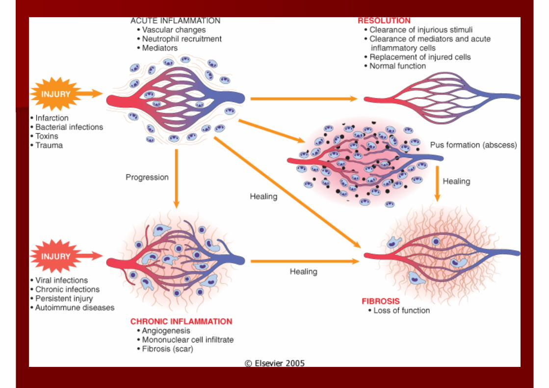
ACUTE INFLAMMATIONACUTE INFLAMMATION
Rapid response to an injurious agentRapid response to an injurious agentthat serves to deliver mediators of host that serves to deliver mediators of host defense (leukocytes and plasma proteins) defense (leukocytes and plasma proteins) to the site of injury to the site of injury
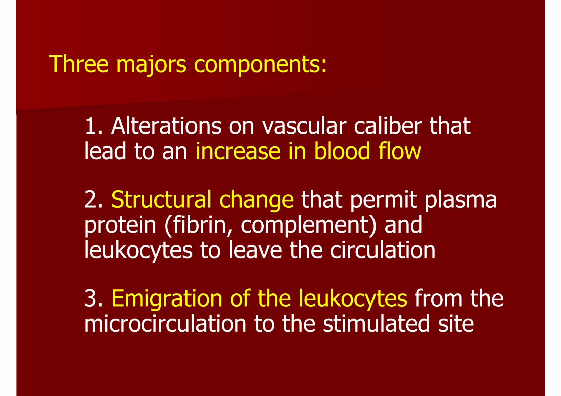
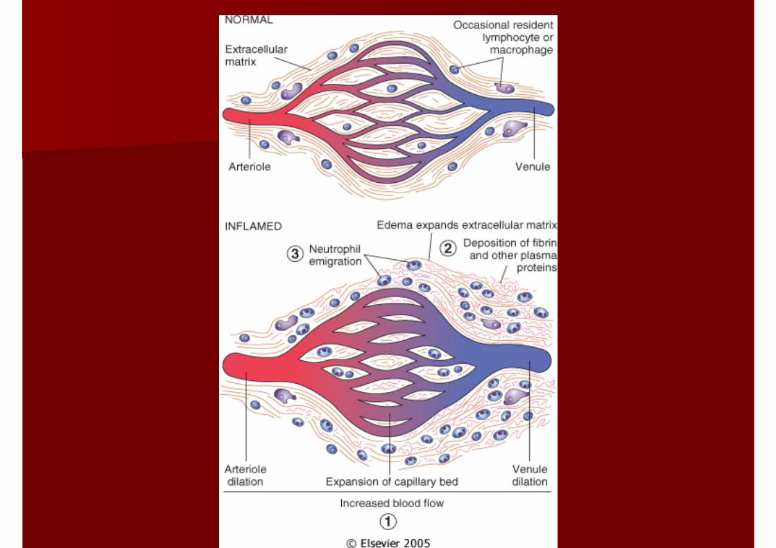
Three majors components:
1. Alterations on vascular caliber that lead to an increase in blood flow
2. Structural change that permit plasma protein (fibrin, complement) and leukocytes to leave the circulation
3. Emigration of the leukocytes from the microcirculation to the stimulated site
Stimuli for Acute inflammationStimuli for Acute inflammation
Infections and microbial toxinsInfections and microbial toxinsTraumaTraumaPhysical and chemical agentsPhysical and chemical agentsTissue necrosisTissue necrosisForeign bodiesForeign bodiesImmune reactionsImmune reactions
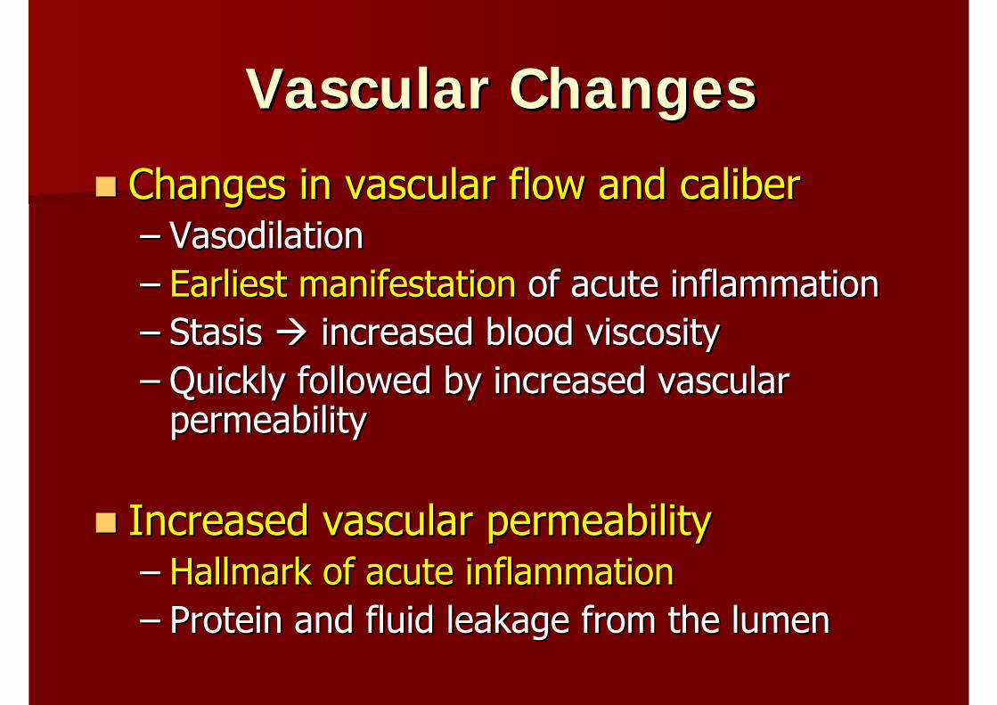
Vascular ChangesVascular Changes
Changes in vascular flow and caliberChanges in vascular flow and caliber–– Vasodilation Vasodilation –– Earliest manifestationEarliest manifestation of acute inflammationof acute inflammation–– Stasis Stasis increased blood viscosity increased blood viscosity –– Quickly followed by increased vascular Quickly followed by increased vascular
permeabilitypermeability
Increased vascular permeabilityIncreased vascular permeability–– Hallmark of acute inflammationHallmark of acute inflammation–– Protein and fluid leakage from the lumenProtein and fluid leakage from the lumen
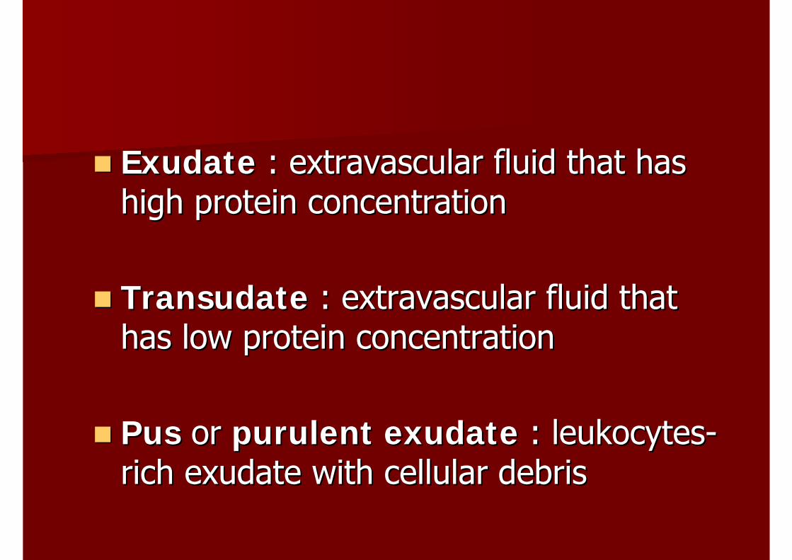
ExudateExudate : extravascular fluid that has : extravascular fluid that has high protein concentrationhigh protein concentration
TransudateTransudate : extravascular fluid that : extravascular fluid that has low protein concentrationhas low protein concentration
PusPus or or purulent exudatepurulent exudate : leukocytes: leukocytes--rich rich exudateexudate with cellular debriswith cellular debris
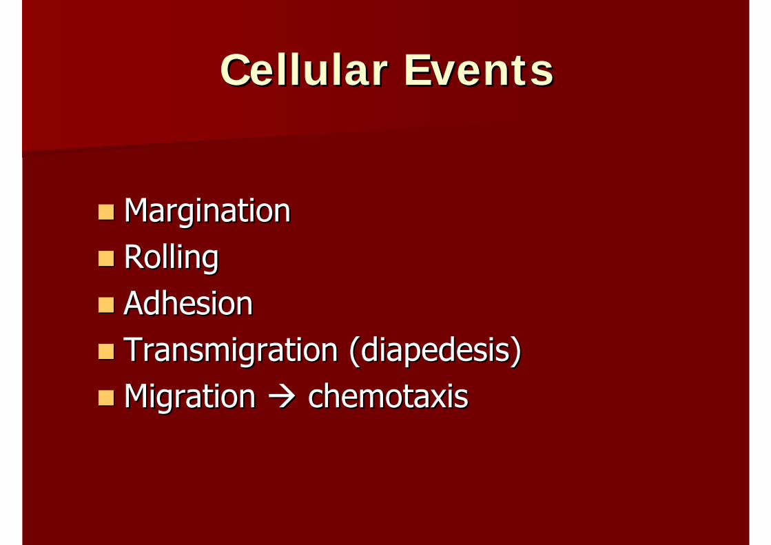
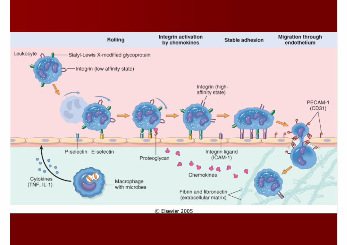
Cellular EventsCellular Events
Margination Margination Rolling Rolling AdhesionAdhesionTransmigration (diapedesis)Transmigration (diapedesis)Migration Migration chemotaxischemotaxis
CHEMICAL MEDIATORS OF CHEMICAL MEDIATORS OF INFLAMMATIONINFLAMMATION
Mediators or originate either from plasma from cellsMediators or originate either from plasma from cellsThe production is triggers by microbial products or The production is triggers by microbial products or by host proteins, other chemical mediatorsby host proteins, other chemical mediatorsMediators perform activity by binding their specific Mediators perform activity by binding their specific receptorsreceptorsOne mediator can stimulate the release of other One mediator can stimulate the release of other mediatorsmediatorsMediators have different effects on different cell Mediators have different effects on different cell typestypesMost mediators are shortMost mediators are short--livedlived
Chemical mediatorsChemical mediators
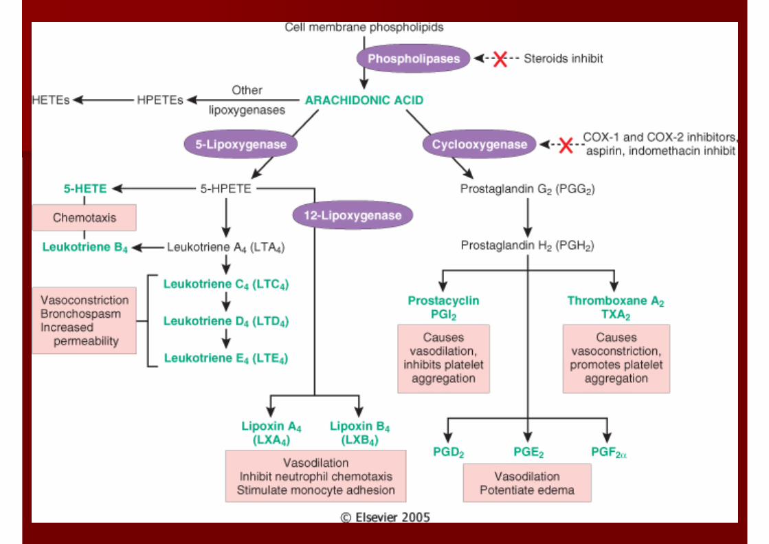
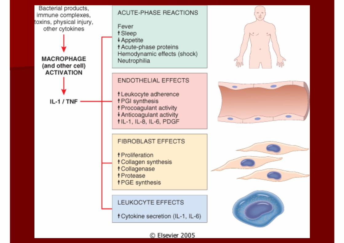
VasoactiveVasoactive aminesamines: : HistamineHistaminePlasma proteinsPlasma proteins: complement system, : complement system, kininkinin system, clotting and system, clotting and fibrinolyticfibrinolytic systemsystemArachidonicArachidonic acid metabolitesacid metabolites: prostaglandins, : prostaglandins, leukotrienesleukotrienes, , thromboxanesthromboxanesCytokines and Cytokines and chemokineschemokines: Interleukin: Interleukin--1(IL1(IL--1), 1), Tumor necrotic factor (TNF)Tumor necrotic factor (TNF)Nitric oxideNitric oxide
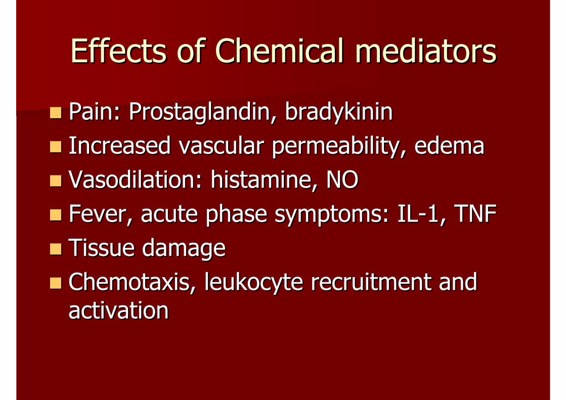
Effects of Chemical mediatorsEffects of Chemical mediators
Pain: Prostaglandin, Pain: Prostaglandin, bradykininbradykininIncreased vascular permeability, edemaIncreased vascular permeability, edemaVasodilationVasodilation: histamine, NO : histamine, NO Fever, acute phase symptoms: ILFever, acute phase symptoms: IL--1, TNF1, TNFTissue damageTissue damageChemotaxisChemotaxis, leukocyte recruitment and , leukocyte recruitment and activationactivation
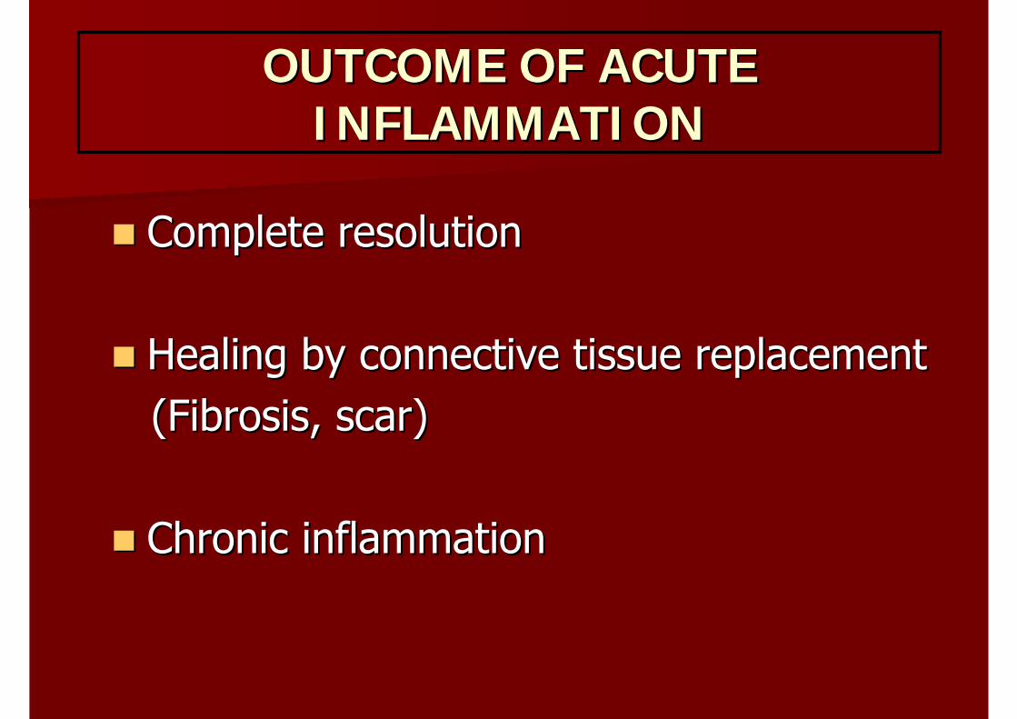
OUTCOME OF ACUTE OUTCOME OF ACUTE INFLAMMATIONINFLAMMATION
Complete resolutionComplete resolution
Healing by connective tissue replacementHealing by connective tissue replacement(Fibrosis, scar)(Fibrosis, scar)
Chronic inflammation Chronic inflammation
MORPHOLOGIC PATTERN OF MORPHOLOGIC PATTERN OF ACUTE INFLAMMATIONACUTE INFLAMMATION
Serous inflammationSerous inflammation–– BurnBurn–– Inflammation in the body cavityInflammation in the body cavity
Fibrinous inflammationFibrinous inflammation–– Severe injury, results in greater vascular Severe injury, results in greater vascular
permeabilitypermeability–– Leakage of fibrinogen (plasma protein)Leakage of fibrinogen (plasma protein)
Suppurative or purulent inflammationSuppurative or purulent inflammation–– Inflammation with pus or purulent exudate Inflammation with pus or purulent exudate
formationformation–– Acute appendicitisAcute appendicitis–– Acute meningitisAcute meningitis–– Abscess : localized collections of purulent Abscess : localized collections of purulent
inflammatory tissueinflammatory tissue–– FibrinopurulentFibrinopurulent inflammation inflammation
Ulcers Ulcers –– Local defect or excavation of the surface of Local defect or excavation of the surface of
an organ or tissue an organ or tissue –– Most common encounter inMost common encounter in
Oral mucosaOral mucosaSubcutaneous tissueSubcutaneous tissue
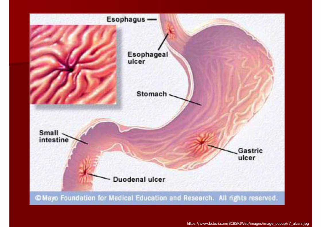
https://www.bcbsri.com/BCBSRIWeb/images/image_popup/r7_ulcers.jpg
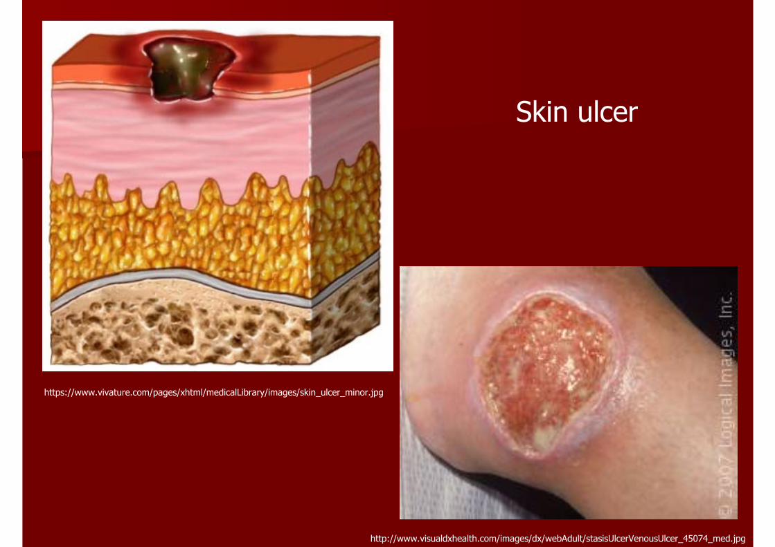
https://www.vivature.com/pages/xhtml/medicalLibrary/images/skin_ulcer_minor.jpg
http://www.visualdxhealth.com/images/dx/webAdult/stasisUlcerVenousUlcer_45074_med.jpg
Skin ulcer
CHRONIC INFLAMMATIONCHRONIC INFLAMMATION
Inflammation of prolonged duration Inflammation of prolonged duration (weeks or months) in which active inflammation, (weeks or months) in which active inflammation, tissue destruction, and attempts at repair are tissue destruction, and attempts at repair are proceeding simultaneously. proceeding simultaneously.
Cause of chronic inflammationCause of chronic inflammation
Persistent infectionPersistent infection
Prolonged exposure to potentially toxic Prolonged exposure to potentially toxic agents, either exogenous or endogenousagents, either exogenous or endogenous
Autoimmunity Autoimmunity
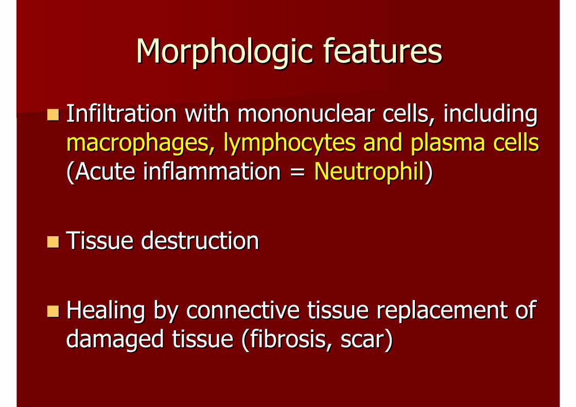
Morphologic featuresMorphologic features
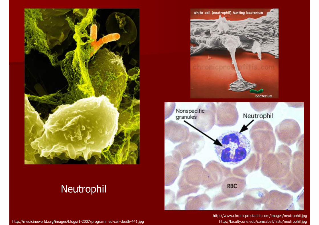
Infiltration with mononuclear cells, including Infiltration with mononuclear cells, including macrophages, lymphocytes and plasma cells macrophages, lymphocytes and plasma cells (Acute inflammation = (Acute inflammation = NeutrophilNeutrophil))
Tissue destructionTissue destruction
Healing by connective tissue replacement of Healing by connective tissue replacement of damaged tissue (fibrosis, scar)damaged tissue (fibrosis, scar)
http://www.chronicprostatitis.com/images/neutrophil.jpghttp://faculty.une.edu/com/abell/histo/neutrophil.jpghttp://medicineworld.org/images/blogs/1-2007/programmed-cell-death-441.jpg
Neutrophil
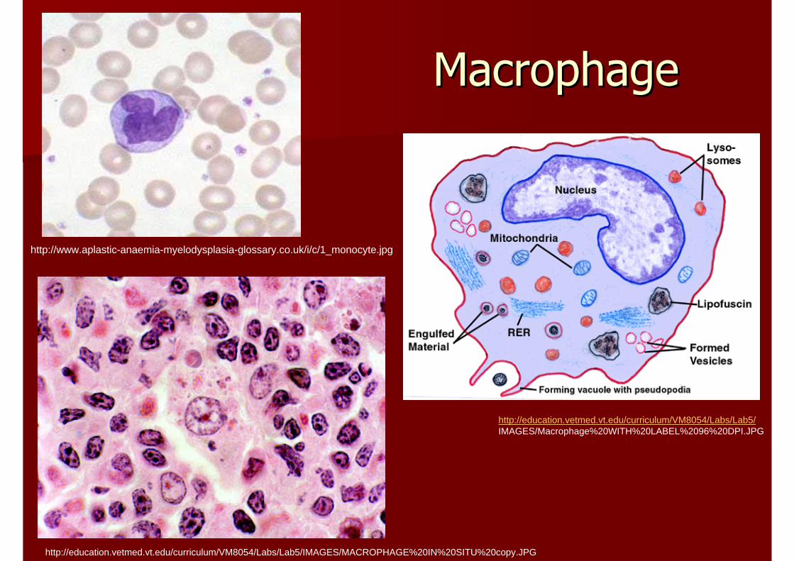
MacrophageMacrophage
http://www.aplastic-anaemia-myelodysplasia-glossary.co.uk/i/c/1_monocyte.jpg
http://education.vetmed.vt.edu/curriculum/VM8054/Labs/Lab5/IMAGES/MACROPHAGE%20IN%20SITU%20copy.JPG
http://education.vetmed.vt.edu/curriculum/VM8054/Labs/Lab5/IMAGES/Macrophage%20WITH%20LABEL%2096%20DPI.JPG
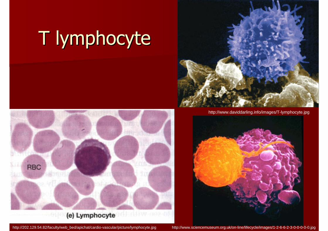
T lymphocyteT lymphocyte
http://202.129.54.82/faculty/web_bed/apichat/cardio-vascular/picture/lymphocyte.jpg
http://www.daviddarling.info/images/T-lymphocyte.jpg
http://www.sciencemuseum.org.uk/on-line/lifecycle/images/1-2-6-6-2-3-0-0-0-0-0.jpg
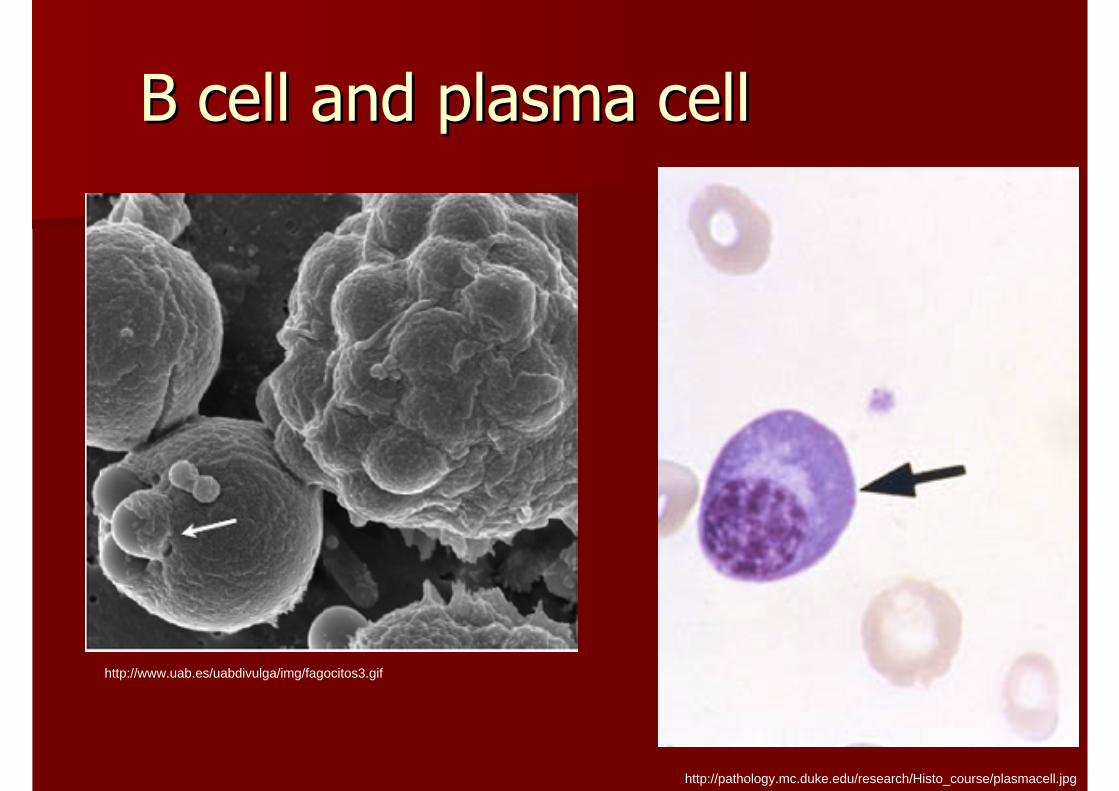
B cell and plasma cellB cell and plasma cell
http://pathology.mc.duke.edu/research/Histo_course/plasmacell.jpg
http://www.uab.es/uabdivulga/img/fagocitos3.gif
https://www.med.illinois.edu/m34/clerkships/surgery/student/other/path/slides/Cholelithiasis%20&%20Chronic%20Cholecystitis.jpg
http://www.pathology.vcu.edu/education/gi/MacronodularCirrhosisHepatitisC.jpg
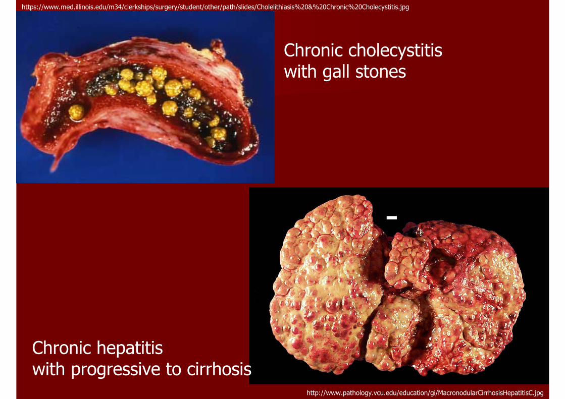
Chronic cholecystitiswith gall stones
Chronic hepatitis with progressive to cirrhosis
TOPICSTOPICS
InflammationInflammation–– Acute inflammationAcute inflammation–– Chronic inflammationChronic inflammation
Tissue repairTissue repair
Principle of Infectious DiseasePrinciple of Infectious Disease
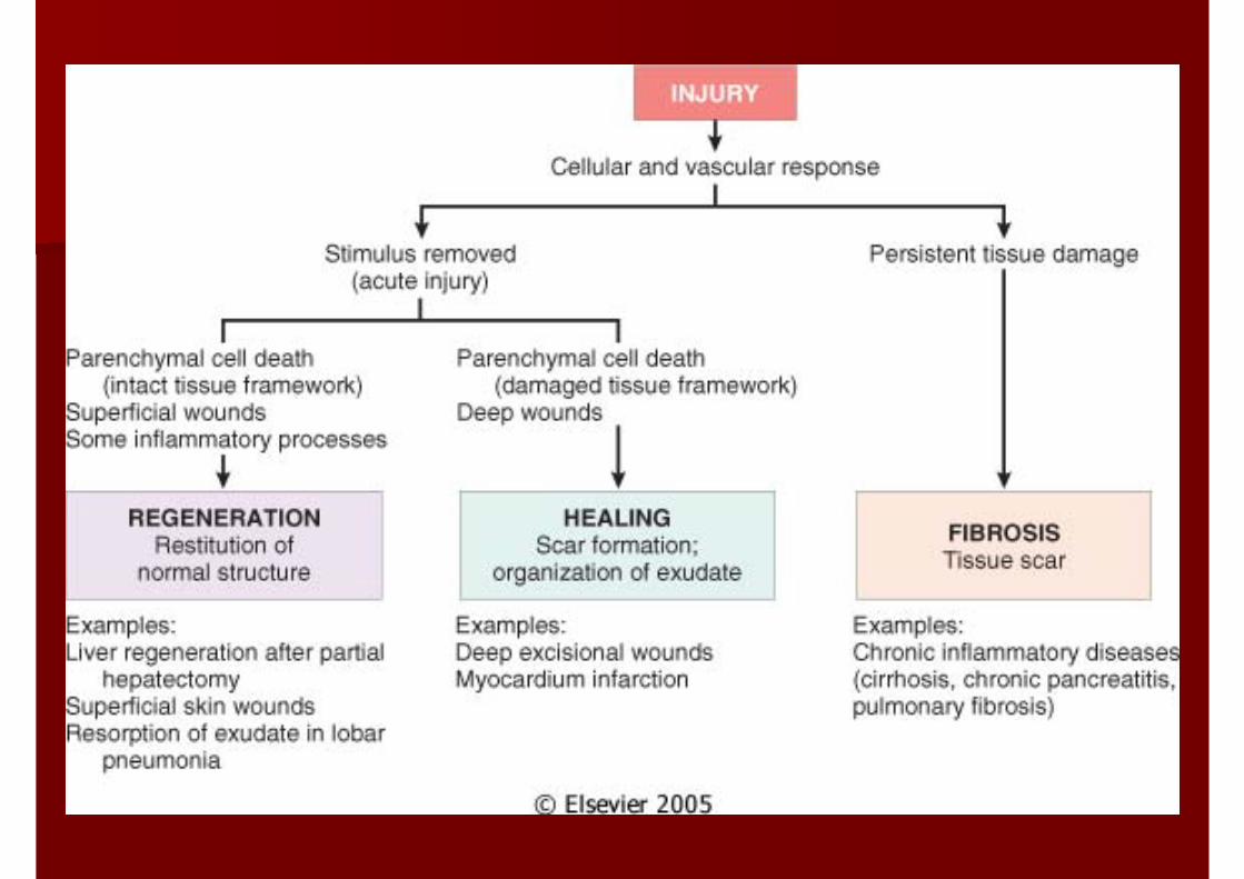
REPAIRREPAIR
Maintenance of normal structure and Maintenance of normal structure and function and survival of the organism function and survival of the organism
RegenerationRegeneration
Healing: scar formation and fibrosisHealing: scar formation and fibrosis
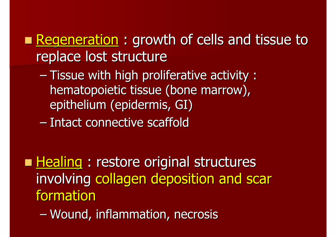
RegenerationRegeneration : growth of cells and tissue to : growth of cells and tissue to replace lost structurereplace lost structure–– Tissue with high proliferative activity : Tissue with high proliferative activity :
hematopoietic tissue (bone marrow), hematopoietic tissue (bone marrow), epithelium (epidermis, GI)epithelium (epidermis, GI)
–– Intact connective scaffoldIntact connective scaffold
HealingHealing : restore original structures : restore original structures involving involving collagen deposition and scar collagen deposition and scar formationformation–– Wound, inflammation, necrosisWound, inflammation, necrosis
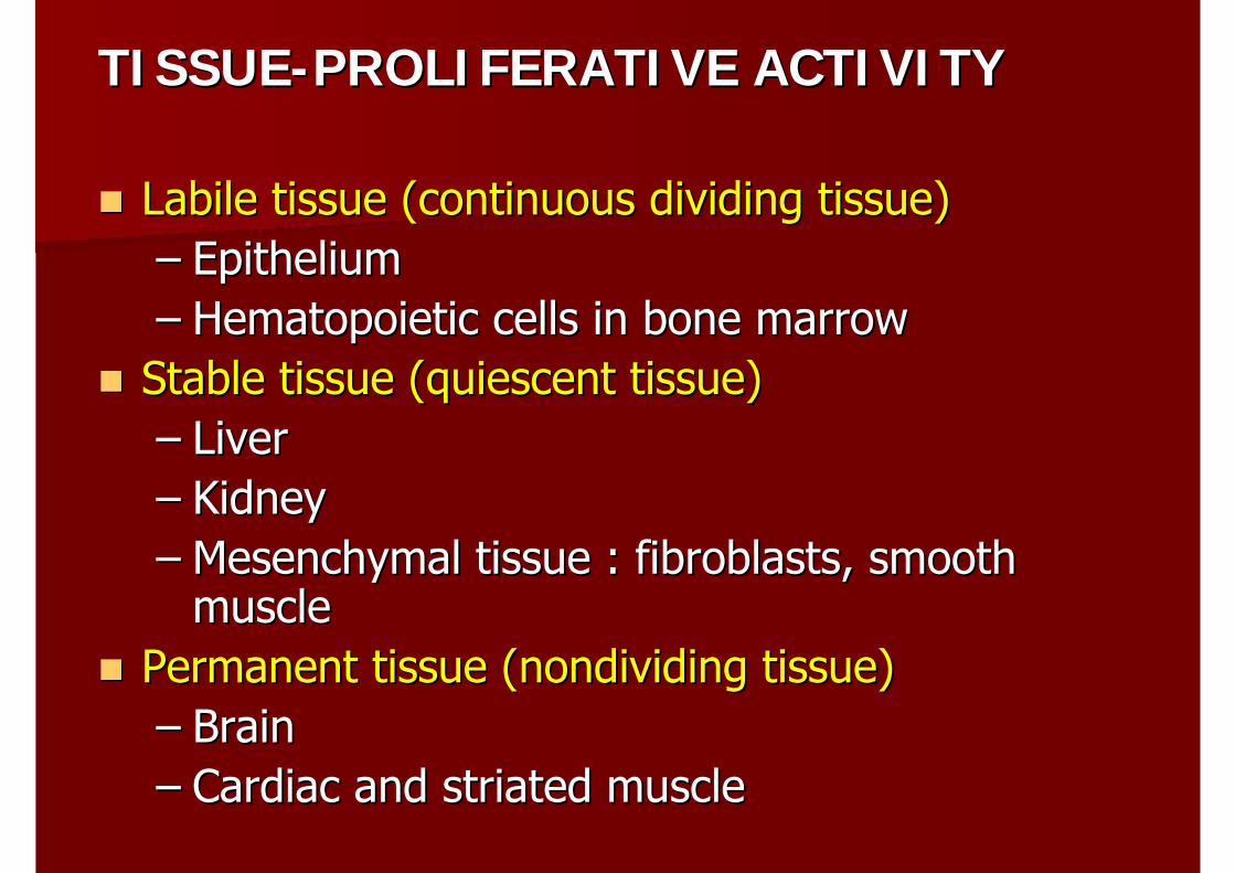
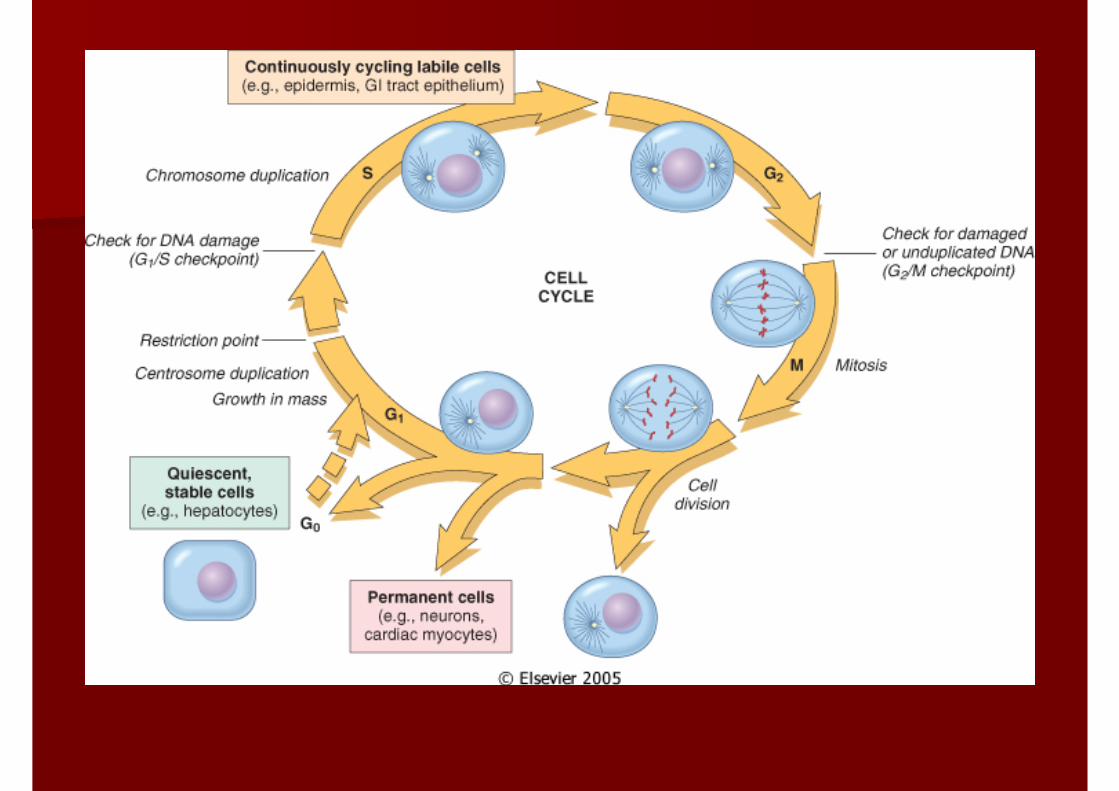
TISSUETISSUE--PROLIFERATIVE ACTIVITYPROLIFERATIVE ACTIVITY
Labile tissue (continuous dividing tissue)Labile tissue (continuous dividing tissue)–– EpitheliumEpithelium–– Hematopoietic cells in bone marrowHematopoietic cells in bone marrow
Stable tissue (quiescent tissue)Stable tissue (quiescent tissue)–– LiverLiver–– Kidney Kidney –– Mesenchymal tissue : fibroblasts, smooth Mesenchymal tissue : fibroblasts, smooth
musclemusclePermanent tissue (Permanent tissue (nondividingnondividing tissue)tissue)–– Brain Brain –– Cardiac and striated muscleCardiac and striated muscle
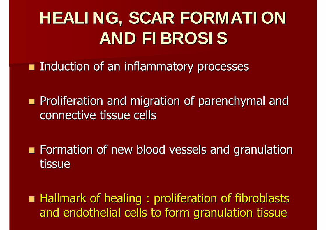
HEALING, SCAR FORMATION HEALING, SCAR FORMATION AND FIBROSISAND FIBROSIS
Induction of an inflammatory processesInduction of an inflammatory processes
Proliferation and migration of parenchymal and Proliferation and migration of parenchymal and connective tissue cellsconnective tissue cells
Formation of new blood vessels and granulation Formation of new blood vessels and granulation tissuetissue
Hallmark of healing : proliferation of fibroblasts Hallmark of healing : proliferation of fibroblasts and endothelial cells to form granulation tissueand endothelial cells to form granulation tissue
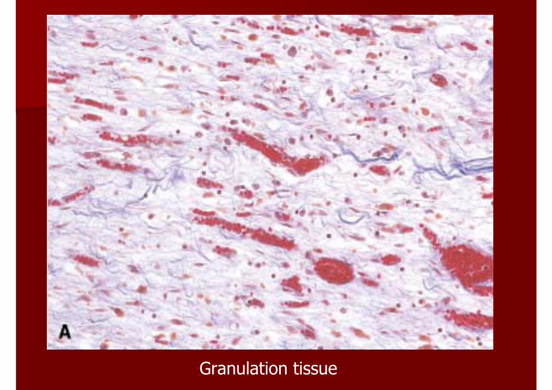
Granulation tissue
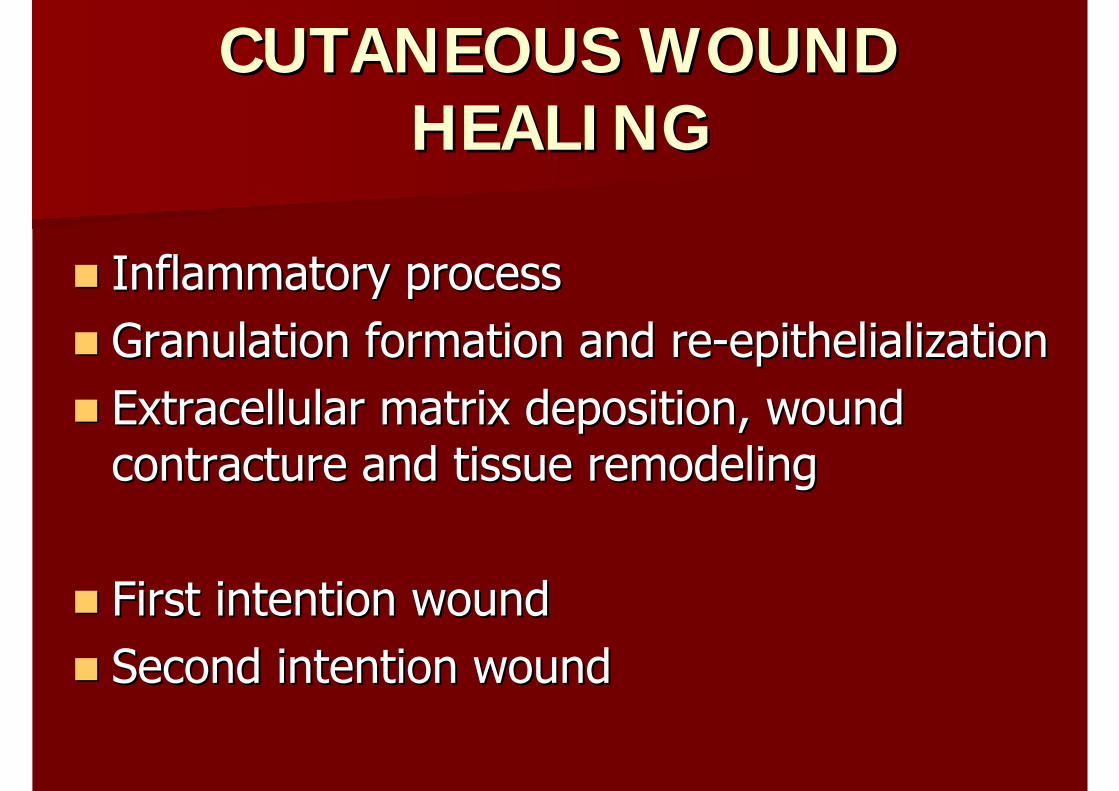
CUTANEOUS WOUND CUTANEOUS WOUND HEALINGHEALING
Inflammatory processInflammatory processGranulation formation and reGranulation formation and re--epithelializationepithelializationExtracellular matrix deposition, wound Extracellular matrix deposition, wound contracture and tissue remodelingcontracture and tissue remodeling
First intention woundFirst intention woundSecond intention woundSecond intention wound
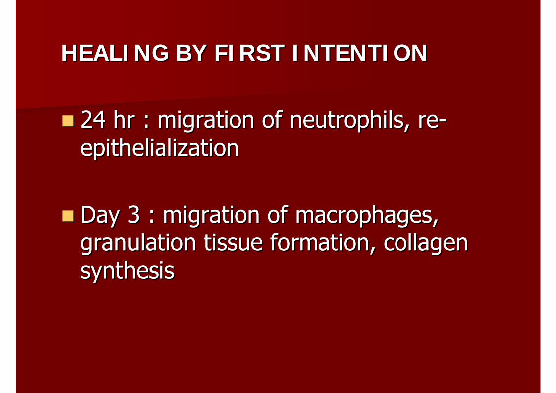
HEALING BY FIRST INTENTIONHEALING BY FIRST INTENTION
24 hr : migration of neutrophils, re24 hr : migration of neutrophils, re--epithelializationepithelialization
Day 3 : migration of macrophages, Day 3 : migration of macrophages, granulation tissue formation, collagen granulation tissue formation, collagen synthesissynthesis
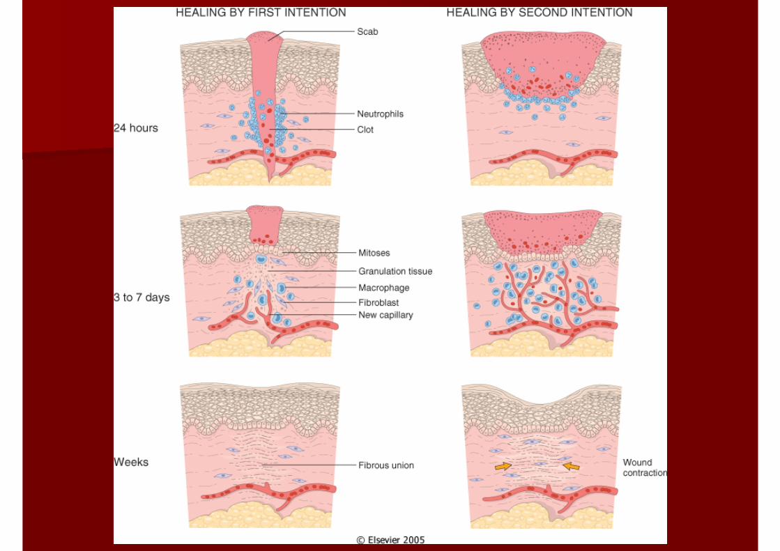
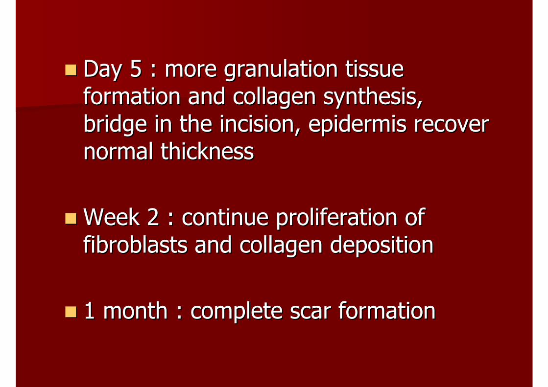
Day 5 : more granulation tissue Day 5 : more granulation tissue formation and collagen synthesis, formation and collagen synthesis, bridge in the incision, epidermis recover bridge in the incision, epidermis recover normal thicknessnormal thickness
Week 2 : continue proliferation of Week 2 : continue proliferation of fibroblasts and collagen depositionfibroblasts and collagen deposition
1 month : complete scar formation1 month : complete scar formation
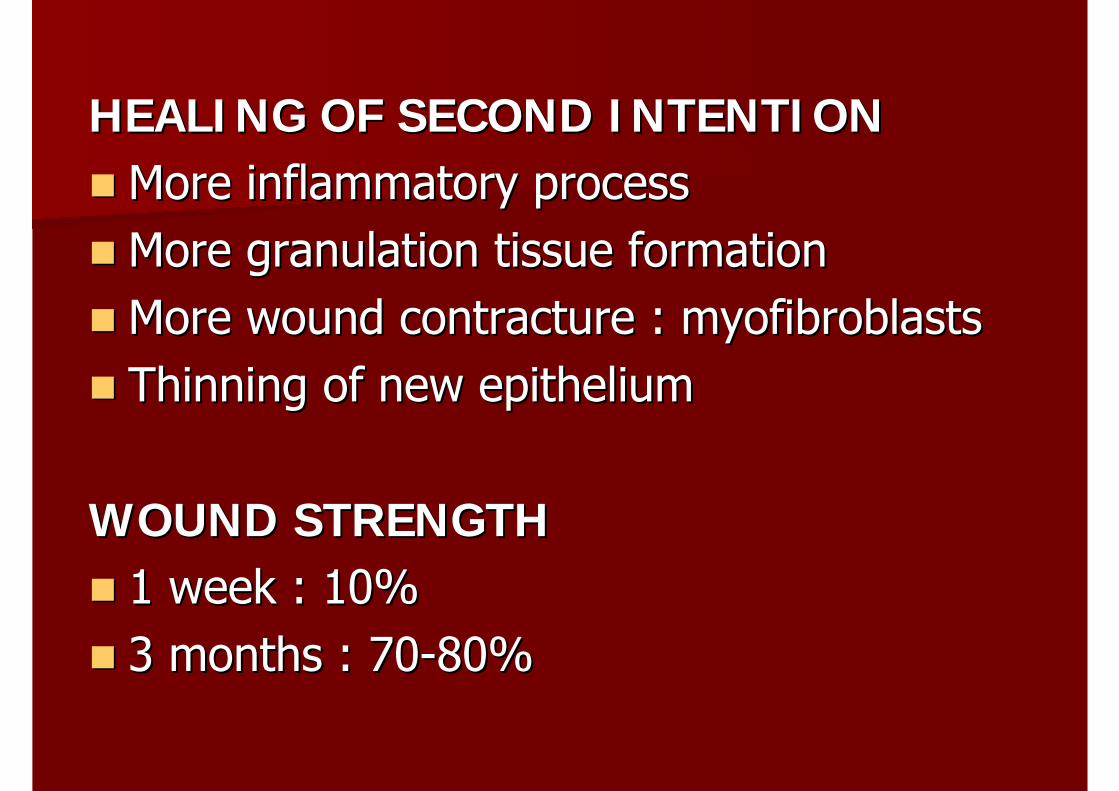
HEALING OF SECOND INTENTIONHEALING OF SECOND INTENTIONMore inflammatory processMore inflammatory processMore granulation tissue formationMore granulation tissue formationMore wound contracture : More wound contracture : myofibroblastsmyofibroblastsThinning of new epitheliumThinning of new epithelium
WOUND STRENGTHWOUND STRENGTH1 week : 10%1 week : 10%3 months : 703 months : 70--80%80%
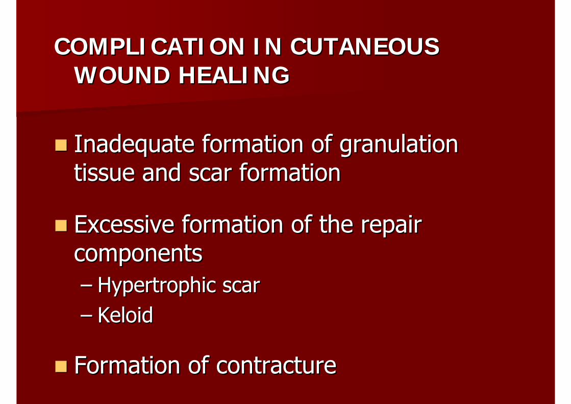
COMPLICATION IN CUTANEOUS COMPLICATION IN CUTANEOUS WOUND HEALINGWOUND HEALING
Inadequate formation of granulation Inadequate formation of granulation tissue and scar formationtissue and scar formation
Excessive formation of the repair Excessive formation of the repair componentscomponents–– Hypertrophic scar Hypertrophic scar –– Keloid Keloid
Formation of contractureFormation of contracture
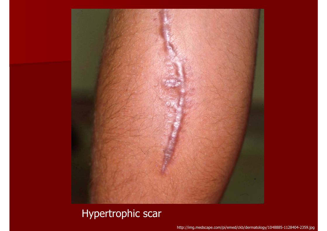
http://img.medscape.com/pi/emed/ckb/dermatology/1048885-1128404-2359.jpg
Hypertrophic scar
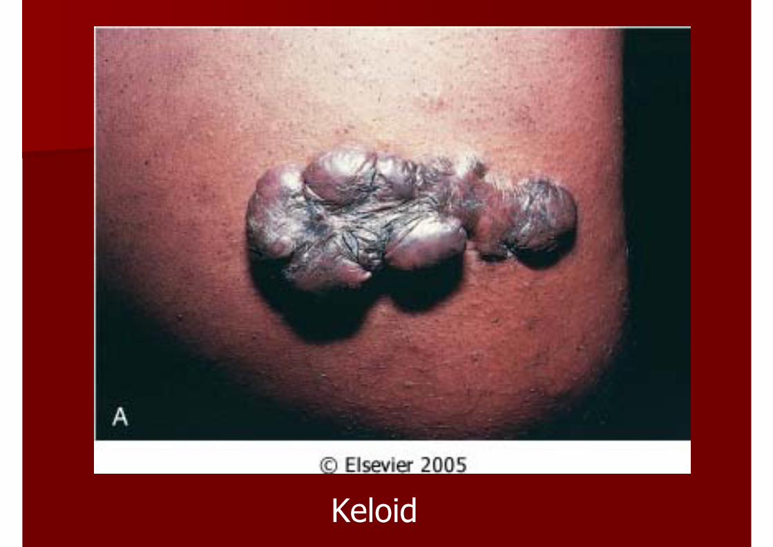
Keloid
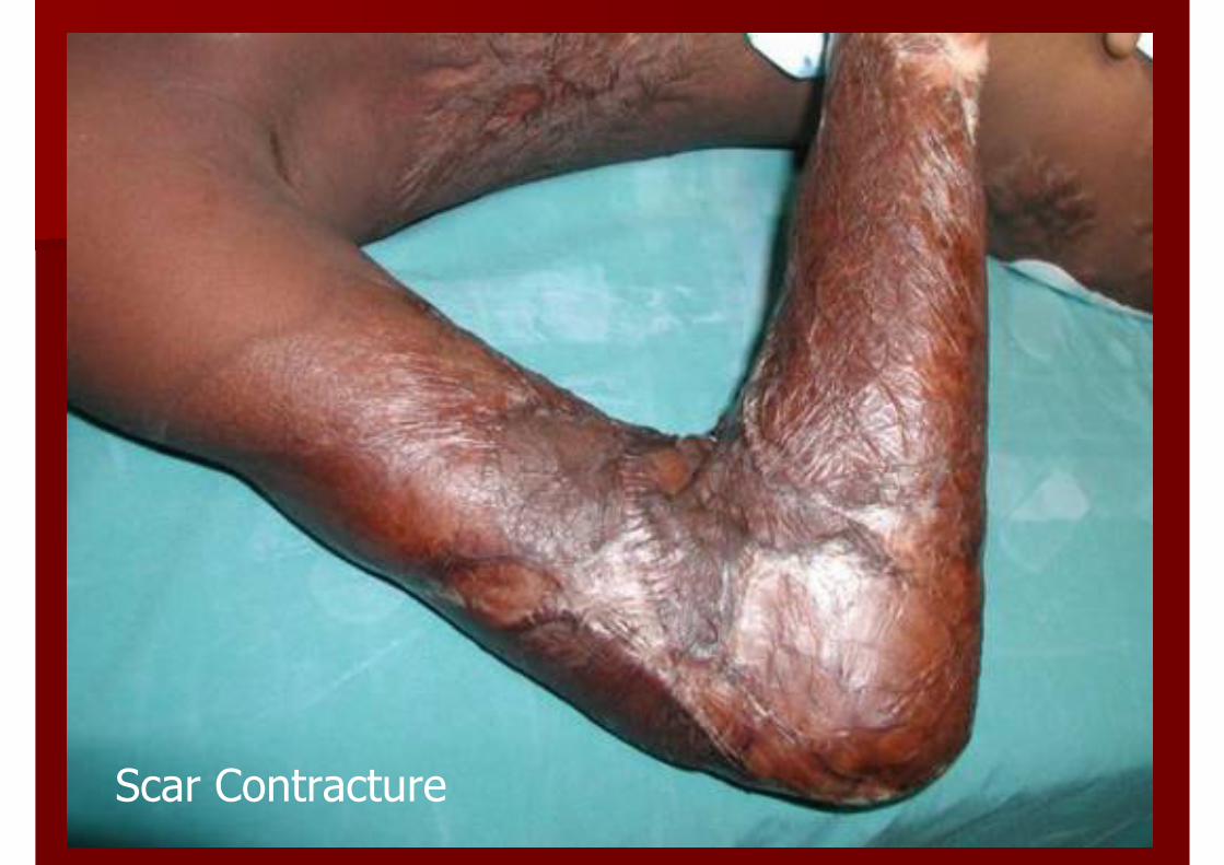
Scar Contracture
Principle of InfectionPrinciple of Infection
Overview and History of Infectious diseaseOverview and History of Infectious diseaseClassificationClassificationPathogenesis of Infectious DiseasePathogenesis of Infectious DiseasePathologyPathologyClinical EvaluationClinical Evaluation
Overview and HistoryOverview and History
Infectious diseaseInfectious disease หมายถึงหมายถึง โรคที่เกิดจากโรคที่เกิดจากสิ่งมีชีวิตที่สามารถกอโรคไดสิ่งมีชีวิตที่สามารถกอโรคได ( (Pathogen)Pathogen)
In the year In the year 1796, 1796, JennerJenner คนพบวามีคนพบวามี cross cross reactive immunity reactive immunity ระหวางระหวาง CowpoxCowpox และและ Small pox Small pox เปนจดุเริ่มตนการพัฒนาวัคซีนเปนจดุเริ่มตนการพัฒนาวัคซีนปองกันโรคฝดาษปองกันโรคฝดาษ ( (Small pox)Small pox)
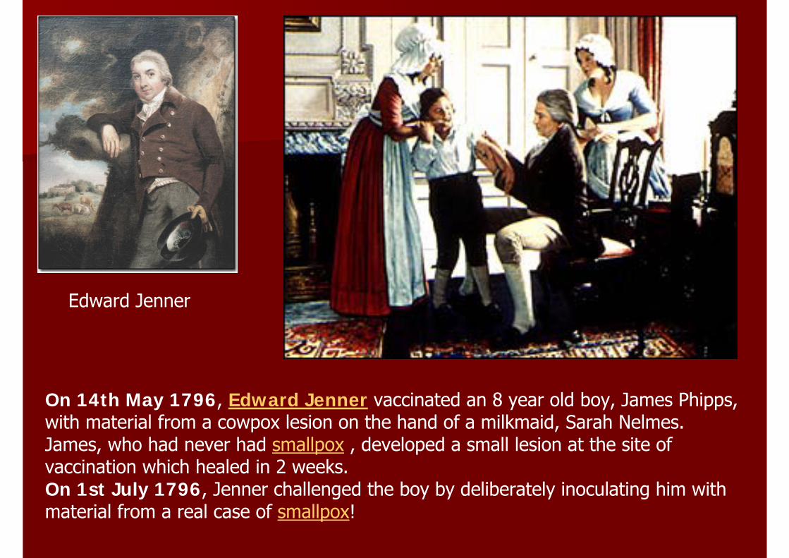
Edward Jenner
On 14th May 1796, Edward Jenner vaccinated an 8 year old boy, James Phipps,with material from a cowpox lesion on the hand of a milkmaid, Sarah Nelmes.James, who had never had smallpox , developed a small lesion at the site of vaccination which healed in 2 weeks.On 1st July 1796, Jenner challenged the boy by deliberately inoculating him withmaterial from a real case of smallpox!
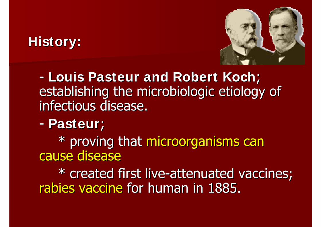
History:History:
-- Louis Pasteur and Robert KochLouis Pasteur and Robert Koch; ; establishing the microbiologic etiology of establishing the microbiologic etiology of infectious disease.infectious disease.-- PasteurPasteur; ;
* proving that * proving that microorganisms can microorganisms can cause diseasecause disease
* created first live* created first live--attenuated vaccines; attenuated vaccines; rabies vaccinerabies vaccine for human in 1885.for human in 1885.
ClassificationClassification
According to According to PathogenicityPathogenicityAccording to Site of Multiplication According to Site of Multiplication According to StructureAccording to Structure
Classification according to Classification according to PathogenicityPathogenicity
-- High virulenceHigh virulence-- Low virulence; opportunistic infectionLow virulence; opportunistic infection
Classification according to Site of Classification according to Site of MultiplicationMultiplication
-- Obligate intracellular organismsObligate intracellular organisms-- Facultative intracellular organismFacultative intracellular organism-- ExtracellularExtracellular organismsorganisms
Obligate Intracellular OrganismsObligate Intracellular Organisms
-- เจริญเติบโตและแบงตัวในเจริญเติบโตและแบงตัวใน host cell host cell เทานั้นเทานั้น-- PrionsPrions-- All virusesAll viruses-- All All rickettsiaerickettsiae-- All All chlamydiaechlamydiae-- Some protozoaSome protozoa
Facultative Intracellular OrganismsFacultative Intracellular Organisms
-- แบงตัวไดทั้งในแบงตัวไดทั้งใน และนอกและนอก host cellhost cell-- MycobacteriaMycobacteria; M. tuberculosis; M. tuberculosis-- BrucellaBrucella sppspp..-- ActinomycesActinomyces-- KlebsiellaKlebsiella rhinoscleromatisrhinoscleromatis-- FrancisellaFrancisella tularensistularensis-- Pseudomonas Pseudomonas malleimallei andand P. P. pseudomalleipseudomallei
Fungi;Fungi;-- CoccidioidesCoccidioides immitisimmitis-- HistoplasmaHistoplasma capsulatumcapsulatum-- Cryptococcus Cryptococcus neoformanneoforman-- BlastomycesBlastomyces dermatidisdermatidis-- ParacoccidioidesParacoccidioides brasilliensisbrasilliensis-- SporothrixSporothrix schenskischenski
Some protozoaSome protozoa
ExtracellularExtracellular OrganismsOrganisms
-- แบงตัวนอกแบงตัวนอก host cell host cell เทานั้นเทานั้น-- MycoplasmaMycoplasma-- All bacteria All bacteria exceptexcept facultative intracellular facultative intracellular organismsorganisms-- Fungi; Fungi; Candida Candida albicansalbicans, , AspergillusAspergillus sppspp, , MucorMucor sppspp..-- Some protozoa Some protozoa exceptexcept TrypanosomaTrypanosoma sppspp., ., Plasmodium Plasmodium sppspp., ., ToxoplasmaToxoplasma sppspp..-- All All metazoametazoa
Classification according to StructureClassification according to Structure
-- PrionPrion -- FungiFungi-- VirusesViruses -- Protozoa, Protozoa, metazoametazoa-- BacteriaBacteria -- EctoparasiteEctoparasite-- RickettsiaRickettsia, , chlamydiachlamydia, , mycoplasmamycoplasma
PrionsPrions::-- 27 27 kDkD nucleic acidnucleic acid--free free prionprion-- are apparently composed of abnormal are apparently composed of abnormal
forms of host protein; forms of host protein; prionprion proteinprotein-- these agents cause transmissible these agents cause transmissible
spongioformspongioform encephalopathiesencephalopathies; ; kurukuru, CJD, , CJD, bovine bovine spongioformspongioform encephalopathy (mad encephalopathy (mad cow)cow)
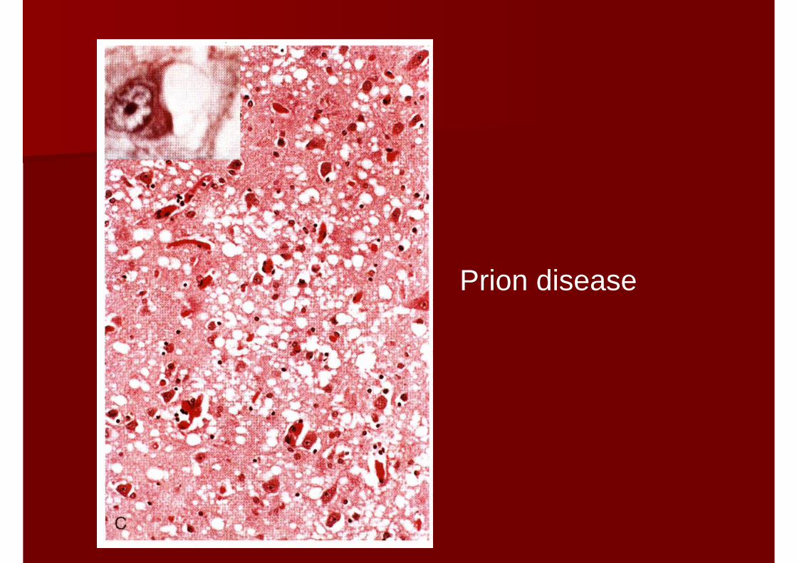
Prion disease
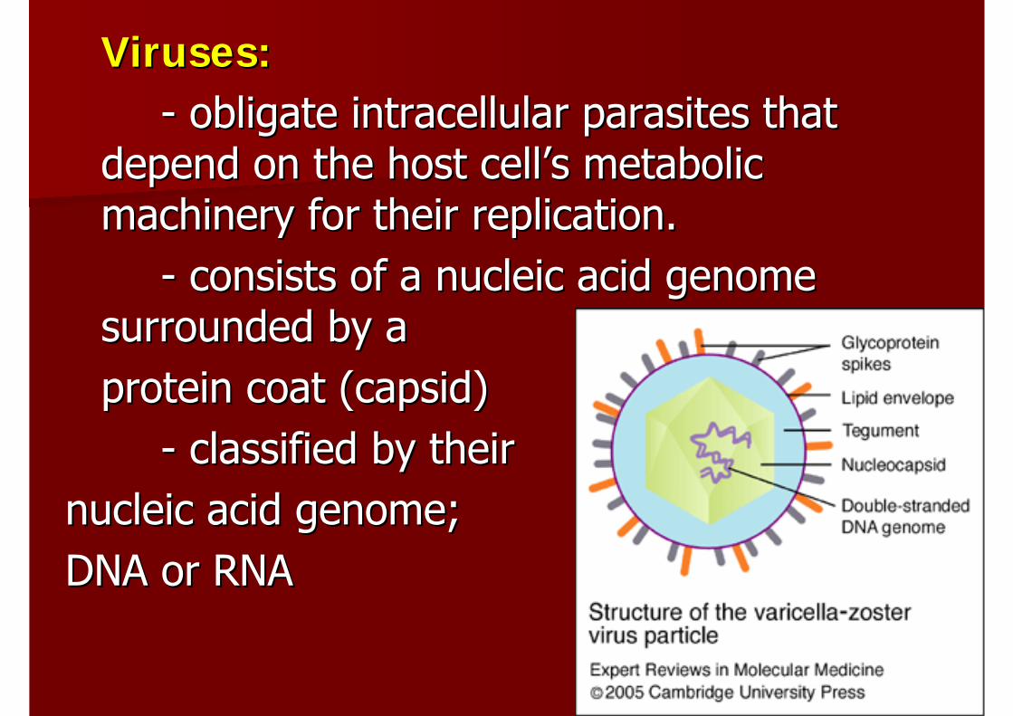
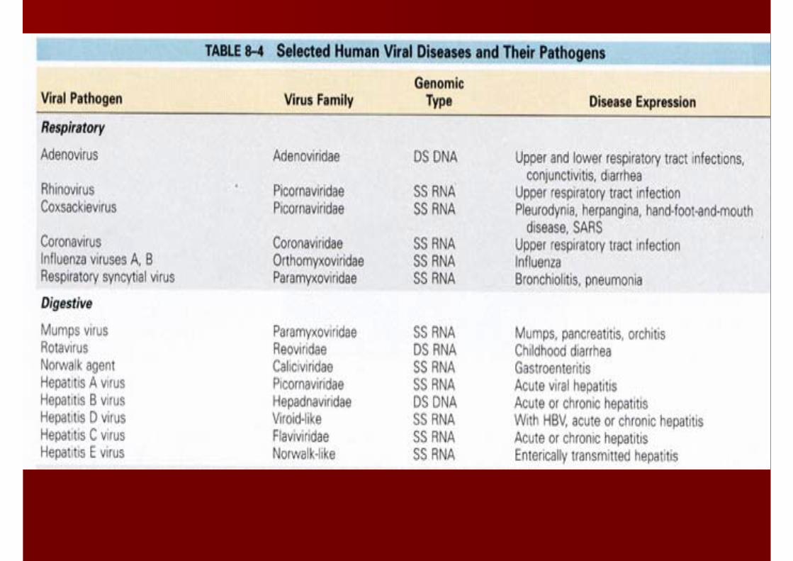
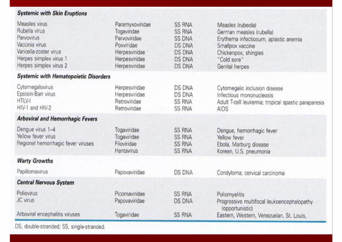
Viruses:Viruses:-- obligate intracellular parasites that obligate intracellular parasites that
depend on the host celldepend on the host cell’’s metabolic s metabolic machinery for their replication.machinery for their replication.
-- consists of a nucleic acid genome consists of a nucleic acid genome surrounded by a surrounded by a protein coat (protein coat (capsidcapsid))
-- classified by their classified by their nucleic acid genome; nucleic acid genome; DNA or RNA DNA or RNA
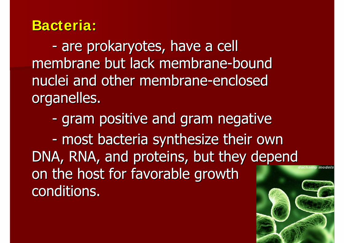
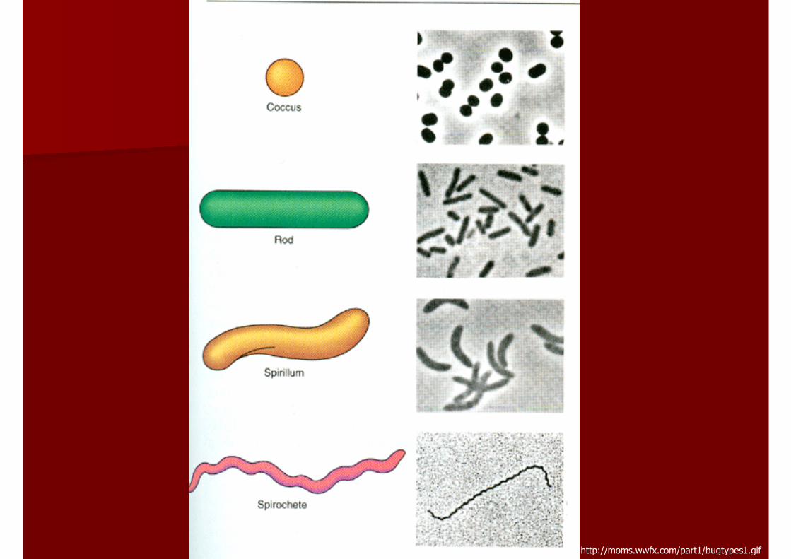
Bacteria:Bacteria:-- are prokaryotes, have a cell are prokaryotes, have a cell
membrane but lack membranemembrane but lack membrane--bound bound nuclei and other membranenuclei and other membrane--enclosed enclosed organelles.organelles.
-- gram positive and gram negativegram positive and gram negative-- most bacteria synthesize their own most bacteria synthesize their own
DNA, RNA, and proteins, but they depend DNA, RNA, and proteins, but they depend on the host for favorable growth on the host for favorable growth conditions.conditions.
http://moms.wwfx.com/part1/bugtypes1.gif
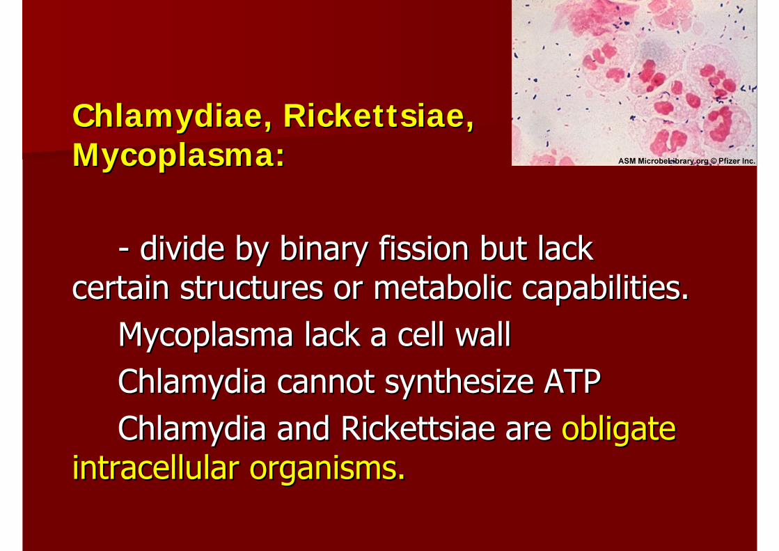
ChlamydiaeChlamydiae, , RickettsiaeRickettsiae, , MycoplasmaMycoplasma::
-- divide by binary fission but lack divide by binary fission but lack certain structures or metabolic capabilities.certain structures or metabolic capabilities.
MycoplasmaMycoplasma lack a cell walllack a cell wallChlamydia cannot synthesize ATPChlamydia cannot synthesize ATPChlamydia and Chlamydia and RickettsiaeRickettsiae are are obligate obligate
intracellular organisms.intracellular organisms.
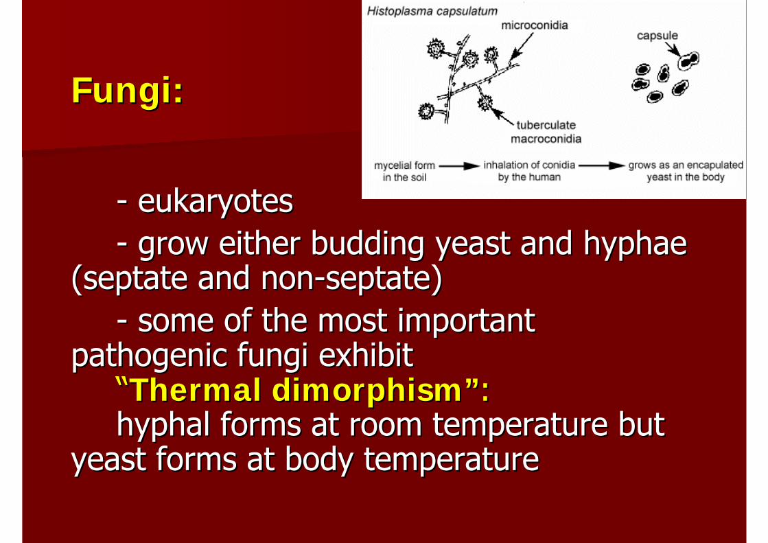
Fungi:Fungi:
-- eukaryoteseukaryotes-- grow either budding yeast and grow either budding yeast and hyphaehyphae
((septateseptate and nonand non--septateseptate))-- some of the most important some of the most important
pathogenic fungi exhibit pathogenic fungi exhibit ““Thermal dimorphismThermal dimorphism””::hyphalhyphal forms at room temperature but forms at room temperature but
yeast forms at body temperatureyeast forms at body temperature
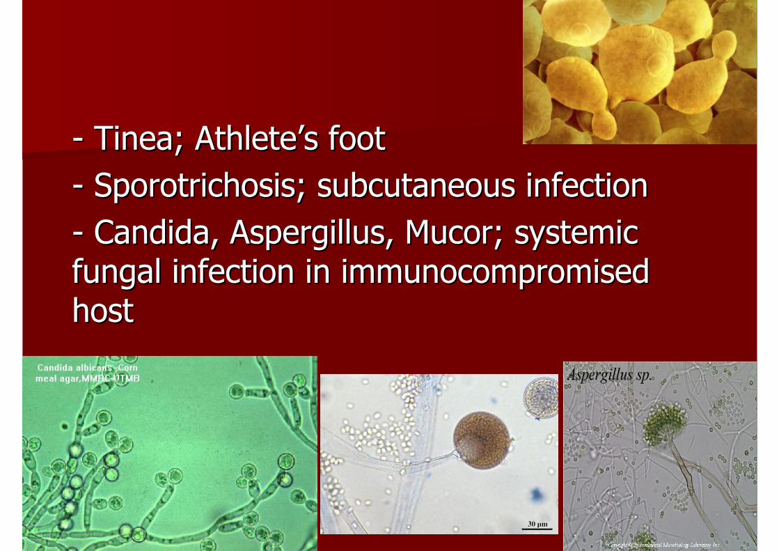
-- TineaTinea; Athlete; Athlete’’s foots foot-- SporotrichosisSporotrichosis; subcutaneous infection; subcutaneous infection-- Candida, Candida, AspergillusAspergillus, , MucorMucor; systemic ; systemic fungal infection in fungal infection in immunocompromisedimmunocompromisedhosthost
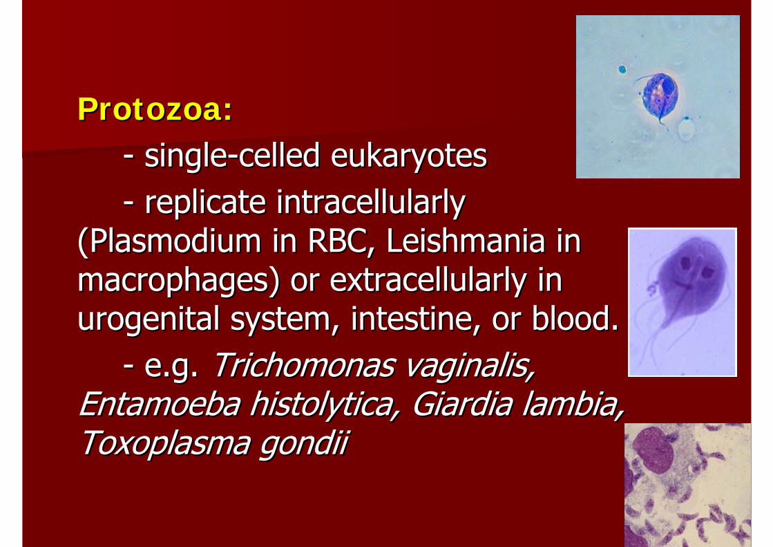
Protozoa:Protozoa:-- singlesingle--celled eukaryotescelled eukaryotes-- replicate replicate intracellularlyintracellularly
(Plasmodium in RBC, (Plasmodium in RBC, LeishmaniaLeishmania in in macrophages) or macrophages) or extracellularlyextracellularly in in urogenitalurogenital system, intestine, or blood.system, intestine, or blood.
-- e.g. e.g. TrichomonasTrichomonas vaginalisvaginalis, , EntamoebaEntamoeba histolyticahistolytica, , GiardiaGiardia lambialambia, , ToxoplasmaToxoplasma gondiigondii
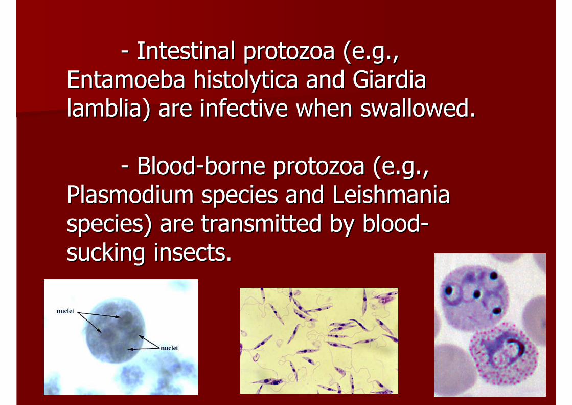
-- Intestinal protozoa (e.g., Intestinal protozoa (e.g., EntamoebaEntamoeba histolyticahistolytica and and GiardiaGiardialamblialamblia) are infective when swallowed.) are infective when swallowed.
-- BloodBlood--borne protozoa (e.g., borne protozoa (e.g., Plasmodium species and Plasmodium species and LeishmaniaLeishmaniaspecies) are transmitted by bloodspecies) are transmitted by blood--sucking insects.sucking insects.
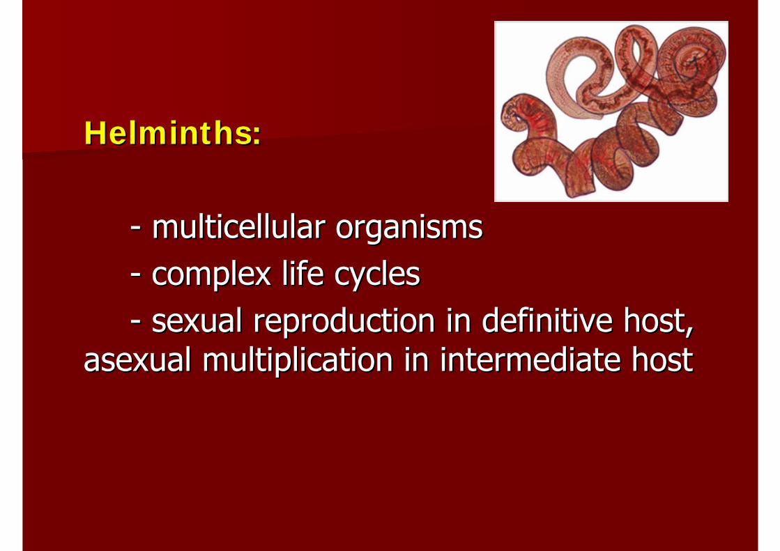
HelminthsHelminths::
-- multicellularmulticellular organismsorganisms-- complex life cyclescomplex life cycles-- sexual reproduction in definitive host, sexual reproduction in definitive host,
asexual multiplication in intermediate hostasexual multiplication in intermediate host
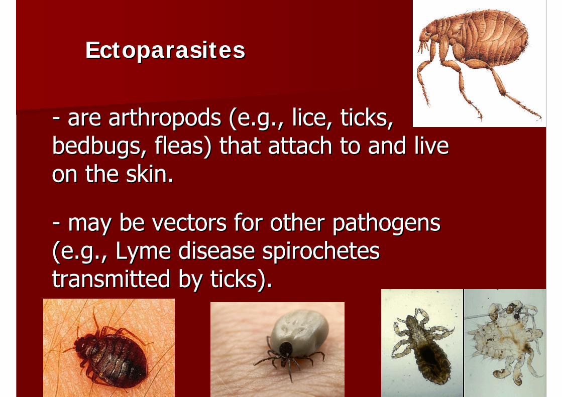
EctoparasitesEctoparasites
-- are arthropods (e.g., lice, ticks, are arthropods (e.g., lice, ticks, bedbugs, fleas) that attach to and live bedbugs, fleas) that attach to and live on the skin.on the skin.
-- may be vectors for other pathogens may be vectors for other pathogens (e.g., (e.g., LymeLyme disease spirochetes disease spirochetes transmitted by ticks).transmitted by ticks).
Pathogenesis of Infectious DiseasePathogenesis of Infectious Disease
Host factorsHost factors–– General factorGeneral factor–– Internal factorInternal factor
Pathogenic organism factorsPathogenic organism factors
Host factors:Host factors:1. 1. General factors:General factors: socioeconomic status, socioeconomic status, behavior pattern, occupationbehavior pattern, occupation
2. 2. Internal factors:Internal factors: Natural defense Natural defense mechanism; skin and normal flora, mechanism; skin and normal flora, respiratory tract and respiratory tract and mucociliarymucociliary mechanism, mechanism, HClHCl production in stomach, or normal flushing production in stomach, or normal flushing action of urineaction of urine
Internal factors:Internal factors: Inflammation; acute Inflammation; acute inflammation, inflammation, phagocytosisphagocytosis, complement, , complement, and production of interferonand production of interferon
Internal factors:Internal factors: The immune response; The immune response; HMI and CMIHMI and CMI
HMI: Ag & HMI: Ag & AbAb (B(B--cell)cell)CMI: TCMI: T-- cell, macrophagescell, macrophages
Organism factors:Organism factors:1. Route of entry1. Route of entry
2. Mode of transmission; congenital 2. Mode of transmission; congenital transfer (Rubella, CMV, HIV, HSV), directly transfer (Rubella, CMV, HIV, HSV), directly contact, food and water, airborne, animal, contact, food and water, airborne, animal, sexual transmissionsexual transmission
3. Spread and Dissemination; localized 3. Spread and Dissemination; localized and disseminated infection and disseminated infection
-- viremiaviremia, , bacteremiabacteremia, , fungemiafungemia
-- septicemia: invasion of the bloodstream septicemia: invasion of the bloodstream by virulent microorganisms from a focus of by virulent microorganisms from a focus of infection that is accompanied by fever, infection that is accompanied by fever, chills, tachycardia, hypotensionchills, tachycardia, hypotension
4. Number of organism4. Number of organism--numerous low virulent organism can numerous low virulent organism can cause severe diseasecause severe disease
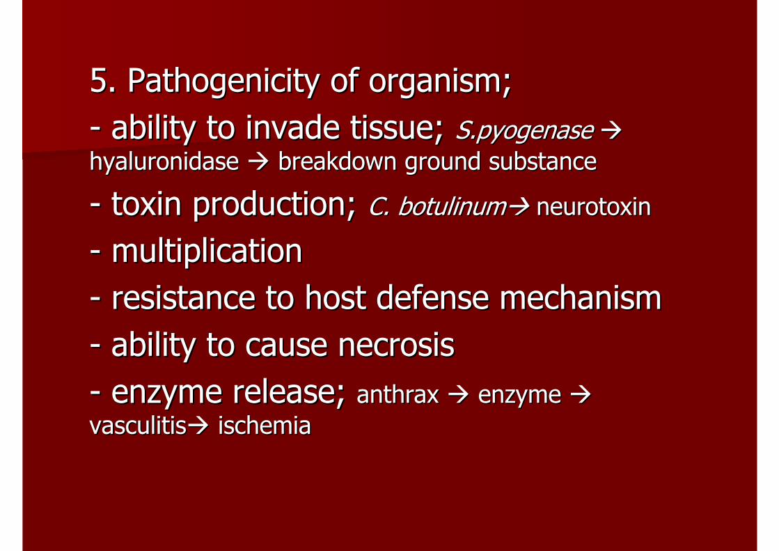
5. 5. PathogenicityPathogenicity of organism; of organism; -- ability to invade tissue; ability to invade tissue; S.pyogenaseS.pyogenasehyaluronidasehyaluronidase breakdown ground substancebreakdown ground substance
-- toxin production; toxin production; C. C. botulinumbotulinum neurotoxinneurotoxin
-- multiplicationmultiplication-- resistance to host defense mechanism resistance to host defense mechanism -- ability to cause necrosisability to cause necrosis-- enzyme release; enzyme release; anthrax anthrax enzyme enzyme vasculitisvasculitis ischemiaischemia
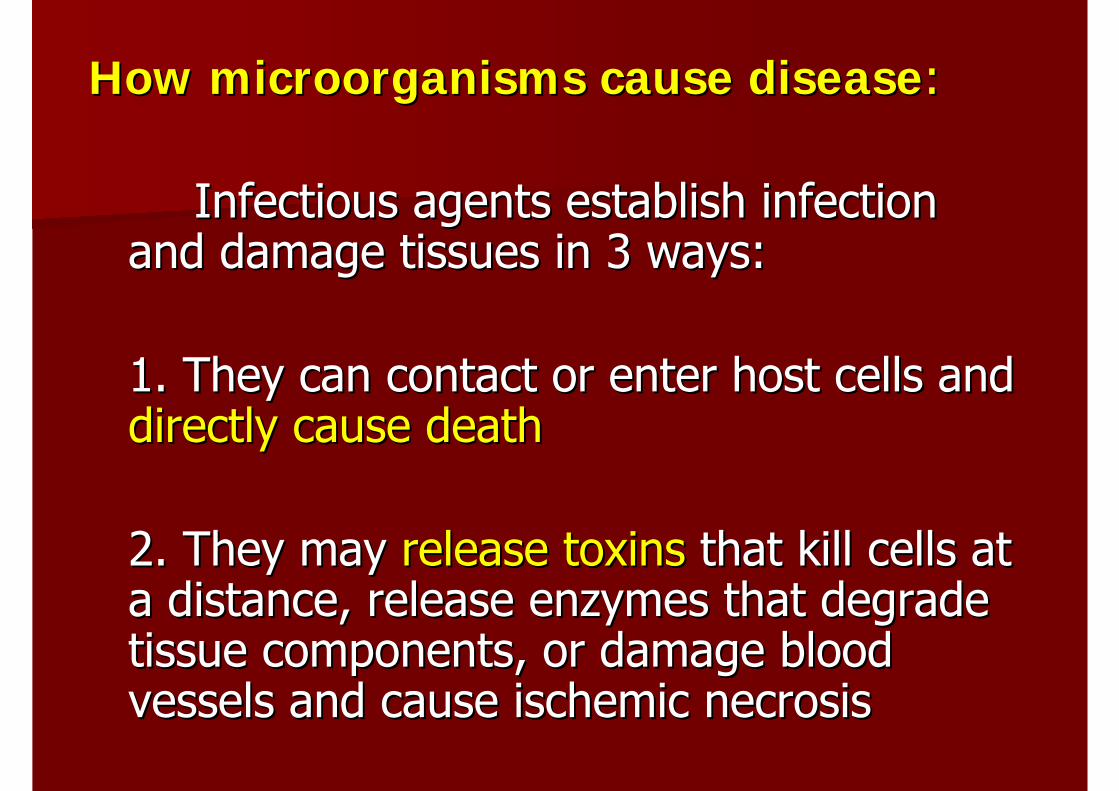
How microorganisms cause diseaseHow microorganisms cause disease::
Infectious agents establish infection Infectious agents establish infection and damage tissues in 3 ways:and damage tissues in 3 ways:
1. They can contact or enter host cells and 1. They can contact or enter host cells and directly cause deathdirectly cause death
2. They may 2. They may release toxinsrelease toxins that kill cells at that kill cells at a distance, release enzymes that degrade a distance, release enzymes that degrade tissue components, or damage blood tissue components, or damage blood vessels and cause ischemic necrosisvessels and cause ischemic necrosis
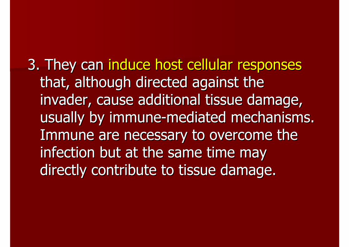
3. They can 3. They can induce host cellular responsesinduce host cellular responsesthat, although directed against the that, although directed against the invader, cause additional tissue damage, invader, cause additional tissue damage, usually by immuneusually by immune--mediated mechanisms. mediated mechanisms. Immune are necessary to overcome the Immune are necessary to overcome the infection but at the same time may infection but at the same time may directly contribute to tissue damage.directly contribute to tissue damage.
Clinical EvaluationClinical Evaluation
Clinical historyClinical historyPhysical examinationPhysical examinationLaboratory investigationLaboratory investigation
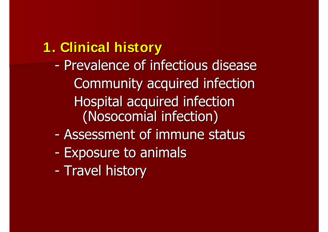
1. Clinical history1. Clinical history-- Prevalence of infectious diseasePrevalence of infectious disease
Community acquired infectionCommunity acquired infectionHospital acquired infection Hospital acquired infection ((NosocomialNosocomial infection)infection)
-- Assessment of immune statusAssessment of immune status-- Exposure to animalsExposure to animals-- Travel historyTravel history
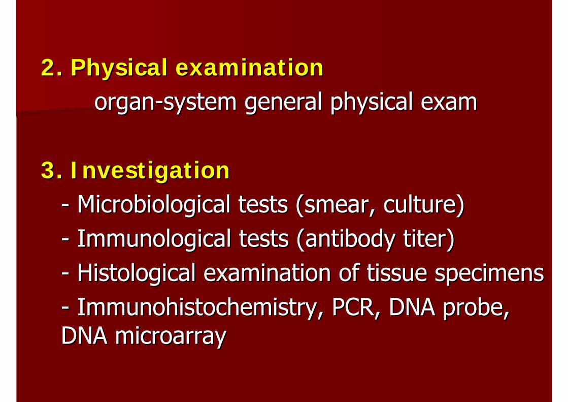
2. Physical examination2. Physical examinationorganorgan--system general physical examsystem general physical exam
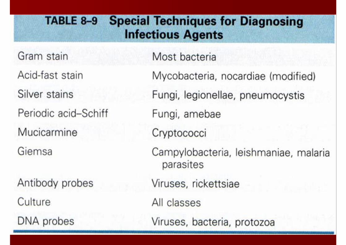
3. Investigation3. Investigation-- Microbiological tests (smear, culture)Microbiological tests (smear, culture)-- Immunological tests (antibody titer)Immunological tests (antibody titer)-- Histological examination of tissue specimensHistological examination of tissue specimens-- ImmunohistochemistryImmunohistochemistry, PCR, DNA probe, , PCR, DNA probe, DNA DNA microarraymicroarray
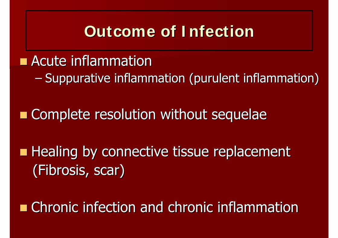
Outcome of InfectionOutcome of Infection
Acute inflammationAcute inflammation–– SuppurativeSuppurative inflammation (purulent inflammation)inflammation (purulent inflammation)
Complete resolution without Complete resolution without sequelaesequelae
Healing by connective tissue replacementHealing by connective tissue replacement(Fibrosis, scar)(Fibrosis, scar)
Chronic infection and chronic inflammation Chronic infection and chronic inflammation
ReferencesReferences
สุภรณสุภรณ พงศะพงศะบุตรบุตร,, บรรณาธิการบรรณาธิการ,, ““ตําราพยาธวิิทยาทัว่ไปตําราพยาธวิิทยาทัว่ไป..””,,
ภาควิชาพยาธิวิทยาและนิติเวชศาสตรภาควิชาพยาธิวิทยาและนิติเวชศาสตร,, โกโกลบอลพริ้นทลบอลพริ้นท,, 2551, 2551, หนาหนา
4141--89,89, 9393--118.118.
Kumar V., Kumar V., CotranCotran R.S., Robbins S.L., R.S., Robbins S.L., ““Robbins Basic Pathology, Robbins Basic Pathology,
77thth editionedition..”” Saunders, 2003, p.33Saunders, 2003, p.33--78, 30778, 307--322.322.