Embed Size (px)
Citation preview

PROGRAMA DE DOCTORADO: INVESTIGACIÓN EN MEDICINA
Título de la tesis
Análisis de un programa de vigilancia para el diagnóstico precoz de carcinoma hepatocelular en pacientes con cirrosis hepática
Nombre del Autor
Alejo Mancebo Mata


PROGRAMA DE DOCTORADO: INVESTIGACIÓN EN MEDICINA
Título de la tesis
Análisis de un programa de vigilancia para el diagnóstico precoz de carcinoma hepatocelular en pacientes con cirrosis hepática
Nombre del Autor
Alejo Mancebo Mata

FOR
-MA
T-V
OA
-010
-BIS
RESUMEN DEL CONTENIDO DE TESIS DOCTORAL
1.- Título de la Tesis Español/Otro Idioma: Análisis de un programa de vigilancia para el diagnóstico precoz de carcinoma hepatocelular en pacientes con cirrosis hepática.
Inglés: Analysis of a surveillance program for an early diagnosis of liver cancer in patients with liver cirrhosis
2.- Autor Nombre: Alejo Mancebo Mata
DNI/Pasaporte/NIE:
Programa de Doctorado:Investigación en Medicina Órgano responsable: Comisión Académica del Programa de Doctorado Investigación en Medicina
RESUMEN (en español)
Introducción: La cirrosis es un factor de riesgo de desarrollo de carcinoma hepatocelular (CHC). El CHC es un tumor con una elevada mortalidad, pero diversas opciones terapéuticas aumentan la supervivencia si se diagnostica en estadio inicial. Las guías de práctica clínica recomiendan realizar vigilancia semestral para detectar CHC en los pacientes con cirrosis. Objetivos: Los tres estudios que componen la presente tesis doctoral analizan diversos aspectos de los programas de cribado sobre los que todavía existe incertidumbre: la incidencia de CHC en la cirrosis alcohólica, la adherencia de los pacientes a los programas de cribado, y las causas asociadas al fracaso del programa para detectar tumores diagnosticados en estadio inicial. Material y métodos: Se analizó una cohorte de pacientes cirróticos consecutivamente incorporados a un programa para el diagnóstico precoz del CHC, seguidos de manera prospectiva en el Hospital Universitario Central de Asturias. El programa se inició en septiembre de 1992 y hasta agosto de 2016 incluyó a 1.259 pacientes, de los que 188 desarrollaron CHC. El cribado consistía en la realización de una ecografía abdominal y la medición del nivel de alfa-fetoproteína. Entre 1992 y 2004 los pacientes fueron aleatorizados a recibir vigilancia semestral (n=183) o trimestral (n=183). Desde 2004 todos los pacientes recibieron vigilancia semestral. Se recogieron variables clínicas y de laboratorio a la inclusión en el programa, durante el transcurso del mismo, y al diagnóstico del tumor o la censura del
paciente. El primer estudio incorporó a los pacientes del programa con cirrosis alcohólica. El segundo estudio incluyó a todos los pacientes seguidos de manera semestral en el programa. El tercer estudio incluyó a todos los pacientes diagnosticados de CHC. Resultados: En el primer estudio se observó que la incidencia anual de CHC en pacientes con cirrosis alcohólica Child-Pugh A o B era del 2,5%. Además, se determinó que los pacientes con plaquetas ≥ 125.000 y menores de 55 años, tenían un bajo riesgo de desarrollar CHC, del 0,3% anual.
En el segundo estudio se observó que sólo el 16% de los pacientes presentaba mala adherencia a los controles del programa de cribado, definida como la inasistencia a 2 controles consecutivos. Los pacientes que, a la entrada en el programa, tenían historia de consumo de drogas por vía parenteral, eran bebedores activos, no habían sufrido descompensaciones de su enfermedad hepática y tenían una ratio AST/ALT ≥ 1,6, presentaron mala adherencia con mayor frecuencia.
En el tercer estudio se observó que el 26% de los CHC descubiertos en el programa de vigilancia se diagnosticaron fuera de los criterios de Milán, considerándose fracasos del programa. El estadio Child-Pugh B/C y un nivel de AFP ≥ 100 ng/ml al diagnóstico del CHC, se asociaron de manera independiente con el fracaso. El estadio Child-Pugh B a la entrada en el programa y las descompensaciones de la enfermedad hepática durante el seguimiento, fueron predictores tempranos del fracaso. Conclusiones: Los pacientes con cirrosis alcohólica tienen un riesgo de desarrollar CHC del 2,5% anual. Este umbral es superior al 1,5%. recomendado por la guía de la AASLD para incorporar a los pacientes a los programas de cribado. Los pacientes con cirrosis alcohólica con plaquetas normales y menores de 55 años tienen un riesgo inferior a dicho umbral.
Los pacientes con cirrosis tienen una buena adherencia a los controles consecutivos del programa de cribado. El consumo activo de alcohol y una historia de consumo de drogas por vía intravenosa son los mejores predictores de mala adherencia.
Una cuarta parte de los CHC descubiertos en el programa de vigilancia se diagnostican fuera de los criterios de Milán. El estadio Child-Pugh B a la entrada en el programa y las descompensaciones de la enfermedad hepática durante el seguimiento se pueden considerar marcadores tempranos de fracaso del programa.
RESUMEN (en Inglés)
Introduction: The most well established risk factor for developing hepatocelular carcinoma (HCC) is cirrhosis. HCC is a tumour with a high rate of mortality, but various treatment options
increase survival in early stages. Clinical Practice Guidelines endorse bi-annual surveillance for early detection of HCC in patients with liver cirrhosis. Purpose The three studies included in the present doctoral thesis discuss various uncertain aspects of surveillance programs: incidence of HCC among patients with alcoholic cirrhosis, patient adherence to screening for HCC and the frequency and risk factors associated with program failure to detect tumors at an early stage. Patients and Methods: We conducted a prospective cohort study of patients with cirrhosis who were consecutively enrolled in a surveillance program for early detection of HCC at the Hospital Universitario Central de Asturias. The program started in september 1992 included 1,259 patients until August 2016, of which 188 developed an HCC. Surveillance was based on abdominal ultrasound (US) and alpha-fetoprotein tests. Patients were randomized to receive surveillance at six-month intervals (n=183) surveillance or three-month intervals (n=183) between 1992 and 2004. After 2004, all patients were surveyed every 6 months. Variables were recorded at entry into the program, during follow-up and at HCC diagnosis. The first study analyzed patients with alcoholic cirrhosis only. The second one investigated all patients with semiannual follow-ups. The third one included all HCC diagnosed in the surveillance program. Results: In the first study, it was observed that the annual incidence of HCC among patients with alcoholic cirrhosis of Child–Pugh class A or B is around 2.5%. Patients under the age of 55 years and with a platelet count ≥ 125 103/mm had a very low risk of developing an HCC, 0.3%.
The second investigation found that only 16% of patients had suboptimal adherence, defined as failure to complete 2 consecutive abdominal US. Active or previous intravenous drug use, active alcohol consumption, absence of liver decompensation before the inclusion in the program and aspartate transaminase/alanine transaminase ratio ≥ 1.6 were independent predictors of suboptimal adherence.
The third study noted that 26% of HCC tumors were beyond the Milan criteria. Child-Pugh B/C and AFP≥100 ng/ml, both at diagnosis, were independently associated with failure. However, Child-Pugh B at entry and development of liver-related complications during follow-up can be early predictors of failure.
Conclusions: Patients with alcoholic liver cirrhosis have an annual incidence of HCC of 2.5%, a risk higher than the 1,5% threshold recommended by AASLD to initiate surveillance for an
early diagnosis of HCC. Patients with normal platelet count and under the age of 55 are below this threshold. The adherence to the process of HCC surveillance can be considered as adequate among cirrhotic patients. Active alcohol consumption and a history of intravenous drug use are the strongest predictors of suboptimal adherence. Approximately 25% of HCC cases diagnosed among patients included in a surveillance program were beyond the Milan criteria. Child-Pugh B at entry and development of liver-related complications during follow-up can be early predictors of failure.
SR. PRESIDENTE DE LA COMISIÓN ACADÉMICA DEL PROGRAMA DE DOCTORADO EN INVESTIGACIÓN EN MEDICINA

A mi familia

Agradecimientos El primer agradecimiento, es, sin lugar a dudas, para mis directores y tutores de tesis. Para el Dr. Rodrigo, por la paciencia y el optimismo con el que transmitía la necesidad de seguir avanzando paso a paso en una tesis que se inició hace 4 largos años ya. Para el Dr. Manuel Rodríguez, a quien en realidad le pertenece. Por tener la visión de recoger datos de los pacientes a quienes diariamente atendía y la perseverancia de rellenar, durante más de 2 décadas, las famosas carpetas azules, amarillas y la base de datos. Por no desesperarse con los numerosos correos que le envié, con los miles de bocetos de artículos o posters que me iba corrigiendo. Por encontrar tiempo entre su inagotable agenda de congresos, clases y seminarios para los cientos de reuniones que están detrás de esta tesis. Por animarme a seguir investigando pese a que ya se hubiese publicado el primer artículo. Esta tesis también pertenece al equipo que, cada semana, cada mes, rellenó las carpetas y la base de datos. Sin pedir nada a cambio, a base de trabajo fuera de su jornada laboral. Ellas y ellos son Luisa González, Carmen Navascués, Valle, Ramón. Esta tesis también le pertenece a las ecografistas, a Maribel, a Nieves. Que son parte del programa de cribado y que salvan vidas a oscuras en una pequeña habitación. Y aunque está en la trinchera final del programa de cribado, con los hepatomas, Maria Varela también forma parte de esta tesis. Porque “amadrinó” y supervisó los artículos. Porque se preocupó de los trabajos como si fuesen suyos propios. No me puedo olvidar de mis “coerres”. Que sin lugar a dudas eran mucho más precisas y pacientes que yo en la recogida de datos durante nuestra breve estancia en el HUCA. Que me alegraron la residencia. Y a quienes guardaré siempre un cariño especial. En parte, el sistema público de salud es la piedra angular de estos trabajos. Porque atiende a una población de pacientes con cirrosis con escasos recursos, con problemas de alcoholismo, con bajas, en paro. Porque no pregunta qué tipo de seguro tiene cada persona para atenderle. Y porque demuestra que la salud está por encima de los beneficios a corto plazo. Por último, nada hubiese sido posible sin el apoyo de mi familia. De mis padres, a quienes quité horas de compañía. De mi abuela, a quien llena de ilusión que sus nietos hagan trabajos. De mi hermano, de Cintzia y mis sobrinos, que me llenaron de alegría los fines de semana. Y, evidentemente, de Alba. A quien “aturullé” durante estos 4 largos años con cada uno de los pasitos, adelante unas veces, atrás otras, de las publicaciones y comunicaciones. Y quien llena de amor la casa donde, durante cientos de horas, se fue labrando este trabajo.
Abreviaturas
AASLD: American Association for the Study of Liver Diseases.
AFP: alfa-fetoproteína.
AST: aspartato aminotransferasa.
ALT: alanina aminotransferasa.
BCLC: Barcelona Clinic Liver Cancer.
CHC: carcinoma hepatocelular.
GGT: γ-glutamil transferasa.
FA: fosfatasa alcalina.
HUCA: Hospital Universitario Central de Asturias.
EASL: European Association for the Study of the Liver.
ECO: ecografía abdominal.
EHGNA: enfermedad de hígado graso no alcohólico.
IMC: Índice de masa corporal.
RIC: rango intercuartílico.
RVS: respuesta viral sostenida.
TACE: quimioembolización transarterial.
VHB: Virus de la hepatitis B.
VHC: Virus de la hepatitis C.
VIH: virus de la inmunodeficiencia humana.
WT: wilde type.

Índice
Introducción .........................................................................................................................17 Epidemiología del CHC ...............................................................................................................18 Etiología del CHC ........................................................................................................................ 20 Tratamiento del CHC .................................................................................................................. 22 Los programas de vigilancia para el diagnóstico precoz del CHC .......................................... 23
Incidencia del CHC en la cirrosis de causa alcohólica ............................................................... 24 Adherencia de los pacientes al programa de vigilancia .............................................................. 26 Efectividad del programa de cribado .............................................................................................. 28
Objetivos ...............................................................................................................................31
Material y métodos .............................................................................................................. 33 Cohorte a Estudio ........................................................................................................................ 33 Análisis de la incidencia anual de CHC en pacientes con cirrosis alcohólica ........................ 34
Pacientes y métodos ............................................................................................................................ 34 Adherencia de los pacientes al programa de vigilancia ............................................................ 36
Pacientes y métodos ............................................................................................................................ 36 Incidencia y factores de riesgo del fracaso del programa ......................................................... 38
Pacientes y métodos ............................................................................................................................ 38 Diagnóstico y estadificación del CHC ....................................................................................... 40
Análisis estadístico ...............................................................................................................41
Resultados............................................................................................................................ 43 Análisis de la incidencia anual de CHC en pacientes con cirrosis alcohólica ........................ 43
Datos clínicos y de laboratorio ......................................................................................................... 43 Incidencia y características de los CHC......................................................................................... 43 Análisis de riesgo ................................................................................................................................. 44 Modelo para predecir la incidencia de Carcinoma Hepatocelular........................................... 45
Adherencia de los pacientes al programa de vigilancia ............................................................ 49 Datos clínicos y de laboratorio ......................................................................................................... 49 Análisis y predictores de una mala adherencia ............................................................................. 49 Características de los CHC ................................................................................................................ 50
Incidencia y factores de riesgo del fracaso del programa ......................................................... 56 Datos clínicos y de laboratorio ......................................................................................................... 56 Características de los CHC ................................................................................................................ 56 Análisis y predictores de fracaso del programa ............................................................................ 57
Justificación de la unidad temática de la tesis .................................................................. 63
Discusión ............................................................................................................................. 65
Conclusiones ....................................................................................................................... 73
Copia de los trabajos ........................................................................................................... 75
Informe del factor de impacto ........................................................................................... 109
Bibliografía ......................................................................................................................... 111

17
Introducción
Cada año más de medio millón de personas en el mundo son diagnosticadas de carcinoma
hepatocelular (CHC). El CHC es una enfermedad mortal, con una supervivencia a 5 años
sin tratamiento de alrededor del 5%. Aunque representa el 5º tumor con más incidencia en
varones, y el 9º en mujeres, a nivel global es el 2º más común como causa de muerte.1 En el
80-90% de todos los casos, el CHC se desarrollará sobre hígados cirróticos.2
La cirrosis es un factor de riesgo de desarrollo de CHC independientemente de la
causa. El CHC puede empeorar la función hepática, siendo una de las principales causas de
muerte relacionadas con la cirrosis hepática. Además, la cirrosis puede limitar las opciones
terapéuticas de tratamiento del CHC.
La cirrosis se desarrolla durante largos periodos de tiempo en los que persiste una
enfermedad hepática crónica. Se caracteriza por una disminución progresiva de la
proliferación hepatocitaria, que expresa el agotamiento de la capacidad regenerativa del
hígado.3 La cirrosis se caracteriza por el aumento del tejido fibroso y la destrucción de las
células hepáticas, proveyendo el sustrato necesario para el desarrollo de nódulos tumorales.4
No se conocen en profundidad las vías metabólicas de desarrollo del CHC. La
telomerasa, que juega un papel fundamental en mantener la longitud y la estabilidad de los
cromosomas de las células que proliferan, como los hepatocitos, es significativamente más
corta que en el tejido cirrótico no tumoral.5 Además, parece existir una disfunción de la
telomerasa6 y una activación de las células estrelladas, que causa un aumento en la producción
de citoquinas, factores de crecimiento y estrés oxidativo.7 En la génesis del CHC también se
han descrito diversas vías oncogénicas y alteraciones en el DNA, incluyendo la pérdida de
función del gen supresor de tumores p53.8
Cuando el CHC es diagnosticado por síntomas, las posibilidades terapéuticas son
muy limitadas.9 Sin embargo, diversos estudios han mostrado que cuando los CHC son
diagnosticados en estadios iniciales, se pueden ofertar tratamientos con intención curativa.10
Dada la relación entre la cirrosis y el desarrollo de CHC, hay una fuerte lógica en favor del
establecimiento de programas de vigilancia para el diagnóstico precoz del CHC en pacientes
con cirrosis.
Introducción
18
Epidemiología del CHC
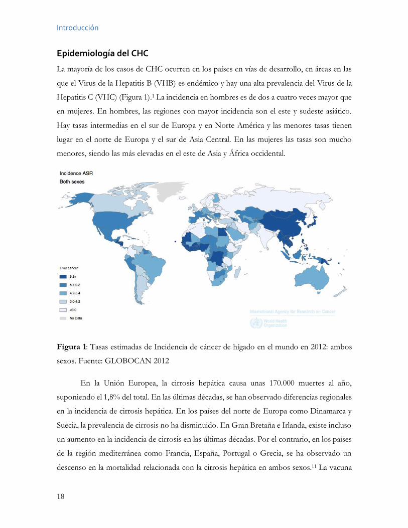
La mayoría de los casos de CHC ocurren en los países en vías de desarrollo, en áreas en las
que el Virus de la Hepatitis B (VHB) es endémico y hay una alta prevalencia del Virus de la
Hepatitis C (VHC) (Figura 1).1 La incidencia en hombres es de dos a cuatro veces mayor que
en mujeres. En hombres, las regiones con mayor incidencia son el este y sudeste asiático.
Hay tasas intermedias en el sur de Europa y en Norte América y las menores tasas tienen
lugar en el norte de Europa y el sur de Asia Central. En las mujeres las tasas son mucho
menores, siendo las más elevadas en el este de Asia y África occidental.
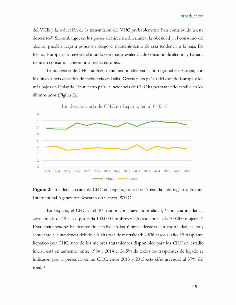
Figura 1: Tasas estimadas de Incidencia de cáncer de hígado en el mundo en 2012: ambos
sexos. Fuente: GLOBOCAN 2012
En la Unión Europea, la cirrosis hepática causa unas 170.000 muertes al año,
suponiendo el 1,8% del total. En las últimas décadas, se han observado diferencias regionales
en la incidencia de cirrosis hepática. En los países del norte de Europa como Dinamarca y
Suecia, la prevalencia de cirrosis no ha disminuido. En Gran Bretaña e Irlanda, existe incluso
un aumento en la incidencia de cirrosis en las últimas décadas. Por el contrario, en los países
de la región mediterránea como Francia, España, Portugal o Grecia, se ha observado un
descenso en la mortalidad relacionada con la cirrosis hepática en ambos sexos.11 La vacuna
Introducción
19
del VHB y la reducción de la transmisión del VHC probablemente han contribuido a este
descenso.12 Sin embargo, en los países del área mediterránea, la obesidad y el consumo del
alcohol pueden llegar a poner en riesgo el mantenimiento de esta tendencia a la baja. De
hecho, Europa es la región del mundo con más prevalencia de consumo de alcohol y España
tiene un consumo superior a la media europea.
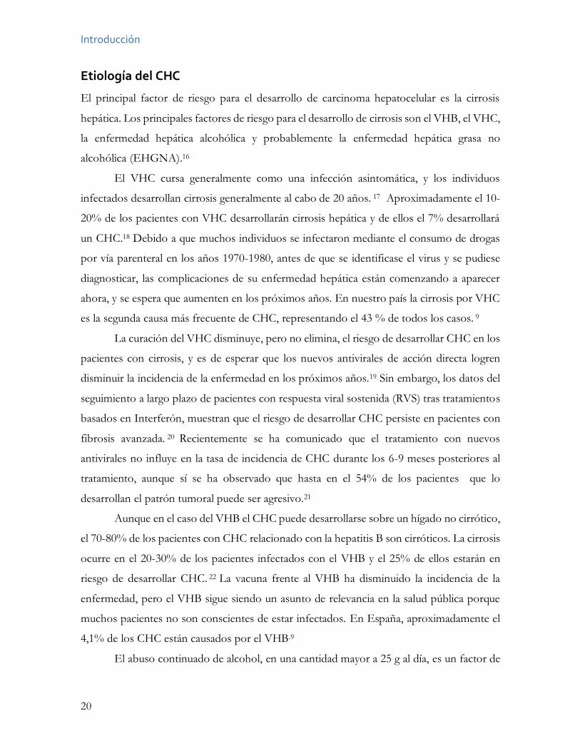
La incidencia de CHC también tiene una notable variación regional en Europa, con
los niveles más elevados de incidencia en Italia, Grecia y los países del este de Europa y los
más bajos en Holanda. En nuestro país, la incidencia de CHC ha permanecido estable en los
últimos años (Figura 2).
Figura 2: Incidencia cruda de CHC en España, basada en 7 estudios de registro. Fuente:
International Agency for Research on Cancer, WHO
En España, el CHC es el 10º tumor con mayor mortalidad,13 con una incidencia
aproximada de 12 casos por cada 100.000 hombres y 3,5 casos por cada 100.000 mujeres.14
Esta incidencia se ha mantenido estable en las últimas décadas. La mortalidad es muy
semejante a la incidencia debido a la alta tasa de mortalidad: 4.536 casos al año. El trasplante
hepático por CHC, uno de los mejores tratamientos disponibles para los CHC en estadio
inicial, está en aumento: entre 1984 y 2014 el 26,5% de todos los trasplantes de hígado se
indicaron por la presencia de un CHC, entre 2013 y 2015 esta cifra ascendió al 37% del
total.15
0
2
4
6
8
10
12
14
16
1993 1994 1995 1996 1997 1998 1999 2000 2001 2002 2003 2004 2005 2006 2007
Incidencia cruda de CHC en España, [edad 0-85+]
Hombres Mujeres
Introducción
20
Etiología del CHC
El principal factor de riesgo para el desarrollo de carcinoma hepatocelular es la cirrosis
hepática. Los principales factores de riesgo para el desarrollo de cirrosis son el VHB, el VHC,
la enfermedad hepática alcohólica y probablemente la enfermedad hepática grasa no
alcohólica (EHGNA).16
El VHC cursa generalmente como una infección asintomática, y los individuos
infectados desarrollan cirrosis generalmente al cabo de 20 años. 17 Aproximadamente el 10-
20% de los pacientes con VHC desarrollarán cirrosis hepática y de ellos el 7% desarrollará
un CHC.18 Debido a que muchos individuos se infectaron mediante el consumo de drogas
por vía parenteral en los años 1970-1980, antes de que se identificase el virus y se pudiese
diagnosticar, las complicaciones de su enfermedad hepática están comenzando a aparecer
ahora, y se espera que aumenten en los próximos años. En nuestro país la cirrosis por VHC
es la segunda causa más frecuente de CHC, representando el 43 % de todos los casos. 9
La curación del VHC disminuye, pero no elimina, el riesgo de desarrollar CHC en los
pacientes con cirrosis, y es de esperar que los nuevos antivirales de acción directa logren
disminuir la incidencia de la enfermedad en los próximos años.19 Sin embargo, los datos del
seguimiento a largo plazo de pacientes con respuesta viral sostenida (RVS) tras tratamientos
basados en Interferón, muestran que el riesgo de desarrollar CHC persiste en pacientes con
fibrosis avanzada. 20 Recientemente se ha comunicado que el tratamiento con nuevos
antivirales no influye en la tasa de incidencia de CHC durante los 6-9 meses posteriores al
tratamiento, aunque sí se ha observado que hasta en el 54% de los pacientes que lo
desarrollan el patrón tumoral puede ser agresivo.21
Aunque en el caso del VHB el CHC puede desarrollarse sobre un hígado no cirrótico,
el 70-80% de los pacientes con CHC relacionado con la hepatitis B son cirróticos. La cirrosis
ocurre en el 20-30% de los pacientes infectados con el VHB y el 25% de ellos estarán en
riesgo de desarrollar CHC. 22 La vacuna frente al VHB ha disminuido la incidencia de la
enfermedad, pero el VHB sigue siendo un asunto de relevancia en la salud pública porque
muchos pacientes no son conscientes de estar infectados. En España, aproximadamente el
4,1% de los CHC están causados por el VHB.9
El abuso continuado de alcohol, en una cantidad mayor a 25 g al día, es un factor de
Introducción
21
riesgo de cirrosis,23 aunque sólo el 13% de los pacientes con consumo excesivo de alcohol
desarrollarán enfermedad hepática alcohólica.24 Existe una relación dosis-respuesta entre la
cantidad de alcohol consumida y el desarrollo de cirrosis, siendo el riesgo más elevado en las
mujeres que consumen 30 g de alcohol al día y en los hombres que consumen 40 g de alcohol
diarios o más.25 El consumo de 60 g de alcohol al día aumenta el riesgo de mortalidad por
cirrosis alcohólica 35 veces en mujeres y 27 en hombres.26 Además, diversos factores pueden
aumentar la fibrosis hepática en los pacientes con consumo crónico de alcohol, como la
obesidad, el tabaquismo o la coexistencia con otras causas de enfermedad hepática.27 Varios
estudios también parecen mostrar que la cafeína protege frente al desarrollo de cirrosis en
los bebedores crónicos.28
La historia natural de la enfermedad hepática por alcohol transcurre desde un hígado
normal, hacia la esteatosis, fibrosis, y finalmente la cirrosis. La esteatosis simple es reversible
tras varias semanas de abstinencia, aunque si continúa el abuso de alcohol, en el 20% de
casos puede aparecer fibrosis o cirrosis en un periodo de 10 años.29 Algunos pacientes
también pasarán por una hepatitis alcohólica.
España es el décimo país que más alcohol consume a nivel mundial, con unos 11
litros de alcohol por persona y año, lo que explica la alta prevalencia de cirrosis por alcohol.
La cirrosis alcohólica, la fase final de la enfermedad hepática por alcohol, es un factor de
riesgo conocido para el desarrollo de CHC.30 Al igual que en Francia,11 en España la cirrosis
por alcohol es la principal causa de hepatocarcinoma, con un 37% del total9 y causa una
mortalidad de 4-5 personas por cada 100.000 habitantes.11
La Enfermedad del Hígado Graso No Alcohólico (EHGNA) se caracteriza por la
acumulación de grasa en al menos en el 5% de los hepatocitos, en ausencia de un consumo
significativo de alcohol, infección viral u otra etiología específica. La EHGNA puede derivar
en fibrosis avanzada y cirrosis.31 Diversos estudios muestran que los pacientes obesos 32 o
con diabetes mellitus tipo 2 tienen el doble de riesgo de desarrollar CHC.33 En España,
aproximadamente el 6% de los CHC se desarrollan sobre cirrosis por EHGNA.9
En los pacientes con cirrosis, el riesgo acumulado a 5 años de desarrollar CHC varía
entre el 5 y el 30%, en función de la etiología. Los pacientes con cirrosis por VHC, VHB y
Colangitis Biliar Primaria estadio IV tienen un riesgo elevado de desarrollar CHC.16,34 La
incidencia de cáncer de hígado en los pacientes hepatitis autoinmune parece ser menor.35
Introducción
22
Igualmente, se han descrito cánceres de hígado en pacientes con cirrosis por otras causas,
como déficit de alfa 1 antitripsina, hemocromatosis, o enfermedad de Wilson. En nuestro
país, el 10% de los CHC están causados por alguna de estas enfermedades.9
Tratamiento del CHC
La supervivencia de los pacientes con hepatocarcinoma depende del estadio tumoral al
diagnóstico, de la función hepática, de la presencia de síntomas y de la comorbilidad como
factor de riesgo competitivo y como factor limitante para administrar tratamientos. Cuando
el cáncer se diagnostica por síntomas, tiene invasión vascular o extrahepática, la
supervivencia al año es del 50%. Aquellos pacientes que permanecen postrados o en reposo
más del 50% del tiempo, o que tienen enfermedad hepática en estadio Child-Pugh C, tienen
una mediana de supervivencia de 3 meses, y sólo el 10% seguirán vivos al año.36
Los pacientes con tumores en estadio intermedio, sin síntomas derivados del cáncer,
con una cirrosis hepática compensada y sin invasión vascular, pero con tumores grandes, no
trasplantables o multifocales, son candidatos a recibir quimioembolización transarterial
(TACE), que mejora la mediana de supervivencia de 16 a más de 40 meses en pacientes bien
seleccionados.37-38 Cuando el cáncer no es subsidiario de quimioembolización, pero los
pacientes no tienen síntomas derivados del tumor, y la cirrosis está compensada, la terapia
oral con sorafenib disminuye el riesgo de muerte en un 31% (HR 0,69, IC 95% 0,55-0,87;
p< ,001).39
Sin embargo, la sobrevida es notablemente mayor cuando los tumores se diagnostican
en estadio inicial. Cuando el CHC se diagnostica en estadios iniciales y no existe invasión
vascular o metástasis, tanto la resección como la ablación ofrecen una supervivencia de hasta
el 90% a 5 años. Cuando existe hipertensión portal o bilirrubina elevada, el trasplante
hepático es el tratamiento de elección, y ofrece una supervivencia a 5 años del 75%. En caso
de no poder trasplantarse o realizarse una resección quirúrgica, la ablación tumoral por
radiofrecuencia en tumores iniciales proporciona una supervivencia de un 50% a 5 años.40
En otros tumores para los que se han desarrollado programas de cribado (mama,
colon, próstata y cérvix) existen tratamientos efectivos para casos incluso moderadamente
avanzados. En el caso del CHC, se trata de un tumor agresivo, con una elevada mortalidad.
Introducción
23
Sin embargo, diversas opciones terapéuticas aumentan la supervivencia si se descubre en
estadios iniciales. Ese hecho ha llevado al establecimiento de programas de vigilancia para el
diagnóstico precoz del CHC. Dado que el 80-90% de los tumores se diagnostican sobre
hígados cirróticos, los pacientes con cirrosis son la población diana de estos programas de
vigilancia.
Los programas de vigilancia para el diagnóstico precoz del CHC
Los programas de vigilancia para el diagnóstico precoz del cáncer tienen como objetivo
reducir la mortalidad por el cáncer. Deberían ofertar a los pacientes una ganancia de al menos
3 meses en la esperanza de vida.41 La decisión de incluir a pacientes en un programa para la
vigilancia precoz para el cáncer depende del riesgo de desarrollarlo: cuánto más riesgo tenga
el paciente más efectivo será el programa de vigilancia. La guía de la American Association
for the Study of Liver Diseases (AASLD) recomienda realizar cribado del hepatocarcinoma
en los pacientes con cirrosis con una incidencia de al menos el 1,5% anual, basándose en
diversos estudios de coste-efectividad. 42,43,44
Sólo existen dos estudios randomizados sobre el cribado de CHC, realizados en
China en pacientes con hepatitis crónica por VHB. Un estudio randomizado controlado en
19.000 pacientes, aunque el estudio fue criticado por fallos metodológicos, mostró que un
programa de vigilancia para el diagnóstico precoz del CHC, basado en la realización de una
ecografía y la medición de la alfafetoproteina cada 6 meses, se asociaba a una reducción del
37% en la mortalidad por CHC.45 Otro estudio randomizado mostró una mejoría en el
estadio tumoral al diagnóstico, pero no demostró mejoría en la supervivencia.46 Sin embargo,
este estudio fue criticado porque con frecuencia los pacientes con CHC no recibían el
tratamiento adecuado.
En los pacientes con cirrosis no existen estudios randomizados mostrando el
beneficio de la vigilancia. Además, hoy en día se enfrentaría a dilemas éticos, dado que la
mayoría de los pacientes informados prefieren la vigilancia.47 Pero existe evidencia indirecta
de múltiples estudios de cohortes y series de casos no controlados que indican que los
tumores detectados dentro de un programa para el diagnóstico precoz del CHC en los
pacientes con cirrosis tienen un estadio más inicial 48 , 49 y presentan una mejoría en la
Introducción
24
supervivencia frente a los diagnosticados fuera del programa.50,51
Dos metanálisis han estudiado la efectividad de los programas de vigilancia del CHC
en los pacientes con cirrosis. Un metanálisis, que incluyó estudios en pacientes con cirrosis
y hepatitis crónica por VHB o VHC, mostró los beneficios de los programas de vigilancia
en el diagnóstico de los tumores en estadio más inicial, pero no pudo demostrar beneficio
en la supervivencia de los pacientes diagnosticados en el programa de vigilancia.52 Otro
metanálisis, que incluyó exclusivamente estudios en pacientes con cirrosis, demostró que los
programas de vigilancia del hepatocarcinoma se asocian con mejoría en el diagnostico precoz
de los tumores, en el tratamiento aplicado y en la supervivencia de los pacientes con cirrosis. 53
Las guías de la AASLD,42 y de la Asociación Europea para el estudio del Hígado
(EASL)54 recomiendan realizar vigilancia con ecografía cada 6 meses a los pacientes con
cirrosis hepática, independientemente de la causa de la misma.
Existen múltiples interrogantes sobre los programas de cribado: la efectividad del
cribado, la población en riesgo, el papel de la AFP, cómo realizar el cribado en las
poblaciones especiales como obesos, etc.
Los tres estudios que componen la presente Tesis Doctoral analizan diversos
aspectos de los programas de cribado sobre los que todavía existe incertidumbre: la
incidencia de CHC en la cirrosis alcohólica, la adherencia de los pacientes a los programas
de cribado, y las causas asociadas al fracaso del programa a la hora de detectar tumores en
estadio inicial.
Incidencia del CHC en la cirrosis de causa alcohólica
Dado que la efectividad de los programas de vigilancia del CHC depende de la incidencia del
tumor en la población diana, es esencial cuantificar el riesgo de desarrollar CHC en los
pacientes con cirrosis. La incidencia anual de CHC en los pacientes con cirrosis por VHB y
VHC varía en distintos estudios entre el 2,2-3,7%55 y el 2-8% respectivamente.56 Tanto en
los pacientes con cirrosis por VHB, por VHC, como por Colangitis Biliar Primaria en estadio
IV, existe evidencia de que el riesgo de desarrollar CHC excede el umbral del 1,5% anual.16
En el resto de etiologías de la cirrosis, la incidencia de CHC no está tan bien documentada.
Los pacientes con enfermedad hepática crónica avanzada relacionada con el alcohol,
Introducción
25
tienen 59 veces más riesgo de desarrollar CHC que la población general.57 Sin embargo, la
incidencia de CHC en los pacientes con cirrosis de causa alcohólica no es bien conocida.
Muchos de los estudios son previos al descubrimiento del VHC, en 1989, y por tanto sus
resultados no son válidos. 58 Otros análisis posteriores incluyen entre los pacientes con
cirrosis alcohólica aquellos con anticuerpos frente al VHC y antígeno de superficie del VHB,
y por tanto sus resultados no son comparables.59,60 Otros estudios analizan la incidencia en
cohortes de pacientes con cirrosis sin informar de la incidencia en los pacientes de causa
alcohólica.61 Finalmente, algunos estudios dan datos sobre incidencia de CHC en pacientes
con cirrosis alcohólica, pero sólo informan sobre incidencia acumulada. En España, un
estudio del año 2006 observó una incidencia de CHC en pacientes con cirrosis alcohólica del
7,1% tras 5 años de seguimiento.62 En Japón, 2 estudios dieron resultados discordantes, con
una incidencia acumulada a 3 años del 1% en un estudio63 y del 2,4% en otro. 64
Los estudios de cohorte que analizan la incidencia anual de CHC en pacientes con
cirrosis alcohólica aportan resultados muy dispares. Un estudio francés de cohorte con
seguimiento prospectivo en ámbito hospitalario mostró una incidencia anual del 5,6%.65 Un
estudio americano de registro, de una cohorte de veteranos de guerra con cirrosis seguidos
en centros de salud, observó una incidencia del 0,6%.66 La diferencia en los resultados puede
estar influenciada por la elevada edad media de los pacientes en el estudio francés y por los
sesgos en la recogida de datos inherente a los estudios de registro en el segundo caso.
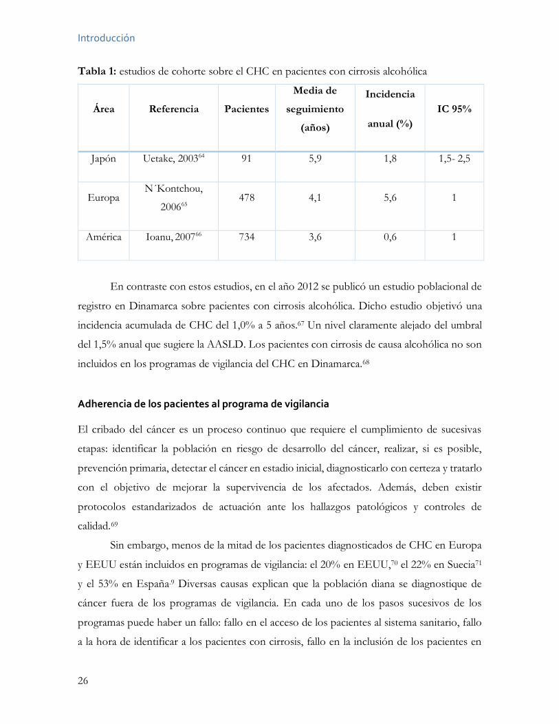
En la Tabla 1 se resumen los estudios de cohorte sobre el CHC en pacientes con
cirrosis alcohólica que aportan datos sobre incidencia anual.
Introducción
26
Tabla 1: estudios de cohorte sobre el CHC en pacientes con cirrosis alcohólica
Área Referencia Pacientes
Media de
seguimiento
(años)
Incidencia
anual (%) IC 95%
Japón Uetake, 200364 91 5,9 1,8 1,5- 2,5
Europa N´Kontchou,
200665 478 4,1 5,6 1
América Ioanu, 200766 734 3,6 0,6 1
En contraste con estos estudios, en el año 2012 se publicó un estudio poblacional de
registro en Dinamarca sobre pacientes con cirrosis alcohólica. Dicho estudio objetivó una
incidencia acumulada de CHC del 1,0% a 5 años.67 Un nivel claramente alejado del umbral
del 1,5% anual que sugiere la AASLD. Los pacientes con cirrosis de causa alcohólica no son
incluidos en los programas de vigilancia del CHC en Dinamarca.68
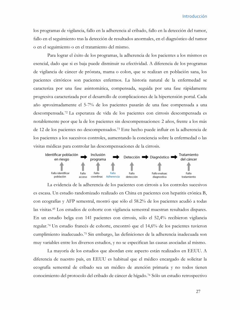
Adherencia de los pacientes al programa de vigilancia El cribado del cáncer es un proceso continuo que requiere el cumplimiento de sucesivas
etapas: identificar la población en riesgo de desarrollo del cáncer, realizar, si es posible,
prevención primaria, detectar el cáncer en estadio inicial, diagnosticarlo con certeza y tratarlo
con el objetivo de mejorar la supervivencia de los afectados. Además, deben existir
protocolos estandarizados de actuación ante los hallazgos patológicos y controles de
calidad.69
Sin embargo, menos de la mitad de los pacientes diagnosticados de CHC en Europa
y EEUU están incluidos en programas de vigilancia: el 20% en EEUU,70 el 22% en Suecia71
y el 53% en España.9 Diversas causas explican que la población diana se diagnostique de
cáncer fuera de los programas de vigilancia. En cada uno de los pasos sucesivos de los
programas puede haber un fallo: fallo en el acceso de los pacientes al sistema sanitario, fallo
a la hora de identificar a los pacientes con cirrosis, fallo en la inclusión de los pacientes en
Introducción
27
los programas de vigilancia, fallo en la adherencia al cribado, fallo en la detección del tumor,
fallo en el seguimiento tras la detección de resultados anormales, en el diagnóstico del tumor
o en el seguimiento o en el tratamiento del mismo.
Para lograr el éxito de los programas, la adherencia de los pacientes a los mismos es
esencial, dado que si es baja puede disminuir su efectividad. A diferencia de los programas
de vigilancia de cáncer de próstata, mama o colon, que se realizan en población sana, los
pacientes cirróticos son pacientes enfermos. La historia natural de la enfermedad se
caracteriza por una fase asintomática, compensada, seguida por una fase rápidamente
progresiva caracterizada por el desarrollo de complicaciones de la hipertensión portal. Cada
año aproximadamente el 5-7% de los pacientes pasarán de una fase compensada a una
descompensada.72 La esperanza de vida de los pacientes con cirrosis descompensada es
notablemente peor que la de los pacientes sin descompensaciones: 2 años, frente a los más
de 12 de los pacientes no descompensados.73 Este hecho puede influir en la adherencia de
los pacientes a los sucesivos controles, aumentando la conciencia sobre la enfermedad o las
visitas médicas para controlar las descompensaciones de la cirrosis.
La evidencia de la adherencia de los pacientes con cirrosis a los controles sucesivos
es escasa. Un estudio randomizado realizado en China en pacientes con hepatitis crónica B,
con ecografías y AFP semestral, mostró que sólo el 58.2% de los pacientes acudió a todas
las visitas.45 Los estudios de cohorte con vigilancia semestral muestran resultados dispares.
En un estudio belga con 141 pacientes con cirrosis, sólo el 52,4% recibieron vigilancia
regular.74 Un estudio francés de cohorte, encontró que el 14,6% de los pacientes tuvieron
cumplimiento inadecuado.75 Sin embargo, las definiciones de la adherencia inadecuada son
muy variables entre los diversos estudios, y no se especifican las causas asociadas al mismo.
La mayoría de los estudios que abordan este aspecto están realizados en EEUU. A
diferencia de nuestro país, en EEUU es habitual que el médico encargado de solicitar la
ecografía semestral de cribado sea un médico de atención primaria y no todos tienen
conocimiento del protocolo del cribado de cáncer de hígado.76 Sólo un estudio retrospectivo
Introducción
28
diferencia si el fallo a la adherencia al programa de cribado se debe a que el paciente no acude
o a que el médico encargado no solicita el estudio.77 En el contexto europeo, no existe una
evidencia sólida de la adherencia de los pacientes al programa de vigilancia del cáncer de
hígado.
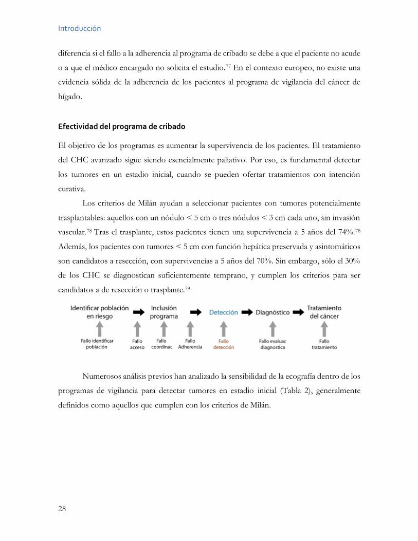
Efectividad del programa de cribado El objetivo de los programas es aumentar la supervivencia de los pacientes. El tratamiento
del CHC avanzado sigue siendo esencialmente paliativo. Por eso, es fundamental detectar
los tumores en un estadio inicial, cuando se pueden ofertar tratamientos con intención
curativa.
Los criterios de Milán ayudan a seleccionar pacientes con tumores potencialmente
trasplantables: aquellos con un nódulo < 5 cm o tres nódulos < 3 cm cada uno, sin invasión
vascular.78 Tras el trasplante, estos pacientes tienen una supervivencia a 5 años del 74%.78
Además, los pacientes con tumores < 5 cm con función hepática preservada y asintomáticos
son candidatos a resección, con supervivencias a 5 años del 70%. Sin embargo, sólo el 30%
de los CHC se diagnostican suficientemente temprano, y cumplen los criterios para ser
candidatos a de resección o trasplante.79
Numerosos análisis previos han analizado la sensibilidad de la ecografía dentro de los
programas de vigilancia para detectar tumores en estadio inicial (Tabla 2), generalmente
definidos como aquellos que cumplen con los criterios de Milán.
Introducción
29
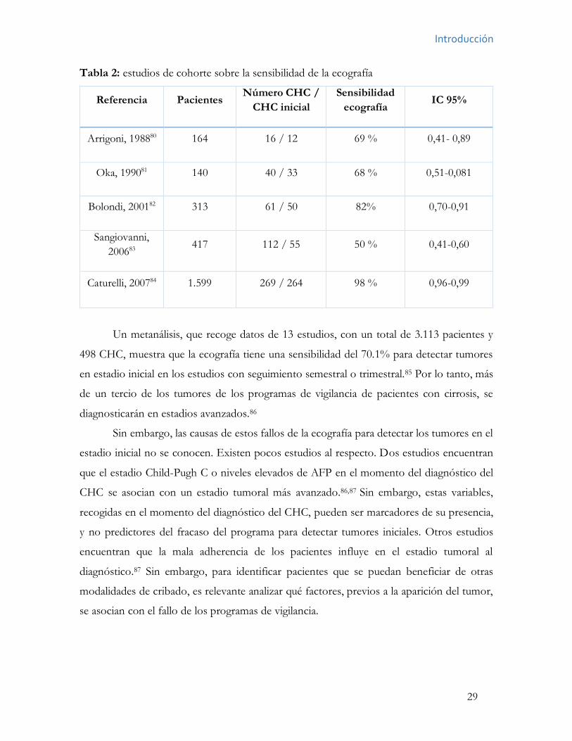
Tabla 2: estudios de cohorte sobre la sensibilidad de la ecografía
Referencia Pacientes Número CHC /
CHC inicial Sensibilidad
ecografía IC 95%
Arrigoni, 198880 164 16 / 12 69 % 0,41- 0,89
Oka, 199081 140 40 / 33 68 % 0,51-0,081
Bolondi, 200182 313 61 / 50 82% 0,70-0,91
Sangiovanni, 200683
417 112 / 55 50 % 0,41-0,60
Caturelli, 200784 1.599 269 / 264 98 % 0,96-0,99
Un metanálisis, que recoge datos de 13 estudios, con un total de 3.113 pacientes y
498 CHC, muestra que la ecografía tiene una sensibilidad del 70.1% para detectar tumores
en estadio inicial en los estudios con seguimiento semestral o trimestral.85 Por lo tanto, más
de un tercio de los tumores de los programas de vigilancia de pacientes con cirrosis, se
diagnosticarán en estadios avanzados.86
Sin embargo, las causas de estos fallos de la ecografía para detectar los tumores en el
estadio inicial no se conocen. Existen pocos estudios al respecto. Dos estudios encuentran
que el estadio Child-Pugh C o niveles elevados de AFP en el momento del diagnóstico del
CHC se asocian con un estadio tumoral más avanzado.86,87 Sin embargo, estas variables,
recogidas en el momento del diagnóstico del CHC, pueden ser marcadores de su presencia,
y no predictores del fracaso del programa para detectar tumores iniciales. Otros estudios
encuentran que la mala adherencia de los pacientes influye en el estadio tumoral al
diagnóstico.87 Sin embargo, para identificar pacientes que se puedan beneficiar de otras
modalidades de cribado, es relevante analizar qué factores, previos a la aparición del tumor,
se asocian con el fallo de los programas de vigilancia.

30
Objetivos
31
Objetivos
El objetivo del primer estudio de la presente tesis doctoral fue determinar la
incidencia de CHC en una población de pacientes con cirrosis de origen alcohólico seguidos
de manera prospectiva. También intentó conocer si existen pacientes con distinto riesgo de
desarrollar hepatocarcinoma.
El objetivo del segundo estudio de esta Tesis Doctoral fue determinar la adherencia
de los pacientes incluidos en un programa de vigilancia para el diagnóstico precoz de CHC.
Igualmente tiene como objetivos determinar si la adherencia de los pacientes al programa de
vigilancia influye en el estadio tumoral al diagnóstico, en el tratamiento aplicado a los tumores
y en la supervivencia de los pacientes con hepatocarcinoma, así como analizar las variables
potencialmente asociadas con una adherencia subóptima.
El objetivo del tercer artículo de la presente Tesis Doctoral fue analizar de manera
prospectiva el fracaso del programa de vigilancia, definido como la proporción de tumores
diagnosticados fuera de los criterios de Milán, e identificar factores asociados con dicho fallo.

32
33
Material y métodos
Cohorte a Estudio
En los tres trabajos se analizó la misma cohorte de pacientes. En septiembre de 1992, se
puso en marcha en la Unidad de Hepatología del Servicio de Aparato Digestivo del Hospital
Universitario Central de Asturias (HUCA) un programa para el diagnóstico precoz del CHC
en los pacientes con cirrosis. Desde entonces, todos los pacientes consecutivos
diagnosticados de cirrosis o atendidos por dicha causa en la Unidad, fueron incluidos de
manera prospectiva en el programa e introducidos en una base de datos. Hasta agosto de
2016 el programa incorporó a 1.259 pacientes con cirrosis, de los que 188 desarrollaron
CHC.
Los pacientes fueron incluidos en el programa si presentaban: a) un estadio Child-
Pugh A ó B; b) una edad comprendida entre 35 y 75 años; c) ausencia de CHC conocido; d)
posibilidad de asistir a las consultas sucesivas y e) ausencia de una enfermedad concomitante
con una esperanza de vida menor a 1 año.
El diagnóstico de cirrosis se realizó mediante biopsia hepática en 407 pacientes
(32,3%), por la presencia de varices esofágicas en 612 pacientes (48,6%), por complicaciones
de la hipertensión portal en 82 pacientes (6,5%) y por la presencia en la ecografía de un
hígado de morfología cirrótica asociado a signos de hipertensión portal en 158 pacientes
(12,5%).
La etiología de la cirrosis se estableció de acuerdo a la causa de la misma: alcohólica,
si existía un consumo diario de alcohol ≥ 60 g en hombres y ≥ 40 g en mujeres, por VHC
en caso de positividad en suero de anticuerpos anti-VHC y RNA del VHC, por VHB en caso
de positividad en suero del antígeno de superficie del VHB u otras causas: hemocromatosis,
hepatitis autoinmune, colangitis biliar primaria, déficit de alfa 1 antitripsina, EHGNA o
enfermedad de Wilson, sobre la base de los criterios universalmente aceptados.
La historia de consumo de alcohol, tabaco o drogas por vía parenteral se recogía
mediante una entrevista con el paciente y los familiares y se llevó a cabo por un hepatólogo
del equipo. Los pacientes que recibían tratamiento para la diabetes o con niveles de glucemia
en ayunas ≥ 126 mg/dL fueron considerados diabéticos. El genotipado de HFE para
detectar la mutación C282Y se realizó mediante reacción en cadena de la polimerasa.
Material y métodos
34
Los pacientes fueron censurados en la fecha de diagnóstico de CHC, la fecha de
progresión a estadío C de Child-Pugh, la fecha de inclusión en lista para el trasplante
hepático, la fecha de la última visita, la fecha de la transferencia a otro centro sanitario, la
fecha del desarrollo de una comorbilidad severa o la fecha de la muerte, lo que sucediese
primero. Para confirmar el estado del paciente al final del periodo de estudio se consultó el
Índice Nacional de Defunciones,
(www.msssi.gob.es/estadEstudios/estadisticas/estadisticas/estMinisterio/IND_TipoDifus
ion.htm), que incluye la fecha de todas las muertes notificadas en España y se actualiza
constantemente.
Se recogieron variables clínicas y de laboratorio en el momento de la inclusión en el
programa, durante el transcurso del mismo, y en el momento del diagnóstico del tumor o la
censura del paciente. Variables como las complicaciones de la enfermedad hepática, el
estadio Child-Pugh, o la asistencia a los controles sucesivos, se recogían en cada visita.
El primer estudio se publicó en 2013 y sólo incluyó a los pacientes con cirrosis
alcohólica. El segundo estudio se publicó en 2017 e incluyó a todos los pacientes seguidos
de manera semestral en el programa hasta el febrero de 2013. El tercer estudio se envió para
su publicación en 2017, e incluyó a todos los tumores diagnosticados durante el trascurso
del programa de vigilancia.
Análisis de la incidencia anual de CHC en pacientes con cirrosis alcohólica
Pacientes y métodos
Este primer estudio se cerró en marzo de 2010 e incluyó a todos los pacientes con cirrosis
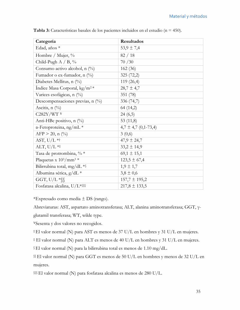
alcohólica consecutivamente incluidos en el programa, un total de 450 pacientes. Se
analizaron 20 variables demográficas, clínicas y bioquímicas recogidas en el momento de la
inclusión (Tabla 3).
Material y métodos
35
Tabla 3: Características basales de los pacientes incluidos en el estudio (n = 450). Categoría Resultados Edad, años * 53,9 ± 7,4 Hombre / Mujer, % 82 / 18 Child-Pugh A / B, % 70 /30 Consumo activo alcohol, n (%) 162 (36) Fumador o ex-fumador, n (%) 325 (72,2) Diabetes Mellitus, n (%) 119 (26,4) Índice Masa Corporal, kg/m2 * 28,7 ± 4,7 Varices esofágicas, n (%) 351 (78) Descompensaciones previas, n (%) 336 (74,7) Ascitis, n (%) 64 (14,2) C282Y/WT ¥ 24 (6,5) Anti-HBc positivo, n (%) 53 (11,8) α-Fetoproteina, ng/mL * 4,7 ± 4,7 (0,1-73,4) AFP > 20, n (%) 3 (0,6) AST, U/L *† 47,9 ± 24,7 ALT, U/L *‡ 33,2 ± 14,9 Tasa de protrombina, % * 69,1 ± 15,1 Plaquetas x 103/mm3 * 123,5 ± 67,4 Bilirrubina total, mg/dL *§ 1,9 ± 1,7 Albumina sérica, g/dL * 3,8 ± 0,6 GGT, U/L *§§ 157,7 ± 195,2 Fosfatasa alcalina, U/L*§§§ 217,8 ± 133,5
*Expresado como media ± DS (rango).
Abreviaturas: AST, aspartato aminotransferasa; ALT, alanina aminotransferasa; GGT, γ-
glutamil transferasa; WT, wilde type. ¥Sesenta y dos valores no recogidos. † El valor normal (N) para AST es menos de 37 U/L en hombres y 31 U/L en mujeres. ‡ El valor normal (N) para ALT es menos de 40 U/L en hombres y 31 U/L en mujeres. § El valor normal (N) para la bilirrubina total es menos de 1.10 mg/dL. §§ El valor normal (N) para GGT es menos de 50 U/L en hombres y menos de 32 U/L en
mujeres. §§§ El valor normal (N) para fosfatasa alcalina es menos de 280 U/L.
Material y métodos
36
Adherencia de los pacientes al programa de vigilancia Pacientes y métodos
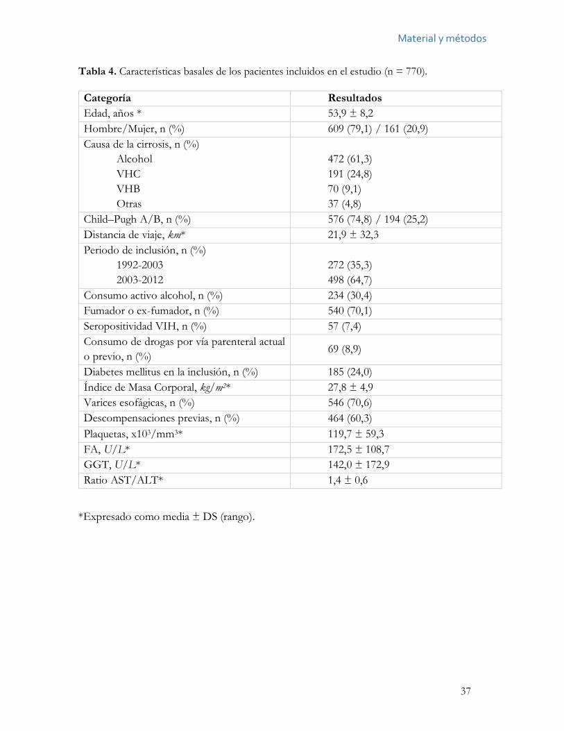
Este segundo estudio incluyó a los 770 pacientes con cirrosis de cualquier etiología
consecutivamente incluidos en el programa hasta febrero de 2013 y seguidos de manera
semestral. El seguimiento se cerró en marzo de 2014. Se recogieron 17 variables
demográficas, clínicas y bioquímicas en la inclusión (Tabla 4). La proximidad al centro
sanitario se calculó a partir del código postal del domicilio del paciente.
Se dividió a los pacientes en 2 cohortes distintas de acuerdo con su adherencia al
programa: la mala adherencia se definió como la no asistencia a la realización de dos
controles consecutivos, mientras que se clasificó como pacientes con buena adherencia a
aquellos que durante el seguimiento no fallaron a 2 controles consecutivos. Este criterio fue
escogido arbitrariamente.
Para la censura de los pacientes, además de los criterios previamente descritos -
pérdida, estadio Child-Pugh C, traslado a otro hospital, CHC, inclusión en lista para el
trasplante hepático, última visita, transferencia a otro centro sanitario, desarrollo de una
comorbilidad severa o muerte-, también se censuró a los pacientes con 2 fallos consecutivos
a los controles.
Material y métodos
37
Tabla 4. Características basales de los pacientes incluidos en el estudio (n = 770).
Categoría Resultados Edad, años * 53,9 ± 8,2 Hombre/Mujer, n (%) 609 (79,1) / 161 (20,9) Causa de la cirrosis, n (%)
Alcohol VHC VHB Otras
472 (61,3) 191 (24,8) 70 (9,1) 37 (4,8)
Child–Pugh A/B, n (%) 576 (74,8) / 194 (25,2) Distancia de viaje, km* 21,9 ± 32,3 Periodo de inclusión, n (%)
1992-2003 2003-2012
272 (35,3) 498 (64,7)
Consumo activo alcohol, n (%) 234 (30,4) Fumador o ex-fumador, n (%) 540 (70,1) Seropositividad VIH, n (%) 57 (7,4) Consumo de drogas por vía parenteral actual o previo, n (%) 69 (8,9)
Diabetes mellitus en la inclusión, n (%) 185 (24,0) Índice de Masa Corporal, kg/m2* 27,8 ± 4,9 Varices esofágicas, n (%) 546 (70,6) Descompensaciones previas, n (%) 464 (60,3) Plaquetas, x103/mm3* 119,7 ± 59,3 FA, U/L* 172,5 ± 108,7 GGT, U/L* 142,0 ± 172,9 Ratio AST/ALT* 1,4 ± 0,6
*Expresado como media ± DS (rango).
Material y métodos
38
Incidencia y factores de riesgo del fracaso del programa
Pacientes y métodos
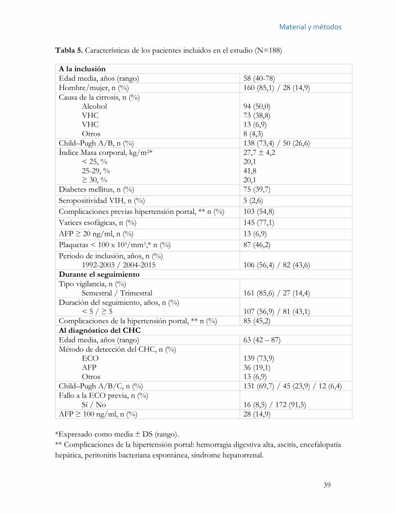
El tercer estudio incluyó a los 188 pacientes que desarrollaron CHC dentro del programa
hasta diciembre de 2014. Se recogieron 21 variables demográficas, clínicas y bioquímicas de
manera prospectiva (Tabla 5). Doce variables se recolectaron en el momento de la inclusión
en el programa, tres durante el seguimiento y 6 en el momento del diagnóstico del CHC.
Para el análisis estadístico se clasificó a los pacientes en 2 cohortes distintas en
función del estadio tumoral al diagnóstico: el fracaso del programa fue definido como la
detección de un CHC fuera de los criterios de Milán, y el no fracaso del programa fue
definido como el diagnóstico de los CHC dentro de los criterios de Milán.
Material y métodos
39
Tabla 5. Características de los pacientes incluidos en el estudio (N=188) A la inclusión Edad media, años (rango) 58 (40-78) Hombre/mujer, n (%) 160 (85,1) / 28 (14,9) Causa de la cirrosis, n (%)
Alcohol VHC VHC Otros
94 (50,0) 73 (38,8) 13 (6,9) 8 (4,3)
Child–Pugh A/B, n (%) 138 (73,4) / 50 (26,6) Índice Masa corporal, kg/m2*
< 25, % 25-29, % ≥ 30, %
27,7 ± 4,2 20,1 41,8 20,1
Diabetes mellitus, n (%) 75 (39,7) Seropositividad VIH, n (%) 5 (2,6) Complicaciones previas hipertensión portal, ** n (%) 103 (54,8) Varices esofágicas, n (%) 145 (77,1) AFP ≥ 20 ng/ml, n (%) 13 (6,9) Plaquetas < 100 x 103/mm3,* n (%) 87 (46,2) Periodo de inclusión, años, n (%)
1992-2003 / 2004-2015 106 (56,4) / 82 (43,6)
Durante el seguimiento Tipo vigilancia, n (%)
Semestral / Trimestral 161 (85,6) / 27 (14,4)
Duración del seguimiento, años, n (%) < 5 / ≥ 5
107 (56,9) / 81 (43,1)
Complicaciones de la hipertensión portal, ** n (%) 85 (45,2) Al diagnóstico del CHC Edad media, años (rango) 63 (42 – 87) Método de detección del CHC, n (%)
ECO AFP Otros
139 (73,9) 36 (19,1) 13 (6,9)
Child–Pugh A/B/C, n (%) 131 (69,7) / 45 (23,9) / 12 (6,4) Fallo a la ECO previa, n (%)
Sí / No 16 (8,5) / 172 (91,5)
AFP ≥ 100 ng/ml, n (%) 28 (14,9) *Expresado como media ± DS (rango). ** Complicaciones de la hipertensión portal: hemorragia digestiva alta, ascitis, encefalopatía hepática, peritonitis bacteriana espontánea, síndrome hepatorrenal.
Material y métodos
40
Diagnóstico y estadificación del CHC
Los pacientes fueron seguidos de manera ambulatoria por hepatólogos del Servicio de
Aparato Digestivo del HUCA. El cribado consistía en la realización de una ecografía
abdominal y la medición del nivel de alfa-fetoproteína (AFP). En cada visita clínica, se realizó
una anamnesis, una exploración física y se revisaron los resultados de los análisis y la
ecografía solicitados previamente. También se notificaba al paciente las fechas del siguiente
control con analítica, la fecha de la ecografía y de la visita médica. En caso de que el paciente
no acudiese al control clínico, el médico le avisaba telefónicamente y se le notificaba una
nueva citación para realizar la ecografía, la analítica y la visita clínica. Durante el estudio, el
hospital no realizó recordatorios de las citas.
Las ecografías semestrales eran realizadas en el Hospital por digestólogos con amplia
experiencia en la ecografía de cribado. Si el paciente requería un ingreso debido a un episodio
de descompensación en la fecha del control ecográfico, se computaba como visita realizada.
Las pruebas de imagen realizadas en dicho ingreso eran valoradas como realizadas con
intención de cribado y registradas a tal efecto.
Cuando la AFP era > 20 ng/ml y más del doble del valor previo, se realizaba una
tomografía axial computarizada multifásica (TAC), para descartar la presencia de CHC.
Cuando se detectaba un nódulo en la ecografía, se realizaban estudios de imagen dinámicos.
Todos los pacientes con una lesión focal detectada en la ecografía o con un aumento del
nivel de AFP y un TAC negativo se monitorizaron trimestralmente mediante ecografía y
AFP al menos durante 1 año.
Desde 1992 a 2001, el CHC se diagnosticó mediante confirmación histológica o
cuando se observaba una lesión hepática junto con un nivel de AFP ≥400 ng/mL. Desde
2001 a 2005, el diagnóstico de CHC se realizó de acuerdo a los criterios de la European
Association for the Study of Liver Disease criteria (EASL),88 y desde 2005 hasta la actualidad,
se siguieron los criterios de la AASLD.42
El estadiaje tumoral se basó al menos en dos técnicas de imagen, que fueron
cambiando a lo largo del estudio, de acuerdo al criterio de referencia más reciente durante
las últimas 2 décadas. El estadio tumoral se definió siguiendo el sistema de estatificación del
Barcelona Clinic Liver Cancer (BCLC)89 y los criterios de Milán.78
Material y métodos
41
Análisis estadístico Los estadísticos descriptivos se comunicaron usando la media ± desviación estándar,
medianas con rango intercuartílico (RIC) o N (%). La comparación entre dos grupos se
obtuvo utilizando el test chi-cuadrado o la distribución t-student. El método Kaplan-Meier
se utilizó para calcular las probabilidades acumuladas. La comparación entre las curvas de
supervivencia se utilizó usando la prueba de log-rank. En las variables continuas, los puntos
de cohorte para la predicción de la variable a estudio, se determinaron maximizando el índice
de Youden (sensibilidad + especificidad - 1), calculado a partir de las curvas Receiving
Operator Curves.
Los parámetros con valores P <0.10 en el análisis univariado se incluyeron en el
análisis multivariado. Antes de incluir las variables en el modelo de regresión multivariable
se analizó la co-linealidad y se excluyeron las variables con un factor de inflación de la
varianza > 5. Se utilizó la regresión de Cox para estimar el cociente de riesgos instantáneos
(Hazard Ratio, HR). Un valor de P < 0.05 se consideró estadísticamente significativo. Todas
las probabilidades tenían 2 colas con un intervalo de confianza del 95%.
Para clasificar a los pacientes según el Índice de Masa Corporal (IMC) se distribuyó
a los pacientes según la clasificación de la Organización Mundial de la Salud y se excluyeron
a los pacientes con ascitis.
Para el análisis estadístico se utilizó el programa SPSS 19.0 (Chicago, IL).

Resultados
42
Resultados
43
Resultados
Análisis de la incidencia anual de CHC en pacientes con cirrosis alcohólica
Datos clínicos y de laboratorio
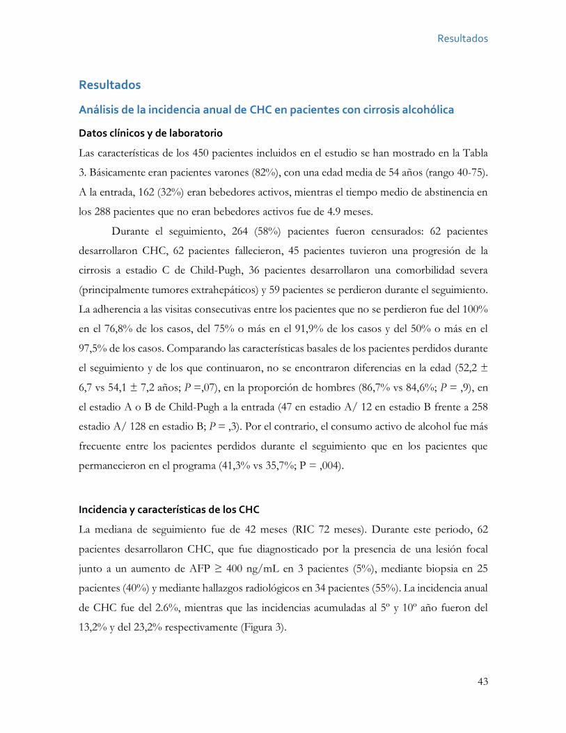
Las características de los 450 pacientes incluidos en el estudio se han mostrado en la Tabla
3. Básicamente eran pacientes varones (82%), con una edad media de 54 años (rango 40-75).
A la entrada, 162 (32%) eran bebedores activos, mientras el tiempo medio de abstinencia en
los 288 pacientes que no eran bebedores activos fue de 4.9 meses.
Durante el seguimiento, 264 (58%) pacientes fueron censurados: 62 pacientes
desarrollaron CHC, 62 pacientes fallecieron, 45 pacientes tuvieron una progresión de la
cirrosis a estadio C de Child-Pugh, 36 pacientes desarrollaron una comorbilidad severa
(principalmente tumores extrahepáticos) y 59 pacientes se perdieron durante el seguimiento.
La adherencia a las visitas consecutivas entre los pacientes que no se perdieron fue del 100%
en el 76,8% de los casos, del 75% o más en el 91,9% de los casos y del 50% o más en el
97,5% de los casos. Comparando las características basales de los pacientes perdidos durante
el seguimiento y de los que continuaron, no se encontraron diferencias en la edad (52,2 ±
6,7 vs 54,1 ± 7,2 años; P =,07), en la proporción de hombres (86,7% vs 84,6%; P = ,9), en
el estadio A o B de Child-Pugh a la entrada (47 en estadio A/ 12 en estadio B frente a 258
estadio A/ 128 en estadio B; P = ,3). Por el contrario, el consumo activo de alcohol fue más
frecuente entre los pacientes perdidos durante el seguimiento que en los pacientes que
permanecieron en el programa (41,3% vs 35,7%; P = ,004).
Incidencia y características de los CHC
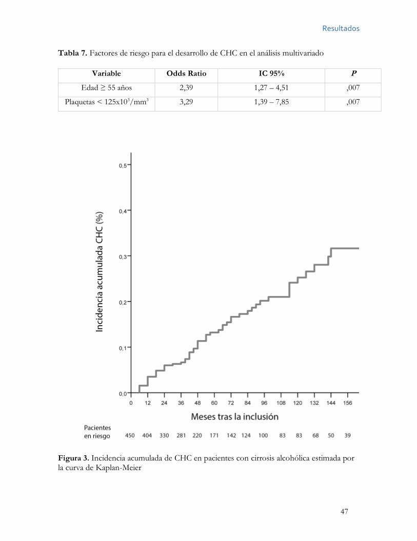
La mediana de seguimiento fue de 42 meses (RIC 72 meses). Durante este periodo, 62
pacientes desarrollaron CHC, que fue diagnosticado por la presencia de una lesión focal
junto a un aumento de AFP ≥ 400 ng/mL en 3 pacientes (5%), mediante biopsia en 25
pacientes (40%) y mediante hallazgos radiológicos en 34 pacientes (55%). La incidencia anual
de CHC fue del 2.6%, mientras que las incidencias acumuladas al 5º y 10º año fueron del
13,2% y del 23,2% respectivamente (Figura 3).
Resultados
44
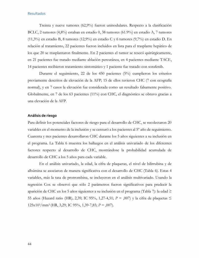
Treinta y nueve tumores (62,9%) fueron uninodulares. Respecto a la clasificación
BCLC, 2 tumores (4,8%) estaban en estadio 0, 38 tumores (61.9%) en estadio A, 7 tumores
(11,3%) en estadio B, 8 tumores (12,9%) en estadio C y 6 tumores (9,7%) en estadio D. En
relación al tratamiento, 22 pacientes fueron incluidos en lista para el trasplante hepático de
los que 20 se trasplantaron finalmente. En 2 pacientes el tumor se resecó quirúrgicamente,
en 21 pacientes fue tratado mediante ablación percutánea, en 4 pacientes mediante TACE,
14 pacientes recibieron tratamiento sintomático y 1 paciente fue tratado con sorafenib.
Durante el seguimiento, 22 de los 450 pacientes (5%) cumplieron los criterios
previamente descritos de elevación de la AFP; 15 de ellos tuvieron CHC (7 con ecografía
normal), y en 7 casos la elevación fue considerada como un resultado falsamente positivo.
Globalmente, en 7 de los 63 pacientes (11%) con CHC, el diagnóstico se obtuvo gracias a
una elevación de la AFP.
Análisis de riesgo
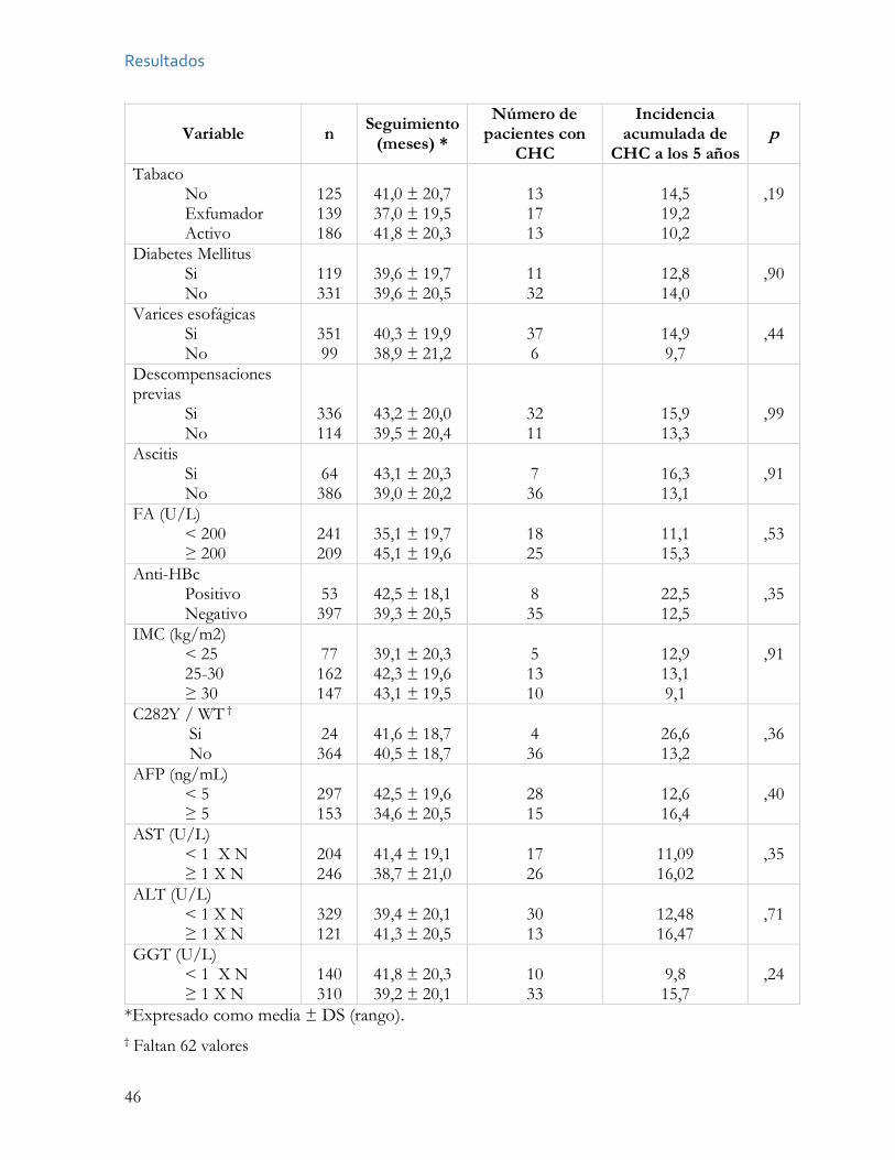
Para definir los potenciales factores de riesgo para el desarrollo de CHC, se recolectaron 20
variables en el momento de la inclusión y se censuró a los pacientes al 5º año de seguimiento.
Cuarenta y tres pacientes desarrollaron CHC durante los 5 años siguientes a su inclusión en
el programa. La Tabla 6 muestra los hallazgos en el análisis univariado de los diferentes
factores respecto al desarrollo de CHC, mostrándose la probabilidad acumulada de
desarrollo de CHC a los 5 años para cada variable.
En el análisis univariado, la edad, la cifra de plaquetas, el nivel de bilirrubina y de
albúmina se asociaron de manera significativa con el desarrollo de CHC (Tabla 6). Estas 4
variables, más la tasa de protrombina, se incluyeron en el análisis multivariado. Usando la
regresión Cox se observó que sólo 2 parámetros fueron significativos para predecir la
aparición de CHC en los 5 años siguientes a su inclusión en el programa (Tabla 7): la edad ≥
55 años (Hazard ratio (HR), 2,39; IC 95%, 1,27-4,51; P = ,007) y la cifra de plaquetas ≤
125x103/mm3 (HR, 3,29; IC 95%, 1,39-7,85; P = ,007).
Resultados
45
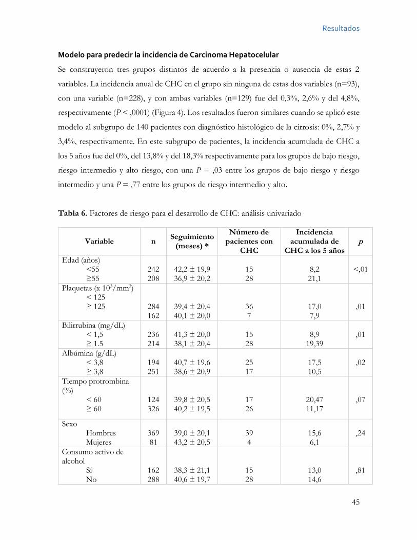
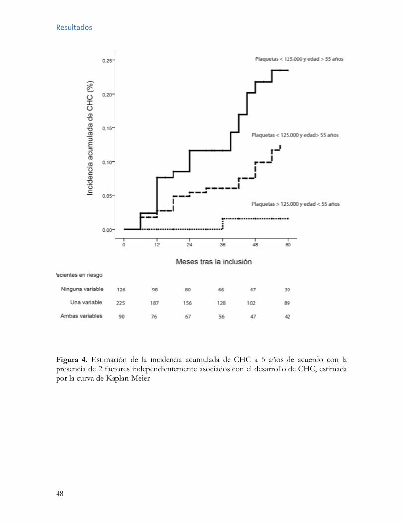
Modelo para predecir la incidencia de Carcinoma Hepatocelular
Se construyeron tres grupos distintos de acuerdo a la presencia o ausencia de estas 2
variables. La incidencia anual de CHC en el grupo sin ninguna de estas dos variables (n=93),
con una variable (n=228), y con ambas variables (n=129) fue del 0,3%, 2,6% y del 4,8%,
respectivamente (P < ,0001) (Figura 4). Los resultados fueron similares cuando se aplicó este
modelo al subgrupo de 140 pacientes con diagnóstico histológico de la cirrosis: 0%, 2,7% y
3,4%, respectivamente. En este subgrupo de pacientes, la incidencia acumulada de CHC a
los 5 años fue del 0%, del 13,8% y del 18,3% respectivamente para los grupos de bajo riesgo,
riesgo intermedio y alto riesgo, con una P = ,03 entre los grupos de bajo riesgo y riesgo
intermedio y una P = ,77 entre los grupos de riesgo intermedio y alto.
Tabla 6. Factores de riesgo para el desarrollo de CHC: análisis univariado
Variable n Seguimiento (meses) *
Número de pacientes con
CHC
Incidencia acumulada de
CHC a los 5 años p
Edad (años) <55 ≥55
242 208
42,2 ± 19,9 36,9 ± 20,2
15 28
8,2 21,1
<,01
Plaquetas (x 103/mm3) < 125 ≥ 125
284 162
39,4 ± 20,4 40,1 ± 20,0
36 7
17,0 7,9
,01
Bilirrubina (mg/dL) < 1,5 ≥ 1.5
236 214
41,3 ± 20,0 38,1 ± 20,4
15 28
8,9
19,39
,01
Albúmina (g/dL) < 3,8 ≥ 3,8
194 251
40,7 ± 19,6 38,6 ± 20,9
25 17
17,5 10,5
,02
Tiempo protrombina (%)
< 60 ≥ 60
124 326
39,8 ± 20,5 40,2 ± 19,5
17 26
20,47 11,17
,07
Sexo Hombres Mujeres
369 81
39,0 ± 20,1 43,2 ± 20,5
39 4
15,6 6,1
,24
Consumo activo de alcohol
Sí No
162 288
38,3 ± 21,1 40,6 ± 19,7
15 28
13,0 14,6
,81
Resultados
46
Variable n Seguimiento (meses) *
Número de pacientes con
CHC
Incidencia acumulada de
CHC a los 5 años p
Tabaco No Exfumador Activo
125 139 186
41,0 ± 20,7 37,0 ± 19,5 41,8 ± 20,3
13 17 13
14,5 19,2 10,2
,19
Diabetes Mellitus Si No
119 331
39,6 ± 19,7 39,6 ± 20,5
11 32
12,8 14,0
,90
Varices esofágicas Si No
351 99
40,3 ± 19,9 38,9 ± 21,2
37 6
14,9 9,7
,44
Descompensaciones previas
Si No
336 114
43,2 ± 20,0 39,5 ± 20,4
32 11
15,9 13,3
,99
Ascitis Si No
64 386
43,1 ± 20,3 39,0 ± 20,2
7 36
16,3 13,1
,91
FA (U/L) < 200 ≥ 200
241 209
35,1 ± 19,7 45,1 ± 19,6
18 25
11,1 15,3
,53
Anti-HBc Positivo Negativo
53 397
42,5 ± 18,1 39,3 ± 20,5
8 35
22,5 12,5
,35
IMC (kg/m2) < 25 25-30 ≥ 30
77 162 147
39,1 ± 20,3 42,3 ± 19,6 43,1 ± 19,5
5 13 10
12,9 13,1 9,1
,91
C282Y / WT † Si No
24 364
41,6 ± 18,7 40,5 ± 18,7
4 36
26,6 13,2
,36
AFP (ng/mL) < 5 ≥ 5
297 153
42,5 ± 19,6 34,6 ± 20,5
28 15
12,6 16,4
,40
AST (U/L) < 1 X N ≥ 1 X N
204 246
41,4 ± 19,1 38,7 ± 21,0
17 26
11,09 16,02
,35
ALT (U/L) < 1 X N ≥ 1 X N
329 121
39,4 ± 20,1 41,3 ± 20,5
30 13
12,48 16,47
,71
GGT (U/L) < 1 X N ≥ 1 X N
140 310
41,8 ± 20,3 39,2 ± 20,1
10 33
9,8 15,7
,24
*Expresado como media ± DS (rango). † Faltan 62 valores
Resultados
47
Tabla 7. Factores de riesgo para el desarrollo de CHC en el análisis multivariado
Variable Odds Ratio IC 95% P
Edad ≥ 55 años 2,39 1,27 – 4,51 ,007
Plaquetas < 125x103/mm3 3,29 1,39 – 7,85 ,007
Figura 3. Incidencia acumulada de CHC en pacientes con cirrosis alcohólica estimada por la curva de Kaplan-Meier
Resultados
48
Figura 4. Estimación de la incidencia acumulada de CHC a 5 años de acuerdo con la presencia de 2 factores independientemente asociados con el desarrollo de CHC, estimada por la curva de Kaplan-Meier
Resultados
49
Adherencia de los pacientes al programa de vigilancia
Datos clínicos y de laboratorio
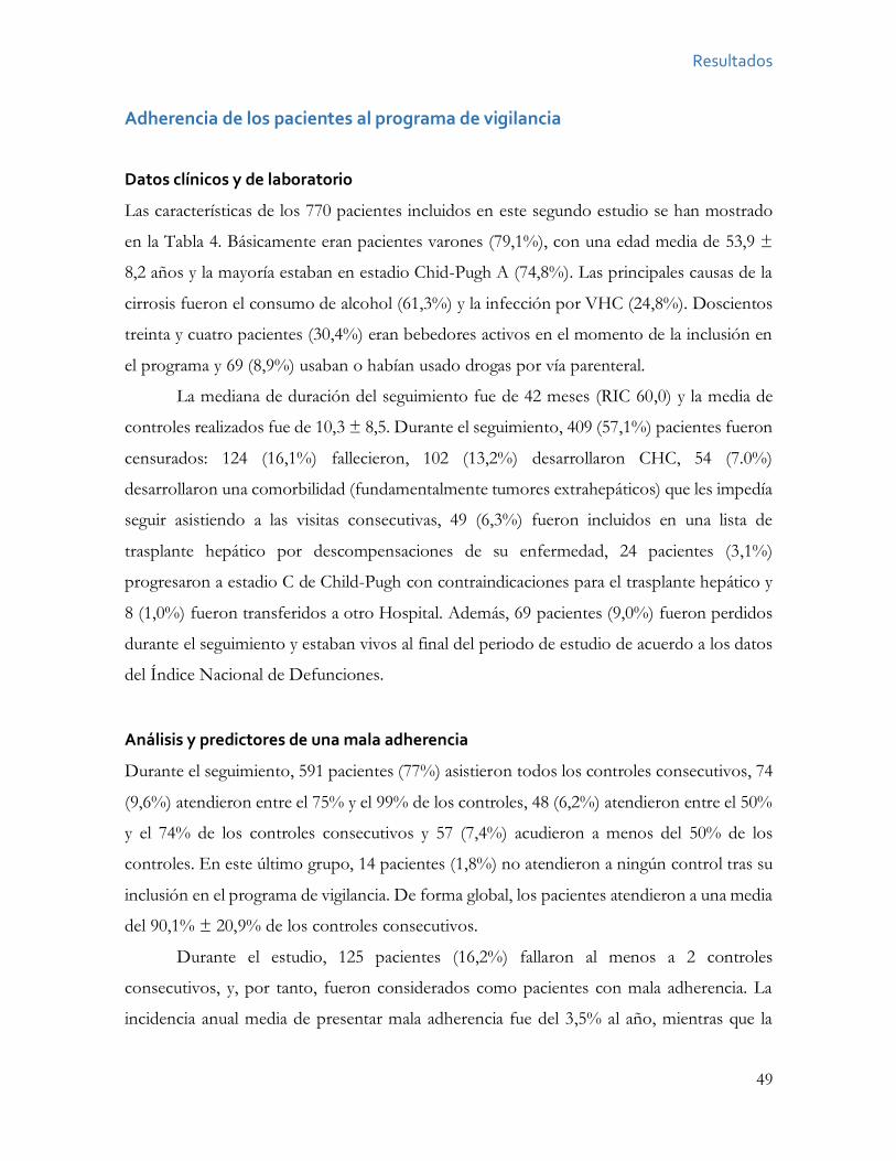
Las características de los 770 pacientes incluidos en este segundo estudio se han mostrado
en la Tabla 4. Básicamente eran pacientes varones (79,1%), con una edad media de 53,9 ±
8,2 años y la mayoría estaban en estadio Chid-Pugh A (74,8%). Las principales causas de la
cirrosis fueron el consumo de alcohol (61,3%) y la infección por VHC (24,8%). Doscientos
treinta y cuatro pacientes (30,4%) eran bebedores activos en el momento de la inclusión en
el programa y 69 (8,9%) usaban o habían usado drogas por vía parenteral.
La mediana de duración del seguimiento fue de 42 meses (RIC 60,0) y la media de
controles realizados fue de 10,3 ± 8,5. Durante el seguimiento, 409 (57,1%) pacientes fueron
censurados: 124 (16,1%) fallecieron, 102 (13,2%) desarrollaron CHC, 54 (7.0%)
desarrollaron una comorbilidad (fundamentalmente tumores extrahepáticos) que les impedía
seguir asistiendo a las visitas consecutivas, 49 (6,3%) fueron incluidos en una lista de
trasplante hepático por descompensaciones de su enfermedad, 24 pacientes (3,1%)
progresaron a estadio C de Child-Pugh con contraindicaciones para el trasplante hepático y
8 (1,0%) fueron transferidos a otro Hospital. Además, 69 pacientes (9,0%) fueron perdidos
durante el seguimiento y estaban vivos al final del periodo de estudio de acuerdo a los datos
del Índice Nacional de Defunciones.
Análisis y predictores de una mala adherencia
Durante el seguimiento, 591 pacientes (77%) asistieron todos los controles consecutivos, 74
(9,6%) atendieron entre el 75% y el 99% de los controles, 48 (6,2%) atendieron entre el 50%
y el 74% de los controles consecutivos y 57 (7,4%) acudieron a menos del 50% de los
controles. En este último grupo, 14 pacientes (1,8%) no atendieron a ningún control tras su
inclusión en el programa de vigilancia. De forma global, los pacientes atendieron a una media
del 90,1% ± 20,9% de los controles consecutivos.
Durante el estudio, 125 pacientes (16,2%) fallaron al menos a 2 controles
consecutivos, y, por tanto, fueron considerados como pacientes con mala adherencia. La
incidencia anual media de presentar mala adherencia fue del 3,5% al año, mientras que la
Resultados
50
incidencia acumulada al 5º, 10º y 15º año fue del 18,0%, 26,6% y del 29,7% respectivamente
(Figura 5).
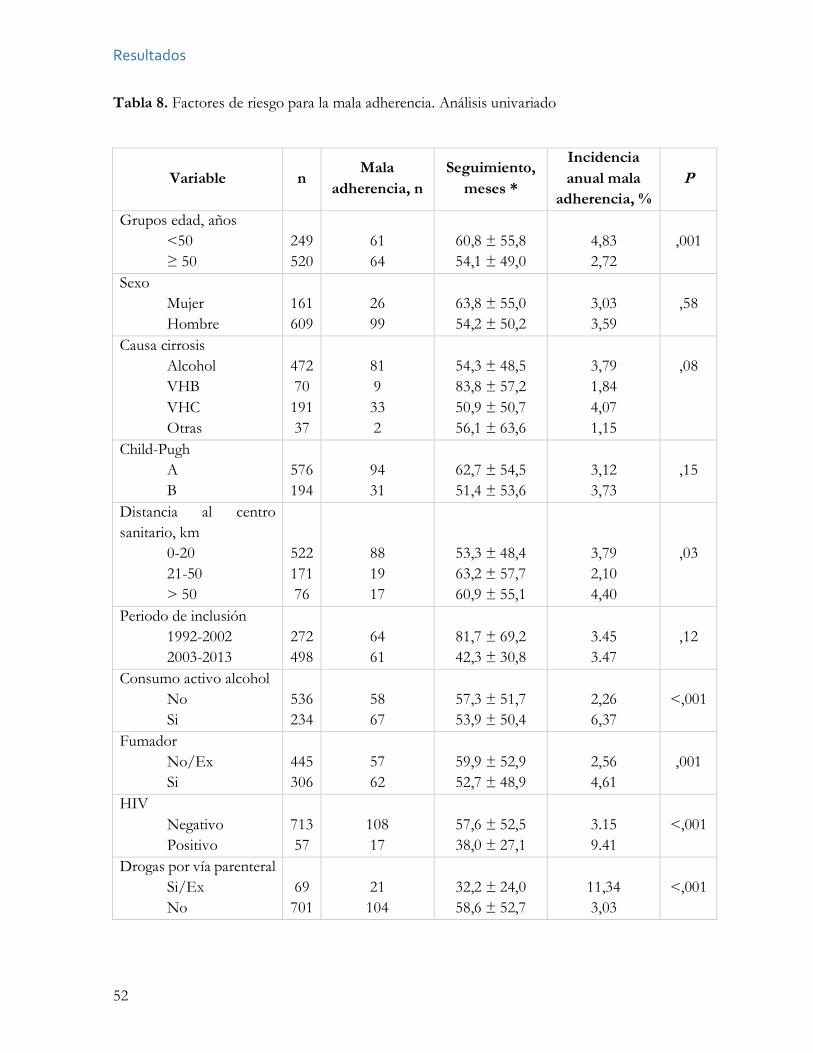
La Tabla 8 muestra la comparativa entre los pacientes con mala y buena adherencia.
En el análisis univariado, la edad < 50 años, la distancia de viaje al hospital > 50 km, el
consumo activo de alcohol, la historia de tabaquismo previa, la seropositividad para el VIH,
la historia de consumo de drogas por vía parenteral, la ausencia de descompensaciones de la
enfermedad hepática, los niveles elevados de FA o GGT y una ratio AST/ALT ≥ 1.6 se
asociaron de manera significativa con una mala adherencia.
En el estudio multivariado se identificaron las siguientes variables como factores de
riesgo independientes para presentar mala adherencia (Tabla 9): el consumo activo o previo de
drogas por vía parenteral [HR, 5,33; 95% IC 95%, 3,07-9,23; P <,001], el consumo activo de
alcohol (HR, 3,03; IC 95%, 2,03-4,51; P < ,001), la ausencia de descompensaciones de la
hipertensión portal previas a la inclusión en el programa de vigilancia (HR, 1,65; IC 95%, 1,07-
2,55; P =,022) y una ratio AST/ALT ≥ 1.6 (HR, 1,82; IC 95%, 1,23-2,70; P = ,003).
Durante el estudio, 179 pacientes (23%) fallaron a un control, con una incidencia
acumulada al 5º, 10º y 15º año del 26,1%, 34,1% y del 36,9%, respectivamente. Las variables
asociadas en el análisis multivariado con el fallo a un control fueron muy semejantes a las
asociadas al fallo a 2 controles consecutivos, es decir: consumo activo o previo de drogas por
vía parenteral (HR, 3,30; IC 95%, 2,06- 5,29; P < ,001), consumo activo de alcohol (HR, 2,79;
IC 95%, 1,96-3,95; P < ,001), Índice de Masa Corporal < 25 (HR, 1,83; IC 95%, 1,29-2,59; P <
,001) y ratio AST/ALT ≥ 1,6 (HR, 1,68; IC 95%, 1,18-2,39; P = ,004).
Características de los CHC
Un total de 107 pacientes desarrollaron CHC, 98 en el grupo de buena adherencia y 9 en el
de mala adherencia. El diagnóstico fue sospechado por una elevación de la AFP en 21
pacientes (19,6%), por la presencia de un nódulo en la ecografía en 72 pacientes (67,3%), por
ambas en 7 pacientes (6,5%) y de manera incidental en otros 7 pacientes (6,5%). La
proporción de tumores en estadio inicial (BCLC 0 o A) fue del 76,1% en el grupo con CHC
detectado por elevación de AFP, del 72,2% en el grupo con CHC sospechado por la
ecografía, del 28,6% en los CHC sospechados por ambos métodos y del 57,1% en aquellos
diagnosticados de manera incidental.
Resultados
51
Respecto a los CHC diagnosticados en el grupo de mala adherencia, sólo 2 de los 9
pacientes (22,2%) fueron diagnosticados mediante ecografías semestrales tras la rentrada del
paciente al programa de vigilancia. Cuatro pacientes (44,4%) fueron diagnosticados durante
la admisión por descompensaciones de su enfermedad hepática y 3 (33,3%) incidentalmente.
Estos pacientes fallaron a una media de 5,5 controles antes del diagnóstico del CHC, y
globalmente, asistieron al 49,3% de los controles durante una media de 4,4 ± 4,2 años de
seguimiento.
Al diagnóstico de CHC 65 tumores (60,6%) eran uninodulares. De acuerdo al sistema
BCLC, 17 tumores (15,9%) estaban en estadio 0, 57 (53,3%) en estadio A, 15 (14,0%) en estadio
B, 13 (12,1%) en estadio C y 5 (4,7%) en estadio D. Se diagnosticaron más pacientes en estadio
intermedio o avanzado (B/C/D) en el grupo de mala adherencia que en el grupo de buena
adherencia (66,5% frente a 27,5%; P = ,015). Setenta y ocho tumores (72,9%) cumplieron los
criterios de Milán y 29 (27,1%) los excedieron. Cinco pacientes estaban en estadio BCLC B, C o
D y cumplían los criterios de Milán mientras que un paciente en estadio BCLCA excedía los
criterios de Milán. Los pacientes con mala adherencia fueron diagnosticados fuera de los criterios
de Milán con mayor frecuencia que los pacientes con buena adherencia (55,5% frente a 24,4%,
P= ,045).
Respecto al tratamiento de los pacientes con CHC, 4 (3,7%) fueron tratados por
resección hepática, 33 (30,8%) mediante trasplante hepático, 29 (27,1%) mediante ablación local,
12 (11,2%) mediante TACE, 6 (5,6%) recibieron sorafenib y 21 (19,6%) tratamiento sintomático.
Los pacientes con buena adherencia fueron tratados más frecuentemente con tratamientos con
potencial curativo que aquellos con mala adherencia, aunque las diferencias no fueron
significativas (63,2%% frente a 33,3%; P = ,078). La supervivencia tras el diagnóstico de CHC
fue mayor en los pacientes en el grupo de buena adherencia (mediana 24,7 meses, RIC 43,2) que
en aquellos con mala adherencia (mediana 14,2 meses, RIC 39,6 meses), aunque las diferencias
no fueron significativas (P = ,160) (Figura 6).
Los pacientes que fallaron a un control también tuvieron tumores más avanzados que
aquellos que asistieron a todos los controles consecutivos (53,4% y 27,2% diagnosticados en
estadio BCLC B/C/D respectivamente; P = ,042). De todas maneras, no se observaron
diferencias entre ambos grupos respecto a la proporción de tumores tratados con intención
curativa (46,6% frente a 66,3%; P = ,143) o en la supervivencia tras el diagnóstico del tumor (P
= ,621).
Resultados
52
Tabla 8. Factores de riesgo para la mala adherencia. Análisis univariado
Variable n Mala
adherencia, n Seguimiento,
meses *
Incidencia anual mala
adherencia, % P
Grupos edad, años <50 ≥ 50
249 520
61 64
60,8 ± 55,8 54,1 ± 49,0
4,83 2,72
,001
Sexo Mujer Hombre
161 609
26 99
63,8 ± 55,0 54,2 ± 50,2
3,03 3,59
,58
Causa cirrosis Alcohol VHB VHC Otras
472 70 191 37
81 9 33 2
54,3 ± 48,5 83,8 ± 57,2 50,9 ± 50,7 56,1 ± 63,6
3,79 1,84 4,07 1,15
,08
Child-Pugh A B
576 194
94 31
62,7 ± 54,5 51,4 ± 53,6
3,12 3,73
,15
Distancia al centro sanitario, km
0-20 21-50 > 50
522 171 76
88 19 17
53,3 ± 48,4 63,2 ± 57,7 60,9 ± 55,1
3,79 2,10 4,40
,03
Periodo de inclusión 1992-2002 2003-2013
272 498
64 61
81,7 ± 69,2 42,3 ± 30,8
3.45 3.47
,12
Consumo activo alcohol No Si
536 234
58 67
57,3 ± 51,7 53,9 ± 50,4
2,26 6,37
<,001
Fumador No/Ex Si
445 306
57 62
59,9 ± 52,9 52,7 ± 48,9
2,56 4,61
,001
HIV Negativo Positivo
713 57
108 17
57,6 ± 52,5 38,0 ± 27,1
3.15 9.41
<,001
Drogas por vía parenteral Si/Ex No
69 701
21
104
32,2 ± 24,0 58,6 ± 52,7
11,34 3,03
<,001
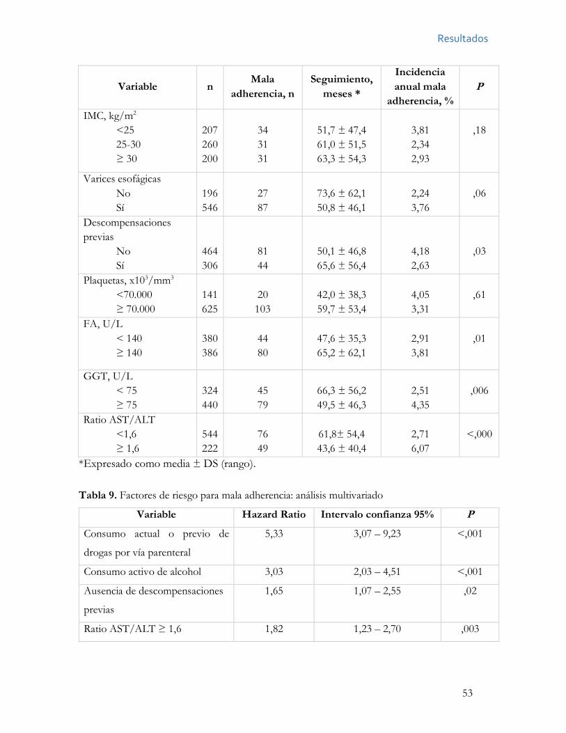
Resultados
53
Variable n Mala
adherencia, n Seguimiento,
meses *
Incidencia anual mala
adherencia, % P
IMC, kg/m2 <25 25-30 ≥ 30
207 260 200
34 31 31
51,7 ± 47,4 61,0 ± 51,5 63,3 ± 54,3
3,81 2,34 2,93
,18
Varices esofágicas No Sí
196 546
27 87
73,6 ± 62,1 50,8 ± 46,1
2,24 3,76
,06
Descompensaciones previas
No Sí
464 306
81 44
50,1 ± 46,8 65,6 ± 56,4
4,18 2,63
,03
Plaquetas, x103/mm3 <70.000 ≥ 70.000
141 625
20
103
42,0 ± 38,3 59,7 ± 53,4
4,05 3,31
,61
FA, U/L < 140 ≥ 140
380 386
44 80
47,6 ± 35,3 65,2 ± 62,1
2,91 3,81
,01
GGT, U/L < 75 ≥ 75
324 440
45 79
66,3 ± 56,2 49,5 ± 46,3
2,51 4,35
,006
Ratio AST/ALT <1,6 ≥ 1,6
544 222
76 49
61,8± 54,4 43,6 ± 40,4
2,71 6,07
<,000
*Expresado como media ± DS (rango).
Tabla 9. Factores de riesgo para mala adherencia: análisis multivariado
Variable Hazard Ratio Intervalo confianza 95% P
Consumo actual o previo de
drogas por vía parenteral
5,33 3,07 – 9,23 <,001
Consumo activo de alcohol 3,03 2,03 – 4,51 <,001
Ausencia de descompensaciones
previas
1,65 1,07 – 2,55 ,02
Ratio AST/ALT ≥ 1,6 1,82 1,23 – 2,70 ,003
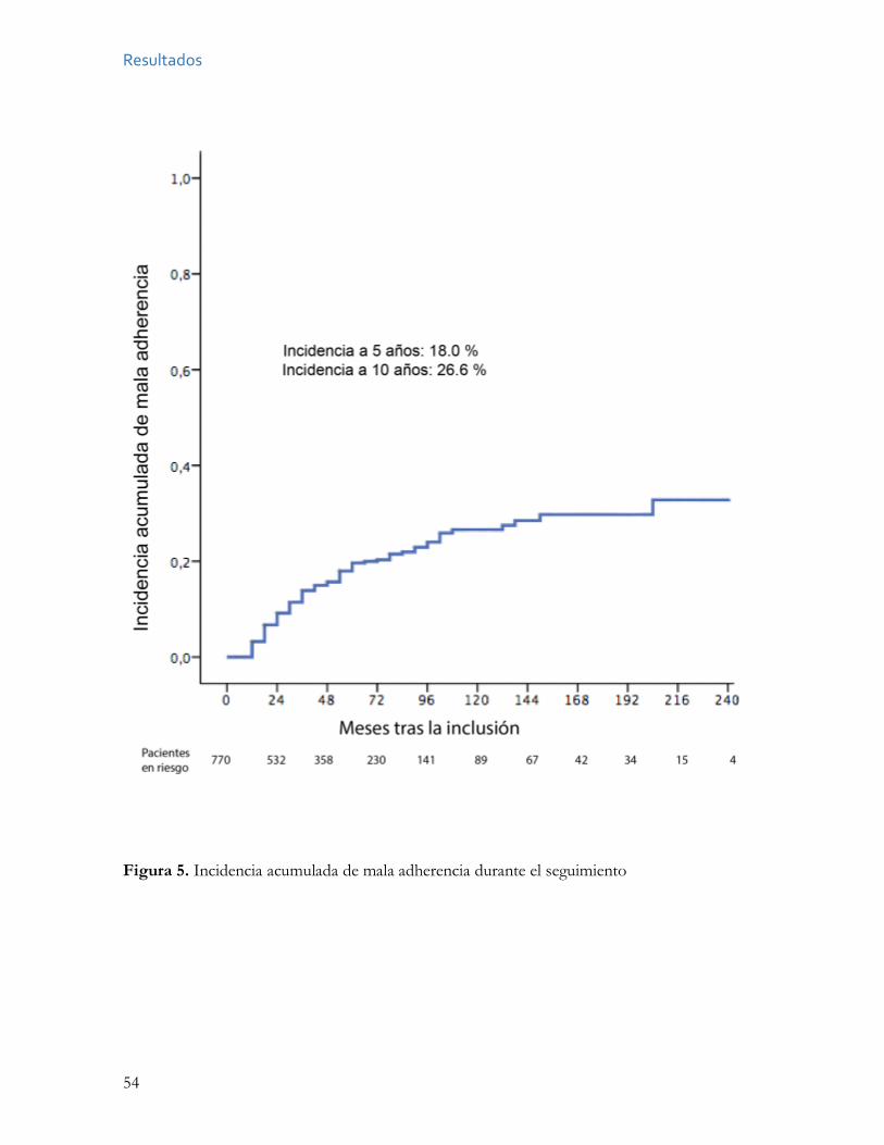
Resultados
54
Figura 5. Incidencia acumulada de mala adherencia durante el seguimiento
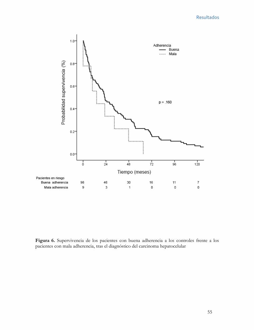
Resultados
55
Figura 6. Supervivencia de los pacientes con buena adherencia a los controles frente a los pacientes con mala adherencia, tras el diagnóstico del carcinoma hepatocelular
Resultados
56
Incidencia y factores de riesgo del fracaso del programa
Datos clínicos y de laboratorio
De los 1.242 pacientes incluidos en el programa de vigilancia, 188 (15,1%) desarrollaron
CHC durante una mediana de seguimiento de 480 meses (RIC 72,0). La incidencia acumulada
a 10 años de desarrollar CHC fue del 25,0%, y el número mediano de controles realizados
hasta el diagnóstico de CHC fue de 10,0 (RIC 13,0).
Los datos demográficos, clínicos y de laboratorio de estos 188 pacientes se mostraron
en la Tabla 5. Los pacientes básicamente eran hombres (85,1%), y las principales causas de
la cirrosis fueron el alcohol (50,0%), el VHC (39,8%) y el VHB (6,9%). En la inclusión, el
39,7% de los pacientes eran diabéticos. Durante el seguimiento y antes del diagnóstico del
tumor, el 45,2% de los pacientes tuvieron al menos un episodio de descompensación.
Finalmente, en el momento del diagnóstico del CHC, la edad media era de 62,6 ± 8,9 años
y la mayoría de los pacientes estaban en estadio A de Child-Pugh.
Características de los CHC
La detección del CHC se basó en la ecografía abdominal en 139 pacientes (73,9%), en la
elevación de AFP en 36 pacientes (19,1%) y en otros métodos en 13 pacientes (6,9%). El
CHC se diagnosticó tras detectar una lesión focal hepática en una prueba de imagen junto a
una elevación de AFP ≥400 ng/mL en 7 pacientes (3,8%), mediante biopsia hepática en 45
pacientes (23,9%), y por test de imagen dinámicos no invasivos en 136 pacientes (72,3%).
Sesenta y siete CHC fueron diagnosticados durante la primera década del estudio
(35,6%) y 121 (64,4%) fueron diagnosticados durante la segunda década. Veintisiete CHC
(14,4%) fueron diagnosticados en el grupo con seguimiento trimestral y 161 (85,6%) en el
grupo con seguimiento semestral.
En total, 138 (73,4%) de los 188 tumores fueron diagnosticados dentro de los
criterios de Milán, y 50 tumores (26,6%) fueron diagnosticados fuera de dichos criterios y
por tanto fueron considerados fracasos del programa. De acuerdo con el sistema BCLC, 28
tumores fueron diagnosticados en estadio 0 (14,9%), 106 tumores en estadio A (56,3%), 22
en estadio B (11,8%), 24 en estadio C (12,8%) y 8 en estadio D (4,2%). Aunque hubo una
mayor proporción de tumores en estadio BCLC 0 en la segunda década del estudio (2004-
Resultados
57
2016) que la primera (1992-2003), estas diferencias no fueron significativas (16,5% vs 11,9%,
P = ,39). En total, 120 tumores fueron uninodulares (63,5%) y el tamaño medio de estos
tumores uninodulares fue de 2,9± 1,4 cm. Los diámetros de los tumores uninodulares no
variaron entre los grupos con seguimiento trimestral o semestral (2,8 ± 1,3 cm vs. 3,1 ± 1,8
cm, P =,48) ni entre la primera o la segunda década del programa (3,0 ± 1,4 cm vs. 2,9 ± 1,5
cm; P = ,93).
Al diagnóstico del CHC la AFP media fue de 8,8 ng/mL (rango 1– 18.430).
Veintiocho pacientes (14,9%) tuvieron niveles de AFP ≥100 ng/mL y 9 (4,7%) ≥1.000
ng/mL. El nivel de AFP al diagnóstico fue significativamente mayor en los pacientes con
CHC detectado por elevación de AFP que en aquellos detectados mediante ecografía
abdominal (659,4±172,5 vs. 48,5±184,0, P <,001).
Respecto al tratamiento de los pacientes con CHC, 118 (62,7%) recibieron terapias
con intención curativa: 5 (2,6%) fueron tratados mediante resección hepática, 52 (27,6%)
mediante trasplante hepático, 3 estaban todavía en lista de trasplante hepático en el momento
de escribir la presente tesis doctoral,36 (19,1%) fueron tratados mediante etanolización y 22
(11,7%) recibieron ablación por radiofrecuencia. Por otro lado, 6 pacientes (13,8%)
recibieron tratamiento con TACE, 8 (4,2%) fueron tratados con sorafenib y 36 (18,1%)
recibieron tratamiento sintomático.
Sólo 2 pacientes (4,0%) entre los 50 fracasos del programa fueron tratados con
intención curativa -resección, trasplante o ablación-, frente a los 116 tratados con intención
curativa entre los 138 tumores que cumplían los criterios de Milán (84,0%) (P < ,001).
La supervivencia tras el diagnóstico de CHC fue más corta en los pacientes con
fracaso del programa (mediana 7,6 meses, RIC 25,5) que en aquellos en los que el programa
no fracasó (mediana 33,9 meses, RIC 55,6, P < ,001) (Figura 7).
Análisis y predictores de fracaso del programa
Análisis univariado
La Tabla 10 muestra la comparación en el análisis univariado entre los fracasos del programa
y aquellos en los que el programa no fracasó. Entre las variables incluidas en la inclusión en
el programa, sólo el estadio B de Child-Pugh (P =,03) se asoció con el fracaso del programa.
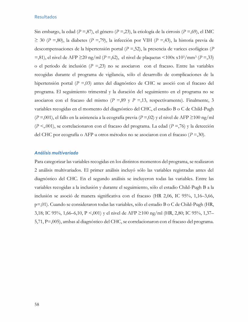
Resultados
58
Sin embargo, la edad (P =,87), el género (P =,23), la etiología de la cirrosis (P =,69), el IMC
≥ 30 (P =,80), la diabetes (P =,79), la infección por VIH (P =,43), la historia previa de
descompensaciones de la hipertensión portal (P =,52), la presencia de varices esofágicas (P
=,81), el nivel de AFP ≥20 ng/ml (P =,62), el nivel de plaquetas <100x x103/mm3 (P =,33)
o el periodo de inclusión (P =,23) no se asociaron con el fracaso. Entre las variables
recogidas durante el programa de vigilancia, sólo el desarrollo de complicaciones de la
hipertensión portal (P =,03) antes del diagnóstico de CHC se asoció con el fracaso del
programa. El seguimiento trimestral y la duración del seguimiento en el programa no se
asociaron con el fracaso del mismo (P =,89 y P =,13, respectivamente). Finalmente, 3
variables recogidas en el momento del diagnóstico del CHC, el estadio B o C de Child-Pugh
(P =,001), el fallo en la asistencia a la ecografía previa (P =,02) y el nivel de AFP ≥100 ng/ml
(P <,.001), se correlacionaron con el fracaso del programa. La edad (P =,76) y la detección
del CHC por ecografía o AFP u otros métodos no se asociaron con el fracaso (P =,30).
Análisis multivariado
Para categorizar las variables recogidas en los distintos momentos del programa, se realizaron
2 análisis multivariados. El primer análisis incluyó sólo las variables registradas antes del
diagnóstico del CHC. En el segundo análisis se incluyeron todas las variables. Entre las
variables recogidas a la inclusión y durante el seguimiento, sólo el estadio Child-Pugh B a la
inclusión se asoció de manera significativa con el fracaso (HR 2,06, IC 95%, 1,16–3,66,
p=,01). Cuando se consideraron todas las variables, sólo el estadio B o C de Child-Pugh (HR,
3,18; IC 95%, 1,66–6,10, P <,001) y el nivel de AFP ≥100 ng/ml (HR, 2,80; IC 95%, 1,37–
5,71, P=,005), ambas al diagnóstico del CHC, se correlacionaron con el fracaso del programa.
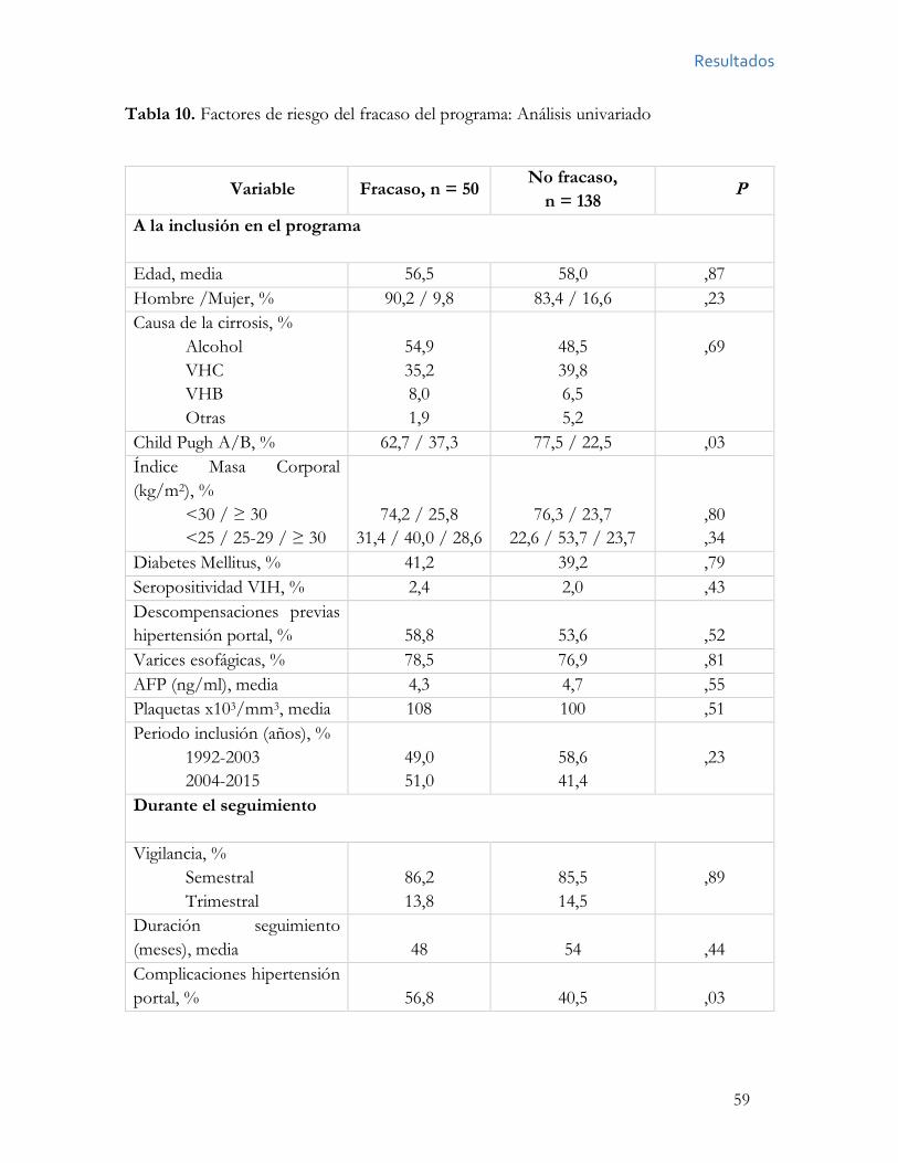
Resultados
59
Tabla 10. Factores de riesgo del fracaso del programa: Análisis univariado
Variable Fracaso, n = 50 No fracaso, n = 138
P
A la inclusión en el programa Edad, media 56,5 58,0 ,87 Hombre /Mujer, % 90,2 / 9,8 83,4 / 16,6 ,23 Causa de la cirrosis, %
Alcohol VHC VHB Otras
54,9 35,2 8,0 1,9
48,5 39,8 6,5 5,2
,69
Child Pugh A/B, % 62,7 / 37,3 77,5 / 22,5 ,03 Índice Masa Corporal (kg/m2), %
<30 / ≥ 30 <25 / 25-29 / ≥ 30
74,2 / 25,8 31,4 / 40,0 / 28,6
76,3 / 23,7 22,6 / 53,7 / 23,7
,80 ,34
Diabetes Mellitus, % 41,2 39,2 ,79 Seropositividad VIH, % 2,4 2,0 ,43 Descompensaciones previas hipertensión portal, %
58,8
53,6
,52
Varices esofágicas, % 78,5 76,9 ,81 AFP (ng/ml), media 4,3 4,7 ,55 Plaquetas x103/mm3, media 108 100 ,51 Periodo inclusión (años), %
1992-2003 2004-2015
49,0 51,0
58,6 41,4
,23
Durante el seguimiento Vigilancia, %
Semestral Trimestral
86,2 13,8
85,5 14,5
,89
Duración seguimiento (meses), media
48
54
,44
Complicaciones hipertensión portal, %
56,8
40,5
,03
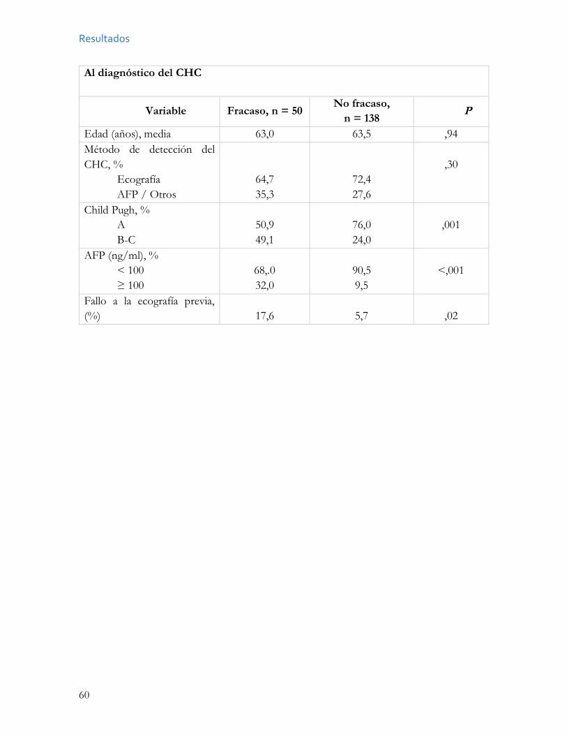
Resultados
60
Al diagnóstico del CHC
Variable Fracaso, n = 50 No fracaso, n = 138 P
Edad (años), media 63,0 63,5 ,94 Método de detección del CHC, %
Ecografía AFP / Otros
64,7 35,3
72,4 27,6
,30
Child Pugh, % A B-C
50,9 49,1
76,0 24,0
,001
AFP (ng/ml), % < 100 ≥ 100
68,.0 32,0
90,5 9,5
<,001
Fallo a la ecografía previa, (%)
17,6
5,7
,02
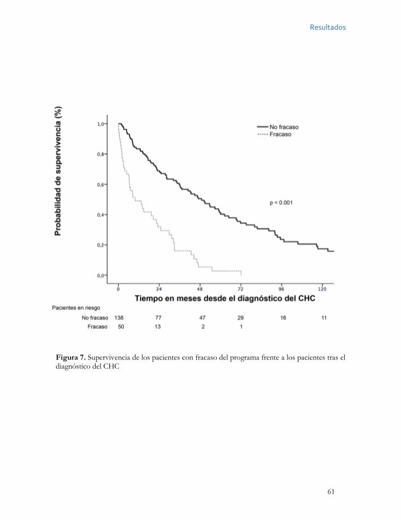
Resultados
61
Figura 7. Supervivencia de los pacientes con fracaso del programa frente a los pacientes tras el diagnóstico del CHC

62
63
Justificación de la unidad temática de la tesis
El CHC es una de las principales complicaciones de la cirrosis hepática. Los pacientes con
cirrosis, independientemente de su causa, deben ser incluidos en programas para la vigilancia
precoz del hepatocarcinoma, basado en la realización de ecografías semestrales.
Los tres estudios que configuran esta tesis doctoral, abordan tres problemas de los
programas de vigilancia del CHC en los pacientes con cirrosis. Los tres estudios analizan una
misma cohorte de pacientes con cirrosis, seguida de manera prospectiva en el Hospital
Universitario Central de Asturias durante más de 20 años.
El objetivo del primer estudio de la tesis doctoral fue determinar la incidencia de
CHC entre una población de pacientes con cirrosis de origen alcohólico y los factores de
riesgo asociados.
El objetivo del segundo estudio fue determinar la adherencia de los pacientes al
programa de vigilancia, y analizar qué factores se asocian con una mala adherencia.
El objetivo del tercer estudio fue analizar el fracaso del programa de vigilancia,
definido como la proporción de tumores diagnosticados fuera de los criterios de Milán, y
analizar qué factores son capaces de predecir el fracaso del mismo.

64
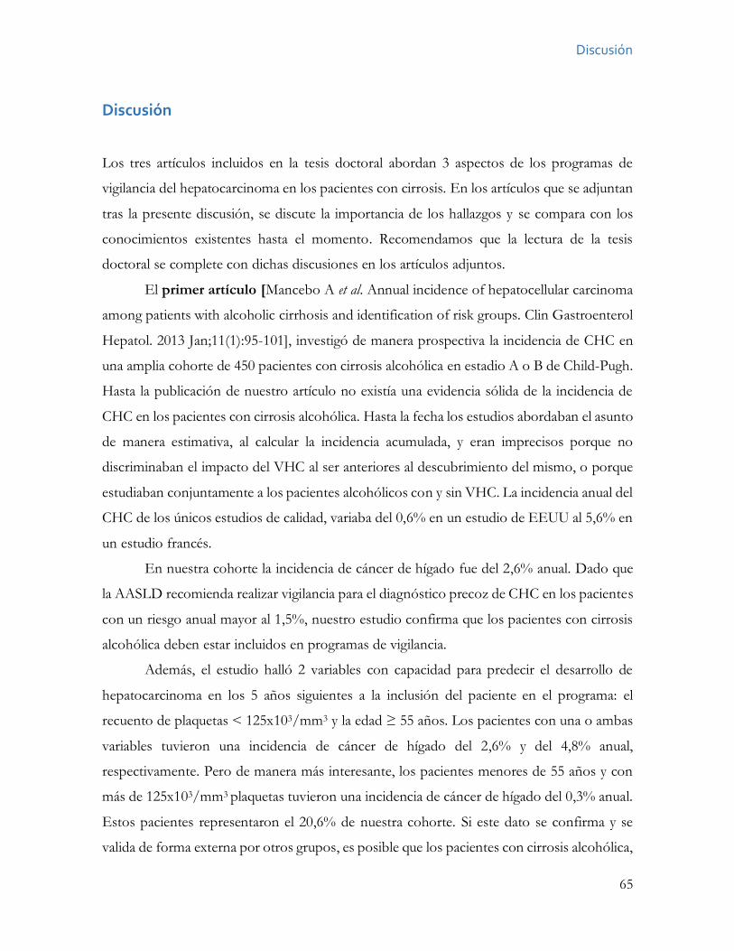
Discusión
65
Discusión
Los tres artículos incluidos en la tesis doctoral abordan 3 aspectos de los programas de
vigilancia del hepatocarcinoma en los pacientes con cirrosis. En los artículos que se adjuntan
tras la presente discusión, se discute la importancia de los hallazgos y se compara con los
conocimientos existentes hasta el momento. Recomendamos que la lectura de la tesis
doctoral se complete con dichas discusiones en los artículos adjuntos.
El primer artículo [Mancebo A et al. Annual incidence of hepatocellular carcinoma
among patients with alcoholic cirrhosis and identification of risk groups. Clin Gastroenterol
Hepatol. 2013 Jan;11(1):95-101], investigó de manera prospectiva la incidencia de CHC en
una amplia cohorte de 450 pacientes con cirrosis alcohólica en estadio A o B de Child-Pugh.
Hasta la publicación de nuestro artículo no existía una evidencia sólida de la incidencia de
CHC en los pacientes con cirrosis alcohólica. Hasta la fecha los estudios abordaban el asunto
de manera estimativa, al calcular la incidencia acumulada, y eran imprecisos porque no
discriminaban el impacto del VHC al ser anteriores al descubrimiento del mismo, o porque
estudiaban conjuntamente a los pacientes alcohólicos con y sin VHC. La incidencia anual del
CHC de los únicos estudios de calidad, variaba del 0,6% en un estudio de EEUU al 5,6% en
un estudio francés.
En nuestra cohorte la incidencia de cáncer de hígado fue del 2,6% anual. Dado que
la AASLD recomienda realizar vigilancia para el diagnóstico precoz de CHC en los pacientes
con un riesgo anual mayor al 1,5%, nuestro estudio confirma que los pacientes con cirrosis
alcohólica deben estar incluidos en programas de vigilancia.
Además, el estudio halló 2 variables con capacidad para predecir el desarrollo de
hepatocarcinoma en los 5 años siguientes a la inclusión del paciente en el programa: el
recuento de plaquetas < 125x103/mm3 y la edad ≥ 55 años. Los pacientes con una o ambas
variables tuvieron una incidencia de cáncer de hígado del 2,6% y del 4,8% anual,
respectivamente. Pero de manera más interesante, los pacientes menores de 55 años y con
más de 125x103/mm3 plaquetas tuvieron una incidencia de cáncer de hígado del 0,3% anual.
Estos pacientes representaron el 20,6% de nuestra cohorte. Si este dato se confirma y se
valida de forma externa por otros grupos, es posible que los pacientes con cirrosis alcohólica,
Discusión
66
con menos de 55 años y niveles de plaquetas > 125x103/mm3, debido al bajo riesgo de
desarrollar CHC, no precisen cribado si no controles analíticos periódicos para detectar en
qué momento pasan a cumplir alguno de los requisitos previos para su inclusión en el
programa de vigilancia.
Numerosos estudios han demostrado que tanto la edad, como un mayor grado de
hipertensión portal, reflejado de manera indirecta en el recuento de plaquetas, son factores
de riesgo de hepatocarcinoma en cirrosis de cualquier etiología. Los programas de cribado
de cáncer deben ser coste-efectivos, y para ello sigue siendo de vital importancia descubrir
qué pacientes se benefician realmente de dicha vigilancia, y en cuáles el riesgo es tan bajo que
el cribado puede ser contraproducente, por los falsos positivos y la incertidumbre que crea
a los pacientes.
En el mismo momento que nuestro grupo envió el estudio para su publicación, se
publicó un estudio retrospectivo danés,67 basado en un registro poblacional de pacientes con
cirrosis por alcohol, que incluyó a pacientes diagnosticados de cirrosis alcohólica desde 1997
mediante la codificación ICD-8 ó ICD-10. En dicho estudio, la incidencia acumulada a los 5
años de hepatocarcinoma fue del 1,0%, un nivel claramente inferior al umbral fijado por la
AASLD para recomendar la vigilancia de CHC. En un comentario sobre artículo,90 Amit G
Singal señala que los estudios de registro están expuestos a un sesgo: aunque los códigos
ICD-8 e ICD-10 alcanzan un valor predictivo del 85% para cirrosis cuando esta codificación
se realiza en unidades de hepatología,91 éste puede disminuir cuando se realiza en centros de
salud de atención primaria, dado que se pueden codificar como cirróticos pacientes con
hepatitis alcohólica aguda, infraestimando el riesgo de desarrollar CHC. Además, los
pacientes con cirrosis descompensada o ictericia son reconocidos más frecuentemente como
cirróticos por los médicos de atención primaria, mientras que los pacientes con enfermedad
hepática compensada suelen pasar desapercibidos.77 Aunque los pacientes con enfermedad
hepática avanzada tienen mayor riesgo de CHC, también tienen mayor probabilidad de morir
como consecuencia de su enfermedad hepática. Esto puede influir en los resultados. Por
otro lado, en el estudio danés se incluyeron pacientes con notables limitaciones funcionales
o comorbilidades, que no deben ser incluidos en los programas de vigilancia, dada la elevada
mortalidad a corto plazo o la imposibilidad de aplicar tratamientos efectivos para el CHC.
Otra limitación a señalar, es que la incidencia de CHC en el estudio danés fue más elevada
Discusión
67
en el primer año que en los consiguientes (9,8 vs 3,8/1.000 personas-año), lo que puede
reflejar que se tratase de CHC prevalentes no detectados al diagnóstico de la cirrosis.
Morris Sherman señala otras limitaciones del estudio danés.92 La investigación no
especifica si se realizaba cribado de CHC a estos pacientes, y por tanto es posible que muertes
atribuidas a la cirrosis, en pacientes no incluidos en los programas de vigilancia, en realidad
fuesen ocasionadas por un CHC no diagnosticado. Además, en el estudio existe una notable
mortalidad global: el 67% de los pacientes murió en los 7 años posteriores al diagnóstico del
CHC, presumiblemente por la enfermedad hepática o por enfermedades relacionadas por el
alcohol. La elevada mortalidad de los pacientes por enfermedad hepática avanzada, al
competir con el CHC, puede reducir la incidencia de CHC en los estudios.
Recientemente se ha comunicado un estudio de registro realizado en Suecia, donde
es obligatorio reportar el hepatocarcinoma.93 El estudio compara 3 registros nacionales: el
Registro de Cáncer, la Causa de Muerte y los diagnósticos del ingreso del Registro Nacional
de Pacientes, y observaron que hasta un 37-45% de los CHC no se habían reportado en el
Registro de Cáncer. Los autores concluyen que el estudio de registro puede infraestimar la
incidencia de CHC. Los autores especulan que el menor número de biopsias hepáticas
obtenidas para diagnosticar el CHC debido a los nuevos criterios diagnósticos basados en
las pruebas de imagen dinámicas puede explicar la infraestimación de la incidencia de CHC
en los estudios de registro.
Por último, es necesario tener en cuenta que el estudio de Jepsen et al67 es difícilmente
comparable por el método estadístico utilizado. El autor principal del estudio, en un artículo
posterior, discute los resultados tan dispares ofrecidos por los métodos estadísticos
habituales, el Kaplan-Meier y regresión Cox, frente al modelo que usaron en su publicación,
el Modelo multiestado.94 Los estudios de incidencia basados en Kaplan-Meier dan resultados
más elevados que los basados en modelos multiestado, porque el método Kaplan-Meier
asume que todos los pacientes censurados tienen el mismo riesgo de desarrollar el evento
que los que siguen bajo observación, aunque eso sólo es cierto en una parte de ellos. Los
pacientes trasplantados, por ejemplo, no seguirían estando en riesgo, y sí aquellos que
abandonan el programa por finalización del periodo de estudio. Estas diferencias se observan
gráficamente en un reciente estudio italiano con 494 pacientes con cirrosis por VHC seguidos
durante 25 años, que encontró, utilizando modelos multiestado, una incidencia acumulada
Discusión
68
menor del 1% anual,95 cifra muy por debajo del 3-5% que se observa en este tipo de cohortes
en los estudios que usan Kaplan Meier. Dado que la práctica totalidad de los estudios en los
que se basan las guías para hacer recomendaciones utilizan el método Kaplan-Meier, si el
modelo multiestado se acaba imponiendo, se precisará establecer nuevos estándares en las
guías de práctica clínica.
Tres años después de la publicación nuestro estudio, se publicó, en forma de resumen
en un congreso, los resultados preliminares de una cohorte francesa de 492 pacientes con
cirrosis alcohólica.96 Las características de dicha cohorte son semejantes a la nuestra: la edad
media de la cohorte francesa era de 59 años (56 en nuestra cohorte), el 67% eran varones
(frente al 82% en nuestra cohorte) y el 65% estaban en abstinencia a la entrada en el programa
de cribado (frente al 63%). Durante un seguimiento medio de 12,4 meses, encontraron una
incidencia acumulada de CHC a un año del 1,3%, muy semejante a la incidencia acumulada
a un año del 1,6% de nuestra cohorte. Estos hallazgos vienen a confirmar los resultados de
nuestra serie y recordar la importancia de que los pacientes con cirrosis por alcohol sean
incluidos en programas de vigilancia.
El segundo estudio de la Tesis Doctoral (Mancebo A et al. Adherence to a
semiannual surveillance program for hepatocellular carcinoma in patients with liver cirrhosis.
J Clin Gastroenterol. 2016), analizó de forma prospectiva la adherencia de los pacientes a los
controles semestrales. Antes de nuestra investigación, no había evidencia sólida sobre este
aspecto de los programas de vigilancia. La mayoría de los estudios que abordan el fallo en el
proceso de cribado están realizados en pacientes con seguimiento anual.76 La definición de
mala adherencia es muy heterogénea entre los distintos estudios. 74,75 Y las investigaciones
no especifican qué eslabón de la cadena del proceso de vigilancia explica que el paciente no
realice el control: si el paciente no estaba en vigilancia, si el control no había sido solicitado
por el médico, o si el paciente no había acudido al mismo. Además, muchos estudios estaban
realizados en EEUU, donde los médicos de atención primaria realizan el seguimiento de una
parte importante de los pacientes con cirrosis. Esto puede influir en los resultados dado que
los médicos de cabecera tienen un menor conocimiento de las guías de práctica clínica.70
En nuestro estudio, se incluyeron 770 pacientes, con cirrosis de cualquier etiología,
con seguimiento semestral. Durante un seguimiento mediano de 42,0 meses, el 77% de los
Discusión
69
pacientes acudió a todos los controles sucesivos. Para definir mala adherencia, se
compararon las características de los tumores de los pacientes que habían fallado a un control
durante el seguimiento, con aquellos que habían fallado a dos controles consecutivos. Se
observó que ésta segunda definición se asociaba de una manera más sólida con las
características de los tumores. Por tanto, se definió mala adherencia como el fallo a 2
controles consecutivos. Con esta definición, sólo el 16.2% de los pacientes tuvieron mala
adherencia. A pesar de la periodicidad semestral de las visitas, estas cifras de adherencia son
comparables a la de los programas de vigilancia del cáncer de mama o colon, que suelen
realizar controles cada 1-2 años.97
Los pacientes que a su entrada en el programa presentaban historia de consumo de
drogas por vía parenteral, que eran bebedores activos, que no habían presentado
descompensaciones de su enfermedad hepática y que tenían una ratio AST/ALT ≥ 1,6
tuvieron mala adherencia con más frecuencia durante el seguimiento. Es posible que, en este
subgrupo de pacientes con factores de riesgo para presentar una mala adherencia a las visitas
planificadas de manera semestral, deba hacerse un especial hincapié en medidas de
información e implicación del paciente. Lograr la abstinencia alcohólica y explicar a los
pacientes sin descompensaciones el transcurso de la enfermedad, son pasos que pueden ser
útiles para aumentar la adherencia.
Comparados con los pacientes que no fallaron a 2 controles consecutivos durante el
seguimiento, los pacientes con mala adherencia tuvieron un estadio tumoral más avanzado
al diagnóstico del CHC, fueron tratados con menos frecuencia con intención curativa y
sobrevivieron menos, 14,2 meses frente a los 24,7 meses de los pacientes con buena
adherencia, aunque esas diferencias no fueron estadísticamente significativas. Es destacable
que sólo 9 pacientes tuvieron CHC y mala adherencia. Este escaso número de tumores puede
explicar la falta de significación.
Recientemente se ha publicado un estudio norteamericano de una cohorte de
veteranos de guerra que aborda las barreras que identifican los pacientes para recibir
vigilancia de CHC.98 Dicho estudio realizó encuestas a 541 pacientes con cirrosis seguidos
en un hospital urbano, y encontró un elevado desconocimiento de los pacientes respecto a
la enfermedad. El 48% creía que una dieta sana hacía innecesaria la vigilancia del CHC, y el
34% creía que la falta de síntomas o una exploración física normal excluían la necesidad de
Discusión
70
vigilancia. El 50% de los pacientes identificaba barreras para recibir vigilancia, destacando la
complejidad del proceso de citación (30,5%), el coste de los test de vigilancia (25,3%) y
dificultades para el transporte (17,3%). Los pacientes que sabían que la cirrosis aumentaba el
riesgo de CHC eran más adherentes a los controles sucesivos. Como otros estudios previos,99
estos hallazgos recalcan la necesidad de aumentar el conocimiento de los pacientes de la
enfermedad, y la utilidad de implementar recordatorios de las citas a los pacientes.
El tercer artículo de la Tesis Doctoral (A Mancebo et al. Incidence and Risk Factors
Associated with Hepatocellular Carcinoma Surveillance Failure) estudia el fracaso del
programa de vigilancia a la hora de detectar tumores dentro de los criterios de Milán. Existe
una amplia evidencia de numerosos estudios de cohorte, que muestran que la sensibilidad de
la ecografía para detectar tumores dentro de los criterios de Milán es aproximadamente del
60-70% cuando se solicitan controles semestrales. Un metanálisis que recoge los trabajos
con seguimiento semestral o menor (7 estudios), observa que la sensibilidad de la ecografía
es del 70.1% para detectar tumores en estadio inicial.
En la misma línea que estudios previos, en nuestra cohorte el programa falla con más
frecuencia en aquellos pacientes con estadio Child-Pugh B o C y con AFP ≥ 100 ng/ml al
diagnóstico del CHC. Estos hallazgos, pueden ser un marcador de la presencia del tumor,
más que una causa del fracaso. La elevación de la AFP puede ser un marcador de la
agresividad tumoral, reflejando la presencia de un tumor de crecimiento rápido o difuso no
visualizado en la ecografía. El empeoramiento de la función hepática puede ser igualmente
consecuencia de la presencia del tumor.
Lo más relevante en los programas de vigilancia, es poder determinar con antelación
qué pacientes tienen más riesgo de que la ecografía no detecte tumores en estadio inicial. No
existen datos sobre este aspecto en las diversas investigaciones que analizan los programas
de vigilancia. En nuestro estudio, observamos que los pacientes en estadio Child-Pugh B a
la entrada en el programa y aquellos que presentaron descompensaciones durante el
seguimiento, son diagnosticados con más frecuencia fuera de los criterios de Milán. Estos
dos hallazgos son más útiles para seleccionar a los pacientes con un mayor riesgo de fracaso
del programa.
Discusión
71
Estos dos resultados muestran que, en los pacientes con enfermedad hepática
avanzada, la ecografía no es un buen método diagnóstico. Tal vez la causa esté relacionada
con el patrón nodular ecográfico de los pacientes con estadios avanzados de la cirrosis, con
hígados atróficos, nodulares y con una mala transmisión sónica. Es posible que en un futuro
se deba tener en cuenta la opinión del ecografista sobre la calidad de la prueba, para valorar
si estos pacientes se pueden beneficiar de métodos alternativos de cribado.
Los trabajos presentados en la tesis doctoral tienen algunas limitaciones. Las variables
recogidas durante el seguimiento de los pacientes son escasas, y algunas, como si el paciente
dejaba de fumar o si mantenía o no el consumo enólico no se recogieron. Tampoco se
recogió de manera prospectiva el IMC o la diabetes. Es posible que estas variables puedan
influir en la incidencia de CHC o en la adherencia de los pacientes a los controles. Además,
el estudio tiene una duración de más de 20 años, y durante el mismo las recomendaciones de
las guías fueron variando. Finalmente, la opinión del ecografista respecto a la calidad de las
exploraciones, como el patrón nodular hepático o la mala transmisión sónica, no fue
recogida.
Optimizar los programas de cribado es más necesario que nunca en la era de los
nuevos antivirales de acción directa contra el VHC. La respuesta viral sostenida (RVS) se
asocia a reducciones en la mortalidad,100 progresión de fibrosis 101 o necesidad de trasplante
hepático. Sin embargo, la hepatocarcinogénesis, aunque se reduce, no desaparece. Los
pacientes con VHC y fibrosis avanzada que logran RVS tienen un riesgo de desarrollar CHC
del 1,5%, frente al 6,2% de los pacientes que no logran RVS.19 Por eso, las guías recomiendan
realizar cribado de CHC a los pacientes con cirrosis por VHC y fibrosis avanzada.54 Tras la
irrupción de los nuevos antivirales de acción directa es de esperar que la población con VHC
y RVS se expanda en las próximas décadas. En España, se estima que unos 56.000 a 113.000
pacientes con VHC tienen cirrosis, de los que unos 1.130-2.200 desarrollarán CHC.102 Sin
embargo, sólo están diagnosticados entre un 24% y un 35% de los pacientes con infección
activa.103 Esta situación puede someter a presión a los programas de vigilancia, dado que aún
hoy el 53% de los CHC se siguen diagnosticando fuera de los programas de cribado.9
Todo ello pone de manifiesto la importancia de identificar a los pacientes que
realmente se benefician de los programas de vigilancia. Diversos estudios han elaborado
Discusión
72
puntuaciones para calcular el riesgo individual de hepatocarcinogénesis en los pacientes con
VHC, basándose en parámetros como la edad > 50 años, una cifra baja de plaquetas, el
consumo de alcohol, el nivel de gamma-glutamil transpeptidasa o la ausencia de RVS.104
En definitiva, para distribuir los recursos de una manera coste-efectiva es más
necesario que nunca identificar a los pacientes en riesgo de desarrollar CHC, a quienes incluir
en los programas de vigilancia, y a los pacientes con bajo riesgo de desarrollar CHC, que no
deben ser incluidos.
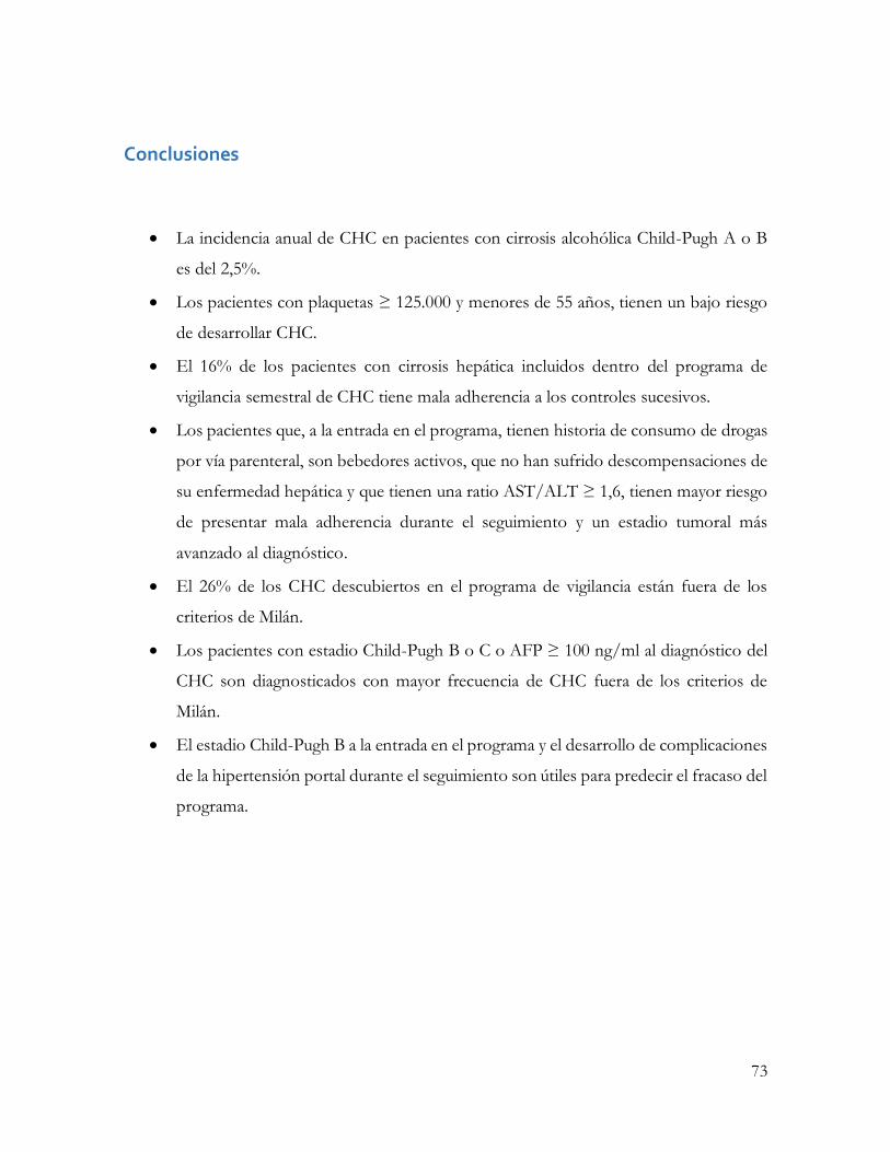
73
Conclusiones
• La incidencia anual de CHC en pacientes con cirrosis alcohólica Child-Pugh A o B
es del 2,5%.
• Los pacientes con plaquetas ≥ 125.000 y menores de 55 años, tienen un bajo riesgo
de desarrollar CHC.
• El 16% de los pacientes con cirrosis hepática incluidos dentro del programa de
vigilancia semestral de CHC tiene mala adherencia a los controles sucesivos.
• Los pacientes que, a la entrada en el programa, tienen historia de consumo de drogas
por vía parenteral, son bebedores activos, que no han sufrido descompensaciones de
su enfermedad hepática y que tienen una ratio AST/ALT ≥ 1,6, tienen mayor riesgo
de presentar mala adherencia durante el seguimiento y un estadio tumoral más
avanzado al diagnóstico.
• El 26% de los CHC descubiertos en el programa de vigilancia están fuera de los
criterios de Milán.
• Los pacientes con estadio Child-Pugh B o C o AFP ≥ 100 ng/ml al diagnóstico del
CHC son diagnosticados con mayor frecuencia de CHC fuera de los criterios de
Milán.
• El estadio Child-Pugh B a la entrada en el programa y el desarrollo de complicaciones
de la hipertensión portal durante el seguimiento son útiles para predecir el fracaso del
programa.

74

75
Copia de los trabajos

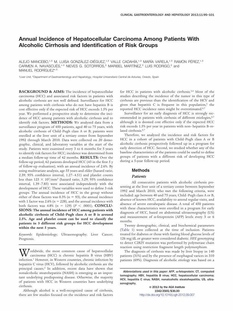
Annual Incidence of Hepatocellular Carcinoma Among Patients WithAlcoholic Cirrhosis and Identification of Risk Groups
ALEJO MANCEBO,*,‡ M. LUISA GONZÁLEZ–DIÉGUEZ,*,‡ VALLE CADAHÍA,*,‡ MARÍA VARELA,*,‡ RAMÓN PÉREZ,*,‡
CARMEN A. NAVASCUÉS,*,‡ NIEVES G. SOTORRÍOS,‡ MARIBEL MARTÍNEZ,‡ LUIS RODRIGO,‡ andMANUEL RODRÍGUEZ*,‡
*Liver Unit, ‡Department of Gastroenterology and Hepatology, Hospital Universitario Central de Asturias, Oviedo, Spain
BACKGROUND & AIMS: The incidence of hepatocellularcarcinoma (HCC) and associated risk factors in patients withalcoholic cirrhosis are not well defined. Surveillance for HCCamong patients with cirrhosis who do not have hepatitis B iscost effective only if the expected risk of HCC exceeds 1.5% peryear. We performed a prospective study to determine the inci-dence of HCC among patients with alcoholic cirrhosis and toidentify risk factors. METHODS: We analyzed data from asurveillance program of 450 patients, aged 40 to 75 years, withalcoholic cirrhosis of Child–Pugh class A or B; patients wereenrolled at the liver unit of a tertiary center from September1992 through March 2010. Data were collected on 20 demo-graphic, clinical, and laboratory variables at the start of thestudy. Patients were examined every 3 to 6 months for 5 yearsto identify risk factors for HCC; incidence was determined froma median follow-up time of 42 months. RESULTS: Over thefollow-up period, 62 patients developed HCC (43 in the first 5 yof follow-up evaluation), with an annual incidence of 2.6%. Byusing multivariate analysis, age 55 years and older (hazard ratio,2.39; 95% confidence interval, 1.27– 4.51) and platelet countsless than 125 ! 103/mm3 (hazard ratio, 3.29; 95% confidenceinterval, 1.39 –7.85) were associated independently with thedevelopment of HCC. These variables were used to define 3 riskgroups. The annual incidence of HCC in the group withouteither of these factors was 0.3% (n " 93), the annual incidencewith 1 factor was 2.6% (n " 228), and the annual incidence withboth factors was 4.8% (n " 129) (P # .0001). CONCLU-SIONS: The annual incidence of HCC among patients withalcoholic cirrhosis of Child–Pugh class A or B is around2.5%. Age and platelet count can be used to classify thepatients in 3 different risk groups for HCC developmentwithin the next 5 years.
Keywords: Epidemiology; Ultrasonography; Liver Cancer;Prognosis.
Worldwide, the most common cause of hepatocellularcarcinoma (HCC) is chronic hepatitis B virus (HBV)
infection.1 However, in Western countries, chronic infection byhepatitis C virus (HCV), followed by alcoholic cirrhosis are theprincipal causes.2 In addition, recent data have shown thatnonalcoholic steatohepatitis (NASH) is emerging as an impor-tant underlying predisposing disease. Otherwise, the majorityof patients with HCC in Western countries have underlyingcirrhosis.
Although alcohol is a well-recognized cause of cirrhosis,there are few studies focused on the incidence and risk factors
for HCC in patients with alcoholic cirrhosis.3,4 Most of thestudies describing the incidence of the tumor in this type ofcirrhosis are previous than the identification of the HCV andgiven that hepatitis C is frequent in this population,5 thereported HCC incidence rates might be overestimated.6,7
Surveillance for an early diagnosis of HCC is strongly rec-ommended in patients with cirrhosis of different etiologies,6,7
although it is deemed cost effective only if the expected HCCrisk exceeds 1.5% per year in patients with non– hepatitis B–re-lated cirrhosis.6,7
Therefore, we analyzed the incidence and risk factors forHCC in a cohort of patients with Child–Pugh class A or Balcoholic cirrhosis prospectively followed up in a program forearly detection of HCC. Second, we studied whether any of thebaseline characteristics of the patients could be useful to definegroups of patients with a different risk of developing HCCduring a 5-year follow-up period.
MethodsPatientsAll consecutive patients with alcoholic cirrhosis pre-
senting at the liver unit of a tertiary center between September1992 and March 2010, who met the following criteria, wereincluded: age between 40 and 75 years, Child–Pugh class A or B,absence of known HCC, availability to attend regular visits, andabsence of severe extrahepatic disease. A total of 450 patientswith these characteristics were enrolled in a program for earlydiagnosis of HCC, based on abdominal ultrasonography (US)and measurement of !-fetoprotein (AFP) levels every 3 or 6months.
Twenty demographic, clinical, and biochemical parameters(Table 1) were collected at the time of inclusion. Patientstreated for diabetes or those with fasting blood glucose levels of126 mg/dL or greater were considered diabetic. HFE genotypingto detect C282Y mutation was performed by polymerase chainreaction using restriction fragment length polymorphism.
The diagnosis of cirrhosis was made by liver biopsy in 140patients (31%) and by the presence of esophageal varices in 310patients (69%). Diagnosis of alcoholic etiology was based on a
Abbreviations used in this paper: AFP, !-fetoprotein; CT, computedtomography; HBV, hepatitis B virus; HCC, hepatocellular carcinoma;HCV, hepatitis C virus; NASH, nonalcoholic steatohepatitis; US, ultra-sonography.
© 2013 by the AGA Institute1542-3565/$36.00
http://dx.doi.org/10.1016/j.cgh.2012.09.007
CLINICAL GASTROENTEROLOGY AND HEPATOLOGY 2013;11:95–101

daily intake of alcohol greater than 80 g in men and greaterthan 60 g in women for 10 years or longer, the absence ofhepatitis B surface antigen and antibody against HCV, and theexclusion of other known causes of liver disease. History ofalcohol intake was assessed at enrollment by means of aninterview with patients and their families performed by one ofthe senior hepatologists involved in the study.
Follow-up EvaluationAll patients were followed up prospectively every 3 or 6
months with a physical examination, abdominal US, determi-nation of AFP level, and blood count and routine biochemicaltests. When the AFP value was greater than 20 ng/mL and morethan twice the previous value, a computed tomography (CT)was performed to rule out the presence of HCC. Otherwise,when a new nodule was detected by US, subsequent tests wereperformed to confirm or exclude the presence of HCC. Allpatients with a focal lesion in the US or with an AFP levelincrease and a negative CT were monitored closely for at least 1year.
From 1992 to 2001 the diagnosis of HCC was made bymeans of histologic confirmation or when a hepatic focal lesionwith an AFP level of 400 ng/mL or greater was observed. From
2001 to 2005, HCC diagnosis was made according to the Eu-ropean Association for the Study of Liver disease criteria6 andfrom 2005 to the end of the study period, the American Asso-ciation for the Study of Liver Diseases criteria7 were applied.Tumor staging was based on at least 2 abdominal imagingtechniques, which were changed throughout the study accord-ing to the state of the art during the past 2 decades.
Statistical AnalysisThe Kaplan–Meier method was used to determine the
cumulative probability of the appearance of HCC during thefollow-up period. The comparison of the survival curves ob-tained was made by means of the log-rank test. For continuousvariables, the optimal threshold for the prediction of HCCdevelopment at 5 years was determined by maximizing theYouden index (sensitivity $ specificity % 1), calculated from thereceiver operating characteristics curves. With regard to bodymass index, patients were distributed following the WorldHealth Organization’s classification and those with ascites wereexcluded. Patients who progressed to a Child–Pugh class C andthose who were lost during the follow-up period, developed asevere comorbidity, or died, were censored at that time. For theanalysis of risk factors, the observation was censored after 5years of follow-up evaluation. Parameters with P values lessthan .10 in the univariate analysis were considered for themultivariate analysis, which was performed by means of theCox proportional hazards model. A P value less than .05 wasconsidered statistically significant. Data analysis was performedusing the SPSS statistical package 17.0 (Chicago, IL).
ResultsClinical and Laboratory DataThe baseline features of the 450 patients are shown in
Table 1. Basically, there was a majority of men (82%), with amean age of 54 years (range, 40 –75 y). At entry, 162 (32%)patients were active drinkers, whereas the mean time of absti-nence in the 288 patients who were no longer actively drinkingwas 4.9 & 1.3 months.
During follow-up evaluation, 264 (58.6%) patients were cen-sored: 62 patients developed HCC, 62 patients died, 45 patientshad cirrhosis that progressed to Child–Pugh stage C, 36 pa-tients developed a severe comorbidity (mainly an extrahepaticmalignancy), and 59 patients were completely lost during fol-low-up evaluation. Adherence to the scheduled appointmentsamong those patients who were not lost during follow-upevaluation was 100% in 76.8%, 75% or greater in 91.9%, and 50%or greater in 97.5% of patients. Comparing baseline character-istics of patients who were completely lost with those of pa-tients remaining in the study, no differences were found withrespect to age (52.2 & 6.7 vs 54.1 & 7.2 y; P " .07), proportionof men (86.7% vs 84.6%; P " .9), and Child–Pugh class A or B atentry (47 class A/12 class B vs 268 class A/128 class B; P " .3).However, active drinking was more frequent among patientslost during follow-up evaluation than in patients who remainedin the program (41.3% vs 35.7%; P " .004).
Incidence and Characteristics ofHepatocellular CarcinomaThe median follow-up period was 42 months (25th
percentile, 18 mo; 75th percentile, 90 mo). During that period
Table 1. Baseline Characteristics of the Patients Enrolled inthe Study (N " 450)
Category Results
Age, ya 53.9 & 7.4Male/female, % 82/18Child–Pugh class A/B, % 70/30Active consumer of alcohol, n (%) 162 (36)Active or previous smoker, n (%) 325 (72.2)Diabetes mellitus, n (%) 119 (26.4)Body mass index, kg/m2a 28.7 & 4.7Esophageal varices, n (%) 351 (78)Previous decompensation, n (%) 336 (74.7)Ascites, n (%) 64 (14.2)C282Y/WTb 24 (6.5)Anti-HBc presence, n (%) 53 (11.8)AFP level, ng/mLa 4.7 & 4.7 (0.1%73.4)AFP '20 ng/mL, n (%) 3 (0.6)AST level, U/La,c 47.9 & 24.7ALT level, U/La,d 33.2 & 14.9Prothrombin time activity, %a 69.1 & 15.1Platelet count, ! 103/mm3a 123.5 & 67.4Total bilirubin level, mg/dLa,e 1.9 & 1.7Serum albumin level, g/dLa 3.8 & 0.6GGT level, U/La,f 157.7 & 195.2Alkaline phosphatase level, U/La,g 217.8 & 133.5
ALT, alanine aminotransferase; anti-HBc, antibodies against hepatitisB core antigen; AST, aspartate aminotransferase; GGT, (-glutamyltransferase; WT, wild type.aExpressed as mean & SD (range).bSixty-two missing values.cThe normal value (N) for AST is less than 37 U/L in men and lessthan 31 U/L in women.dThe normal value (N) for ALT is less than 40 U/L in men and less than31 U/L in women.eThe normal value (N) for total bilirubin is less than 1.10 mg/dL.fThe normal value (N) for GGT is less than 50 U/L in men and lessthan 32 U/L in women.gThe normal value (N) for alkaline phosphatase is less than 280 U/L.
96 MANCEBO ET AL CLINICAL GASTROENTEROLOGY AND HEPATOLOGY Vol. 11, No. 1
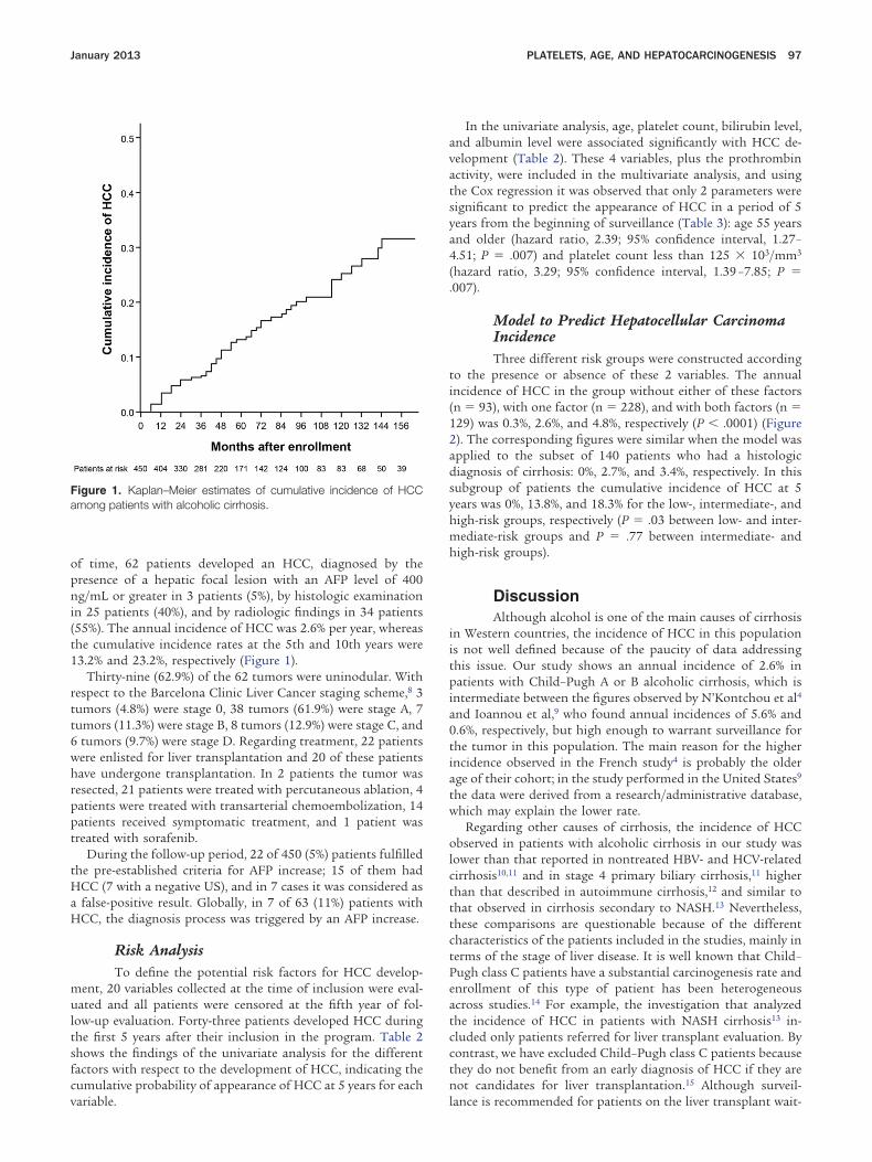
of time, 62 patients developed an HCC, diagnosed by thepresence of a hepatic focal lesion with an AFP level of 400ng/mL or greater in 3 patients (5%), by histologic examinationin 25 patients (40%), and by radiologic findings in 34 patients(55%). The annual incidence of HCC was 2.6% per year, whereasthe cumulative incidence rates at the 5th and 10th years were13.2% and 23.2%, respectively (Figure 1).
Thirty-nine (62.9%) of the 62 tumors were uninodular. Withrespect to the Barcelona Clinic Liver Cancer staging scheme,8 3tumors (4.8%) were stage 0, 38 tumors (61.9%) were stage A, 7tumors (11.3%) were stage B, 8 tumors (12.9%) were stage C, and6 tumors (9.7%) were stage D. Regarding treatment, 22 patientswere enlisted for liver transplantation and 20 of these patientshave undergone transplantation. In 2 patients the tumor wasresected, 21 patients were treated with percutaneous ablation, 4patients were treated with transarterial chemoembolization, 14patients received symptomatic treatment, and 1 patient wastreated with sorafenib.
During the follow-up period, 22 of 450 (5%) patients fulfilledthe pre-established criteria for AFP increase; 15 of them hadHCC (7 with a negative US), and in 7 cases it was considered asa false-positive result. Globally, in 7 of 63 (11%) patients withHCC, the diagnosis process was triggered by an AFP increase.
Risk AnalysisTo define the potential risk factors for HCC develop-
ment, 20 variables collected at the time of inclusion were eval-uated and all patients were censored at the fifth year of fol-low-up evaluation. Forty-three patients developed HCC duringthe first 5 years after their inclusion in the program. Table 2shows the findings of the univariate analysis for the differentfactors with respect to the development of HCC, indicating thecumulative probability of appearance of HCC at 5 years for eachvariable.
In the univariate analysis, age, platelet count, bilirubin level,and albumin level were associated significantly with HCC de-velopment (Table 2). These 4 variables, plus the prothrombinactivity, were included in the multivariate analysis, and usingthe Cox regression it was observed that only 2 parameters weresignificant to predict the appearance of HCC in a period of 5years from the beginning of surveillance (Table 3): age 55 yearsand older (hazard ratio, 2.39; 95% confidence interval, 1.27–4.51; P " .007) and platelet count less than 125 ! 103/mm3
(hazard ratio, 3.29; 95% confidence interval, 1.39 –7.85; P ".007).
Model to Predict Hepatocellular CarcinomaIncidenceThree different risk groups were constructed according
to the presence or absence of these 2 variables. The annualincidence of HCC in the group without either of these factors(n " 93), with one factor (n " 228), and with both factors (n "129) was 0.3%, 2.6%, and 4.8%, respectively (P # .0001) (Figure2). The corresponding figures were similar when the model wasapplied to the subset of 140 patients who had a histologicdiagnosis of cirrhosis: 0%, 2.7%, and 3.4%, respectively. In thissubgroup of patients the cumulative incidence of HCC at 5years was 0%, 13.8%, and 18.3% for the low-, intermediate-, andhigh-risk groups, respectively (P " .03 between low- and inter-mediate-risk groups and P " .77 between intermediate- andhigh-risk groups).
DiscussionAlthough alcohol is one of the main causes of cirrhosis
in Western countries, the incidence of HCC in this populationis not well defined because of the paucity of data addressingthis issue. Our study shows an annual incidence of 2.6% inpatients with Child–Pugh A or B alcoholic cirrhosis, which isintermediate between the figures observed by N’Kontchou et al4
and Ioannou et al,9 who found annual incidences of 5.6% and0.6%, respectively, but high enough to warrant surveillance forthe tumor in this population. The main reason for the higherincidence observed in the French study4 is probably the olderage of their cohort; in the study performed in the United States9
the data were derived from a research/administrative database,which may explain the lower rate.
Regarding other causes of cirrhosis, the incidence of HCCobserved in patients with alcoholic cirrhosis in our study waslower than that reported in nontreated HBV- and HCV-relatedcirrhosis10,11 and in stage 4 primary biliary cirrhosis,11 higherthan that described in autoimmune cirrhosis,12 and similar tothat observed in cirrhosis secondary to NASH.13 Nevertheless,these comparisons are questionable because of the differentcharacteristics of the patients included in the studies, mainly interms of the stage of liver disease. It is well known that Child–Pugh class C patients have a substantial carcinogenesis rate andenrollment of this type of patient has been heterogeneousacross studies.14 For example, the investigation that analyzedthe incidence of HCC in patients with NASH cirrhosis13 in-cluded only patients referred for liver transplant evaluation. Bycontrast, we have excluded Child–Pugh class C patients becausethey do not benefit from an early diagnosis of HCC if they arenot candidates for liver transplantation.15 Although surveil-lance is recommended for patients on the liver transplant wait-
Figure 1. Kaplan–Meier estimates of cumulative incidence of HCCamong patients with alcoholic cirrhosis.
January 2013 PLATELETS, AGE, AND HEPATOCARCINOGENESIS 97
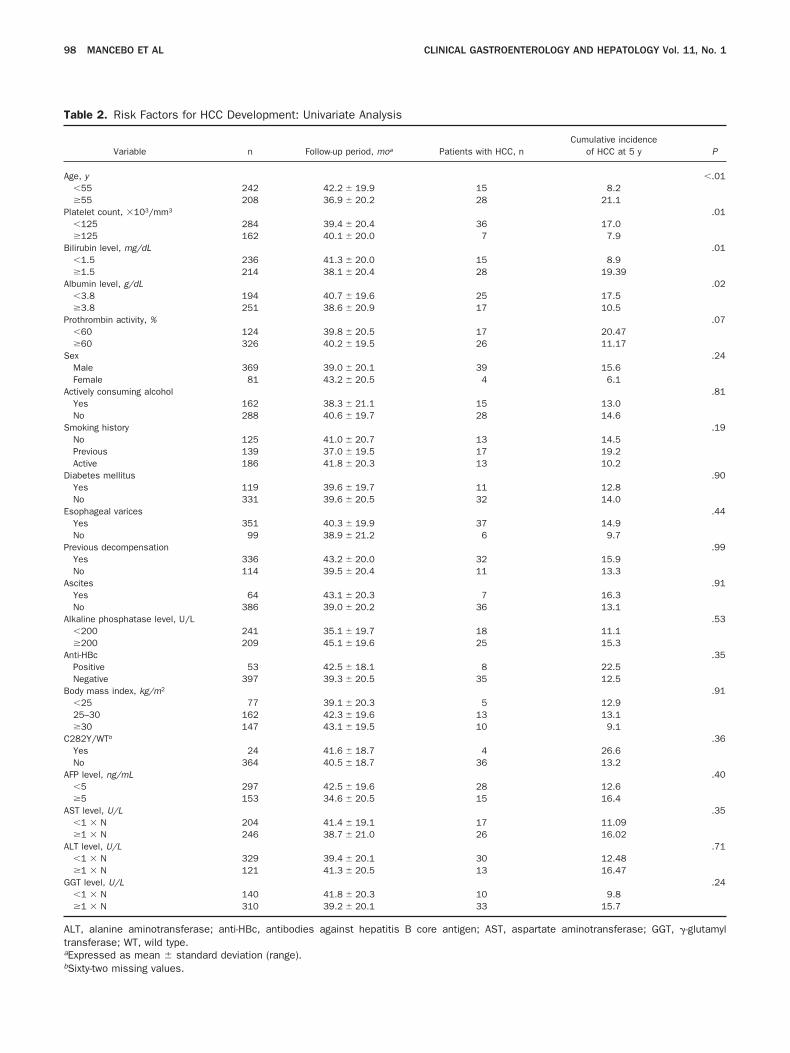
Table 2. Risk Factors for HCC Development: Univariate Analysis
Variable n Follow-up period, moa Patients with HCC, nCumulative incidence
of HCC at 5 y P
Age, y #.01#55 242 42.2 & 19.9 15 8.2"55 208 36.9 & 20.2 28 21.1
Platelet count, !103/mm3 .01#125 284 39.4 & 20.4 36 17.0"125 162 40.1 & 20.0 7 7.9
Bilirubin level, mg/dL .01#1.5 236 41.3 & 20.0 15 8.9"1.5 214 38.1 & 20.4 28 19.39
Albumin level, g/dL .02#3.8 194 40.7 & 19.6 25 17.5"3.8 251 38.6 & 20.9 17 10.5
Prothrombin activity, % .07#60 124 39.8 & 20.5 17 20.47"60 326 40.2 & 19.5 26 11.17
Sex .24Male 369 39.0 & 20.1 39 15.6Female 81 43.2 & 20.5 4 6.1
Actively consuming alcohol .81Yes 162 38.3 & 21.1 15 13.0No 288 40.6 & 19.7 28 14.6
Smoking history .19No 125 41.0 & 20.7 13 14.5Previous 139 37.0 & 19.5 17 19.2Active 186 41.8 & 20.3 13 10.2
Diabetes mellitus .90Yes 119 39.6 & 19.7 11 12.8No 331 39.6 & 20.5 32 14.0
Esophageal varices .44Yes 351 40.3 & 19.9 37 14.9No 99 38.9 & 21.2 6 9.7
Previous decompensation .99Yes 336 43.2 & 20.0 32 15.9No 114 39.5 & 20.4 11 13.3
Ascites .91Yes 64 43.1 & 20.3 7 16.3No 386 39.0 & 20.2 36 13.1
Alkaline phosphatase level, U/L .53#200 241 35.1 & 19.7 18 11.1"200 209 45.1 & 19.6 25 15.3
Anti-HBc .35Positive 53 42.5 & 18.1 8 22.5Negative 397 39.3 & 20.5 35 12.5
Body mass index, kg/m2 .91#25 77 39.1 & 20.3 5 12.925–30 162 42.3 & 19.6 13 13.1"30 147 43.1 & 19.5 10 9.1
C282Y/WTb .36Yes 24 41.6 & 18.7 4 26.6No 364 40.5 & 18.7 36 13.2
AFP level, ng/mL .40#5 297 42.5 & 19.6 28 12.6"5 153 34.6 & 20.5 15 16.4
AST level, U/L .35#1 ! N 204 41.4 & 19.1 17 11.09"1 ! N 246 38.7 & 21.0 26 16.02
ALT level, U/L .71#1 ! N 329 39.4 & 20.1 30 12.48"1 ! N 121 41.3 & 20.5 13 16.47
GGT level, U/L .24#1 ! N 140 41.8 & 20.3 10 9.8"1 ! N 310 39.2 & 20.1 33 15.7
ALT, alanine aminotransferase; anti-HBc, antibodies against hepatitis B core antigen; AST, aspartate aminotransferase; GGT, (-glutamyltransferase; WT, wild type.aExpressed as mean & standard deviation (range).bSixty-two missing values.
98 MANCEBO ET AL CLINICAL GASTROENTEROLOGY AND HEPATOLOGY Vol. 11, No. 1
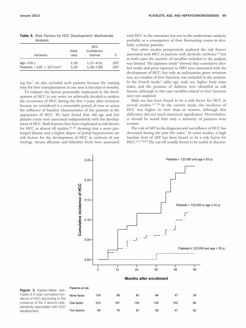
ing list,7 we also excluded such patients because the waitingtime for liver transplantation in our area is less than 6 months.
To evaluate the factors potentially implicated in the devel-opment of HCC in our series, we arbitrarily decided to analyzethe occurrence of HCC during the first 5 years after inclusionbecause we considered it a reasonable period of time to assessthe influence of baseline characteristics of the patients in theappearance of HCC. We have found that old age and lowplatelet count were associated independently with the develop-ment of HCC. Both features have been implicated as risk factorsfor HCC in almost all studies,16 –20 showing that a more pro-longed disease and a higher degree of portal hypertension arerisk factors for the development of HCC in cirrhosis of anyetiology. Serum albumin and bilirubin levels were associated
with HCC in the univariate but not in the multivariate analysis,probably as a consequence of their fluctuating course in alco-holic cirrhotic patients.
Two other studies prospectively analyzed the risk factorsassociated with HCC in patients with alcoholic cirrhosis,3,4 butin both cases the number of variables included in the analysiswas limited. The Japanese study3 showed that cumulative alco-hol intake and prior exposure to HBV were associated with thedevelopment of HCC, but only an indocyanine green retentiontest, as a marker of liver function, was included in the analysis.In the French study,4 older age, male sex, higher body massindex, and the presence of diabetes were identified as riskfactors, although in this case variables related to liver functionwere not analyzed.
Male sex has been found to be a risk factor for HCC inseveral studies.4,17,18 In the current study, the incidence ofHCC was higher in men than in women, although thisdifference did not reach statistical significance. Nevertheless,it should be noted that only a minority of patients werewomen.
The role of AFP in the diagnosis and surveillance of HCC hasdecreased during the past few years.7 In some studies, a highbaseline level of AFP has been found to be a risk factor forHCC.14,17,18,20 The cut-off usually found to be useful in discrim-
Table 3. Risk Factors for HCC Development: MultivariateAnalysis
VariablesOddsratio
95%Confidence
interval P
Age "55 y 2.39 1.27–4.51 .007Platelets #125 ! 103/mm3 3.29 1.39–7.85 .007
Figure 2. Kaplan–Meier esti-mates of 5-year cumulative inci-dence of HCC according to thepresence of the 2 factors inde-pendently associated with HCCdevelopment.
January 2013 PLATELETS, AGE, AND HEPATOCARCINOGENESIS 99
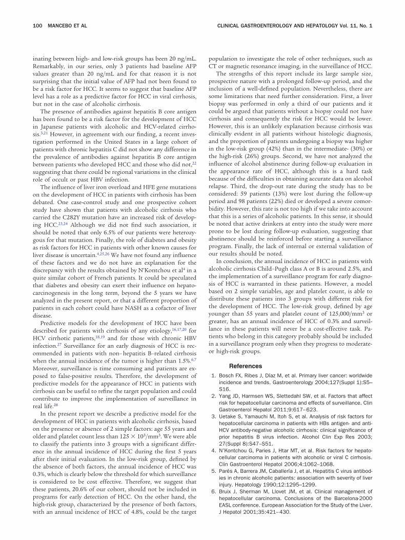
inating between high- and low-risk groups has been 20 ng/mL.Remarkably, in our series, only 3 patients had baseline AFPvalues greater than 20 ng/mL and for that reason it is notsurprising that the initial value of AFP had not been found tobe a risk factor for HCC. It seems to suggest that baseline AFPlevel has a role as a predictive factor for HCC in viral cirrhosis,but not in the case of alcoholic cirrhosis.
The presence of antibodies against hepatitis B core antigenhas been found to be a risk factor for the development of HCCin Japanese patients with alcoholic and HCV-related cirrho-sis.3,21 However, in agreement with our finding, a recent inves-tigation performed in the United States in a large cohort ofpatients with chronic hepatitis C did not show any difference inthe prevalence of antibodies against hepatitis B core antigenbetween patients who developed HCC and those who did not,22
suggesting that there could be regional variations in the clinicalrole of occult or past HBV infection.
The influence of liver iron overload and HFE gene mutationson the development of HCC in patients with cirrhosis has beendebated. One case-control study and one prospective cohortstudy have shown that patients with alcoholic cirrhosis whocarried the C282Y mutation have an increased risk of develop-ing HCC.23,24 Although we did not find such association, itshould be noted that only 6.5% of our patients were heterozy-gous for that mutation. Finally, the role of diabetes and obesityas risk factors for HCC in patients with other known causes forliver disease is uncertain.4,25,26 We have not found any influenceof these factors and we do not have an explanation for thediscrepancy with the results obtained by N’Kontchou et al4 in aquite similar cohort of French patients. It could be speculatedthat diabetes and obesity can exert their influence on hepato-carcinogenesis in the long term, beyond the 5 years we haveanalyzed in the present report, or that a different proportion ofpatients in each cohort could have NASH as a cofactor of liverdisease.
Predictive models for the development of HCC have beendescribed for patients with cirrhosis of any etiology,16,17,20 forHCV cirrhotic patients,18,19 and for those with chronic HBVinfection.27 Surveillance for an early diagnosis of HCC is rec-ommended in patients with non– hepatitis B–related cirrhosiswhen the annual incidence of the tumor is higher than 1.5%.6,7
Moreover, surveillance is time consuming and patients are ex-posed to false-positive results. Therefore, the development ofpredictive models for the appearance of HCC in patients withcirrhosis can be useful to refine the target population and couldcontribute to improve the implementation of surveillance inreal life.28
In the present report we describe a predictive model for thedevelopment of HCC in patients with alcoholic cirrhosis, basedon the presence or absence of 2 simple factors: age 55 years andolder and platelet count less than 125 ! 103/mm3. We were ableto classify the patients into 3 groups with a significant differ-ence in the annual incidence of HCC during the first 5 yearsafter their initial evaluation. In the low-risk group, defined bythe absence of both factors, the annual incidence of HCC was0.3%, which is clearly below the threshold for which surveillanceis considered to be cost effective. Therefore, we suggest thatthese patients, 20.6% of our cohort, should not be included inprograms for early detection of HCC. On the other hand, thehigh-risk group, characterized by the presence of both factors,with an annual incidence of HCC of 4.8%, could be the target
population to investigate the role of other techniques, such asCT or magnetic resonance imaging, in the surveillance of HCC.
The strengths of this report include its large sample size,prospective nature with a prolonged follow-up period, and theinclusion of a well-defined population. Nevertheless, there aresome limitations that need further consideration. First, a liverbiopsy was performed in only a third of our patients and itcould be argued that patients without a biopsy could not havecirrhosis and consequently the risk for HCC would be lower.However, this is an unlikely explanation because cirrhosis wasclinically evident in all patients without histologic diagnosis,and the proportion of patients undergoing a biopsy was higherin the low-risk group (42%) than in the intermediate- (30%) orthe high-risk (26%) groups. Second, we have not analyzed theinfluence of alcohol abstinence during follow-up evaluation inthe appearance rate of HCC, although this is a hard taskbecause of the difficulties in obtaining accurate data on alcoholrelapse. Third, the drop-out rate during the study has to beconsidered: 59 patients (13%) were lost during the follow-upperiod and 98 patients (22%) died or developed a severe comor-bidity. However, this rate is not too high if we take into accountthat this is a series of alcoholic patients. In this sense, it shouldbe noted that active drinkers at entry into the study were moreprone to be lost during follow-up evaluation, suggesting thatabstinence should be reinforced before starting a surveillanceprogram. Finally, the lack of internal or external validation ofour results should be noted.
In conclusion, the annual incidence of HCC in patients withalcoholic cirrhosis Child–Pugh class A or B is around 2.5%, andthe implementation of a surveillance program for early diagno-sis of HCC is warranted in these patients. However, a modelbased on 2 simple variables, age and platelet count, is able todistribute these patients into 3 groups with different risk forthe development of HCC. The low-risk group, defined by ageyounger than 55 years and platelet count of 125,000/mm3 orgreater, has an annual incidence of HCC of 0.3% and surveil-lance in these patients will never be a cost-effective task. Pa-tients who belong in this category probably should be includedin a surveillance program only when they progress to moderate-or high-risk groups.
References1. Bosch FX, Ribes J, Díaz M, et al. Primary liver cancer: worldwide
incidence and trends. Gastroenterology 2004;127(Suppl 1):S5–S16.
2. Yang JD, Harmsen WS, Slettedahl SW, et al. Factors that affectrisk for hepatocellular carcinoma and effects of surveillance. ClinGastroenterol Hepatol 2011;9:617–623.
3. Uetake S, Yamauchi M, Itoh S, et al. Analysis of risk factors forhepatocellular carcinoma in patients with HBs antigen- and anti-HCV antibody-negative alcoholic cirrhosis: clinical significance ofprior hepatitis B virus infection. Alcohol Clin Exp Res 2003;27(Suppl 8):S47–S51.
4. N’Kontchou G, Paries J, Htar MT, et al. Risk factors for hepato-cellular carcinoma in patients with alcoholic or viral C cirrhosis.Clin Gastroenterol Hepatol 2006;4:1062–1068.
5. Parés A, Barrera JM, Caballería J, et al. Hepatitis C virus antibod-ies in chronic alcoholic patients: association with severity of liverinjury. Hepatology 1990;12:1295–1299.
6. Bruix J, Sherman M, Llovet JM, et al. Clinical management ofhepatocellular carcinoma. Conclusions of the Barcelona-2000EASL conference. European Association for the Study of the Liver.J Hepatol 2001;35:421–430.
100 MANCEBO ET AL CLINICAL GASTROENTEROLOGY AND HEPATOLOGY Vol. 11, No. 1
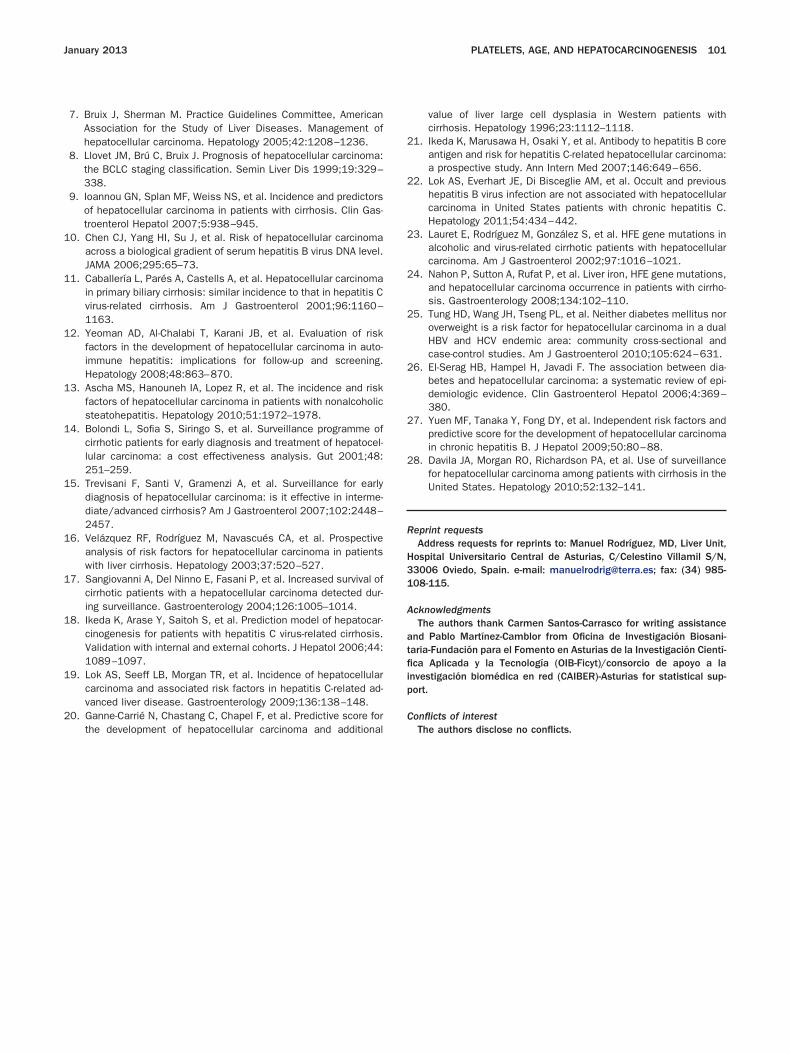
7. Bruix J, Sherman M. Practice Guidelines Committee, AmericanAssociation for the Study of Liver Diseases. Management ofhepatocellular carcinoma. Hepatology 2005;42:1208–1236.
8. Llovet JM, Brú C, Bruix J. Prognosis of hepatocellular carcinoma:the BCLC staging classification. Semin Liver Dis 1999;19:329–338.
9. Ioannou GN, Splan MF, Weiss NS, et al. Incidence and predictorsof hepatocellular carcinoma in patients with cirrhosis. Clin Gas-troenterol Hepatol 2007;5:938–945.
10. Chen CJ, Yang HI, Su J, et al. Risk of hepatocellular carcinomaacross a biological gradient of serum hepatitis B virus DNA level.JAMA 2006;295:65–73.
11. Caballería L, Parés A, Castells A, et al. Hepatocellular carcinomain primary biliary cirrhosis: similar incidence to that in hepatitis Cvirus-related cirrhosis. Am J Gastroenterol 2001;96:1160–1163.
12. Yeoman AD, Al-Chalabi T, Karani JB, et al. Evaluation of riskfactors in the development of hepatocellular carcinoma in auto-immune hepatitis: implications for follow-up and screening.Hepatology 2008;48:863–870.
13. Ascha MS, Hanouneh IA, Lopez R, et al. The incidence and riskfactors of hepatocellular carcinoma in patients with nonalcoholicsteatohepatitis. Hepatology 2010;51:1972–1978.
14. Bolondi L, Sofia S, Siringo S, et al. Surveillance programme ofcirrhotic patients for early diagnosis and treatment of hepatocel-lular carcinoma: a cost effectiveness analysis. Gut 2001;48:251–259.
15. Trevisani F, Santi V, Gramenzi A, et al. Surveillance for earlydiagnosis of hepatocellular carcinoma: is it effective in interme-diate/advanced cirrhosis? Am J Gastroenterol 2007;102:2448–2457.
16. Velázquez RF, Rodríguez M, Navascués CA, et al. Prospectiveanalysis of risk factors for hepatocellular carcinoma in patientswith liver cirrhosis. Hepatology 2003;37:520–527.
17. Sangiovanni A, Del Ninno E, Fasani P, et al. Increased survival ofcirrhotic patients with a hepatocellular carcinoma detected dur-ing surveillance. Gastroenterology 2004;126:1005–1014.
18. Ikeda K, Arase Y, Saitoh S, et al. Prediction model of hepatocar-cinogenesis for patients with hepatitis C virus-related cirrhosis.Validation with internal and external cohorts. J Hepatol 2006;44:1089–1097.
19. Lok AS, Seeff LB, Morgan TR, et al. Incidence of hepatocellularcarcinoma and associated risk factors in hepatitis C-related ad-vanced liver disease. Gastroenterology 2009;136:138–148.
20. Ganne-Carrié N, Chastang C, Chapel F, et al. Predictive score forthe development of hepatocellular carcinoma and additional
value of liver large cell dysplasia in Western patients withcirrhosis. Hepatology 1996;23:1112–1118.
21. Ikeda K, Marusawa H, Osaki Y, et al. Antibody to hepatitis B coreantigen and risk for hepatitis C-related hepatocellular carcinoma:a prospective study. Ann Intern Med 2007;146:649–656.
22. Lok AS, Everhart JE, Di Bisceglie AM, et al. Occult and previoushepatitis B virus infection are not associated with hepatocellularcarcinoma in United States patients with chronic hepatitis C.Hepatology 2011;54:434–442.
23. Lauret E, Rodríguez M, González S, et al. HFE gene mutations inalcoholic and virus-related cirrhotic patients with hepatocellularcarcinoma. Am J Gastroenterol 2002;97:1016–1021.
24. Nahon P, Sutton A, Rufat P, et al. Liver iron, HFE gene mutations,and hepatocellular carcinoma occurrence in patients with cirrho-sis. Gastroenterology 2008;134:102–110.
25. Tung HD, Wang JH, Tseng PL, et al. Neither diabetes mellitus noroverweight is a risk factor for hepatocellular carcinoma in a dualHBV and HCV endemic area: community cross-sectional andcase-control studies. Am J Gastroenterol 2010;105:624–631.
26. El-Serag HB, Hampel H, Javadi F. The association between dia-betes and hepatocellular carcinoma: a systematic review of epi-demiologic evidence. Clin Gastroenterol Hepatol 2006;4:369–380.
27. Yuen MF, Tanaka Y, Fong DY, et al. Independent risk factors andpredictive score for the development of hepatocellular carcinomain chronic hepatitis B. J Hepatol 2009;50:80–88.
28. Davila JA, Morgan RO, Richardson PA, et al. Use of surveillancefor hepatocellular carcinoma among patients with cirrhosis in theUnited States. Hepatology 2010;52:132–141.
Reprint requestsAddress requests for reprints to: Manuel Rodríguez, MD, Liver Unit,
Hospital Universitario Central de Asturias, C/Celestino Villamil S/N,33006 Oviedo, Spain. e-mail: [email protected]; fax: (34) 985-108-115.
AcknowledgmentsThe authors thank Carmen Santos-Carrasco for writing assistance
and Pablo Martínez-Camblor from Oficina de Investigación Biosani-taria-Fundación para el Fomento en Asturias de la Investigación Cientí-fica Aplicada y la Tecnología (OIB-Ficyt)/consorcio de apoyo a lainvestigación biomédica en red (CAIBER)-Asturias for statistical sup-port.
Conflicts of interestThe authors disclose no conflicts.
January 2013 PLATELETS, AGE, AND HEPATOCARCINOGENESIS 101

Adherence to a Semiannual Surveillance Program forHepatocellular Carcinoma in Patients With Liver Cirrhosis
Alejo Mancebo, MD, Marıa Luisa Gonzalez-Dieguez, MD,Carmen A. Navascues, MD, Valle Cadahıa, MD, Marıa Varela, MD,Ramon Perez, PhD, Luis Rodrigo, PhD, and Manuel Rodrıguez, PhD
Background: Patient adherence to screening for hepatocellularcarcinoma (HCC) is not well known. Our aims were to analyze theadherence to a surveillance program in a prospective cohort ofcirrhotic patients and to examine its association with HCC stage atdiagnosis.
Materials and Methods: A total of 770 patients with cirrhosis wereexamined semiannually by ultrasound and alpha-fetoprotein at atertiary center. We collected data on 17 variables at baseline.Suboptimal adherence was defined as failure to complete 2 con-secutive screening rounds.
Results: Over a median follow-up period of 42.0 months (inter-quartile range: 60.0), 125 patients (16.2%) had suboptimal adher-ence. Active or previous intravenous drug use [hazard ratio (HR),5.33; 95% confidence interval (CI), 3.07-9.23], active alcohol con-sumption (HR, 3.03; 95% CI, 2.03-4.51), absence of liver decom-pensation before the inclusion in the program (HR, 1.65; 95% CI,1.07-2.55) and aspartate transaminase/alanine transaminase ratioZ1.6 (HR, 1.82; 95% CI, 1.23-2.70) were independent predictorsof suboptimal adherence. Compared with those with optimaladherence, patients with suboptimal adherence had a moreadvanced HCC stage at diagnosis (P=0.015), they were less fre-quently treated with curative intention (P=0.078) and survivedless (median: 14.2mo; IQR: 36.0 vs. 22.7mo; IQR: 47.4;P=0.160), although these differences were not significant.
Conclusions: The adherence to the process of HCC surveillance canbe considered as adequate among cirrhotic patients. Active alcoholconsumption and a history of intravenous drug use are the stron-gest predictors of suboptimal adherence. These patients have amore advanced HCC stage at diagnosis and tend to be less fre-quently treated with curative intention.
Key Words: hepatocellular carcinoma, adherence, ultrasonography,surveillance, cirrhosis
(J Clin Gastroenterol 2016;00:000–000)
Despite some concerns derived from the scarcity ofstrong evidence, Clinical Practice Guidelines endorse
biannual surveillance for early detection of hepatocellularcarcinoma (HCC) in patients with Child-Pugh A or B livercirrhosis.1,2 Only 2 randomized trials demonstrating HCCscreening effectiveness have been published.3,4 Both studieswere conducted in chronic hepatitis B virus carriers, andthey were criticized because of design flaws.5 However, 1systematic review6 has shown that compared with inci-dentally diagnosed HCC, patients who undergo surveil-lance have earlier-stage HCC and live longer. Moreover, itdoes not seem ethical to conduct new randomized controltrials.7
Surveillance for HCC intends to detect tumors at anearly stage when potentially curative treatments can beapplied.1 However, a short proportion of patients diag-nosed with HCC in western countries are under surveil-lance.8,9 Moreover, there is little evidence about patientcompliance with the HCC surveillance program. Tworandomized controlled trials conducted in China found thatonly 58.2%3 and 40.6%4 of patients completed all of thehalf-yearly visits. Studies examining the causes of failure inHCC surveillance processes in cirrhotic patients have notspecified whether the screening is unsolicited by the physi-cian, whether there is a lack of insurance coverage orwhether the patients did not comply with the scheduledtest.10–14
Our primary objectives were to assess patient adher-ence to the HCC surveillance program and to identifypredictors of noncompliance in a large cohort of cirrhoticsubjects. Our secondary objective was to examine whetheradherence is associated with tumor stage at diagnosis.
MATERIALS AND METHODS
PatientsWe conducted a prospective cohort study of 770
patients with cirrhosis who were consecutively enrolled in asurveillance program for early detection of HCC based onsemiannual screening tests with an alpha-fetoprotein (AFP)assay and ultrasonography (US) between January 1992 andFebruary 2013. Follow-up ended in March 2014. Patientswere included if they had (a) Child-Pugh class A/B cir-rhosis; (b) age between 35 and 75 years; (c) absence ofknown HCC; (d) availability to attend regular visits; and (e)no comorbid illness with a life expectancy less than 1 year.All patients seen at the Liver Unit during the study and whofulfilled the criterion above were enrolled in the program.Throughout the study, patients were seen at the outpatientclinic of a tertiary hospital by senior hepatologists andsurveillance was ordered by clinic providers as part of usual
Received for publication March 31, 2016; accepted September 11, 2016.From the Department of Gastroenterology and Hepatology, Liver
Unit, Hospital Universitario Central de Asturias, University ofOviedo, Oviedo, Spain.
A.M.: drafting of the manuscript. M.L.G.-D.: acquisition, analysis andinterpretation of data. M.V.: acquisition of data and study super-vision. C.A.N., V.C., and R.P.: acquisition of data. L.R.: admin-istrative support. M.R.: acquisition, analysis and interpretation ofdata, statistical analysis, critical revision of the manuscript forimportant intellectual content and study supervision.
The authors declare that they have nothing to disclose.Address correspondence to: Manuel Rodrıguez, MD, Department of
Gastroenterology and Hepatology, Liver Unit, Hospital Uni-versitario Central de Asturias, University of Oviedo, Avenida deRoma, s/n, Oviedo 33011, Spain(e-mail: [email protected]).
Copyright r 2016 Wolters Kluwer Health, Inc. All rights reserved.
ORIGINAL ARTICLE
J Clin Gastroenterol ! Volume 00, Number 00, ’’ 2016 www.jcge.com | 1Copyright r 2016 Wolters Kluwer Health, Inc. Unauthorized reproduction of this article is prohibited.
This paper can be cited using the date of access and the unique DOI number which can be found in the footnotes.
care. At every clinical visit, laboratory tests and abdominalUS were scheduled for the next 6 months and a new clinicalappointment was fixed over the following week. Imagingstudies were performed by trained medical staff belongingto the Institution. Reminder calls were not offered by thehospital during the study.
Patients were classified into 4 categories according tothe cause of cirrhosis: alcoholic (daily ethanol consumptionZ60 g in men and Z40 g in women during a periodZ10 y), hepatitis C virus (HCV) when serum anti-HCVantibodies and HCV-RNA were present, hepatitis B viruswhen hepatitis B surface antigen was present; and othercauses.
Cirrhosis was diagnosed by liver biopsy in 252 patients(32.7%), by the presence of esophageal varices in 398patients (51.8%), by the presence of clinical patterns asso-ciated with decompensated cirrhosis in 55 patients (7.1%)and by US showing a cirrhotic-appearing liver with asso-ciated signs of portal hypertension in 65 patients (8.4%).
Seventeen demographic, clinical and biochemicalparameters (Table 1) were collected at the time of inclusion.The geographic proximity to HCC screening facilities wasdefined as the travel distance to the clinic from the patient’spostal address. Active alcohol consumption was defined asany alcohol drinking within the previous month to the
inclusion and it was assessed by means of an interview withpatients and their relatives. Active and previous intra-venous drug use (IVDU) were combined into a single cat-egory due to the scarce number of active users (n=10).
During the overall follow-up, patient participation wasmonitored at each consecutive visit. If the patient requiredhospital admission due to an episode of liver decom-pensation at the time of the visit, it was computed ascompliant. Liver imaging studies performed during thistime were computed as performed for surveillancepurposes.
Patients were divided into 2 distinct cohorts accordingto their adherence to the program: suboptimal adherencewas defined as failure to complete 2 consecutive abdominalUS while those patients who did not fail to 2 consecutiverounds were considered as having optimal surveillance.This criterion was chosen arbitrary.
To calculate the cumulative probability of suboptimaladherence, patients were censored at the date of HCCdiagnosis, the date of progression to Child-Pugh class C,the date of death, the date of developing a severe comor-bidity, the date of inclusion in a waiting list for livertransplantation, the date at last follow-up visit, the date totransfer to another center, or the date to failure to 2 con-secutive screening tests, whichever came first. Otherwise,patients who were evaluated after failing any control wereincluded in the analysis of the tumor characteristics and inthe analysis of survival. The Spanish National Death Index(www.msssi.gob.es/estadEstudios/estadisticas/estadisticas/estMinisterio/IND_TipoDifusion.htm), which includes thedate of all deaths reported in Spain and is continuouslyupdated, was consulted to ensure the state of the patients atthe end of the follow-up period.
Follow-up EvaluationPatients were monitored for HCC every 6 months
using AFP and US. The attendance of the patients at everyscreening round and consecutive visit were prospectivelyrecorded, and when patients failed a check-up, they wererescheduled for a new appointment.
When the AFP value was >20 ng/mL and more thantwice the previous value, a computed tomography wasperformed to rule out the presence of HCC. Otherwise,when a new nodule was detected by US, subsequentdynamic imaging tests were performed. All patients with afocal lesion in the US or with an AFP level increase and anegative computed tomography were monitored closely forat least 1 year.
From 1992 to 2001, HCC was diagnosed using histo-logic confirmation or when a hepatic focal lesion with anAFPZ400 ng/mL was observed. After 2001, HCC diag-nosis was based on the Clinical Practice Guidelines rec-ommendations.1,2 Tumor staging was based on at least 2abdominal imaging techniques. The Barcelona Clinic LiverCancer (BCLC) system and the Milan Criteria were usedfor tumor staging.15,16
Statistical AnalysisDescriptive statistics are reported using mean±SD,
median values with with interquartile ranges (IQR) or N(%). Comparative results between groups were obtainedusing the w2 test or the student t test. The Kaplan-Meiermethod was used to determine the cumulative probabilityof the suboptimal adherence. The comparison of the sur-vival curves obtained was performed using the log-rank
TABLE 1. Baseline Characteristics of the Patients Enrolled in theStudy (N = 770)
Category Results
Age (y)* 53.9±8.2Male/female [n (%)] 609 (79.1)/161 (20.9)Cirrhosis cause [n (%)]Alcohol 472 (61.3)HCV 191 (24.8)HBV 70 (9.1)Others 37 (4.8)
Child-Pugh class A/B [n (%)] 567 (73.6)/190 (24.6)Travel distance (km)* 21.9±32.3Period of inclusion [n (%)]1992-2003 272 (35.3)2003-2012 498 (64.7)
Active consumer of alcohol [n (%)] 234 (30.4)Active or previous smoker [n (%)] 540 (70.1)HIV seropositivity [n (%)] 57 (7.4)Actual or previous intravenous drug user[n (%)]
69 (8.9)
Diabetes mellitus at inclusion [n (%)] 185 (24.0)Body mass index (kg/m2)* 27.8±4.9Esophageal varices [n (%)] 546 (70.69)Previous decompensation [n (%)] 464 (60.3)Platelet count ("103/mm3)* 119.7±59.3AP level (U/L)*,y 172.5±108.7GGT level (U/L)*,8 142.0±172.9ASTw/ALTz ratio* 1.4± .6
*Expressed as the mean±SD.wThe normal value (N) for AST is <37U/L in men and <31U/L in
women.zThe normal value (N) for ALT is <40U/L in men and <31U/L in
women.yThe normal value (N) for AP is <280U/L.8The normal value (N) for GGT is <50U/L in men and less than 32U/L
in women.ALT indicates alanine aminotransferase; AP, alkaline phosphatase;
AST, aspartate aminotransferase; GGT, g-glutamyl transferase; HBV,hepatitis B virus; HCV, hepatitis C virus; HIV, human immunodeficiencyvirus.
Mancebo et al J Clin Gastroenterol ! Volume 00, Number 00, ’’ 2016
2 | www.jcge.com Copyright r 2016 Wolters Kluwer Health, Inc. All rights reserved.
Copyright r 2016 Wolters Kluwer Health, Inc. Unauthorized reproduction of this article is prohibited.This paper can be cited using the date of access and the unique DOI number which can be found in the footnotes.
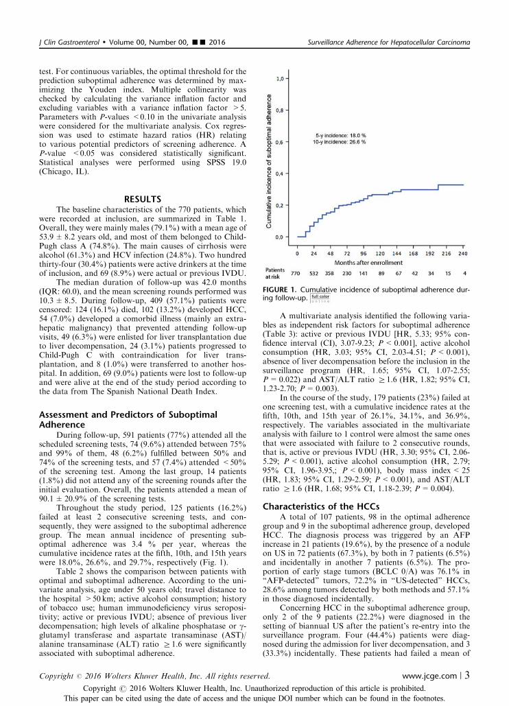
test. For continuous variables, the optimal threshold for theprediction suboptimal adherence was determined by max-imizing the Youden index. Multiple collinearity waschecked by calculating the variance inflation factor andexcluding variables with a variance inflation factor >5.Parameters with P-values <0.10 in the univariate analysiswere considered for the multivariate analysis. Cox regres-sion was used to estimate hazard ratios (HR) relatingto various potential predictors of screening adherence. AP-value <0.05 was considered statistically significant.Statistical analyses were performed using SPSS 19.0(Chicago, IL).
RESULTSThe baseline characteristics of the 770 patients, which
were recorded at inclusion, are summarized in Table 1.Overall, they were mainly males (79.1%) with a mean age of53.9±8.2 years old, and most of them belonged to Child-Pugh class A (74.8%). The main causes of cirrhosis werealcohol (61.3%) and HCV infection (24.8%). Two hundredthirty-four (30.4%) patients were active drinkers at the timeof inclusion, and 69 (8.9%) were actual or previous IVDU.
The median duration of follow-up was 42.0 months(IQR: 60.0), and the mean screening rounds performed was10.3±8.5. During follow-up, 409 (57.1%) patients werecensored: 124 (16.1%) died, 102 (13.2%) developed HCC,54 (7.0%) developed a comorbid illness (mainly an extra-hepatic malignancy) that prevented attending follow-upvisits, 49 (6.3%) were enlisted for liver transplantation dueto liver decompensation, 24 (3.1%) patients progressed toChild-Pugh C with contraindication for liver trans-plantation, and 8 (1.0%) were transferred to another hos-pital. In addition, 69 (9.0%) patients were lost to follow-upand were alive at the end of the study period according tothe data from The Spanish National Death Index.
Assessment and Predictors of SuboptimalAdherence
During follow-up, 591 patients (77%) attended all thescheduled screening tests, 74 (9.6%) attended between 75%and 99% of them, 48 (6.2%) fulfilled between 50% and74% of the screening tests, and 57 (7.4%) attended <50%of the screening test. Among the last group, 14 patients(1.8%) did not attend any of the screening rounds after theinitial evaluation. Overall, the patients attended a mean of90.1±20.9% of the screening tests.
Throughout the study period, 125 patients (16.2%)failed at least 2 consecutive screening tests, and con-sequently, they were assigned to the suboptimal adherencegroup. The mean annual incidence of presenting sub-optimal adherence was 3.4 % per year, whereas thecumulative incidence rates at the fifth, 10th, and 15th yearswere 18.0%, 26.6%, and 29.7%, respectively (Fig. 1).
Table 2 shows the comparison between patients withoptimal and suboptimal adherence. According to the uni-variate analysis, age under 50 years old; travel distance tothe hospital >50 km; active alcohol consumption; historyof tobacco use; human immunodeficiency virus seroposi-tivity; active or previous IVDU; absence of previous liverdecompensation; high levels of alkaline phosphatase or g-glutamyl transferase and aspartate transaminase (AST)/alanine transaminase (ALT) ratio Z1.6 were significantlyassociated with suboptimal adherence.
A multivariate analysis identified the following varia-bles as independent risk factors for suboptimal adherence(Table 3): active or previous IVDU [HR, 5.33; 95% con-fidence interval (CI), 3.07-9.23; P<0.001], active alcoholconsumption (HR, 3.03; 95% CI, 2.03-4.51; P<0.001),absence of liver decompensation before the inclusion in thesurveillance program (HR, 1.65; 95% CI, 1.07-2.55;P=0.022) and AST/ALT ratio Z1.6 (HR, 1.82; 95% CI,1.23-2.70; P=0.003).
In the course of the study, 179 patients (23%) failed atone screening test, with a cumulative incidence rates at thefifth, 10th, and 15th year of 26.1%, 34.1%, and 36.9%,respectively. The variables associated in the multivariateanalysis with failure to 1 control were almost the same onesthat were associated with failure to 2 consecutive rounds,that is, active or previous IVDU (HR, 3.30; 95% CI, 2.06-5.29; P<0.001), active alcohol consumption (HR, 2.79;95% CI, 1.96-3.95,; P<0.001), body mass index<25(HR, 1.83; 95% CI, 1.29-2.59; P<0.001), and AST/ALTratio Z1.6 (HR, 1.68; 95% CI, 1.18-2.39; P=0.004).
Characteristics of the HCCsA total of 107 patients, 98 in the optimal adherence
group and 9 in the suboptimal adherence group, developedHCC. The diagnosis process was triggered by an AFPincrease in 21 patients (19.6%), by the presence of a noduleon US in 72 patients (67.3%), by both in 7 patients (6.5%)and incidentally in another 7 patients (6.5%). The pro-portion of early stage tumors (BCLC 0/A) was 76.1% in“AFP-detected” tumors, 72.2% in “US-detected” HCCs,28.6% among tumors detected by both methods and 57.1%in those diagnosed incidentally.
Concerning HCC in the suboptimal adherence group,only 2 of the 9 patients (22.2%) were diagnosed in thesetting of biannual US after the patient’s re-entry into thesurveillance program. Four (44.4%) patients were diag-nosed during the admission for liver decompensation, and 3(33.3%) incidentally. These patients had failed a mean of
FIGURE 1. Cumulative incidence of suboptimal adherence dur-ing follow-up.
J Clin Gastroenterol ! Volume 00, Number 00, ’’ 2016 Surveillance Adherence for Hepatocellular Carcinoma
Copyright r 2016 Wolters Kluwer Health, Inc. All rights reserved. www.jcge.com | 3Copyright r 2016 Wolters Kluwer Health, Inc. Unauthorized reproduction of this article is prohibited.
This paper can be cited using the date of access and the unique DOI number which can be found in the footnotes.
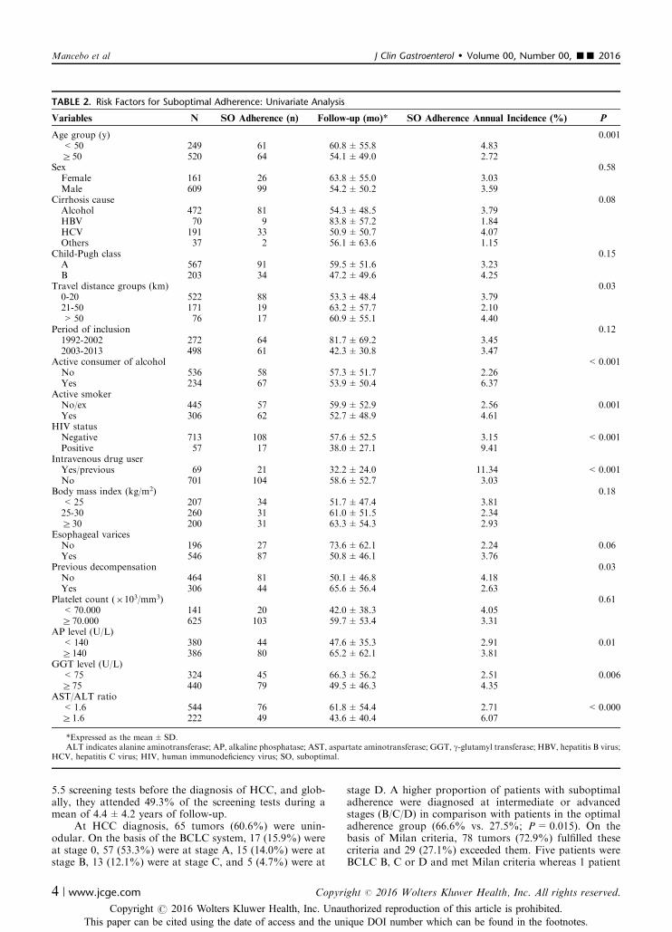
5.5 screening tests before the diagnosis of HCC, and glob-ally, they attended 49.3% of the screening tests during amean of 4.4±4.2 years of follow-up.
At HCC diagnosis, 65 tumors (60.6%) were unin-odular. On the basis of the BCLC system, 17 (15.9%) wereat stage 0, 57 (53.3%) were at stage A, 15 (14.0%) were atstage B, 13 (12.1%) were at stage C, and 5 (4.7%) were at
stage D. A higher proportion of patients with suboptimaladherence were diagnosed at intermediate or advancedstages (B/C/D) in comparison with patients in the optimaladherence group (66.6% vs. 27.5%; P=0.015). On thebasis of Milan criteria, 78 tumors (72.9%) fulfilled thesecriteria and 29 (27.1%) exceeded them. Five patients wereBCLC B, C or D and met Milan criteria whereas 1 patient
TABLE 2. Risk Factors for Suboptimal Adherence: Univariate Analysis
Variables N SO Adherence (n) Follow-up (mo)* SO Adherence Annual Incidence (%) P
Age group (y) 0.001<50 249 61 60.8±55.8 4.83Z50 520 64 54.1±49.0 2.72
Sex 0.58Female 161 26 63.8±55.0 3.03Male 609 99 54.2±50.2 3.59
Cirrhosis cause 0.08Alcohol 472 81 54.3±48.5 3.79HBV 70 9 83.8±57.2 1.84HCV 191 33 50.9±50.7 4.07Others 37 2 56.1±63.6 1.15
Child-Pugh class 0.15A 567 91 59.5±51.6 3.23B 203 34 47.2±49.6 4.25
Travel distance groups (km) 0.030-20 522 88 53.3±48.4 3.7921-50 171 19 63.2±57.7 2.10>50 76 17 60.9±55.1 4.40
Period of inclusion 0.121992-2002 272 64 81.7±69.2 3.452003-2013 498 61 42.3±30.8 3.47
Active consumer of alcohol <0.001No 536 58 57.3±51.7 2.26Yes 234 67 53.9±50.4 6.37
Active smokerNo/ex 445 57 59.9±52.9 2.56 0.001Yes 306 62 52.7±48.9 4.61
HIV statusNegative 713 108 57.6±52.5 3.15 <0.001Positive 57 17 38.0±27.1 9.41
Intravenous drug userYes/previous 69 21 32.2±24.0 11.34 <0.001No 701 104 58.6±52.7 3.03
Body mass index (kg/m2) 0.18<25 207 34 51.7±47.4 3.8125-30 260 31 61.0±51.5 2.34Z30 200 31 63.3±54.3 2.93
Esophageal varicesNo 196 27 73.6±62.1 2.24 0.06Yes 546 87 50.8±46.1 3.76
Previous decompensation 0.03No 464 81 50.1±46.8 4.18Yes 306 44 65.6±56.4 2.63
Platelet count ("103/mm3) 0.61<70.000 141 20 42.0±38.3 4.05Z70.000 625 103 59.7±53.4 3.31
AP level (U/L)<140 380 44 47.6±35.3 2.91 0.01Z140 386 80 65.2±62.1 3.81
GGT level (U/L)<75 324 45 66.3±56.2 2.51 0.006Z75 440 79 49.5±46.3 4.35
AST/ALT ratio<1.6 544 76 61.8±54.4 2.71 <0.000Z1.6 222 49 43.6±40.4 6.07
*Expressed as the mean±SD.ALT indicates alanine aminotransferase; AP, alkaline phosphatase; AST, aspartate aminotransferase; GGT, g-glutamyl transferase; HBV, hepatitis B virus;
HCV, hepatitis C virus; HIV, human immunodeficiency virus; SO, suboptimal.
Mancebo et al J Clin Gastroenterol ! Volume 00, Number 00, ’’ 2016
4 | www.jcge.com Copyright r 2016 Wolters Kluwer Health, Inc. All rights reserved.
Copyright r 2016 Wolters Kluwer Health, Inc. Unauthorized reproduction of this article is prohibited.This paper can be cited using the date of access and the unique DOI number which can be found in the footnotes.
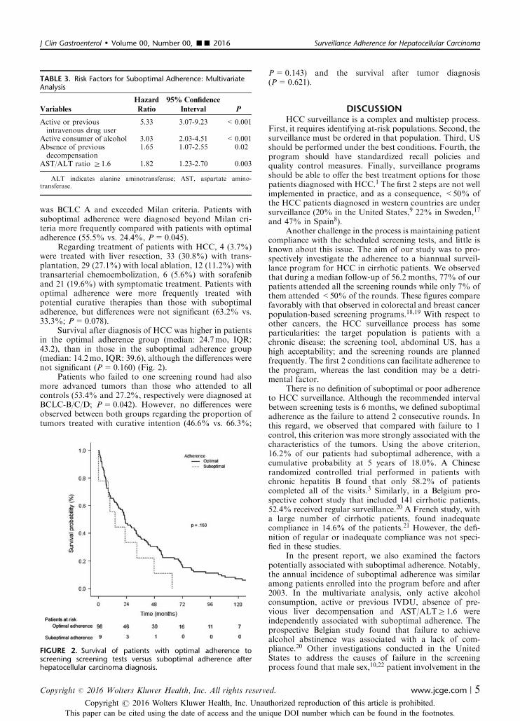
was BCLC A and exceeded Milan criteria. Patients withsuboptimal adherence were diagnosed beyond Milan cri-teria more frequently compared with patients with optimaladherence (55.5% vs. 24.4%, P=0.045).
Regarding treatment of patients with HCC, 4 (3.7%)were treated with liver resection, 33 (30.8%) with trans-plantation, 29 (27.1%) with local ablation, 12 (11.2%) withtransarterial chemoembolization, 6 (5.6%) with sorafeniband 21 (19.6%) with symptomatic treatment. Patients withoptimal adherence were more frequently treated withpotential curative therapies than those with suboptimaladherence, but differences were not significant (63.2% vs.33.3%; P=0.078).
Survival after diagnosis of HCC was higher in patientsin the optimal adherence group (median: 24.7mo, IQR:43.2), than in those in the suboptimal adherence group(median: 14.2mo, IQR: 39.6), although the differences werenot significant (P=0.160) (Fig. 2).
Patients who failed to one screening round had alsomore advanced tumors than those who attended to allcontrols (53.4% and 27.2%, respectively were diagnosed atBCLC-B/C/D; P=0.042). However, no differences wereobserved between both groups regarding the proportion oftumors treated with curative intention (46.6% vs. 66.3%;
P=0.143) and the survival after tumor diagnosis(P=0.621).
DISCUSSIONHCC surveillance is a complex and multistep process.
First, it requires identifying at-risk populations. Second, thesurveillance must be ordered in that population. Third, USshould be performed under the best conditions. Fourth, theprogram should have standardized recall policies andquality control measures. Finally, surveillance programsshould be able to offer the best treatment options for thosepatients diagnosed with HCC.1 The first 2 steps are not wellimplemented in practice, and as a consequence, <50% ofthe HCC patients diagnosed in western countries are undersurveillance (20% in the United States,9 22% in Sweden,17
and 47% in Spain8).Another challenge in the process is maintaining patient
compliance with the scheduled screening tests, and little isknown about this issue. The aim of our study was to pro-spectively investigate the adherence to a biannual surveil-lance program for HCC in cirrhotic patients. We observedthat during a median follow-up of 56.2 months, 77% of ourpatients attended all the screening rounds while only 7% ofthem attended <50% of the rounds. These figures comparefavorably with that observed in colorectal and breast cancerpopulation-based screening programs.18,19 With respect toother cancers, the HCC surveillance process has someparticularities: the target population is patients with achronic disease; the screening tool, abdominal US, has ahigh acceptability; and the screening rounds are plannedfrequently. The first 2 conditions can facilitate adherence tothe program, whereas the last condition may be a detri-mental factor.
There is no definition of suboptimal or poor adherenceto HCC surveillance. Although the recommended intervalbetween screening tests is 6 months, we defined suboptimaladherence as the failure to attend 2 consecutive rounds. Inthis regard, we observed that compared with failure to 1control, this criterion was more strongly associated with thecharacteristics of the tumors. Using the above criterion,16.2% of our patients had suboptimal adherence, with acumulative probability at 5 years of 18.0%. A Chineserandomized controlled trial performed in patients withchronic hepatitis B found that only 58.2% of patientscompleted all of the visits.3 Similarly, in a Belgium pro-spective cohort study that included 141 cirrhotic patients,52.4% received regular surveillance.20 A French study, witha large number of cirrhotic patients, found inadequatecompliance in 14.6% of the patients.21 However, the defi-nition of regular or inadequate compliance was not speci-fied in these studies.
In the present report, we also examined the factorspotentially associated with suboptimal adherence. Notably,the annual incidence of suboptimal adherence was similaramong patients enrolled into the program before and after2003. In the multivariate analysis, only active alcoholconsumption, active or previous IVDU, absence of pre-vious liver decompensation and AST/ALTZ1.6 wereindependently associated with suboptimal adherence. Theprospective Belgian study found that failure to achievealcohol abstinence was associated with a lack of com-pliance.20 Other investigations conducted in the UnitedStates to address the causes of failure in the screeningprocess found that male sex,10,22 patient involvement in the
TABLE 3. Risk Factors for Suboptimal Adherence: MultivariateAnalysis
VariablesHazardRatio
95% ConfidenceInterval P
Active or previousintravenous drug user
5.33 3.07-9.23 <0.001
Active consumer of alcohol 3.03 2.03-4.51 <0.001Absence of previousdecompensation
1.65 1.07-2.55 0.02
AST/ALT ratio Z1.6 1.82 1.23-2.70 0.003
ALT indicates alanine aminotransferase; AST, aspartate amino-transferase.
FIGURE 2. Survival of patients with optimal adherence toscreening screening tests versus suboptimal adherence afterhepatocellular carcinoma diagnosis.
J Clin Gastroenterol ! Volume 00, Number 00, ’’ 2016 Surveillance Adherence for Hepatocellular Carcinoma
Copyright r 2016 Wolters Kluwer Health, Inc. All rights reserved. www.jcge.com | 5Copyright r 2016 Wolters Kluwer Health, Inc. Unauthorized reproduction of this article is prohibited.
This paper can be cited using the date of access and the unique DOI number which can be found in the footnotes.
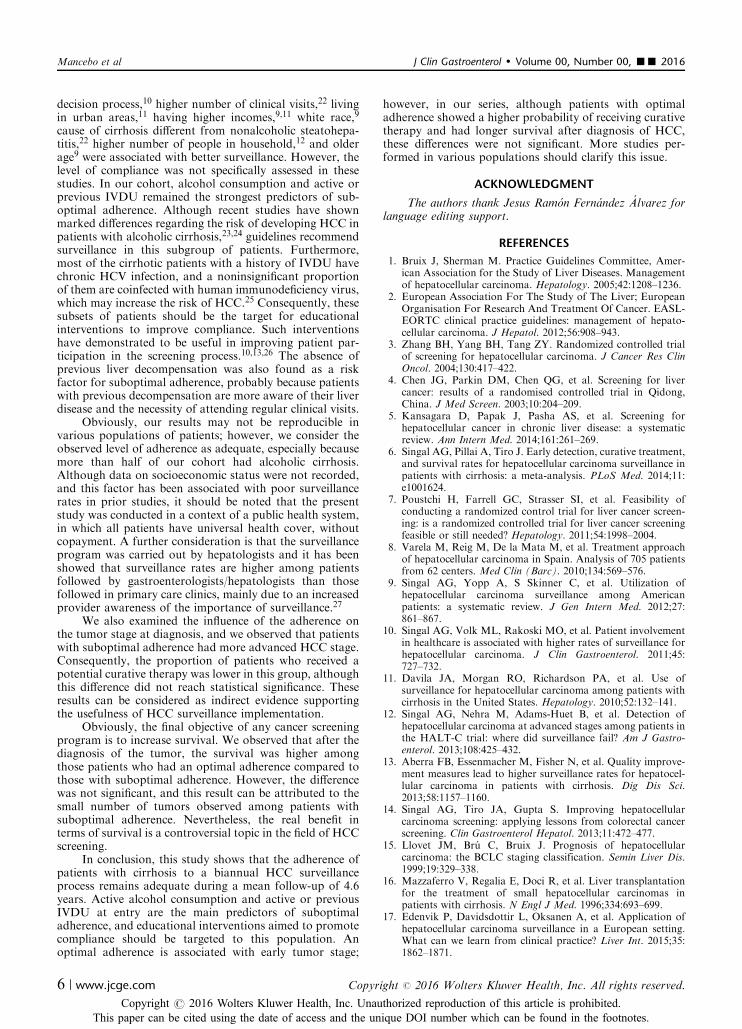
decision process,10 higher number of clinical visits,22 livingin urban areas,11 having higher incomes,9,11 white race,9
cause of cirrhosis different from nonalcoholic steatohepa-titis,22 higher number of people in household,12 and olderage9 were associated with better surveillance. However, thelevel of compliance was not specifically assessed in thesestudies. In our cohort, alcohol consumption and active orprevious IVDU remained the strongest predictors of sub-optimal adherence. Although recent studies have shownmarked differences regarding the risk of developing HCC inpatients with alcoholic cirrhosis,23,24 guidelines recommendsurveillance in this subgroup of patients. Furthermore,most of the cirrhotic patients with a history of IVDU havechronic HCV infection, and a noninsignificant proportionof them are coinfected with human immunodeficiency virus,which may increase the risk of HCC.25 Consequently, thesesubsets of patients should be the target for educationalinterventions to improve compliance. Such interventionshave demonstrated to be useful in improving patient par-ticipation in the screening process.10,13,26 The absence ofprevious liver decompensation was also found as a riskfactor for suboptimal adherence, probably because patientswith previous decompensation are more aware of their liverdisease and the necessity of attending regular clinical visits.
Obviously, our results may not be reproducible invarious populations of patients; however, we consider theobserved level of adherence as adequate, especially becausemore than half of our cohort had alcoholic cirrhosis.Although data on socioeconomic status were not recorded,and this factor has been associated with poor surveillancerates in prior studies, it should be noted that the presentstudy was conducted in a context of a public health system,in which all patients have universal health cover, withoutcopayment. A further consideration is that the surveillanceprogram was carried out by hepatologists and it has beenshowed that surveillance rates are higher among patientsfollowed by gastroenterologists/hepatologists than thosefollowed in primary care clinics, mainly due to an increasedprovider awareness of the importance of surveillance.27
We also examined the influence of the adherence onthe tumor stage at diagnosis, and we observed that patientswith suboptimal adherence had more advanced HCC stage.Consequently, the proportion of patients who received apotential curative therapy was lower in this group, althoughthis difference did not reach statistical significance. Theseresults can be considered as indirect evidence supportingthe usefulness of HCC surveillance implementation.
Obviously, the final objective of any cancer screeningprogram is to increase survival. We observed that after thediagnosis of the tumor, the survival was higher amongthose patients who had an optimal adherence compared tothose with suboptimal adherence. However, the differencewas not significant, and this result can be attributed to thesmall number of tumors observed among patients withsuboptimal adherence. Nevertheless, the real benefit interms of survival is a controversial topic in the field of HCCscreening.
In conclusion, this study shows that the adherence ofpatients with cirrhosis to a biannual HCC surveillanceprocess remains adequate during a mean follow-up of 4.6years. Active alcohol consumption and active or previousIVDU at entry are the main predictors of suboptimaladherence, and educational interventions aimed to promotecompliance should be targeted to this population. Anoptimal adherence is associated with early tumor stage;
however, in our series, although patients with optimaladherence showed a higher probability of receiving curativetherapy and had longer survival after diagnosis of HCC,these differences were not significant. More studies per-formed in various populations should clarify this issue.
ACKNOWLEDGMENT
The authors thank Jesus Ramon Fernandez Alvarez forlanguage editing support.
REFERENCES
1. Bruix J, Sherman M. Practice Guidelines Committee, Amer-ican Association for the Study of Liver Diseases. Managementof hepatocellular carcinoma. Hepatology. 2005;42:1208–1236.
2. European Association For The Study of The Liver; EuropeanOrganisation For Research And Treatment Of Cancer. EASL-EORTC clinical practice guidelines: management of hepato-cellular carcinoma. J Hepatol. 2012;56:908–943.
3. Zhang BH, Yang BH, Tang ZY. Randomized controlled trialof screening for hepatocellular carcinoma. J Cancer Res ClinOncol. 2004;130:417–422.
4. Chen JG, Parkin DM, Chen QG, et al. Screening for livercancer: results of a randomised controlled trial in Qidong,China. J Med Screen. 2003;10:204–209.
5. Kansagara D, Papak J, Pasha AS, et al. Screening forhepatocellular cancer in chronic liver disease: a systematicreview. Ann Intern Med. 2014;161:261–269.
6. Singal AG, Pillai A, Tiro J. Early detection, curative treatment,and survival rates for hepatocellular carcinoma surveillance inpatients with cirrhosis: a meta-analysis. PLoS Med. 2014;11:e1001624.
7. Poustchi H, Farrell GC, Strasser SI, et al. Feasibility ofconducting a randomized control trial for liver cancer screen-ing: is a randomized controlled trial for liver cancer screeningfeasible or still needed? Hepatology. 2011;54:1998–2004.
8. Varela M, Reig M, De la Mata M, et al. Treatment approachof hepatocellular carcinoma in Spain. Analysis of 705 patientsfrom 62 centers. Med Clin (Barc). 2010;134:569–576.
9. Singal AG, Yopp A, S Skinner C, et al. Utilization ofhepatocellular carcinoma surveillance among Americanpatients: a systematic review. J Gen Intern Med. 2012;27:861–867.
10. Singal AG, Volk ML, Rakoski MO, et al. Patient involvementin healthcare is associated with higher rates of surveillance forhepatocellular carcinoma. J Clin Gastroenterol. 2011;45:727–732.
11. Davila JA, Morgan RO, Richardson PA, et al. Use ofsurveillance for hepatocellular carcinoma among patients withcirrhosis in the United States. Hepatology. 2010;52:132–141.
12. Singal AG, Nehra M, Adams-Huet B, et al. Detection ofhepatocellular carcinoma at advanced stages among patients inthe HALT-C trial: where did surveillance fail? Am J Gastro-enterol. 2013;108:425–432.
13. Aberra FB, Essenmacher M, Fisher N, et al. Quality improve-ment measures lead to higher surveillance rates for hepatocel-lular carcinoma in patients with cirrhosis. Dig Dis Sci.2013;58:1157–1160.
14. Singal AG, Tiro JA, Gupta S. Improving hepatocellularcarcinoma screening: applying lessons from colorectal cancerscreening. Clin Gastroenterol Hepatol. 2013;11:472–477.
15. Llovet JM, Bru C, Bruix J. Prognosis of hepatocellularcarcinoma: the BCLC staging classification. Semin Liver Dis.1999;19:329–338.
16. Mazzaferro V, Regalia E, Doci R, et al. Liver transplantationfor the treatment of small hepatocellular carcinomas inpatients with cirrhosis. N Engl J Med. 1996;334:693–699.
17. Edenvik P, Davidsdottir L, Oksanen A, et al. Application ofhepatocellular carcinoma surveillance in a European setting.What can we learn from clinical practice? Liver Int. 2015;35:1862–1871.
Mancebo et al J Clin Gastroenterol ! Volume 00, Number 00, ’’ 2016
6 | www.jcge.com Copyright r 2016 Wolters Kluwer Health, Inc. All rights reserved.
Copyright r 2016 Wolters Kluwer Health, Inc. Unauthorized reproduction of this article is prohibited.This paper can be cited using the date of access and the unique DOI number which can be found in the footnotes.
18. Zavoral M, Suchanek S, Zavada F, et al. Colorectal cancerscreening in Europe. World J Gastroenterol. 2009;15:5907–5915.
19. Clarke TC, Soler-Vila H, Fleming LE, et al. Trends inadherence to recommended cancer screening: the US popula-tion and working cancer survivors. Front Oncol. 2012;2:190.
20. Henrion J, Libon E, De Maeght S, et al. Surveillance forhepatocellular carcinoma: compliance and results according tothe aetiology of cirrhosis in a cohort of 141 patients. ActaGastroenterol Belg. 2000;63:5–9.
21. Trinchet JC, Chaffaut C, Bourcier V, et al. Ultrasonographicsurveillance of hepatocellular carcinoma in cirrhosis: arandomized trial comparing 3- and 6-month periodicities.Hepatology. 2011;54:1987–1997.
22. Singal AG, Li X, Tiro J, et al. Racial, social, and clinicaldeterminants of hepatocellular carcinoma surveillance. Am JMed. 2015;128:90.e1–e7.
23. Jepsen P, Ott P, Andersen PK, et al. Risk for hepatocellularcarcinoma in patients with alcoholic cirrhosis: a Danishnationwide cohort study. Ann Intern Med. 2012;156:841–847.
24. Mancebo A, Gonzalez-Dieguez ML, Cadahıa V, et al. Annualincidence of hepatocellular carcinoma among patients withalcoholic cirrhosis and identification of risk groups. ClinGastroenterol Hepatol. 2013;11:95–101.
25. Ioannou GN, Bryson CL, Weiss NS, et al. The prevalence ofcirrhosis and hepatocellular carcinoma in patients with humanimmunodeficiency virus infection. Hepatology. 2013;57:249–257.
26. Kennedy NA, Rodgers A, Altus R, et al. Optimisation ofhepatocellular carcinoma surveillance in patients with viralhepatitis: a quality improvement study. Intern Med J. 2013;43:772–777.
27. Singal AG, Yopp A, SSkinner C, et al. Utilization ofhepatocellular carcinoma surveillance among American patients:a systematic review. J Gen Intern Med. 2012;27:861–867.
J Clin Gastroenterol ! Volume 00, Number 00, ’’ 2016 Surveillance Adherence for Hepatocellular Carcinoma
Copyright r 2016 Wolters Kluwer Health, Inc. All rights reserved. www.jcge.com | 7Copyright r 2016 Wolters Kluwer Health, Inc. Unauthorized reproduction of this article is prohibited.
This paper can be cited using the date of access and the unique DOI number which can be found in the footnotes.

91
Title: Incidence and Risk Factors Associated with Hepatocellular Carcinoma Surveillance Failure
Authors: Alejo Mancebo,1 María Varela,1 María Luisa González-Diéguez,1 Carmen A. Navascués,1
Valle Cadahía,1 Alicia Mesa-Álvarez,2 Luis Rodrigo1 and Manuel Rodríguez.1
Affiliations: 1 Liver Unit, Division of Gastroenterology and Hepatology, Hospital Universitario Central
de Asturias, Universidad de Oviedo, Spain.
2 Department of Radiology, Hospital Universitario Central de Asturias.
Abstract Background: Surveillance for hepatocellular carcinoma (HCC) intends to detect tumors at
an early stage to improve survival.
Aim: We performed a study to assess the frequency and risk factors associated with program
failure.
Methods: We analyzed data from 188 consecutive patients diagnosed with HCC within a
surveillance program conducted among 1242 cirrhotic patients based on ultrasonography
(US) and alpha-fetoprotein (AFP) testing every 3 or 6 months. Program failure was defined
as the detection of HCC exceeding the Milan criteria. Variables recorded at entry into the
program, during follow-up and at HCC diagnosis were analyzed.
Results: At diagnosis, 50 (26.6%) HCC tumors were beyond the Milan criteria. In univariate
analysis, Child-Pugh B at entry (p=0.03), development of complications of portal
hypertension before tumor diagnosis (p=0.03) and failure to complete the prior screening
round (p=0.02), Child-Pugh B/C (p=0.001) and AFP≥100 ng/ml (p=0.03) at diagnosis,
were associated with failure. In multivariate analysis, only Child-Pugh B/C (HR, 3.18; 95%
CI, 1.66–6.10, p<0.001) and AFP≥100 ng/ml, both at diagnosis (HR, 2.80; 95% CI, 1.37–
5.71, p=0.005), were independently associated with failure. Survival was higher among
patients with tumors within the Milan criteria than those with program failure (33.9 vs. 7.6
months, p<0.001).
Conclusions: Approximately 25% of HCC cases diagnosed among patients included in a
92
surveillance program were beyond the Milan criteria. Child-Pugh B/C and AFP≥100 ng/ml
at diagnosis were associated with program failure. However, Child-Pugh B at entry and
development of liver-related complications during follow-up can be early predictors of
failure.
Introduction
Hepatocellular carcinoma (HCC) is the third leading cause of cancer death worldwide and is
one of the main causes of death in cirrhotic patients.1 Despite some concerns regarding the
effectiveness of surveillance programs,2 Clinical Practice Guidelines (CPG) endorse bi-
annual surveillance for early detection of HCC in patients with Child-Pugh A or B liver
cirrhosis.3,4 However, in the United States and Europe, most tumors in cirrhotic patients are
diagnosed outside surveillance programs.5,6
Surveillance for HCC intends to detect tumors at an early stage, when potentially
curative treatments can be applied. Although several controlled studies and systematic
reviews7,8 have demonstrated the survival benefit associated with tumors diagnosed within a
surveillance program, ultrasonography (US) has a sensitivity of 63% in detecting tumors at
the initial stages.9 Thus, up to one-third of HCCs diagnosed in surveillance programs are
detected at advanced stages.10
The aim of the present study was to prospectively analyze the failure rate, defined as
the proportion of tumors diagnosed beyond the Milan criteria,11 of a surveillance program
and to identify potential factors associated with program failure.
Materials and methods
Patients We conducted a monocentric, prospective cohort study of 1242 patients with cirrhosis who
were consecutively enrolled into a surveillance program for early detection of HCC based
on quarterly or semi-annual US and alpha-fetoprotein screening tests, between September
1992 and December 2014. Patients were included if they had (a) Child-Pugh class A/B
cirrhosis; (b) age between 35 and 75 years; (c) absence of known HCC; (d) availability to
93
attend regular visits; and (e) no comorbid disease associated with a life expectancy of less
than 1 year.
Patients were randomized to receive surveillance at six-month intervals (n=183)
surveillance or three-month intervals (n=183) between 1992 and 2004. After 2004, all
patients were surveyed every 6 months. All US examinations were performed at the
recruitment center by expert operators and those explorations done at community health
centers were not considered. The characteristics of this cohort of cirrhotic patients have
recently been reported in two studies,12,13 but essentially they were predominantly men
(n=965) with alcoholic (n=688) or HCV-induced cirrhosis (n=270) and in Child-Pugh class
A (n=981). Patients were censored at the date of HCC diagnosis, the date of progression to
Child-Pugh class C, the date of death, the date of development of a severe comorbidity, the
date of inclusion on a waiting list for liver transplantation, the date of last follow-up visit, or
the date of transfer to another center, whichever came first.
The present study analyzed 188 patients from this cohort who developed HCC
during follow-up. Twenty-one demographic, clinical and laboratory variables were
prospectively recorded. Twelve variables were collected at inclusion in the program, 3 during
follow-up and 6 at the time of HCC diagnosis (Table 1). At every visit during follow-up,
alpha-fetoprotein (AFP) levels, adherence to consecutive US, liver decompensation and
hepatic function were updated. Complications of portal hypertension during follow-up were
considered when they developed between the inclusion in the program and the screening
round prior to HCC diagnosis. Patients who received diabetes treatment or those whose
fasting basal glucose level was ≥126 md/dL were considered diabetic. The Spanish National
Death Index
(www.msssi.gob.es/estadEstudios/estadisticas/estadisticas/estMinisterio/IND_TipoDifus
ion.htm), which includes the date of all deaths reported in Spain and is continuously updated,
was consulted to ensure the state of the patients at the end of the follow-up period.
For statistical analysis, patients were classified into 2 cohorts according to tumor stage
at diagnosis: program failure was defined as diagnosis of HCC outside the Milan criteria, and
non-failure was defined as diagnosis of HCC within the Milan criteria.
Diagnosis and staging of HCC
94
When the AFP value was >20 ng/mL and greater than twice the previous value, multiphasic
liver computed tomography (CT) was performed to rule out the presence of HCC.
Otherwise, when a new nodule was detected by US, subsequent dynamic imaging tests were
performed. All patients with a focal lesion detected by US or with an increased AFP level
and negative CT were monitored with US and AFP every 3 months for at least 1 year.
From 1992 to 2001, HCC was diagnosed by histological confirmation or when a
hepatic focal lesion with an AFP ≥400 ng/mL was observed. From 2001 to 2005, HCC was
diagnosed according to the European Association for the Study of Liver Disease criteria14,
and from 2005 to the end of the study period, the American Association for the Study of
Liver Diseases criteria were applied.4 Tumor staging was based on at least two abdominal
imaging techniques, which changed throughout the study according to the state of the art
during the past 2 decades. The Barcelona Clinic Liver Cancer (BCLC)15 staging system and
the Milan Criteria11 were used for tumor classification.
Statistical analysis Descriptive statistics are reported as the mean ± standard deviation, median value with
interquartile ranges (IQR) or N (%). Comparative results between groups were obtained via
the chi-square test or Student’s t-test. Differences between subgroups were explored by the
independent-samples t-test procedure after Levene’s test for equality of variances.
Parameters with p-values <.10 in the univariate analysis were considered for multivariate
analysis. Cox regression was used to estimate hazard ratios relating to various potential
predictors of program failure. A p-value less than 0.05 was considered statistically significant.
Survival was calculated by the Kaplan–Meier method, and survival curves were compared by
the log-rank test. With regard to body mass index, patients were distributed following the
World Health Organization’s classification, and those with ascites were excluded. Statistical
analyses were performed using SPSS 21.0 (Chicago, IL, USA).
Results Among the 1242 patients included in the surveillance program, 188 (15.1%) developed HCC
during a median follow-up of 48.0 months (IQR 72.0). The 10-year cumulative incidence of
95
HCC was 25.0%, and the median number of screening rounds performed until HCC
diagnosis was 10.0 (IQR 13.0).
The demographic, clinical and biochemical parameters of these 188 patients are
shown in Table 1. Patients were generally male (85.1%), and the main causes of cirrhosis
were alcohol (50.0%), hepatitis C virus (HCV) (39.8%) and hepatitis B virus (HBV) (6.9%).
At inclusion, 39.7% of the patients were diabetic. During follow-up and before the tumor
diagnosis, 45.2% of the patients had at least one episode of liver decompensation. Finally, at
the time of HCC diagnosis, the median age was 62.6 ± 8.9 years, and most of the patients
had Child-Pugh class A cirrhosis (69.7%).
Characteristics of the HCCs Detection of HCC was based on US in 139 patients (73.9%), on AFP elevation in 36 patients
(19.1%) and on other methods in 13 patients (6.9%). HCC was diagnosed after detection of
a hepatic lesion by imaging together with an AFP elevation of ≥400 ng/mL in 7 patients
(3.8%), by biopsy in 45 patients (23.9%), and by non-invasive dynamic imaging criteria in
136 patients (72.3%).
Sixty-seven HCC cases were diagnosed during the first half of the study (35.6%), and
121 (64.4%) were diagnosed during the second half. Twenty-seven (14.4%) HCC cases were
diagnosed in the three-month group, and 161 (85.6%) were diagnosed in the six-month
group.
In total, 138 (73.4%) of 188 tumors were diagnosed within the Milan criteria, and 50
tumors (26.6%) were beyond these criteria and therefore were considered program failures.
According to the BCLC staging scheme, 28 tumors were diagnosed at stage 0 (14.9%), 106
tumors at stage A (56.3%), 22 at stage B (11.8%), 24 at stage C (12.8%) and 8 at stage D
(4.2%). Although there was a higher proportion of BCLC 0 tumors in the second half of the
study (2004-2016) compared with the first half (1992-2003), the difference was not
significant (16.5% vs. 11.9%, p=0.39). In total, 120 tumors were single-nodule (63.5%), and
the median size of these single-nodule tumors was 2.9 ± 1.4 cm. The diameters of the single-
nodule tumors did not vary between the 3-month and 6-month follow-up groups (2.8 ± 1.3
cm vs. 3.1 ± 1.8 cm, p=0.48) in either the first half or the second half of the program (3.0 ±
1.4 cm vs. 2.9 ± 1.5 cm; p=0.93).
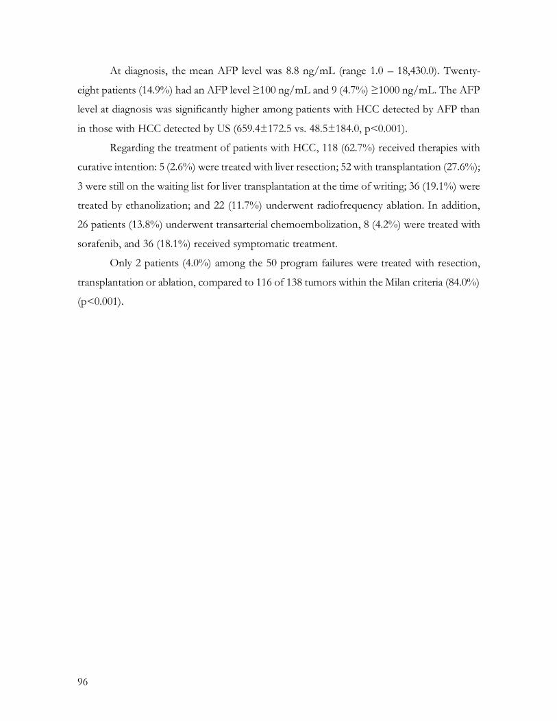
96
At diagnosis, the mean AFP level was 8.8 ng/mL (range 1.0 – 18,430.0). Twenty-
eight patients (14.9%) had an AFP level ≥100 ng/mL and 9 (4.7%) ≥1000 ng/mL. The AFP
level at diagnosis was significantly higher among patients with HCC detected by AFP than
in those with HCC detected by US (659.4±172.5 vs. 48.5±184.0, p<0.001).
Regarding the treatment of patients with HCC, 118 (62.7%) received therapies with
curative intention: 5 (2.6%) were treated with liver resection; 52 with transplantation (27.6%);
3 were still on the waiting list for liver transplantation at the time of writing; 36 (19.1%) were
treated by ethanolization; and 22 (11.7%) underwent radiofrequency ablation. In addition,
26 patients (13.8%) underwent transarterial chemoembolization, 8 (4.2%) were treated with
sorafenib, and 36 (18.1%) received symptomatic treatment.
Only 2 patients (4.0%) among the 50 program failures were treated with resection,
transplantation or ablation, compared to 116 of 138 tumors within the Milan criteria (84.0%)
(p<0.001).
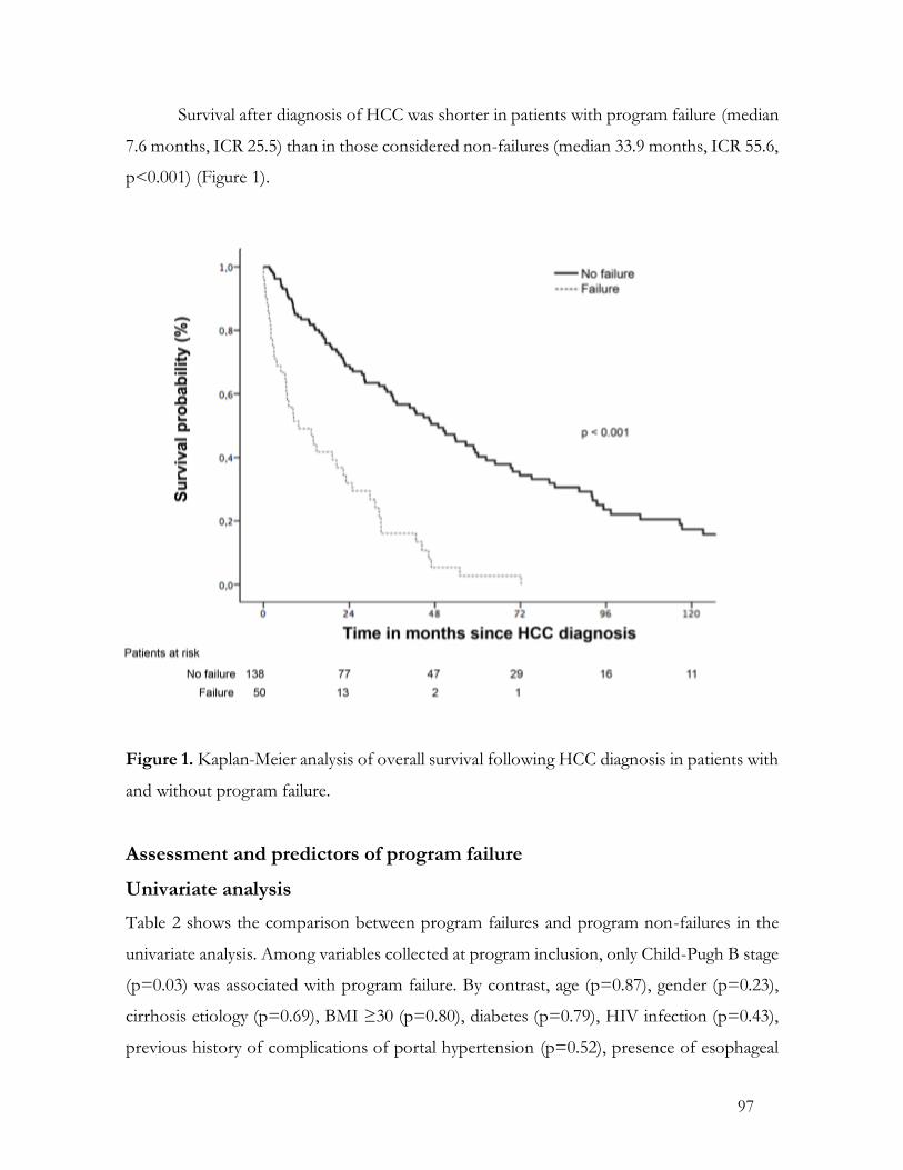
97
Survival after diagnosis of HCC was shorter in patients with program failure (median
7.6 months, ICR 25.5) than in those considered non-failures (median 33.9 months, ICR 55.6,
p<0.001) (Figure 1).
Figure 1. Kaplan-Meier analysis of overall survival following HCC diagnosis in patients with
and without program failure.
Assessment and predictors of program failure
Univariate analysis Table 2 shows the comparison between program failures and program non-failures in the
univariate analysis. Among variables collected at program inclusion, only Child-Pugh B stage
(p=0.03) was associated with program failure. By contrast, age (p=0.87), gender (p=0.23),
cirrhosis etiology (p=0.69), BMI ≥30 (p=0.80), diabetes (p=0.79), HIV infection (p=0.43),
previous history of complications of portal hypertension (p=0.52), presence of esophageal
98
varices (p=0.81), AFP level ≥20 ng/ml (p=0.62), platelet level <100 x103/mm3 (p=0.33) or
inclusion period (p=0.23) were not associated with failure. Among the variables collected
during the surveillance program, only the development of complications of portal
hypertension (p=0.03) previously to HCC diagnosis was associated with program failure.
Quarterly follow-up and duration of follow-up within the program were not associated with
program failure (p=0.89 and p=0.13, respectively). Finally, 3 variables collected at HCC
diagnosis, Child-Pugh class B or C (p=0.001), failure to attend the prior screening round
(p=0.02) and AFP level ≥100 ng/ml (p<0.001), were correlated with program failure. Age
(p=0.76) and detection of HCC by US or by AFP elevation or other methods (p=0.30) were
not associated with failure.
Multivariate analysis To categorize the variables recorded at different times, we performed two multivariate
analyses. The first analysis included only variables registered before HCC diagnosis; all
variables were included in the second analysis. Among the variables collected at inclusion
and during follow-up, only Child-Pugh B stage at inclusion was significantly associated with
failure (hazard ratio [HR] 2.06, 95% CI, 1.16–3.66, p=0.01). When all variables were
considered, only Child-Pugh B or C (HR, 3.18; 95% CI, 1.66–6.10, p<0.001) and AFP level
≥100 ng/ml (HR, 2.80; 95% CI, 1.37–5.71, p=0.005), both at HCC diagnosis, were
correlated with program failure.
Discussion HCC surveillance in cirrhotic patients aims to detect tumors in the initial stages of the disease
to offer treatments that increase patient survival. Although the usefulness of screening
programs remains controversial, several studies have shown survival improvement in
patients under surveillance.8
CPG recommends bi-annual US exams,3,4 although the sensitivity of US in detecting
early-stage tumors is limited. One meta-analysis observed a US sensitivity of 70.1% for
detecting tumors within the Milan criteria when the follow-up was conducted on a 6-month
or shorter basis.9 Another meta-analysis obtained similar data, showing that 70.9% of
99
patients diagnosed within a screening program had tumors diagnosed at an initial stage based
on BCLC or Milan criteria.8 In our cohort, 73.4% of the tumors were diagnosed within the
Milan criteria, and 62.7% of the patients received curative-intended treatment. However,
only 18.6% of the tumors were single-nodule and had a size of ≤ 2 cm, corresponding to
ideal detection of very early tumors. However, we must consider that this study had a
duration of more than 20 years and that we analyzed all patients included in the surveillance
program who were diagnosed with HCC.
Among the variables recorded at inclusion in the surveillance program, only Child-
Pugh B stage predicted program failure. A coarse US pattern, which is more frequent in
patients with advanced liver disease, might hamper early detection of tumors by US. A recent
study has shown that Child Pugh B or C, together with male gender, high BMI, alcohol or
NASH cirrhosis and in-patient status, was associated with inadequate quality of US for
excluding HCC.16 Unfortunately, the US pattern and the quality of the explorations were not
recorded in our series. It could be argued that patients with more advanced cirrhosis could
have worse adherence to the program and consequently a more advanced tumor stage at
diagnosis. However, in the present series the proportion of patients who did not complete
the previous screening round was similar between Child-Pugh class A (5.4%) and B (7.1).
Moreover, in a previous study we observed that the absence of previous liver
decompensation in patients with cirrhosis was associated with suboptimal adherence.13
A retrospective Italian study of 1,170 HCCs diagnosed by bi-annual or annual
surveillance found an association between male gender and program failure, and this
association was attributed to higher BMI among men.10 However, in our cohort, neither
gender nor BMI was related to failure, although the proportion of men was higher in our
series than in the Italian cohort (85.1% vs. 70.9%).
Similar to a previous study,17 we did not find any advantage of 3-month surveillance
in terms of early tumor detection. Curiously, there was not an increase in the proportion of
patients diagnosed within the Milan criteria between the first and second halves of the study.
By contrast, Sangiovanni et al.,18 in a study analyzing tumors diagnosed by annual and bi-
annual follow-up between 1987 and 2001, observed a decrease in mortality in those tumors
diagnosed more recently due to earlier detection. In that study,18 single nodule size decreased
from a mean of 3.7 cm during the first 5 years to 2.2 cm in the last 5 years of the study. In
100
our study, the difference in single nodule sizes between the first and second halves of the
study (3.1 vs. 2.9 cm) was not significant. However, among single tumors ≤2 cm, tumor
diameters were significantly smaller in the second half of the program than in the first half
(1.4±0.2 cm vs. 1.8±0.1 cm, p<0.001).
Poor adherence was related to a higher rate of tumors outside the Milan criteria in a
prospective French study that included HBV and HCV cirrhotic patients.19 In a previous
study of the same cohort,13 we have also reported that patients with suboptimal adherence
to the program have a more advanced HCC stage at diagnosis. Furthermore, in the present
study, we observed that patients who failed to complete the prior screening round were more
frequently diagnosed outside the Milan criteria, although it was not independently associated
with failure. Probably the lack of association was related to the limited number of patients
who did not complete the prior screening round (n= 16). Nevertheless, the findings
emphasize the relevance of patient adherence to the screening process.
We observed that the development of portal hypertension-related complications
during follow-up was significantly associated with screening failure. Similarly, the French
study found that patients who developed decompensation during follow-up had a lower
probability of treatment by transplantation, resection or ablation.19 Again, the most plausible
explanation for this association is that in this subset of patients with more advanced disease,
US is less useful for detecting early tumors.
In our cohort, Child-Pugh class B or C and an AFP level ≥100 ng/ml, both at
diagnosis, were the only variables independently associated with program failure. In the
Italian cohort, Child-Pugh class B or C and AFP >200 ng/ml at tumor diagnosis were also
related to program failure.10 These findings may be considered a consequence rather than a
cause of the presence of a tumor at an advanced stage. Probably, the association of high
levels of AFP with program failure reveals an aggressive biological pattern, or a diffuse
involvement of the liver by the tumor.
HCC surveillance is not recommended for Child-Pugh class C patients, except those
included in a waiting list for liver transplantation.3,4 In our series, 12 patients were in this
class at the time of tumor detection, but all of them had been in class A or B in the previous
screening round, suggesting that the development of the tumor could have contributed to
the liver function impairment.
101
Although AFP testing is not recommended by CPG as a screening method,3,4 tumor
diagnosis was triggered by an AFP elevation in 19.1% of our patients. The proportion of
tumors within the Milan criteria was lower in this group than in the subset of tumors detected
by US (69.4% vs. 75.2%), but these differences were not significant. Although recent studies
have suggested that a serial analysis of AFP levels may increase the sensitivity of surveillance
programs,20-22 better tumor-markers for HCC surveillance and early diagnosis are needed.
Recently, an atypical and aggressive HCC pattern has been described in patients who
developed tumors during or shortly after completion of treatment with direct antiviral agents
for hepatitis C.23 Because our study was closed in June 2016, only 3 patients with HCC have
received such treatment. In the future, it should be clarified if this pattern is treatment-related
or it is a consequence of a more advanced liver disease and a delayed detection.
Surveillance programs may improve if different screening strategies can be
implemented in different patients according to their risk of program failure. Patients with a
higher risk of failure might benefit from closer surveillance and/or other imaging techniques.
Although sensitivity of CT scan in detecting early tumors is higher compared with US,24 the
use of CT is hampered by cost, radiation and a higher rate of false-positive results.25,26
Gadoxetic acid-enhanced MRI has higher sensitivity in detecting very early-stage tumors
than US27, although more studies and cost-effectiveness analyses are needed.
Most patients included in our study were males with a clear predominance of
alcoholic cirrhosis, and thus these results may be not reproducible in different populations.
In addition, some variables, such as BMI and diabetes, were only collected at the time of
inclusion in the program. Finally, the opinion of the sonographer regarding exam quality
factors, such as a coarse US pattern or impaired sonic transmission, was not recorded.
In conclusion, among patients who develop HCC after their inclusion in a
surveillance program, approximately one quarter have a tumor diagnosed beyond the Milan
criteria, which is associated with shorter survival. Child-Pugh class B or C and an AFP level
≥100 ng/ml, both at diagnosis, are associated with program failure. However, variables
collected before HCC diagnosis, such as Child-Pugh class B at inclusion or the development
of portal hypertension-related complications during follow-up, are more useful in selecting
patients with a higher risk of program failure. Current screening strategies are probably not
suitable for these patients, and different modalities should be investigated.
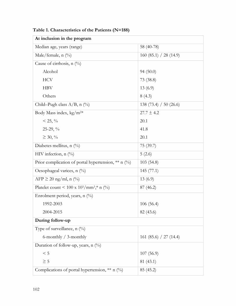
102
Table 1. Characteristics of the Patients (N=188)
At inclusion in the program
Median age, years (range) 58 (40-78)
Male/female, n (%) 160 (85.1) / 28 (14.9)
Cause of cirrhosis, n (%)
Alcohol
HCV
HBV
Others
94 (50.0)
73 (38.8)
13 (6.9)
8 (4.3)
Child–Pugh class A/B, n (%) 138 (73.4) / 50 (26.6)
Body Mass index, kg/m2*
< 25, %
25-29, %
≥ 30, %
27.7 ± 4.2
20.1
41.8
20.1
Diabetes mellitus, n (%) 75 (39.7)
HIV infection, n (%) 5 (2.6)
Prior complication of portal hypertension, ** n (%) 103 (54.8)
Oesophageal varices, n (%) 145 (77.1)
AFP ≥ 20 ng/ml, n (%) 13 (6.9)
Platelet count < 100 x 103/mm3,* n (%) 87 (46.2)
Enrolment period, years, n (%)
1992-2003
2004-2015
106 (56.4)
82 (43.6)
During follow-up
Type of surveillance, n (%)
6-monthly / 3-monthly
161 (85.6) / 27 (14.4)
Duration of follow-up, years, n (%)
< 5
≥ 5
107 (56.9)
81 (43.1)
Complications of portal hypertension, ** n (%) 85 (45.2)
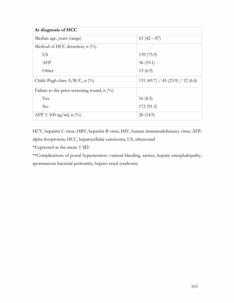
103
At diagnosis of HCC
Median age, years (range) 63 (42 – 87)
Method of HCC detection, n (%)
US
AFP
Other
139 (73.9)
36 (19.1)
13 (6.9)
Child–Pugh class A/B/C, n (%) 131 (69.7) / 45 (23.9) / 12 (6.4)
Failure to the prior screening round, n (%)
Yes
No
16 (8.5)
172 (91.5)
AFP ≥ 100 ng/ml, n (%) 28 (14.9)
HCV, hepatitis C virus; HBV, hepatitis B virus; HIV, human immunodeficiency virus; AFP,
alpha-fetoprotein; HCC, hepatocellular carcinoma; US, ultrasound
*Expressed as the mean ± SD.
**Complications of portal hypertension: variceal bleeding, ascites, hepatic encephalopathy,
spontaneous bacterial peritonitis, hepato-renal syndrome.
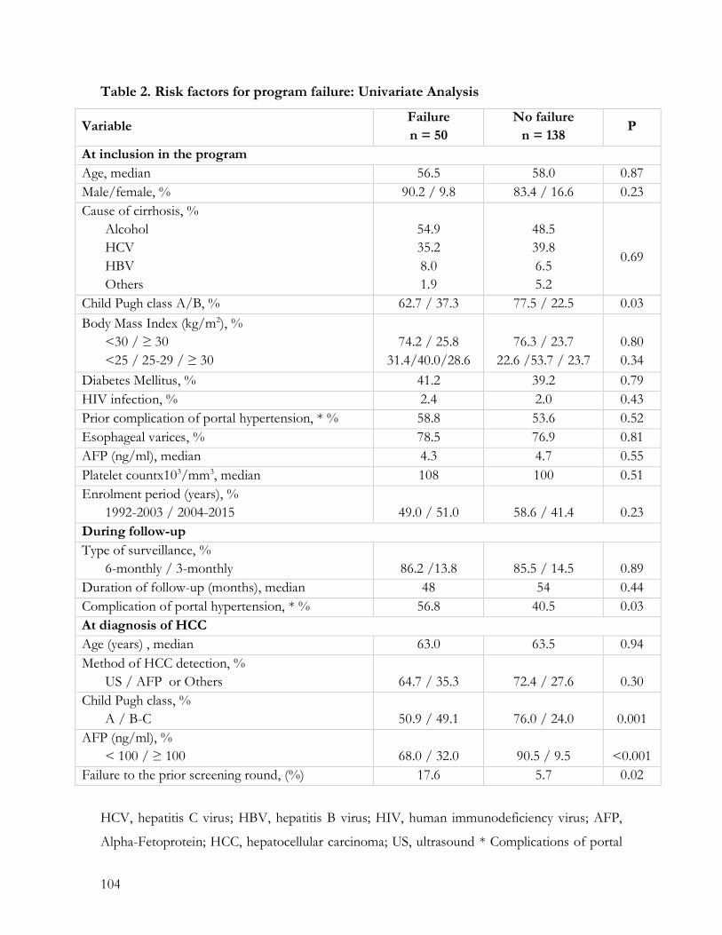
104
Table 2. Risk factors for program failure: Univariate Analysis
Variable Failure n = 50
No failure n = 138
P
At inclusion in the program Age, median 56.5 58.0 0.87 Male/female, % 90.2 / 9.8 83.4 / 16.6 0.23 Cause of cirrhosis, %
Alcohol HCV HBV Others
54.9 35.2 8.0 1.9
48.5 39.8 6.5 5.2
0.69
Child Pugh class A/B, % 62.7 / 37.3 77.5 / 22.5 0.03 Body Mass Index (kg/m2), %
<30 / ≥ 30 <25 / 25-29 / ≥ 30
74.2 / 25.8
31.4/40.0/28.6
76.3 / 23.7
22.6 /53.7 / 23.7
0.80 0.34
Diabetes Mellitus, % 41.2 39.2 0.79 HIV infection, % 2.4 2.0 0.43 Prior complication of portal hypertension, * % 58.8 53.6 0.52 Esophageal varices, % 78.5 76.9 0.81 AFP (ng/ml), median 4.3 4.7 0.55 Platelet countx103/mm3, median 108 100 0.51 Enrolment period (years), %
1992-2003 / 2004-2015
49.0 / 51.0
58.6 / 41.4
0.23 During follow-up Type of surveillance, %
6-monthly / 3-monthly
86.2 /13.8
85.5 / 14.5
0.89 Duration of follow-up (months), median 48 54 0.44 Complication of portal hypertension, * % 56.8 40.5 0.03 At diagnosis of HCC Age (years) , median 63.0 63.5 0.94 Method of HCC detection, %
US / AFP or Others
64.7 / 35.3
72.4 / 27.6
0.30 Child Pugh class, %
A / B-C
50.9 / 49.1
76.0 / 24.0
0.001 AFP (ng/ml), %
< 100 / ≥ 100
68.0 / 32.0
90.5 / 9.5
<0.001 Failure to the prior screening round, (%) 17.6 5.7 0.02
HCV, hepatitis C virus; HBV, hepatitis B virus; HIV, human immunodeficiency virus; AFP,
Alpha-Fetoprotein; HCC, hepatocellular carcinoma; US, ultrasound * Complications of portal
105
hypertension: variceal bleeding, ascites, hepatic encephalopathy, spontaneous bacterial
peritonitis, hepato-renal syndrome.
References 1. El-Serag HB. Hepatocellular carcinoma. N Engl J Med 2011;365:1118–27.
2. Kansagara D, Papak J, Pasha AS, et al. Screening for hepatocellular cancer in chronic liver
disease: A Systematic Review. Ann Intern Med. 2014;161:261-9.
3. European Association For The Study Of The Liver, European Organisation For Research
And Treatment Of Cancer. EASL-EORTC. Clinical practice guidelines: management of
hepatocellular carcinoma. J Hepatol 2012;56:908–43.
4. Bruix J, Sherman M. Practice Guidelines Committee, American Association for the Study
of Liver Diseases. Management of hepatocellular carcinoma. Hepatology 2005;42:1208 –36.
5. Varela M, Reig M, de la Mata M, et al. Treatment approach of hepatocellular carcinoma in
Spain. Analysis of 705 patients from 62 centers. Med Clin (Barc). 2010;134:569-76.
6. Singal AG, Yopp A, S Skinner C, et al. Utilization of hepatocellular carcinoma surveillance
among American patients: a systematic review. J Gen Intern Med. 2012;27: 861-7.
7. Mourad A, Deuffic-Burban S, Ganne-Carrié N, et al. Hepatocellular carcinoma screening
in patients with compensated hepatitis C virus (HCV)-related cirrhosis aware of their HCV
status improves survival: a modeling approach. Hepatology. 2014;59:1471-81.
8. Singal AG, Pillai A, Tiro J. Early detection, curative treatment, and survival rates for
hepatocellular carcinoma surveillance in patients with cirrhosis: a meta-analysis. PLoS Med.
2014;1;11:e1001624.
9. Singal A, Volk ML, Waljee A, et al. Meta-analysis: surveillance with ultrasound for early-
stage hepatocellular carcinoma in patients with cirrhosis. Aliment Pharmacol Ther.
2009;30:37-47.
10. Del Poggio P, Olmi S, Ciccarese F, et al. Factors that affect efficacy of ultrasound
surveillance for early stage hepatocellular carcinoma in patients with cirrhosis. Clin
Gastroenterol Hepatol. 2014;12:1927-33.
11. Mazzaferro V, Regalia E, Doci R, et al. Liver transplantation for the treatment of small
hepatocellular carcinomas in patients with cirrhosis. N Engl J Med. 1996;334:693-9.
106
12. Mancebo A, González-Diéguez ML, Cadahía V, et al. Annual incidence of hepatocellular
carcinoma among patients with alcoholic cirrhosis and identification of risk groups. Clin
Gastroenterol Hepatol. 2013;11:95-101.
13. Mancebo A, González-Diéguez ML, Navascués CA, et al. Adherence to a semiannual
surveillance program for hepatocellular carcinoma in patients with liver cirrhosis. J Clin
Gastroenterol. 2016;[Epub ahead of print]
14. Bruix J, Sherman M, Llovet JM, et al. Clinical management of hepatocellular carcinoma.
Conclusions of the Barcelona-2000 EASL conference. European Association for the Study
of the Liver. J Hepatol 2001;35:421– 30.
15. Llovet JM, Bru C, Bruix J. Prognosis of hepatocellular carcinoma: the BCLC staging
classification. Semin Liver Dis 1999;19:329– 38.
16. Simmons O, Fetzer DT, Yokoo T, et al. Predictors of adequate ultrasound quality for
hepatocellular carcinoma surveillance in patients with cirrhosis. Aliment Pharmacol Ther.
2017;45:169-77.
17. Trinchet JC, Chaffaut C, Bourcier V, et al. Ultrasonographic surveillance of hepatocellular
carcinoma in cirrhosis: a randomized trial comparing 3 and 6 month periodicity. Hepatology
2011;54:1987–97.
18. Sangiovanni A, Del Ninno E, Fasani P, et al. Increased survival of cirrhotic patients with
a hepatocellular carcinoma detected during surveillance. Gastroenterology 2004;126:1005-
14.
19. C. Costentin, R. Layese, V. Bourcier, et al. Prospective evidence that hepatocellular
carcinoma surveillance in patients with compensated viral cirrhosis increases the probability
of curative treatment and survival taking into account lead-time bias (ANRS CO12 CIRVIR
cohort). J Hepatol 2016;64: S183–S212.
20. Bird TG, Dimitropoulou P, Turner RM, et al. Alpha-Fetoprotein detection of
hepatocellular carcinoma leads to a standardized analysis of dynamic AFP to improve
screening based detection. PLoS One. 2016;16;11:e0156801
21. Lee E, Edward S, Singal AG, et al. Improving screening for hepatocellular carcinoma by
incorporating data on levels of α-fetoprotein, over time. Clin Gastroenterol Hepatol.
2013;11:437-40.
107
22. Chang TS, Wu YC, Tung SY, et al. Alpha-fetoprotein measurement benefits
hepatocellular carcinoma surveillance in patients with cirrhosis. Am J Gastroenterol.
2015;110:836-44
23. A Romano, F Capra, S Piovesan, et al. Incidence and pattern of "de novo" hepatocellular
carcinoma in HCV patients treated with oral DAAs. Hepatology 2016;64:S10A.
24. Colli A, Fraquelli M, Casazza G, et al. Accuracy of ultrasonography, spiral CT, magnetic
resonance, and alpha-fetoprotein in diagnosing hepatocellular carcinoma: a systematic
review. Am J Gastroenterol. 2006;101:513-23.
25. Pocha C, Dieperink E, McMaken KA, et al. Surveillance for hepatocellular cancer with
ultrasonography vs. computed tomography -- a randomised study. Aliment Pharmacol Ther.
2013;38:303-12.
26. Lencioni R, Crocetti L, Della Pina MC, et al. Guidelines for imaging focal lesions in liver
cirrhosis. Exp Rev Gastroenterol Hepatol 2008;2:697–703.
27. Young-Suk L, So Yeon K, Jae Ho B, et al. Surveillance for hepatocellular carcinoma with
magnetic resonance imaging using a liver-specific contrast agent gadoxetic acid and
ultrasonography in high-risk patients with cirrhosis. Hepatology 2014;60:843

108
109
Informe del factor de impacto
• Primer artículo (Mancebo A et al. Annual incidence of hepatocellular carcinoma
among patients with alcoholic cirrhosis and identification of risk groups)
o Publicado en: Clinical Gastroenterology and Hepatology, año 2013
o Factor de impacto 4.14
o Citado 41 veces
• Segundo artículo (Mancebo A et al. Adherence to a semiannual surveillance
program for hepatocellular carcinoma in patients with liver cirrhosis.)
o Publicado en: Journal of Clinical Gastroenterology, año 2016
o Factor de impacto: 3.16

110
Bibliografía
111
Bibliografía
1 Ferlay J, Soerjomataram I, Dikshit R, Eser S, Mathers C, Rebelo M, et al. Cancer incidence and
mortality worldwide: sources, methods and major patterns in GLOBOCAN 2012. Int J Cancer.
2015;136:E359-86. 2 Singal AG, El-Serag HB. Hepatocellular carcinoma from epidemiology to prevention:
translating knowledge into practice. Clin Gastroenterol Hepatol. 2015;13:2140-51. 3 Delhaye M, Louis H, Degraef C, Le Moine O, Devière J, Gulbis B, et al. Relationship between
hepatocyte proliferative activity and liver functional reserve in human cirrhosis. Hepatology
1996;23:1003-11. 4 Caillot F, Derambure C, Bioulac-Sage P, Francois A, Scotte M, Goria O, et al. Transient and
etiology-related transcription regulation in cirrhosis prior to hepatocellular carcinoma
occurrence. World J Gastroenterol 2009;15:300-9. 5 Wiemann SU, Satyanarayana A, Tsahuridu M, Tillmann HL, Zender L, Klempnauer J, et al.
Hepatocyte telomere shortening and senescence are general markers of human liver cirrhosis.
FASEB J 2002;16:935-42. 6 Farazi PA, Glickman J, Horner J, Dephinho RA. Cooperative interactions of p53 mutation,
telomere dysfunction, and chronic liver damage in hepatocellular carcinoma progression. Cancer
Res 2006;66:4766-73. 7 Bataller R, Brenner DA. Liver fibrosis. J Clin Invest 2005;115:209-18. 8 Carson DA, Lois A. Cancer progression and p53. Lancet 1995;346:1009-11. 9 Rodríguez de Lopez C, Reig M, Matilla A, Ferrer MT, Dueñas E, Mínguez B, et al. La principal
causa de diagnóstico de carcinoma hepatocelular (CHC) fuera de los programas de cribado es la
cirrosis hepática alcohólica no conocida. Análisis de 686 casos en 74 centros. Gastroenterol
Hepatol. 2016;39:S25. 10 Leykum LK, El-Serag HB, Cornell J, Papadopoulos KP. Screening for hepatocellular
carcinoma among veterans with hepatitis C on disease stage, treatment received, and survival.
Clin Gastroenterol Hepatol 2007;5:508-12. 11 Blachier M, Leleu H, Peck-Radosavljevic M, Valla DC, Roudot-Thoraval F. The burden of
liver disease in Europe: A review of available epidemiological data. J Hepatol. 2013;58:593-608. 12 Chiang CJ, Yang YW, You SL, et al. Thirty-year outcomes of the national hepatitis B
immunization program in Taiwan. JAMA. 2013;310:974-6.
Bibliografía
112
13 Calleja JL, Crespo J. Asociación Española para el Estudio del Hígado. Libro Blanco de la
Hepatología en España, 1a ed. Madrid; 2015. 14 Sociedad Española de Oncología médica. Las cifras del cáncer en España 2014. Depósito
Legal: M-2399-2016. 15 Organización Nacional de Trasplantes. Memoria del Registro Español de Trasplante Hepático,
2015. (http://www.sethepatico.org). 16 Fattovich G, Stroffolini T, Zagni I, Donato F. Hepatocellular carcinoma in cirrhosis: incidence
and risk factors. Gastroenterology. 2004;127(5 Suppl 1):35-50. 17 Freeman AJ, Dore GJ, Law MG, Thorpe M, Von Overbeck J, Lloyd AR. Estimating
progression to cirrhosis in chronic hepatitis C virus infection. Hepatology. 2001;34:809-16. 18 Di Bisceglie AM. Hepatitis C and hepatocellular carcinoma. Hepatology. 1997;26(Suppl 1):34-
8. 19 Morgan RL, Baack B, Smith BD, Yartel A, Pitasi M, Falck-Ytter Y. Eradication of hepatitis C
virus infection and the development of hepatocellular carcinoma: a meta-analysis of
observational studies. Ann Intern Med. 2013;158:329-37. 20 Li DK, Chung RT. Impact of hepatitis C virus eradication on hepatocellular carcinogenesis.
Cancer. 2015;121:2874-82. 21 Antonietta R, Franco C, Sara P. Incidence and pattern of “de novo” hepatocellular carcinoma
in HCV patients treated with oral DAAs. Hepatology. 2016;64:S10A. 22 Cadranel JF, Lahmek P, Causse X, Bellaiche G, Bettan L, Fontanges T, et al. Epidemiology of
chronic hepatitis B infection in France: risk factors for significant fibrosis--results of a
nationwide survey. Aliment Pharmacol Ther. 2007;26:565-76. 23 Corrao G, Bagnardi V, Zambon A, Torchio P. Meta-analysis of alcohol intake in relation to
risk of liver cirrhosis. Alcohol Alcohol. 1998;33:381–92. 24 World Health Organization (2014). Global status report on alcohol and health. WHO Library
Cataloguing-in-Publication Data, Geneva. ISBN 978 92 4 156475 5. 25 Rehm J, Taylor B, Mohapatra S, Irving H, Baliunas D, Patra J, et al. Alcohol as a risk factor
for liver cirrhosis: a systematic review and meta-analysis. Drug Alcohol Rev. 2010;29:437-45. 26 Kamper-Jørgensen M, Grønbæk M, Tolstrup J, Becker U. Alcohol and cirrhosis: dose-
response or threshold effect? J Hepatol. 2004;41:25-30. 27 Diehl AM. Obesity and alcoholic liver disease. Alcohol. 2004;34:81-7.
Bibliografía
113
28 Friedrich K1, Smit M1, Wannhoff A. Coffee consumption protects against progression in liver
cirrhosis and increases long-term survival after liver transplantation. J Gastroenterol Hepatol.
2016;3:1470-5. 29 Teli MR, James OF, Burt AD, Bennett MK, Day CP. The natural history of nonalcoholic fatty
liver: a follow-up study. Hepatology. 1995;22:1714-9. 30 Donato F, Tagger A, Gelatti U, Parrinello G, Boffetta P, Albertini A, et al. Alcohol and
hepatocellular carcinoma: the effect of lifetime intake and hepatitis virus infections in men and
women. Am J Epidemiol. 2002;155:323-31. 31 Neuschwander-tetri BA, Caldwell SH. Nonalcoholic steatohepatitis: summary of an AASLD
single topic conference. Hepatology. 2003;37:1202-19. 32 Wolk A, Gridley G, Svensson M, Nyrén O, McLaughlin JK, Fraumeni JF, et al. A prospective
study of obesity and cancer risk (Sweden). Cancer Causes Control. 2001;12:13-21. 33 El-Serag HB, Tran T, Everhart JE. Diabetes increases the risk of chronic liver disease and
hepatocellular carcinoma. Gastroenterology. 2004;126:460-8. 34 Silveira MG, Suzuki A, Lindor Kd. Surveillance for hepatocellular carcinoma in patients with
primary biliary cirrhosis. Hepatology. 2008;48:1149-56. 35 Teufel A, Weinmann A, Centner C, Piendl A, Lohse AW, Galle PR, et al. Hepatocellular
carcinoma in patients with autoimmune hepatitis. World J Gastroenterol. 2009;15:578-82. 36 El-Serag HB, Marrero JA, Rudolph L, Reddy KR. Diagnosis and treatment of hepatocellular
carcinoma. Gastroenterology. 2008;134:1752-63. 37 Takayasu K, Arii S, Kudo M, Ichida T, Matsui O, Izumi N, et al. Superselective transarterial
chemoembolization for hepatocellular carcinoma. Validation of treatment algorithm proposed
by Japanese guidelines. J Hepatol. 2012;56:886-92. 38 Burrel M, Reig M, Forner A, Barrufet M, de Lope CR, Tremosini S, et al. Survival of patients
with hepatocellular carcinoma treated by transarterial chemoembolisation (TACE) using Drug
Eluting Beads. Implications for clinical practice and trial design. J Hepatol. 2012;56:1330-5. 39 Llovet JM, Ricci S, Mazzaferro V, Hilgard P, Gane E, Blanc JF, et al. Sorafenib in advanced
hepatocellular carcinoma. N Engl J Med. 2008;359:378-90. 40 Llovet JM, Bruix J, Capurro S, VIlana R. Long-term survival after ethanol injection for small
hepatocellular carcinoma in 100 cirrhotic patients. Relevance of maintained success. J Hepatol.
1999;30:100A. 41 Naimark D, Naglie G, Detsky AS. The meaning of life expectancy: what is a clinically
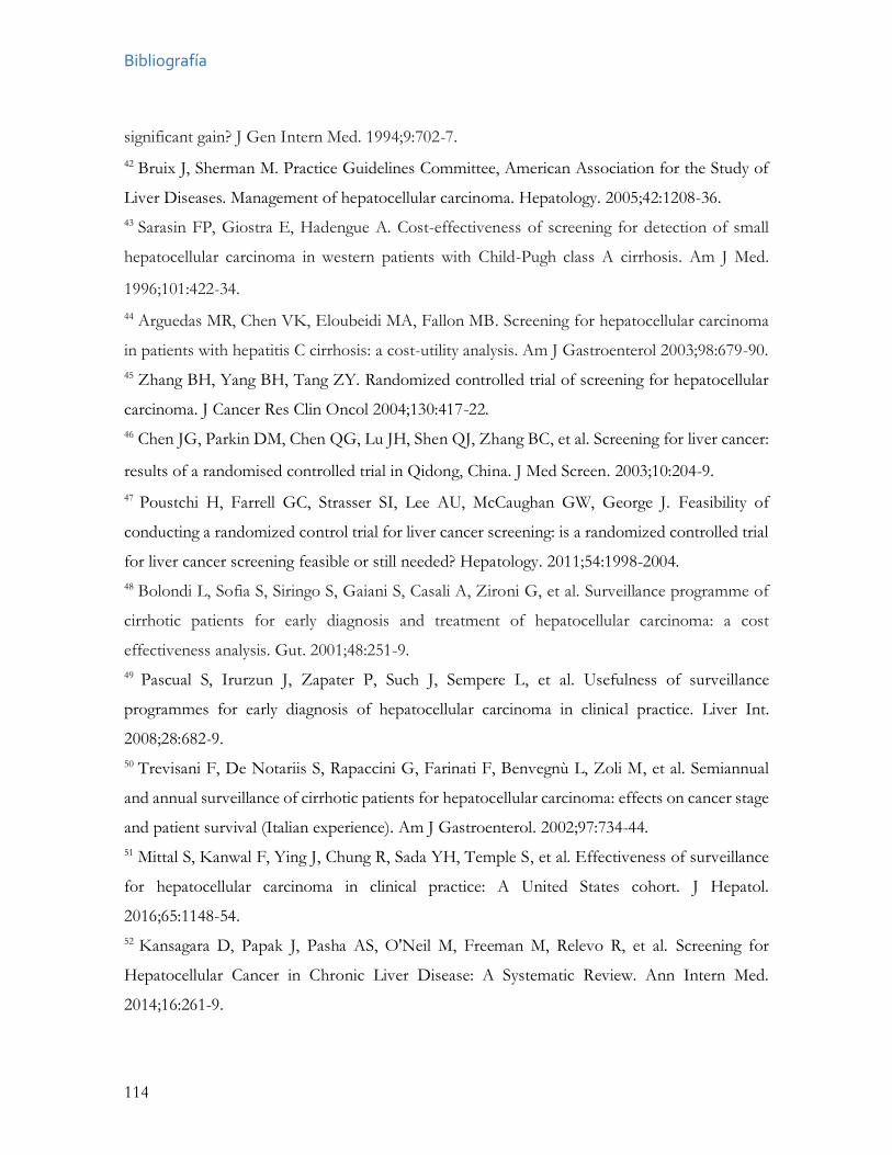
Bibliografía
114
significant gain? J Gen Intern Med. 1994;9:702-7. 42 Bruix J, Sherman M. Practice Guidelines Committee, American Association for the Study of
Liver Diseases. Management of hepatocellular carcinoma. Hepatology. 2005;42:1208-36. 43 Sarasin FP, Giostra E, Hadengue A. Cost-effectiveness of screening for detection of small
hepatocellular carcinoma in western patients with Child-Pugh class A cirrhosis. Am J Med.
1996;101:422-34. 44 Arguedas MR, Chen VK, Eloubeidi MA, Fallon MB. Screening for hepatocellular carcinoma
in patients with hepatitis C cirrhosis: a cost-utility analysis. Am J Gastroenterol 2003;98:679-90. 45 Zhang BH, Yang BH, Tang ZY. Randomized controlled trial of screening for hepatocellular
carcinoma. J Cancer Res Clin Oncol 2004;130:417-22. 46 Chen JG, Parkin DM, Chen QG, Lu JH, Shen QJ, Zhang BC, et al. Screening for liver cancer:
results of a randomised controlled trial in Qidong, China. J Med Screen. 2003;10:204-9. 47 Poustchi H, Farrell GC, Strasser SI, Lee AU, McCaughan GW, George J. Feasibility of
conducting a randomized control trial for liver cancer screening: is a randomized controlled trial
for liver cancer screening feasible or still needed? Hepatology. 2011;54:1998-2004. 48 Bolondi L, Sofia S, Siringo S, Gaiani S, Casali A, Zironi G, et al. Surveillance programme of
cirrhotic patients for early diagnosis and treatment of hepatocellular carcinoma: a cost
effectiveness analysis. Gut. 2001;48:251-9. 49 Pascual S, Irurzun J, Zapater P, Such J, Sempere L, et al. Usefulness of surveillance
programmes for early diagnosis of hepatocellular carcinoma in clinical practice. Liver Int.
2008;28:682-9. 50 Trevisani F, De Notariis S, Rapaccini G, Farinati F, Benvegnù L, Zoli M, et al. Semiannual
and annual surveillance of cirrhotic patients for hepatocellular carcinoma: effects on cancer stage
and patient survival (Italian experience). Am J Gastroenterol. 2002;97:734-44. 51 Mittal S, Kanwal F, Ying J, Chung R, Sada YH, Temple S, et al. Effectiveness of surveillance
for hepatocellular carcinoma in clinical practice: A United States cohort. J Hepatol.
2016;65:1148-54. 52 Kansagara D, Papak J, Pasha AS, O'Neil M, Freeman M, Relevo R, et al. Screening for
Hepatocellular Cancer in Chronic Liver Disease: A Systematic Review. Ann Intern Med.
2014;16:261-9.
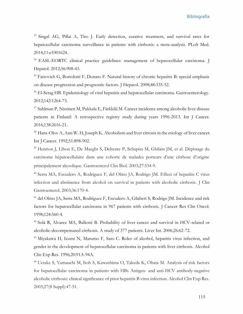
Bibliografía
115
53 Singal AG, Pillai A, Tiro J. Early detection, curative treatment, and survival rates for
hepatocellular carcinoma surveillance in patients with cirrhosis: a meta-analysis. PLoS Med.
2014;11:e1001624. 54 EASL-EORTC clinical practice guidelines: management of hepatocellular carcinoma. J
Hepatol. 2012;56:908-43. 55 Fattovich G, Bortolotti F, Donato F. Natural history of chronic hepatitis B: special emphasis
on disease progression and prognostic factors. J Hepatol. 2008;48:335-52. 56 El-Serag HB. Epidemiology of viral hepatitis and hepatocellular carcinoma. Gastroenterology.
2012;142:1264-73. 57 Sahlman P, Nissinen M, Pukkala E, Färkkilä M. Cancer incidence among alcoholic liver disease
patients in Finland: A retrospective registry study during years 1996-2013. Int J Cancer.
2016;138:2616-21. 58 Hans-Olov A, Ann W. H, Joseph K. Alcoholism and liver cirrosis in the etiology of liver cancer.
Int J Cancer. 1992;51:898-902. 59 Henrion J, Libon E, De Maeght S, Deltenre P, Schapira M, Ghilain JM, et al. Dèpistage du
carcinome hèpatocellulaire dans une cohorte de malades porteurs d’une cirrhose d’origine
principalement alcoolique. Gastroenterol Clin Biol. 2003;27:534-9. 60 Serra MA, Escudero A, Rodríguez F, del Olmo JA, Rodrigo JM. Effect of hepatitis C virus
infection and abstinence from alcohol on survival in patients with alcoholic cirrhosis. J Clin
Gastroenterol. 2003;36:170-4. 61 del Olmo JA, Serra MA, Rodríguez F, Escudero A, Gilabert S, Rodrigo JM. Incidence and risk
factors for hepatocellular carcinoma in 967 patients with cirrhosis. J Cancer Res Clin Oncol.
1998;124:560-4. 62 Solà R, Alvarez MA, Ballesté B. Probability of liver cancer and survival in HCV-related or
alcoholic-decompensated cirrhosis. A study of 377 patients. Liver Int. 2006;26:62-72. 63 Miyakawa H, Izumi N, Marumo F, Sato C. Roles of alcohol, hepatitis virus infection, and
gender in the development of hepatocellular carcinoma in patients with liver cirrhosis. Alcohol
Clin Exp Res. 1996;20:91A-94A. 64 Uetake S, Yamauchi M, Itoh S, Kawashima O, Takeda K, Ohata M. Analysis of risk factors
for hepatocellular carcinoma in patients with HBs Antigen- and anti-HCV antibody-negative
alcoholic cirrhosis: clinical significance of prior hepatitis B virus infection. Alcohol Clin Exp Res.
2003;27(8 Suppl):47-51.
Bibliografía
116
65 N'Kontchou G, Paries J, Htar MT, Ganne-Carrie N, Costentin L, Grando-Lemaire V, et al.
Risk factors for hepatocellular carcinoma in patients with alcoholic or viral C cirrhosis. Clin
Gastroenterol Hepatol. 2006;4:1062-8. 66 Ioannou GN, Splan MF, Weiss NS, McDonald GB, Beretta L, Lee SP. Incidence and
predictors of hepatocellular carcinoma in patients with cirrhosis. Clin Gastroenterol Hepatol.
2007;5:938-45. 67 Jepsen P, Ott P, Andersen PK, Sørensen HT, Vilstrup H. Risk for hepatocellular carcinoma
in patients with alcoholic cirrhosis: a Danish nationwide cohort study. Ann Intern Med. 2012
19;156:841-7. 68 Fialla AD, de Muckadel OB, Touborg Lassen A. Incidence, etiology and mortality of cirrosis:
A population-based cohort study. Scand J Gastroenterol. 2012;47:702-9. 69 Singal AG, Tiro JA, Gupta S. Improving hepatocellular carcinoma screening: applying lessons
from colorectal cancer screening. Clin Gastroenterol Hepatol. 2013;11:472-7. 70 Singal AG, Yopp A, S Skinner C, Packer M, Lee WM, Tiro JA.. Utilization of hepatocellular
carcinoma surveillance among American patients: a systematic review. J Gen Intern Med.
2012;27:861-7. 71 Edenvik P, Davidsdottir L, Oksanen A, Isaksson B, Hultcrantz R, Stål P. Application of
hepatocellular carcinoma surveillance in a European setting. What can we learn from clinical
practice? Liver Int. 2015;35:1862-71. 72 D’Amico G, Morabito A, Pagliaro L, Marubi E. Survival and prognostic indicators in
compensated and decompensated cirrhosis. Dig Dis Sci. 1986;31:468-75. 73 D'Amico G, Garcia-Tsao G, Pagliaro L. Natural history and prognostic indicators of survival
in cirrhosis: a systematic review of 118 studies. J Hepatol. 2006;44:217-31. 74 Henrion J, Libon E, De Maeght S, Schapira M, Ghilain JM, Maisin JM, et al. Surveillance for
hepatocellular carcinoma: compliance and results according to the aetiology of cirrhosis in a
cohort of 141 patients. Acta Gastroenterol Belg. 2000;63:5-9. 75 Trinchet JC, Chaffaut C, Bourcier V, Degos F, Henrion J, Fontaine H, et al. Ultrasonographic
surveillance of hepatocellular carcinoma in cirrhosis: a randomized trial comparing 3- and 6-
month periodicities. Hepatology. 2011;54:1987-97. 76 Singal AG, Nehra M, Adams-Huet B, Yopp AC, Tiro JA, Marrero JA, et al. Detection of
hepatocellular carcinoma at advanced stages among patients in the HALT-C trial: where did
surveillance fail? Am J Gastroenterol. 2013;108:425-32.
Bibliografía
117
77 Singal AG, Yopp AC, Gupta S, Skinner CS, Halm EA, Okolo E, et al. Failure rates in the
hepatocellular carcinoma surveillance process. Cancer Prev Res (Phila). 2012;5:1124-30. 78 Mazzaferro V, Regalia E, Doci R, Andreola S, Pulvirenti A, Bozzetti F, et al. Liver
transplantation for the treatment of small hepatocellular carcinomas in patients with cirrhosis.
N Engl J Med. 1996;334:693-9. 79 Marrero JA, Fontana RJ, Barrat A, Askari F, Conjeevaram HS, Su GL, et al. Prognosis of
hepatocellular carcinoma: comparison of 7 staging systems in an American cohort. Hepatology.
2005;41:707-16. 80 Arrigoni A, Andriulli A, Gindro T, Piantino P, Capussotti L, Rizzetto M. Pattern analysis of
serum alpha-fetoprotein in the early diagnosis of hepatocellular carcinoma in liver cirrhosis. Int
J Biol Markers. 1988;3:172-6. 81 Oka H, Kurioka N, Kim K, Kanno T, Kuroki T, Mizoguchi Y, et al. Prospective study of early
detection of hepatocellular carcinoma in patients with cirrhosis. Hepatology. 1990;12:680-7. 82 Bolondi L, Sofia S, Siringo S, Gaiani S, Casali A, Zironi G, et al. Surveillance programme of
cirrhotic patients for early diagnosis and treatment of hepatocellular carcinoma: a cost
effectiveness analysis. Gut. 2001;48:251-9. 83 Sangiovanni A, Prati GM, Fasani P, Ronchi G, Romeo R, Manini M, et al. The natural history
of compensated cirrhosis due to hepatitis C virus: a 17-year cohort study of 214 patients.
Hepatology. 2006;43:1303-10. 84 Caturelli E, Bartolucci F, Biasini E, Vigliotti ML, Andriulli A, Siena DA, et al. Diagnosis of
liver nodules observed in chronic liver disease patients during ultrasound screening for early
detection of hepatocellular carcinoma. Am J Gastroenterol. 2002;97:397-405. 85 Singal A, Volk ML, Waljee A, Salgia R, Higgins P, Rogers MA, et al. Meta-analysis: surveillance
with ultrasound for early-stage hepatocellular carcinoma in patients with cirrhosis. Aliment
Pharmacol Ther. 2009;30:37-47. 86 Del Poggio P, Olmi S, Ciccarese F, Di Marco M, Rapaccini GL, Benvegnù L, et al. Factors
that affect efficacy of ultrasound surveillance for early stage hepatocellular carcinoma in patients
with cirrhosis. Clin Gastroenterol Hepatol. 2014;12:1927-33. 87 Costentin C, Layese R, Bourcier V, Corvi L, Petro-Sanchez V, Marcellin P, et al. Prospective
evidence that hepatocellular carcinoma surveillance in patients with compensated viral cirrhosis
increases the probability of curative treatment and survival taking into account lead-time bias
(ANRS CO12 CIRVIR cohort). J Hepatol. 2016;64:S194-5.
Bibliografía
118
88 Bruix J, Sherman M, Llovet JM, Beaugrand M, Lencioni R, Burroughs AK, et al. Clinical
management of hepatocellular carcinoma. Conclusions of the Barcelona-2000 EASL conference.
European Association for the Study of the Liver. J Hepatol. 2001;35:421-30. 89 Llovet JM, Brú C, Bruix J. Prognosis of hepatocellular carcinoma: the BCLC staging
classification. Semin Liver Dis. 1999;19:329-38. 90 Mayorga C, Yopp AC, Singal AG. Hepatocellular carcinoma surveillance: all cirrhotic patients
may not be equal. Gastroenterology. 2012;143:1393-5. 91 Vestberg K, Thulstrup AM, Sørensen HT, Ottesen P, Sabroe S, Vilstrup H. Data quality of
administratively collected hospital discharge data for liver cirrhosis epidemiology. J Med Syst.
1997;21:11-20. 92 Sherman M. Whither hepatocellular carcinoma screening?. Hepatology. 2012;56:2412-4. 93 Törner A, Stokkeland K, Svensson Å, Dickman PW1, Hultcrantz R, Montgomery S, et al. The
underreporting of hepatocellular carcinoma to the cancer register and a log-linear model to
estimate a more correct incidence. Hepatology. 2016 Aug 17. doi: 10.1002/hep.28775. 94 Jepsen P, Vilstrup H, Andersen PK. The clinical course of cirrhosis: The importance of
multistate models and competing risks analysis. Hepatology. 2015;62:292-302. 95 D'Amico G, Pasta L, Morabito A, D'Amico M, Caltagirone M, Malizia G, et al. Competing
risks and prognostic stages of cirrhosis: a 25-year inception cohort study of 494 patients. Aliment
Pharmacol Ther. 2014;39:1180-93. 96 Ganne N, Bourcier V, Chaffaut C, Archambaud I, Oberti F, Perarnau JM, et al. Incidence of
hepatocellular carcinoma (HCC) and complications in alcoholic compensated cirrhosis.
preliminary results of a multicenter prospective french and belgian cohort (INCA CIRRAL).
Journal of Hepatol. 2015;62:S213-4. 97 Clarke TC, Soler-Vila H, Fleming LE, Christ SL, Lee DJ, Arheart KL. Trends in adherence to
recommended cancer screening: the US population and working cancer survivors. Front Oncol.
2012;2:190. 98 Farvardin S, Patel J, Khambaty M, Yerokun OA, Mok H, Tiro JA, et al. Patient-reported
barriers are associated with lower hepatocellular carcinoma surveillance rates in patients with
cirrhosis. Hepatology. 2016. doi: 10.1002/hep.28770. 99 Beste LA, Ioannou GN, Yang Y, Chang MF, Ross D, Dominitz JA. Improved surveillance for
hepatocellular carcinoma with a primary care-oriented clinical reminder. Clin Gastroenterol
Hepatol. 2015;13:172-9.
Bibliografía
119
100 McCombs J, Matsuda T, Tonnu-Mihara I, Saab S, Hines P, L'italien G, et al. The risk of long-
term morbidity and mortality in patients with chronic hepatitis C: results from an analysis of data
from a Department of Veterans Affairs Clinical Registry. JAMA Intern Med. 2014;174:204-12. 101 Maylin S, Martinot-Peignoux M, Moucari R, Boyer N, Ripault MP, Cazals-Hatem D, et al.
Eradication of Hepatitis C virus in patients successfully treated for chronic Hepatitis C.
Gastroenterology. 2008;135:821-9. 102 Ministerio de Sanidad, Servicios Sociales e Igualdad. Plan estratégico para el abordaje de la
Hepatitis C en el Sistema Nacional de Salud. Mayo 2015, Ministerio de Sanidad, Servicios
Sociales e Igualdad; Gobierno de España. 103 Cornberg M, Razavi HA, Alberti A, Bernasconi E, Buti M, Cooper C, et al. A systematic
review of hepatitis C virus epidemiology in Europe, Canada and Israel. Liver Int. 2011;31:30-60. 104 Ganne-Carrié N, Layese R, Bourcier V, Cagnot C, Marcellin P, Guyader D, et al. Nomogram
for individualized prediction of hepatocellular carcinoma occurrence in hepatitis C virus
cirrhosis (ANRS CO12 CirVir). Hepatology. 2016;64:1136-47.