Embed Size (px)
Citation preview

LUND UNIVERSITY
PO Box 117221 00 Lund+46 46-222 00 00
Proteome-wide selected reaction monitoring assays for the human pathogenStreptococcus pyogenes.
Karlsson, Christofer; Malmström, Lars; Aebersold, Ruedi; Malmström, Johan
Published in:Nature Communications
DOI:10.1038/ncomms2297
Published: 2012-01-01
Link to publication
Citation for published version (APA):Karlsson, C., Malmström, L., Aebersold, R., & Malmström, J. (2012). Proteome-wide selected reactionmonitoring assays for the human pathogen Streptococcus pyogenes. Nature Communications, 3, [1301]. DOI:10.1038/ncomms2297
General rightsCopyright and moral rights for the publications made accessible in the public portal are retained by the authorsand/or other copyright owners and it is a condition of accessing publications that users recognise and abide by thelegal requirements associated with these rights.
• Users may download and print one copy of any publication from the public portal for the purpose of privatestudy or research. • You may not further distribute the material or use it for any profit-making activity or commercial gain • You may freely distribute the URL identifying the publication in the public portal

Take down policyIf you believe that this document breaches copyright please contact us providing details, and we will removeaccess to the work immediately and investigate your claim.
Download date: 26. Jun. 2018
1
Proteome-wide selected reaction monitoring assays for the human pathogen Streptococcus pyogenes
Christofer Karlsson1, #
, Lars Malmström2, #
, Ruedi Aebersold2, 3
, Johan Malmström1,4
*
1) Department of Immunotechnology, Lund University, Lund SE-221 84, Sweden
2) Institute of Molecular Systems Biology, ETH Zurich, Zurich CH-8093, Switzerland.
3) Faculty of Science, University of Zurich, Zurich CH-8006, Switzerland
4) Biognosys AG, Schlieren CH-8952, Switzerland
#) Equal author contribution
*) Corresponding author:
Johan Malmström, Ph.D Department of Immunotechnology
BMC, D13
SE-221 84 Lund, Sweden
Phone: +4646-2220830
Fax: +4646-2224200
Keywords: targeted proteomics, Streptococcus pyogenes, mass spectrometry, selected reaction
monitoring (SRM), multiple reaction monitoring (MRM), SRM assay, proteome-wide
Running title: Proteome wide analysis of Streptococcus pyogenes

2
Abstract
Selected reaction monitoring (SRM) mass spectrometry (MS) is a targeted proteomics
technology used to identify and quantify proteins with high sensitivity, specificity and high
reproducibility. Execution of SRM-MS relies on protein-specific SRM assays, a set of
experimental parameters that requires considerable effort to develop. Here we present a
proteome-wide SRM assay repository for the important gram-positive human pathogen
group A Streptococcus (GAS). Using a multi-layered approach we generated SRM assays
for 10412 distinct GAS peptides followed by extensive testing of the SRM assays in more
than 200 different GAS protein pools. Based on the number of SRM assay observations we
created a rule-based SRM assay scoring model to select the most suitable assays per
protein for a given cellular compartment and bacterial state. The resource described here
represents an important tool for deciphering the GAS proteome using SRM and we
anticipate that concepts described here can be extended to other pathogens.
Bacterial infections are a major cause of disease and mortality aggravated by the emerging
resistance to antibiotics. During an infection, pathogenic bacteria can rapidly alter their
proteome composition to adapt to hostile environments and evade immune response1-4
. How the
bacteria regulate their proteome composition in vivo to accomplish host environment adaption
and immune response evasion is, however, still unclear. Quantitative and comprehensive in vivo
proteome-wide analysis of large cohorts of clinically isolated bacterial strains would
considerably improve our understanding of how these processes are accomplished and how they
are influenced by underlying genetic differences and environmental factors. For example
advances in understanding underlying genetic differences between clinical GAS strains have
revealed that mutations in the regulatory system covRS is linked to a severe disease outcome and
as reviewed by Cole et al5.
3
Modern proteomics technologies allow quantitative measurement of the vast majority of proteins
in bacterial proteomes as recently reviewed6. The conceptual advance of directed mass
spectrometry technologies using liquid chromatography coupled to tandem MS (LC-MS/MS)7
has resulted in several proteome-wide absolute quantification studies of how bacteria adapt to
new environments in vitro8-10
. The development of SRM-MS analysis has recently become a
viable complement to data dependent and directed MS analysis because data sets with
unprecedented reproducibility across multiple samples and a large dynamic range can be
achieved. SRM is a targeted MS technology where preselected pairs of peptide precursor ion and
fragment ion mass masses, also known as transitions, are explicitly monitored over time in a
triple quadrupole (QQQ) MS instrument. The non-scanning mode of measurement of the most
intense peptides and peptide fragments for each protein results in the lowest limit of detection of
any LC-based MS technique. Using SRM to study pathogen virulence mechanisms is attractive
as bacterial proteomes have an estimated dynamic range of 4-5 orders of magnitude10
which is
smaller than the linear dynamic range of SRM11
A key characteristic of SRM-MS analysis is the
accurate protein quantification capability, where the quantification variance is similar to ELISA
in bacterial proteomes12
. The accurate protein quantification capability along with the
reproducible mode of analysis results in comprehensive data matrices (protein quantity vs.
sample) with very few missing values, as the same peptide species are measured in all samples
13,14. The consistency and completeness of such data sets is important for the analysis of, for
example, large collections of clinically isolated strains or for studying small genetic differences
resulting in single amino acid substitutions.
The execution of SRM experiments is dependent on a priori knowledge regarding which
peptides and transitions to target. This knowledge is typically obtained by creating deep
proteome maps using multidimensional peptide fractionation strategies followed by data
dependent LC-MS/MS analysis. From such proteome maps, proteotypic peptides (PTP’s)
uniquely identifying proteins of interest, and suitable transitions are selected and optimized15
.
4
The transitions are subsequently used by QQQ mass spectrometers, where peptide ions are
isolated in the first quadrupole, fragmented in the second and the resulting peptide-fragments are
isolated and monitored in the third quadrupole, providing a high degree of selectivity and
sensitivity for the detection of the targeted peptides. Several transitions per peptide are
commonly used to increase the confidence level that the targeted peptide is in fact identified and
accurately quantified. Sets of transitions for a single precursor peptide along with the precise
retention time are collectively referred to as an SRM assay.
The limited availability of SRM assays is a prohibitive bottleneck for carrying out SRM-MS
analyses and necessitates time consuming and expensive SRM assay development. Although
there is currently a considerable amount of effort put into the construction of large-scale
transitions atlases to facilitate the step from selecting target proteins to actually measuring
them16
, a proteome-wide repository for a bacterial pathogen has not been reported to date.
In the work described here we demonstrate the construction of a proteome-wide SRM assay
repository for the important human pathogen Group A Streptococcus (GAS). GAS is a gram-
positive bacterium responsible for common and relatively mild clinical conditions such as
pharyngitis and streptococcal skin infections17,18
. GAS can also cause severe and potentially life-
threatening conditions such as septic shock and necrotizing fasciitis, resulting in more than
500000 deaths every year, thus making GAS one of the more important human pathogens.
The work described here outlines a multi-layered approach to generate SRM assays for 10412
distinct GAS peptides. To improve the usability of the repository we performed extensive testing
of all SRM assays in different bacterial states and cellular compartments. Based on the
performance of the individual assays as a function of biological matrix, we calculated an assay
score based on a rule based assay-scoring model. This score ranks individual assays based on
their detectability. The assay score ranked, proteome-wide SRM assay repository presented here
provides an important resource for understanding GAS proteome spatial distribution,

5
organization of related protein functions and protein abundance range. Furthermore, we define a
transportability index indicating the portability of individual SRM assays across related
genomes. We anticipate that the resource described here will become an important resource for
understanding GAS biology and that it can be used as a basis for the construction of SRM-wide
assay repositories for other pathogens, emerging pathogens and commensal bacteria.

6
Results
Construction of a proteome-wide GAS SF370 SRM assay repository
We selected GAS SF370 as the model strain for the construction of a proteome-wide GAS SRM
assay repository. Previous LC-MS/MS analysis on GAS SF370 resulted in the identification of
946 of the 1905 GAS SF370 open reading frames (ORFs)7. The data resulting from these
measurements was stored in a publically available instance of PeptideAtlas19
. In this study we
expanded the available PeptideAtlas instance by resorting to sub-cellular fractionation from
several GAS strains grown under various environmental conditions (Figure 1a). In total 433
high-resolution LC-MS/MS measurements using 231 unique protein pools resulted in the
identification of 8320 proteotypic peptides (PTP’s) for GAS. The PTP’s were ranked according
to decreasing extracted ion chromatogram (XIC) intensities, estimating protein abundance as
previously described10
, and served as the basis for the construction of the proteome-wide SRM
assay repository.
The construction of the proteome-wide SRM assay repository relied on a two-legged strategy.
The first leg, outlined in Figure 1b, involved the construction of SRM assays based on a MS/MS
spectral library. We constructed the spectral library for high abundant PTP’s identified by
several MS/MS spectra from the large-scale proteome inventory as previously described20
. For
PTP’s without a sufficient number of fragment ion spectra to create a reliable spectral library,
the corresponding PTP’s were chemically synthesized. For proteins that remained undetected in
the proteome mapping data sets we predicted the most suitable PTP’s using APEX21
and
synthesized corresponding peptides, generating in total 2489 synthetic peptides. The synthesized
peptides were analyzed by shotgun MS/MS and the resulting data were amended to the spectral
library. The strongest conserved transitions and the retention time (RT) were extracted from the
spectral library and stored, enabling RT normalized SRM assays to be downloaded directly into

7
the SRM methods used by the MS22
. The efforts resulted in 10412 SRM assays and the
transitions were ranked according to intensity as described earlier20
.
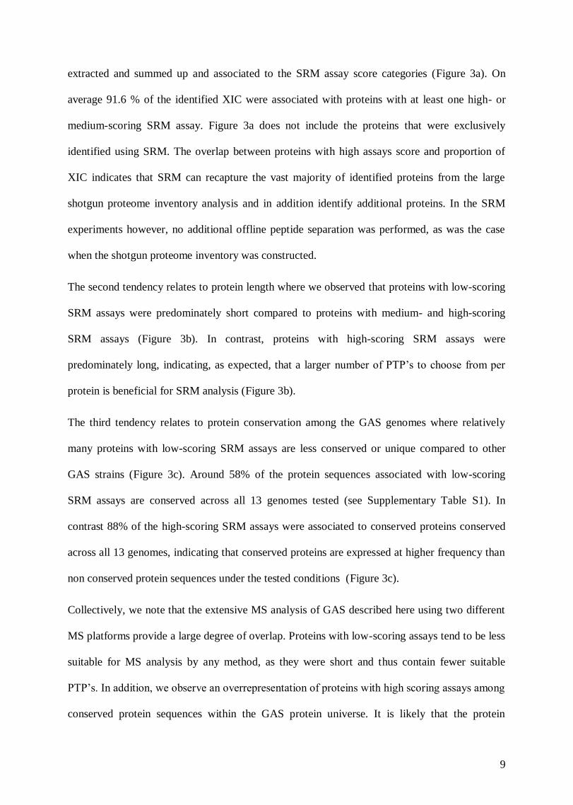
The second leg included iterative testing of the assays with SRM using a QQQ instrument to
increase the confidence of individual SRM assays (Figure 1c). We tested 7621 distinct peptide
sequences with their corresponding SRM assays, represented by a total of 79277 transitions, in
cell lysates from GAS grown under different conditions. The conditions included different
growth phases, oxidative stress, exposure to human plasma supplement, or antibiotics (Figure
1a). All SRM assays were tested at least two times and several more than hundred times (Figure
2a), resulting in 957850 individual ion chromatograms. The most frequently observed SRM
assays and the most intense transitions associated with them were ranked as described
previously20
. We used this information to build an SRM assay score using a rule-based scoring
model. The model divides the assays into three categories, low-, medium- and high-scoring. The
scoring indicates the ability of an SRM assay to detect the corresponding peptide in tryptic GAS
digests from cellular compartments and different bacterial states (Figure 1c). The major assay
score parameter is based on the SRM false discovery rate (FDR) thresholds of peptide
identification in complex biological peptide mixtures. The high scoring assays represent cases
where the peptide was detected with high confidence in complex GAS peptide mixtures (FDR of
≤1%). The medium scoring assays represent cases where the peptide was detected with lower
confidence (FDR 1>2%). These SRM assay score categories received and arbitrary score of 100
and 50 respectively. SRM assays developed on synthetic peptides were included in the medium
scoring SRM assays. The fine-tuning of the assay score within these two categories was based
on the number of times the peptides were observed, minus the number of attempted observations
divided by two. Thus, the higher the frequency with which an SRM assay was observed with
high probability, the higher the assay score. The low scoring SRM assays represent cases where
the peptide remained undetected in complex GAS peptide mixtures. These SRM assays were
scored based on the number of transitions per SRM assay, which positively influences the low-
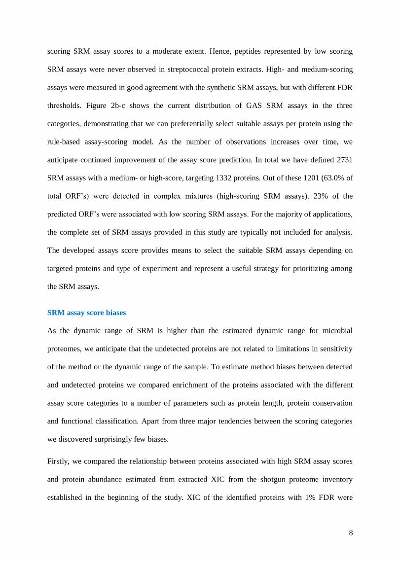
8
scoring SRM assay scores to a moderate extent. Hence, peptides represented by low scoring
SRM assays were never observed in streptococcal protein extracts. High- and medium-scoring
assays were measured in good agreement with the synthetic SRM assays, but with different FDR
thresholds. Figure 2b-c shows the current distribution of GAS SRM assays in the three
categories, demonstrating that we can preferentially select suitable assays per protein using the
rule-based assay-scoring model. As the number of observations increases over time, we
anticipate continued improvement of the assay score prediction. In total we have defined 2731
SRM assays with a medium- or high-score, targeting 1332 proteins. Out of these 1201 (63.0% of
total ORF’s) were detected in complex mixtures (high-scoring SRM assays). 23% of the
predicted ORF’s were associated with low scoring SRM assays. For the majority of applications,
the complete set of SRM assays provided in this study are typically not included for analysis.
The developed assays score provides means to select the suitable SRM assays depending on
targeted proteins and type of experiment and represent a useful strategy for prioritizing among
the SRM assays.
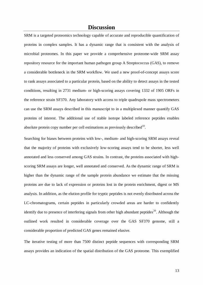
SRM assay score biases
As the dynamic range of SRM is higher than the estimated dynamic range for microbial
proteomes, we anticipate that the undetected proteins are not related to limitations in sensitivity
of the method or the dynamic range of the sample. To estimate method biases between detected
and undetected proteins we compared enrichment of the proteins associated with the different
assay score categories to a number of parameters such as protein length, protein conservation
and functional classification. Apart from three major tendencies between the scoring categories
we discovered surprisingly few biases.
Firstly, we compared the relationship between proteins associated with high SRM assay scores
and protein abundance estimated from extracted XIC from the shotgun proteome inventory
established in the beginning of the study. XIC of the identified proteins with 1% FDR were
9
extracted and summed up and associated to the SRM assay score categories (Figure 3a). On
average 91.6 % of the identified XIC were associated with proteins with at least one high- or
medium-scoring SRM assay. Figure 3a does not include the proteins that were exclusively
identified using SRM. The overlap between proteins with high assays score and proportion of
XIC indicates that SRM can recapture the vast majority of identified proteins from the large
shotgun proteome inventory analysis and in addition identify additional proteins. In the SRM
experiments however, no additional offline peptide separation was performed, as was the case
when the shotgun proteome inventory was constructed.
The second tendency relates to protein length where we observed that proteins with low-scoring
SRM assays were predominately short compared to proteins with medium- and high-scoring
SRM assays (Figure 3b). In contrast, proteins with high-scoring SRM assays were
predominately long, indicating, as expected, that a larger number of PTP’s to choose from per
protein is beneficial for SRM analysis (Figure 3b).
The third tendency relates to protein conservation among the GAS genomes where relatively
many proteins with low-scoring SRM assays are less conserved or unique compared to other
GAS strains (Figure 3c). Around 58% of the protein sequences associated with low-scoring
SRM assays are conserved across all 13 genomes tested (see Supplementary Table S1). In
contrast 88% of the high-scoring SRM assays were associated to conserved proteins conserved
across all 13 genomes, indicating that conserved proteins are expressed at higher frequency than
non conserved protein sequences under the tested conditions (Figure 3c).
Collectively, we note that the extensive MS analysis of GAS described here using two different
MS platforms provide a large degree of overlap. Proteins with low-scoring assays tend to be less
suitable for MS analysis by any method, as they were short and thus contain fewer suitable
PTP’s. In addition, we observe an overrepresentation of proteins with high scoring assays among
conserved protein sequences within the GAS protein universe. It is likely that the protein

10
sequences with exclusively low scoring SRM assays are not expressed under the tested growth
conditions. Membrane proteins were not specifically addressed in the sub-cellular fractionation,
however, we observe no overall bias against membrane proteins. Nevertheless, detection of
certain membrane protein species may require specific digestion/extraction methods23,24
.
Spatial distribution of proteins with high scoring assays
The iterative testing of the developed SRM assays on more than 540 LC-SRM-MS
measurements represents a comprehensive proteome-wide targeted measurement of the GAS
SF370 encoding genome. As we tested the SRM assays in enriched fractions of the intracellular,
surface associated and secreted protein pools we could also estimate the predominant
localization for proteins associated with high scoring SRM assays (Figure 4). The majority of
the proteins, 934 proteins were predominately present in the intracellular pool (Figure 4a and b).
A smaller fraction, 115 and 28 proteins, were predominately found in the surface associated and
secreted protein pools respectively (Figure 4c and d) leaving 124 proteins that were relatively
evenly split between two or more compartments (Figure 4e and f).
To visualize the proteome distribution we used Cystoscape25
with the Cerebral26
plugin (Figure
4g, Supplementary Figure S1). The proteins were grouped according to cellular functions using
the National pathogen microbe data resource (NMPDR) subsystem information27
(now part of
PATRICs Bioinformatics Resource Center28
) and selected cellular location for the individual
functional categories based on the protein expression profiles shown in Figure 4a-d, represented
as circles in Figure 4g. Cellular functional categories containing either proteins with
contradicting cellular location from cluster Figure 4e-f or different proteins with contradicting
cellular location were considered to have unknown localization and are represented as rectangles
in Figure 4g. The size of the circles/rectangles indicates the number of member proteins ranging
from 1 to 34. The edges between the circles/rectangles represent protein members that belong to
more than one NMPDR subsystem, whereas the location of the rectangles within the network

11
view is influenced by the edges. The color scheme represents continuous decreasing average
SRM assay score, where red indicates NMPDR subsystems with high average SRM assay score
(>119) and black indicating proteins with predominately low scoring SRM assays (<10). It can
be noted that the majority of circles and rectangles are red, demonstrating the coverage of high-
scoring assays across the GAS proteome. Several black protein groups were not detectable under
the tested growth conditions. In general these subsystems contain relatively few members. In
summary, the iterative testing of the SRM assays in three subcellular compartments provides an
overview of the subcellular protein distribution for GAS strain SF370. The majority of the nodes
have a relatively high proportion of high-scoring SRM assays. More information regarding the
subcellular localization for individual proteins can be found in Supplementary Data 1.
Transportability of SRM assays across the GAS pan-genome and related species.
We used GAS SF370 as a model strain when developing the proteome-wide SRM assay
repository. However, as there is substantial genetic variation within and between genomes from
different GAS serotypes29-32
, it is important to know which SRM assays target proteins in other
GAS strains. To explore the transportability of this resource to other GAS strains and closely or
distantly related species, we selected in total 75 taxa of low GC Gram-positive bacteria,
Firmictutes, (see Supplementary Figure S2) and mapped medium- and high-scoring SF370
assays onto respective genome. To estimate the taxon evolutionary relationship, a phylogenetic
tree was constructed based on respective rpoB gene sequence (Supplementary Figure S2). There
was a large attrition of assays with increasing evolutionary distance (Figure 5a) and depending
taxonomic rank (Figure 5b). Nevertheless, transportability within the species rank was high (Fig
5b, c) with average genome coverage’s of 59-70% (1167-1332 ORFs) demonstrating that the
developed SRM-assays will target a majority of the currently defined GAS pan-genome products
independent on serotype or strain (Figure 5c). Transportability in the genus rank was the most
diverse (8-31% genome coverage) (Figure 5b) with Group C streptococci genomes having the

12
highest degree of average coverage and also being the closest related taxa based on ropB
homologies (Figure 5a).
To address the identity of proteins with transferable assays we calculated the number of species
with at least a single high scoring peptide belonging NMPDR subsystem. We note that the most
frequent NMPDR functions with SRM assays with high-degree of transportability are as
expected ribosomal proteins, universal GTPases and proteins involved in central metabolism. In
contrast the NMPDR subsytems with lowest level of transportability are Phage capsid proteins,
Custered, Regularly Interspaced Short Palindromic Repeats (CRISPR) associated proteins and
Streptococcus pyogenes Virulome (see Supplementary Figure S3a, b for more information). The
SRM assay transportability is an important parameter when targeting GAS proteins in microbial
communities as for example in the oral cavity where many bacterial species are present33
.
SRM assay repository and availability
The availability of the SRM assays (transitions, retention time and collision energy), their
NMPDR subsystems, measured subcellular localization and degree of transferability is found in
Supplementary Data 2. The full list of SRM assays can be downloaded from the PeptideAtlas
Public SRM Transition Lists at
https://db.systemsbiology.net/sbeams/cgi/PeptideAtlas/GetTransitionLists under the accession
number PATR00014.
13
Discussion
SRM is a targeted proteomics technology capable of accurate and reproducible quantification of
proteins in complex samples. It has a dynamic range that is consistent with the analysis of
microbial proteomes. In this paper we provide a comprehensive proteome-wide SRM assay
repository resource for the important human pathogen group A Streptococcus (GAS), to remove
a considerable bottleneck in the SRM workflow. We used a new proof-of-concept assays score
to rank assays associated to a particular protein, based on the ability to detect assays in the tested
conditions, resulting in 2731 medium- or high-scoring assays covering 1332 of 1905 ORFs in
the reference strain SF370. Any laboratory with access to triple quadrupole mass spectrometers
can use the SRM assays described in this manuscript to in a multiplexed manner quantify GAS
proteins of interest. The additional use of stable isotope labeled reference peptides enables
absolute protein copy number per cell estimations as previously described10
.
Searching for biases between proteins with low-, medium- and high-scoring SRM assays reveal
that the majority of proteins with exclusively low-scoring assays tend to be shorter, less well
annotated and less conserved among GAS strains. In contrast, the proteins associated with high-
scoring SRM assays are longer, well annotated and conserved. As the dynamic range of SRM is
higher than the dynamic range of the sample protein abundance we estimate that the missing
proteins are due to lack of expression or proteins lost in the protein enrichment, digest or MS
analysis. In addition, as the elution profile for tryptic peptides is not evenly distributed across the
LC-chromatograms, certain peptides in particularly crowded areas are harder to confidently
identify due to presence of interfering signals from other high abundant peptides20
. Although the
outlined work resulted in considerable coverage over the GAS SF370 genome, still a
considerable proportion of predicted GAS genes remained elusive.
The iterative testing of more than 7500 distinct peptide sequences with corresponding SRM
assays provides an indication of the spatial distribution of the GAS proteome. This exemplified

14
by high enrichments of cell walled anchored proteins in the surface compartment, and virulence
factors in the secreted fraction. However, a substantial number of ribosomal proteins and central
metabolic enzymes are detected in the extracellular fractions. In fact, around 10% of the detected
proteins displayed similar abundance levels in more than one cellular compartment. There are
probably several reasons for this, which are difficult to distinguish. Similar phenomena has been
described earlier34-36
, and could be explained by spontaneous cell lysis or artifacts related to
experimental procedures. However, it is also likely that certain proteins are present in more than
one cellular compartment natively. This has been described in detail for several GAS proteins as
examples, glycolytic proteins implicated as virulence factors located on the surface37-39
or cell
wall anchored proteins proteolytically released into the growth media 40-42
.
We believe the SRM assay repository will become a useful resource for addressing central
medical and molecular microbiological related questions regarding GAS in general as the
transportability of the SRM assays across the known GAS protein universe was high. Relatively
little effort is required to also cover other strain-specific SRM assays in the respiratory. Defining
GAS proteome composition differences between clinical isolates and mutant strains in vitro and
in vivo are examples of how SRM assay repository could be used. Awareness of SRM assay
transportability to closely related species is essential if targeting GAS proteins in microbial
ecologies, such as in pharyngitis in vivo.
In conclusion we have in this work provided a proteome-wide SRM assay repository resource
for one of the most important human pathogens to facilitate SRM-MS analysis for this
bacterium. As several assays can be transported to other species we expect that the reach of the
resource extend beyond GAS. We believe that the iterative testing of all SRM assays and the
construction of a novel SRM assay score model along with estimating protein specific biases for
the differential scoring SRM assays increases the usefulness of the described resource.

15
Methods
Bacterial culture conditions
S. pyogenes M1 strains SF370, MGAS5005 and AP1 (strain 40/58 from the WHO Collaborating
Centre for Reference and Research on Streptococci, Prague, Czech Republic) was cultured
(37°C; 5% CO2) in C-medium43
, Todd-Hewitt (TH) broth (Difco Laboratories) or in TH with
supplements as indicated below or in Protein-reduced TH (PR-TH) broth36
for secreted protein
isolation. Supplements were added to TH as indicated at the following concentrations: 1, 5, 10,
20 or 50 % (V/V) citrate treated human plasma (Skåne University Hospital, SUS) 50 % (V/V)
citrate treated mouse plasma from CD1 mice (SeraLab), 4 mg/ml human serum albumin (Sigma-
Aldrich), 4 mg/ml essentially fatty acid free (∼0.005%) human serum albumin (Sigma-Aldrich),
0.3 mg/ml human fibrinogen (Sigma-Aldrich), 1.2 mg/ml human IgG (Sigma-Aldrich),
rifampicin at the following concentrations 0.25, 1.25, 2.5, 12.5 or 25 ng/ul, erythromycin at the
following concentrations 0.1, 0.5, 1, 5 or 10 (μg/ml), hydrogen peroxide at the following
concentrations 0.5 mM or 5 mM. Cultures were also grown at the following conditions: strict
anaerobically (Elektrotek Workstation), room atmosphere, or pH at levels 5.5, 6.4, 7.3, 8.1 or 9
Subcellular protein isolation
Bacteria were generally harvested at exponential (OD620 nm=0.4-0.5) or at stationary phase
(OD620 nm=0.7-0.8) by centrifugation 10 minutes at 2500 x g. To isolate intracellular proteins
samples were treated as earlier described1. For surface-associated protein isolation, TBS washed
cells were re-suspended in 20 mM Tris-HCl, 150 mM NaCl, 10 mM CaCl2, 1 M D-arabinose,
pH 7.6 to a concentration of 1.6 x 10⁹ colony forming units (CFU) per ml. Samples were treated
with 10 g sequencing grade trypsin (Promega) per ml for 15 min at 37 °C44,45
. Cells were
removed by centrifugation at 1000 x g for 15 min at 4 °C and the resulting supernatant was
treated as described below in the ’Protein digestion & peptide cleaning’ section except for more
extensive washes during peptide cleaning for arabinose removal. Secreted proteins were isolated

16
from 22 m filtered culture supernatants that were concentrated with Amicon Ultra-15
Centrifugal Filter Units, 30 MWCO (Millipore). The resulting concentrate was diafiltrated in the
same filter unit type twice with 50 mM Tris-HCl, pH 8.35 and then once with 6 M Urea, 0.2 M
Tris-HCl, pH 8.35.
Protein digestion & peptide cleaning
The proteins were reduced with 5 mM dithiothreitol (DTT) for 45 min at 37 °C, and alkylated
with 25 mM iodoacetamide for 45 min before diluting the sample with 100 mM ammonium
bicarbonate to a final urea concentration below 1.5M. Proteins were digested by incubation with
trypsin (1/100, w/w) for at least 6 h at 37 °C. The peptides were cleaned up by C18 reversed-
phase spin columns according to the manufacturer’s instructions (Harvard Apparatus).
Shotgun tandem mass spectrometry analysis
The shotgun tandem and targeted mass spectrometry analysis was performed as previously
described1. Briefly, the hybrid Orbitrap-LTQ XL mass spectrometer (Thermo Electron, Bremen,
Germany) was coupled online to a split-less Eksigent 2D NanoLC system (Eksigent
technologies, Dublin, CA, USA). Peptides were loaded with a constant flow rate of 10 µl/min
onto a pre-column (Zorbax 300SB-C18 5 x 0.3 mm, 5 µm, Agilent technologies, Wilmington,
DE, USA) and subsequently separated on a RP-LC analytical column (Zorbax 300SB-C18 150
mm x 75 µm, 3.5 µm, Agilent technologies) with a flow rate of 350 nl/min. The peptides were
eluted with a linear gradient from 95% solvent A (0.1% formic acid in water) and 5% solvent B
(0.1% formic acid in acetonitrile) to 40% solvent B over 55 minutes. The mass spectrometer was
operated in data-dependent acquisition mode to automatically switch between Orbitrap-MS
(from m/z 400 to 2000) and LTQ-MS/MS. Four MS/MS spectra were acquired in the linear ion
trap per each Fourier Transform-MS scan which was acquired at 60,000 FWHM nominal
resolution settings using the lock mass option (m/z 445.120025) for internal calibration. The
dynamic exclusion list was restricted to 500 entries using a repeat count of two with a repeat

17
duration of 20 seconds and with a maximum retention period of 120 seconds. Precursor ion
charge state screening was enabled to select for ions with at least two charges and rejecting ions
with undetermined charge state. The normalized collision energy was set to 30%, and one
microscan was acquired for each spectrum.
The data analysis was performed as previously described 1. Briefly, the resulting MS2 data were
searched with X! Tandem search engine, version 2009.04.01.1 with the k-score plugin46
, a
common peptide and protein list was generated using the Trans-Proteomic pipeline, version
4.4.047
. All searches were performed with full-tryptic cleavage specificity, up to 2 allowed
missed cleavages, a precursor mass error of 15 ppm and an error tolerance of 0.5 Da for the
fragment ions. Because of the sample preparation cysteine carbamidomethylation was defined as
fixed modification in the search parameters. A protein database with sequences for GAS SF370
(Genome ID 79812 from PATRIC) was used to match the individual spectra to certain peptides.
The database was extended by decoy sequences to validate the resulting peptide-spectrum
matches (PSMs)48
. A 1% false-discovery rate (FDR) was then used to generate the final protein
list with ProteinProphet. MS1-based quantification was done using SuperHirn49
. Features were
detected using SuperHirn using a retention time tolerance of 1, MS1 m/z tolerance of 10, MS2
PPM m/z tolerance of 30. Only features with charge 1-5 were included. Any feature for which
more than one peptide could be identified at the 1% FDR, hence mapping to more than one
protein, were discarded.
Generation of proteome-wide SRM assays
We generated experimentally validated SRM assay for three proteotypic peptides for each
protein in the SF370 proteome. Transitions were generated from experimental MS2 spectra
either from off gel electrophoresis fractionated cell lysates for a pool of GAS SF370 grown
under various conditions or from crude synthetic peptides purchased from JPT (Berlin,
Germany). The transitions were scored in a scoring scheme that favored y-ions over b-ions,

18
required both Q1/Q3 to be between 400 and 1500 M/Z, Q3 larger than Q1 was favored and
precursor charge of 2 was preferred over other charge states. The four best transitions for each
peptide were measured in none-scheduled SRM mode against the sample where the peptide was
identified by MS2.
SRM analysis
SRM transition assays were constructed by testing the twenty most abundant peptide fragments
for selected proteotypic peptides identified with high confidence in the LC-MS/MS experiments.
Spiked in the RT-peptides (Biognosys AG, Zurich, Switzerland) allowed normalization of the
retention time as previously described20
. The SRM measurements were performed on a TSQ
Vantage triple quadropole mass spectrometer (Thermo Electron, Bremen, Germany) equipped
with a nanoelectrospray ion source (Thermo Electron). Chromatographic separations of peptides
were performed on an Eksigent 1D NanoLC system (Eksigent technologies) using the same
chromatographic conditions as described above for the Eksigent 2D NanoLC system connected
to the hybrid Orbitrap-LTQ XL mass spectrometer. The LC was operated with a flow rate of 400
nl/min. The mass spectrometer was operated in SRM mode, with both Q1 and Q3 settings at unit
resolution (FWHM 0.7 Da). A spray voltage of +1700 V was used with a heated ion transfer
setting of 270ºC for desolvation. Data were acquired using the Xcalibur software (version 2.1.0).
The dwell time was set to 10 ms and the scan width to 0.01 m/z. All collision energies were
calculated using the formula: CE = (Parent m/z) x 0.034 + 3.314.
The data analysis was performed as previously described20
using a 1% FDR. The resulting
peptide abundances were exported into a database, where protein abundances were inferred by
summing up the abundances for the peptides uniquely mapping to each protein22
.

19

20
References
1. Malmström, J. A. et al. Streptococcus pyogenes in human plasma: adaptive mechanisms
analyzed by mass spectrometry based proteomics. J Biol Chem 287, 1415–1425 (2011).
2. Hecker, M., Becher, D., Fuchs, S. & Engelmann, S. A proteomic view of cell physiology
and virulence of Staphylococcus aureus. Int J Med Microbiol 300, 76–87 (2010).
3. Poetsch, A., Haussmann, U. & Burkovski, A. Proteomics of corynebacteria: From
biotechnology workhorses to pathogens. Proteomics 11, 3244–3255 (2011).
4. Chao, T.-C. & Hansmeier, N. The current state of microbial proteomics: Where we are
and where we want to go. Proteomics 12, 638–650 (2012).
5. Cole, J. N., Barnett, T. C., Nizet, V. & Walker, M. J. Molecular insight into invasive
group A streptococcal disease. Nat Rev Microbiol 9, 724–736 (2011).
6. Malmström, L., Malmström, J. A. & Aebersold, R. Quantitative proteomics of microbes:
Principles and applications to virulence. Proteomics 11, 2947–2956 (2011).
7. Lange, V. et al. Targeted quantitative analysis of Streptococcus pyogenes virulence
factors by multiple reaction monitoring. Mol Cell Proteomics 7, 1489–1500 (2008).
8. Kuhner, S. et al. Proteome Organization in a Genome-Reduced Bacterium. Science 326,
1235–1240 (2009).
9. Schmidt, A. et al. Absolute quantification of microbial proteomes at different states by
directed mass spectrometry. Mol Syst Biol 7, 510 (2011).
10. Malmström, J. A. et al. Proteome-wide cellular protein concentrations of the human
pathogen Leptospira interrogans. Nature 460, 762–765 (2009).
11. Stahl-Zeng, J. et al. High sensitivity detection of plasma proteins by multiple reaction
monitoring of N-glycosites. Mol Cell Proteomics 6, 1809–1817 (2007).
12. Teleman, J. et al. Automated selected reaction monitoring software for accurate label-free
protein quantification. J Proteome Res 11, 3766–3773 (2012).
13. Malmström, J. A., Lee, H. & Aebersold, R. Advances in proteomic workflows for
systems biology. Curr Opin Biotechnol 18, 378–384 (2007).
14. Lange, V., Picotti, P., Domon, B. & Aebersold, R. Selected reaction monitoring for
quantitative proteomics: a tutorial. Mol Syst Biol 4, 222 (2008).
15. Kuster, B., Schirle, M., Mallick, P. & Aebersold, R. Scoring proteomes with proteotypic
peptide probes. Nat Rev Mol Cell Biol 6, 577–583 (2005).
16. Picotti, P. et al. A database of mass spectrometric assays for the yeast proteome. Nat
Methods 5, 913–914 (2008).
17. Carapetis, J. R., Steer, A. C., Mulholland, E. K. & Weber, M. The global burden of group
A streptococcal diseases. The Lancet infectious diseases 5, 685–694 (2005).
18. Cunningham, M. W. Pathogenesis of group A streptococcal infections. Clin Microbiol
Rev 13, 470–511 (2000).
19. Deutsch, E. W., Lam, H. & Aebersold, R. PeptideAtlas: a resource for target selection for
emerging targeted proteomics workflows. EMBO Rep 9, 429–434 (2008).
20. Malmström, L., Malmström, J. A., Selevsek, N., Rosenberger, G. & Aebersold, R.
Automated Workflow for Large-Scale Selected Reaction Monitoring Experiments. J
Proteome Res 11, 1644–1653 (2012).
21. Vogel, C. & Marcotte, E. M. Calculating absolute and relative protein abundance from
mass spectrometry-based protein expression data. Nat Protoc 3, 1444–1451 (2008).
22. Malmström, L., Marko-Varga, G., Westergren-Thorsson, G., Laurell, T. & Malmström, J.
A. 2DDB - a bioinformatics solution for analysis of quantitative proteomics data. BMC
Bioinformatics 7, 158 (2006).
23. Wu, C. C. & Yates, J. R. The application of mass spectrometry to membrane proteomics.

21
Nat Biotechnol 21, 262–267 (2003).
24. Wiśniewski, J. R., Zougman, A., Nagaraj, N. & Mann, M. Universal sample preparation
method for proteome analysis. Nat Methods 6, 359–362 (2009).
25. Shannon, P. et al. Cytoscape: a software environment for integrated models of
biomolecular interaction networks. Genome Res 13, 2498–2504 (2003).
26. Barsky, A., Gardy, J. L., Hancock, R. E. W. & Munzner, T. Cerebral: a Cytoscape plugin
for layout of and interaction with biological networks using subcellular localization
annotation. Bioinformatics 23, 1040–1042 (2007).
27. McNeil, L. K. et al. The National Microbial Pathogen Database Resource (NMPDR): a
genomics platform based on subsystem annotation. Nucleic Acids Res 35, D347–53
(2007).
28. Gillespie, J. J. et al. PATRIC: the comprehensive bacterial bioinformatics resource with a
focus on human pathogenic species. Infect Immun 79, 4286–4298 (2011).
29. Beres, S. B. et al. Molecular complexity of successive bacterial epidemics deconvoluted
by comparative pathogenomics. Proc Natl Acad Sci USA 107, 4371–4376 (2010).
30. Beres, S. B. et al. Molecular genetic anatomy of inter- and intraserotype variation in the
human bacterial pathogen group A Streptococcus. Proc Natl Acad Sci USA 103, 7059–
7064 (2006).
31. Sumby, P. et al. Evolutionary origin and emergence of a highly successful clone of
serotype M1 group a Streptococcus involved multiple horizontal gene transfer events. J
Infect Dis 192, 771–782 (2005).
32. McShan, W. M. et al. Genome sequence of a nephritogenic and highly transformable
M49 strain of Streptococcus pyogenes. J Bacteriol 190, 7773–7785 (2008).
33. Human Microbiome Project Consortium. Structure, function and diversity of the healthy
human microbiome. Nature 486, 207–214 (2012).
34. Chhatwal, G. S. Anchorless adhesins and invasins of Gram-positive bacteria: a new class
of virulence factors. Trends Microbiol 10, 205–208 (2002).
35. Barinov, A. et al. Prediction of surface exposed proteins in Streptococcus pyogenes, with
a potential application to other Gram-positive bacteria. Proteomics 9, 61–73 (2009).
36. Lei, B., Mackie, S., Lukomski, S. & Musser, J. M. Identification and immunogenicity of
group A Streptococcus culture supernatant proteins. Infect Immun 68, 6807–6818 (2000).
37. Boël, G., Jin, H. & Pancholi, V. Inhibition of cell surface export of group A streptococcal
anchorless surface dehydrogenase affects bacterial adherence and antiphagocytic
properties. Infect Immun 73, 6237–6248 (2005).
38. Lottenberg, R. et al. Cloning, sequence analysis, and expression in Escherichia coli of a
streptococcal plasmin receptor. J Bacteriol 174, 5204–5210 (1992).
39. Cork, A. J. et al. Defining the structural basis of human plasminogen binding by
streptococcal surface enolase. J Biol Chem 284, 17129–17137 (2009).
40. Raeder, R., Woischnik, M., Podbielski, A. & Boyle, M. D. P. A secreted streptococcal
cysteine protease can cleave a surface-expressed M1 protein and alter the
immunoglobulin binding properties. Research in Microbiology 149, 539–548 (1998).
41. Nelson, D. C., Garbe, J. & Collin, M. Cysteine proteinase SpeB from Streptococcus
pyogenes - a potent modifier of immunologically important host and bacterial proteins.
Biol Chem 392, 1077–1088 (2011).
42. Berge, A. & Björck, L. Streptococcal cysteine proteinase releases biologically active
fragments of streptococcal surface proteins. J Biol Chem 270, 9862–9867 (1995).
43. Collin, M. & Olsén, A. EndoS, a novel secreted protein from Streptococcus pyogenes
with endoglycosidase activity on human IgG. EMBO J 20, 3046–3055 (2001).
44. Solis, N., Larsen, M. R. & Cordwell, S. J. Improved accuracy of cell surface shaving
proteomics in Staphylococcus aureus using a false-positive control. Proteomics 10, 2037–
2049 (2010).

22
45. Severin, A. et al. Proteomic analysis and identification of Streptococcus pyogenes
surface-associated proteins. J Bacteriol 189, 1514–1522 (2007).
46. Craig, R. & Beavis, R. C. A method for reducing the time required to match protein
sequences with tandem mass spectra. Rapid Commun Mass Spectrom 17, 2310–2316
(2003).
47. Keller, A., Eng, J., Zhang, N., Li, X.-J. & Aebersold, R. A uniform proteomics MS/MS
analysis platform utilizing open XML file formats. Mol Syst Biol 1, 2005.0017 (2005).
48. Elias, J. E. & Gygi, S. P. Target-decoy search strategy for increased confidence in large-
scale protein identifications by mass spectrometry. Nat Methods 4, 207–214 (2007).
49. Mueller, L. N. et al. SuperHirn - a novel tool for high resolution LC-MS-based
peptide/protein profiling. Proteomics 7, 3470–3480 (2007).

23
Acknowledgements
JM is funded by the Swedish Research Council (project 2008-3356), the Crafoord Foundation
(ref nr 20100892) and the Swedish Foundation for Swedish Research (FFL4). CK is supported
by the Swedish Research Council postdoctoral fellowship (project 2010-996). RA is supported
by the European Research Council (grant #ERC-2008-AdG 233226), SystemsX.ch, the Swiss
initiative for systems biology, the Swiss National Science Foundation grant Nr. 3100A0-130530
and by the European Union Seventh Framework Program PROSPECTS (Proteomics
Specification in Space and Time, Grant HEALTH-F4-2008). We thank Karin M. Hansson and
Mats Mågård for excellent technical assistance

24
Author Contributions
Conceived and designed the experiments: CK LM RA JM. Performed the experiments: CK JM.
Bioinformatic analysis: LM. Analyzed the data: CK LM RA JM. Wrote the paper: CK LM RA
JM.

25
Additional information
The authors declare no competing financial interest.

26
Figures Legends
Figure 1: Construction of a proteome-wide SRM assay repository. a) Graphical
representation of the enriched Group A Streptococci (GAS) cellular compartments. Repeated
enrichment of protein pools from the cellular compartments and bacterial states were digested
using trypsin and analyzed by liquid chromatography tandem mass spectrometry (LC-MS/MS).
b) Outline of the strategy used to construct a spectral library from where the low scoring selected
reaction monitoring (SRM) assays were extracted. For high-abundant proteotypic peptides
(PTP’s) the SRM assays were determined directly in biological samples, whereas for medium-
and low-abundant PTP peptides were synthesized and analyzed with LC-MS/MS. c) To increase
the confidence of the individual SRM assays the low-scoring SRM assays were tested
extensively in complex mixture of GAS tryptic digest using SRM.
Figure 2: Rule based assay score modeling can select high performing assays depending on
cellular compartment and cellular state. SRM assays were tested repeatedly in mixtures of
GAS tryptic digests from different subcellular compartments and bacterial states. a) Statistics
over the repetitive testing of all SRM assays. b) The assays were divided up into three
categories, low-, medium-, and high-scoring SRM assays based on a rule-based assay score
model. c) Assay score distribution for high scoring SRM assays.
Figure 3: Protein identification biases across functional categories and ORF properties.
Iterative testing of the developed SRM assays in complex biological mixtures of GAS tryptic
digests resulted in subdivision of the proteins into three SRM assays-score categories; proteins
with low-, medium- or high-scoring SRM assays. Using the SRM assay score categories we
determined biases among associated proteins within the three categories. a) Proportion of

27
extracted ion chromatogram (XIC) intensities associated with proteins with at least one high or
medium-scoring SRM assay or proteins with low-scoring SRM assays. NMPDR was used to
categorize proteins; Protein Metabolism includes categories Amino Acids and Derivatives and
Protein Metabolism; Miscellaneous includes categories Clustering-based subsystems,
Miscellaneous, Phages, Prophages, Transposable elements, Plasmids, Regulation and Cell
signaling, Respiration, Stress Response, Cell Division and Cell Cycle and Cell Wall and
Capsule; Carbohydrate Metabolism includes category Carbohydrates; RNA & DNA Metabolism
includes categories Nucleosides and Nucleotides, DNA Metabolism and RNA Metabolism;
Other Metabolism includes categories Phosphorus Metabolism, Potassium metabolism, Fatty
Acids, Lipids, and Isoprenoids, Cofactors, Vitamins, Prosthetic Groups and Pigments, Sulfur
Metabolism, Iron acquisition and metabolism, Nitrogen Metabolism, Membrane Transport and
Metabolism of Aromatic Compounds Virulence includes categories Virulence and Virulence,
Disease and Defense.
b) Genome-wide correlation between SRM assay-score and ORF length. c) Correlation between
SRM assay score and relative degree of protein conservation across 13 GAS strains as
determined with TOP-BLAST hits with SF370 as reference.
Figure 4: Spatial distribution of proteins with high scoring SRM assays. Testing of all SRM
assays in three subcellular compartments enabled the construction of a subcellular distribution
map for the proteins with high scoring assays. a-b) predominately intracellular proteins, c)
surface-associated proteins, d) secreted proteins and e-f) proteins with split subcellular
compartmentalization. Red lines in panels a-f represent the average distribution of the clusters.
Subsequently the identified proteins were grouped into NMPDR subsystems and visualized
using Cytoscape. g) Outline of the GAS proteome network topology, where circles represent
NMPDR subsystems where all proteins predominantly have the same subcellular location,

28
secreted, surface associated or intracellular, according to the subcellular protein profiles in
Figure 4a-d. Rectangles represent NMPDR subsystems where an equal number of members have
opposing subcellular location profiles. The localization of the rectangles in the network is
influenced by the edges, which represent protein members that belong to more than one
NMPDR subsystem. Increasing node size represents increasing number of member proteins. The
color represents average SRM assay score, where red indicates NMPDR subsystems with high-
average SRM assay score and black indicating NMPDR subsystems with low average SRM
assays score. For full details of NMPDR subsystems see Supplementary Figure S1.
Figure 5: SRM assay transportability to related species. All SRM assays were developed on
basis of the GAS strain SF370 genome. The degree of SRM assay transportability within
selected species was determined phylogeny clustering of respective rpoB gene and mapping
medium- and high-scoring SRM assays on to respective genome. a) Transportability for the
SRM assays across 75 genomes within the Firmicutes phylum. b) Average ORF genome
coverage of high- and medium-scoring SRM assays within taxonomic ranks. Boxes extend from
the 25th to 75th percentiles and error bars represent minimum to maximum values. c) View of
SRM assay transportability for 13 GAS genomes deposited in the public domain.

29
Figure 1

30
Figure 2

31
Figure 3

32
Figure 4

33
Figure 5