Embed Size (px)
Citation preview
277
Psidii Guajavae Folium
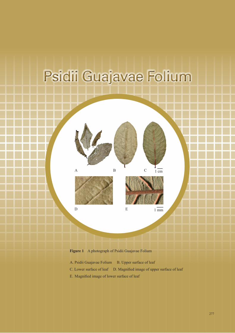
Figure 1 A photograph of Psidii Guajavae Folium
A. Psidii Guajavae Folium B. Upper surface of leaf
C. Lower surface of leaf D. Magnified image of upper surface of leaf
E. Magnified image of lower surface of leaf
277
A
D
B C
E
1 cm
1 mm

278
1. NAMES
Official Name: Psidii Guajavae Folium
Chinese Name: 番石榴葉
Chinese Phonetic Name: Fanshiliuye
2. SOURCE
Psidii Guajavae Folium is the dried leaf of Psidium guajava L. (Myrtaceae). The leaf is collected in
spring and summer, washed clean, then dried under the sun to obtain Psidii Guajavae Folium.
3. DESCRIPTION
Mostly crumpled, rolled or broken. When intact, oblong to elliptic, 3-14 cm long, 2-7 cm wide, apex
rounded or acuminate, base obtuse to rounded, margin entire. The upper surface yellowish-green to
pale brown, glabrous or nearly glabrous, sparsely dark brown glandular-punctate. The lower surface
greyish-brown to dark green, densely covered with pubescence, principal and lateral veins slightly
protuberant. Petioles 2-10 mm long. Texture leathery. Odour delicately aromatic; taste astringent (Fig. 1).
4. IDENTIFICATION
4.1 Microscopic Identification (Appendix III)
Transverse sectionUpper epidermis consists of 1 layer of flat polygonal cells, covered with cuticle; with
unicellular non-glandular hairs on the surface. Lower epidermal cells slightly small, sparsely
covered with non-glandular hairs. Hypodermis consists of 2-3 layers of collenchymatous cells,
orderly arranged, with secretory glands. Palisade tissue consists of 2 layers of columnar cells,
discontinue at the midrib. Spongy tissue consists of slightly small cells, with secretory glands.
Vascular bundle of the midrib bicollateral, surrounded by pericycle fibres. Xylem vessels radially
arranged. Phloem narrow. Clusters of calcium oxalate in rosette aggregate and prisms of calcium
oxalate scattered in the mesophyll. Collenchymatous cells subrounded, located beneath the lower
epidermis (Fig. 2).
Psidii Guajavae Folium

279
PowderColour yellowish-brown to yellowish-green. Non-glandular hairs unicellular. Clusters of
calcium oxalate numerous, in rosette aggregate, scattered or embedded in parenchymatous
cells, 6-44 μm in diameter; polychromatic under the polarized microscope. Lower epidermal
cells polygonal, with paracytic stomata, with 2 subsidiary cells. Secretory glands usually
broken, the intact ones subrounded. Calcium oxalate prism sheath occasionally visible,
Prisms of calcium oxalate scattered or embedded in parenchymatous cells, sometimes
arranged in rows; polychromatic under the polarized microscope. Fibres slender, some with
the walls slightly undulantly curved; white under the polarized microscope. Vessels mostly
spiral, 6-40 μm in diameter (Fig. 3).
Psidii Guajavae Folium

280
Figure 2 Microscopic features of transverse section of Psidii Guajavae Folium
A. Sketch B. Section illustration C. Prism and clusters of calcium oxalate (under the light microscope) D. Prism and clusters of calcium oxalate (under the polarized microscope)E. Non-glandular hair F. Secretory gland
1. Upper epidermis 2. Hypodermis 3. Palisade tissue 4. Spongy tissue5. Non-glandular hair 6. Pericycle fiber 7. Phloem 8. Xylem 9. Prism of calcium oxalate10. Cluster of calcium oxalate 11. Collenchyma 12. Secretory gland 13. Lower epidermis
12345678
9101112131234
5
678
910111213B
D
E
100 μm
50 μm 50 μmC
F
A
Psidii Guajavae Folium

281
Figure 3 Microscopic features of powder of Psidii Guajavae Folium
1. Non-glandular hair 2. Clusters of calcium oxalate 3. Lower epidermis cells with paracytic stomata4. Secretory gland 5. Calcium oxalate prisms sheath 6. Pericycle fibres 7. Spiral vessel
a. Features under the light microscope b. Features under the polarized microscope
1a 2a
4a
6a 6b 7a
5a 5b
2b 3a
50 μm
Psidii Guajavae Folium

282
4.2 Thin-Layer Chromatographic Identification [Appendix IV(A)]
Standard solutionsGuaijaverin standard solution
Weigh 1.0 mg of guaijaverin CRS (Fig. 4) and dissolve in 10 mL of ethanol (50%).
Hyperoside standard solution
Weigh 1.0 mg of hyperoside CRS (Fig. 4) and dissolve in 10 mL of ethanol (50%).
Developing solvent systemPrepare a mixture of dichloromethane, ethyl acetate, formic acid, ethanol and water
(6.5:3.5:3:1:0.5, v/v).
Spray reagentWeigh 1 g of aluminium trichloride and dissolve in 100 mL of ethanol.
Test solutionWeigh 0.5 g of the powdered sample and place it in a 50-mL conical flask, then add 10 mL of
ethanol (50%). Sonicate (350 W) the mixture for 30 min. Filter through a 0.45-µm PTFE filter.
ProcedureCarry out the method by using a HPTLC silica gel F254 plate, a twin trough chamber and a
freshly prepared developing solvent system as described above. Apply separately guaijaverin
standard solution, hyperoside standard solution and the test solution (2 μL each) to the plate.
Before the development, add the developing solvent to one of the troughs of the chamber and
place the HPTLC plate in the other trough. Cover the chamber with a lid and let equilibrate
for about 15 min. Carefully tilt the chamber to allow sufficient solvent to pass from the trough
containing the solvent to the other containing the HPTLC plate for development. Develop over a
path of about 7 cm. After the development, remove the plate from the chamber, mark the solvent
front and dry in air. Spray the plate evenly with the spray reagent and heat at about 105ºC
(about 3 min). Examine the plate under UV light (366 nm). Calculate the Rf values by using the
equation as indicated in Appendix IV (A).
Psidii Guajavae Folium

283
Figure 4 Chemical structures of (i) guaijaverin and (ii) hyperoside
(i)
(ii)
Front
Start
1 2 3
Figure 5 A reference HPTLC chromatogram of Psidii Guajavae Folium extract observed under UV light (366 nm) after staining
1. Hyperoside standard solution 2. Guaijaverin standard solution 3. Test solution
Psidii Guajavae Folium

284
For positive identification, the sample must give spots or bands with chromatographic
characteristics, including the colour and the Rf values, corresponding to those of guaijaverin and
hyperoside (Fig. 5).
4.3 High-Performance Liquid Chromatographic Fingerprinting (Appendix XII)
Standard solutions Guaijaverin standard solution for fingerprinting, Std-FP (15 mg/L)
Weigh 0.3 mg of guaijaverin CRS and dissolve in 20 mL of ethanol (50%).
Hyperoside standard solution for fingerprinting, Std-FP (10 mg/L)
Weigh 0.1 mg of hyperoside CRS and dissolve in 10 mL of ethanol (50%).
Test solutionWeigh 0.1 g of the powdered sample and place it in a 50-mL centrifuge tube, then add 10 mL
of ethanol (50%). Sonicate (120 W) the mixture for 30 min. Centrifuge at about 4000 × g for
10 min. Filter through a 0.45-µm PTFE filter.
Chromatographic systemThe liquid chromatograph is equipped with a DAD (254 nm) and a column (4.6 × 250 mm)
packed with ODS bonded silica gel (5 µm particle size). The flow rate is about 1.0 mL/min.
Programme the chromatographic system as follows (Table 1) –
Table 1 Chromatographic system conditions
Time(min)
2.5 mM Sodium acetate solution(%, v/v)
Acetonitrile(%, v/v) Elution
0 – 5 86 14 isocratic
5 – 60 86 → 82 14 → 18 linear gradient
System suitability requirementsPerform at least five replicate injections, each using 10 µL of guaijaverin Std-FP and hyperoside
Std-FP. The requirements of the system suitability parameters are as follows: the RSD of the
peak areas of guaijaverin and hyperoside should not be more than 5.0%; the RSD of the retention
times of guaijaverin and hyperoside peaks should not be more than 2.0%; the column efficiencies
determined from guaijaverin and hyperoside peaks should not be less than 25000 theoretical plates.
The R value between peak 2 and the closest peak; and the R value between peak 5 and the closest
peak in the chromatogram of the test solution should not be less than 1.5 (Fig. 6).
Psidii Guajavae Folium

285
ProcedureSeparately inject guaijaverin Std-FP, hyperoside Std-FP and the test solution (10 µL each)
into the HPLC system and record the chromatograms. Measure the retention times of
guaijaverin and hyperoside peaks in the chromatograms of guaijaverin Std-FP, hyperoside
Std-FP and the retention times of the six characteristic peaks (Fig. 6) in the chromatogram
of the test solution. Identify guaijaverin and hyperoside peaks in the chromatogram of the
test solution by comparing its retention time with that in the chromatograms of guaijaverin
Std-FP and hyperoside Std-FP. The retention times of guaijaverin and hyperoside peaks in
the chromatograms of the test solution and the corresponding Std-FP should not differ by
more than 2.0%. Calculate the RRTs of the characteristic peaks by using the equation as
indicated in Appendix XII.
The RRTs and acceptable ranges of the six characteristic peaks of Psidii Guajavae Folium extract
are listed in Table 2.
Table 2 The RRTs and acceptable ranges of the six characteristic peaks of Psidii Guajavae
Folium extract
Peak No. RRT Acceptable Range1 (ellagic acid) 0.91 ± 0.032 (marker, hyperoside) 1.00 -3 (isoquercitrin) 1.06 ± 0.034 (reynoutrin) 1.23 ± 0.035 (guaijaverin) 1.32 ± 0.036 (avicularin) 1.43 ± 0.03
Psidii Guajavae Folium

286
5. TESTS
5.1 Heavy Metals (Appendix V): meet the requirements.
5.2 Pesticide Residues (Appendix VI): meet the requirements.
5.3 Mycotoxins (Appendix VII): meet the requirements.
5.4 Sulphur Dioxide Residues (Appendix XVI): meet the requirements.
5.5 Foreign Matter (Appendix VIII): not more than 5.0%.
5.6 Ash (Appendix IX)
Total ash: not more than 6.0%.
Acid-insoluble ash: not more than 0.5%.
Figure 6 A reference fingerprint chromatogram of Psidii Guajavae Folium extract
For positive identification, the sample must give the above six characteristic peaks with RRTs
falling within the acceptable range of the corresponding peaks in the reference fingerprint
chromatogram (Fig. 6).
Psidii Guajavae Folium

287
5.7 Water Content (Appendix X)
Oven dried method: not more than 11.0%.
6. EXTRACTIVES (Appendix XI)
Water-soluble extractives (cold extraction method): not less than 10.0%.
Ethanol-soluble extractives (cold extraction method): not less than 22.0%.
7. ASSAY
Carry out the method as directed in Appendix IV (B).
Standard solutionMixed guaijaverin and hyperoside standard stock solution, Std-Stock (100 mg/L each)
Weigh accurately 1.0 mg of guaijaverin CRS and 1.0 mg of hyperoside CRS, and dissolve in 10 mL
of ethanol (50%).
Mixed guaijaverin and hyperoside standard solution for assay, Std-AS
Measure accurately the volume of the mixed guaijaverin and hyperoside Std-Stock, dilute with ethanol
(50%) to produce a series of solutions of 0.5, 1, 5, 10, 25 mg/L for both guaijaverin and hyperoside.
Test solutionWeigh accurately 0.1 g of the powdered sample and place it in a 50-mL centrifuge tube, then add 10 mL
of ethanol (50%). Sonicate (120 W) the mixture for 30 min. Centrifuge at about 4000 × g for 10 min.
Transfer the supernatant to a 25-mL volumetric flask. Repeat the extraction for two more times with
10 mL of ethanol (50%) and 5 mL of ethanol (50%) respectively. Combine the supernatants and make up
to the mark with ethanol (50%). Filter through a 0.45-µm PTFE filter.
Chromatographic systemThe liquid chromatograph is equipped with a DAD (254 nm) and a column (4.6 × 250 mm) packed
with ODS bonded silica gel (5 µm particle size). The flow rate is about 1.0 mL/min. Programme the
chromatographic system as follows (Table 3) –
Psidii Guajavae Folium

288
System suitability requirementsPerform at least five replicate injections, each using 20 µL of the mixed guaijaverin and hyperoside
Std-AS (5 mg/L each). The requirements of the system suitability parameters are as follows: the RSD
of the peak areas of guaijaverin and hyperoside should not be more than 5.0%; the RSD of the retention
times of guaijaverin and hyperoside peaks should not be more than 2.0%; the column efficiencies
determined from guaijaverin and hyperoside peaks should not be less than 30000 and 20000 theoretical
plates respectively.
The R value between guaijaverin peak and the closest peak; and the R value between hyperoside peak
and the closest peak in the chromatogram of the test solution should not be less than 1.5 (Fig. 7).
Calibration curvesInject a series of the mixed guaijaverin and hyperoside Std-AS (20 µL each) into the HPLC system and
record the chromatograms. Plot the peak areas of guaijaverin and hyperoside against the corresponding
concentrations of the mixed guaijaverin and hyperoside Std-AS. Obtain the slopes, y-intercepts and the
r2 values from the corresponding 5-point calibration curves.
ProcedureInject 20 µL of the test solution into the HPLC system and record the chromatogram. Identify guaijaverin
and hyperoside peaks (Fig. 7) in the chromatogram of the test solution by comparing their retention times
with those in the chromatogram of the mixed guaijaverin and hyperoside Std-AS. The retention times of
guaijaverin and hyperoside peaks in the chromatograms of the test solution and the Std-AS should not
differ by more than 5.0%. Measure the peak areas and calculate the concentrations (in milligram per litre)
of guaijaverin and hyperoside in the test solution, and calculate the percentage contents of guaijaverin and
hyperoside in the sample by using the equations as indicated in Appendix IV (B).
Limits The sample contains not less than 0.16% of guaijaverin (C20H18O11) and not less than 0.10% of
hyperoside (C21H20O12), calculated with reference to the dried substance.
Table 3 Chromatographic system conditions
Time(min)
2.5 mM Sodium acetate solution(%, v/v)
Acetonitrile (%, v/v) Elution
0 – 5 86 14 isocratic
5 – 60 86 → 82 14 → 18 linear gradient
Psidii Guajavae Folium

289
Figure 7 A reference assay chromatogram of Psidii Guajavae Folium extract
Psidii Guajavae Folium


![Banana Leaf[1]](https://img.pdfslide.tips/doc/110x75/557b2b89d8b42a726a8b54e6/banana-leaf1-558491c49acfa.jpg)