Embed Size (px)
Citation preview
Quiz_20.09.04
1235-1245 h
Quiz_20.09.04
• β lactam antibiotics cross barriers of the bacterial cell to reach their targets. Draw two diagrams cell envelopes: one for G-negative cells, the other for G-positive cells. Label each diagram fully by identifying all macromolecules found in every layer. [5 points]
• Show the pathway for traverse of a β lactam across the two cell walls. List the cell’s barriers against antibiotic activity. [3 points]
• Inside a Living Cell: coloured diagrams to be submitted [2 points]
Bacterial transport
•MFS
•Group translocation
•ABC transporters
•OM receptors
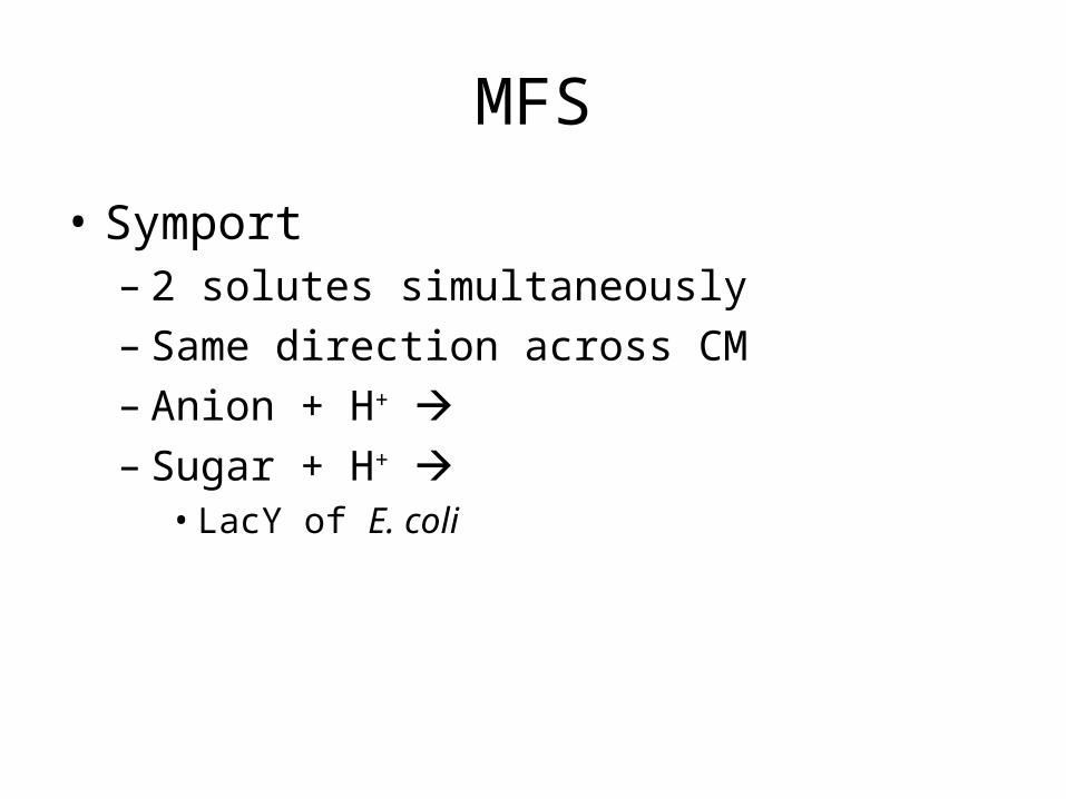
MFS
• Symport– 2 solutes simultaneously– Same direction across CM– Anion + H+ – Sugar + H+
• LacY of E. coli
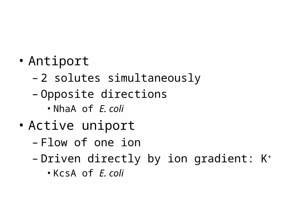
• Antiport– 2 solutes simultaneously– Opposite directions
• NhaA of E. coli
• Active uniport– Flow of one ion– Driven directly by ion gradient: K+
• KcsA of E. coli
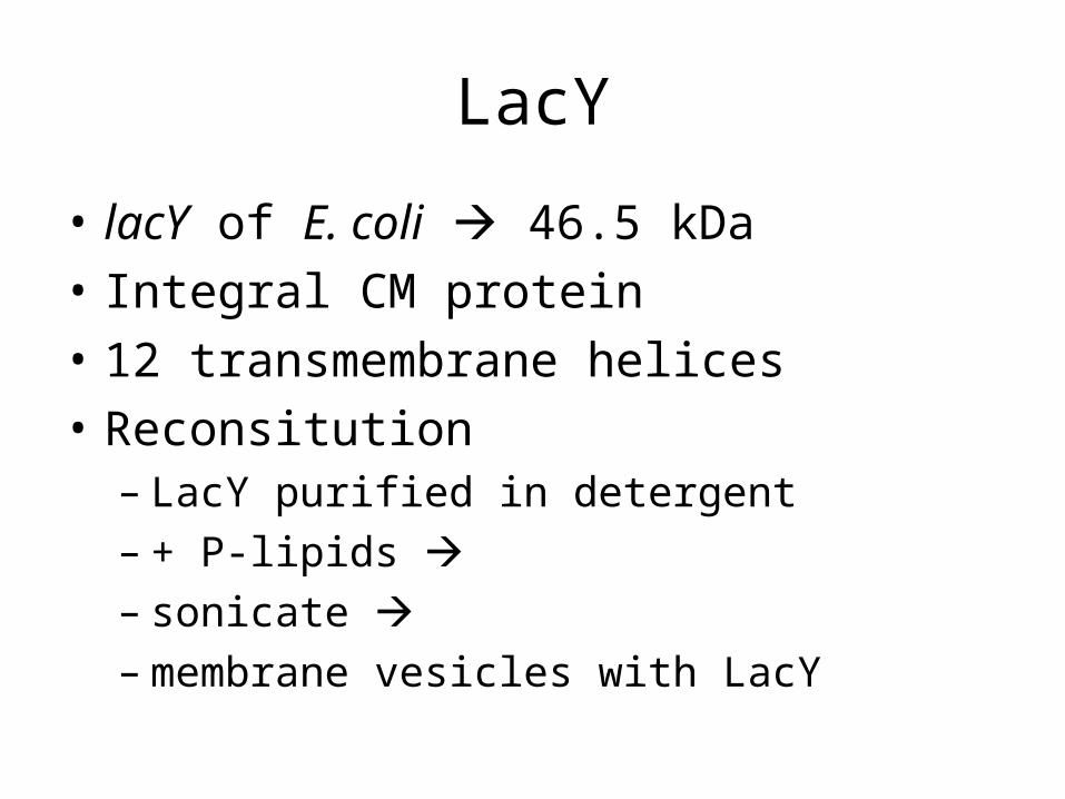
LacY
• lacY of E. coli 46.5 kDa
• Integral CM protein
• 12 transmembrane helices
• Reconsitution– LacY purified in detergent– + P-lipids – sonicate – membrane vesicles with LacY
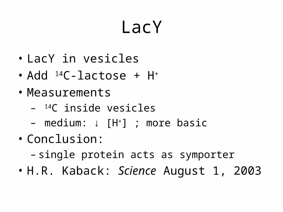
LacY
• LacY in vesicles
• Add 14C-lactose + H+
• Measurements– 14C inside vesicles– medium: ↓ [H+] ; more basic
• Conclusion: – single protein acts as symporter
• H.R. Kaback: Science August 1, 2003
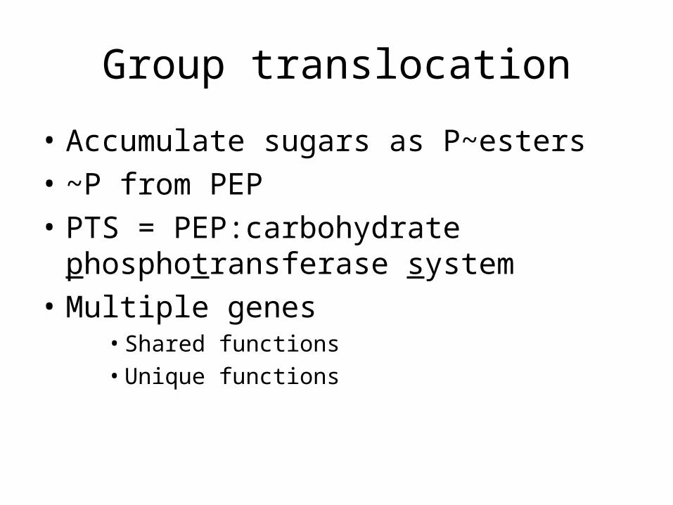
Group translocation
• Accumulate sugars as P~esters
• ~P from PEP
• PTS = PEP:carbohydrate phosphotransferase system
• Multiple genes• Shared functions• Unique functions
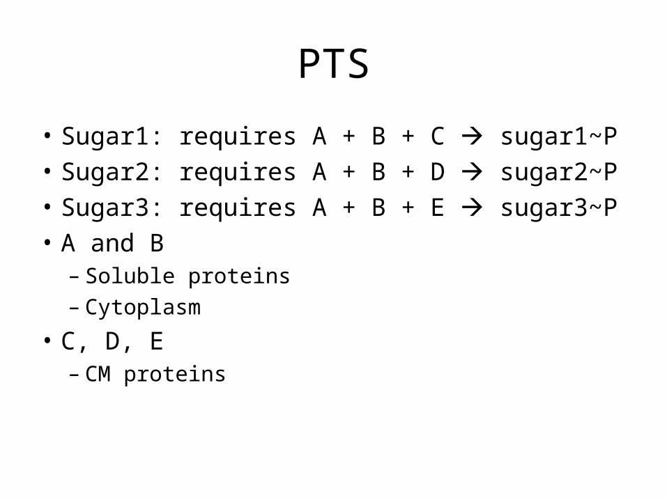
PTS
• Sugar1: requires A + B + C sugar1~P
• Sugar2: requires A + B + D sugar2~P
• Sugar3: requires A + B + E sugar3~P
• A and B– Soluble proteins– Cytoplasm
• C, D, E– CM proteins
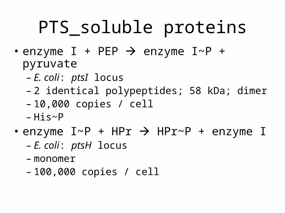
PTS_soluble proteins• enzyme I + PEP enzyme I~P + pyruvate
– E. coli: ptsI locus– 2 identical polypeptides; 58 kDa; dimer– 10,000 copies / cell– His~P
• enzyme I~P + HPr HPr~P + enzyme I– E. coli: ptsH locus– monomer– 100,000 copies / cell
PTS_membrane proteins
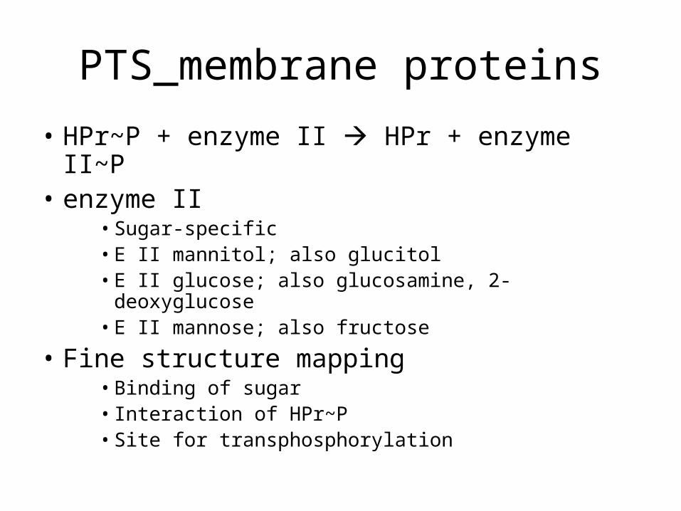
• HPr~P + enzyme II HPr + enzyme II~P• enzyme II
• Sugar-specific • E II mannitol; also glucitol• E II glucose; also glucosamine, 2-deoxyglucose• E II mannose; also fructose
• Fine structure mapping• Binding of sugar• Interaction of HPr~P• Site for transphosphorylation
PTS_membrane proteins
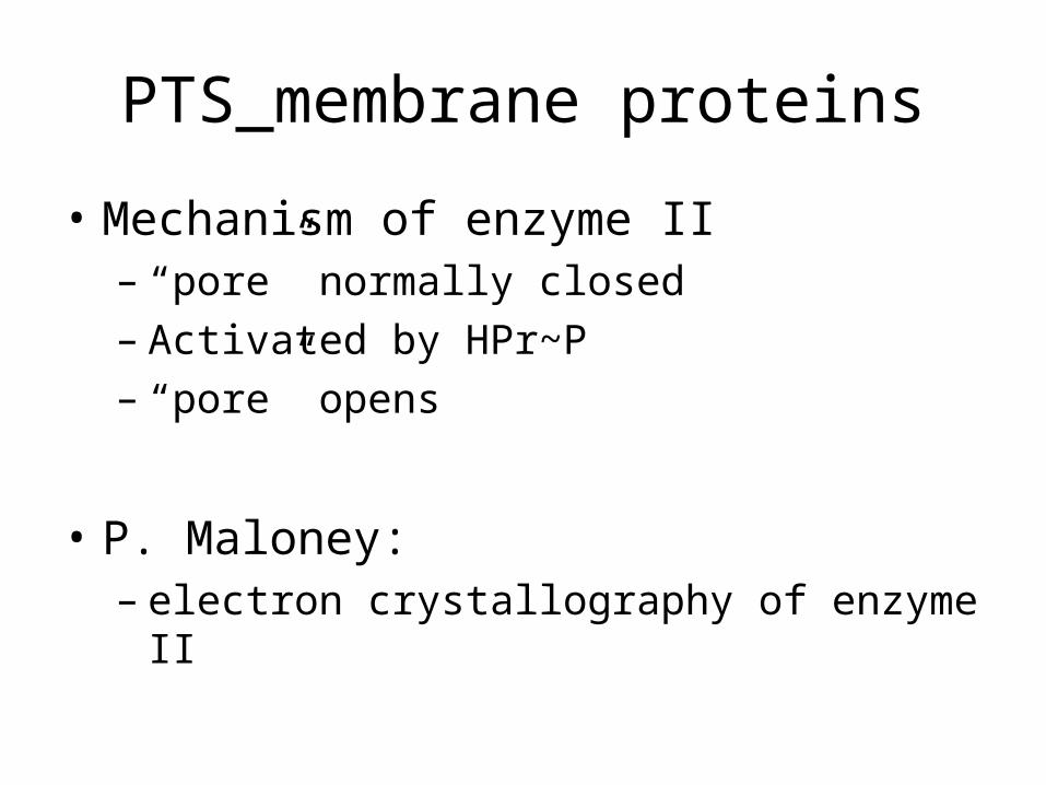
• Mechanism of enzyme II– “pore” normally closed– Activated by HPr~P– “pore” opens
• P. Maloney: – electron crystallography of enzyme II
ABC transporters
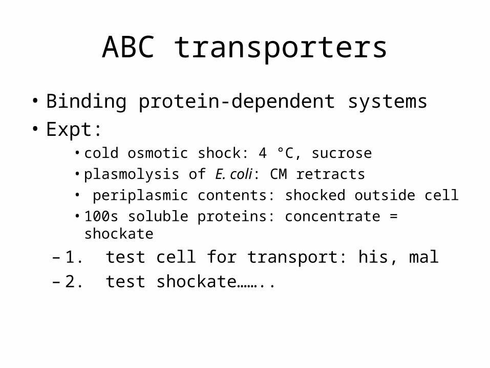
• Binding protein-dependent systems
• Expt: • cold osmotic shock: 4 °C, sucrose • plasmolysis of E. coli: CM retracts • periplasmic contents: shocked outside cell• 100s soluble proteins: concentrate = shockate
– 1. test cell for transport: his, mal– 2. test shockate……..
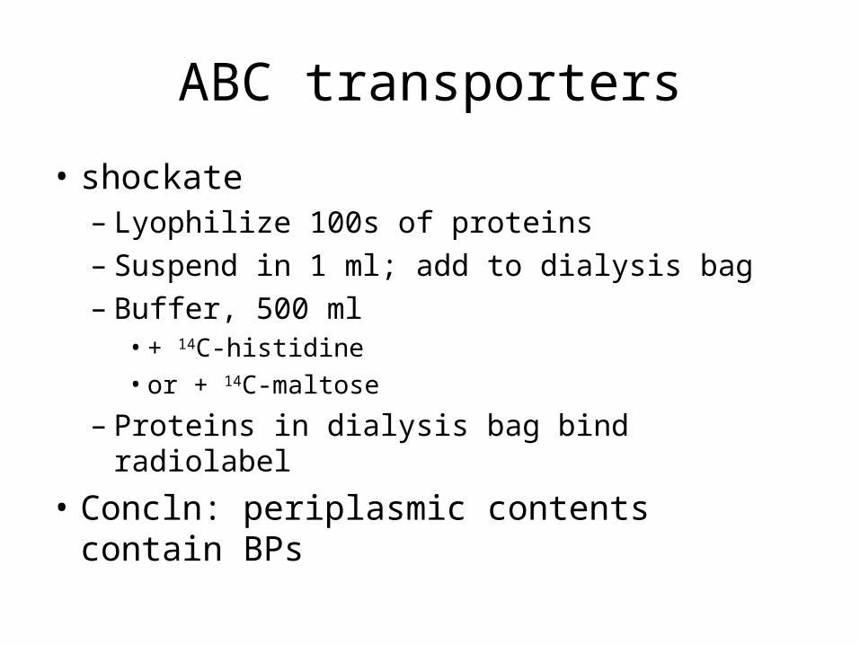
ABC transporters
• shockate– Lyophilize 100s of proteins– Suspend in 1 ml; add to dialysis bag– Buffer, 500 ml
• + 14C-histidine• or + 14C-maltose
– Proteins in dialysis bag bind radiolabel
• Concln: periplasmic contents contain BPs
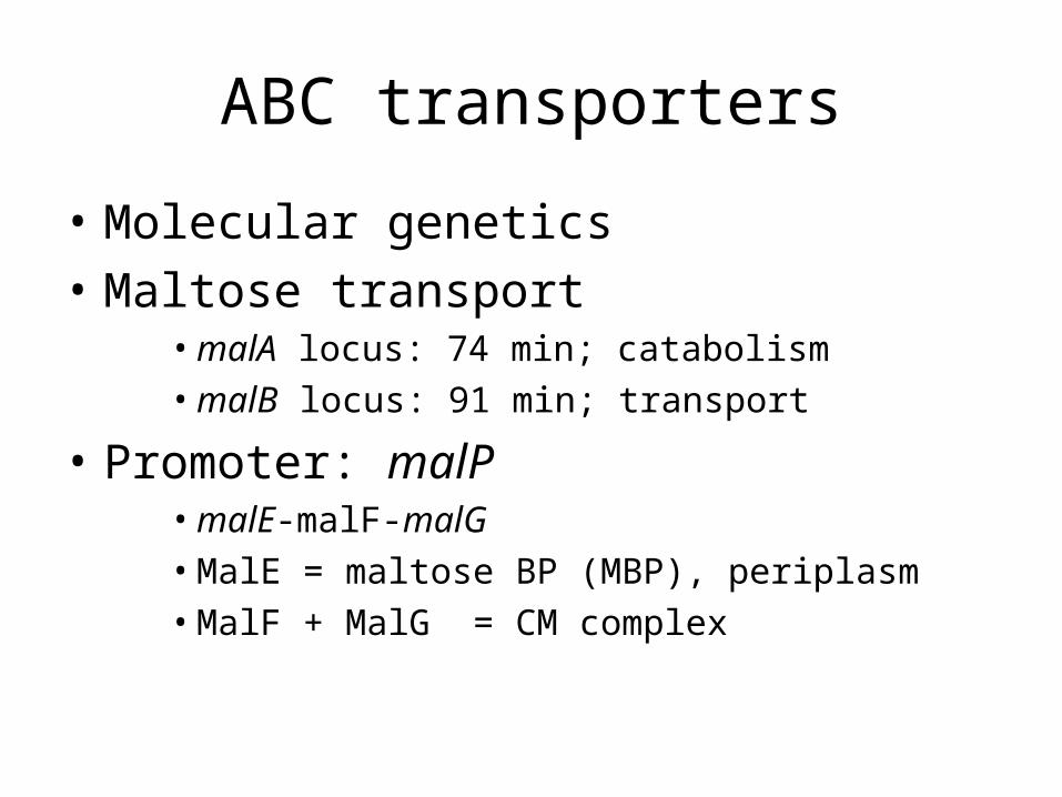
ABC transporters
• Molecular genetics
• Maltose transport• malA locus: 74 min; catabolism• malB locus: 91 min; transport
• Promoter: malP• malE-malF-malG• MalE = maltose BP (MBP), periplasm• MalF + MalG = CM complex
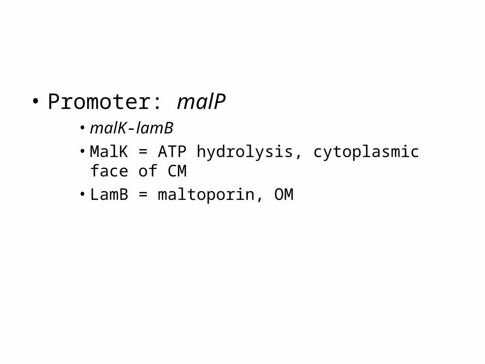
• Promoter: malP • malK-lamB• MalK = ATP hydrolysis, cytoplasmic face of CM• LamB = maltoporin, OM
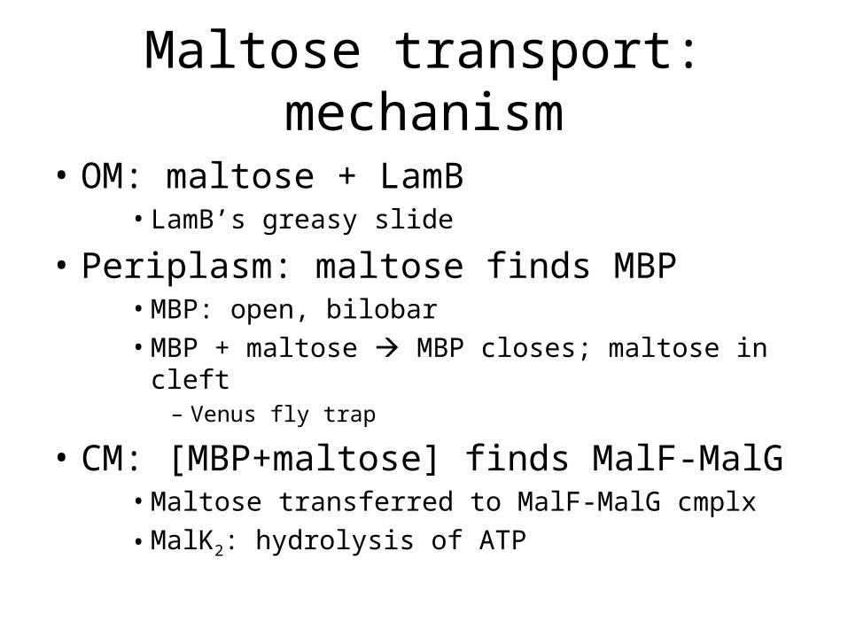
Maltose transport: mechanism
• OM: maltose + LamB• LamB’s greasy slide
• Periplasm: maltose finds MBP• MBP: open, bilobar• MBP + maltose MBP closes; maltose in cleft
– Venus fly trap
• CM: [MBP+maltose] finds MalF-MalG• Maltose transferred to MalF-MalG cmplx
• MalK2: hydrolysis of ATP
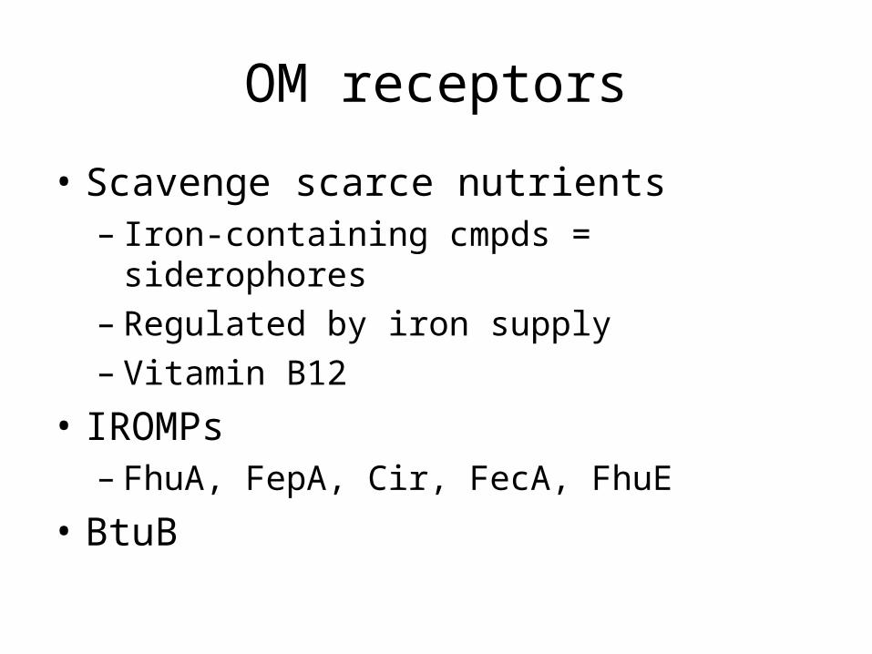
OM receptors
• Scavenge scarce nutrients– Iron-containing cmpds = siderophores– Regulated by iron supply– Vitamin B12
• IROMPs– FhuA, FepA, Cir, FecA, FhuE
• BtuB
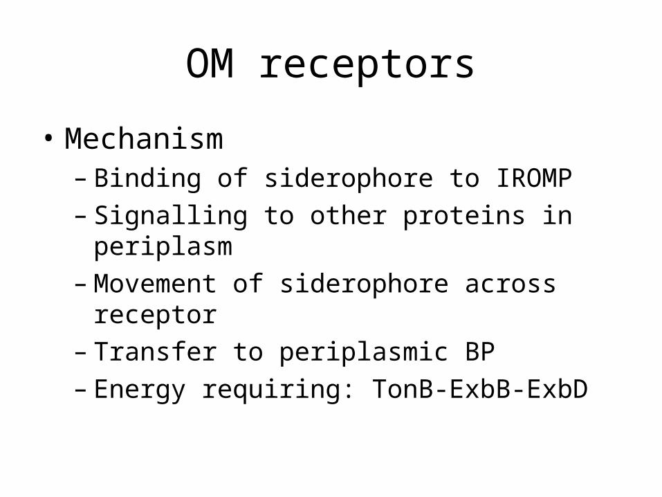
OM receptors
• Mechanism– Binding of siderophore to IROMP– Signalling to other proteins in periplasm– Movement of siderophore across receptor– Transfer to periplasmic BP– Energy requiring: TonB-ExbB-ExbD
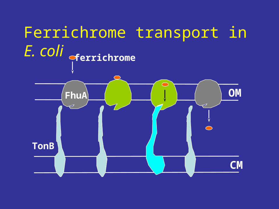
FhuA
TonB
ferrichrome
CM
OM
Ferrichrome transport in E. coli
OM receptors
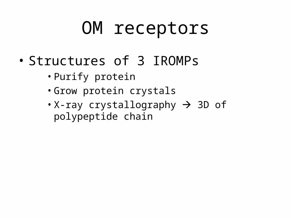
• Structures of 3 IROMPs• Purify protein • Grow protein crystals• X-ray crystallography 3D of polypeptide chain
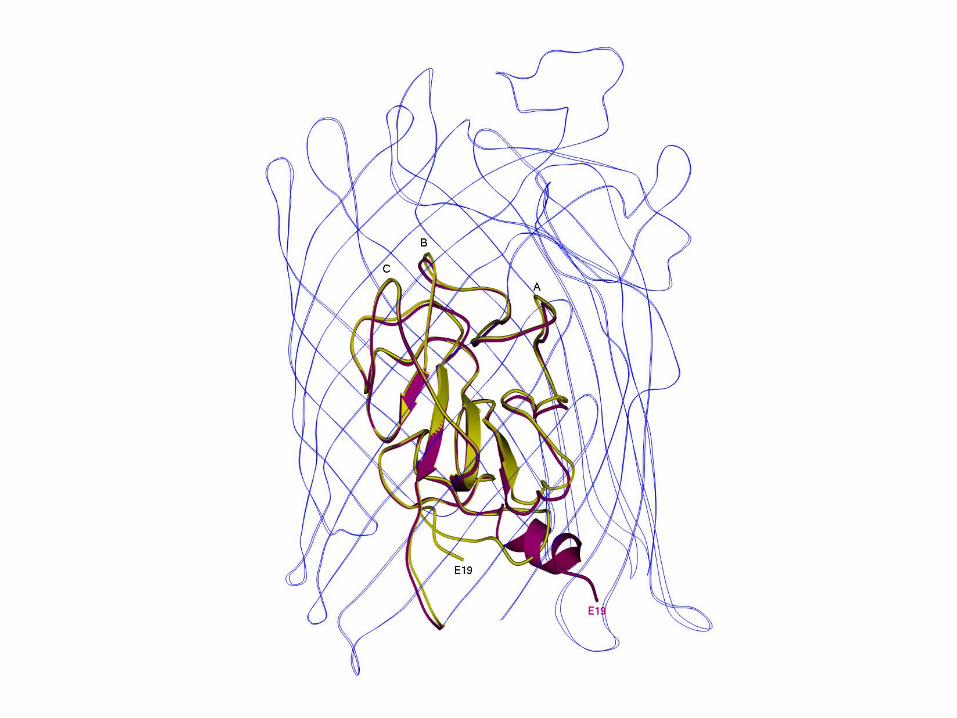
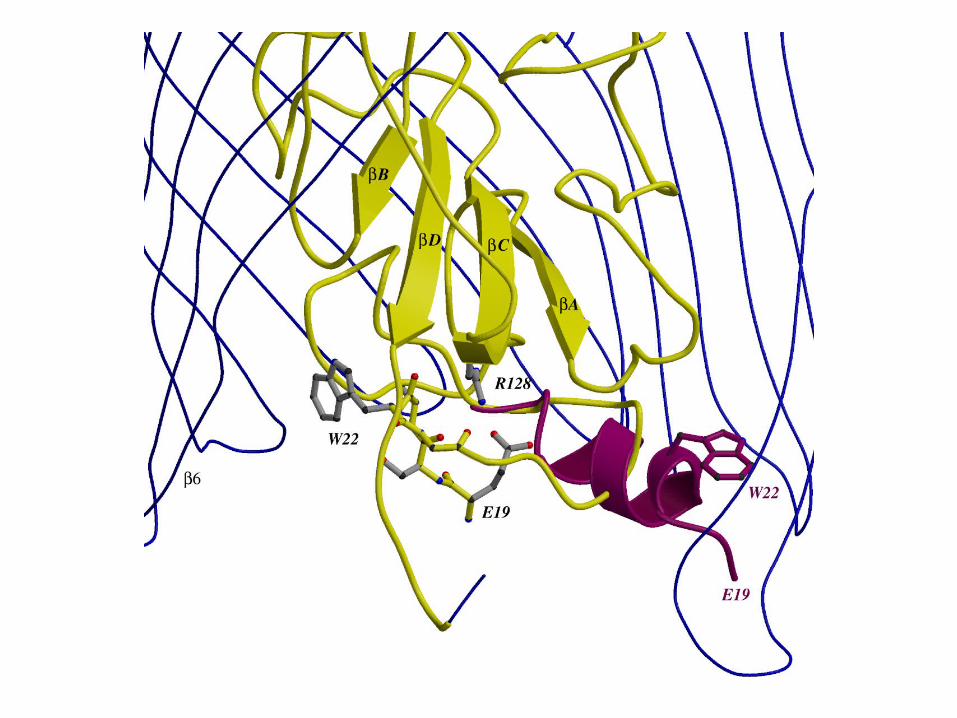
OM receptor
FhuA: 2 domains• N terminus: cork• C terminus:
– β barrel, 22 strands; – Surface-exposed loops– Short connecting turns: periplasm
• Signalling: • Minus siderophore = α helix• Plus siderophore = helix random coil• Tells energy transducing system to activate