Embed Size (px)
Citation preview
Posted at the Institutional Resources for Unique Collection and Academic Archives at Tokyo Dental College,
Available from http://ir.tdc.ac.jp/
Title
Radicular cyst and granuloma : A
clinicopathological study of1590 cases and a
literature review
Author(s)Sanuki, T; Matsuzaka, K; Inoue, K; Hashimoto, K;
Inoue, T
Journal 日本口腔検査学会雑誌, 6(1): 44-49
URL http://hdl.handle.net/10130/3294
Right
44
Original
Radicular cyst and granuloma: A clinicopathological study of
1590 cases and a literature review
Sanuki T, Matsuzaka K, Inoue K, Hashimoto K, Inoue TDepartment of Clinical Pathophysiology, Tokyo Dental College
*:2-9-18 Misaki-cho, Chiyoda-ku, Tokyo, 101-0061 JAPAN
TEL: +81-3-6380-9252 FAX : +81-3-6380-9606
e-mail: [email protected]
Abstract
Radicular cysts are the most common type of cyst found in the jaw. Various hypotheses
regarding their pathogenesis have been proposed, but there is no consensus. Therefore,
the purpose of this study was to evaluate the pathogenesis of radicular cysts via a
clinicopathological study. Radicular cysts and granulomas of 1590 cases seen at the
Tokyo Dental College were evaluated histopathologically. Various materials were present
in the lumens and cyst walls of radicular cysts and granulomas in 39.7% (597/1590) of
those cases, especially, foreign bodies such as root canal filling materials or intracanal
medicaments were recognized in 14.0% of these cases (223/1590). These results
suggest that the failure of root canal treatment frequently plays a role in the formation of
radicular cysts and granulomas.
Key words:periapical lesion, radicular cyst, pathogenesis, dental treatment, root canal
treatment
Reseaved:December 20th 2013 accepted:March 20th 2014
Introduction
Radicular cysts are the most common type of cyst
found in the jaw, and are classified as inflammatory
cysts according to the World Health Organization1)2).
Clinically, radicular cysts are associated with teeth
that have caries, have undergone previous restorative
care, have sustained trauma or are apparent failures
of root canal treatments3). Pathologically, the causes
of radicular cysts have been classified as biological
factors such as bacterial infections, chemical factors
such as stimulation of intracanal medicaments, and
physical factors such as extrusions of instruments
and overfilling of root canal filling materials. In
particular, the relationship between these factors
and dental treatments has been suggested. However,
there is no consensus about the pathogenesis
of radicular cysts related to dental treatments4).
Therefore, to determine the relationship(s) between
the pathogenesis of radicular cysts and dental
treatments, we statistically retrieved the ratio of
these materials which were included in radicular
cysts and granulomas seen at the Tokyo Dental
College and reviewed their origin.
JJ S E D P Vol. 6 No. 1: , 2014
45
Materials and Methods
This study included 1590 patients diagnosed and
treated for radicular cysts and radicular granulomas
at the Tokyo Dental College Chiba and the Suidobashi
Hospital from 2007 to 2011. Specimens were
routinely fixed in 10% buffered formalin and were
embedded in paraffin. Sections 4 mm thick were
serially cut and stained with hematoxylin and eosin.
Those specimens were retrieved histologically and
materials observed in the cystic lumens or cyst
walls of those radicular cysts and granulomas were
categorized into 3 types: “foreign body”, “hard tissue”
and “bacterial cluster”. Subsequently, the ratio of
each category in the 1590 cases was estimated.
Furthermore, in some cases that recognized
the presence of the above-described features,
immunohistochemical staining was performed to
demonstrate the foreign body reaction using an anti-
CD68 antibody (1:50, DAKO, Glostrup, Denmark),
which is expressed on the surface of macrophages.
We also evaluated the protective response in the
lining epithelium by examining the expression of
β -Defensin 2, which is an antimicrobial peptide
secreted by stratified squamous epithelium using
an anti-human β -defensin 2 antibody (1:50,
R&D Systems, Inc., Minneapolis, MN, USA). After
deparaffinization, the sections were microwaved for
30 min at 97 °C for antigen retrieval. Endogenous
peroxidase activity was blocked by incubating the
sections with 3% H2O2 in methanol for 30 min.
To prevent non-specific reactions, sections were
incubated with 3% bovine serum albumin for 30
min in a humidity chamber. After washing in PBS,
sections were incubated with the primary antibodies
overnight at 4°C. The sections were then incubated
with a horseradish-peroxidase-conjugated secondary
antibody [Histofine MAX-PO(MULTI), Nichirei,
Tokyo, Japan] for 30 min in the humidity chamber.
Finally, they were visualized using 0.01% 3,3’
-diaminobenzidine, and were counterstained with
hematoxylin.
Results
Histologically, foreign bodies such as root canal filling
materials or intracanal medicaments (223/1590,
14.0%), hard tissue particles such as cementum or
dentin (202/1590, 12.7%) and microbial clusters
composed of micrococcus and fungus (172/1590,
10.8%) were observed in radicular cysts and
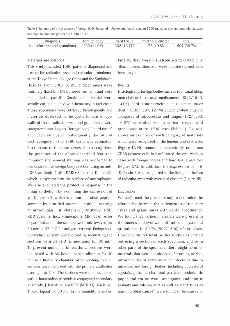
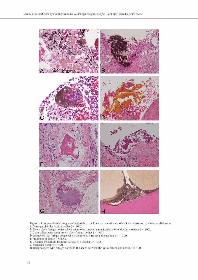
granulomas in the 1590 cases (Table 1). Figure 1
shows an example of each category of materials
which were recognized in the lumens and cyst walls
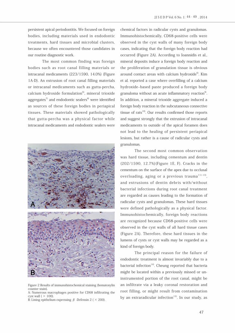
(Figure 1A-H). Immunohistochemically, numerous
CD68-positive cells had infiltrated the cyst walls in
cases with foreign bodies and hard tissue particles
(Figure 2A). In addition, the expression of β
-Defensin 2 was recognized in the lining epithelium
of radicular cysts with microbial clusters (Figure 2B).
Discussion
We performed the present study to determine the
relationship between the pathogenesis of radicular
cysts and granulomas with dental treatments.
We found that various materials were present in
the lumens and cyst walls of radicular cysts and
granulomas in 39.7% (597/1590) of the cases.
However, the retrieval in this study was carried
out using a section of each specimen, and so in
other parts of the specimens there might be other
materials that were not observed. According to Nair,
intraradicular or extraradicular infections due to
microbes and foreign bodies, including cholesterol
crystals, gutta-percha, food particles, endodontic
paper and cotton wool, amalgams, endodontic
sealants and calcium salts as well as scar tissues as
non-microbial causes5) were found to be causes of
diagnosis foreign body hard tissue microbial cluster totalradicular cyst and granuloma 223 (14.9%) 202 (12.7%) 172 (10,8%) 597 (39.7%)
Table 1 Summary of the presence of foreign body, bacterial infection and hard tissue in 1590 radicular cyst and granuloma cases
at Tokyo Dental College since 2007 to20011
44 - 49
46
Figure 1. Example of each category of materials in the lumens and cyst walls of radicular cysts and granulomas (H-E stain).A: Gutta-percha-like foreign bodies ( × 200).B: Blown-black foreign bodies which seem to be intracanal medicaments or endodontic sealers ( × 100).C: Giant cell phagositizing brown-black foreign bodies ( × 400).D: Orange oil-like foreign bodies which seem to be intracanal medicaments ( × 200).E: Fragment of dentin ( × 200).F: Detached cementum from the surface of the apex ( × 100).G: Microbial cluster ( × 400).H: Bacteria laced with foreign bodies in the space between the gutta-percha and dentin ( × 400).
Sanuki et al. Radicular cyst and granuloma: A clinicopathological study of 1590 cases and a literature review
JJ S E D P Vol. 6 No. 1: , 2014
47
persistent apical periodontitis. We focused on foreign
bodies, including materials used in endodontic
treatments, hard tissues and microbial clusters,
because we often encountered those candidates in
our routine diagnostic work.
The most common finding was foreign
bodies such as root canal filling materials or
intracanal medicaments (223/1590, 14.0%) (Figure
1A-D). An extrusion of root canal filling materials
or intracanal medicaments such as gutta-percha,
calcium hydroxide formulation6), mineral trioxide
aggregates7) and endodontic sealers8) were identified
as sources of these foreign bodies in periapical
tissues. These materials showed pathologically
that gutta-percha was a physical factor while
intracanal medicaments and endodontic sealers were
chemical factors in radicular cysts and granulomas.
Immunohistochemically, CD68-positive cells were
observed in the cyst walls of many foreign body
cases, indicating that the foreign body reaction had
occurred (Figure 2A). According to Ioannidis et al.,
mineral deposits induce a foreign body reaction and
the proliferation of granulation tissue is obvious
around contact areas with calcium hydroxide6). Kim
et al. reported a case where overfilling of a calcium
hydroxide–based paste produced a foreign body
granuloma without an acute inflammatory reaction9).
In addition, a mineral trioxide aggregate induced a
foreign body reaction in the subcutaneous connective
tissue of rats10). Our results confirmed those reports
and suggest strongly that the extrusion of intracanal
medicaments to outside of the apical foramen does
not lead to the healing of persistent periapical
lesions, but rather is a cause of radicular cysts and
granulomas.
The second most common observation
was hard tissue, including cementum and dentin
(202/1590, 12.7%)(Figure 1E, F). Cracks in the
cementum on the surface of the apex due to occlusal
overloading, aging or a previous trauma11)-13),
and extrusions of dentin debris with/without
bacterial infections during root canal treatment
are regarded as causes leading to the formation of
radicular cysts and granulomas. These hard tissues
were defined pathologically as a physical factor.
Immunohistochemically, foreign body reactions
are recognized because CD68-positive cells were
observed in the cyst walls of all hard tissue cases
(Figure 2A). Therefore, these hard tissues in the
lumens of cysts or cyst walls may be regarded as a
kind of foreign body.
The principal reason for the failure of
endodontic treatment is almost invariably due to a
bacterial infection14). Cheung reported that bacteria
might be located within a previously missed or un-
instrumented portion of the root canal, might be
an infiltrate via a leaky coronal restoration and
root filling, or might result from contamination
by an extraradicular infection15). In our study, as
Figure 2 Results of immunohistochemical staining (hematoxylin counter stain).A: Numerous macrophages positive for CD68 infiltrating the cyst wall ( × 100).B: Lining epithelium expressing β -Defensin 2 ( × 200).
44 - 49
48
biological factors, microbial clusters were observed
in 172/1590 cases (10.8%) (Figure 1G). In particular,
there were microbial clusters in the space between
the dentin and gutta-percha (Figure 1H), which
indicates that the bacteria might infiltrate via a
leaky root filling. Immunohistochemically, the
expression of β -Defensin 2 was recognized in the
lining epithelium of all radicular cyst cases with
microbial clusters as a protective reaction (Figure
2B). However, the expression of β -Defensin 2 was
consistently just a reaction against bacterial infection,
and therefore, it seemed not to be enough to expel
the bacteria. Although the details of microbes
observed in this study were not determined, various
microbes are regarded as causes of persistent apical
periodontitis including radicular cysts. For example,
the genera Actinomyces5)15)-18), Enterococcus and
Propionibacterium19)20) and Candida albicans5)21)-23)
have been reported. To eradicate those microbes,
optimal intracanal medicaments or antibiotics which
are available for each microbe should be selected
during the root canal treatment.
In conclusion, the results suggest that the
above-described 3 materials in cystic lumens or
cyst walls are related to the extrusion of intracanal
medicaments, cracks in the cementum on the surface
of the apex by occlusal overloading, the extrusion
of dentin debris during root canal treatment and/
or bacterial infiltration via leaky root fillings.
Those materials are typically derived from dental
treatments. In fact, these iatrogenic factors are
responsible for the formation of radicular cysts and
granulomas. To reduce iatrogenic factors, diagnosis
not only by experience as usual but also by objective
evaluation and evidence-based treatment, such as
occlusal equilibration using a bite pressure meter,
isolation of causative microorganisms by intracanal
bacterial culture and optimal selection of antibiotics
based on drug sensitivity tests during root canal
treatment, are needed for clinical dentistry in the
future.
References1) Main DMG: Epithelial jaw cysts: 10 years of WHO
classification. J Oral Pathol Med, 14:1–7, 19852) Kramer IR, Pindborg JJ, Shear M: The WHO histological
typing of odontogenic tumours: a commentary on the second edition, Cancer, 70: 2988–2994, 1992
3) Marx: Oral and maxillofacial pathology: A Rationale for Diagnosis and Treatment, Quintessence Publishing Co, Inc., Illinois, 574-575, 2003
4) Lin LM, Ricucci D, Lin J, Rosenberg PA: Nonsurgical root canal therapy of large cyst-like inflammatory periapical lesions and inflammatory apical cysts, J Endod, 5: 607-615, 2009
5) Nair PN: On the causes of persistent apical periodontitis: a review, Int Endod J, 39: 249-281, 2006
6) Ioannidis K, Thomaidis V, Fiska A, Lambrianidis T: Lack of periradicular healing and gradually increasing swelling two years after intentional extrusion of calcium hydroxide into periapical lesion: report of a case, Oral Surg Oral Med Oral Pathol Oral Radiol Endod, 109: e86-91, 2010
7) Chang SW, Oh TS, Lee W, Cheung GS, Kim HC: Long-term observation of the mineral trioxide aggregate extrusion into the periapical lesion: a case series, Int J Oral Sci, 5: 54-57, 2013
8) Froes FG, Miranda AM, Abad Eda C, Riche FN, Pires FR: Non-surgical management of paraesthesia and pain associated with endodontic sealer extrusion into the mandibular canal, Aust Endod J, 35: 183-186, 2009
9) Kim JW, Cho KM, Park SH, Song SG, Park MS, Jung HR, Song JY, Kim YS, Lee SK: Overfilling of calcium hydroxide-based paste Calcipex II produced a foreign body granuloma without acute inflammatory reaction, Oral Surg Oral Med Oral Pathol Oral Radiol Endod, 107: e73-76, 2009
10) Batur YB, Acar G, Yalcin Y, Dindar S, Sancakli H, Erdemir U: The cytotoxic evaluation of mineral trioxide aggregate and bioaggregate in the subcutaneous connective tissue of rats, Med Oral Patol Oral Cir Bucal, 18: e745-751, 2013
11) Tai TF, Chiang CP, Lin CP, Lin CC, Jeng JH: Persistent endodontic lesion due to complex cementodentinal tears in a maxillary central incisor--a case report, Oral Surg Oral Med Oral Pathol Oral Radiol Endod, 103:e55-60, 2007
12) Noma N, Kakigawa H, Kozono Y, Yokota M: Cementum crack format ion by repeated loading in vitro, J Periodontol, 78:764-769, 2007
13) Stewart ML, McClanahan SB: Cemental tear: a case report, Int Endod J, 39: 81-86, 2006
14) Nair R: Pathology of apical periodontitis, In: Ørstavik D, Pitt Ford TR, eds, Essential Endodontology: Prevention and Treatment of Apical Periodontitis, 2nd ed,, MA: Blackwell Science, Boston, 68–88, 2008
15) Cheung GS: Endodontic failures: changing the approach, Int Dent J, 46: 131–138, 1996
16) Nair PN, Pajarola G, Luder HU: Ciliated epithelium-lined radicular cysts, Oral Surg Oral Med Oral Pathol Oral Radiol Endod, 94: 485-493, 2002
17) Tek M, Metin M, Sener I, Bereket C, Tokac M, Kazancioglu HO, Ezirganli S: The predominant bacteria isolated from radicular cysts, Head Face Med, 9: 25, 2013
18) Ricucci D, Siqueira JF Jr: Apical actinomycosis as a continuum of intraradicular and extraradicular infection:
Sanuki et al. Radicular cyst and granuloma: A clinicopathological study of 1590 cases and a literature review
JJ S E D P Vol. 6 No. 1: , 2014
49
case report and critical review on its involvement with treatment failure J Endod, 34: 1124-1129, 2008
19) Wang QQ, Zhang CF, Chu CH, Zhu XF: Prevalence of Enterococcus faecalis in saliva and filled root canals of teeth associated with apical periodontitis, Int J Oral Sci, 4: 19-23, 2012
20) Lee SH, Baek DH: Antibacterial and neutralizing effect of human β -defensins on Enterococcus faecalis and Enterococcus faecalis lipoteichoic acid, J Endod, 38: 351-356, 2012
21) Miranda TT, Vianna CR, Rodrigues L, Monteiro AS, Rosa CA, Corrêa A Jr: Diversity and frequency of yeasts from the dorsum of the tongue and necrotic root canals associated with primary apical periodontitis, Int Endod J, 42: 839-344, 2009
22) Zhang S, Wang QQ, Zhang CF, Soo I: Identification of dominant pathogens in periapical lesions associated with persistent apical periodontitis, Chin J Dent Res, 13: 115-121, 2010
23) Poptani B, Sharaff M, Archana G, Parekh V: Detection of Enterococcus faecalis and Candida albicans in previously root-filled teeth in a population of Gujarat with polymerase chain reaction, Contemp Clin Dent, 4: 62-66, 2013
44 - 49