Embed Size (px)
Citation preview
Produced with the kind Support of
Radionuclide Metabolic Therapy Clinical Aspects,
Dosimetry and Imaging
A Technologist’s Guide
Publications · Brochures
European Association of Nuclear Medicine
®EANMEANM
Produced with the kind Support of
Peștean, Claudiu (Cluj – Napoca)
Veloso Jéronimo, Vanessa (Loures)
Hogg, Peter (Manchester)
Contributors
Berhane Menghis, Ruth (Liverpool)
Bodet-Milin, Caroline (Nantes)
Chiti, Arturo (Milan)
Cutler, Cathy S. (Missouri)
do Rosário Vieira, Maria (Lisbon)
Faivre-Chauvet, Alain (Nantes)
Flux, Glenn (Sutton, Surrey)
Gorgan, Ana (Cluj – Napoca)
Kraeber-Bodéré, Françoise (Nantes)
Larg, Maria Iulia (Cluj – Napoca)
Lowry, Brian A. (Liverpool)
Lupu, Nicoleta (Cluj – Napoca)
Mantel, Eleanor S. (Philadelphia)
Mayes, Christopher (Liverpool)
Pallardy, Amandine (Nantes)
Peștean, Claudiu (Cluj – Napoca)
Piciu, Doina (Cluj – Napoca)
Rauscher, Aurore (Nantes)
Silva, Nadine (Lisbon)
Sjögreen Gleisner, Katarina (Lund)
Strigari, Lidia (Rome)
Szczepura, Katy (Manchester)
Testanera, Giorgio (Milan)
Vinjamuri, Sobhan (Liverpool)
Williams, Jessica (Philadelphia)
Editors
EANM
3
Foreword 5
Introduction 6Claudiu Peştean, Vanessa Veloso Jerónimo and Peter Hogg
Acronyms 7
Section I
1. Principles in Radionuclide Therapy (*) 9
Eleanor S. Mantel and Jessica Williams
2. Biological Efects of Ionising Radiation 18
Katy Szczepura
3. Dosimetry in Molecular Radiotherapy 29
Katarina Sjögreen Gleisner, Lidia Strigari, Glenn Flux
4. Special Considerations in Radiation Protection:
Minimising Exposure of Patients, Staf and Members of the Public 41
Claudiu Peştean and Maria Iulia Larg
Section II
Preamble on a Multiprofessional Approach in Radionuclide Metabolic Therapy 54
Giorgio Testanera and Arturo Chiti
1. Radionuclide Therapy in Thyroid Carcinoma 61
Doina Piciu
2. Radionuclide Therapy in Benign Thyroid Disease 77
Doina Piciu
3. Radionuclide Therapy in Neuroendocrine Tumours 81
Ruth Berhane Menghis, Brian A. Lowry, Christopher Mayes and Sobhan Vinjamuri
4. Radionuclide Therapy in Hepatocellular Carcinoma 93
Nadine Silva and Maria do Rosário Vieira
5. Radioimmunotherapy in Lymphomas 105
Aurore Rauscher, Caroline Bodet-Milin, Amandine Pallardy, Alain Faivre-Chauvet, Françoise Kraeber-Bodéré
6. Radionuclide Therapy of Refractory Metastatic Bone Pain 112
Brian A. Lowry, Ruth B. Menghis, Christopher Mayes and Sobhan Vinjamuri
7. Radiosynovectomy 121
Christopher Mayes, Brian A. Lowry, Ruth Berhane Menghis and Sobhan Vinjamuri
8. Nursing Implications for Patients Undergoing Radionuclide Metabolic Therapy 129
Ana Gorgan, Nicoleta Lupu and Claudiu Peștean
Section III
1. Future Perspectives in Radionuclide Therapy (*) 136
Cathy S. Cutler
Imprint 153
(*) Articles were written with the kind support
of and in cooperation with:
Contents
Prin
ted
in a
cco
rdan
ce w
ith t
he
Au
stria
n E
co-L
abel
for p
rinte
d m
atte
rs.
4
The editors would like to express sincere gratitude
to the following people, because without their generous help
this book would not have come to fruition:
Reviewer: Lisa Bodei
English language editing: Rick Mills
Project management: Katharina Leissing
Acknowledgements

EANM
5
Foreword
Nuclear Medicine is a composite discipline
in which radionuclide therapy has a role of
growing importance, with an increasing im-
pact on clinical practice. Advances in radio-
pharmaceutical production and the imple-
mentation of a multidisciplinary approach
in clinical medicine have propelled radio-
nuclide therapy methods and application
towards personalised treatment, therapeutic
eicacy, patient comfort and radiation safety.
Technologists are key igures in this process,
since their competencies allow them to play
an important role in every step necessary for
successful treatment, from radiopharmaceu-
tical preparation to administration. They are
also the main actors in pre- and post-thera-
peutic imaging. The current book is specii-
cally aimed at radiographers and technolo-
gists working in, or intending to work in, a
Nuclear Medicine department with radionu-
clide therapy facilities, though it is also likely
to be valuable for other healthcare profes-
sionals working in, or willing to work in, this
challenging environment.
A successful radionuclide therapy unit is not
only fundamental for patient care in Oncol-
ogy, Endocrinology and Orthopaedics, but
can also serve as a gold standard in an inter-
professional and multidisciplinary approach
to clinical medicine.
Following the successful PET-CT Tech Guide
series, this book is the joint work of many
professionals from diferent nations and
ields of interest within Nuclear Medicine.
I want to really thank all those people who
have contributed to this work as authors and
reviewers, without whom the book would
not have been possible. I am proud to be
able to welcome and thank our colleagues
from the SNM (Society of Nuclear Medicine,
United States) for their high-quality contribu-
tions. I also wish to extend particular grati-
tude to the EANM Dosimetry and Therapy
Committee for their availability and expertise
in the required ields. Special thanks are due
to Claudiu Peştean, Vanessa Veloso Jerónimo
and Professor Peter Hogg, for their incredible
enthusiasm and competence in dealing with
the editorial duties and organisational work.
Finally, I remain extremely grateful to the
EANM Executive Committee, the EANM Exec-
utive Secretariat, the Technologist Commit-
tee and all the EANM committees involved in
the project.
With my warmest regards
Giorgio Testanera
Chair, EANM Technologist Committee
6
This year we have focussed on radionuclide
therapy for our book. For decades, radionu-
clide therapy has been used to help manage
a range of malignant and benign diseases,
and for many pathologies its utility is well
known and well documented. In the early
years the radionuclide range and the pathol-
ogies which could be managed (treatment
and/or palliation) were limited. However,
signiicant progress continues to be made,
and there has been considerable expansion
in the available therapeutic radionuclides
and the pathologies which can be managed
by them. On this basis we feel it is timely for
this book to be published. The book brings
together experts in the ield of radionuclide
therapy from Europe and America in order
that they can share their theoretical knowl-
edge and clinical/practical experience. These
experts emanate from a range of professional
backgrounds and include medical physicists,
nuclear medicine physicians, radiographers,
nuclear medicine technologists and others.
The book commences with background
information about radionuclide therapy
(Section I). If you are new to radionuclide
therapy, then we suggest that special atten-
tion is paid to the irst four chapters as the
information covered will give you a good
theoretical grounding; with this in mind you
can progress to read about the clinical and
technical aspects of radionuclide therapy in
Section II. Here, attention is paid to how to
conduct radiotherapy procedures and also
the pathologies which it can treat and/or
where it can be used for palliation. This sec-
tion contains signiicant input from those
who practice radionuclide therapy, especially
nuclear medicine physicians and others who
have a range of relevant clinical skills. In Sec-
tion III we consider the future, and particular
emphasis is placed on molecular therapy.
The inal chapter considers new radionu-
clides which may become valuable in the
future.
We should like to thank the authors who
have taken the time to write the chapters
and also the reviewers who have provided
constructive feedback to the authors. We
acknowledge that writing and reviewing
involves a considerable efort and we hope
that the readers will ind this book useful for
training and practical purposes.
IntroductionClaudiu Peştean, Vanessa Veloso Jerónimo and Peter Hogg
EANM
7
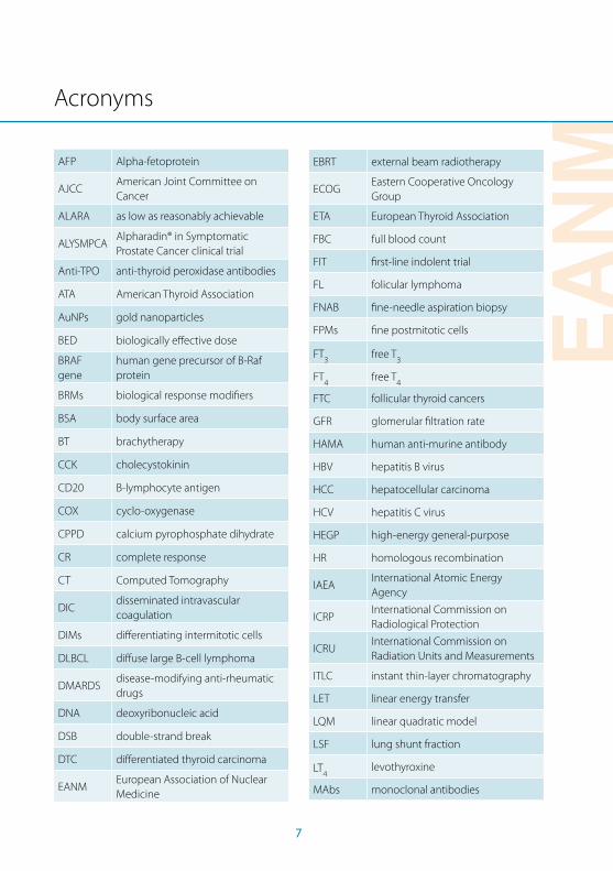
AFP Alpha-fetoprotein
AJCCAmerican Joint Committee on Cancer
ALARA as low as reasonably achievable
ALYSMPCAAlpharadin® in Symptomatic Prostate Cancer clinical trial
Anti-TPO anti-thyroid peroxidase antibodies
ATA American Thyroid Association
AuNPs gold nanoparticles
BED biologically efective dose
BRAF gene
human gene precursor of B-Raf protein
BRMs biological response modiiers
BSA body surface area
BT brachytherapy
CCK cholecystokinin
CD20 B-lymphocyte antigen
COX cyclo-oxygenase
CPPD calcium pyrophosphate dihydrate
CR complete response
CT Computed Tomography
DICdisseminated intravascularcoagulation
DIMs diferentiating intermitotic cells
DLBCL difuse large B-cell lymphoma
DMARDSdisease-modifying anti-rheumatic drugs
DNA deoxyribonucleic acid
DSB double-strand break
DTC diferentiated thyroid carcinoma
EANMEuropean Association of Nuclear Medicine
EBRT external beam radiotherapy
ECOGEastern Cooperative Oncology Group
ETA European Thyroid Association
FBC full blood count
FIT irst-line indolent trial
FL folicular lymphoma
FNAB ine-needle aspiration biopsy
FPMs ine postmitotic cells
FT3
free T3
FT4
free T4
FTC follicular thyroid cancers
GFR glomerular iltration rate
HAMA human anti-murine antibody
HBV hepatitis B virus
HCC hepatocellular carcinoma
HCV hepatitis C virus
HEGP high-energy general-purpose
HR homologous recombination
IAEAInternational Atomic Energy Agency
ICRPInternational Commission on Radiological Protection
ICRUInternational Commission on Radiation Units and Measurements
ITLC instant thin-layer chromatography
LET linear energy transfer
LQM linear quadratic model
LSF lung shunt fraction
LT4
levothyroxine
MAbs monoclonal antibodies
Acronyms
8
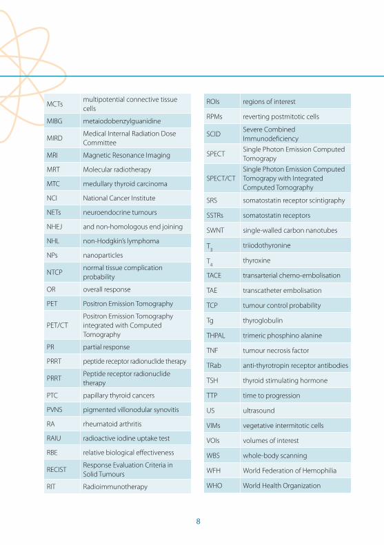
MCTsmultipotential connective tissue cells
MIBG metaiodobenzylguanidine
MIRDMedical Internal Radiation Dose Committee
MRI Magnetic Resonance Imaging
MRT Molecular radiotherapy
MTC medullary thyroid carcinoma
NCI National Cancer Institute
NETs neuroendocrine tumours
NHEJ and non-homologous end joining
NHL non-Hodgkin’s lymphoma
NPs nanoparticles
NTCPnormal tissue complication probability
OR overall response
PET Positron Emission Tomography
PET/CTPositron Emission Tomography integrated with Computed Tomography
PR partial response
PRRT peptide receptor radionuclide therapy
PRRTPeptide receptor radionuclide therapy
PTC papillary thyroid cancers
PVNS pigmented villonodular synovitis
RA rheumatoid arthritis
RAIU radioactive iodine uptake test
RBE relative biological efectiveness
RECISTResponse Evaluation Criteria in Solid Tumours
RIT Radioimmunotherapy
ROIs regions of interest
RPMs reverting postmitotic cells
SCIDSevere Combined Immunodeiciency
SPECTSingle Photon Emission Computed Tomograpy
SPECT/CTSingle Photon Emission Computed Tomograpy with Integrated Computed Tomography
SRS somatostatin receptor scintigraphy
SSTRs somatostatin receptors
SWNT single-walled carbon nanotubes
T3
triiodothyronine
T4
thyroxine
TACE transarterial chemo-embolisation
TAE transcatheter embolisation
TCP tumour control probability
Tg thyroglobulin
THPAL trimeric phosphino alanine
TNF tumour necrosis factor
TRab anti-thyrotropin receptor antibodies
TSH thyroid stimulating hormone
TTP time to progression
US ultrasound
VIMs vegetative intermitotic cells
VOIs volumes of interest
WBS whole-body scanning
WFH World Federation of Hemophilia
WHO World Health Organization
EANM
9
Section I1. Principles in Radionuclide Therapy
Eleanor S. Mantel and Jessica Williams
Introduction
Not long after the discovery of radium by
Marie and Pierre Curie in 1898, Alexander Gra-
ham Bell predicted its use to treat tumours
in 1903. Within 10 years, radium had indeed
been used to treat a multitude of diseases [1].
The discovery of radium thus directly led to
the progress in radionuclide therapy. While
radionuclide therapy has evolved over the
years, the basic theory has stayed the same.
Radionuclide therapy uses ionising radiation
to kill or shrink abnormal cells and tumours
by damaging the cells’ DNA, which causes
them to stop growing. Although these treat-
ments are delivered systemically, they are
cell speciic, targeting distant metastases
throughout the body as well as the primary
tumour. The cessation of cell growth allows
not only for palliative therapy, but the com-
plete ablation of certain cancerous disease
processes while eliciting a low or no physi-
ological response from the patient. This is
attributed to the patient-speciic dosing,
which potentially keeps the levels of toxicity
to a minimum. This, in turn, helps to improve
the patient’s tolerance to treatment and may
improve prognosis and outcome.
All of the therapies discussed in this chapter
are accomplished by utilising beta-emitting
isotopes to deliver a high localised radiation
dose. Beta particle emission occurs when the
ratio of neutrons to protons in the nucleus is
too high. An excess neutron is transformed
into a proton and an electron. The pro-
ton stays in the nucleus and the electron is
expelled. Often, gamma-ray emission ac-
companies the emission of a beta particle.
Typically, if there are gamma emissions, they
follow the decay of the beta. It is this gamma
ray emission that allows some of the isotopes,
at lower doses, to be used for diagnostic and
dosimetric purposes prior to treatment. Ev-
ery isotope has a unique energy that is pro-
duced. The energy of the particle itself deter-
mines how much speed a beta particle has,
how far it can penetrate and how much en-
ergy it emits to the tissue. As with all radioac-
tive emitters, beta emitters must be shielded.
Beta shielding is, generally, achieved using
Lucite or plexiglass. Lucite and plexiglass are
the materials of choice for shielding in an at-
tempt to reduce the number of bremsstrah-
lung interactions and production of X-rays.
However, in some high-dose or high-energy
therapies that have a component of gamma
decay, lead shielding may be used as well.
Generally, medical therapies have a ben-
eit versus risk ratio that must be considered.
When considering a radionuclide therapy
regimen, one needs to review the patient’s
medical history to determine whether he or
she is an ideal candidate for the treatment.
Each speciic radionuclide therapy has its
own set of relative and absolute contraindi-
cations. Some common contraindications
in female patients include pregnancy and
breastfeeding [2-6]. Adherence to radiation
safety precautions and instructions is es-
sential for all radiotherapy administrations.
For patients unable to adhere to certain
10
restraints following the treatment (for exam-
ple, patients with urinary incontinence [2]),
an in-patient admission might be necessary.
Admission to the shielded isolation ward
may be required for patients receiving treat-
ments with certain high-energy isotopes or
at high doses, depending on the regulatory
requirements [2].
Radioisotopes and their uses in
radionuclide therapy
In this chapter, we will explore a number of
isotopes that can be used for radionuclide
therapy. Since each of the isotopes can be
used either independently or bound to an-
other chemical compound, they are em-
ployed to treat diferent disease processes.
Iodine-131
A commonly used isotope for radiotherapy is
iodine-131 (131I). 131I is a beta emitter with a
principal gamma ray of 364 keV (81% abun-
dance) and beta particles with an energy of
0.61 MeV. Its half-life is 8.1 days and it has an
average range in tissue of 0.4 mm. 131I is pro-
duced by the irradiation of tellurium-130 in a
nuclear reactor. While some patients may be
treated on an out-patient basis, regulatory re-
quirements may necessitate those receiving
high doses of 131I to have an extended stay
in a lead-lined room as an in-patient. Patients
receiving these higher doses require special
handling in an efort to minimise radiation
exposure to those around them.
Given in its natural form, 131I-sodium iodide
is used to treat residual thyroid cancer and
metastatic disease by ablating any residual
tissue after partial or complete thyroidec-
tomy. 131I-sodium iodide is also used to treat
non-cancerous diseases such as hyperthy-
roidism and non-toxic multinodular goitre.
The dose is administered orally and can be in
capsule or liquid form since this radionuclide
is readily absorbed from the gastrointestinal
tract into the salivary glands, gastric mucosa
and thyroid tissue. Because both malignant
and benign disease processes are treated
utilising 131I, the dose will difer from one pa-
tient to another. Doses generally range from
1.11 to 11.1 GBq.
Prognosis following 131I therapy varies among
cancer patients. While patient age, gender,
pathology of the cancer, grading and size all
have an efect on how thyroid cancers be-
have, they still have a favourable prognosis.
The well-diferentiated types of thyroid can-
cer, follicular and papillary, generally have
better outcomes than the other types (med-
ullary and anaplastic), with 10-year survival
rates of between 92% and 98%. However,
5-20% of patients with well-diferentiated
thyroid cancer will experience loco-regional
relapse that requires additional treatments
and/or surgery [7].
Patients with hyperthyroidism that is treated
with 131I-sodium iodide have a high response
rate. Those given a higher dose of 131I-sodium
iodide (600 MBq) have a higher cure rate but
also an increased incidence of hypothyroid-
ism [8].
Section I 1. Principles in Radionuclide Therapy
EANM
11
131I-labelled metaiodobenzylguanidine
(MIBG) is an analogue of noradrenaline [2]
and therefore is taken up by the adrenergic
nervous system. It is this uptake that makes 131 I-MIBG the tracer of choice when treating
a number of diferent cancers and diseases.
When 131I is tagged to MIBG, it can be used
to treat stage III or IV neuroblastoma, inoper-
able phaeochromocytoma, inoperable carci-
noid tumour, inoperable paraganglioma and
metastatic or recurrent medullary thyroid
cancer [2]. The dose is administered via slow
intravenous infusion and ranges between 3.7
and 11.2 GBq. Approximately 60% of patients
treated for phaeochromocytoma respond to
this therapy. Thirty percent see a regression
in tumour size while 30% experience symp-
tom relief; unfortunately, however, about a
third of the patients have no response at all
[7].
131I-Lipiodol is another form of 131I radiother-
apy and is used to treat inoperable primary
hepatic carcinoma. Lipiodol is a naturally io-
dinated fatty acid ethyl ester of poppy seed
oil. This iodine-labelled product consists of
fat droplets of approximately 20–200 µm in
diameter. Lipiodol is administered directly
into the hepatic artery in a volume of 2–3 ml.
The standard dose administered is 0.9–2.4
GBq. 131I-Lipiodol targets cancer cells, causing
cytotoxicity in the tumour cells while spar-
ing the normal cells and tissues surrounding
the tumour. While a majority of the dose is
retained in the liver, lung ibrosis is a com-
mon complication when high lung uptake
is identiied. 131I-Lipiodol was found by Raoul
et al. to have reduced tumour size in approxi-
mately 55% of those treated [9].
131I-tositumomab is a murine monoclonal an-
tibody that is used to treat CD20-positive, fol-
licular, non-Hodgkin’s lymphoma refractory
to rituximab and in relapse. The therapy is ad-
ministered intravenously. On the basis of the
initial dosimetric calculations, most patients
receive a therapeutic dose of 2,590–3,330
MBq, but there is a wide variation in dose
range. Patients who have previously received
murine antibodies must have a negative se-
rum human anti-murine antibody (HAMA)
result before proceeding with additional
treatment regimens. Use of 131I-tositumomab
is also contraindicated in patients with hy-
persensitivity to murine (mouse) proteins.
It has been found that patients undergoing
this therapy have a 63% response rate with a
median duration of 25.3 months [7].
Yttrium-90
Yttrium-90 (90Y) is a beta emitter with a half-
life of 2.7 days and an energy of 2.27 MeV. It
has an average soft tissue range of 3.6 mm. 90Y is produced by high-purity separation
from strontium-90 (90Sr), a ission product of
uranium in a nuclear reactor.
90Y-ibritumomab tiuxetan is a CD20-directed
radiotherapeutic antibody used to treat pa-
tients with relapsed or refractory, low-grade
or follicular B-cell non-Hodgkin’s lymphoma
(NHL) and previously untreated follicular NHL
who achieve a partial or complete response to
irst-line chemotherapy [6]. 90Y-ibritumomab
12
tiuxetan is contraindicated in patients with
hypersensitivity to murine (mouse) proteins.
Four hours prior to administration of the 90Y-
ibritumomab tiuxetan, the patient receives
rituximab therapy with the goal of clearing
the majority of normal B cells so that the
therapeutic dose is more focussed on the
tumour cells. 90Y-ibritumomab tiuxetan is ad-
ministered intravenously through a 0.22-µm
low-protein-binding in-line ilter between
the syringe and the infusion port. The dose
itself is based on the patient’s platelet count
and actual body weight. Post-treatment eval-
uation is an important component of this
treatment as thrombocytopenia and neutro-
penia are common adverse events, occurring
in approximately 90% of patients. Seventy
percent of rituxan-refractory patients treated
with 90Y-labelled rituxan showed an overall
response that lasted 7.7 months [7].
90Y-microspheres are utilised as a treatment
in patients with non-resectable hepatomas
and liver metastases [10,11]. Microspheres
are a single-use, permanently implanted
radiotherapy source. The radioactive micro-
spheres are delivered directly to the liver via
an intra-arterial catheter in the hepatic ar-
tery that supplies blood to the tumour. The
microspheres are not metabolised nor are
they excreted; therefore they remain within
the tumour and continue to have a radio-
therapeutic efect. Generally, the doses for
these therapies are 1.5–2.5 GBq but choice
of dose is patient dependent. It is important
to determine the percent of lung shunting
present prior to the administration of the
treatment in order to ensure that the micro-
spheres will localise in the intended tumour
site and not end up in the stomach, lungs
or small intestines. Cumulative dose is also
taken into account in patients receiving
consecutive treatments. Doses for patients
with increased shunting or previous therapy
administrations will be decreased appropri-
ately.
There are two types of microsphere: resin
and glass. Resin microspheres are 20–60 µm
in diameter. The activity for a single resin mi-
crosphere is 40–70 Bq, with a total number
of particles implanted of 30–60 x 106. Resin
microspheres are routinely administered to
patients with liver metastases from colorec-
tal carcinoma. Response rates have been
shown to be higher in patients receiving this
therapy in conjunction with a chemothera-
py regimen as compared to those receiving
just the chemotherapy regimen, and it has
also been shown that the former group has
an improvement in time to progression [11].
The glass microspheres are 20-30 µm in di-
ameter, with each milligram containing ap-
proximately 22,000–73,000 microspheres.
The activity for a single glass microsphere
is 2,500 Bq. Glass microspheres are used for
compromised portal venous low or portal
vein thrombosis and in patients diagnosed
with hepatocellular carcinoma (HCC), typi-
cally secondary to viral hepatitis or cirrhosis.
According to clinical studies, the median
survival rate depends on the dose adminis-
tered. A 3.6-month median survival rate was
documented for patients receiving a dose of
Section I 1. Principles in Radionuclide Therapy
EANM
13
less than 80 Gy, while those receiving a dose
greater than or equal to 80 Gy had a median
survival rate of 11.1 months [10].
90Y-silicate/citrate as a colloid is suitable
for the treatment of inlammation of the
synovium in the knee only [12]. This treat-
ment is more commonly known as radiation
synovectomy/radiosynoviorthesis. There are
multiple indications for use of 90Y-silicate/
citrate for the treatment of joint pain arising
from arthropathies, including: rheumatoid
arthritis, spondylarthropathy (e.g. reactive
or psoriatic arthritis), inlammatory joint dis-
eases (e.g. Lyme disease) , Behçet´s disease,
persistent synovial efusion, haemophilic
arthritis, calcium pyrophosphate dihydrate
(CPPD) arthritis, pigmented villonodular sy-
novitis (PVNS), persistent efusion after joint
prosthesis, undiferentiated arthritis where
the arthritis is characterised by synovitis, sy-
novial thickening or efusion.
The route of administration is intra-articular
injection. In order to avoid leakage of the ra-
diocolloid from the joint space and to ensure
that it is absorbed by the phagocytes within
the joint, the particle size must be between
10 and 20 µm. The size of the particles is also
important in ensuring that the radiocolloid
remains uniformly within the joint space
without causing an inlammatory response.
The dose commonly used is 185–222 MBq.
It was found by Gencoglu et al. that 58% of
patients had a good clinical response to this
therapy, with the remainder having a fair or
poor response [13].
Phosphorus-32
Phosphorus-32 (32P) is a reactor-produced, pure
beta-emitting radionuclide with a half-life of
14.3 days. The maximum beta particle energy
is 1.71 MeV. The particle range in tissue is 8 mm.
32P-sodium phosphate can be used for two
diferent therapies: palliation of pain from
bone metastasis and myeloproliferative
diseases (polycythaemia vera and essential
thrombocythaemia) [3,14].
Since 32P-sodium phosphate accumulates in
the hydroxyapatite crystal of the bones in a
similar manner to phosphate, it is an ideal
tracer for palliation of pain from bone me-
tastasis. This therapy can be administered ei-
ther intravenously or orally. The intravenous
therapeutic dose is 117–370 MBq, while the
oral dose range is commonly 370–740 MBq.
Another use for 32P is in the treatment of
polycythaemia vera and essential thrombo-
cythaemia [3]. 32P has been used successfully
to treat polycythaemia vera since 1939. Doses
are based on the patient’s weight and blood
counts. Intravenously administered doses
range from 37 to 740 MBq, with an average
dose range of 37–296 MBq. Some patients
require repeat treatments and doses are ad-
justed as necessary depending on the patient.
32P-chromic phosphate as a colloid is used in
several contexts: for treatment of malignant
efusions/malignant diseases of the serosal
cavities and of cystic neoplasms and for ra-
diosynoviorthesis.
14
While the treatment of choice for malignant
efusions, in both the chest and the abdo-
men, is typically chemotherapy, 32P-chromic
phosphate does provide a possible alterna-
tive for some patients. This radiocolloidal
suspension is administered via intracavitary
injection directly into the serosal cavity (pleu-
ral, pericardial or peritoneal). Pre-treatment
imaging with 99mTc-sulphur colloid is required
to determine the overall distribution of the
tracer as well as to quantify the loculation, if
present. Common dose ranges are depen-
dent on the area/cavity being treated. The
suggested dose range for intraperitoneal
administrations is 370–740 MBq, while for
intrapleural administration it is slightly lower,
at 222–555 MBq, and pericardial doses range
from 185 to 370 MBq.
When 32P-chromic phosphate is used for
radiosynovectomy/radiosynoviorthesis the
size of the colloid is extremely important. A
particle size of 2–10 µm is needed to ensure
that the particles can be engulfed by the
phagocytes and do not leak from the joint
space, potentially causing an inlammatory
response. The dose, which is injected directly
into the synovial joint, is determined by the
size of the joint and ranges from 10–20 MBq
for proximal interphalangeal joints to 185–
222 MBq for knee joints.
Rhenium-186
Rhenium-186 (186Re) is produced in a high
lux reactor and emits a beta particle with an
energy of 1.07 MeV, with a soft tissue range
of 1.1 mm and a 9% abundant gamma emis-
sion with a photopeak of 0.137 MeV. The half-
life is 3.7 days.
186Re-etidronate is used for the palliation of
pain from bone metastases, osteoblastic me-
tastases or mixed osteoblastic lesions seen
as areas of intense uptake on a bone scan,
with the primary malignancy being either
prostate or breast carcinoma. Low blood cell
counts may be a relative contraindication
when deciding to utilise this therapy option
and should be evaluated carefully. The rec-
ommended dose is 1,295 MBq and it is ad-
ministered intravenously [4]. Patients should
be informed that their bone pain may actu-
ally increase in the irst week after adminis-
tration of therapy owing to a phenomenon
called “pain lare”. This should subside within
2–4 weeks post administration.
186Re-sulphide as a colloid is used for radio-
synovectomy/radiosynoviorthesis therapy. 186Re-sulphur colloid is best used for hip,
shoulder, elbow, wrist, ankle and subtalar
joints [12]. The activity administered and the
total volume administered are dependent
on the joint to be injected. As with the other
radiosynoviorthesis therapies, the route of
administration is intra-articular. Dose ranges
vary depending on the joint of interest: A typ-
ical hip and shoulder dose range is 74–185
MBq with a recommended volume of 3 ml.
The wrist and subtalar dose range is 37–74
MBq with a smaller dose volume of 1–1.5 ml.
The elbow dose range is 74–111 MBq with
Section I 1. Principles in Radionuclide Therapy
EANM
15
a volume similar to that of the wrist volume
of 1–2 ml. The ankle dose is 74 MBq with a
recommended volume of 1–1.5 ml. The total
activity of one session should not exceed 370
MBq [12].
Erbium-169
Erbium-169 (169Er) is a beta emitter with an
energy of 0.34 MeV that is produced in a high
lux reactor. The soft tissue range is 0.3 mm.
The half-life is 9.4 days.
169Er-citrate colloid is used primarily for ra-
diosynovectomy/radiosynoviorthesis and is
most appropriately used for metacarpopha-
langeal, metatarsophalangeal and digital
interphalangeal joints. The dose is admin-
istered intra-articularly and the site deter-
mines how much volume can be injected:
the metacarpophalangeal dose is 20–40
MBq with a recommended volume of 1 ml,
the metatarsophalangeal dose is 30–40 MBq
with a recommended volume of 1 ml, and
the proximal interphalangeal dose is 10–20
MBq with a recommended volume of 0.5 ml.
The total dose injected should not exceed
750 MBq at any one time [12]. In a double-
blind study it was found that treatment
with 169Er-citrate colloid had positive results
in 58% of cases through destruction of the
rheumatoid pannus [15].
Strontium-89
Strontium-89 (89Sr) is a beta emitter with an
energy of 1.46 MeV and a half-life of 50.5
days. The soft tissue range is 2.4 mm. This
radionuclide interacts within the body in a
similar way to calcium analogues. It rapidly
clears from the blood and is absorbed in
metastatic bony lesions.
89Sr-chloride is another tracer utilised for pal-
liation of pain from metastasis to the bones
or mixed osteoblastic lesions from primary
breast carcinoma or hormonally resistant
prostate cancer or any other tumour pre-
senting osteoblastic lesions seen as areas of
increased uptake on bone scans [4,14]. The
recommended dose is 150 MBq and admin-
istration is via slow intravenous infusion. Pa-
tients will generally begin to feel some pain
relief 7–20 days after administration of the
therapeutic dose. Low blood counts are a rel-
ative contraindication for this treatment. All
patients should be monitored for changes in
blood counts following this therapy.
Samarium-153
Samarium-153 (153Sm) is a beta-emitting ra-
dionuclide produced from neutron irradia-
tion of samarium-152 oxide. 153Sm has an en-
ergy of 0.81 MeV and a soft tissue range of 0.6
mm. Its half-life is 1.9 days.
153Sm-lexidronam is another therapeutic ra-
diotracer used for palliation of pain from me-
tastases to the bones or mixed osteoblastic
lesions from primary prostate or breast cancer
or any other tumour presenting osteoblastic
lesions seen as areas of increased uptake on
bone scans. The recommended dose is cal-
culated as 37 MBq/kg and it is administered
16
intravenously. Since the administration of
this radiotherapeutic agent can cause bone
marrow suppression, it is important that the
patient’s blood count is monitored following
the injection. Each patient’s dose will be dif-
ferent as it is weight based. Patients may be-
gin to experience pain relief as soon as one
week after administration, and signiicant
pain relief may last an average of 16 weeks,
decreasing or eliminating the need for opi-
ate medications [16]. This therapy appears to
be a safe and eicacious method for treat-
ing patients with bone pain [17]. The shorter
half-life permits a high dose to be delivered
over a short period; this allows for multiple
therapies if the pain is recurrent, which pa-
tients seem to tolerate well [18].
EANM
17
References Section I, Chapter 1
References1. Graham LS, Kereiakes JG, Harris C, Cohen MB. Nuclear
medicine from Becquerel to the present. Radiographics. 1989; 9:118-202.
2. EANM procedure guidelines for 131I-meta-iodobenzyl-guanidine (131I-mIBG) therapy.
3. EANM procedure guideline for 32P phosphate treatment of myeloproliferative diseases.
4. EANM procedure guideline for treatment of refractory metastatic bone pain.
5. EANM procedure guideline for the treatment of liver cancer and liver metastases with intra-arterial radioac-tive compounds.
6. EANM procedure guideline of radio-immunotherapy for B-cell lymphoma and 90Y-radiolabeled ibritumomab tiuxetan (Zevalin®).
7. Alazraki NP, Shumate MJ, Kooby DA. A clinician’s guide to nuclear oncology – practical molecular imaging and radionuclide therapies. The Society of Nuclear Medi-cine, 2007.
8. Boelaert K, Syed AA, Manji N, Sheppard MC, Holder RL, Gough SC, Franklyn JA. Prediction of cure and risk of hypothyroidism in patients receiving 131I for hyperthy-roidism. Clin Endocrinol (Oxf ). 2009;70:129-38.
9. Raoul JL, Messner M, Boucher E, Bretagne JF, Campion JP, Boudjema K. Preoperative treatment of hepatocellular carcinoma with intra-arterial injection of 131I-labelled lipiodol. Br J Surg. 2003;90:1379-83.
10. TheraSphere® yttrium-90 glass microspheres package insert.
11. SirSphere® microspheres (yttrium-90 microspheres) package insert.
12. EANM procedure guidelines for radiosynovectomy.
13. Gencoglu EA, Aras G, Kucuk O, Atay G, Tutak I, Ataman S, et al. Utility of Tc-99m human polyclonal immunoglobu-lin G scintigraphy for assessing the eicacy of yttrium-90 silicate therapy in rheumatoid knee synovitis. Clin Nucl Med. 2002;27:395-400.
14. Shackett P. Nuclear medicine technology: procedures and quick reference, 2nd edn. Philadelphia: Lippincott Williams and Wilkins, 2009.
15. Delbarre F, Menkes C, Le Go A. Proof, by a double-blind study, of the eicacy of synoviorthesis by erbium-169 in rheumatoid arthritis of the ingers. C R Acad Sci Hebd Seances Acad Sci D. 1977;284:1001-4.
16. Quadramet package insert.
17. Seraini AN. Samarium Sm-153 lexidronam for the pal-liation of bone pain associated with metastases. Cancer. 2000;88(12 Suppl):2934-9.
18. Sartor O, Reid RH, Bushnell DL, Quick DP, Ell PJ. Safety and eicacy of repeat administration of samarium Sm-153 lexidronam to patients with metastatic bone pain. Cancer. 2007;109:637-43.
18
Section I2. Biological Efects of Ionising Radiation
Katy Szczepura
Introduction
The biological efects of ionising radiation
are complicated and are inluenced by many
factors, including the amount and rate of
energy imparted to the tissue, the type of
radiation, the cell and tissue type involved,
age and gender, and variation in individual
sensitivity to radiation [1, 2].
This chapter discusses the biological efects
of radiation, the deinitions of patient dose
and the calculation of risk, the way in which
radiation causes damage, the efects that
may occur and the diferent efects on tissues
according to tissue type.
Deinitions of dose in biological tissues
When ionising radiation interacts with a ma-
terial, the ionisation and excitation that occur
cause energy to be deposited within that
material, known as dose. There are diferent
deinitions of radiation dose, depending on
the situation under consideration.
Absorbed dose
The energy that is deposited per unit mass
of a material during interactions of ionising
radiation is known as absorbed dose. This is a
measurable quantity and is deined as:
D = E
m
where D = absorbed dose, E = energy (joules)
and m = mass (kg). Therefore the SI units of
absorbed dose are J kg-1; this is known as a
Gray (Gy).
When the energy is absorbed in an organ it
is known as organ dose and is deined as ab-
sorbed dose averaged over the whole organ.
Kerma
Another quantity used is kerma, which is also
measured in Grays. Kerma is an acronym for
kinetic energy released per unit mass (or in
matter or material ). It describes the energy
transferred per unit mass of irradiated mate-
rial.
For low-energy ionising radiation, D and
kerma are approximately the same, but at
higher energies (>1 MeV) they start to dif-
fer. This is due to the fact that at higher inci-
dent energies, the secondary electrons that
are produced may themselves have high
energy and so deposit their energy outside
the mass of interest, or may themselves pro-
duce bremsstrahlung radiation. This energy
is included in the kerma measurement, but
not in absorbed dose. Therefore the energy
released and the energy absorbed are not the
same in the mass of interest. Absorbed dose
is the most useful measurement when con-
sidering biological efects of radiation.
Equivalent dose
It is important to consider the type of radia-
tion that is depositing the energy to the tis-
sue. Even for the same amount of energy,
diferent radiation types cause difering
amounts of damage.
Section I 2. Biological Efects of Ionising Radiation
EANM
19
Non-charged ionising radiation, such as
photons, are known as indirectly ionising
radiation; this is because an interaction of a
photon and an atom will only cause a single
ionisation, and most of the ionisation that
then occurs is due to the secondary electron
that is released.
Charged particles interact by means of the
coulombic forces between the moving
charged particle and the electrons in the
atoms. With electrons only a small amount
of energy is deposited per event and many
thousands of events can occur along the
path of the charged particle, which is rela-
tively large compared to cellular dimensions;
the ionisation density is therefore considered
to be low. Heavy charged particles, such as
alpha particles, travel much shorter distances
and therefore the ionising events are more
closely spaced and within distances compa-
rable to a single strand of DNA.
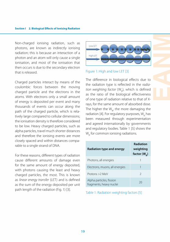
For these reasons, diferent types of radiation
cause diferent amounts of damage even
for the same amount of energy deposited,
with photons causing the least and heavy
charged particles, the most. This is known
as linear energy transfer (LET) and is deined
as the sum of the energy deposited per unit
path length of the radiation (Fig. 1) [3].
Figure 1: High and low LET [3]
The diference in biological efects due to
the radiation type is relected in the radia-
tion weighting factor (WR), which is deined
as the ratio of the biological efectiveness
of one type of radiation relative to that of X-
rays, for the same amount of absorbed dose.
The higher the WR, the more damaging the
radiation [4]. For regulatory purposes, WR has
been measured through experimentation
and agreed internationally by governments
and regulatory bodies. Table 1 [5] shows the
WR for common ionising radiations.
Radiation type and energy
Radiation
weighting
factor (WR
)
Photons, all energies 1
Electrons, muons, all energies 1
Protons >2 MeV 2
Alpha particles, ission fragments, heavy nuclei
20
Table 1: Radiation weighting factors [5]
20
The SI unit of equivalent dose is J kg-1, but
it is given the unit Sievert (Sv) to distinguish
it from absorbed dose. Equivalent dose is
mainly used for radiation protection pur-
poses.
Relative biological efectiveness – weighted
dose
More useful in the context of radionuclide
therapy is relative biological efectiveness
(RBE), deined as the ratio of a dose of a low-
LET reference radiation to the dose of radia-
tion that elicits the same biological response,
quantitatively and qualitatively. The RBE de-
pends on the radiation type, the dose rate,
the distribution of dose in time, the cells (or
tissues) exposed and the type of injury inves-
tigated.
When considering radionuclide therapy, the
ICRP recommends that the organ dose is
weighted by the RBE of the speciic biological
response. It is important to recognise the dif-
ference between WR and RBE when consid-
ering dose to an individual: RBE is a quantity
for deterministic endpoints measured under
a speciic set of experimental conditions and
is tissue and source location dependent,
whereas WR is a single set of values chosen
by committee review and so has less value
when considering dose to an individual.
Guidance on appropriate values for the RBE
for deterministic efects can be found in ICRP
publications 58 [6] and 92 [5] and Interna-
tional Commission on Radiation Units and
Measurements (ICRU) report 67 [7].
Efective dose
Absorbed and equivalent dose take into ac-
count the amount of energy, the mass of the
tissue and the radiation used, but they do
not take into account the type of tissue be-
ing irradiated.
Organs have diferent sensitivities to ra-
diation, known as radiosensitivity, based on
their tissue type, which will be discussed in
more depth later in this chapter. Efective dose
(E) is a calculation that takes into account this
variation in radiosensitivity. It is a calculation
of risk of the patient developing fatal cancer
due to the irradiation.
Each organ is given a tissue weighting factor
(WT), based on that organ’s risk of developing
cancer. A WT
for each organ has been calcu-
lated by the International Commission for Ra-
diological Protection (ICRP) and attempts to
provide a single number that is proportional
to the overall detriment from a particular,
often inhomogeneous, type of radiation ex-
posure. The overall detriment represents a
balance between cancer incidence, cancer
mortality, life shortening and hereditary ef-
fects. Table 2 shows the most recent WT as
published by the ICRP [8].
Section I 2. Biological Efects of Ionising Radiation
EANM
21
Organ
Tissue
weighting
factor (WT)
Sum
of WT
values
Bone marrow (red), colon, lung, stomach, breast, remainder tissuesa
0.12 0.72
Gonads 0.08 0.08
Bladder, oesophagus, liver, thyroid
0.04 0.16
Bone surface, brain, salivary glands, skin
0.01 0.04
Total 1.00 1.00
a Remainder tissues: adrenals, extrathoracic (ET) re-gion, gall bladder, heart, kidneys, lymphatic nodes, muscle, oral mucosa, pancreas, prostate (♂), small in-testine, spleen, thymus, uterus/cervix (♀)
Table 2: Recommended tissue weighting
factors [8]
It is important to recognise that efective
dose is a measurement of risk to an average
person and is based on general populations;
it should never be used to calculate the risk
to an individual as it does not take into ac-
count the individual’s age, gender or radio-
sensitivity.
Risk estimates are taken from groups of peo-
ple with known exposures, such as the atom-
ic bomb survivors of Hiroshima and Nagasaki
in 1945. Diferences in genetics, natural levels
of cancer, diet, smoking, stress and unknown
bias afect these results. The doses, and dose
rates, during these events were much higher
than the regulated dose levels used medi-
cally. There is growing evidence that there
are variations in radiosensitivity that can af-
fect the risk of radiation-induced cancer or,
at higher doses, tissue damage. A proportion
of this range is likely to be due to genetic
diferences, but recent studies demonstrate
that lifestyle factors, particularly tobacco use,
afect an individual’s risk [2].
Efective dose calculation for radionuclides
A schema for calculation of absorbed doses
has been developed by the Medical Internal
Radiation Dose Committee (MIRD) and this
methodology has been widely adopted. In
addition, the ICRP has introduced biokinetic
models and data. This has resulted in estima-
tion of the efective dose for a large num-
ber of radiopharmaceuticals. The igures are
given in mSv/MBq, meaning that an estima-
tion of the efective dose to a single patient
can be made simply by multiplying with the
administered activity. It must be noted that
variations from patient to patient can be very
large; nevertheless, a rough estimation of the
efective dose to the patient can be made. In
order to calculate absorbed dose and organ
doses for an individual patient, one requires
knowledge of the emissions of the radionu-
clide, the activity administered, the activity
in the speciic organ and in all other organs,
the size and shape of the organ, the kinetic
properties of the radiopharmaceutical and its
quality. These factors are not readily available
for the individual patient.
DNA damage
The energy that is transferred during ionising
radiation interactions can be used to break

22
chemical bonds. In living tissue this breaking
of bonds can be detrimental to cells, and can
kill the cell or cause it to reproduce abnor-
mally.
The biological efects of ionising radiation are
mainly due to damage to the nuclear DNA
chain. Ionising radiation can directly interact
with DNA to cause ionisation, thus initiating
the chain of events that lead to biological
changes [9) (10]. This is called direct action,
which is the main process for radiation with
high LET, such as alpha particles or neutrons.
Ionising radiation can also interact with mol-
ecules within the cell, in particular water, to
produce free radicals, which are able to go
on to interact with the DNA chain and cause
damage. This is called indirect action (11].
Deoxyribonucleic acid (DNA)
DNA is a complex macromolecule that con-
tains the genetic instructions used in the de-
velopment and function of all living tissues.
It consists of bases attached to a “backbone”
of alternating sugar and monophosphate
molecules. The bases are: adenine (A), gua-
nine (G), cytosine (C) and thymine (T). A and
G are purines and are the two larger bases. C
and T are pyrimidines and are the two smaller
bases. Due to their size, a large base and a
small base combine opposite to each other
to create the well-known double helix (C
and T together would be too small, A and G
together would be too big). Because of the
chemical structure of the bases, C and G are
always paired, and T and A are always paired
(Fig. 2) [12].
One of the most important features of DNA
is that it can replicate and repair. During a
process called mitosis, the cell divides and
replicates its genetic material, becoming two
distinct cells, hence allowing growth and re-
production of the cell.
Figure 2: DNA double-helix structure [12]
It is well recognised that the most impor-
tant lesion caused by ionising radiation is
the double-strand break (DSB), in which both
strands in the DNA helix are severed. Once
damaged, the DNA will attempt to repair it-
self. There are two main mechanisms of re-
pair: homologous recombination (HR) and
non-homologous end joining (NHEJ). In HR
an undamaged DNA chain, with the same in-
formation, is used as a template to replicate
the damaged part. The damage is removed
and replaced by this replicated piece. NHEJ is
Imag
e p
ub
lish
ed w
ith c
ou
rtes
y o
f th
e ow
ner
Section I 2. Biological Efects of Ionising Radiation
EANM
23
a rough form of repair in which the two dam-
aged ends of the breaks are rejoined [11].
Once the repair mechanisms have been at-
tempted, there are three main outcomes:
the DNA has been repaired correctly and the
cell becomes a viable cell; the DNA has been
repaired incorrectly and the cell becomes a
mutated cell; and the DNA was not able to be
repaired and the cell becomes an unviable
cell. If the DNA is repaired correctly, there is
no lasting efect of the ionising incident, and
so it needs no further consideration.
There are two categories of efects: somatic ef-
fects, where the damage afects the individual
who has been exposed, and genetic or heredi-
tary efects, where the damage occurs in the
cells used for reproduction, known as germ cells,
and so will afect subsequent generations.
Mutation
If the DNA has been repaired incorrectly,
then the “code” within the cell is no longer
correct. This is known as a mutation, and if
this cell then replicates and multiplies, cancer
can result. On occasion the mutation leads to
the cell multiplying with no control and ex-
ceeds the natural rate of cell death; this can
lead to the development of a tumour.
Apoptosis
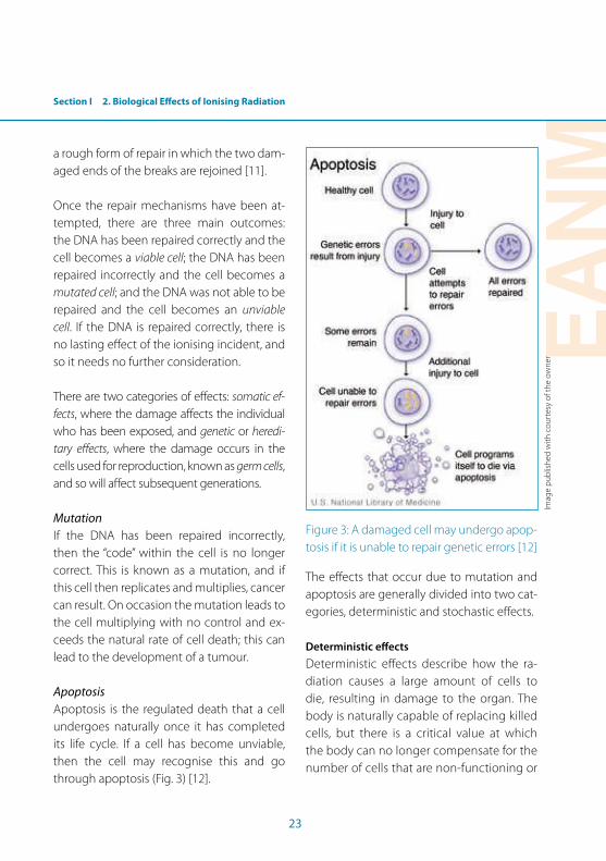
Apoptosis is the regulated death that a cell
undergoes naturally once it has completed
its life cycle. If a cell has become unviable,
then the cell may recognise this and go
through apoptosis (Fig. 3) [12].
Figure 3: A damaged cell may undergo apop-
tosis if it is unable to repair genetic errors [12]
The efects that occur due to mutation and
apoptosis are generally divided into two cat-
egories, deterministic and stochastic efects.
Deterministic efects
Deterministic efects describe how the ra-
diation causes a large amount of cells to
die, resulting in damage to the organ. The
body is naturally capable of replacing killed
cells, but there is a critical value at which
the body can no longer compensate for the
number of cells that are non-functioning or
Imag
e p
ub
lish
ed w
ith c
ou
rtes
y o
f th
e ow
ner
24
have died. Therefore there is a threshold ra-
diation dose below which deterministic ef-
fects are not seen, and as the dose increases
past this threshold value, the number of
cells that die, and so the severity of the ef-
fect, increases.
Deterministic efects can be divided into two
categories, acute and late efects. Acute ef-
fects occur in tissues that are rapidly prolif-
erating, such as in the epithelial surfaces of
the skin or alimentary tract. These cells are
replaced by cells that are more tolerant to
radiation, and these efects therefore subside
after 3-4 weeks. Late efects are typically not
seen for 6 months after irradiation and devel-
op through complex interacting processes
that are not yet well understood [13].
Efects are dependent on the area irradiated
and the dose received. Acute efects include
skin erythema, epilation, fatigue, nausea and
vomiting, while late efects include tissue
necrosis, sterility, cataracts and secondary
cancers [13].
Thresholds and timelines for deterministic
efects
Skin erythema/necrosis/epilation. Erythema
occurs within 1-24 hours of exposure to dos-
es greater than 2 Sv. Breakdown of the skin
surface occurs approximately 4 weeks after
15 Sv has been received. Epilation is revers-
ible after 3 Sv but irreversible after 7 Sv and
occurs 3 weeks following exposure [14].
Cataracts. Cataract occurs due to accumu-
lation of damaged or dead cells within the
lens, the removal of which cannot take place
naturally. Cataracts may occur after 0.5 Gy
has been received, but may take years to de-
velop [15].
Sterility. Radiation can impair the female
germ cell function, leading to impaired fer-
tility or infertility. The dose required to have
this efect decreases with age as the number
of cells decreases. Similarly, radiation expo-
sure to the testes can result in temporary or
permanent limitations in sperm production.
Permanent sterility occurs after 2.5-3.5 Gy has
been received by the gonads.
Acute radiation syndrome (radiation sickness).
Radiation sickness involves nausea, vomit-
ing and diarrhoea developing within hours
or minutes of a radiation exposure owing to
deterministic efects on the bone marrow,
gastrointestinal tract and central nervous
system [15].
Deterministic efects on the foetus. Determin-
istic efects during pregnancy depend not
only on the radiation dose received but also
on the gestational age at which it occurred.
The embryo is relatively radioresistant dur-
ing its pre-implantation phase but highly
radiosensitive whilst organs are forming (at
2-8 weeks) and in the neuronal stem cell
proliferation phase (at 8-15 weeks). Foetal
radiosensitivity falls after this period. High
Section I 2. Biological Efects of Ionising Radiation
EANM
25
levels of radiation exposure in pregnancy
can lead to growth retardation, in particular
microcephaly. The threshold dose for this ef-
fect is high (>20 Gy), with other deterministic
efects (hypospadias, microphthalmia, retinal
degeneration and optic atrophy) having a
lower threshold level of >1 Gy [15].
Stochastic efects
Stochastic means statistical in nature, and
these efects arise through chance. Stochas-
tic efects occur due to mutations in the DNA
chain and have no threshold value. As ab-
sorbed dose increases, the risk of observing
these efects increases, and the risk is consid-
ered proportional to the dose. The severity
of the efect is not related to the absorbed
dose; the person afected will either develop
cancer or they will not.
Stochastic efects are cancer if the efect oc-
curs in the person’s own cells (somatic cells)
and genetic mutations in subsequent gen-
erations if the efect occurs in the germ cells.
The tissue weighting factors discussed previ-
ously in this chapter are based on the risk of
stochastic efects occurring. The risk is based
on the tissue type that the organ consists of.
Radiosensitivity of cells
In 1906, experiments carried out by two
French scientists on rodents deined the ra-
diosensitivity of cells based on their funda-
mental characteristics. The Law of Bergonie
and Tribondeau [16] states that radiosensitiv-
ity is based on three factors:
• The mitotic rate: The rate at which the cells
divide and multiply: the higher the mitotic
rate, the greater the radiosensitivity of the
cell.
• The mitotic future: How long the cell is able
to divide for: the longer the mitotic future,
the greater the radiosensitivity of the cell.
• State of diferentiation: An undiferentiated
cell is an immature, embryonic or primitive
cell. It has a non-speciic appearance with
multiple non-speciic functions. A diferenti-
ated cell is highly distinct or specialised. Dif-
ferentiation predicts how vulnerable a cell
will be to radiation. The more diferentiated
a cell is, the more radioresistant it will be.
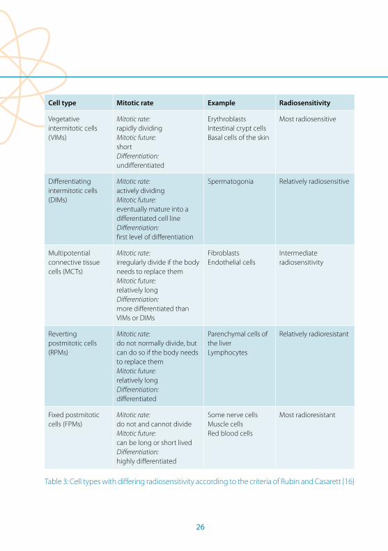
In 1968, Rubin and Casarett [17] deined
ive cell types with difering radiosensitiv-
ity according to the characteristics shown in
Table 3 [16].
26
Cell type Mitotic rate Example Radiosensitivity
Vegetative intermitotic cells (VIMs)
Mitotic rate:
rapidly dividingMitotic future:
shortDiferentiation:
undiferentiated
ErythroblastsIntestinal crypt cells Basal cells of the skin
Most radiosensitive
Diferentiating intermitotic cells (DIMs)
Mitotic rate:
actively dividingMitotic future:
eventually mature into a diferentiated cell lineDiferentiation:
irst level of diferentiation
Spermatogonia Relatively radiosensitive
Multipotential connective tissue cells (MCTs)
Mitotic rate:
irregularly divide if the body needs to replace themMitotic future:
relatively longDiferentiation:
more diferentiated than VIMs or DIMs
FibroblastsEndothelial cells
Intermediate radiosensitivity
Reverting postmitotic cells (RPMs)
Mitotic rate:
do not normally divide, but can do so if the body needs to replace themMitotic future:
relatively longDiferentiation:
diferentiated
Parenchymal cells of the liverLymphocytes
Relatively radioresistant
Fixed postmitotic cells (FPMs)
Mitotic rate:
do not and cannot divideMitotic future:
can be long or short livedDiferentiation:
highly diferentiated
Some nerve cellsMuscle cellsRed blood cells
Most radioresistant
Table 3: Cell types with difering radiosensitivity according to the criteria of Rubin and Casarett [16]

Section I 2. Biological Efects of Ionising Radiation
EANM
27
A more modern way of considering the bio-
logical efects on a cellular level is the Micha-
lowski classiication of cells. Under this clas-
siication, cells fall into three categories:
• Stem cells – continuously divide and re-
produce to give rise to both new stem
cells and cells that eventually give rise to
mature functional cells.
• Maturing cells arising from stem cells that
through progressive division eventually
diferentiate into end-stage mature func-
tional cells.
• Mature adult functional cells that do not
divide.
Summary
Ionising radiation causes damage to tis-
sues through ionisation, either directly with
the cell or indirectly with water, leading to
free radicals. The main target for damage is
the DNA chain, where the ionisation causes
breaks within the chain. Once damaged, the
DNA will repair correctly, repair incorrectly
or be so damaged that it will become non-
functioning or die. Incorrect repair causes
mutations, known as stochastic efects,
whereas cell death leads to deterministic ef-
fects such as necrosis, which is required for
efective radiotherapy.
Dose calculation is complex and highly de-
pendent on the individual circumstances.
Diferent terms exist, and are used depend-
ing on whether radiation protection or radio-
therapy is under consideration. When con-
sidering populations, efective dose is useful
in generalising risk, but when considering an
individual one needs to look at the individual
organ dose and consider the RBE for the ion-
ising radiation being used.
Recent studies have shown that radiosen-
sitivity is dependent on many factors when
considering an individual, and that as well as
age and gender, lifestyle factors and genetics
testing may inluence dose planning in the
future [2].
28
References Section I, Chapter 2
References1. Wall BF, Health Protection Agency . Centre for Radiation
CaEH. Radiation risks from medical X-ray examinations as a function of the age and sex of the patient. Didcot: Centre for Radiation Chemical and Environmental Haz-ards; 2011. iii, 66 p.
2. Radiation AGoI. Human Radiosensitivity - RCE 21. Ox-fordshire: Health Protection Agency, 2013.
3. Szczepura K. Linear Energy Transfer. 2013.
4. Allisy-Roberts P, Williams JR, Farr RFPfmi. Farr’s physics for medical imaging. 2nd ed. / Penelope Allisy-Roberts, Jerry Williams. ed. Edinburgh: Saunders; 2008.
5. Task Group on Radiation Quality Efects in Radiological Protection CoRE, I.ternational Commission on Radio-logical Protection. Relative biological efectiveness (RBE), quality factor (Q), and radiation weighting factor (w(R)). A report of the International Commission on Radio-logical Protection. Ann ICRP. 2003;33(4):1-117. PubMed PMID: 14614921.
6. International Commission on Radiological Protection C. RBE for deterministic efects : a report of a Task Group of Committee 1 of the International Commission on Radiological Protection. Oxford: published for the In-ternational Commission on Radiological Protection by Pergamon; 1990. v,57 p p.
7. International Commission on Radiation Units and M. Absorbed-dose speciication in nuclear medicine. Ash-ford, Kent: Nuclear Technology Publishing; 2002. 120 p.
8. Wrixon AD. New ICRP recommendations. J Radiol Prot. 2008;28(2):161-8. doi: 10.1088/0952-4746/28/2/R02. PubMed PMID: 18495983.
9. Valentin J. Low-dose extrapolation of radiation-related cancer risk. Ann ICRP. 2005;35(4):1-140. doi: 10.1016/j.icrp.2005.11.002. PubMed PMID: 16782497.
10. Lehnert S. Biomolecular action of ionizing radiation. Bristol: Institute of Physics; 2008.
11. Urbano KV. Advances in genetics research. Hauppauge, N.Y.: Lancaster : Nova Science ; Gazelle [distributor]; 2011. xii, 290 p.
12. Medicine UNLo. What is DNA? 2013.
13. DeVita VT, Hellman S, Rosenberg SA. Cancer : principles & practice of oncology. 7th ed. Philadelphia, Pa.: Lip-pincott Williams & Wilkins; 2005. 1 CD ROM p.
14. Balter S, Hopewell JW, Miller DL, Wagner LK, Zelefsky MJ. Fluoroscopically guided interventional proce-dures: a review of radiation efects on patients’ skin and hair. Radiology. 2010;254(2):326-41. doi: 10.1148/radiol.2542082312. PubMed PMID: 20093507.
15. Stewart FA, Akleyev AV, Hauer-Jensen M, Hendry JH, Kleiman NJ, Macvittie TJ, et al. ICRP publication 118: ICRP statement on tissue reactions and early and late efects of radiation in normal tissues and organs--threshold doses for tissue reactions in a radiation protection context. Ann ICRP. 2012;41(1-2):1-322. doi: 10.1016/j.icrp.2012.02.001. PubMed PMID: 22925378.
16. Bushberg JT, Seibert JA, Leidholdt EM, Boone JM. The essential physics of medical imaging. 3rd ed., Interna-tional ed. ed. Philadelphia, Pa. ; London: Wolters Kluwer/Lippincott Williams & Wilkins; 2012.
17. Rubin P, Casarett GW. Clinical radiation pathology. Phila-delphia: W. B. Saunders Co.; 1968.

EANM
29
Section I3. Dosimetry in Molecular Radiotherapy
Katarina Sjögreen Gleisner, Lidia Strigari, Glenn Flux
Introduction1 It is unthinkable that a patient would be
treated with external beam radiotherapy
(EBRT) or brachytherapy (BT) without prior
treatment planning, and prescriptions are
always made in terms of the absorbed dose
delivered to target tissues and organs at risk.
Molecular radiotherapy (MRT) is very similar
to BT in that the radiation source is located
inside the patient’s body. However, in MRT
the source is difuse and therapy is normally
given systemically as a radiopharmaceutical,
whereas in BT solid radiation sources are used.
Unlike in EBRT and BT, where the irradia-
tion geometry and hence the absorbed
dose distribution can be planned in ad-
vance, in MRT this distribution depends on
the amount of radiopharmaceutical that
accumulates over time in diferent tissues,
something which varies between patients
1. Quantities
The activity of a radioactive sample is the mean number of decays per second. The unit is Becquerel (Bq) which equals 1/s.
The cumulated activity is the number of decays that occur in a given region over a period of time. The unit is Bq s, or Bq h.
When ionising radiation travels through matter, it interacts and deposits energy. The energy imparted is the sum of all energy deposits in a given volume. The absorbed dose is the quotient of the mean energy imparted and the mass of the volume. The unit of ab-sorbed dose is Gray (Gy), which equals 1 J/kg.
The term ‘dose’ alone can be confusing and is best avoided, as this can refer to either the level of activ-ity administered or the absorbed dose subsequently delivered.
and therefore needs to be measured. Thus
for MRT, the determination of the absorbed
dose is more cumbersome. However, it is
reasonable to assume that the efects of
therapy, in terms of response and toxicity,
are primarily dependent on the absorbed
doses delivered, as for EBRT and BT, rather
than on the level of activity administered.
MRT dosimetry constitutes the bridge be-
tween the administered activity and the
prediction of treatment efects, and has to
be taken into consideration along with ra-
diobiological characteristics of tissues. As
in other radiation treatment modalities, it is
essential that dosimetry in MRT is regarded
as teamwork between biomedical technolo-
gists, radiographers, nuclear medicine tech-
nologists, medical physicists, and physicians
specialised in oncology or nuclear medicine.
Internal dosimetry
Following administration, the radiopharma-
ceutical distributes in the patient’s body ac-
cording to pharmacokinetics speciic to the
radiopharmaceutical and the individual pa-
tient. A particle emitted at radioactive decay
may impart its energy in close vicinity to the
point of decay, i.e. in the same tissue in which
it was emitted, or it may travel some distance
before losing its energy, perhaps in another
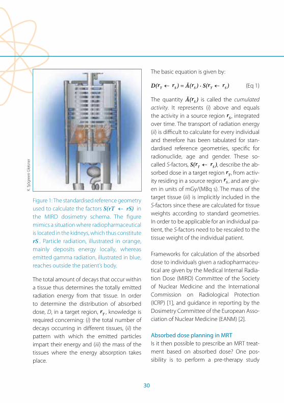
tissue (Fig. 1, K. Sjögreen Gleisner).
30
Figure 1: The standardised reference geometry
used to calculate the factors S(rT ← rS) in
the MIRD dosimetry schema. The igure
mimics a situation where radiopharmaceutical
is located in the kidneys, which thus constitute
rS . Particle radiation, illustrated in orange,
mainly deposits energy locally, whereas
emitted gamma radiation, illustrated in blue,
reaches outside the patient’s body.
The total amount of decays that occur within
a tissue thus determines the totally emitted
radiation energy from that tissue. In order
to determine the distribution of absorbed
dose, D, in a target region, rT , knowledge is
required concerning: (i) the total number of
decays occurring in diferent tissues, (ii) the
pattern with which the emitted particles
impart their energy and (iii) the mass of the
tissues where the energy absorption takes
place.
The basic equation is given by:
D(rT
← rS ) = Ã(rS ) · S(r
T ← rS ) (Eq 1)
The quantity Ã(rS ) is called the cumulated
activity. It represents (i) above and equals
the activity in a source region rS , integrated
over time. The transport of radiation energy
(ii) is diicult to calculate for every individual
and therefore has been tabulated for stan-
dardised reference geometries, speciic for
radionuclide, age and gender. These so-
called S-factors, S(rT
← rS ), describe the ab-
sorbed dose in a target region rT , from activ-
ity residing in a source region rS , and are giv-
en in units of mGy/(MBq s). The mass of the
target tissue (iii) is implicitly included in the
S-factors since these are calculated for tissue
weights according to standard geometries.
In order to be applicable for an individual pa-
tient, the S-factors need to be rescaled to the
tissue weight of the individual patient.
Frameworks for calculation of the absorbed
dose to individuals given a radiopharmaceu-
tical are given by the Medical Internal Radia-
tion Dose (MIRD) Committee of the Society
of Nuclear Medicine and the International
Commission on Radiological Protection
(ICRP) [1], and guidance in reporting by the
Dosimetry Committee of the European Asso-
ciation of Nuclear Medicine (EANM) [2].
Absorbed dose planning in MRT
Is it then possible to prescribe an MRT treat-
ment based on absorbed dose? One pos-
sibility is to perform a pre-therapy study
K. S
jög
reen
Gle
isn
er
Section I 3. Dosimetry in Molecular Radiotherapy
EANM
31
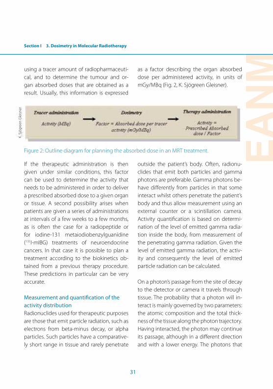
using a tracer amount of radiopharmaceuti-
cal, and to determine the tumour and or-
gan absorbed doses that are obtained as a
result. Usually, this information is expressed
as a factor describing the organ absorbed
dose per administered activity, in units of
mGy/MBq (Fig. 2, K. Sjögreen Gleisner).
Figure 2: Outline diagram for planning the absorbed dose in an MRT treatment.
If the therapeutic administration is then
given under similar conditions, this factor
can be used to determine the activity that
needs to be administered in order to deliver
a prescribed absorbed dose to a given organ
or tissue. A second possibility arises when
patients are given a series of administrations
at intervals of a few weeks to a few months,
as is often the case for a radiopeptide or
for iodine-131 metaiodiobenzylguanidine
(131I-mIBG) treatments of neuroendocrine
cancers. In that case it is possible to plan a
treatment according to the biokinetics ob-
tained from a previous therapy procedure.
These predictions in particular can be very
accurate.
Measurement and quantiication of the
activity distribution
Radionuclides used for therapeutic purposes
are those that emit particle radiation, such as
electrons from beta-minus decay, or alpha
particles. Such particles have a comparative-
ly short range in tissue and rarely penetrate
outside the patient’s body. Often, radionu-
clides that emit both particles and gamma
photons are preferable. Gamma photons be-
have diferently from particles in that some
interact whilst others penetrate the patient’s
body and thus allow measurement using an
external counter or a scintillation camera.
Activity quantiication is based on determi-
nation of the level of emitted gamma radia-
tion inside the body, from measurement of
the penetrating gamma radiation. Given the
level of emitted gamma radiation, the activ-
ity and consequently the level of emitted
particle radiation can be calculated.
On a photon’s passage from the site of decay
to the detector or camera it travels through
tissue. The probability that a photon will in-
teract is mainly governed by two parameters:
the atomic composition and the total thick-
ness of the tissue along the photon trajectory.
Having interacted, the photon may continue
its passage, although in a diferent direction
and with a lower energy. The photons that
K. S
jög
reen
Gle
isn
er
32
provide correct information on the location
of decay are those that penetrate without
interaction, the so-called primaries. Photon
attenuation is the reduction of the amount of
such primaries. Scattered photons are those
that are detected but have interacted along
their passage and thus carry false informa-
tion about the point of decay. Attenuation
thus causes a loss of detected counts, so that
a factor used for attenuation correction must
have a value greater than one. Scatter causes
a falsely increased count rate, and it is desir-
able to remove such counts.
Practically, there are several methods for de-
termining the amount of activity in a patient,
and the choice of method depends on the
level of accuracy required. All methods in-
volve repeated patient measurements, since
the aim is to follow the activity retention over
time. The timing of the measurements needs
to be distributed with regard to typical bioki-
netic behaviour of the radiopharmaceutical.
It is an advantage if exactly the same equip-
ment can be used for all measurements. It is
also essential that clinical protocols are care-
fully set up, and that patient positioning fol-
lows agreed routines.
Probe-based measurements
The activity retention in the body can be
assessed by serial measurements using a
standard probe dose-rate meter. The irst
measurement will represent 100% of the ad-
ministered activity if it is made shortly after
infusion, but prior to voiding. By curve itting
of the retention curve and knowledge of the
amount of administered activity, the total-
body cumulated activity can be determined.
It is important that the patient counter ge-
ometry is kept constant for all measurements
that presence of metal near the patient or
detector is avoided to keep the room scatter
low and that care is taken to remove any pos-
sibly contaminated material in the room. For
a gamma emitter such as 131I, the total-body
retention curve can be used for estimation of
the red marrow absorbed dose [3, 4].
Image-based activity quantiication
In image-based activity quantiication the
underlying idea is that each image element
can be viewed as a detector, so that the pixel
or voxel count rate relects the number of
detected photons in a particular position. By
delineating a region or volume in an image,
the total number of photons from a given
anatomical region can be determined. The
delineation of organs and tissues is central
in the determination of the activity (Fig. 3,
K. Sjögreen Gleisner).
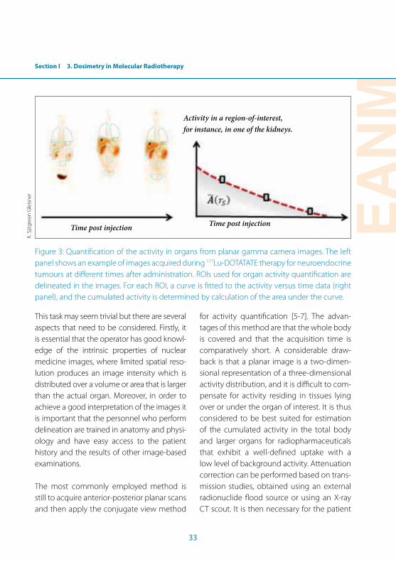
Section I 3. Dosimetry in Molecular Radiotherapy
EANM
33
Activity in a region-of-interest,
for instance, in one of the kidneys.
Time post injectionTime post injection
Figure 3: Quantiication of the activity in organs from planar gamma camera images. The left
panel shows an example of images acquired during 177Lu-DOTATATE therapy for neuroendocrine
tumours at diferent times after administration. ROIs used for organ activity quantiication are
delineated in the images. For each ROI, a curve is itted to the activity versus time data (right
panel), and the cumulated activity is determined by calculation of the area under the curve.
This task may seem trivial but there are several
aspects that need to be considered. Firstly, it
is essential that the operator has good knowl-
edge of the intrinsic properties of nuclear
medicine images, where limited spatial reso-
lution produces an image intensity which is
distributed over a volume or area that is larger
than the actual organ. Moreover, in order to
achieve a good interpretation of the images it
is important that the personnel who perform
delineation are trained in anatomy and physi-
ology and have easy access to the patient
history and the results of other image-based
examinations.
The most commonly employed method is
still to acquire anterior-posterior planar scans
and then apply the conjugate view method
for activity quantiication [5-7]. The advan-
tages of this method are that the whole body
is covered and that the acquisition time is
comparatively short. A considerable draw-
back is that a planar image is a two-dimen-
sional representation of a three-dimensional
activity distribution, and it is diicult to com-
pensate for activity residing in tissues lying
over or under the organ of interest. It is thus
considered to be best suited for estimation
of the cumulated activity in the total body
and larger organs for radiopharmaceuticals
that exhibit a well-deined uptake with a
low level of background activity. Attenuation
correction can be performed based on trans-
mission studies, obtained using an external
radionuclide lood source or using an X-ray
CT scout. It is then necessary for the patient
K. S
jög
reen
Gle
isn
er
34
to maintain a similar position in the transmis-
sion and radionuclide imaging sessions. To-
mographic imaging with SPECT and PET en-
ables the activity distribution to be resolved
in three dimensions. From such 3D images,
measurements of the activity concentration
can be determined for the body region cov-
ered by the scan. The preferred method for
activity quantiication is to include it as part
of the image reconstruction, provided that
corrections for attenuation and scatter can
be included [8]. For such corrections, an at-
tenuation map is required, and can be ob-
tained from a CT study which is registered
to the SPECT or PET study. In this regard,
hybrid SPECT/CT and PET/CT systems ofer
signiicant advantages for the purposes of
dosimetry. Although in principle SPECT and
PET images allow determination of the activ-
ity concentration in a volume represented by
each voxel, tissues that are small in compari-
son to the spatial resolution will be afected
by resolution-induced “spill-out” of activity,
with an underestimated activity concentra-
tion as a result. For SPECT the spatial resolu-
tion can be in the order of 1 cm or more, and
the activity concentration in smaller organs
such as the kidneys and tumours may be
underestimated. This is the so-called partial
volume efect and it should also be corrected
for. Methods involving a combination of pla-
nar and tomographic imaging techniques
have been explored, whereby the activity re-
tention curve is measured by planar imaging
and its amplitude is adjusted to absolute val-
ues of the activity concentration or absorbed
dose rate as determined from SPECT/CT [9].
Radiopharmaceuticals used in radionuclide
therapies generally have a relatively long bio-
logical half-life, which has to some extent lim-
ited more widespread use of luorine-18 PET
imaging for dosimetry. However, an increas-
ing number of research studies are using
longer-lived PET radionuclides for dosimetry
purposes, including iodine-124 (4.2 days),
zirconium-89 (3.3 days), copper-64 (12.7 h)
and yttrium-86 (14.7 h) [10-12]. Also it has re-
cently been determined that yttrium-90 can
be imaged using PET [13], and this is now the
subject of many research studies.
Calculation of the absorbed dose
From the values of the activity in a tissue at
diferent times, the cumulated activity is de-
termined by integration, and the mean ab-
sorbed dose can be determined using Eq. 1.
Generally, this method is used when activity
measurements are made using a probe for
the total body, or when using planar imag-
ing for whole-organ dosimetry. Quantitative
SPECT/CT or PET/CT allows for dosimetry in
smaller volumes using voxel-based methods
(Fig. 4, K. Sjögreen Gleisner).
Section I 3. Dosimetry in Molecular Radiotherapy
EANM
35
Figure 4: The left panel shows a transverse slice of a SPECT/CT study acquired during 177Lu-
DOTATATE therapy for neuroendocrine tumours. If quantitative tomographic reconstruction is
available, then quantiication of the activity, or activity concentration, in organs and tumours
can be performed by delineating volumes of interest (VOIs) in SPECT/CT images. Such activity
values can be used to calculate the absorbed dose according to the MIRD schema (Eq. 1). Another
possibility (right panel) is to calculate the absorbed dose rate distribution on a voxel-by-voxel
basis by using the SPECT/CT images as input to a Monte Carlo code that mimics the particle
emission and subsequent interactions in the patient’s body. The VOIs are then applied in the
resulting absorbed dose rate image.
From the 3D distribution of activity concen-
tration values, the absorbed dose rate dis-
tribution can be calculated using so-called
point dose kernels or voxel S values, describ-
ing the energy deposition pattern around a
point source located in water (or bone). This
method assumes that the anatomical region
is homogeneous in terms of density, such as
soft tissues within the trunk. For body regions
where the density is heterogeneous, as in
the lungs, a direct Monte Carlo calculation is
preferable. Here, the activity distribution from
SPECT or PET is used as input to a Monte Carlo
dose calculation code. If co-registration is
available, the dose rate maps from SPECT/CT
or PET/CT images acquired on diferent occa-
sions can be used to calculate a 3D map of
the absorbed dose [14] where organ delinea-
tion can be made. Otherwise, organ delinea-
tion needs to be performed in each separate
image. Whichever dosimetry method is used,
a determination of the mass of the target re-
gion is required. The mass can be determined
using the CT image either on a voxel basis by
applying a calibration relationship, such as is
normally done in EBRT [14], or by delineating
K. S
jög
reen
Gle
isn
er
36
the target region and assuming a reference
value for the mass density.
Radiobiology
The aim of radiobiology is to establish rela-
tionships between the absorbed doses de-
livered and the radiobiological efects in tis-
sues and tumours. Currently in MRT few such
relationships have been established, but this
is an area of intensive research. Radiobio-
logical models in MRT can be adopted from
EBRT and BT through calculation of the bio-
logically efective dose (BED). The BED takes
into consideration the absorbed dose, the
absorbed dose rate and the capacity of tu-
mours and tissues to repair induced radiation
damage. Ideally the correlation between pre-
and post-treatment dosimetry should be as-
sessed to increase the predictive capability of
absorbed dose-response relationships.
The ‘four Rs’ of radiobiology
The ‘four Rs’ of radiobiology summarise the
main features of cell survival of tumours and
normal tissues [15]:
• Repair: Powerful repair mechanisms coun-
teract DNA damage induced by ionising
radiation with a repair half-time, Trep
, from
minutes to hours;
• Reoxygenation: Oxygen stabilises the ef-
fect of radiation. Hypoxic cells are the
main obstacle to tumour curability, but, as
the radiation dose is usually delivered over
several days, there is suicient time for its
reoxygenation.
• Repopulation: During MRT, the cells in
some tumours and some normal tissues
grow, partially counteracting the cell kill-
ing. Repopulation in normal tissues is also
an important mechanism to counteract
acute side-efects.
• Redistribution: Cells have diferent ra-
diosensitivity at diferent parts of the cell
cycle. The highest radiation sensitivity is in
the early S and late G2/M phase.
All of these mechanisms can be included in
the linear quadratic model (LQM).
The linear quadratic model
The LQM applies to diferent treatment regi-
mens, including MRT, and allows the use of
BED, a quantity with the dimensions of Gy.
This is useful to compare diferent irradiation
schemes and to evaluate the impact of treat-
ment in both tumours and normal tissues.
The radiosensitivity of tumours and tissues
can be considered using the parameters α,
which represents the radiosensitivity and is
lower for radioresistant cells, and the α/β ra-
tio, which indicates the response to fraction-
ation and to diferent dose rates. This ratio is
approximately 10 Gy for tumours and 3 Gy for
late-responding tissues.
Radiobiological models
The most commonly used radiobiological
models are the tumour control probability
(TCP) and the normal tissue complication
probability (NTCP). The TCP depends on N*,
the initial number of clonogenic cells, which

Section I 3. Dosimetry in Molecular Radiotherapy
EANM
37
is the fraction of cells that have the potential
to proliferate and give rise to a tumour. Usu-
ally N* is considered proportional to the tu-
mour volume. Palpable tumours are usually
characterised by 109 cells/cm3 (large masses
may contain 1012 cells/cm3, while microscop-
ic tumours/micrometastases contain about
106 cells/cm3). As expected, a TCP value cal-
culated at the same absorbed dose and with
the same radiobiological parameters (α and
β) decreases when N* increases, as more cells
require a higher absorbed dose for eradica-
tion. Obviously, tumour response also de-
pends on concurrently given agents, such as
chemotherapy.
Sparing of normal tissues is essential for a
good therapeutic outcome. This is largely
dependent on the tissue architecture, which
is basically classiied as serial or parallel. In se-
rial organs (for example the spinal cord and
intestines) even a small volume irradiated
beyond a threshold absorbed dose can lead
to whole organ failure. In parallel organs such
as the liver, kidneys and lungs the efect de-
pends on the irradiated volume.
NTCP models can predict the incidence of
acute/late complications for serial/parallel
organs. In EBRT early/acute reactions such
as tiredness, nausea, vomiting, skin redden-
ing, haematological toxicity and erythema
occur typically during radiotherapy or within
3 months, while late reactions, such as ibro-
sis and liver/kidney failure, could occur even
6 months thereafter. The majority of MRT
clinical data on toxicity are available for only
a few organs [16]. The maximum tolerated
dose depends on tissue and patient char-
acteristics and can be lowered by the use of
chemotherapy agents.
Normal tissues are more spared than tu-
mours by a continuous low dose rate, and
the LQM predicts a greater beneit due to
multifractionated treatments, using an α/β of
3 Gy. However, full understanding of a dose-
efect relationship is limited by a lack of re-
ported clinical data. An improvement to the
dose-response models should include the
efects of non-uniform dose distributions [17,
18] as well as clinical risk factors.
Clinical evidence for dosimetry
A BED response curve for kidney damage
has been reported after peptide receptor ra-
dionuclide therapy used for neuroendocrine
tumours at high cumulative activities [19].
The LQM supports the evidence that multi-
ple-cycle schemes decrease the incidence of
nephrotoxicity.
Red marrow toxicity data have mostly been
obtained from treatments with 131I for thy-
roid carcinoma and from 131I-mIBG therapy
of neuroblastoma. In many studies, red mar-
row dosimetry is approached with great
caution, and a maximum absorbed dose of
2 Gy to the blood (as a red marrow surro-
gate) is generally accepted [20] for radioio-
dine treatment of diferentiated thyroid can-
cer. Recently a higher limit of 3 Gy has been
proposed [21].

38
Lung toxicity data have also been mainly
derived from the treatment of metastatic
thyroid carcinoma with 131I or from loco-
regional liver treatments using 131I-Lipiodol
or 90Y-labelled microspheres. In the literature,
lung-related constraints are based on difer-
ent criteria [21-23]. Radiation pneumonitis
has also been seen in patients receiving 90Y-
TheraSphere as single treatment at doses ≥30 Gy. An empiric lung shunt [24] cannot
be considered a dosimetric constraint. Severe
radiation-induced lung toxicity, expected
at doses of 25–27 Gy, were not observed in
some studies, probably due to the overes-
timated normal lung absorbed dose [22].
In non-resectable primary or metastatic liver
tumours, radioembolisation is an increasingly
used loco-regional treatment. The partition
model permits a distinction between tumour
and normal liver doses [25], allowing the use
of constraints for normal tissues. Moreover,
TCP/NTCP models have been proposed [26].
Pre-treatment imaging seems feasible and at-
tractive for radiobiological treatment optimi-
sation [26-28]. Multi-cycle treatments would
allow administration of higher activities and
could improve the risk-beneit balance [29].
Other organs that afect the quality of life
should also be included in the evaluation of
treatment. Cells that are actively dividing and
not as well diferentiated, such as gonads,
haematopoietic cells and the gastrointestinal
tract, are most susceptible to radiation dam-
age. In young and long-surviving patients, the
gonads should be considered for assessment
of temporary and permanent sterility risks.
Conclusion
After almost 70 years of the use of radionuclides
to treat cancer, there is now a rapidly increasing
awareness of the need to administer activities
according to a predicted pattern of absorbed
dose delivery, and to assess after a therapeutic
procedure whether that predicted absorbed
dose has in fact been delivered. This funda-
mental change in practice is being spurred
on by the need for evidence-based medicine
and the emerging interest in molecular imag-
ing and personalised treatment. Whilst there
is evidently an opportunity for much research,
it remains the case that only with this cancer
therapy can the agent responsible for treat-
ment be imaged in vivo – this ofers a poten-
tial for individualised treatment planning that
is unprecedented. The greatest challenges at
present are the need to quantify activity im-
aged by a gamma camera that is not well
suited to the task (standardized uptake values
have long been established for quantiication
with PET) and the development of radiobiol-
ogy to understand the clinical implications of
an absorbed dose. This ield has been the slow-
est to develop of all cancer treatments, in part
due to the relatively small numbers of patients
treated at individual centres, but also due to
the need for a multidisciplinary approach to
what, in many centres, will be a new service.
As dosimetry is at the heart of molecular radio-
therapy, so imaging is at the heart of dosimetry.
It is likely that changes in clinical practice will
raise challenges and opportunities previously
not encountered, but that the outcome will be
signiicantly greater beneit to patients.
EANM
39
References Section I, Chapter 3
References 1. Bolch WE, Eckerman KF, Sgouros G, Thomas SR. MIRD
pamphlet No. 21: a generalized schema for radiophar-maceutical dosimetry -- standardization of nomencla-ture. J Nucl Med. 2009;50:477-84.
2. Lassmann M, Chiesa C, Flux G, Bardies M. EANM Dosim-etry Committee guidance document: good practice of clinical dosimetry reporting. Eur J Nucl Med Mol Imag-ing. 2011;38:192-200.
3. Buckley SE, Chittenden SJ, Saran FH, Meller ST, Flux GD. Whole-body dosimetry for individualized treatment planning of 131I-MIBG radionuclide therapy for neu-roblastoma. J Nucl Med. 2009;50:1518-24.
4. Hindorf C, Glatting G, Chiesa C, Linden O, Flux G. EANM Dosimetry Committee guidelines for bone marrow and whole-body dosimetry. Eur J Nucl Med Mol Imag-ing.37:1238-50.
5. Fleming JS. A technique for the absolute measurement of activity using a gamma camera and computer. Phys Med Biol. 1979;24:176-80.
6. Thomas SR, Maxon HR, Kereiakes JG. In vivo quantitation of lesion radioactivity using external counting methods. Med Phys. 1976;3:253-5.
7. Siegel JA, Thomas SR, Stubbs JB, Stabin MG, Hays MT, Koral KF, et al. MIRD pamphlet no. 16: Techniques for quantitative radiopharmaceutical biodistribution data acquisition and analysis for use in human radiation dose estimates. J Nucl Med. 1999;40:37S-61S.
8. Dewaraja YK, Frey EC, Sgouros G, Brill AB, Roberson P, Zanzonico PB, et al. MIRD pamphlet No. 23: quantitative SPECT for patient-speciic 3-dimensional dosimetry in internal radionuclide therapy. J Nucl Med.53:1310-25.
9. Garkavij M, Nickel M, Sjogreen-Gleisner K, Ljungberg M, Ohlsson T, Wingardh K, et al. 177Lu- [DOTA0,Tyr3] octreotate therapy in patients with disseminated neuro-endocrine tumors: Analysis of dosimetry with impact on future therapeutic strategy. Cancer. 2010;116:1084-92.
10. Barone R, Borson-Chazot F, Valkema R, Walrand S, Chauvin F, Gogou L, et al. Patient-speciic dosimetry in predicting renal toxicity with (90)Y-DOTATOC: relevance of kidney volume and dose rate in inding a dose-efect relationship. J Nucl Med. 2005;46 Suppl 1:99S-106S.
11. Rizvi SN, Visser OJ, Vosjan MJ, van Lingen A, Hoekstra OS, Zijlstra JM, et al. Biodistribution, radiation dosimetry and scouting of 90Y-ibritumomab tiuxetan therapy in patients with relapsed B-cell non-Hodgkin’s lymphoma using 89Zr-ibritumomab tiuxetan and PET. Eur J Nucl Med Mol Imaging.39:512-20.
12. Schwartz J, Humm JL, Divgi CR, Larson SM, O’Donoghue JA. Bone marrow dosimetry using 124I-PET. J Nucl Med.53:615-21.
13. Lhommel R, van Elmbt L, Gofette P, Van den Eynde M, Jamar F, Pauwels S, et al. Feasibility of 90Y TOF PET-based dosimetry in liver metastasis therapy using SIR-Spheres. Eur J Nucl Med Mol Imaging.37:1654-62.
14. Sjogreen-Gleisner K, Rueckert D, Ljungberg M. Registra-tion of serial SPECT/CT images for three-dimensional dosimetry in radionuclide therapy. Phys Med Biol. 2009;54:6181-200.
15. Withers HR. The four r’s of radiotherapy. Adv Radiol Biol. 1975;5:241-7.
16. Strigari L, Benassi M, Chiesa C, Cremonesi M, Bodei L, D’Andrea M. Dosimetry in nuclear medicine therapy: radiobiology application and results. Q J Nucl Med Mol Imaging. 2011;55:205-21.
17. Jackson A, Ten Haken RK, Robertson JM, Kessler ML, Kutcher GJ, Lawrence TS. Analysis of clinical complica-tion data for radiation hepatitis using a parallel architec-ture model. Int J Radiat Oncol Biol Phys. 1995;31:883-91.
18. Strigari L, D’Andrea M, Maini CL, Sciuto R, Benassi M. Biological optimization of heterogeneous dose distribu-tions in systemic radiotherapy. Med Phys. 2006;33:1857-66.
19. Wessels BW, Konijnenberg MW, Dale RG, Breitz HB, Cre-monesi M, Meredith RF, et al. MIRD pamphlet No. 20: the efect of model assumptions on kidney dosimetry and response -- implications for radionuclide therapy. J Nucl Med. 2008;49:1884-99.
20. Benua RS, Cicale NR, Sonenberg M, Rawson RW. The rela-tion of radioiodine dosimetry to results and complica-tions in the treatment of metastatic thyroid cancer. Am J Roentgenol Radium Ther Nucl Med. 1962;87:171-82.
40
21. Dorn R, Kopp J, Vogt H, Heidenreich P, Carroll RG, Gulec SA. Dosimetry-guided radioactive iodine treatment in patients with metastatic diferentiated thyroid cancer: largest safe dose using a risk-adapted approach. J Nucl Med. 2003;44:451-6.
22. Sgouros G, Song H, Ladenson PW, Wahl RL. Lung toxicity in radioiodine therapy of thyroid carcinoma: develop-ment of a dose-rate method and dosimetric implica-tions of the 80-mCi rule. J Nucl Med. 2006;47:1977-84.
23. Song H, He B, Prideaux A, Du Y, Frey E, Kasecamp W, et al. Lung dosimetry for radioiodine treatment plan-ning in the case of difuse lung metastases. J Nucl Med. 2006;47:1985-94.
24. SIR-Spheres® Yttrium-90 microspheres (package insert). In: Limited SM, editor. North Sydney, NSW, Australia; 2009.
25. Ho S, Lau WY, Leung TW, Chan M, Ngar YK, Johnson PJ, et al. Partition model for estimating radiation doses from yttrium-90 microspheres in treating hepatic tumours. Eur J Nucl Med. 1996;23:947-52.
26. Strigari L, Sciuto R, Rea S, Carpanese L, Pizzi G, Soriani A, et al. Eicacy and toxicity related to treatment of hepa-tocellular carcinoma with 90Y-SIR spheres: radiobiologic considerations. J Nucl Med. 2010;51:1377-85.
27. Ahmadzadehfar H, Sabet A, Biermann K, Muckle M, Brockmann H, Kuhl C, et al. The signiicance of 99mTc-MAA SPECT/CT liver perfusion imaging in treatment planning for 90Y-microsphere selective internal radia-tion treatment. J Nucl Med. 2010;51:1206-12.
28. Lambert B, Mertens J, Sturm EJ, Stienaers S, Defreyne L, D’Asseler Y. 99mTc-labelled macroaggregated albumin (MAA) scintigraphy for planning treatment with 90Y mi-crospheres. Eur J Nucl Med Mol Imaging. 2010;37:2328-33.
29. Cremonesi M, Ferrari M, Bartolomei M, Orsi F, Bonomo G, Arico D, et al. Radioembolisation with 90Y-micro-spheres: dosimetric and radiobiological investigation for multi-cycle treatment. Eur J Nucl Med Mol Imaging. 2008;35:2088-96.

EANM
41
Section I4. Special Considerations in Radiation Protection: Minimising Exposure of Patients, Staf and Members of the Public
Claudiu Peştean and Maria Iulia Larg
Introduction
Nuclear medicine involves the use of radio-
active tracers for medical purposes in diag-
nostic and therapeutic procedures. Besides
its beneit, the radiation may afect the hu-
man body in diferent ways depending on
the type of emitted rays, the activity of the
radioactive sources, the physiological dis-
tribution and elimination of the radiotracer
within the human body after administration,
etc. Because the efects of the radiation on
the human body may be destructive, it is
necessary to implement special precautions
when using radioactive substances.
Radiation protection’s main target is to
achieve a dose as low as reasonably achiev-
able (the ALARA principle) for all the cat-
egories of individuals involved in procedures
where ionising radiation is used (occupation-
al staf, patients and public). To minimise the
radiation exposure from a radioactive source,
it is necessary to appropriately inluence spe-
ciic parameters, including:
• Time: as short a time as possible should be
spent near a radioactive source.
• Distance: the maximum possible distance
should be maintained from the radioac-
tive source; this might involve using re-
mote handling devices or keeping a safe
distance from radioactive patients,
• Shielding: in some instances it is manda-
tory to use speciic shielding devices when
radioactive sources are manipulated.
• Potential for contamination: because un-
sealed radioactive sources are used, it is
necessary to minimise the possibility of
contamination (internal and/or external) [1].
Radiation protection concerns must be re-
lected in all aspects of activity in a nuclear
medicine facility [1]:
• The facility should be appropriately de-
signed and suitably located within the
hospital, with appropriate circuits for pa-
tients and staf, radiopharmaceuticals, and
radioactive wastes or supplies. The facility
should have enough space and a suicient
number of rooms. It is mandatory to have
specially designed spaces for all opera-
tions (including the storage and disposal
of radioactive waste). A nuclear medicine
facility needs to be authorised by compe-
tent authorities.
• All the equipment should be authorised
and meet stipulated speciications. Equip-
ment for safe handling should be available
in the department for all procedures; ra-
diation monitoring instruments should be
available as required by national regulations.
• All staf must be competent and have re-
ceived special training to ensure that they
can work with radioactive material with
minimal risk.
• Patients must be adequately informed
about all relevant aspects of the adminis-
tration of radioactive tracers.

42
Regulations and principles in radiation
protection
All activities that involve the use of ionising
radiations are performed under rules and
guidance set out by regulatory bodies. In
Europe, the European Commission issues
speciic directives for activities using ionising
radiations. All member states have speciic
regulations which comply with the recom-
mendations of the European Commission.
These recommendations are designed to
ensure the safety of all categories of individu-
als afected by activities using ionising radia-
tions. Protection against ionising radiations
is the goal of activity of many representative
organisations at an international level, be-
yond Europe. The International Commission
on Radiological Protection (ICRP) is the com-
mission that developed and elaborated the
international system of radiological protec-
tion. This system is used all over the world as
a reference for all standards, legislation, pro-
grammes and practice of radiation protec-
tion. Documents like the Euratom Basic Safe-
ty Standards Directive [2], ICRP Publication
103 [3], and Council Directive 97/43/Euratom
[4] are designed to ensure that activities (in
general) and medical practices (in particular)
using ionising radiation are performed in a
way that respects the safety standards re-
lated to radiation protection.
All procedures in a nuclear medicine depart-
ment involve the use of radioactive trac-
ers for the purpose of diagnosis or therapy.
In diagnostic procedures, gamma photon
or positron emitters are used to obtain the
images, while in therapeutic procedures,
beta emitters are administered. In both situ-
ations, radiation protection has an important
place in daily practice and staf must be well
trained to respect the principles of radiation
protection. Special precautions are needed
when therapeutic procedures are performed
in a nuclear medicine department because
high activities are administered and because
of the high energy of emitted particles. It is
necessary to have speciic procedures that
allow therapy to be performed in safe condi-
tions for patients and their families and also
for professional staf and the environment
generally.
Three categories of exposure may occur
when a medical procedure is performed us-
ing ionising radiation:
• Occupational exposure: exposure of work-
ers incurred during the course of their
work [2].
• Medical exposure: this includes the expo-
sure of patients as part of their own medi-
cal diagnosis or treatment, the exposure of
individuals as part of occupational health
surveillance, the exposure of individuals
as part of health screening programmes,
the exposure of healthy individuals or pa-
tients voluntarily participating in medical
or biomedical, diagnostic or therapeutic,
research programmes, and the exposure
of individuals as part of medicolegal pro-
cedures [4].
EANM
43
4. Special Considerations in Radiation Protection: Minimising Exposure of Patients, Staf and Members of the Public
Section I
• Public exposure: the radiation exposure to
individuals, excluding any medical or oc-
cupational exposure [2].
The irst key principle of radiation protection
is justiication. Every exposure must be justi-
ied; this means that the beneit must exceed
the detriment caused by irradiation. From a
nuclear medicine point of view, those pro-
cedures which are appropriate need to be
continued, but when an equal outcome (in
terms of diagnostic information or therapeu-
tic efect) can be obtained without using irra-
diation, the exposure cannot be justiied. The
second principle is optimisation. This means
that even when the exposure is justiied, it
must be kept at a level “as low as reasonably
achievable taking social and economic fac-
tors into account” [5]. Optimisation in radio-
nuclide therapy should include aspects such
as: safe handling of radioactive sources, safe
administration, adequate patient informa-
tion, application of dose constraints to fam-
ily members and the general public, suitable
care of the hospitalised patient and adequate
emergency procedures. Another principle of
radiation protection is limitation. Limitation
basically refers to the limitation of exposure
for those categories of individuals to whom
the principle applies: professionals (occupa-
tional exposure) and public (public exposure).
According to Article 80, Justiication, of the
Euratom Basic Safety Standards Directive, a
medical exposure shall demonstrate a suf-
icient beneit against the detriment that
it produces. A detailed description of this
principle is as follows: the beneit should in-
clude all the potential diagnostic and thera-
peutic beneits, including the beneits to
health or well-being of an individual and the
beneits to society; the detriments shall be
smaller than the beneits taking into account
the eicacy, beneits and all the risks of the
alternatives that have the same objective (di-
agnostic or therapeutic), without or with less
exposure to ionising radiation. When we talk
about detriment, we should also consider the
detriment produced by exposure to staf and
other individuals. The Basic Safety Standard
Directive states that before it is generally ad-
opted, any new technique that involves the
use of ionising radiation must be justiied; it
also states that existing techniques shall be
reviewed when new and important evidence
about their eicacy or consequences is ac-
quired. It is very important is to understand
that when an exposure cannot be justiied, it
shall be prohibited [2].
Even when justiied, all procedures involving
ionising radiation must be optimised as the
exposure should be as low as is reasonably
achievable. Article 5 of the Euratom Basic
Safety Standard Directive states that in all
situations, radiation protection shall be op-
timised with the intent that the magnitude
and likelihood of exposures and the number
of individuals exposed are kept as low as
reasonably achievable, economic and soci-
etal factors being taken into account. Article
81 of the same document explains how the
optimisation in medical exposure is to be
achieved:
44
• Doses applied for the purposes of radio-
diagnostic and interventional radiology
should be kept as low as is reasonably
achievable consistent with acquiring the
required imaging information, while in
radiotherapeutic procedures the doses
should achieve the intended result for the
target volumes, while ensuring that expo-
sure of non-target tissues is kept as low as
possible.
• The national regulatory body and the gov-
ernment should promote the establish-
ment, regular review and use of reference
levels having regard to the available Euro-
pean reference levels.
• When research projects are performed
and ionising radiation is used, individuals
shall participate voluntarily and shall be
appropriately informed about the risk of
exposure; dose constraints shall be estab-
lished for individuals for whom no direct
medical beneit is expected.
• The optimisation process also covers the
selection of equipment, consistent pro-
duction of adequate diagnostic informa-
tion or therapeutic outcome, and other
“practical aspects” (e.g. quality control and
the evaluation of patient and staf doses or
administered activities).
• For carers and comforters, appropriate
guidance and dose constraints shall be
established.
• In the case of treatment or diagnostic pro-
cedures with radionuclides, the practitio-
ner shall provide the patient with written
information about the restriction of doses
to persons in contact with the patient and
about the risk of ionising radiation; this
information shall be provided before the
patient leaves the hospital or the depart-
ment.
We have already mentioned the term “dose
constraints”. According to the Euratom Basic
Safety Standard Directive, a dose constraint
is “a constraint set as a prospective upper
bound of individual dose used to deine the
range of options considered in the process
of optimisation related to a given radiation
source”. Constraints are a part of optimisa-
tion; they may be applied to a department,
to a procedure or to an individual patient.
The sum of doses from constraints must al-
ways be below the dose limit established for
the category of individuals to whom the con-
straint is applied. In general, dose constraints
are expressed as an individual efective dose
over a year or any other appropriate shorter
period of time [5].
“Limitation” refers only to the public and oc-
cupational exposure, not the medical expo-
sure [4]. According to the ICRP Publication
103 (The 2007 Recommendations of the In-
ternational Commission on Radiological Pro-
tection), and also according to the Euratom
Basic Safety Standards Directive, in planned
occupational or public exposure, dose limits
apply to the sum of all planned individual
EANM
45
4. Special Considerations in Radiation Protection: Minimising Exposure of Patients, Staf and Members of the Public
Section I
exposures. For occupational exposure, the
dose limit is expressed as an efective dose
and the limit shall be 20 millisievert (mSv) in a
year. This limit is mediated in a period of time
of 5 years, i.e. 100 mSv in 5 years. In some spe-
cial situations a maximum efective dose of
50 mSv in any single year is deemed accept-
able, but the average dose over any 5 con-
secutive years shall not exceed 20 mSv per
year. In the case of pregnant workers, addi-
tional limits are taken into account: exposure
during the duration of the pregnancy shall
not exceed 1 mSv. The limit for the lens of the
eye is an equivalent dose of 150 mSv per year
(this limit is currently being reviewed by the
ICRP, according to ICRP Publication 103), the
limit for the skin is 500 mSv per year (aver-
aged on any cm2 of the skin) and the limit for
extremities (arms, forearms, feet and ankles)
is 500 mSv per year. For the public, the limits
are more restrictive, as would be expected.
The limit for public exposure is 1 mSv in a
year. According to the same documents and
recommendations, for public exposure, the
limit of the efective dose shall be 1 mSv in
a year; in special circumstances the efec-
tive dose for public exposure is allowed to
exceed this limit, but only on condition that
over any 5 consecutive years, the averaged
dose does not exceed 1 mSv per year. A limit
for the equivalent dose to the eye has also
been established, i.e. 15 mSv per year (this
limit is currently being reviewed by ICRP, ac-
cording to the ICRP Publication 103).
These are only the main aspects of and
recommendations regarding radiation
protection. In the documents that have al-
ready been mentioned, many other impor-
tant aspects are mentioned that must be
considered with reference to nuclear medi-
cine practice in general and radionuclide
therapeutic procedures in particular.
In relation to the justiication, there are other
important aspects regarding responsibilities,
while in relation to optimisation, the above-
mentioned documents contain important
recommendations about dose constraints
which must be well known to practitioners.
Dose limits are explained in great detail, with
provision of important information on the
dose limits in pregnancy and during lactation
and the dose limits for apprentices and stu-
dents. These documents represent the start-
ing point for any radiation protection perspec-
tive regarding practice in nuclear medicine.
Radiation protection in departments
performing radionuclide therapy
procedures
In order to keep the exposure as low as pos-
sible, respecting the recommendations and
regulations previously discussed, adoption of
a suitable approach to radiation protection is
necessary. In a nuclear medicine department
where metabolic radionuclide therapy is per-
formed in addition to diagnostic procedures,
account needs to be taken of various speciic
aspects relating to the department and its
internal management, to the staf and pro-
cedures, and to the patient and his/her fam-
ily (or members of the public who will come
into contact with the patient).
46
The department and internal management
Departments where therapeutic procedures
are performed need to have a special design
in order to satisfy the special needs of ther-
apy. The minimum requirements for such a
department, from a radiation protection per-
spective, are:
• Special and separate designed circuits for
radioactive and non-radioactive patients
• Specially designed storage room for iso-
topes
• Specially designed room and facilities for
administration of radioisotopes
• Specially designed rooms for in-patients
with separate facilities where the patient
will remain after the therapeutic activity
has been administered
• Specially designed storage rooms for ra-
dioactive waste
• Special decontamination room: for wet
and dry decontamination
• A separate system for collection and stor-
age of residual water and other household
waste until decontamination prior to its re-
lease into the general waste system.
According to the Euratom Basic Safety Stan-
dards Directive, where necessary, the work
area in a nuclear medicine department shall
be designed as a controlled area (an area
with controlled access where special rules
are necessary to ofer protection against ion-
ising radiation or to prevent the spread of
radioactive contamination) and a supervised
area (an area where appropriate supervision
is needed to ensure protection against ionis-
ing radiation).
Workers should be classiied in two catego-
ries: category A workers, liable to receive an
efective dose greater than 6 mSv per year or
an equivalent dose greater than three-tenths
of the dose limits for the lens of the eye, skin
and extremities, and category B workers, who
are liable to receive an efective dose exceed-
ing 1 mSv per year but do not reach the value
of 6 mSv [2].
The internal rules, protocols and guidelines
followed and used in the department should
ensure that the procedures respect all radia-
tion protection principles and that all activity
complies with international recommenda-
tions and national legislation relating to ra-
diation protection.
The staf and procedures
Due to the complexity and the potential risk of
radionuclide therapy procedures, it is impor-
tant that the staf of the nuclear medicine de-
partment are well-trained specialists and that
procedures are performed safely and in accor-
dance with good practice. The hazard for staf
is inluenced by the chemical form of the ra-
diopharmaceutical, by its physical properties
(half-life, radiation energy and type) and by its
biological properties (after administration, the
EANM
47
4. Special Considerations in Radiation Protection: Minimising Exposure of Patients, Staf and Members of the Public
Section I
radiopharmaceutical may afect personnel
depending on its biodistribution and biologi-
cal half-life). The hazard is also inluenced by
the time spent close to unsealed sources (e.g.
capsules, vials or even irradiated patients) and
is proportional to the activity of the source.
The radiation exposure of nuclear medicine
staf may be determined both by external ir-
radiation (when radiopharmaceuticals are
manipulated for preparation and administra-
tion, when patients are examined for post-
therapeutic assessment, when health care is
provided in emergency situations after the
patient has been irradiated, etc.) and by in-
ternal irradiation (due to ingestion of nuclides
via contamination of the skin and hands, ac-
cidental punctures during manipulation and
administration, or inhalation of volatile radio-
pharmaceuticals, e.g. 131I radioiodine).
In order to reduce the irradiation as far as
possible during daily practice, nuclear medi-
cine staf should use appropriate techniques
and procedures and adhere to relevant regu-
lations. The following list provides an over-
view and explanation of issues which nuclear
medicine staf should take into account in
order to reduce the irradiation to their own
beneit and that of the other members of the
medical team:
• Experience is invaluable and may amelio-
rate the exposure: an experienced tech-
nologist will perform procedures more
quickly and with a lower likelihood of ac-
cidents (e.g. misadministration, contami-
nation).
• It should be ensured that appropriate
steps are followed for the diferent proce-
dures. All necessary information, includ-
ing documentation and blood samples,
should be obtained from patients prior to
administration of the therapeutic radionu-
clide. It is also mandatory to provide the
patient with all the necessary information
about the procedure and to ensure that
the patient understands all aspects of the
procedure and has no further questions
or doubts; in this way technologists can
minimise contact with patients after the
administration.
• Technologists should wear appropriate
protective equipment when entering con-
trolled areas and during the manipulation,
preparation and administration of radio-
pharmaceuticals. This equipment includes
gloves, a single-use protective coat, mask,
single-use shoes and special glasses
• To reduce irradiation of the hands, it is very
useful to manipulate unshielded sources
or radioactive vials using long forceps or
tongs; this is in accordance with the “in-
verse square law”, according to which if
the distance from the radiation source is
doubled, the radiation intensity will be de-
creased by a factor of 4. In the same way,
during post-therapeutic assessment it is
useful for technologists to maintain the
maximum possible distance.
• It is mandatory always to manipulate the
sources with appropriate shielding. Most

48
therapeutic radiopharmaceuticals are
beta (β-) emitters, so the best way to shield
the source is to use low Z materials in or-
der to reduce the probability of producing
bremsstrahlung X-rays (which are more
penetrating than β- particles). Perspex is
suitable for this purpose. When high ac-
tivities are used, and in therapeutic admin-
istrations this is a common situation, or
when, in addition to β- particles, the radio-
nuclide emits gamma (γ) photons, it is use-
ful to have a mixed shielding from Perspex
and lead [6]. It is important that, when
necessary, mobile shielding walls are used
to ofer protection against the radiation
emitted by the patient (this depends on
the energy and, implicitly, the radionuclide
used and the activity administered).
Surveillance of personnel exposure is very
important; thus, all members of staf should
wear personal dosimeters and be monitored
in order to ensure that the limits established
by national regulations are respected. It is
also important for technologists to wear
inger dosimeters during procedures that
involve source manipulation and adminis-
tration. The ideal dosimeter should be: inde-
pendent of radiation energy and geometry
of the irradiation, capable of distinguish-
ing doses from diferent types of radiation
(β, γ, X), capable of detecting a large range
of energies, easy to use, low cost, immedi-
ately readable, small, robust and indepen-
dent of environmental conditions. Com-
mon types of personal dosimeter are ilm
badges, thermoluminescent dosimeters
and electronic personal dosimeters using
as detectors G-M tubes or silicon solid-state
diodes. When there is a risk of internal con-
tamination, measurements of internal con-
tamination may be performed.
• In order to prevent any unnecessary expo-
sure and to keep the exposure of person-
nel as low as possible, it is important that
scheduled environmental measurements
are performed at representative points in
the department to assess the environmen-
tal radiation level. Surfaces should be kept
clean and decontamination procedures
should be appropriately performed.
• Nuclear medicine staf should be suitably
trained so that when accidental contami-
nation occurs, they respect stipulated pro-
cedures in order to minimise the radiation
exposure to themselves and others.
• When health care procedures are neces-
sary after therapy, the nursing or medical
staf should be instructed regarding radia-
tion protection (e.g. permissible time and
distance in proximity to the radioactive
patient, special care regarding the risk of
contamination with radioactive saliva or
urine).
• No pregnant person should work in areas
where the exposure is higher than 1 mSv
over the period of pregnancy; no breast-
feeding person should work in areas where
there is risk of internal contamination.
EANM
49
4. Special Considerations in Radiation Protection: Minimising Exposure of Patients, Staf and Members of the Public
Section I
The patient and his/her family (or members
of the public who will come into contact with
the patient)
According to legal and guidance documents
considered in this chapter, any therapeutic
administration of radionuclides must be
justiied. For this purpose, clinicians follow
dedicated protocols and clinical guidelines
for pathologies that may be treated by met-
abolic radionuclide therapy.
It is well known that no therapeutic admin-
istration should be performed in a pregnant
patient. In the case of a breastfeeding person,
the therapy can be performed under certain
circumstances, respecting constraints relat-
ing to breastfeeding and taking into account
special concerns regarding radiation protec-
tion of the child (e.g. the exposure limit of
the child should not exceed 1 mSv).
From a radiation protection point of view, the
nuclear medicine staf must be aware of sev-
eral aspects relevant to avoidance of unnec-
essary exposure or minimisation of exposure
of the patient and persons who come into
contact with the patient after administration
of the therapy. The technologist should pay
special attention to these aspects; he/she
and the medical team have the responsibil-
ity of instructing the patient and of making
the patient aware of the importance of these
aspects of radiation protection.
The patient must be prepared appropriately
for the therapy. When necessary, the patient
should be in the fasting condition (for oral
administration of the radiopharmaceutical,
to avoid malabsorption, e.g. for 131I radioio-
dine therapy). Contraindications to certain
pharmaceuticals, substances, treatments
or food are to be respected (in the case of
radioiodine therapy, administration/use of
the following should be avoided: thyroid
hormones, iodinated contrast media, anti-
arrhythmic medications such as amioda-
rone, iodine-containing disinfectants, food
containing iodine in excess).
The nuclear medicine staf member will in-
struct the patient on certain measures that
may be taken in order to minimise the efect
of radiation on non-target organs, e.g. to eat
sour candies or lemon for the salivary glands,
and to be appropriately hydrated in order to
ensure good clearance of the radiotracer
and to minimise the efects of the radiation
on the kidneys and urinary system.
The patient and his family should under-
stand all aspects of the therapeutic pro-
cedure and all the necessary conditions
regarding visits, contact with other persons
and hygienic measures and should be ready
to respect them.
Some procedures are performed during
hospitalisation and some under out-patient
conditions. Usually, for radioiodine therapy,
any administration of radioiodine that ex-
ceeds 30 mCi (1.1 GBq) is performed dur-
ing hospitalisation; however, some publica-
tions and studies assert that higher activi-
ties (3.7–5.5 GBq) can be administered on
50
an outpatient basis with adapted radiation
protection guidelines and proper oversight
by professionals [7]. In other types of radio-
nuclide therapy, hospitalisation is not neces-
sary, but the patient should remain under
medical surveillance for up to several hours.
The decision to perform the therapy on an
out-patient basis or to discharge a hospital-
ised patient after therapy can be taken only
if dose constraints and dose limits for family
members or close persons and the public
will not be exceeded due to residual activity.
The values to be observed are established by
competent authorities and national regula-
tions [8]. In Europe, the situation regarding
treatment delivery on an out-patient basis
and time of discharge varies from country
to country. In some countries the activity
of radioiodine that may be administered on
an out-patient basis is up to 30 mCi, while in
others even hyperthyroidism is treated dur-
ing hospitalisation of the patient.
During the hospitalisation, the patient will
be instructed how to use the toilet (e.g. the
toilet should be lushed 2-3 times, and men
should sit down to avoid spillage), how to
maintain personal hygiene in order to mi-
nimise contamination (hands should be
washed with soap and water after using the
toilet), to wear footwear when leaving the
bed and not to leave the room unnecessar-
ily.
A hospitalised patient should be discharged
only if he or she will be able to respect the
instructions regarding radiation protection,
taking into account socio-economic and
environmental factors like the living space,
the number of rooms, the quality of sanitary
installation, the number of family members
etc. [8].
In the majority of therapeutic procedures,
including those involving radionuclides that
emit only beta (β-) particles (e.g. metastatic
bone pain palliation with 89Sr), the patient
needs to give special attention only to urine
contamination (clothes and toilets) in order
to ensure the radiation protection of other
persons [9]. The situation in respect of radio-
iodine therapy is more complex, and the pa-
tient should respect some other instructions
after discharge:
• The patient should stay as far as possible
from other persons at home (more than 1
m and for extended periods of time, more
than 2 m).
• The patient should urinate sitting down,
lush the toilet 2-3 times after urinating,
and wash the hands afterwards.
• Direct contact with children should be
avoided, and a greater distance should
be maintained from them; in addition, ar-
rangements should be made for children
to be cared for in another house by other
individuals for the irst 2 days (if this is not
possible for psychological reasons, con-
tact should be as brief as possible).
EANM
51
4. Special Considerations in Radiation Protection: Minimising Exposure of Patients, Staf and Members of the Public
Section I
• While physical contact with adults is not
contraindicated, it should be restricted to
about half an hour per day (hugging and
sex).
• The patient should sleep alone, with beds
at least 2 m apart; beds in adjacent rooms
should not be against the same wall.
• Regarding elderly persons, those over 60
years old will be encouraged to take only
those measures that are easy for them.
• In the case of a pregnant partner, the
sleeping instructions outlined above
should be respected in order to keep the
dose to the unborn child as low as is rea-
sonably achievable
• Breastfeeding must be stopped before
therapy and should not be restarted after
returning home
• Conception should be avoided for
4 months after therapy
• For short visits (up to a few hours), visi-
tors should keep a safe distance and avoid
direct contact with the patient; visits by
young children and pregnant women are
discouraged.
• Cutlery, crockery and towels must not be
used by other persons (after washing they
are safe).
• During the irst week, public transporta-
tion should be restricted to a maximum of
2 hours per trip; as far as possible, the pa-
tient should maintain a suitable distance
from other passengers, and the passengers
should also change seats during the trip.
• Social events should be avoided.
• While at work, a distance of 2 m should be
maintained from other individuals most of
the time; if the work involves contact with
children under 10 years old, the patient
must stay of work; similarly, if the work
would be afected by ionising radiation
(e.g. development of photographic plates,
radioimmunoassay), the patient should
stay at home.
The length of time for which patients should
follow these instructions has been estimated
in diferent ways. One of these estimations
has been made as a function of the efec-
tive dose measured at 1 m distance from any
point of the patient’s body [8]:
• At an efective dose rate of <40 µSv h-1,
corresponding to an estimated residual
activity of <800 MBq, the recommended
period for compliance with instructions is
3 weeks.
• At an efective dose rate of <20 µSv h-1,
corresponding to an estimated residual
activity of <400 MBq, the recommended
period for compliance with instructions is
2 weeks.
52
• At an efective dose rate of <10 µSv h-1,
corresponding to an estimated residual
activity of <200 MBq, the recommended
period for compliance with instructions is
1 week.
• At an efective dose rate of <5 µSv h-1, cor-
responding to an estimated residual activ-
ity of <100 MBq, the recommended period
for compliance with instructions is 4 days.
• At an efective dose rate of <3 µSv h-1,
corresponding to an estimated residual
activity of <60 MBq, the recommended
period for compliance with instructions is
24 hours.
Radiation protection is a very important as-
pect of metabolic radionuclide therapy and
it must be ensured not only that the patient
beneits from the therapy but that the pro-
cedure is done properly, thereby minimising
exposure of the patient, the medical staf, the
patient’s family and the public.

EANM
53
References Section I, Chapter 4
References1. Khalil M, ed. Basic sciences of nuclear medicine, 1st edn.
New York: Springer; 2011.
2. Draft Euratom Basic Safety Standard Directive. 2010 [ac-cessed December 2012]. http://ec.europa.eu/energy/nuclear/radiation_protection/doc/art31/2010_02_24_draft_euratom_basic_safety_standards_directive.pdf
3. Horea Hulubei Foundation. The 2007 Recommenda-tions of the International Commission on Radiological Protection. ICRP Publication 103. Bucharest: Anima; 2010.
4. Council Directive 97/43 Euratom. 1997 [accessed De-cember 2012]. http://ec.europa.eu/energy/nuclear/radioprotection/doc/legislation/9743_en.pdf
5. Harding LK. Radiation protection legislation. Eur J Nucl Med. 1998;25:187–91.
6. Huggett S, ed. Best practice in nuclear medicine. Part 2. A technologist’s guide. Vienna: European Association of Nuclear Medicine; 2007.
7. Salvatori M, Lucignani G. Radiation exposure, protection and risk from nuclear medicine procedures. Eur J Nucl Med. 2010;37:1225-31.
8. European Commission. Radiation protection following iodine-131 therapy (exposure due to out-patients or dis-charged in-patients). 1998 [accessed December 2012]. http://ec.europa.eu/energy/nuclear/radiation_protec-tion/doc/publication/097_en.pdf
9. European Association of Nuclear medicine. EANM pro-cedure guideline for treatment of refractory metastatic bone pain. Eur J Nucl Med Mol Imaging. 2008;35:1934-40. Available from: http://www.eanm.org/publications/guidelines/gl_radio_treatment.pdf?PHPSESSID=t998d8u5a99j92gahi2970g1c0
54
Section IIPreamble on a Multiprofessional Approach in Radionuclide Metabolic Therapy
Giorgio Testanera and Arturo Chiti
Introduction
Whilst the science of nuclear medicine con-
tinues to be further perfected and described
within the literature, a signiicant limitation in
translating this knowledge into practice is as-
sociated with the ways in which people work
together to deliver a service which integrates
and collaborates efectively with relevant
medical and non-medical disciplines. Put
simply, the science can be faultless but the
practice can be deicient because of human
limitations. As nuclear medicine has evolved,
it has grown closer to those it provides ser-
vices to. Clear examples of this include oncol-
ogy for radiotherapy planning (PET/CT) and
radionuclide therapy for treatment or pallia-
tion of benign and malignant pathology. In
both instances, personnel external to nuclear
medicine have become centrally involved in
the organisation and delivery of key aspects
of the nuclear medicine service to improve
the pathway and enhance the outcome
whilst addressing patients’ needs more sen-
sitively. With these considerations in mind, in
this preamble we recognise the importance
of efective inter-professional working both
within and external to the nuclear medicine
department and highlight some key human-
related factors that need to be considered for
the delivery of a successful multidisciplinary
externally integrated service.
Nuclear medicine comprises a broad pro-
fessional mix. Each professional group has
a speciic formative training programme
and subsequently holds a range of respon-
sibilities within the department. Whilst
considerable variations in individual respon-
sibilities exist between and within countries,
one thing all nuclear medicine departments
have in common is the continued need for
a diverse range of professional groups. This
professional range is necessary to deliver a
service which draws expertise from diferent
bodies of knowledge.
Daily practice is a synergy between medical
specialists, nurses, technologists, radiophar-
macists and physicists, with every profes-
sional igure covering fundamental elements
of the work lowchart, from radiopharmaceu-
tical preparation to reporting.
In diagnostic nuclear medicine the goal is to
have an appropriate report on high-standard
quality images, while in radionuclide therapy
the goal is to achieve appropriate treatment
of the disease through delivery of a radia-
tion dose at the desired cytotoxic level, with
the deined endpoints being cure, disease
control (stabilisation) or palliation. Likewise
it is fundamental to avoid or minimise toxic
efects (spare normal organs), both in the
acute time frame and as long-term complica-
tions [1]. These topics are discussed in more
breadth and depth in dedicated chapters of
this book.
Facility and personnel
The facilities required depend on national
legislation for the emission of beta- and
beta- and gamma-emitting therapy agents.
If required by law, the patient should be
admitted to an approved isolation facility
Section II Preamble on a Multiprofessional Approach in Radionuclide Metabolic Therapy
EANM
55
comprising an appropriately shielded room
and en suite bathroom facilities. Radiopro-
tection issues are critical not only due to pa-
tient emission but also because of the rela-
tively long half-life and the higher possibility
of contamination.
The facility in which treatment is adminis-
tered must have appropriate personnel and
radiation safety equipment; procedures must
be available for waste handling and disposal,
handling of contamination, monitoring per-
sonnel for accidental contamination and
controlling contamination spread [2].
It is really important for all the personnel
involved to have a good knowledge of the
facility and of the principles and legislation
underlying the facility structure in order to
be able to interact eiciently.
Facilities are considered not only the shield-
ed hospital rooms, but also the radiophar-
macy laboratory for radiopharmaceutical
preparation, speciic patient circuits within
the department and the nuclear medicine
diagnostic department in which pre- and
post-treatment dosimetry imaging is per-
formed.
Patients and personnel
Patients are the main subject of all radionu-
clide therapy procedures and it is important
that staf behave correctly in order to ensure
that they are introduced into a friendly and
easy-to-understand environment. Illness and
radioactivity are two of the most scary events
for people living within the “Western culture”
and this has to be borne in mind in patient
management. Treatment must be explained
to the patient by the physician, but in addi-
tion all personnel who deal with the patient
(nurses, technologists) must be aware of the
procedure that he or she is undergoing in or-
der to be able to answer questions properly.
Radiation safety measures must be orally ex-
plained and presented to the patient in writ-
ten form (lealet). The ways in which these
rules are explained will inluence patient
compliance with them. It is also important
not to ask patients to do impossible things [3].
Patients and personnel have to follow the
same goals of efective therapy and respect
for radiation safety procedures within a good
practice environment.
Personnel and personnel
Generally speaking, each professional group
has a lead person and together these people
manage the service. A medical lead (doctor)
specialised in nuclear medicine is necessary
and holds medical responsibility for the ser-
vice. The service usually also has a manager;
often the manager is a nuclear medicine
technologist, physicist or radiographer. As
mentioned earlier, nuclear medicine is now
integrated more closely with other disci-
plines (e.g. oncology) and these disciplines
will also have their professional groups and
managerial systems.

56
The delivery of a successful patient-focussed
service will require good team working [4]. To
achieve this, all team members will require
varying levels of skill and knowledge about
other professional groups, in relation to their
areas of expertise and the roles they can fulil.
One way of achieving efective inter-profes-
sional working is through inter-professional
education. Leadership skills are also vital so
that work gets done eiciently and all those
involved contribute efectively. In the rest of
this preamble we shall consider what team
working, leadership and inter-professional
working/education are and why they are so
important.
Some examples of team working procedures
directly related to radionuclide therapy are
as follows (these examples are not meant to
be exhaustive, but only to illustrate concepts
expressed in situations encountered in daily
practice):
• Clinical audit: Carried out by physicians but
with the cooperation of nurses, since the
hospitalisation must be geared to speciic
patient needs and special care may be re-
quired to ensure patient comfort.
• Pre-therapeutic scans: Performed by the
technologist, after indication by a physi-
cian, in order to simulate the biodistri-
bution of radiopharmaceuticals using a
diagnostic activity. This is very important
in order to guarantee a high-standard
scan and to identify possible abnormali-
ties in patient uptake that can change the
choice of therapeutic modality (e.g. owing
to unexpected metastasis). The technolo-
gist must be aware of the diagnosis and
prognosis of the patient as these are pre-
requisites for ensuring that the scan is per-
formed adequately.
• Pre-therapeutic dosimetry: Performed by a
physicist together with the technologist,
after indication by a physician, using a
tracer activity in order to assess individual
kinetics and plan efective treatment with-
in the dose limits for radiosensitive organs.
Good practice is fundamental since small
errors in instrument calibration and mea-
surements with the probe and gamma
camera may lead to wide diferences in
therapeutic dose estimation. Technolo-
gist cooperation is fundamental to ind
time slots in the diagnostic schedule for
performance of dosimetry calculation, and
coordination with nurses is required for
scheduling of blood sampling for analysis
of activity in the blood.
• Administration: The radiopharmaceutical is
prepared by radiopharmacists and given
by a nurse, technologist or radiographer
with appropriate skills, depending on na-
tional regulations. Generally, physicians act
as a support but they also can be the main
actors. All those involved, including the
patient, must be aware of the radiophar-
maceutical characteristics, the dose to be
administered, possible adverse events and
the level of radiation risk.
Section II Preamble on a Multiprofessional Approach in Radionuclide Metabolic Therapy
EANM
57
• Hospitalisation: Nurses monitor patients’
early symptoms and make patients as
comfortable as possible without forget-
ting radiation protection and contamina-
tion risks. Suitable advice and explanations
must be given to patients to ensure that
they have a correct approach to the envi-
ronment and respect for radiation protec-
tion rules. Physicists will cooperate with
patients in minimising risk and teaching
them the best way to deal with patient
emergencies without increasing person-
nel exposure to radiation.
• Post-therapy dosimetry and follow-up:
Physicists must perform evaluations and
controls before discharging patients. The
technologist’s role is to scan the patient
with the gamma camera to evaluate bio-
distribution. The technologist must know
the administered dose and patient audit
to perform a good scan for the indication
requested. The patient needs to know the
appropriate radiation safety procedures to
be followed at home, and it is important to
have a printed lealet available as a sound
means of explanation.
As stated above, legislation regulating radio-
nuclide therapy may vary greatly between
countries, and competencies, skills and
knowledge of professional groups may difer
accordingly.
The need for good inter-disciplinary work-
ing has been recognised for many years, and
centres of excellence have been established
to enhance inter-professional practice and
facilitate/publish research [5]; a multitude
of articles about this subject have been
published in the medical, nursing and al-
lied health literature. Underpinning this is a
growing realisation that inter-professional
education supports good inter-professional
working, and consequently medical, nursing
and allied health curricula have evolved to
accommodate this; the relationship between
inter-professional education and inter-pro-
fessional collaboration in practice is well
documented [6,7]. Various deinitions have
been proposed on what constitutes inter-
professional education, but an appropriate
one is that put forward by the Centre for the
Advancement of Inter-professional Educa-
tion: “when two or more professions learn
with, from and about each other to improve
collaboration and the quality of care… and
includes all such learning in academic and
work-based settings before and after qualii-
cation, adopting an inclusive view of profes-
sional” (2006).
Inter-professional learning is not simply
about learning alongside other professionals
in a classroom or lecture theatre. It concerns
learning with other professionals to
understand much more about them with
the intention of improving collaboration and
mutual respect and ultimately the quality
of patient care. However, the importance
of team skill development within hospitals
seems to be culturally sensitive, so much so
that while some countries (e.g. the United
States) attach high importance to team
58
working abilities, other countries have not yet
recognised their value. Numerous textbooks
have been published on team working, and
a tremendous amount of journal literature
exists on this topic, too [8].
It is important that, besides theories, the fo-
cus is kept on treating the patient success-
fully with the lowest dose possible and with
the least possible radiation risk for operators.
Involvement of diferent medical specialists
Medicine is increasingly becoming a multi-
disciplinary science, and this enhances the
quality of care provided to patients. Radio-
nuclide therapy is no exception, and agree-
ment among disciplines must be sought on
why, when and how the use of radionuclide
targeted techniques can be of value. Difer-
ent diseases can be successfully treated with
nuclear medicine techniques, and diferent
specialists should be involved in patient
management depending upon the disease
under consideration. Most patients who can
beneit from radionuclide therapy are suf-
fering from cancer, but it should not be for-
gotten that nuclear medicine therapy helps
many patients afected by hyperthyroidism
and diseases of the joints. The clinical sce-
nario always requires careful evaluation of
all the possible therapeutic strategies that
a nuclear medicine physician can apply to
his or her patients, and cases which do not
it into existing guidelines should always be
discussed within multidisciplinary boards. In
this context, some discussion about leader-
ship may arise.
Leadership is a poorly understood concept
and often people misguidedly directly associ-
ate leadership solely with management roles
[9,10]. It is true to say that managers should
be leaders; it is also true to say that leaders
do not need to be managers – indeed, most
leaders are not managers [11]. Leadership is
about getting things done with and through
other people: it concerns inluencing others
to do things. The purpose of leadership train-
ing is clear – efective leadership has been
proven to enhance productivity, staf loyalty
and performance, and many other important
factors. Many theories exist about leadership
but perhaps the most pertinent for nuclear
medicine would be transformational leader-
ship [12], as this is ideally suited to peer lead-
ership. It is important to recognise the con-
cept of peer leadership within and external to
nuclear medicine as for many aspects of work
it is the team and speciic team members
who take responsibility rather than one indi-
vidual. For instance, when nuclear medicine
articulates with oncology for performance of
radionuclide therapy procedures, the oncol-
ogy team “work with” the nuclear medicine
team to deliver the joint service in order to
achieve the required outcomes. It might be
that neither nuclear medicine nor oncology
is “in charge”, but together they apply their
talents to achieve outcomes. Making sure
these teams work efectively together can be
diicult to achieve as conlict of role can arise
and confusion can ensue about who should
do what and who is capable of doing what.
Transformational leadership theory sits well
in this context.
Section II Preamble on a Multiprofessional Approach in Radionuclide Metabolic Therapy
EANM
59
As you read our book on radionuclide meta-
bolic therapy we hope you will remember
that efective inter-professional working is
essential for the delivery of an eicient and
successful service.
At this point we acknowledge that the issues
raised in our preamble have signiicant bod-
ies of knowledge associated with them and
we do not claim to be experts in these ields.
In this preamble we appreciate that we have
only skimmed the surface of the theory and
practice that would support this, but we an-
ticipate that in the next Technologist's Guide
an expert in inter-professional working will
be invited to contribute an evidence-based
chapter.
60
References Section II, Preamble
References 1. Thomas SR. Options for radionuclide therapy: from ixed
activity to patient-speciic treatment planning. Cancer Biother Radiopharm. 2002;17:71-82.
2. Giammarile F, Bodei L, Chiesa C, Flux G, Forrer F, Kraeber-Bodere F, et al.; Therapy, Oncology and Dosimetry Com-mittees. EANM procedure guideline for the treatment of liver cancer and liver metastases with intra-arterial radioactive compounds. Eur J Nucl Med Mol Imaging. 2011;38:1393-406.
3. Van Den Broek W. CTE session 7, EANM congress 2005.
4. Tutty L. Best practice in nuclear medicine, part 1. A tech-nologist guide. EANM 2006.
5. http://www.caipe.org.uk/, accessed 5.4.2013
6. Wahlström O, Sandén I, Hammar M. Multiprofes-sional education in the medical curriculum. Med Educ 1997;31:425-9.
7. Learning together to work together for health (report of a WHO study group of multiprofessional education of health personnel: the team approach). Geneva: World Health Organisation, 1988.
8. http://www.belbin.com/, accessed 5.4.2013
9. Hogg P, Hogg D, Henwood S. Consultant radiographer leadership – A discussion. Radiography 2008;14 Suppl 1:e39-45.
10. Hogg P, Hogg D, Bentley HB. Leadership in the devel-opment of the radiographic profession. Imaging and Oncology, 54-60, ISBN 1 871 101 360.
11. Hogg P. The role of leadership in nuclear medicine. In: European Association of Nuclear Medicine Annual Conference. European Association of Nuclear Medicine, Birmingham, UK; 2011:S192.
12. Kouzes JM, Posner BZ. The leadership challenge, 4th edn. San Francisco: Wiley
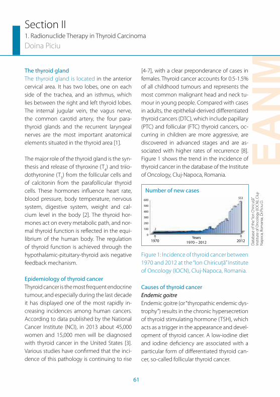
EANM
61
Section II1. Radionuclide Therapy in Thyroid Carcinoma
Doina Piciu
The thyroid gland
The thyroid gland is located in the anterior
cervical area. It has two lobes, one on each
side of the trachea, and an isthmus, which
lies between the right and left thyroid lobes.
The internal jugular vein, the vagus nerve,
the common carotid artery, the four para-
thyroid glands and the recurrent laryngeal
nerves are the most important anatomical
elements situated in the thyroid area [1].
The major role of the thyroid gland is the syn-
thesis and release of thyroxine (T4) and triio-
dothyronine (T3) from the follicular cells and
of calcitonin from the parafollicular thyroid
cells. These hormones inluence heart rate,
blood pressure, body temperature, nervous
system, digestive system, weight and cal-
cium level in the body [2]. The thyroid hor-
mones act on every metabolic path, and nor-
mal thyroid function is relected in the equi-
librium of the human body. The regulation
of thyroid function is achieved through the
hypothalamic-pituitary-thyroid axis negative
feedback mechanism.
Epidemiology of thyroid cancer
Thyroid cancer is the most frequent endocrine
tumour, and especially during the last decade
it has displayed one of the most rapidly in-
creasing incidences among human cancers.
According to data published by the National
Cancer Institute (NCI), in 2013 about 45,000
women and 15,000 men will be diagnosed
with thyroid cancer in the United States [3].
Various studies have conirmed that the inci-
dence of this pathology is continuing to rise
[4-7], with a clear preponderance of cases in
females. Thyroid cancer accounts for 0.5-1.5%
of all childhood tumours and represents the
most common malignant head and neck tu-
mour in young people. Compared with cases
in adults, the epithelial-derived diferentiated
thyroid cancers (DTC), which include papillary
(PTC) and follicular (FTC) thyroid cancers, oc-
curring in children are more aggressive, are
discovered in advanced stages and are as-
sociated with higher rates of recurrence [8].
Figure 1 shows the trend in the incidence of
thyroid cancer in the database of the Institute
of Oncology, Cluj-Napoca, Romania.
Years
1970 – 2012
Number of new cases
600
500
400
300
200
100
0
2
553
1970 2012
← ←
Figure 1: Incidence of thyroid cancer between
1970 and 2012 at the “Ion Chiricuță” Institute
of Oncology (IOCN), Cluj-Napoca, Romania.
Causes of thyroid cancer
Endemic goitre
Endemic goitre (or “thyropathic endemic dys-
trophy”) results in the chronic hypersecretion
of thyroid stimulating hormone (TSH), which
acts as a trigger in the appearance and devel-
opment of thyroid cancer. A low-iodine diet
and iodine deiciency are associated with a
particular form of diferentiated thyroid can-
cer, so-called follicular thyroid cancer.
Dat
abas
e o
f th
e “Io
n C
hiri
cuță
” In
stitu
te o
f On
colo
gy
(IOC
N),
Clu
j-N
apo
ca, R
om
ania
, Dr.P
iciu
D.
62
Ionising radiation
Ionising radiation is one of the most studied
causes of thyroid cancer, which scientiic and
clinical communities accept is directly relat-
ed to a personal history of irradiation.
The human environment accounts for up to
85% of the annual human radiation dose, the
sources being construction materials, food,
drink, soil and the cosmos. Human activities
account for the remaining 15%, the predomi-
nant source being medical procedures (14%
of total); only a small amount of exposure is
due to fallout from past testing of nuclear
weapons, though nuclear accidents have
been associated with a clear increase in the
incidence of thyroid cancer [9].
Chronic stimulation of TSH
As mentioned above, chronic TSH stimula-
tion is considered a trigger for the appear-
ance of metaplasia. This condition is also
important in the follow-up of patients with
DTC, where the replacement dose of thyroid
hormone must produce suppression of TSH
in order to decrease the probability of dis-
ease recurrence.
Environmental factors
Environmental risk factors include radiation
from soil, water, drinks, construction materi-
als and professional exposure, as well as low-
iodine diet and intake.
Immunological factors
The association of chronic lymphocytic thy-
roiditis (Hashimoto’s thyroiditis) with thy-
roid cancer is well known. This pathology is
frequently associated with thyroid nodules;
the long evolution of nodular thyroid and the
occurrence of chronic thyroid insuiciency
may lead to cancer.
Genetic factors
Genetic mutations are responsible for the
development of medullary thyroid carcino-
mas (MTC) in more than 25% of cases. If the
abnormal gene, usually located in codon 634
of chromosome 10, runs in the family, then
the individual may be at very high risk of de-
veloping MTC. Such persons should consider
approaching a medical practitioner for infor-
mation on serum calcitonin tests and genetic
testing. People with certain inherited medi-
cal conditions, including Gardner’s syndrome
and familial polyposis (genetic disorders in
which large numbers of precancerous pol-
yps develop throughout the colon and up-
per intestine), are also at higher risk of PTC.
Cowden disease, a rare inherited disorder
that causes lesions on the face, hands and
feet and inside the mouth, also increases the
risk of developing thyroid cancer and breast
cancer. In addition, mutations of BRAF genes,
which may be determined even from the cy-
tology specimen, are related to DTC.
Symptoms of thyroid cancer
The symptoms of thyroid cancer frequently
are not expressed for many years during the
evolution of the disease. A nodule in the cer-
vical area is the most common presenting
feature. A complete clinical examination is
able to suggest the diagnosis of thyroid pa-
thology, but the positive diagnosis of thyroid
Section II 1. Radionuclide Therapy in Thyroid Carcinoma
EANM
63
cancer is sometimes very diicult. The guide-
lines published by clinicians and scientists
have sought to optimise working practice
and procedures in order to assist in diagnosis.
Only local compression and invasion are
indings speciic to thyroid cancer. The fol-
lowing symptoms may be present owing to
thyroid dysfunction but they are not speciic
to thyroid carcinoma:
• Respiratory insuiciency
• Hoarseness
• Deglutition diiculties
• Tremor
• Sweating
• Diarrhoea/constipation
• Anxiety, depression, lethargy
• Heat/cold intolerance
• Coarse skin
• Weight loss/weight gain
• Pretibial oedema
• Muscle weakness
• Increased/reduced heart rate
• Dry hair
• Swallowing diiculties
• Rarely, pain in the anterior cervical area
These signs and symptoms may very seri-
ously afect the patient’s status.
Diagnosis of thyroid cancer
Clinical examination
The location of the thyroid gland in the ante-
rior neck region makes it very easy to inspect
the organ so as to determine the presence or
absence of disease. A large goitre may cause
tracheal deviation. Palpation of the cervi-
cal lymph nodes should be done carefully,
taking into account the known pathways of
lymphatic drainage: lateral cervical, supra-
clavicular and submental. Inspection of the
gland may reveal enlargement, asymmetry
and a full, bounding pulse. The physician
may observe mobility of the gland and dif-
iculties in swallowing. Palpation will reveal
the texture (soft/irm), the surface (smooth/
seedy/lumpy) and the volume (increased in
diferent grades or decreased/atrophic). The
presence of regional pathological lymph
nodes should be noted, as should spontane-
ous pain or pain on palpation, mobility over
the neck structures and mobility upon swal-
lowing. During the clinical examination it is
important to note any history of irradiation
and possible inherited associated patholo-
gies.
Thyroid ultrasound
Thyroid ultrasound is a widespread tech-
nique that is used as a irst-line diagnostic
procedure for detecting and characterising
nodular thyroid disease (more than 46% of
thyroid nodules greater than 1 cm are not de-
tected by palpation). There are some special
patterns on an ultrasound image that sug-
gest malignant transformation, the most im-
portant being a solid nodule, with peri- and
intranodular circulation, a taller-than-wide
shape (sensitivity, 40.0%; speciicity, 91.4%),
a spiculated margin (sensitivity, 48.3%; speci-
icity, 91.8%), marked hypoechogenicity
(sensitivity, 41.4%; speciicity, 92.2%), micro-
calciication (sensitivity, 44.2%; speciicity,
90.8%), and macrocalciication (sensitivity,
9.7%; speciicity, 96.1%) [10].
64
Fine-needle aspiration biopsy
Fine-needle aspiration biopsy (FNAB) is rou-
tinely recommended for all nodules exceed-
ing 1 cm in maximum diameter. Its sensitivity
has been reported to be 83%, with an associ-
ated speciicity of 72–100% [11].
FNAB is a simple, rapid, cost-efective di-
agnostic method used in the evaluation of
the thyroid nodule. According to the latest
studies [12], the use of various immunohis-
tochemical markers in cytological samples
(BRAF, RAS, RET/PTC etc.) might increase the
accuracy of the positive diagnosis.
Serum hormonal tests
Current guidelines [13,14] recommend that
serum TSH be used as the irst-line test for
detecting both overt and subclinical hypo-
or hyperthyroidism in ambulatory patients
with stable thyroid status and intact hypo-
thalamic/pituitary function. Thyroid hor-
mone tests, [T4, T
3, free T
4 (FT
4) and free T
3
(FT3)] are conducted to determine the cause
of an abnormal TSH test and to check how
well treatment of thyroid disease is working.
In practice, measurement of FT4 and FT
3 is a
more reliable test of thyroid function than
measurement of total hormone levels.
The tissue-speciic origin of thyroglobulin
(Tg) is used as a tumour marker for DTC. Be-
cause Tg protein is tissue speciic, the detec-
tion of Tg in non-thyroidal tissues or luids
(such as pleural luid) indicates the pres-
ence of metastatic thyroid cancer (except
struma ovarii). Current guidelines do not
routinely recommend preoperative serum
Tg measurement. Serum Tg measurements
performed at 4-6 weeks after surgery have
been shown to be of important prognostic
value. Antithyroglobulin antibodies interfere
with the accurate measurement of serum Tg.
Serial antithyroglobulin antibody monitor-
ing necessitates the use of the same method
each time, because assays vary in sensitivity,
speciicity and absolute values.
Thyroid scintigraphy
Thyroid scintigraphy uses radioactive trac-
ers for imaging of the thyroid gland and for
functional assessment during thyroid uptake
of the radiopharmaceutical. Diferent trac-
ers are in use for diferent purposes. Thyroid
scintigraphy is not the irst-line method for
the evaluation of thyroid nodules but it has a
very well established place in the diagnostic
protocol. Whole body radioiodine scintigra-
phy is the most important imaging method
during the follow-up of DTC.
Computed tomography, magnetic
resonance imaging and positron emission
tomography
These modalities are not usually indicated in
the evaluation of thyroid nodules and thyroid
cancers; rather, they are reserved for speciic
well-documented situations.
Overall, clinical examination, TSH determina-
tion, thyroid ultrasound and FNAB are gen-
erally suicient in permitting correct assess-
ment of thyroid nodules and cancer.
Section II 1. Radionuclide Therapy in Thyroid Carcinoma
EANM
65
Classiication of thyroid tumours
Well-diferentiated malignant neoplasms
(85% of thyroid cancers):
• Papillary thyroid carcinoma (PTC)
• Follicular thyroid carcinoma (FTC)
Poorly diferentiated malignant neoplasms:
• Medullary thyroid carcinoma (MTC)
• Anaplastic thyroid carcinoma
Other thyroid tumours:
• Teratoma
• Primary lymphoma and plasmacytoma
• Ectopic thymoma
• Angiosarcoma
• Smooth muscle tumours
• Peripheral nerve sheath tumours
• Paraganglioma
• Solitary ibrous tumour
• Follicular dendritic cell tumour
• Langerhans cell histiocytosis
• Secondary tumours of the thyroid
The TNM Staging System
The most common system used to describe
the stages of thyroid cancer is the American
Joint Committee on Cancer (AJCC) TNM
Staging for Thyroid Cancer [15], where T in-
dicates the size of the main (primary) tumour
and whether it has grown into nearby areas,
N describes the extent of spread to nearby
(regional) lymph nodes and M indicates
whether the cancer has spread (metasta-
sised) to other organs of the body. (The most
common sites of spread of thyroid cancer
are the lungs, the brain, the liver and the
bones.)
T categories for thyroid cancer (other than
anaplastic thyroid cancer)
Note: All categories may be subdivided:
• (s) solitary tumour
• (m) multifocal tumour (the largest focus
determines the classiication)
TX: Primary tumour cannot be assessed
T0: No evidence of primary tumour
T1: Tumour 2 cm or smaller, limited to the
thyroid
T1a: Tumour 1 cm or smaller, limited to the
thyroid
T1b: Tumour larger than 1 cm but not larger
than 2 cm, limited to the thyroid
T2: Tumour between 2 and 4 cm in greatest
dimension, limited to the thyroid
T3: Tumour more than 4 cm in greatest
dimension, limited to the thyroid; any
tumour that displays minimal extrathy-
roidal extension (e.g. extension to the
sternothyroid muscle or perithyroid soft
tissues)
T4a: Tumour of any size that has grown ex-
tensively beyond the thyroid gland into
the nearby tissues of the neck, such
as the larynx, trachea, oesophagus or
recurrent laryngeal nerve (also called
moderately advanced disease)
T4b: Tumour of any size that has grown ei-
ther back toward the spine or into near-
by large blood vessels (also called very
advanced disease)
66
T categories for anaplastic thyroid cancer
All anaplastic thyroid cancers are considered
T4 tumours at the time of diagnosis.
T4a: Tumour still within the thyroid
T4b: Tumour has grown beyond the thyroid
N categories for thyroid cancer
NX: Regional (nearby) lymph nodes cannot
be assessed
N0: No spread to nearby lymph nodes
N1: Cancer spread to nearby lymph nodes
N1a: Spread to pretracheal, paratracheal and
prelaryngeal lymph nodes
N1b: Spread to other cervical lymph nodes or
to lymph nodes behind the throat (ret-
ropharyngeal) or in the upper mediasti-
num
M categories for thyroid cancer
M0: No distant metastasis
M1: Spread to other parts of the body, such
as distant lymph nodes, brain, internal
organs and bones
Thyroid cancer staging
Papillary or follicular thyroid cancer in
patients younger than 45 years old
All people younger than 45 years old hav-
ing these types of cancer are considered to
have stage I cancer if they have no distant
spread, and stage II cancer if they have dis-
tant spread:
Stage I (any T, any N, M0)
Stage II (any T, any N, M1)
Papillary or follicular thyroid cancer in
patients aged 45 years old and older
Stage I (T1, N0, M0)
Stage II (T2, N0, M0)
Stage III:
• T3, N0, M0
• T1 to T3, N1a, M0
Stage IVA:
• T4a, any N, M0
• T1 to T3, N1b, M0
Stage IVB (T4b, any N, M0):
Stage IVC (any T, any N, M1)
Medullary thyroid cancer
Age is not a factor in the staging of medullary
thyroid cancer
Stage I (T1, N0, M0)
Stage II:
• T2, N0, M0
• T3, N0, M0:
Stage III (T1 to T3, N1a, M0)
Stage IVA:
• T4a, any N, M0
• T1 to T3, N1b, M0
Stage IVB (T4b, any N, M0)
Stage IVC (any T, any N, M1)
Anaplastic (undiferentiated) thyroid cancer
All anaplastic thyroid cancers are considered
to be stage IV, relecting the poor prognosis
of this type of cancer.
Stage IVA (T4a, any N, M0)
Stage IVB (T4b, any N, M0)
Stage IVC (any T, any N, M1)
Section II 1. Radionuclide Therapy in Thyroid Carcinoma
EANM
67
Prognostic factors
The majority of thyroid cancers are not ag-
gressive and have an excellent prognosis.
Prognostic factors include:
• Age
• Sex
• Histological type
• Histological grade
• Tumour size
• Extrathyroidal extension
• Distant metastases
• Lymph node involvement
• Multicentricity
• Incomplete resection
Depending on histology and stage, dif-
ferent treatment approaches are adopted
for thyroid carcinoma. There are multiple
guidelines for diagnosis and therapy, many
of them published by national and interna-
tional committees of experts in endocrinol-
ogy, endocrine surgery, nuclear medicine
and oncology. All of these algorithms have
been reviewed frequently with the intention
of minimising suboptimal diagnosis, surgery
or irradiation and providing the best option
for multimodal treatment.
Surgical Treatment
Surgery is the most important treatment op-
tion in thyroid cancer management. It consists
in total thyroidectomy, total lobectomy or pro-
phylactic total thyroidectomy in MTC. Lymph-
adenectomy in thyroid cancer is well deined
regarding the ablation of the pretracheal
lymph node group (level VI, Delphian lymph
nodes) and the selective resection of lateral-
cervical lymph nodes. The type of surgical pro-
cedure is dictated by the type of tumour and
the risk category, as follows [14]:
Diferentiated thyroid carcinoma (DTC) –
nodules with undeined cytology from FNAB:
• undeined solitary nodule <4 cm – total
lobectomy
• undeined solitary nodule >4 cm or per-
sonal history of irradiation or familial histo-
ry of thyroid cancer – total thyroidectomy
• undeined multiple nodules or bilateral
nodules – total thyroidectomy
Diferentiated thyroid carcinoma (DTC) – nod-
ules with suggestive cytology for malignancy:
• thyroid cancer >1 cm – total thyroidctomy
• total lobectomy only if:
- tumour <1 cm
- low risk histology
- unifocal
- intrathyroid papillary carcinoma,
without previous irradiation and
without lymph node metastases or
local invasion.
The type of lymphadenectomy depends on
the tumour type, tumour size and presence
of lymph node metastases.
Medullary thyroid carcinoma (MTC) is treated
by total thyroidectomy and level VI lymph
node dissection; uni- or bilateral cervical
68
lymph node resection is also recommended
depending upon presence of the sporadic
or hereditary form, tumour size and lymph
node involvement.
Anaplastic thyroid carcinoma requires total
thyroidectomy and level VI lymph node dis-
section, assuming that the staging indicates
these to be appropriate; frequently, however,
this cancer is classiied as being at a stage un-
suitable for surgical resection.
Thyroid hormone treatment
Synthetic levothyroxine is typically adminis-
tered in a dose of 2-2.5 µg/kg body weight; ac-
cording to the risk group and time since thera-
py, the dose might be suppressive, with the aim
of maintaining the TSH level below 0.1 mIU/L.
Radioiodine therapy
Radiopharmaceutical
Iodine-131 (131I) sodium iodide capsules are
used for oral administration, with a speciic
activity of 1.11 GBq, 1.85 GBq, 2.59 GBq, 3.7
GBq or 5.55 GBq at calibration time, as clearly
stated by the producer.
Principle131I-sodium iodide is administered systemi-
cally for selective irradiation of post-surgical
thyroid remnants of DTC or other suitable
cases of DTC not treated by surgery, or for the
irradiation of iodine-avid metastases.
Recommendations
The European Thyroid Association (ETA) [14]
recommendations are:
1. Very low risk group – no indication for
postoperative 131I
• Unifocal diferentiated microcarcinoma
(≤1 cm)
• No extension beyond the thyroid capsule
• No lymph node metastases
• Complete surgery
2. Low-risk group – probable indication – no
consensus
• Less than total thyroidectomy, no lymph
node dissection
• Age <18 years
• Unfavourable histology, T1 >1 cm, T2,
N0, M0
3. High-risk group – deinite indication
• T3, T4, N1, M1
• Incomplete surgery
• High risk of recurrence
The American Thyroid Association (ATA) [13]
categorised their recommendations into
seven ratings (A–F, plus I for recommenda-
tion neither for nor against) according to the
strength of the available medical evidence.
For assessment of risk recurrence, they pro-
posed a three-level stratiication into low-,
intermediate- and high-risk groups.

Section II 1. Radionuclide Therapy in Thyroid Carcinoma
EANM
69
According to the ATA, radioiodine ablation is
recommended for:
• Patients with stage III or IV disease
• All patients with stage II disease who are
younger than 45 years
• Most patients with stage II disease who
are 45 years old or older
• Selected patients with stage I disease
who have higher risk factors, i.e. multi-
focal (>2 foci) disease, nodal metastases,
extrathyroid and vascular invasion and/
or aggressive histology.
ETA/ATA recommendations for management
of metastatic and recurrent disease
For local and regional recurrence:
• Surgery and 131I
• External beam therapy of 40-45 Gy in
25-30 sessions only if surgery is not pos-
sible and there is no 131I uptake
For distant metastases:
• Lungs: 3.7-7.4 GBq every 4-8 months
during the irst 2 years
• Bones: surgery and 131I, external beam
therapy
• Brain: surgery and external beam ther-
apy, 131I only after external radiotherapy
• Doses are repeated until the disease is
cured
• Radioiodine therapy is stopped if there
are signs that the tumour is no longer
iodine avid
Risk of second malignancies:
• ETA – the risk of second malignancy
is signiicantly higher at a dose above
22.2 GBq 131I [14].
• ATA – the risk of second malignancy is
dose related, and long-term follow-up
studies demonstrate a very low risk.
There appears to be an increased risk of
breast cancer, but it is unclear whether
this is a result of screening, radioiodine
therapy or other factors.
Protocol
There are multiple choices for establishing
the necessary dose for radioiodine therapy,
but administration of a ixed dose of 131I is the
simplest and most widely used method.
The indications for radioiodine are as follows:
a) Remnant ablation. The dose for remnant
ablation may be 1.1, 1.85, 2.59 or 3.7 GBq,
according to the volume of the tissue, the
uptake and the level of Tg.
b) Lymph node metastases may be treated
with 3.7–6.475 GBq of 131I. Cancers that
extend through the thyroid capsule and
have been incompletely resected are
treated with 3.7-7.4 GBq.
c) Patients with distant metastases are usu-
ally treated with 7.4 GBq of 131I, which typi-
cally will not induce radiation sickness or
produce serious damage to other struc-
tures but may exceed generally accepted
70
safety limits for the blood in the elderly
and those with impaired kidney function.
d) Difuse pulmonary metastases are treated
with 5.55 GBq or less of 131I to avoid lung
injury, which may occur when more than
2.96 GBq remains in the whole body 48 h
after the treatment.
The administered activity of radioactive io-
dine therapy should be adjusted for paediat-
ric patients.
After the administration, the patient remains
in a fasting condition for two more hours
and after that he/she is advised to hydrate
consistently and to use “chewing drops” to
stimulate saliva production and thereby re-
duce the impact of irradiation on the salivary
gland.
The administration of 131I for the treatment of
DTC is typically performed under hospitali-
sation and patients are discharged in accor-
dance with national radioprotection regula-
tions; however, in some countries the treat-
ment may be done on an ambulatory basis
even at doses of 131I higher than 1.1 GBq.
All patients receive paper and electronic in-
formation about the diagnosis, the treatment
options, follow-up, side-efects and personal
indications about individual medication.
The rooms must have separate washing and
toilet facilities and the patient is under con-
tinuous surveillance by the staf, on separate
cameras. Upon discharge, the patient is ad-
vised how to minimise the radiation risk to
other persons and for what period of time,
depending on the dosimetry measurements
at the time of discharge.
Post-treatment whole-body 131I scan
Whole-body radioiodine imaging should be
performed several days (1-3) following treat-
ment to document 131I uptake by the tumour
and to assess the extent of the disease.
Special recommendations:
• Do not use CT with contrast media for
staging; it is reserved for speciic situations
where the 131I therapy is not scheduled.
• Avoid 131I whole-body scan before radioio-
dine treatment unless distant metastases
are suspected and an important active
thyroid remnant must be removed by sur-
gery.
• Do not use any medication (blocking
agents with iodine, amiodarone etc.) that
may inluence the radioiodine uptake.
• Use the lowest eicient radioiodine activity.
• At discharge it is necessary to give the pa-
tient information regarding radioprotec-
tion issues.
A total response and disease-free state are
indicated by the absence of tumour mass
on clinical examination and ultrasound, ab-
sence of symptoms, undetectable Tg in the
presence of high TSH and absence of anti-Tg
antibodies.
Section II 1. Radionuclide Therapy in Thyroid Carcinoma
EANM
71
Strict follow-up is very important for early
detection of disease recurrence and rapid in-
stitution of appropriate treatment.
External beam radiotherapy
External beam radiotherapy (EBRT) has only
a limited indication in DTC, being appropri-
ate when the tumour is inoperable or there is
local invasion without a favourable response
to radioiodine therapy. Similarly, in MTC, EBRT
is limited to inoperable tumours and cases
with regional invasion. In anaplastic thyroid
carcinoma, by contrast, EBRT is frequently
used owing to the very severe outcome of
the disease and the lack of other therapeutic
options.
Chemotherapy
Thyroid carcinoma is widely considered to
be radio- and chemoresistant. In DTC, che-
motherapy is reserved for those situations in
which the tumour loses ainity for radioio-
dine, while in MTC it is used in some forms of
abdominal metastasis. Anaplastic thyroid car-
cinoma shows rapid and aggressive growth,
and response rates to systemic chemothera-
py are very limited.
Clinical trials
When DTC shows continued evolution, but
no longer responds to radioiodine and pres-
ents a negative 131I whole-body scan, new
drugs undergoing clinical trials might repre-
sent a reasonable treatment choice.
The role of supporting staf in
administering radionuclide therapy
Patient preparation
Patients must have undergone total or near-
total thyroidectomy and have a conirmed
pathological diagnosis of DTC.
For successful radioiodine therapy, the TSH
value must exceed 40 mIU/L. This value may
be obtained:
• At 4-6 weeks after surgery in the absence
of thyroid therapy replacement
• After 2-4 weeks of thyroid hormone with-
drawal, in the case of replacement thyroid
hormone therapy;
• After administration of recombinant TSH
in two intramuscular injections of 0.9 ml
solution on two consecutive days, if the
patient is under hormonal treatment.
It is important to be familiar with the TSH
stimulation procedures and to be able to ver-
ify that the patient has understood and con-
sented to the procedure. The recombinant
TSH stimulation method will involve a range
of clinical professionals in ensuring accurate
dose preparation and administration.
Patients must fast before therapy and must
be checked carefully by all the staf. It must
be ensured that the patient is not taking
any of an extensive inventory of long-term
medications that might interfere with iodine
uptake. During the preceding 3 months, the
72
patient must not have received blocking
agents or iodinated contrast media used in
X-ray examinations. Dietary restrictions con-
sist in avoidance of iodised salt, seafood, nuts
and other foods rich in iodine for 1 week
prior to therapy.
Precautions
In the case of female patients of childbearing
potential, a routine pregnancy test must be
done within 72 h prior to the administration
of 131I. No radioiodine therapy may be admin-
istered in pregnant patients.
All institutional and departmental documen-
tation must be explained and signed before
therapy in order to limit the exposure after
iodine administration.
In order to avoid or reduce the stunning ef-
fect, pretherapy 131I whole-body scan is not
routinely recommended. It is possible to:
• Use 123I or low doses (74 or 111 MBq) of 131I
• Use a shortened interval (of not more than
72 h) between the
diagnostic 131I dose and the therapy dose.
A close relationship between all staf mem-
bers involved in the diagnostic and treat-
ment of this pathology is essential in achiev-
ing an optimal outcome for patients.
Patient surveillance
Patient surveillance in the period follow-
ing radioiodine administration is an issue
relevant to physicians, nurses, technologists
and others. Radiation protection-related
problems are very important for both pa-
tients and staf, and have already been dis-
cussed in Chapter 4 of this book.
Contamination and vomiting, as well as vari-
ous prostheses and metallic clips (which may
be contaminated by the iodine from the
bloodstream), are factors that may inluence
the accuracy of scanning results [16] and
therefore it is very important that staf pay at-
tention to such circumstances.
Whole-body scanning (WBS) – technical
issues
131I-NaI is absorbed from the gastrointestinal
tract after oral administration. The process of
organiication consists in trapping of 131I in
the thyroid gland and in all DTC cells dissemi-
nated in the body.
The whole-body acquisition allows data to
be acquired from a patient who is larger than
the ield of view of the gamma camera. The
patient’s bed is moved into/out of the gantry
during acquisition of the image.
The scan is performed at 24, 48, 72 h or some-
times even 5-7 days after administration of
the radioiodine capsule, the time interval
depending on the activity of the radioiodine
dose and the amount of remnant tissue. The
patient’s position for scanning is supine, with
slight neck extension. Scanning technique
entails use of the following:
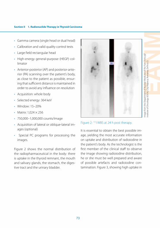
Section II 1. Radionuclide Therapy in Thyroid Carcinoma
EANM
73
• Gamma camera (single head or dual head)
• Calibration and valid quality control tests
• Large ield rectangular head
• High-energy general-purpose (HEGP) col-
limator
• Anterior-posterior (AP) and posterior-ante-
rior (PA) scanning over the patient’s body,
as close to the patient as possible, ensur-
ing that suicient distance is maintained in
order to avoid any inluence on resolution
• Acquisition: whole body
• Selected energy: 364 keV
• Window: 15–20%
• Matrix: 1,024 × 256
• 750,000–1,000,000 counts/image
• Acquisition of lateral or oblique-lateral im-
ages (optional)
• Special PC programs for processing the
images.
Figure 2 shows the normal distribution of
the radiopharmaceutical in the body: there
is uptake in the thyroid remnant, the mouth
and salivary glands, the stomach, the diges-
tive tract and the urinary bladder.
Figure 2: 131I WBS at 24 h post therapy.
It is essential to obtain the best possible im-
age, yielding the most accurate information
on uptake and distribution of radioiodine in
the patient’s body. As the technologist is the
irst member of the clinical staf to observe
the image showing radioiodine distribution,
he or she must be well prepared and aware
of possible artefacts and radioiodine con-
tamination. Figure 3, showing high uptake in
Imag
e fr
om
the
Dep
artm
ent o
f Nu
clea
r Med
icin
e o
f th
e “Io
n C
hiri
cuță
” In
stitu
te o
f On
colo
gy
(IOC
N),
Clu
j-Nap
oca
, Ro
man
ia, D
r.Pic
iu D
.

74
the ocular areas due to contact lens contami-
nation, illustrates how a mistake may occur
and the importance of recognising artefacts
and taking appropriate corrective action.
Figure 3: 131I WBS post therapy: high uptake is
seen in the ocular areas due to contact lens
contamination.
Figures 4 and 5 further serve to illustrate the
sort of mistake that may arise if inadequate
precautions are taken to limit possible con-
tamination. Figure 4 shows a 131I WBS at 48 h
post therapy that demonstrates thyroid rem-
nant uptake in the thyroid area; in addition,
pathological uptake is evident in the lower
right lobe of the lung. This image was highly
suggestive for a macronodular right lung me-
tastasis. Because the value of Tg (28.5 ng/L,
N.V. <0.1 ng/L in athyreotic patients) was not
very high, as is usually the case in patients
with lung metastasis, we supposed that ei-
ther the lung tumor was not related to the
thyroid pathology or that this was a metasta-
sis of the thyroid carcinoma with low expres-
sion of Tg. Before testing other pathologies,
it was important to double check the pos-
sibility of radioiodine contamination of the
patient. In this second inspection we found
that the patient had placed a handkerchief in
the pocket of the disposable robe. Figure 5
was obtained immediately after changing
the disposal robe, and we observed that the
uptake from the right lung has disappeared. Imag
e fr
om
the
Dep
artm
ent o
f Nu
clea
r Med
icin
e o
f th
e “Io
n C
hiri
cuță
” In
stitu
te o
f On
colo
gy
(IOC
N),
Clu
j-Nap
oca
, Ro
man
ia, D
r.Pic
iu D
.
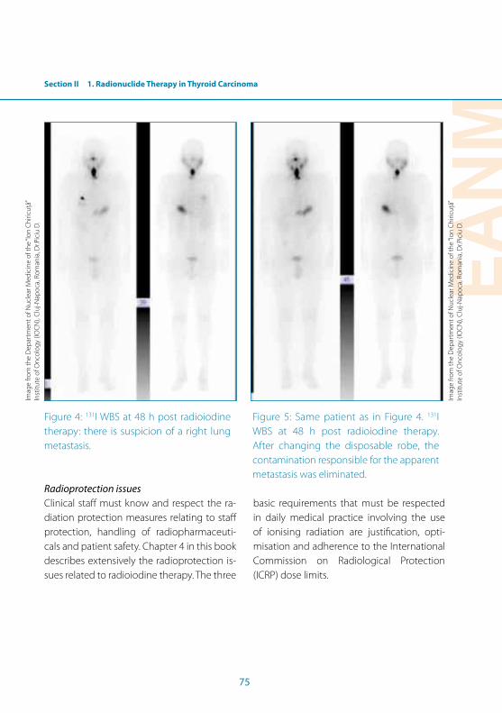
Section II 1. Radionuclide Therapy in Thyroid Carcinoma
EANM
75
Figure 4: 131I WBS at 48 h post radioiodine
therapy: there is suspicion of a right lung
metastasis.
Figure 5: Same patient as in Figure 4. 131I
WBS at 48 h post radioiodine therapy.
After changing the disposable robe, the
contamination responsible for the apparent
metastasis was eliminated.
Radioprotection issues
Clinical staf must know and respect the ra-
diation protection measures relating to staf
protection, handling of radiopharmaceuti-
cals and patient safety. Chapter 4 in this book
describes extensively the radioprotection is-
sues related to radioiodine therapy. The three
basic requirements that must be respected
in daily medical practice involving the use
of ionising radiation are justiication, opti-
misation and adherence to the International
Commission on Radiological Protection
(ICRP) dose limits.
Imag
e fr
om
the
Dep
artm
ent o
f Nu
clea
r Med
icin
e o
f th
e “Io
n C
hiri
cuță
” In
stitu
te o
f On
colo
gy
(IOC
N),
Clu
j-Nap
oca
, Ro
man
ia, D
r.Pic
iu D
.
Imag
e fr
om
the
Dep
artm
ent o
f Nu
clea
r Med
icin
e o
f th
e “Io
n C
hiri
cuță
” In
stitu
te o
f On
colo
gy
(IOC
N),
Clu
j-Nap
oca
, Ro
man
ia, D
r.Pic
iu D
.
76
References Section II, Chapter 1
References1. Bliss RD, Gauger PG, Delbridge LW. Surgeon’s approach
to the thyroid gland: surgical anatomy and the impor-tance of technique. World J Surg. 2000;24:891-7.
2. Piciu D. Thyroid gland. in Piciu D, ed. Nuclear endocri-nology. Berlin Heidelberg New York: Springer; 2012, pp 53-163.
3. National Cancer Institute. 2013. http://www.cancer.gov/cancertopics/wyntk/thyroid.
4. Enewold L, Zhu K, Ron E, Marrogi AJ, Stojadinovic A, Peoples GE, Devesa SS. Rising thyroid cancer incidence in the United States by demographic and tumor char-acteristics, 1980–2005. Cancer Epidemiol Biomarkers Prev 2009;18:784–91.
5. Davies L, Welch HG. Increasing incidence of thy-roid cancer in the United States, 1973–2002. JAMA 2006;295:2164–7.
6. Colonna M, Guizard AV, Schvartz C, Velten M, Raverdy N, Molinie F, et al. A time trend analysis of papillary and follicular cancers as a function of tumour size: a study of data from six cancer registries in France (1983–2000). Eur J Cancer 2007;43:891–900.
7. Hogan AR, Zhuge Y, Perez EA, Koniaris LG, Lew JI, Sola JE. Pediatric thyroid carcinoma: incidence and outcomes in 1753 patients. J Surg Res 2009;156:167-72.
8. Piciu D, Piciu A, Irimie A. Thyroid cancer in children: a 20-year study at a Romanian oncology institute. Endocr J. 2012;59:489-96.
9. Sources of radiation exposure. http://www.epa.gov/radiation/sources/index.html
10. Wémeau JL, Sadoul JL, d’Herbomez M, Monpeyssen H, Tramalloni J, Leteurtre E, et al. Guidelines of the French Society of Endocrinology for the management of thy-roid nodules. Ann Endocrinol 2011;72:251-81.
11. Wojtczak B, Sutkowski K, Bolanowski M, Łukieńczuk T, Lipiński A, Kaliszewski K, et al. The prognostic value of ine-needle aspiration biopsy of the thyroid gland - analysis of results of 1078 patients. Neuro Endocrinol Lett. 2012;33:511-6.
12. Koperek O, Kornauth C, Capper D, Berghof AS, Asari R, Niederle B, et al. Immunohistochemical detection of the BRAF V600E-mutated protein in papillary thyroid carcinoma. Am J Surg Pathol. 2012;36:844-50.
13. Cooper DS, Doherty GM, Haugen BR, Kloos RT, Lee SL, Mandel SJ, et al. Revised American Thyroid Associa-tion management guidelines for patients with thyroid nodules and diferentiated thyroid cancer. Thyroid 2009;19:1167-214.
14. Pacini F, Schlumberger M, Dralle H, Elisei R, Smit JW, Wiersinga W. European consensus for the management of patients with diferentiated thyroid carcinoma of the follicular epithelium. Eur J Endocrinol 2006;154:787-803.
15. Edge S, Byrd DR, Compton CC, Fritz AG, Greene FL, Trotti A, eds. AJCC Cancer Staging Manual, 7th edn. New York: Springer, 2010:1-646..
16. Piciu D, Piciu A, Irimie A. Cerebral false-positive radio-iodine uptake. Acta Endocrinologica (Buc) 2012;8:495.

EANM
77
Section II2. Radionuclide Therapy in Benign Thyroid Disease
Doina Piciu
Introduction
Radioiodine (iodine 131, 131I) was the irst
radiopharmaceutical to be used in humans
for the treatment of benign conditions of the
thyroid gland, the irst procedure being per-
formed during the 1940s.
A frequent indication for radioiodine therapy
is hyperthyroidism, which is a consequence
of excessive thyroid hormone action. Patients
with large non-toxic goitre, including even
those with a euthyroid status, may beneit
from a reduction in thyroid volume after ra-
dioiodine therapy (shrinkage efect).
The further treatment options for hyper-
thyroidism include antithyroid drugs and
surgery, and the choice of therapy depends
upon the speciic circumstances. Medical
treatment (drugs) is frequently the irst-line
therapy, with the primary intention of stabi-
lising symptoms and allowing for a proper
assessment of deinitive therapeutic strate-
gies. During the next 3-6 months, according
to disease evolution and the recurrence or
otherwise of hyperthyroidism upon with-
holding of medication, surgery or radioio-
dine therapy may be proposed. There are,
however, centres in the world, mainly in the
United States, where radioiodine is started as
irst-line therapy. Surgery is usually reserved
for large, compressive, unaesthetic goitres
and for those patients with thyroid nodules
in which there is a high index of suspicion of
future malignant transformation.
In patients with hyperthyroidism, the aim
of the 131I treatment is to achieve a non-hy-
perthyroid status, which can be euthyroid or
hypothyroid, the latter being subsequently
treated by thyroid hormone substitutive
medication, such as levothyroxine (LT4).
Indications and contra-indications for 131I
therapy
Indications for 131I therapy are:
• Graves’ disease (autoimmune hyperthy-
roidism)
• Toxic multinodular goitre
• Solitary hyperfunctioning nodule
• Non-toxic multinodular goitre
• Goitre recurrence after surgery
• With special precautions, amiodarone-
induced hyperthyroidism
Contra-indications for 131I therapy may be ab-
solute or relative. Absolute contra-indications
are pregnancy and breastfeeding; the physi-
cian may, however, advise the radioiodine
treatment after termination of pregnancy and
cessation of breastfeeding if the patient’s con-
dition is life threatening. Relative contra-indica-
tions are uncontrolled hyperthyroidism and ac-
tive thyroid orbitopathy (especially in smokers).
Clinical staf, including all those involved with
patient care, should take appropriate steps to
determine whether any contra-indications
exist. When they do exist, the physician
should be informed prior to the commence-
ment of radionuclide therapy.
78
Facility requirements
In some countries the national regulations
dictate that radionuclide treatments may be
conducted only in in-patients while in oth-
ers they may be permitted on an ambulatory
out-patient basis. The clinical facility in which
thyroid disease is to be treated with radionu-
clides must have appropriate authorisation.
It must also have appropriate personnel, ra-
diation safety equipment and clear, well-de-
ined procedures for handling and disposal
of waste. This facility may be an endocrinol-
ogy department or a nuclear medicine de-
partment or any other specialised unit that
respects the above requirements.
The involvement of technologists in radionu-
clide therapy procedures varies signiicantly,
from no involvement to leading adminis-
tration procedures. At the minimum they
should have knowledge and skill in the cor-
rect use of the equipment and the radiation
safety of the facility.
Patient preparation
• Written informed consent must be ob-
tained from the patient before the pro-
cedure commences. Special recommen-
dations and radioprotection information
must also be discussed/given to patients
before commencement.
• Depending upon national regulations,
recommendations relating to conception
may have to be provided. Generally, it is
suggested that after 131I therapy, contra-
ception should be used for 4 months.
• In female patients of childbearing poten-
tial, a routine pregnancy test should be
performed before the administration of 131I.
• Antithyroid drugs must be stopped 5–7
days before radioiodine therapy.
• Beta-blockers, e.g. metoprolol, atenolol
and propranolol (drugs used to manage
heart problems) should be stopped 24 h
before therapy.
• Propylthiouracil (a drug to reduce thyroid
metabolism) should be stopped at least 2
weeks before therapy.
• In patients with Graves’ ophthalmopa-
thy, if the patient is already taking steroid
therapy, oral prednisolone (a drug used to
treat auto-immune and inlammatory con-
ditions) is administered. It is not routine to
start corticotherapy.
• In patients with thyrotoxicosis induced by
amiodarone (an anti-arrhythmic drug) or in
those receiving compounds that contain
iodine (e.g. radiographic contrast agents),
radioiodine can be administered as deini-
tive therapy if the drug has been stopped
suiciently long for the excess iodine load
to be eliminated – at least 3–6 months.
Assessment of disease
• Laboratory testing, including free thyroxine
(FT4), free triiodothyronine (FT
3), thyroid-
stimulating hormone (TSH), anti-thyroid
Section II 2. Radionuclide Therapy in Benign Thyroid Disease
EANM
79
peroxidase antibodies (anti-TPO) and anti-
thyrotropin receptor antibodies (TRab).
• 99mTc thyroid scintigraphy or a radioactive
iodine uptake test (RAIU, with scanning 24
h after intake of the radioactive iodine) can
be undertaken according to availability.
Uptake measurements are not usually re-
quired when ixed activities are used.
• Determination of the thyroid target vol-
ume can be done using ultrasonography,
while evaluation of intrathoracic exten-
sion in those with a large goitre can be
achieved using magnetic resonance im-
aging or computed tomography (without
contrast agents).
• Fine-needle aspiration biopsy (FNAB) is
performed when nodules meet criteria
suggestive of malignancy.
• In patients with Graves’ ophthalmopathy,
the status of thyroid eye disease activity
is established by an experienced ophthal-
mologist.
• For children between 5 and 15 years of
age, radioiodine therapy may be consid-
ered. The side-efects should be clearly dis-
cussed by all staf involved in the treatment
of a young patient with Graves’ disease.
Radiopharmaceutical and administration
Radioiodine is available in solid (capsules)
and liquid forms, both of which can be used
for oral administrations.
The ixed dose method is usually based on an
estimation of the size of the gland, which
can be achieved by ultrasound. Frequently
the recommended activity is 3.7-5.55 MBq/
gram of thyroid, adjusted with the 24-h 131I
RAIU (radioiodine uptake). In patients with
autonomous nodules, the activity currently
prescribed varies between 200 and 800 MBq,
with the intention of achieving an irradiation
dose of 200-300 Gy at nodule level.
The calculated dose method might be used
in the selection of a more accurate dose of
radioiodine for treatment. There are multiple
methods for dose calculation, but the sim-
plest formula is as follows:
MBq= (V×25×Gy) ÷ [(RAIU (24h) x Tef
]
where MBq = the calculated activity in MBq,
V = the gland volume estimated at ultra-
sound in ml (cm3), Gy = the estimated dose
at thyroid level, RAIU (24h) = % of thyroid up-
take at 24 h, Tef
= efective half-time, and 25
is a constant.
Those involved with performing or assisting
with radionuclide therapy procedures must
be able to use and calculate this and similar
basic formulas.
Side-efects of 131I therapy
Patients with a large goitre may notice tran-
sient swelling of the goitre and dyspnoea. In
some patients a thyroid storm may develop.
This rare condition must be treated with
intravenous infusion of antithyroid drugs,
80
corticosteroids and beta-blockers. This situa-
tion is extremely rare and does not represent
a reason for not recommending the therapy.
Hypothyroidism is the main side-efect of ra-
dioiodine treatment. Its rate varies and its inci-
dence continues to increase over time, so that
life-long follow-up is essential. According to
published literature, the rate of hypothyroidism
at 1 year after radioiodine therapy is very similar
to the rate following the surgical approach.
Administration of prednisone (a drug used to
treat allergic reactions) helps prevent exacer-
bation of ophthalmopathy, and this is now
the standard approach in patients who have
clinically active ophthalmopathy at the time
of treatment.
The determination of periorbital inlamma-
tory tissue by nuclear testing with 99mTc-DTPA
may identify the cases that will beneit from
steroid treatment.
Follow-up after radioiodine treatment
As mentioned above, regular review of thy-
roid function tests in patients who have un-
dergone radioiodine treatment for thyroid
disease is essential to assess the eicacy of
the treatment and for timely detection of
development of hypothyroidism. The level of
serum TSH increases slowly, so determination
of TSH is less useful for monitoring during the
irst 3-6 months, when determination of FT4
should be used instead. In those with persis-
tent hyperthyroidism, radioiodine treatment
can be repeated after 6–12 months. Annual
laboratory tests (including TSH) are necessary
throughout life, even in patients with euthy-
roidism after 131I therapy.
Further readingStokkel MP, Handkiewicz Junak D, Lassmann M, Dietlein M, Luster M. EANM procedure guidelines for therapy of benign thyroid disease. Eur J Nucl Med Mol Imaging. 2010;37:2218–28.
Piciu D. Thyroid gland. in Piciu D, ed. Nuclear endocrinology. Berlin Heidelberg New York: Springer; 2012:53-163.
Riccabona G. Use of radionuclides in the diagnosis and treatment of thyroid diseases. Wien Med Wochenschr. 1977;127:97-101.
ATA/AACE guidelines. Hyperthyroidism and other causes of thyrotoxicosis: management guidelines of the American Thyroid Association and American Association of Clinical Endocrinologists. Endocrine Practice 2011; 17 no. 3:11-13.
EANM
81
Section II3. Radionuclide Therapy in Neuroendocrine Tumours
Ruth Berhane Menghis, Brian A. Lowry, Christopher Mayes and Sobhan Vinjamuri
Deinition and classiication of
neuroendocrine tumours
Neuroendocrine tumours (NETs) are a rare,
slow growing and heterogeneous group
of tumours that originate from the difuse
neuroendocrine system and the conined
neuroendocrine system. The difuse and
conined neuroendocrine systems are rep-
resented by the pituitary, the parathyroid
and the adrenal medulla, as well as the en-
docrine islets in the pancreatic tissue, and
the disseminated endocrine cells in the di-
gestive and respiratory tracts [1].
NETs are classiied according to their embry-
ological origin into tumours of the foregut
(oesophagus, lungs, stomach, duodenum
and pancreas), midgut (appendix, ileum,
caecum and ascending colon) and hind-
gut (distal large bowel and rectum) [2]. This
classiication does not, however, take into
consideration the biological diferences of
NETs. Optimal and revised current practice
is to describe NETs according to their loca-
tion of primary origin (e.g. duodenum and
caecum) and to include information on
biological activity which results in hormone
secretion or symptomatic presentation (e.g.
vipoma, gastrinoma and insulinoma).
The irst WHO classiication of NETs was
published in 1980. In this classiication the
term “carcinoid” was used to describe most
tumours. In order to standardise terminol-
ogy, this classiication was updated in 2000,
in 2004 and in 2010. The 2010 updated
classiication is based on tumour histopa-
thology (well diferentiated/poorly diferen-
tiated), proliferative activity (G1, G2, G3) and
TNM staging [3].
Pathology
At presentation the vast majority of tumours
are either benign or relatively slow growing
(well diferentiated) or retain many multi-
potent diferentiation features [4]. Such fea-
tures include the possession of neuroamine
uptake mechanisms and/or speciic recep-
tors at the cell membrane, such as soma-
tostatin receptors (SSTRs), which can be
used for the identiication, localisation and
therapy of NETs. However, NETs can also
present with an aggressive behaviour and
become highly malignant. The latter are de-
scribed as poorly diferentiated NETs.
SSTRs are G protein-coupled receptors with
a widespread distribution in the human
nervous system and diferent tissues in the
body, including the pancreatic islets, stom-
ach, lungs, kidneys and prostate. Five SSTR
subtype genes have been cloned and char-
acterised. They have been code named sst1,
sst2, sst3, sst4 and sst5 [5].
Of particular interest is the fact that a large
number of NETs express SSTRs and this forms
the foundation for the functional imaging
carried out in nuclear medicine. Of the ive
major subtypes of SSTR, SSTR2 and SSTR5
are the ones most commonly expressed in
NETs; however, there is considerable varia-
82
tion in SSTR subtype expression among the
diferent tumour types and among tumours
of the same type [6].
It is also particularly important to evaluate
the proliferation index of neoplastic cells by
measuring the percentage of tumour cells
with Ki-67 positivity. Ki-67 is an antigen ex-
pressed during the G1 and M phases of the
cell cycle. Determination of the Ki-67 plays
an important role and allows selection of
the most appropriate and efective treat-
ment, reserving chemotherapy for NETs
with a high proliferative index which are
likely to respond better when treated with
antiblastic drugs [7]. In contrast, NETs with
a low Ki-67 respond less well to chemother-
apy and are more responsive to treatment
with somatostatin analogues, either cold
somatostatin or possibly radiolabelled ana-
logues (radiometabolic therapy) [8].
Presentation
Clinically, NETs can be symptomatic (i.e.
“functioning”) or silent (i.e. “non-function-
ing”). The functioning NETs can give rise to
clinical syndromes, such as the one called
“carcinoid syndrome”, which is character-
ised by nausea, flushing and diarrhoea.
Other clinical manifestations include
wheezing or asthma-like symptoms as well
as pellagra-like skin lesions. These symp-
toms arise due to specific production, stor-
age and secretion of polypeptides, amines
and hormones, like serotonin. Cardiac in-
volvement can occur as a result of fibrosis
involving the endocardium, primarily on
the right side.
Non-functioning NETs are not associated
with any speciic syndrome and are more
diicult to detect than functioning NETs; for
this reason, patients generally present late,
with large primary tumours and advanced
disease. In the case of non-functioning
NETs, there can be some degree of hormon-
al secretion, but the secretion may be at
subclinical levels or there may be secretion
of inactive compounds. Non-speciic symp-
toms such as weight loss, abdominal bleed-
ing and pain can be related to increased
tumour mass producing mass efect symp-
toms and/or metastases [9].
Diagnosis
The diagnosis of NETs is best carried out by
a multidisciplinary approach and is based
on clinical symptoms, hormone levels, his-
tological characterisation, and radiological
and functional nuclear medicine imaging
assessment.
Nuclear imaging has played an important
role in the diagnosis, staging and follow-up
of NETs and, as mentioned above, has al-
lowed appropriate and optimal functional
assessment of NETs. Scintigraphic imag-
ing with indium-111 (111In)-labelled SST
(Octreoscan®) has been a useful tool in
diagnosing SSTR-positive tumours for de-
cades (Fig. 1). A recent development which
marked an important step forwards in the
Section II 3. Radionuclide Therapy in Neuroendocrine Tumours
EANM
83
diagnostic work-up of NETs is the introduc-
tion of novel somatostatin-based positron
emission tomography (PET) tracers labelled
with generator-produced radiometal gal-
lium-68 (68Ga).
Visualisation of NETs is obtained on 68Ga-DO-
TA-peptide PET/CT scans as PET tracers such
as 68Ga-DOTA-peptides bind to the SSTRs
overexpressed on the surface of NET cells.
Several diferent DOTA-peptides (DOTATOC,
DOTANOC and DOTATATE) have been used
in the clinical setting for either diagnosis
or therapy (Fig. 2). Basically the diference
among these compounds is due to diferent
peptides having diferent ainity for SSTR
subtypes.
Many studies have found a high target/back-
ground ratio which results in better image
contrast and a higher sensitivity for 68Ga-
DOTATOC PET/CT than for somatostatin re-
ceptor scintigraphy (SRS) [10, 11]. The study
with the largest patient population (84 pa-
tients with NETs) reported DOTATOC to have
a superior sensitivity (97%) compared with
CT (61%) and SRS (52%) for the detection of
NET lesions, with special value in the detec-
tion of small tumours and lesions localised in
the nodes [12].
Moreover, further studies have been carried
out to compare 68Ga-DOTATOC with con-
ventional imaging to assess the accuracy of
PET for the detection of bone lesions. In 51
patients with well-diferentiated NETs, PET
with 68Ga-DOTATOC performed better than
CT or SRS for the early detection of second-
ary bone NET lesions (sensitivity of 97% and
speciicity of 92%) [13].
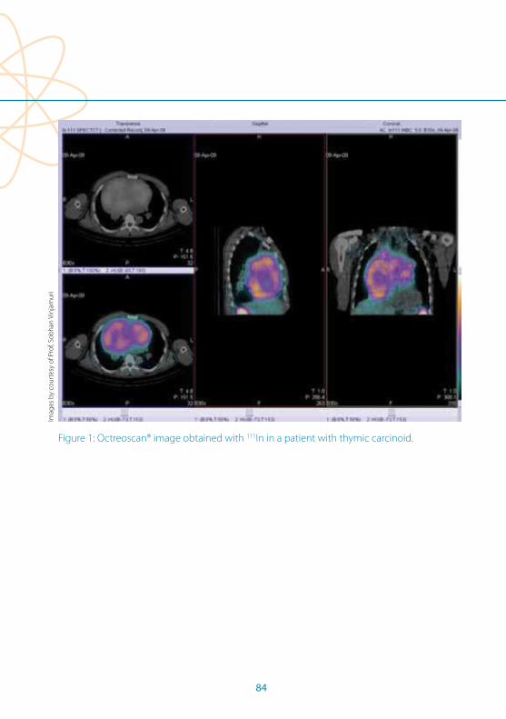
84
Figure 1: Octreoscan® image obtained with 111In in a patient with thymic carcinoid.
Imag
es b
y co
urt
esy
of P
rof.
Sob
han
Vin
jam
uri
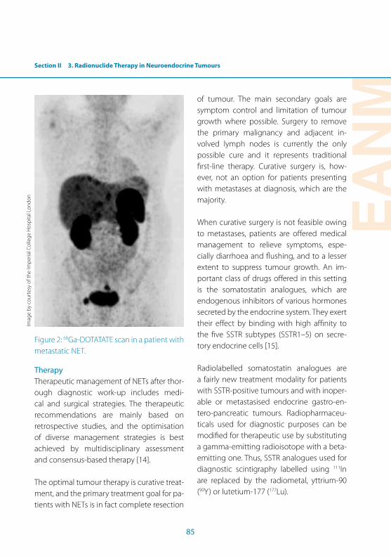
Section II 3. Radionuclide Therapy in Neuroendocrine Tumours
EANM
85
Figure 2: 68Ga-DOTATATE scan in a patient with
metastatic NET.
Therapy
Therapeutic management of NETs after thor-
ough diagnostic work-up includes medi-
cal and surgical strategies. The therapeutic
recommendations are mainly based on
retrospective studies, and the optimisation
of diverse management strategies is best
achieved by multidisciplinary assessment
and consensus-based therapy [14].
The optimal tumour therapy is curative treat-
ment, and the primary treatment goal for pa-
tients with NETs is in fact complete resection
of tumour. The main secondary goals are
symptom control and limitation of tumour
growth where possible. Surgery to remove
the primary malignancy and adjacent in-
volved lymph nodes is currently the only
possible cure and it represents traditional
irst-line therapy. Curative surgery is, how-
ever, not an option for patients presenting
with metastases at diagnosis, which are the
majority.
When curative surgery is not feasible owing
to metastases, patients are ofered medical
management to relieve symptoms, espe-
cially diarrhoea and lushing, and to a lesser
extent to suppress tumour growth. An im-
portant class of drugs ofered in this setting
is the somatostatin analogues, which are
endogenous inhibitors of various hormones
secreted by the endocrine system. They exert
their efect by binding with high ainity to
the ive SSTR subtypes (SSTR1–5) on secre-
tory endocrine cells [15].
Radiolabelled somatostatin analogues are
a fairly new treatment modality for patients
with SSTR-positive tumours and with inoper-
able or metastasised endocrine gastro-en-
tero-pancreatic tumours. Radiopharmaceu-
ticals used for diagnostic purposes can be
modiied for therapeutic use by substituting
a gamma-emitting radioisotope with a beta-
emitting one. Thus, SSTR analogues used for
diagnostic scintigraphy labelled using 111In
are replaced by the radiometal, yttrium-90
(90Y) or lutetium-177 (177Lu).
Imag
e b
y co
urt
esy
of t
he
Imp
eria
l Co
lleg
e H
osp
ital L
on
do
n
86
The rationale for the targeted therapy is
the selective irradiation of tumoral cells by
means of transportation of radioactivity in-
side the tumour cell, following internalisa-
tion of the complex formed by the SSTR and
the beta-emitting particle. Suitable radionu-
clides are 90Y, a pure, high-energy beta emit-
ter (2.27 MeV), 177Lu, a medium-energy beta
emitter (0.5 MeV) with a low abundance, and 111In [16].
111In is a gamma-emitting isotope with a half-
life of 2.8 days. The energy of this gamma-
emitting isotope ranges from 171 to 245 keV,
making it suitable for scintigraphic imaging.
177Lu is a beta- and gamma-emitting isotope
with a maximum energy of 0.49 MeV, a tis-
sue penetration range of 2 mm and a half-
life of 6.7 days. The gamma emission enables
performance of imaging and dosimetry cal-
culations at the same time. Its short tissue
penetration range allows targeted therapies
for small lesions and its longer half-life makes
it suitable for cases where a longer “tumour
residence time” is a clinical requisite.
90Y is a pure beta-emitting isotope with high
energy (2.3 MeV), a half-life of 2.67 days and
a tissue penetration range of 11 mm. Its high
therapeutic potential derives from the emis-
sion of high-energy beta particles causing
damage to large tumours. The absence of
gamma ray emission, however, makes im-
aging and dosimetric calculation with 90Y
diicult. The latter is an important factor for
therapeutic planning.
Radionuclides for therapeutic purposes
should emit beta particles because it is these
particles which have greater therapeutic po-
tential since have suicient energy to cause
cell damage and deliver higher radiation
doses to a larger part of the tumour without
penetrating very far into the surrounding tis-
sue [17].
The irst radiopharmaceutical used in pep-
tide receptor radionuclide therapy (PRRT) was 111In-DTPA-octreotide, already used in the
medical setting as a diagnostic agent with
activities ranging between 10 and 160 GBq
[18, 19]. The therapeutic eicacy of 111In-
DTPA-octreotide was initially promising but
several studies arrived at the conclusion
that 111In-coupled peptides are not ideal for
PRRT because of the small particle range and
therefore short tissue penetration and poor
energy delivery to tumour cells.
Limitations of 111In were overcome when
researchers came up with a modiied so-
matostatin analogue, [Tyr3]octreotide, with
a higher ainity for SSTR2, and a diferent
chelator, DOTA instead of DTPA (which was
used in 111In therapy), to ensure more stable
binding of the intended beta-emitting radio-
nuclide, 177Lu or 90Y.
Clinical applications, contra-indications
and limitations of PRRT
Targeted radionuclide therapy ofers sev-
eral advantages compared with the con-
ventional management options ofered for
NETs. Selective tumour localisation allows
Section II 3. Radionuclide Therapy in Neuroendocrine Tumours
EANM
87
treatment to be administered systemically
while minimising potential toxicity to non-
target, healthy tissues. The fact that potential
toxicity to healthy tissue is spared means that
targeted therapies are usually better toler-
ated than other, systemic treatments such as
chemotherapy and deliver higher absorbed
radiation doses to the tumour than can be
achieved by external beam irradiation. This
applies mostly to NETs, which are relatively
radioresistant compared with other solid
cancers. This kind of therapy relies on the
administration of appropriately radiolabelled
somatostatin analogues and is intended to
control symptoms due to hormonal secre-
tion. Indeed, symptomatic improvement
may be achieved with all 90Y- and 177Lu-
labelled somatostatin analogues that have
been used for PRRT. However, several studies
that have assessed objective responses have
yielded encouraging results as well. Kwekke-
boom et al. reported that despite diferences
in protocols, complete plus partial responses
in diferent studies with 90Y-octreotide have
ranged from 10% to 30%. According to the
same authors, the overall objective tumour
response rate with 177Lu-octreotate, compris-
ing complete and partial responses, was al-
most 30% [20].
A prerequisite for PRRT is the demonstration
that tumoral lesions have suicient uptake
when a diagnostic 111In-octreotide or 68Ga-la-
belled somatostatin analogue scan is carried
out. Adequate uptake of the above-men-
tioned tracers is an essential indicator that
therapeutic levels of internal radiation could
potentially be delivered by administration of 177Lu-octreotate or 90Y radiopeptide therapy.
The intensity of uptake is assessed visually
and considered adequate if lesion uptake is
of greater intensity than that of normal liver.
Once adequate uptake has been conirmed,
further suitability criteria include the con-
irmation of a histopathologically proven,
relatively well diferentiated NET with a Ki-67
score <10 [8].
The kidney is the major critical organ dur-
ing therapy with 90Y or 177Lu. Once iltered
by glomeruli, radiolabelled peptides are re-
absorbed by proximal tubular cells and re-
tained in the interstitium [21]. Another critical
organ to be taken into consideration is the
bone marrow.
In summary, the inclusion criteria for PRRT
are:
• Adequate tumour uptake on octreotide
scan or 68Ga-DOTA-peptide PET/CT
• ECOG performance status of 2 or less
• Life expectancy of at least 6 months
Absolute contra-indications for PRRT are:
• Pregnancy and lactation
Relative contra-indications for PRRT are:
• Severe liver impairment
• Renal impairment
• Impaired haematological function
• Severe cardiac impairment

88
Pre-therapeutic evaluation
PRRT uses radiolabelled somatostatin ana-
logues with high ainity for SSTR2, which is
expressed by the majority of NETs. Currently
the most frequently used radiopharmaceu-
ticals for PRRT of NETs are 90Y-DOTA-Tyr3-
octreotide (90Y-DOTATOC), 90Y-DOTA-Tyr3-oc-
treotate (90Y-DOTATATE) and 177Lu-DOTA-Tyr3-
octreotate (177Lu-DOTATATE).
Adequate tumour uptake on 111In-DTPA-
octreotide (111In-pentetreotide) scintigraphy
(Octreoscan®) is an essential prerequisite
when considering PRRT as a therapeutic
option. Novel somatostatin-based PET im-
aging techniques such as 68Ga-DOTATOC
or 68Ga-DOTATATE are also used to evaluate
adequate tumour uptake before PRRT. In the
pre-therapy imaging evaluation setting, false
negatives may be due to the small size of le-
sions (knowledge of the spatial resolution of
the imaging modality used is important) or
poorly diferentiated forms, which express
fewer receptors.
Other imaging modalities that can be con-
sidered before radionuclide therapy adminis-
tration are chest X-ray, CT or MRI of the lesion
and echocardiography (the latter can be con-
sidered in patients in whom carcinoid heart
disease is suspected).
Further pre-therapy evaluation for PRRT con-
sists in reviewing the blood tests in order
to exclude patients with important renal,
hepatic and haematological impairment.
Of vital importance is the determination of
Ki-67 and correct histopathological evalua-
tion of the tumoral lesion, as well as correct
biochemical assessment, in order to better
categorise the patient.
As mentioned above, radionuclide therapy
is contraindicated in pregnant and lactating
women and patients with less than 6 months’
life expectancy. Patients with carcinoid heart
disease should be monitored closely be-
cause these patients are at high risk of right-
sided heart insuiciency.
Therapeutic procedure
Prior to therapy administration, each patient
has to undergo a consultation with the nu-
clear medicine physician in order to obtain a
thorough medical history, perform a physi-
cal examination, conirm appropriateness
for treatment and acquire formal written in-
formed consent.
Targeted therapy is administered intrave-
nously in subsequent cycles (usually three or
four) in order to reach an adequate cumula-
tive activity to efectively irradiate the tu-
mour. According to several dosimetric stud-
ies, to obtain the disappearance of tumoral
lesions or, at least, a reduction in lesion size,
an absorbed dose of at least 70-80 Gy must
be provided. Such an absorbed dose can be
delivered by giving the patient a cumulative
activity of at least 20 GBq of 177Lu-DOTATATE
[22]. Based on a prospective phase I-II study,
Bodei et al. reported cumulative activities of
up to 29 GBq of 177Lu-DOTATATE to be well
tolerated. In this study, cumulative renal
Section II 3. Radionuclide Therapy in Neuroendocrine Tumours
EANM
89
absorbed doses ranged from 8 to 37 Gy with
no major acute or delayed renal toxicity. A
median decrease in creatinine clearance of
27.9% was recorded after 2 years, with higher
reductions in patients with hypertension and
diabetes [23].
An example of the precautions taken to re-
duce the renal absorbed dose is to give pa-
tients an intravenous infusion of positively
charged amino acids, such as lysine and/
or arginine, 30 min before administration of 177Lu or 90Y and in the 4 h following therapy.
The amino acid infusion aims to optimally
hydrate the patient and competitively inhibit
the renal proximal tubular reabsorption of
the radiopeptide [24]. This precaution has
been shown to reduce the absorbed dose
to the kidneys by between 9% and 53% [22].
As nausea and vomiting are some of the
common side-efects of radionuclide thera-
py, an intravenous injection of an anti-emetic
is also administered. The radiopeptide is then
administered in a saline bag through slow in-
travenous infusion. As ascertained by phase
I studies, the recommended dose per cycle
of 177Lu-DOTATATE is about 5.5-7.4 GBq, while
the cumulative dose is around 22.2-29.6 GBq
[25]. The subsequent doses can be ixed or
escalated according to patient response and
post-therapy image evaluation.
The unsealed source should be administered
in designated facilities incorporating appro-
priate lead shielding, en suite washing facili-
ties and radiation monitoring equipment.
The cycles are separated by at least
8-12 weeks, such intervals being necessary
to ensure adequate recovery from any hae-
matological or other toxicity.
The side-efects of radionuclide therapy can
be divided into acute side-efects and more
delayed efects caused by radiation toxicity.
The most common acute efects, occurring
at the time of injection or up to a few days
after therapy, include nausea, vomiting and
increased pain at tumour sites [26]. These
side-efects are generally mild, are minimised
by a slow infusion and can be prevented or
reduced by symptomatic treatment. Severe
toxicity includes liver, kidney or haematologi-
cal failure and may occur as a result of the
radiation absorbed dose in healthy organs.
Post-therapy evaluation
Follow-up of patients who undergo targeted
radiopeptide therapy consists in anatomical,
functional, biochemical and clinical response
evaluations.
The anatomical response is evaluated by
means of radiological imaging (Fig. 3), most
notably CT or MRI. The image response cri-
teria used when applying CT are based on
the RECIST criteria (Response Evaluation Cri-
teria in Solid Tumors), according to which re-
sponse can be subdivided into:
• CR (complete response)
• PR (partial response)
• PD (progressive disease)
• SD (stable disease)
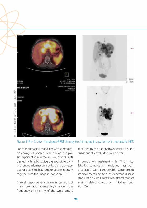
90
Figure 3: Pre- (bottom) and post-PRRT therapy (top) imaging in a patient with metastatic NET.
Functional imaging modalities with somatosta-
tin analogues labelled with 111In or 68Ga play
an important role in the follow-up of patients
treated with radionuclide therapy. More com-
prehensive information may be gained by eval-
uating factors such as tumour uptake intensity,
together with the image response on CT.
Clinical response evaluation is carried out
in symptomatic patients. Any change in the
frequency or intensity of the symptoms is
recorded by the patient in a special diary and
subsequently evaluated by a doctor.
In conclusion, treatment with 90Y- or 177Lu-
labelled somatostatin analogues has been
associated with considerable symptomatic
improvement and, to a lesser extent, disease
stabilisation with limited side-efects that are
mainly related to reduction in kidney func-
tion [20].
Imag
es b
y co
urt
esy
of I
mp
eria
l Co
lleg
e H
osp
ital L
on
do
n
EANM
91
References Section II, Chapter 3
References 1. Arnold R, Frank M, Kajdan U. Management of gastroen-
teropancreatic endocrine tumors: the place of soma-tostatin analogues. Digestion. 1994;55 Suppl 3:107–13.
2. Williams ED, Sandler M. The classiication of carcinoid tumours. Lancet. 1963;I:238–9.
3. Rindi G, Arnold R, Bosman, FT, Capella C, Klimstra DS, Klöppel G, et al. Nomenclature and classiication of neuroendocrine neoplasms of the digestive system. In: WHO classiication of tumours of the digestive system. Bosman FT, Carneiro F, Hruban RH, Theise D, eds. Lyon: International Agency for Research on Cancer 2010:13-14.
4. Kaltsas G, Mukherjee JJ, Plowman PN, Grossman AB. The role of chemotherapy in the nonsurgical management of malignant neuroendocrine tumours. Clin Endocrinol. 2001;55: 575-87.
5. Hoyer D, Lubbert H, Bruns C. Molecular pharmacology of somatostatin receptors, Naunyn Schmiedeberg’s Arch Pharmacol. 1995;350:441–53.
6. De Herder WW, Holand LJ, van der Lely AJ, Lamberts SW. Peptide receptors in gut endocrine tumours. Bailliere’s Clin Gastroenterol. 1996;10:571–87.
7. Bajetta E, Catena L, Procopio G, Bichisao E, Ferrari L, Della Torre S, et al, Is the new WHO classication of neuroen-docrine tumours useful for selecting an appropriate treatment? Ann Oncol. 2005;16:1374-80.
8. De Herder WW, Holand LJ, Van der Lely AJ, Lamberts SW. Somatostatin receptors in gastroentero-pancre-atic neuroendocrine tumours. Endocr Relat Cancer. 2003;10:451-8.
9. Metz DC, Jensen RT. Gastrointestinal neuroendocrine tumors: pancreatic endocrine tumors. Gastroenterology. 2008;135:1469–92.
10. Hofmann M, Maecke H, Börner R, Weckesser E, Schöf-ski P, Oei L, et al. Biokinetics and imaging with the so-matostatin receptor PET radioligand 68Ga-DOTATOC: preliminary data. Eur J Nucl Med. 2001;28:1751–7.
11. Kowalski J, Henze M, Schuhmacher J, Macke HR, Hof-mann M, Haberkorn U. Evaluation of positron emission tomography imaging using [68Ga]-DOTA-D Phe(1)-Tyr(3)-octreotide in comparison to [111In]-DTPAOC SPECT: irst results in patients with neuroendocrine tumors. Mol Imaging Biol. 2003;5:42–8.
12. Gabriel M, Decristoforo C, Kendler D, Dobrozemsky G, Heute D, Uprimny C, et al. 68Ga-DOTA-Tyr3-octreotide PET in neuroendocrine tumors: comparison with so-matostatin receptor scintigraphy and CT. J Nucl Med. 2007;48:508–18.
13. Putzer D, Gabriel M, Henninger B, Kendler D, Uprimny C, Dobrozemsky G, et al. Bone metastases in patients with neuroendocrine tumor: 68Ga-DOTA-Tyr3-octreotide PET in comparison to CT and bone scintigraphy. J Nucl Med. 2009;50:1214–21.
14. Pavel M, Baudin E, Couvelard A, Krenning E, Öberg K, Steinmüller T, and all other Barcelona Consensus Confer-ence participants 2011.
15. Moller LN, Stidsen CE, Hartmann B, Holst JJ. Somatosta-tin receptors. Biochim Biophys Acta. 2003;1616:1–84.
16. Breeman WA, de Jong M, Kwekkeboom DJ, Valkema R, Bakker WH, Kooij PP, et al. Somatostatin receptor mediated imaging and therapy: basic science, current knowledge, limitations and future perspectives. Eur J Nucl Med. 2001;28:1421-9.
17. Smith MC, Liu J, Chen T, Schran H, Yeh CM, Jamar F et al. OctreoTher: ongoing early clinical development of a somatostatin-receptor-targeted radionuclide antineo-plastic therapy. Digestion. 2000;62 Suppl 1:69–72.
18. Fjälling M, Andersson P, Forssell-Aronsson E, Grétars-dóttir J, Johansson V, Tisell LE, et al. Systemic radio-nuclide therapy using indium-111-DTPA-D-Phe1-oc-treotide in midgut carcinoid syndrome. J Nucl Med. 1996;37:1519–21.
92
19. De Jong M, Valkema R, Jamar F, Kvols LK, Kwekkeboom DJ, Breeman WA, et al. Somatostatin receptor-targeted radionuclide therapy of tumors: preclinical and clinical indings. Semin Nucl Med. 2002;32:133–40.
20. Kwekkeboom DJ, de Herder WW, van Eijck CHJ, Kam BL, van Essen M, Teunissen JJM, Krenning EP. Peptide receptor radionuclide therapy in patients with gastro-enteropancreatic neuroendocrine tumours. Semin Nucl Med. 2010;40:78-88.
21. Behr TM, Goldenberg DM, Becker W. Reducing the renal uptake of radiolabeled antibody fragments and peptides for diagnosis and therapy: present sta-tus, future prospects and limitations. Eur J Nucl Med. 1998;25:201–12.
22. De Jong M, Valkema R, Jamar F, Kvols LK, Kwekkeboom DJ, Breeman WA, et al. Somatostatin receptor-targeted radionuclide therapy of tumors: preclinical and clinical indings. Semin Nucl Med. 2002;32:133-40.
23. Bodei L, Cremonesi M, Grana GM, Fazio N, Iodice S, Baio SM, et al. Peptide receptor radionuclide therapy with 177Lu-Dotatate: the IEO phase I-II study. Eur J Nuc Med. 2011;38:2125-35.
24. Bernard BF, Krenning EP, Breeman WA, Rolleman EJ, Bak-ker WH, Visser TJ, et al. D-lysine reduction of indium-111 octreotide and yttrium-90 octreotide renal uptake. J Nucl Med. 1997;38:1929-33.
25. Cybulla M, Weiner SM, Otte A. End-stage renal disease after treatment with 90Y-DOTATOC. Eur J Nucl Med. 2001;28:1552–4.
26. Waldherr C, Pless M, Maecke HR, Schumacher T, Craz-zolara A, Nitzsche EU, et al, Tumor response and clinical beneit in neuroendocrine tumors after 7.4 GBq (90)Y-DOTATOC, J Nucl Med. 2002;43:610–6.

EANM
93
Section II4. Radionuclide Therapy in Hepatocellular Carcinoma
Nadine Silva and Maria do Rosário Vieira
Epidemiology
Each year, the number of new cases of can-
cer and cancer-related deaths increases. It
is estimated that there are 10.9 million new
cancer diagnoses and 6.7 million cancer-
related deaths around the world each year
[1]. Liver cancer is the sixth most common
cancer worldwide, with 749,000 new cases
per year, and is the third leading cause of
cancer-related deaths, with 695,245 cases
annually [2]. Hepatocellular carcinoma (HCC)
represents more than 90% of primary liver
cancers [1].
The worldwide distribution of HCC is related
to the prevalence of risk factors [3,4]. The
highest incidence rates, approximately 85%,
occur in East Asia and sub-Saharan Africa [1],
and the dominant risk factor in these geo-
graphical areas is chronic hepatitis B virus
(HBV) infection [3]. In the United States and
France, the prevalence of HCC is related to
cirrhosis due to alcohol abuse [3], while in
Spain, Italy and Japan the primary risk factor
for HCC appears to be hepatitis C virus (HCV)
infection [3].
The incidence of HCC is increasing world-
wide, and it is estimated that by 2020, the
incidence in Europe will be 78,000 new cases
annually [1].
Risk factors
The predominant risk factors are HBV infec-
tion, HCV infection, alcoholism and alatoxin
intake [1,3,4]. It is estimated that 54% of HCC
cases are related to HBV infection, while 31%
are related to HCV infection and almost 15%
are due to other causes [1].
All the aforementioned risk factors can in-
duce a process of chronic inlammation that
is capable of causing hepatocyte necrosis
and further regeneration, leading to collagen
deposition and cirrhosis [4]. One-third of cir-
rhotic patients will develop HCC [1]. Patient
survival is related to cirrhosis, as it is a pro-
gressive disease and inluences the success
of treatments [1].
Since the risk factors are recognised and HCC
is typically initially “silent”, often being diag-
nosed at an advanced stage, it is important
to enter patients at higher risk of developing
HCC into surveillance programmes in order
to detect tumours early [1,4,5]. The overall
5-year survival rate for HCC is below 10%, and
without treatment the 5-year survival rate is
less than 5% [5,6].
Diagnosis
Nowadays, the goal is to diagnose tumours
sized less than 2 cm with the purpose of ap-
plying potentially curative procedures [1,4].
Alpha-fetoprotein (AFP) is a serum glyco-
protein that may be increased in HCC [4,7].
However, serum levels of AFP remain normal
in about 30% of HCC patients [7] and some
other tumours may be associated with ele-
vated serum AFP. Despite this, increased AFP
values can be helpful in identifying patients
with a higher risk of HCC [4,7].
94
It is possible to detect HCC at an early stage
with ultrasound (US), but also with dynamic
imaging techniques such as four-phase com-
puted tomography (CT) or dynamic contrast-
enhanced magnetic resonance imaging
(MRI) [1,4,8].
Liver biopsy is not necessary if clinical, labo-
ratory and radiography are positive for HCC
[7] but is recommended for tumours occur-
ring in non-cirrhotic livers [1].
Therapeutic approach
The therapeutic approach to HCC will de-
pend on the tumour size, the extent of liver
disease and the presence of metastases [9].
Staging the disease is the irst step in achiev-
ing an appropriate therapeutic approach.
There are several potential curative therapies,
such as surgical resection, liver transplanta-
tion and percutaneous ablation [4,5].
Surgical resection is the irst-line treatment
option for patients with well-deined and
solitary liver tumours, as well as good liver
function [4]. Selected candidates may have
a positive outcome with resection, with a
5-year survival rate of 30-50% [1]. Unfortu-
nately, this is an option for only 10-20% of
HCC patients, and recurrence after resection
is a possibility [1,3,9]. In cases of recurrence,
patients need to be re-staged and treatment
based upon the results of that re-staging [1].
Liver transplantation ofers less risk of re-
currence, with the same chance of survival.
On the other hand, the waiting list is often
several months and this may lead to tumour
progression, resulting in exclusion from the
transplantation list [4].
For both of the aforementioned approach-
es there are some neo-adjuvant therapies
which may be helpful by delaying disease
progression, such as transarterial chemo-
embolisation (TACE) and transcatheter em-
bolisation (TAE) [1,4].
Local ablation by percutaneous ethanol in-
jection or radiofrequency achieves good re-
sponses in patients with tumours less than 2
cm, with a 90% complete response rate [1].
Although there are options which provide a
high complete response rate for HCC at early
stages, there is a lack of efective options for
unresectable HCC associated with liver dis-
ease [3]. For most such patients, treatment
options are palliative [9].
HCC has a poor response to systemic chemo-
therapy [3,6,9]. TACE and TAE are the most com-
monly used options for patients with interme-
diate stage HCC [1,4]. Although these proce-
dures can achieve partial responses in 15-55%
of patients, both are often associated with mild
to moderate adverse events, such as abdomi-
nal pain, fever, nausea and vomiting [1,3].
Some patients with advanced stage HCC
may beneit from therapy with sorafenib,
which is the only systemic approach with
demonstrated value in the management of
HCC [1].
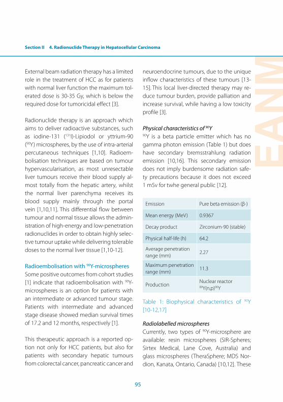
Section II 4. Radionuclide Therapy in Hepatocellular Carcinoma
EANM
95
External beam radiation therapy has a limited
role in the treatment of HCC as for patients
with normal liver function the maximum tol-
erated dose is 30-35 Gy, which is below the
required dose for tumoricidal efect [3].
Radionuclide therapy is an approach which
aims to deliver radioactive substances, such
as iodine-131 (131I)-Lipiodol or yttrium-90
(90Y) microspheres, by the use of intra-arterial
percutaneous techniques [1,10]. Radioem-
bolisation techniques are based on tumour
hypervascularisation, as most unresectable
liver tumours receive their blood supply al-
most totally from the hepatic artery, whilst
the normal liver parenchyma receives its
blood supply mainly through the portal
vein [1,10,11]. This diferential low between
tumour and normal tissue allows the admin-
istration of high-energy and low-penetration
radionuclides in order to obtain highly selec-
tive tumour uptake while delivering tolerable
doses to the normal liver tissue [1,10-12].
Radioembolisation with 90Y-microspheres
Some positive outcomes from cohort studies
[1] indicate that radioembolisation with 90Y-
microspheres is an option for patients with
an intermediate or advanced tumour stage.
Patients with intermediate and advanced
stage disease showed median survival times
of 17.2 and 12 months, respectively [1].
This therapeutic approach is a reported op-
tion not only for HCC patients, but also for
patients with secondary hepatic tumours
from colorectal cancer, pancreatic cancer and
neuroendocrine tumours, due to the unique
inlow characteristics of these tumours [13-
15]. This local liver-directed therapy may re-
duce tumour burden, provide palliation and
increase survival, while having a low toxicity
proile [3].
Physical characteristics of 90Y90Y is a beta particle emitter which has no
gamma photon emission (Table 1) but does
have secondary bremsstrahlung radiation
emission [10,16]. This secondary emission
does not imply burdensome radiation safe-
ty precautions because it does not exceed
1 mSv for twhe general public [12].
Emission Pure beta emission (β-)
Mean energy (MeV) 0.9367
Decay product Zirconium-90 (stable)
Physical half-life (h) 64.2
Average penetration range (mm)
2.27
Maximum penetration range (mm)
11.3
ProductionNuclear reactor89Y(n,p)90Y
Table 1: Biophysical characteristics of 90Y
[10-12,17]
Radiolabelled microspheres
Currently, two types of 90Y-microsphere are
available: resin microspheres (SIR-Spheres;
Sirtex Medical, Lane Cove, Australia) and
glass microspheres (TheraSphere; MDS Nor-
dion, Kanata, Ontario, Canada) [10,12]. These
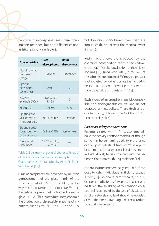
96
two types of microsphere have diferent pro-
duction methods, but also diferent charac-
teristics, as shown in Table 2.
CharacteristicsGlass
microspheres
Resin
ricrospheres
No. of spheres per dose (range)
3-8x10⁶ 30-60x106
Speciic activity per sphere (Bq)
2500 50
Activity available (GBq)
3, 5, 7, 10, 15, 20
3
Size (µm) 20-30 20-60
Splitting one vial for two or more patients
Not possible Possible
Solution used for suspension of the spheres
Saline (0.9%) Sterile water
Associated impurities
88Y, 152Eu, 154Eu, 57Co, 60Co
88Y
Table 2: Summary of general characteristics of
glass and resin microspheres (adapted from
Giammarile et al. [10], Murthy et al. [17] and
Vente et al. [18])
Glass microspheres are obtained by neutron
bombardment of the glass matrix of the
spheres, in which 89Y is embedded; in this
way, 89Y is converted to radioactive 90Y and
the radioisotope cannot be leached from the
glass [11,12]. This procedure may enhance
the production of detectable amounts of im-
purities, such as 88Y, 152Eu, 154Eu, 57Co and 60Co,
but dose calculations have shown that these
impurities do not exceed the medical event
limits [12].
Resin microspheres are produced by the
chemical incorporation of 90Y in the carbox-
ylic group after the production of the micro-
spheres [10]. Trace amounts (up to 0.4% of
the administered dose) of 90Y may be present
and excreted by urine during the irst 24 h.
Resin microspheres have been shown to
have detectable amounts of 88Y [12].
Both types of microsphere are biocompat-
ible, non-biodegradable devices and are not
excreted or metabolised. These devices de-
cay to ininity, delivering 94% of their radia-
tion in 11 days [17].
Radiation safety considerations
Patients treated with 90Y-microspheres will
have the activity conined to the liver, though
some may have shunting activity in the lungs
or the gastrointestinal tract. As 90Y is a pure
beta emitter, the only considered dose to an
individual likely to be in contact with the pa-
tient is the bremsstrahlung radiation [12].
Patient instructions are only required if the
dose to other individuals is likely to exceed
1 mSv [12]. For health care workers, no bur-
densome radiation safety precautions need
be taken: the shielding of this radiopharma-
ceutical is achieved by the use of plastic and
acrylic materials and lead should be avoided
due to the bremsstrahlung radiation produc-
tion that may arise [12].
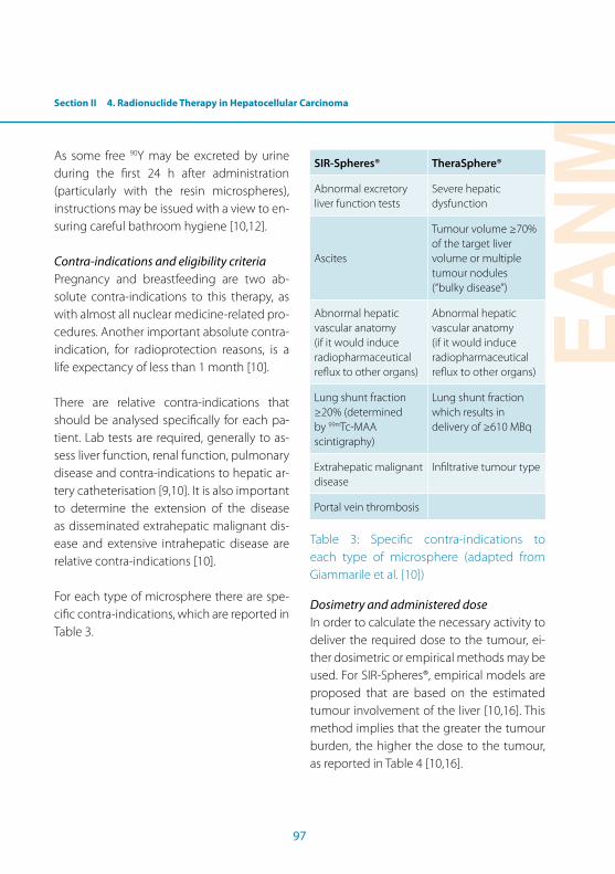
Section II 4. Radionuclide Therapy in Hepatocellular Carcinoma
EANM
97
As some free 90Y may be excreted by urine
during the irst 24 h after administration
(particularly with the resin microspheres),
instructions may be issued with a view to en-
suring careful bathroom hygiene [10,12].
Contra-indications and eligibility criteria
Pregnancy and breastfeeding are two ab-
solute contra-indications to this therapy, as
with almost all nuclear medicine-related pro-
cedures. Another important absolute contra-
indication, for radioprotection reasons, is a
life expectancy of less than 1 month [10].
There are relative contra-indications that
should be analysed speciically for each pa-
tient. Lab tests are required, generally to as-
sess liver function, renal function, pulmonary
disease and contra-indications to hepatic ar-
tery catheterisation [9,10]. It is also important
to determine the extension of the disease
as disseminated extrahepatic malignant dis-
ease and extensive intrahepatic disease are
relative contra-indications [10].
For each type of microsphere there are spe-
ciic contra-indications, which are reported in
Table 3.
SIR-Spheres® TheraSphere®
Abnormal excretory liver function tests
Severe hepatic dysfunction
Ascites
Tumour volume ≥70% of the target liver volume or multiple tumour nodules (“bulky disease”)
Abnormal hepatic vascular anatomy (if it would induce radiopharmaceutical relux to other organs)
Abnormal hepatic vascular anatomy (if it would induce radiopharmaceutical relux to other organs)
Lung shunt fraction ≥20% (determined by 99mTc-MAA scintigraphy)
Lung shunt fraction which results in delivery of ≥610 MBq
Extrahepatic malignant disease
Iniltrative tumour type
Portal vein thrombosis
Table 3: Speciic contra-indications to
each type of microsphere (adapted from
Giammarile et al. [10])
Dosimetry and administered dose
In order to calculate the necessary activity to
deliver the required dose to the tumour, ei-
ther dosimetric or empirical methods may be
used. For SIR-Spheres®, empirical models are
proposed that are based on the estimated
tumour involvement of the liver [10,16]. This
method implies that the greater the tumour
burden, the higher the dose to the tumour,
as reported in Table 4 [10,16].

98
Tumour involvement
(%)
Recommended
activity (GBq)
<25% 2.0
25-50% 2.5
>50% 3.0
Table 4: Recommended activity for SIR-
Spheres®, accordingly to tumour involvement
[10,16]
Another empirical model suggested for SIR-
Spheres® relates to the body surface area
(BSA), using Eq. 1 to calculate the activity to
be delivered to the patient [10]:
A(GBq) = (BSA – 0.2) + tumor volume
total liver volume (1)
The dosimetric models, which consider the
lung shunt fraction (LSF), are usually used for
TheraSphere® [10,16]. The delivered dose for
TheraSphere® is between 80 and 150 Gy [16].
With glass microspheres the activity required
to deliver the prescribed dose is calculated
using Eq. 2 [16]:
A(GBq) = [Dose(Gy)] [Liver mass(kg)]
49.8 (2)
Whichever type of microsphere is used, the
vendors recommend adjustments in order to
reduce the fraction of activity shunting to the
lungs (the LSF). For TheraSphere, the dose to
the lungs should be less than 30 Gy [10,16].
For SIR-Spheres®, the adjustments are shown
in Table 5 [10].
LSFReduction in the
administered activity (%)
<10% No reduction
10-15% 20%
15-20% 40%
>20% The technique is contra-indicated
Table 5: Adjustment in the administered
activity according to LSF, for SIR-Spheres® [10]
In order to detect and quantify LSF, techne-
tium-99m (99mTc)-labelled albumin macro-
aggregate (MAA) scintigraphy is performed
[10]. The LSF is determined using Eq. 3 [10]:
Lung Shant fraction = lung counts
lung + liver counts (3)
Procedure
As mentioned previously, a pre-therapy eval-
uation must be performed in order to assess
patient eligibility for this speciic treatment.
This evaluation consists in a complete history
and physical examination, chest radiography
and assessment of pulmonary function, liver
sonography or CT, evaluation of serum liver
enzymes, complete blood count, coagula-
tion testing, determination of creatinine lev-
els and assessment of tumour markers [9-11].
Positron emission tomography (PET)/CT with
luorine-18 (18F) luorodeoxyglucose (FDG)
can be performed to exclude extrahepatic
disease and also to evaluate the extension
of hepatic disease. Usually, HCCs have very
low-grade FDG uptake, or even no uptake,
Section II 4. Radionuclide Therapy in Hepatocellular Carcinoma
EANM
99
unless the tumour is of an aggressive type;
nevertheless, 18F-FDG uptake has prognostic
value and is used to check the therapeutic
outcome [10,19].
A CT scan allows accurate calculation of the
liver volume, which is essential for dose cal-
culation [10,11].
Angiography by high-speed multi-slice CT
is usually performed 4-6 weeks before the
therapy with 90Y-microspheres [11]. Angiog-
raphy visualises the hepatic arterial anatomy
and the haemodynamics of the hepatic cir-
culation [11]. Anatomical variations are as-
sessed, as is the presence or absence of por-
tal venous thrombosis [10]. Angiography also
permits the placement of coils in order to oc-
clude aberrant hepatic arteries and embolise
collateral vessels, to ensure that the micro-
spheres remain conined to the liver [11]. Coil
embolisation of the gastroduodenal artery
may be of use in preventing shunting activity
to the gastrointestinal tract [10].
During angiography, a catheter is placed in
the hepatic artery or in any branch of the
hepatic artery (depending on the hepatic
arterial anatomy or on selective treatment
options) [10,11]. This procedure allows intra-
arterial administration of 99mTc-MAA, which
have a comparable size to 90Y-microspheres.
Thus, scintigraphic acquisition is performed
as soon as possible after the administration
of 75-150 MBq of 99mTc-MAA. Planar views
(anterior and lateral) of the upper abdomen,
anterior chest image and hepatic SPECT are
acquired [9-11]. Visual evaluation of lung and
gastrointestinal shunt and lung shunt quan-
tiication are performed, which may lead to
dose adjustment. Assessment of the tumour
uptake within the liver is also performed [10].
It is important to note that imaging with 99mTc-MAA may entail visualisation of the thy-
roid, stomach and urinary bladder because of
free pertechnetate [10].
After LSF assessment and evaluation of the
presence or absence of gastrointestinal
shunt and of whether patients are suiciently
well to undergo the therapy, dose calculation
proceeds [10]. 90Y-microsphere administra-
tion is carried out in the interventional radi-
ology department as it involves intra-arterial
catheter manipulation. Administration is
performed by a nuclear medicine physician
using the vendor’s device and with gentle
infusion in order to prevent backlow of the
radiopharmaceutical [10].
A few hours after administration, in order to
locate the administered activity, bremsstrah-
lung planar images (anterior and right later-
al) may be acquired using a gamma camera
with an energy peak of 100 keV and a wide-
energy window in order to increase sensitiv-
ity [20]. Alternatively, 30-min PET images may
be acquired, which have the advantage of
better spatial resolution [10]. These images
are essential to assess the administered ac-
tivity, as this may be unexpectedly located
outside the liver despite the indings on 99mTc-MAA scintigraphy and the previous liver
embolisation [20]. They are also important in

100
localising the activity in order to prevent and
manage side-efects if there is extrahepatic
activity [20].
Side-efects and follow-up
The overall incidence of complications and
side-efects after the therapy is low [17]. Even
though prophylactic hepatic embolisation
is performed in order to prevent shunting
and to guarantee maximum tumour uptake,
some side-efects may be experienced by
patients because inadvertent deposition of 90Y-microspheres is a possibility despite an-
giographic occlusion techniques [9]. Com-
monly, patients experience fatigue, nausea,
abdominal pain, fever and transitory eleva-
tion of the transaminases [10]. Some other
possible side-efects occur at lower rates
(2-8%), such as chronic abdominal pain, ra-
diation gastritis, gastrointestinal ulceration,
upper gastrointestinal ulceration, haemor-
rhage, pancreatitis, radiation pneumonitis
and radiation-induced liver disease [9,10]. In
addition to careful assessment of extrahepat-
ic shunting, appropriate selection of patients
is essential to minimise side-efects [9,17].
Post-therapeutic follow-up is performed in
order to detect any possible side-efects but
also to evaluate tumour response. It is con-
ducted 30 days after the therapy and at 2- to
3-month intervals thereafter [10].
131I-Lipiodol therapy
Treatment with intra-arterial injection of 131I-Lipiodol was developed in the 1990s
and has yielded valuable clinical results and
demonstrated good tolerance by patients
[21]. Compared with 90Y-microspheres, 131I-
Lipiodol is approximately 10 times less ex-
pensive [19].
131I-Lipiodol is an option in patients with por-
tal vein thrombosis and those who have al-
ready been surgically treated. 131I-Lipiodol is
also under study for application in patients
with liver metastases and as a neoadjuvant
therapeutic before resection and liver trans-
plantation [10].
Physical characteristics of 131I131I is a beta emitter but it is also character-
ised by high gamma ray emission (Table 6).
This allows image acquisition but constrains
the administered activity owing to radiation
exposure [10,21].
Emission Beta, gamma
Mean energy (MeV)β (0.61, 89% abundance), γ (0.36, 81% abundance)
Decay product Xenon-131 (stable)
Physical half-life (days) 8.04
Average penetration range (mm)
β: 0.9 mm
Maximum penetration range (mm)
β: 2.4 mm
Production Nuclear reactor
Table 6: Biophysical characteristics of 131I
[10,19,21]
Section II 4. Radionuclide Therapy in Hepatocellular Carcinoma
EANM
101
131I-Lipiodol
This radiopharmaceutical is available in a
commercial form as Lipiocis (IBA, Brussels,
Belgium) [10]. Lipiodol is an oil compound
with 38-40% iodine fatty acid ethyl esters of
poppy seed oil [10,19]. This compound con-
tains stable iodine-127 (127I), and by a simple
isotopic change it is substituted by 131I [10,19].
After intra-arterial injection, 131I-Lipiodol mi-
grates towards the tumour region. There
may be some unspeciic uptake in non-ma-
lignant endothelial cells, but this compound
is highly selective for malignant cells [10,19].
It is estimated that 24 h after the administra-
tion, more than 75% of Lipiodol is trapped in
the liver, though the remainder reaches the
lungs [10,19,22]. The tumour/non-tumour
uptake ratio is about 15-20:1 and it tends
to increase with time, possibly because of
the lack of macrophagic cells in the tumour,
leading to slow Lipiodol clearance from the
tumour [19].
Other tissues show very little uptake of this
compound, this being true even of the thy-
roid gland [22].
Radiation safety considerations
Because of the gamma emission of 131I, hospi-
talisation is required in order to safeguard in-
dividuals likely to come into contact with the
patient [22]. This hospitalisation can be from
4 days to a week, in a shielded room [22,23].
Discharge will depend on the measured re-
maining activity, and this limit is usually less
than 1 mSv to other individuals.
It is important to note that 131I-Lipiodol is
eliminated mainly via the urinary tract (30-
50% of the administered activity at day 8)
[19]. A small amount of the activity is elimi-
nated in faeces (<3% at day 5) [19]. In this re-
spect, it is important to verbally instruct the
patient on efective bathroom hygiene to
prevent contamination.
Thyroid gland blockage with stable iodine or
potassium perchlorate is usually not recom-
mended [10].
The health facility in which the procedure is
performed must be fully equipped in order
to guarantee radiation safety for both staf
and patients (i.e. shielding rooms, adequate
protection during transportation of the pa-
tient within the institution, implementation
of appropriate contamination procedures
and suitably trained staf ) [10].
Contra-indications and eligibility criteria
Pregnancy and breast-feeding are again two
absolute contra-indications to this therapy.
Another important absolute contra-indica-
tion, for radioprotection reasons, is a life ex-
pectancy of less than 1 month [10].
There are relative contra-indications that
should be analysed individually for each pa-
tient. Lab tests are required, generally to ac-
cess liver function, renal function, pulmonary
disease and contra-indications to hepatic ar-
tery catheterisation [10]. It is also important
to determine the extension of the disease,
as disseminated extrahepatic malignant
102
disease and extensive intrahepatic disease
are relative contra-indications [10].
Appropriate patient selection with respect to
hepatic reserve and overall functional status
will maximise the therapeutic outcome and
minimise the risk to normal hepatic paren-
chyma [10].
Administered dose
The administered activity of 131I-Lipiodol is
2.22 GBq, which corresponds to a hepatic
dose of approximately 50 Gy [10]. Although
this is a ixed activity, it is possible to adjust
it according to tumour load if necessary [10].
Radiopharmaceutical preparation must be
performed in an adequately ventilated cabi-
net in the Nuclear Medicine Department in
order to prevent iodine aerosol inhalation
[19].
As this compound is a viscous oil, it might
ofer some resistance to syringe dispensing
and catheter injection [10].
Procedure
As previously mentioned, a pre-therapy eval-
uation must be performed in order to assess
the patient’s eligibility for this speciic treat-
ment. This evaluation consists in a complete
history and physical examination, pulmonary
function assessment, three-phase contrast
CT, gadolinium-enhanced MRI, assessment
of extent of extrahepatic disease, evalua-
tion of serum liver enzymes, complete blood
count, coagulation testing, determination of
creatinine levels and assessment of tumour
markers [10,19].
As previously mentioned, PET/CT with 18F-
FDG may be of use, especially in order to
evaluate extrahepatic disease [10,19].
Angiography is performed to assess the he-
patic vascular anatomy and to place an intra-
arterial catheter. Due to anatomical variations
the catheter may be placed in other, distally
located hepatic branches [10,19,23].
The activity is administered slowly in the
catheter to prevent relux and consequent
retrograde low into the gastroduodenal
artery [10,23]. This procedure is done under
luoroscopic control [10,23]. After the admin-
istration, the patient is taken to the shielded
room [19,22,23].
Whole-body scintigraphic images are ac-
quired one week after the administration of 131I-Lipiodol. A CT scan may be performed
based on the high iodine content of the ra-
diopharmaceutical as this dispenses supple-
mentary contrast administration [10,23].
Some patients may beneit from one or sev-
eral treatments [19] that can be performed
2, 5, 8 and 12 months after the irst injection
[10].
Side-efects and follow-up
Serious adverse efects are rare [19,22].
Early side-efects, which occur rarely, may
be moderate and temporary pyrexia (29%),

Section II 4. Radionuclide Therapy in Hepatocellular Carcinoma
EANM
103
moderate and temporary alteration on bio-
logical liver test (20%) and hepatic pain on
injection (12.5%) [10,19,22]. The delayed side
efects, which occur more rarely, are moder-
ate and reversible leucopenia (7%) and inter-
stitial pneumopathies (2%) [19].
Follow-up should be performed using the
same modalities as have been employed
to assess the disease (three-phase CT, MRI,
PET/CT, lab tests) and this should be done
1 month after therapy and then at 2- to
3-month intervals [10,19].
104
References Section II, Chapter 4
References1. Llovet J, Ducreux M, Lencioi R, Di Bisceglie AM, Galle PR,
Dufour JF, et al. EASL-EORTIC Clinical Practice Guidelines; management of hepatocellular carcinoma. J Hepatol. 2012;56:908-43.
2. IARC. http://www-dep.iarc.fr/;2013 [accessed 23/03/2013]
3. Geschwind J, Salem R, Carr B, Soulen MC, Thurston KG, Goin KA, et al. Yttrium-90 microspheres for the treat-ment of hepatocellular carcinoma. Gastroenterology. 2004;127:S195-205.
4. Bruix J, Hessheimer AJ, Forner A, Boix L, Vilana R, Llovet JM. New aspects of diagnosis and therapy of hepatocel-lular carcinoma. Oncogene. 2006;25:3848-56.
5. Clark H, Carson W, Kavanagh P, Ho C, Shen P, Zagoria R. Staging and current treatment of hepatocellular carci-noma. Radiographics. 2005;25:S3–24.
6. O’Brien T, Kirk G, Zhang M. Hepatocellular carcinoma: paradigm of preventive oncology. Cancer. 2004;10:67-73.
7. Bialecki E, Di Bisceglie A. Diagnosis of hepatocellular carcinoma. HPB (Oxford). 2005;7:26-34.
8. Dufy J, Hiatt J, Busuttil R. Surgical resection of hepato-cellular carcinoma. Cancer. 2008;14:100-11.
9. Dancey J, Sheperd F, Paul K, Sniderman K, Houle S, Ga-brys J, et al. Treatment of nonresectable hepatocellular carcinoma with intrahepatic 90Y-microspheres. J Nucl Med. 2000;41:1673-81.
10. Giammarile F, Bodei L, Chiesa C, Flux G, Forrer F, Kraeber-Bodere F, et al. EANM procedure guideline for treatment of liver cancer and liver metastases with intra-arterial radioactive compounds. Eur J Nucl Med Mol Imaging. 2011;38:1393-406.
11. Andrews J, Walker SC, Ackermann RJ, Cotton LA, Ens-minger WD, Shapiro B. Hepatic radioembolization with yttrium-90 containing glass microspheres: preliminary results and clinical follow-up. J Nucl Med. 1994;35:1637-44.
12. Gulec S, Siegel J. Posttherapy radiation safety consider-ations in radiomicrosphere treatment with 90Y-micro-spheres. J Nucl Med. 2007;48:2080–6.
13. Gulec S, Fong Y. Yttrium 90 microsphere selective inter-nal radiation treatment of hepatic colorectal metastases. Arch Surg. 2007;142:675–82.
14. Arslan N, Emi M, Alagöz E, Üstünöz B, Oysul K, Arpaci F, et al. Selective intraarterial radionuclide therapy with yttrium-90 (Y-90) microspheres for hepatic neuroen-docrine metastases: initial experience at a single center. Vojnosanit Pregl. 2011;4:341-8.
15. Cao C, Yan T, Morris D, Bester L. Radioembolization with yttrium-90 microspheres for pancreatic cancer liver metastases: results from a pilot study. Tumori. 2010;96:955-8.
16. Dezarn W. Quality assurance issues for therapeutic ap-plication of radioactive microspheres. Int J Radiat Oncol Biol Phys. 2008;71:S147-151.
17. Murthy R, Nunez R, Szklaruk J, Erwin W, Madof D, Gupta S, et al. Yttrium-90 microsphere therapy for hepatic malignancy: device, indications, technical consider-ations and potential complications. Radiographics. 2005;25:S41-55.
18. Vente M, Wondergem M, Van Der Tweel I, Van Der Bosch M, Zonnenberg B, Lam M, et al. Yttrium-90 radioem-bolization for the treatment of liver malignancies: a structured meta-analysis. Eur J Radiol. 2009;19:951-9.
19. Ahmadzadehfar H, Sabet A, Jürgen H, Risse, Risse J. Therapy of hepatocellular carcinoma with iodine-131-Lipiodol. In: Lau JWY, ed. Hepatocellular carcino-ma - Clinical research. ISBN: 978-953-51-0112-3, InTech, Available from: http://www.intechopen.com/books/hepatocellularcarcinoma-clinical-research/therapy-of-hepatocellular-carcinoma-with-iodine-131-lipiodol.
20. Elschot M, Vermolen BJ, Lam MG, de Keizer B, van den Bosch MA, de Jong HW. Quantitative comparison of PET and bremsstrahlung SPECT for imaging the in vivo yttrium-90 microsphere distribution after liver radioem-bolization. PLoS One. 2013;8:e55742.
21. Ruyck K, Lambert B, Bacher K, Gemmel F, De Vos F, Vral A, et al. Biologic dosimetry of 188Re-HDD/Lipiodol ver-sus 131I-Lipiodol therapy in patients with hepatocellular carcinoma. J Nucl Med. 2004;45:612-8.
22. Raoul J, Boucher E, Roland Y, Garin E. 131-iodine Lipiodol therapy in hepatocellular carcinoma. Q J Nucl Med Mol Imaging. 2005;53:348-55.
23. Marelli L, Shusang V, Buscombe JR, Cholongitas E, Sti-gliano R, Davies N, et al. Transarterial injection of 131I-Lipiodol, compared with chemoembolization, in the treatment of unresectable hepatocellular cancer. J Nucl Med. 2009;50:871-7.
EANM
105
Section II5. Radioimmunotherapy in Lymphomas
Aurore Rauscher, Caroline Bodet-Milin, Amandine Pallardy, Alain Faivre-Chauvet, Françoise Kraeber-Bodéré
Introduction
Radioimmunotherapy (RIT) is a targeted
therapy whereby irradiation from radionu-
clides is delivered to tumour targets using
monoclonal antibodies (MAbs) directed
against a tumour antigen. Over the last 20
years, RIT has progressed signiicantly, with
the development of new humanised mAbs,
stable chelates for labelling and pretargeting
techniques [1]. The cytotoxic mechanisms
of RIT involve both radiobiological and im-
munological processes. B-cell non-Hodgkin
lymphoma (NHL) cells express well-charac-
terised antigens, are highly radiosensitive, re-
spond to cold mAbs and represent a relevant
indication for RIT. NHLs can be classiied into
more than 25 histological subtypes accord-
ing to the World Health Organisation (WHO)
classiication, and can be separated into ag-
gressive (65% of NHLs) and indolent forms
(35%) [2]. Difuse large B-cell NHL (DLBCL) is
the most common type of aggressive NHL
(31%), and follicular lymphoma (FL) is the
most common type of indolent NLH (22%).
FL generally shows indolent progression
with response to chemotherapy, but always
relapses. Survival ranges from 5 to 15 years.
The prognosis of DLBCL is diferent, with
50-60% of patients being cured. Treatment
of disseminated NHL includes multi-agent
chemotherapy, with the possibility of high-
dose chemotherapy coupled with stem cell
transplantation in high-risk young people
(<60-65 years). The introduction of rituximab
(MabThera, Rituxan, Roche Ltd., Genentech,
Basel, Switzerland), a monoclonal chimeric
anti-CD20 antibody, resulted in an improve-
ment in outcome in patients with aggressive
or indolent B-NHL when it was combined
with diferent chemotherapy regimens (R-
chemotherapy), as compared with use of
chemotherapy alone [3]. Involved-ield ra-
diation can be proposed for limited stage
FL or treatment of residual masses of DLBCL.
Today, one product targeting the CD20 an-
tigen has been approved in Europe for RIT: 90Y-ibritumomab tiuxetan, (Zevalin, Spectrum
Pharmaceuticals, Henderson, NV, USA). An-
other product targeting CD20 is available in
the United States: 131I-tositumomab, (Bexxar,
GlaxoSmithKline). RIT can be integrated into
clinical practice using non-ablative doses for
treatment of FL patients. Diferent RIT proto-
cols are being assessed in clinical trials in FL
or other lymphoma subtypes: myeloablative
or high-dose treatment, RIT as consolidation
after chemotherapy, RIT in irst-line treat-
ment, fractionated RIT and other MAbs es-
pecially targeting antigens other than CD20.
Clinical application and contra-indications/
limitations to RIT
Clinical application
RIT can be integrated into clinical practice us-
ing non-ablative activities for the treatment
of patients with relapsed or refractory FL or
as consolidation after induction chemother-
apy in the front-line treatment of FL patients.
106
Contra-indications
Exclusion criteria for treatment with Zevalin®
are:
• Pregnancy and continuing breast-feeding
• Known hypersensitivity to 90Y-ibritumom-
ab tiuxetan, yttrium chloride, other murine
proteins or any of their components
• Children and adolescents under 18 years
of age
• Marked bone marrow suppression (<1.5 x
109/L leucocytes; < 100 x 109/L thrombo-
cytes)
• Greater than 25% of bone marrow iniltra-
tion by lymphoma
• Previous external beam radiation involving
>25% of the active bone marrow
• Prior bone marrow or stem cell transplan-
tation
• Detectable human anti-murine antibody
(HAMA), depending on titre
The peripheral blood cell count should not
be lower than the limits stated above. While
lower blood counts do not constitute an ab-
solute contraindication, they do increase the
risk of severe and prolonged bone marrow
suppression.
Therapeutic procedure
Radiopharmaceutical
Approved name: 90Y- ibritumomab tiuxetan
or Zevalin®
Yttrium-90 is a beta-emitter, with a physical
half-life of 64 h, a maximum particle energy
of 2.27 MeV and a maximum range of 11 mm
in soft tissue.
Preparation procedure
Zevalin® is supplied as a kit for the prepara-
tion of 90Y-radiolabelled ibritumomab tiux-
etan. The kit contains one antibody vial with
3.2 mg ibritumomab tiuxetan (1.6 mg/mL),
one 2-mL sodium acetate vial, one 10-mL
formulation bufer vial and a 10-mL empty
reaction vial. The kit must be stored at 2-8°C
and should not be frozen. The radioactive
component, 90Y, must be obtained separately
upon order from the manufacturer. Only
carrier-free 90Y of pharmaceutical grade qual-
ity must be used for antibody labelling, since
metal contamination has a detrimental efect
on the labelling eiciency.
Labelling must be performed only by quali-
ied staf (radiopharmacist, chemist or tech-
nician with qualiication in radiopharmacy)
with appropriate authorisation for the use
and manipulation of radionuclides. Appro-
priate facilities for shielding, calibration and
quality control should be in place. Proper
aseptic technique and precautions for han-
dling radioactive materials must be em-
ployed during the preparation procedure.
Vial rubber stoppers must be cleaned with
a suitable alcohol swab and sterile syringes
must be used. Waterproof gloves, protective
shields for syringes and containers, forceps
and tongs as gripping tools must be used
for the preparation. Owing to the 9.2 mm
Section II 5. Radioimmunotherapy in Lymphomas
EANM
107
length of 90Y beta emission [4], at least 1-cm-
thick Perspex or lead-loaded Perspex shields
should be used during labelling.
The irst step consists in the addition to the
reaction vial of a 1.2-fold excess of sodium
acetate solution compared with the yttrium
volume. The second step involves aseptically
transferring 1,500 MBq of 90Y to the reaction
vial and mixing the solution by inversion (or
‘rolling’ the vial). Then 1.3 mL ibritumomab
tiuxetan solution, previously brought to
room temperature, is transferred to the reac-
tion vial, and after mixing the reaction solu-
tion is left at room temperature for 5 min. A
labelling time longer than 6 min or shorter
than 4 min or foam formation during the mix-
ing will result in inadequate radiolabelling. In
order to chelate free 90Y and inhibit radiolysis
of the radiolabelled antibody, an appropriate
volume of formulation bufer that contains
human serum albumin and DTPA is added to
the reaction vial; this results in a combined
total volume of 10 mL.
The inal formulation after radiolabelling con-
tains 2.08 mg ibritumomab tiuxetan in a total
volume of 10 mL. After radiolabelling, imme-
diate use is recommended, but the solution
may be stored at 2°-8°C (in a refrigerator).
At these temperatures and protected from
light, it can be used up to a maximum of 8 h.
Quality control
The radiochemical purity of the prepared 90Y-radiolabelled Zevalin must be checked
before administration. Instant thin-layer
chromatography (ITLC) is recommended for
this purpose with sodium chloride solution
(0.9%) as mobile phase. DTPA-90Y is carried by
the solvent and has a chromatographic ratio
(Rf ) of 1, and radiolabelled antibody remains
at the deposit with an Rf of 0. The radiochro-
matogram may be analysed in diferent ways:
by cutting the ITLC into two parts and count-
ing each part in an appropriate counter, by
using a radiochromatograph or by using a
phosphorimager. Radiochemical purity is
calculated as follows:
% RCP = net count bottom half / total count x 100
Common sources of error in quality control
are drop size and dead-time error during
measurement of the ITLC strips. If the aver-
age radiochemical purity is less than 95%, the
preparation must not be administered.
Method of administration
Before Zevalin® infusion, the activity in the
10-mL syringe must be measured using a
calibrated ionisation chamber under the ap-
propriate geometric-, volume- and material-
speciic (plastic needles, glass vials) condi-
tions of calibration.
The prepared solution must be given as a
slow intravenous infusion over a minimum
of 10 min. The infusion must not be admin-
istered as an intravenous bolus. Zevalin® may
be infused directly by stopping the low from
an infusion bag and administering it directly
into the line. A 0.2- or 0.22-µm low-protein-
binding ilter must be on line between the
108
patient and the infusion port. The line must
be lushed with at least 10 mL of sodium
chloride 9 mg/mL (0.9%) solution after the
infusion of Zevalin®.
Administration schemes
Zevalin® is administered 6-8 days after a pre-
dose of 2 x 250 mg of rituximab, to improve
biodistribution and tumour targeting. The
whole therapy requires only two outpatient
visits. No dosimetry study is required. When
a dosimetry study is performed for research
purposes, the irst dose of cold MAb is in-
jected with 185 MBq of 111In-ibritumomab
tiuxetan. The injected activity depends on
body weight and platelet count. The thera-
peutic dose is 14.8 MBq/kg (11.1 MBq/kg in
patients with a platelet count of 100,000 to
149,000/mm3) to a maximum total activity of
1,184 MBq [5].
Pre-therapeutic evaluation
Evaluation prior to RIT should be carried out
as proposed in the EANM guideline (6) and
should include:
• A clinical examination.
• Careful recording of the patient’s history,
including information on previous thera-
pies, in particular external beam radiation
therapy involving active bone marrow and
stem cell transplantation.
• A representative (>2 cm long cylinder)
bone marrow biopsy from the iliac crest:
The biopsy must have been performed no
earlier than the last point in time at which
disease progression was detected or in any
case a maximum of 3 months before the
scheduled RIT.
• Exclusion of pregnancy and conirmation
of cessation of breast feeding.
• Recording of current medications, espe-
cially those afecting haematological func-
tion.
• Determination of blood proile, prothrom-
bin time (INR) and serum creatinine and
bilirubin levels within 1 week prior to RIT:
RIT should not be performed if these val-
ues are above 2.5 times the upper normal
limits for the local laboratory.
• Estimation of life expectancy (life expec-
tancy >3 months, Karnofsky index >70%):
Patients with a life expectancy of less than
3-4 weeks or rapid disease progression are
unlikely to beneit from RIT.
Section II 5. Radioimmunotherapy in Lymphomas
EANM
109
Post-therapeutic assessment
Safety
Haematological toxicity is the major side-ef-
fect of RIT, and depends on bone marrow in-
volvement and prior treatment [6, 7]. After a
dose of 14.8 MBq/kg of Zevalin®, a reduction
of 30-70% in leucocyte and platelet counts
from baseline levels is possible, sometimes
occurring very rapidly. Grade 4 neutropenia,
thrombocytopenia and anaemia have been
reported in 30%, 10% and 3% of patients,
respectively, with the nadir in counts occur-
ring 7-9 weeks after Zevalin® injection, i.e.
later than after chemotherapy. Median time
to complete recovery of blood count (hae-
moglobin >12 g/dL, platelets >150,000/μL
and leucocytes >4,300/μL) is 99 days for FL
patients [8]. After RIT, weekly blood tests from
the second post-RIT week are recommend-
ed, until baseline levels have been reached
[9]. If levels drop faster than expected, short-
er-term controls should be instituted. If the
platelet count falls below 30 x 109/L, levels
should be checked at least three times per
week. Platelet transfusions and growth fac-
tors should be administered if indicated. The
patient should also be informed of the in-
creased risk of infection and bleeding.
Non-haematological toxicity is generally low,
including asthenia, anorexia, fever, nausea,
headache, chills, arthralgia and myalgia. Al-
lergic reactions have been observed dur-
ing MAb infusion, in particular after the irst
rituximab injection prior to Zevalin admin-
istration. It is important to highlight that
RIT is well tolerated by older patients and
represents a strong treatment choice in this
group. Immunogenicity with human anti-
mouse and human anti-chimeric antibody
production has been observed, ranging from
1% to 63% between studies.
Secondary myelodysplastic syndrome or
acute myelogenous leukaemia has been re-
ported in 1-3% of cases [7]. The risk appears
to be increased in patients previously treated
with several lines of chemotherapy or radio-
therapy. A cytological and genetic analysis of
bone marrow could be proposed for heavily
pre-treated patients prior to beginning RIT.
In the long-term analysis of the International
Radioimmunotherapy Network, the rate of
secondary solid tumours was 0.8%, including
breast cancer, prostate cancer, glioblastoma
and non-small-cell lung cancer [8].
Eicacy
In a study of 143 patients with relapsed or re-
fractory FL or transformed B-cell NHL, Zeva-
lin® appeared more eicient than rituximab,
yielding signiicantly higher overall response
(OR) and complete response (CR) rates (80%
vs 56%, p=0.002, and 30% vs 16%, p=0.04,
respectively) [10]. Patients refractory to ritux-
imab had a 74% OR and those with throm-
bocytopenia had an OR of 83% [11]. Mean
time to progression (TTP) in responders was
12.6 months. OR was observed in 50% of
patients with bulky lymphoma. Chemother-
apy, administered in patients treated with
RIT, was not associated with higher toxicity
[12]. In a meta-analysis involving relapsed
NHL patients treated with Zevalin® in four

110
clinical trials, long-term responses (TTP >12
months) were seen in 37% of patients [13].
At a median follow-up time of 53.5 months,
the median TTP was 29.3 months. A third of
these patients had been treated with at least
three previous therapies, and 37% of them
had not responded to their last therapy. The
estimated 5-year overall survival was 53% for
all patients treated with Zevalin and 81% for
long-term responders.
The FIT (First-Line Indolent Trial) randomised
phase III trial showed the beneits of Zeva-
lin® as consolidation in previously untreated
FL patients [14]. After completing induction
therapy, patients were randomised to receive
either a standard dose of Zevalin® (n=208) or
no further treatment (n=206). Induction ther-
apies included CVP/COP (n=106), CHOP and
CHOP-like (n=183), ludarabine combina-
tions (n=22), chlorambucil (n=39) and ritux-
imab–chemotherapy combinations (n=59).
A high conversion rate from partial response
(PR) to CR of 77% was observed after RIT,
leading to a high CR rate of 87% after RIT. The
improvement in response was associated
with an increase in progression-free survival
of more than 2 years in the RIT consolidation
arm as compared to the control arm.
After RIT, response should be assessed 3
months after Zevalin® injection using the
international guidelines on lymphoma re-
sponse criteria. The quality of response may
still be improved beyond 3 months after RIT.
Conclusion
The anti-CD20 radiolabelled antibody 90Y-
ibritumomab tiuxetan (Zevalin®) is approved
in Europe for the treatment of patients with
relapsed or refractory FL or as consolidation
after induction chemotherapy in the front-
line treatment in FL patients. In relapsing
refractory FL and transformed NHL, RIT as
monotherapy induces a CR rate of around
30%, with a possibility of durable remissions.
RIT consolidation after induction therapy
signiicantly improves the quality of the re-
sponse. Dose-limiting toxicity of RIT is hae-
matological, depending on bone marrow
involvement and prior treatment. Non-hae-
matological toxicity is generally low.
EANM
111
References Section II, Chapter 5
References1. Goldenberg DM, Sharkey RM. Recent progress in cancer
therapy with radiolabeled monoclonal antibodies. Ther Deliv 2011;2: 675-9.
2. Harris NL, Jafe ES, Diebold J, Flandrin G, Muller-Herme-link HK, Vardiman J, et al. World Health Organization classiication of the hematopoietic and lymphoid tis-sues: Report of the clinical advisory committee meet-ing, Airlie House, Virginia, November 1997. J Clin Oncol 1999;17:3835-49.
3. Coiier B, Reyes F, Groupe d’étude des lymphomes de l’adulte, Best treatment of aggressive non-Hodgkin’s lymphoma : a French perspective. Oncology 2005;19:7-15.
4. Delacroix D, Guerre JP, Leblanc P, Hickman C. Radionu-clides and radioprotection data handbook, 2nd edn. Commissariat à l’Energie Atomique, 2002.
5. Witzig TE, White CA, Wiseman GA, Gordon LI, Emma-nouilides C, Raubitschek A, et al. Phase I/II trial of IDEC-Y2B8 radioimmunotherapy for treatment of relapsed or refractory CD 20+ B-cell non-Hodgkin’s lymphoma. J Clin Oncol 1997;17:3793-803.
6. Witzig TE, White CA, Gordon LI, Wiseman GA, Emma-nouilides C, Murray JL, et al. Safety of yttrium-90 ibri-tumomab tiuxetan radioimmunotherapy for relapsed low-grade, follicular, or transformed non-Hodgkin’s lymphoma. J Clin Oncol 2003;21:1263-70.
7. Czuczman MS, Emmanouilides C, Darif M, Witzig TE, Gordon LI, Revell S, et al. Treatment-related myelodys-plastic syndrome and acute myelogenous leukemia in patients treated with ibritumomab tiuxetan radioim-munotherapy. J Clin Oncol 2007;25:4285–92.
8. Hohloch K, Bischof Delaloye A, Windemuth-Kieselbach C, Gómez-Codina J, Linkesch W, Jurczak W, et al. Radio-immunotherapy confers long-term survival to lympho-ma patients with acceptable toxicity: registry analysis by the International Radioimmunotherapy Network. J Nucl Med 2011;52:1354-60.
9. Tennvall J, Fischer M, Bischof Delaloye A, Bombardieri E, Bodei L, Giammarile F, et al. EANM procedure guideline for radio-immunotherapy for B-cell lymphoma with 90Y-radiolabelled ibritumomab tiuxetan (Zevalin). Eur J Nucl Med Mol Imaging 2007;34:616-22.
10. Witzig TE, Gordon LI, Cabanillas F, Czuczman MS, Em-manouilides C, Joyce R, et al. Randomized controlled trial of yttrium-90-labeled ibritumomab tiuxetan radio-immunotherapy versus rituximab immunotherapy for patients with relapsed or refractory low-grade, follicular, or transformed B-cell non-Hodgkin’s lymphoma. J Clin Oncol 2002;20:2453-63.
11. Witzig TE, Flinn IW, Gordon LI, Emmanouilides C, Czucz-man MS, Saleh MN, et al. Treatment with ibritumomab tiuxetan radioimmunotherapy in patients with ritux-imab-refractory follicular non-Hodgkin’s lymphoma. J Clin Oncol 2002;20:3262-9.
12. Ansell SM, Ristow KM, Habermann TM, Wiseman GA, Witzig TE. Subsequent chemotherapy regimens are well tolerated after radioimmunotherapy with yttrium-90 ibritumomab tiuxetan for non-Hodgkin’s lymphoma, J Clin Oncol 2002;20:3885-90.
13. Witzig TE, Molina A, Gordon LI, Emmanouilides C, Schil-der RJ, Flinn IW, et al. Long-term responses in patients with recurring or refractory B-cell non-Hodgkin lym-phoma treated with yttrium 90 ibritumomab tiuxetan. Cancer 2007;109:1804-10.
14. Morschhauser F, Radford J, Van Hoof A, Vitolo U, Soubey-ran P, Tilly H, et al. Phase III trial of consolidation therapy with yttrium-90-ibritumomab tiuxetan compared with no additional therapy after irst remission in advanced follicular lymphoma. J Clin Oncol 2008;26:5156-64.
112
Section II6. Radionuclide Therapy of Refractory Metastatic Bone Pain
Brian A. Lowry, Ruth B. Menghis, Christopher Mayes and Sobhan Vinjamuri
Introduction
In recent years there has been improvement
in treatment strategies in many types of can-
cer and survival rates have improved. Many
of the more common cancers metastasise to
bone and a signiicant number of patients
may therefore experience pain from such
lesions. Patients are often fearful of meta-
static bone pain, and report it as their worst
symptom. If it is not well controlled, it can be
the factor which most reduces their quality
of life. The majority of patients respond well
to conventional analgesia using the widely
accepted World Health Organisation (WHO)
pain ladder approach, and there is a role for
bisphosphonates in many types of cancer [1].
However, some patients have pain which is
not controlled well by conventional analge-
sia, and may have an unacceptable level of
side-efects from it.
In some cancers, disease-modifying treat-
ments may also help with bone pain, and
the efects of chemotherapy may provide
pain relief in some cases. In more diicult
cases, external beam radiotherapy (EBRT)
can be highly efective, especially in local-
ised disease. When patients have metasta-
ses that are more widespread, EBRT can be
used to treat a wide ield, but toxicity may
begin to outweigh the beneicial efects [1].
As it is a systemic treatment, treatment with
radionuclide therapy is likely to be most use-
ful in patients with multi-site disease. Due to
the fact that the treatment is targeted to
tumour, the potential toxicity is relatively
reduced [1].
Radiopharmaceuticals
Bone metastases may involve mainly osteo-
blastic cells, leading to a sclerotic type lesion,
or mainly osteoclasts, which will result in lytic
lesions. Often there is a mixture of the two.
There are virtually no bone metastatic lesions
that do not have at least an element of os-
teoblastic activity, meaning that substances
involved in bone re-mineralisation, such as
calcium and phosphate, will be preferentially
taken up there [2]. The exact mechanism
of action of the radionuclides used for this
type of therapy is not fully understood, but
it is thought to be related to the reduction of
inlammatory processes at the interface be-
tween neoplastic and normal bone, resulting
in reduced mechanical pressure efects on
the periosteum [3].
The ideal radiopharmaceutical for bone me-
tastases – often termed “bone seeking” – is
one which is preferentially taken up by the
sites of metastatic bone damage and re-
tained there for a prolonged period. Beta par-
ticle emission is preferable, but recently al-
pha emitters have been used and are likely to
be used more widely in the future. The pres-
ence of gamma emissions may be helpful in
enabling imaging of the distribution of the
radiopharmaceutical in the body, but may
present radiation protection problems. They
should deposit energy over a path length
similar to that of the tumour deposit size and
decay to a stable daughter product [2]. The
range of the particle in tissue may inluence
its suitability for a particular situation – more
bulky tumours may be better treated with a
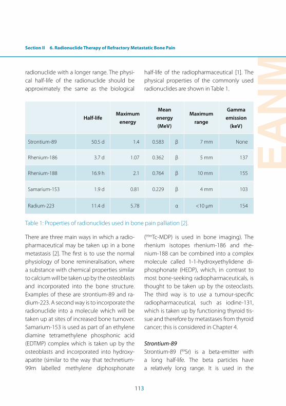
Section II 6. Radionuclide Therapy of Refractory Metastatic Bone Pain
EANM
113
radionuclide with a longer range. The physi-
cal half-life of the radionuclide should be
approximately the same as the biological
half-life of the radiopharmaceutical [1]. The
physical properties of the commonly used
radionuclides are shown in Table 1.
Half-life Maximum
energy
Mean
energy
(MeV)
Maximum
range
Gamma
emission
(keV)
Strontium-89 50.5 d 1.4 0.583 β 7 mm None
Rhenium-186 3.7 d 1.07 0.362 β 5 mm 137
Rhenium-188 16.9 h 2.1 0.764 β 10 mm 155
Samarium-153 1.9 d 0.81 0.229 β 4 mm 103
Radium-223 11.4 d 5.78 α <10 µm 154
Table 1: Properties of radionuclides used in bone pain palliation [2].
There are three main ways in which a radio-
pharmaceutical may be taken up in a bone
metastasis [2]. The irst is to use the normal
physiology of bone remineralisation, where
a substance with chemical properties similar
to calcium will be taken up by the osteoblasts
and incorporated into the bone structure.
Examples of these are strontium-89 and ra-
dium-223. A second way is to incorporate the
radionuclide into a molecule which will be
taken up at sites of increased bone turnover.
Samarium-153 is used as part of an ethylene
diamine tetramethylene phosphonic acid
(EDTMP) complex which is taken up by the
osteoblasts and incorporated into hydroxy-
apatite (similar to the way that technetium-
99m labelled methylene diphosphonate
(99mTc-MDP) is used in bone imaging). The
rhenium isotopes rhenium-186 and rhe-
nium-188 can be combined into a complex
molecule called 1-1-hydroxyethylidene di-
phosphonate (HEDP), which, in contrast to
most bone-seeking radiopharmaceuticals, is
thought to be taken up by the osteoclasts.
The third way is to use a tumour-speciic
radiopharmaceutical, such as iodine-131,
which is taken up by functioning thyroid tis-
sue and therefore by metastases from thyroid
cancer; this is considered in Chapter 4.
Strontium-89
Strontium-89 (89Sr) is a beta-emitter with
a long half-life. The beta particles have
a relatively long range. It is used in the

114
radiopharmaceutical form 89Sr chloride. The
maximum range of the beta particles in tis-
sue is relatively long, at 7 mm. It is handled by
the body in a manner similar to calcium and
is therefore taken up by bone which is more
metabolically active. It is renally excreted and
the glomerular iltration rate of the patient
should be considered. It has mainly been used
to treat metastatic bone pain from hormone-
resistant prostate cancer and has been found
to give pain relief in 60-70% of patients. The
onset of pain relief is typically around 10-14
days and the duration of pain relief may be
prolonged, at up to 15 months. Its toxicity is
usually in the form of myelosuppression, which
typically occurs in the irst 6 weeks post treat-
ment and reverses over the next 6 weeks [2].
Rhenium-186
Rhenium-186 (186Re) is a beta emitter with
a half-life of 3.7 days and also has gamma
emissions which permit imaging. The range
of the beta particles is 5 mm. It has been used
in prostate cancer and breast cancer and
most patients report pain relief within about
2 days of administration. It causes temporary
myelosuppression with a nadir in counts at
around 4 weeks and recovery in 8 weeks [1].
Rhenium-188
Rhenium-188 (188Re) is also a beta emitter.
It has a shorter half-life of 16.9 h and may
produce more rapid pain relief than other ra-
diopharmaceuticals. Its beta particles have a
relatively long range, 10 mm, which may be
useful for some clinical applications. It also
causes myelosuppression [1].
Samarium-153
Samarium-153 (153Sm) emits lower energy
beta particles with a range of only 4 mm. A
high proportion of the radiopharmaceutical
is quickly taken up by bone – preferentially
at sites with increased osteoblastic activ-
ity. The gamma emissions have potential for
imaging. There is typically a quick pain relief
response in most patients – within about
one week – with the duration being in the
order of 8 weeks. Myelosuppression may oc-
cur, with a nadir in blood counts at around
6 weeks [1].
Radium-223
Radium-223 (223Ra) has been the subject of
considerable interest recently. It is an alpha
particle emitter and the energy will therefore
be deposited over a short range: less than a
tenth of a millimetre. It is a calcium mimetic
and will therefore be taken up by the osteo-
blasts and incorporated into hydroxyapatite
at sites of increased bone turnover [4]. A rela-
tively low number of “hits” to a cell by alpha
particles will result in cell death, so there is
potential for an alpha-emitting radiopharma-
ceutical to eiciently kill tumour cells with-
out having a signiicant impact on the bone
marrow.
Clinical applications
The most established indication for radionu-
clide therapy for bone pain is in prostate can-
cer, and 89Sr has been found to be efective
in achieving pain relief in 65-75% of patients.
A sub-group of patients may even report
complete pain relief [5,6]. 89Sr has also been
Section II 6. Radionuclide Therapy of Refractory Metastatic Bone Pain
EANM
115
used both instead of EBRT and as an adjuvant
to it, showing pain relief in most cases [2]. 153Sm has also been used with good efect in
metastatic prostate cancer [2]. 223Ra, with the
trade name Alpharadin®, has been used with
very promising results in hormone-resistant
prostate cancer and, at the time of writing, a
trial called Alpharadin in Symptomatic Pros-
tate Cancer (ALYSMPCA) is soon going to
be formally published. The initial data show
signiicant reductions in the pain reported
by patients and their use of opioid analgesia.
A signiicant survival beneit has also been
found [7]. The radiopharmaceutical is now
going through licensing procedures and is
likely to be brought into widespread clinical
use over the next few years.
In breast cancer, 89Sr, 186Re and 188Re have
all been used and found to be efective in
achieving pain relief in most patients [6].
Signiicant reduction in pain was found in
a small study using 186Re-(Sn)-HEDP. A trial
which included breast cancer patients found
that 153Sm-EDTMP yielded a signiicant re-
duction in pain and opioid use [8].
Toxicity and limitations
The most important consideration for patient
safety with bone-seeking radionuclide thera-
py is the efect on the bone marrow. Almost
all patients will have some myelosuppression
[1], which in most cases will be relatively mild
and transient. The timing of myelosuppres-
sive efects varies with the radiopharmaceu-
tical used, as mentioned above, but typically
the nadir in blood counts is around 4 weeks
and recovery occurs at around 8 weeks post
treatment [2]. Patients with signiicant bone
marrow involvement will be at higher risk of
haematological toxicity and this should be
considered before treatment. Toxicity may
be increased in patients with subclinical dis-
seminated intravascular coagulation (DIC),
which is often present in advanced stage
cancer patients, particularly those with pros-
tate cancer [1].
A signiicant number of patients may expe-
rience a short-term increase in pain, a phe-
nomenon known as “pain lare”. This is more
common with rhenium treatments. There do
not appear to be any clinical features that
identify patients likely to experience pain
lare [2].
Contra-indications and situations requiring
caution
As with many medical radiation exposures,
pregnancy is an absolute contra-indication
and should be excluded according to local
protocol.
Patients with bone metastases are at risk of
developing spinal cord compression, where a
metastatic deposit in the spine enlarges suf-
iciently to exert mechanical pressure on the
nervous tissue. Treatment with bone-seeking
radiopharmaceuticals can potentially worsen
this and it is therefore a contra-indication.
Any health professionals dealing with such
patients should be alert to the symptoms
of this condition, which include worsen-
ing back pain (particularly pain outside the
116
lumbar region), limb weakness, sensory loss
in a dermatomal distribution and bowel and/
or bladder dysfunction. A patient with symp-
toms or signs suspicious of spinal cord com-
pression will require urgent medical review.
As the radiopharmaceuticals are predomi-
nantly renally cleared it is important to con-
sider the patient’s renal function. A patient
with poor renal function will receive a higher
efective dose due to more radiopharma-
ceutical remaining present in the body for
longer. Acute kidney injury will be a contra-
indication to bone-seeking radionuclide
therapy and the patient will require stabilisa-
tion before it can be considered. The pres-
ence of vesico-ureteric low obstruction and/
or bladder outlow obstruction, of particular
concern in prostate cancer, may cause an in-
creased dose to those organs and surround-
ing structures; this may need assessment and
possibly intervention before treatment [1].
Urinary incontinence, again a common sce-
nario in prostate cancer, may cause problems
with radioactive contamination.
Pre-therapeutic evaluation
The indication for treatment with bone-
seeking radionuclides is therefore metastatic
bone pain that is not controlled with con-
ventional analgesia and is limiting activities
of daily living. The patient group presenting
for nuclear medicine therapy is likely to in-
clude those in whom other treatments, such
as bisphosphonates, hormonal treatment
and EBRT, have failed. It should be noted that
evidence shows that patients in somewhat
earlier stages of the development of bone
metastases, with relatively limited and lo-
calised disease, may gain more beneit from
radionuclide treatment [3]. Bearing in mind
the time frames for onset of pain relief and
its duration, consideration should be given
to which radionuclide is most suitable. A ra-
dionuclide with a shorter half-life may give a
quicker response, but with a shorter duration
in a patient with relatively rapidly progress-
ing disease. A radionuclide with a longer half-
life may be appropriate to achieve a longer
duration of efect in other patients. Patients
with a short life expectancy may not be suit-
able for therapy with some of the longer-
lived radionuclides, particularly 89Sr.
A suitably qualiied medical practitioner
should obtain suicient medical history and
perform an appropriate examination prior
to therapy. Enquiries should be made about
symptoms suggestive of conditions which
would contra-indicate radionuclide therapy.
Medication history should be looked at to
see whether there are any medicines which
may afect radiopharmaceutical uptake or
cause interactions. It should be borne in
mind that many patients will take “over the
counter” medicines which do not require
prescriptions. Many may also be taking
“complementary therapies” such as herbal
substances and food supplements.
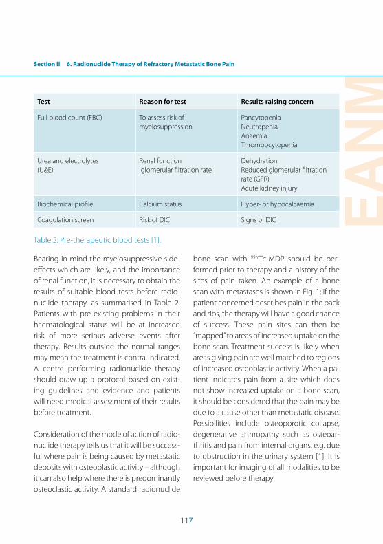
Section II 6. Radionuclide Therapy of Refractory Metastatic Bone Pain
EANM
117
Test Reason for test Results raising concern
Full blood count (FBC) To assess risk of myelosuppression
PancytopeniaNeutropeniaAnaemiaThrombocytopenia
Urea and electrolytes(U&E)
Renal function glomerular iltration rate
DehydrationReduced glomerular iltration rate (GFR)Acute kidney injury
Biochemical proile Calcium status Hyper- or hypocalcaemia
Coagulation screen Risk of DIC Signs of DIC
Table 2: Pre-therapeutic blood tests [1].
Bearing in mind the myelosuppressive side-
efects which are likely, and the importance
of renal function, it is necessary to obtain the
results of suitable blood tests before radio-
nuclide therapy, as summarised in Table 2.
Patients with pre-existing problems in their
haematological status will be at increased
risk of more serious adverse events after
therapy. Results outside the normal ranges
may mean the treatment is contra-indicated.
A centre performing radionuclide therapy
should draw up a protocol based on exist-
ing guidelines and evidence and patients
will need medical assessment of their results
before treatment.
Consideration of the mode of action of radio-
nuclide therapy tells us that it will be success-
ful where pain is being caused by metastatic
deposits with osteoblastic activity – although
it can also help where there is predominantly
osteoclastic activity. A standard radionuclide
bone scan with 99mTc-MDP should be per-
formed prior to therapy and a history of the
sites of pain taken. An example of a bone
scan with metastases is shown in Fig. 1; if the
patient concerned describes pain in the back
and ribs, the therapy will have a good chance
of success. These pain sites can then be
“mapped” to areas of increased uptake on the
bone scan. Treatment success is likely when
areas giving pain are well matched to regions
of increased osteoblastic activity. When a pa-
tient indicates pain from a site which does
not show increased uptake on a bone scan,
it should be considered that the pain may be
due to a cause other than metastatic disease.
Possibilities include osteoporotic collapse,
degenerative arthropathy such as osteoar-
thritis and pain from internal organs, e.g. due
to obstruction in the urinary system [1]. It is
important for imaging of all modalities to be
reviewed before therapy.
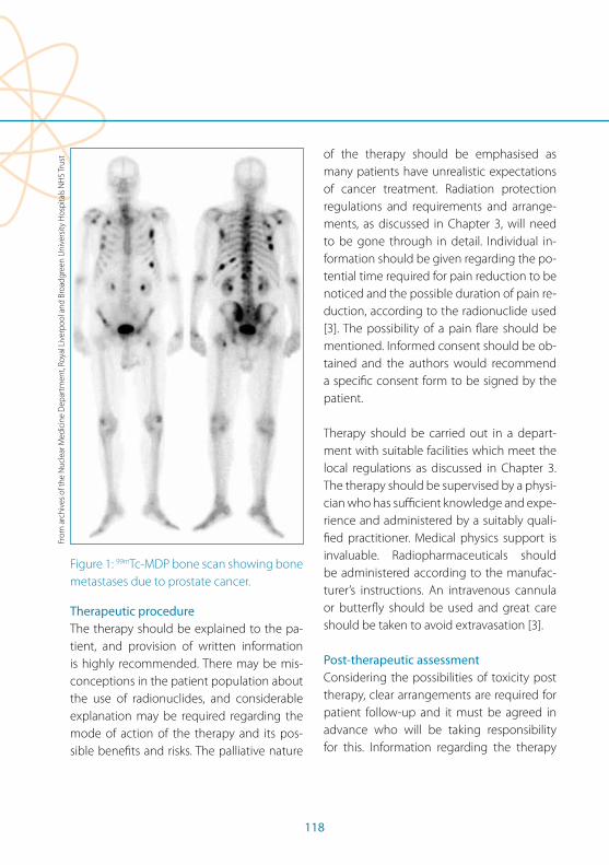
118
Figure 1: 99mTc-MDP bone scan showing bone
metastases due to prostate cancer.
Therapeutic procedure
The therapy should be explained to the pa-
tient, and provision of written information
is highly recommended. There may be mis-
conceptions in the patient population about
the use of radionuclides, and considerable
explanation may be required regarding the
mode of action of the therapy and its pos-
sible beneits and risks. The palliative nature
of the therapy should be emphasised as
many patients have unrealistic expectations
of cancer treatment. Radiation protection
regulations and requirements and arrange-
ments, as discussed in Chapter 3, will need
to be gone through in detail. Individual in-
formation should be given regarding the po-
tential time required for pain reduction to be
noticed and the possible duration of pain re-
duction, according to the radionuclide used
[3]. The possibility of a pain lare should be
mentioned. Informed consent should be ob-
tained and the authors would recommend
a speciic consent form to be signed by the
patient.
Therapy should be carried out in a depart-
ment with suitable facilities which meet the
local regulations as discussed in Chapter 3.
The therapy should be supervised by a physi-
cian who has suicient knowledge and expe-
rience and administered by a suitably quali-
ied practitioner. Medical physics support is
invaluable. Radiopharmaceuticals should
be administered according to the manufac-
turer’s instructions. An intravenous cannula
or butterly should be used and great care
should be taken to avoid extravasation [3].
Post-therapeutic assessment
Considering the possibilities of toxicity post
therapy, clear arrangements are required for
patient follow-up and it must be agreed in
advance who will be taking responsibility
for this. Information regarding the therapy
From
arc
hive
s of
the
Nuc
lear
Med
icin
e D
epar
tmen
t, Ro
yal L
iver
poo
l and
Bro
adgr
een
Uni
vers
ity H
osp
itals
NH
S Tr
ust
Section II 6. Radionuclide Therapy of Refractory Metastatic Bone Pain
EANM
119
and follow-up plans should be conveyed
immediately after its administration to the
patient’s supervising physician, e.g. an on-
cologist or urologist, and to the general prac-
titioner.
It will be important to monitor the patient
for haematological toxicity and this will again
depend on the radionuclide used. Such
monitoring will involve periodic blood tests –
typically every 1-2 weeks – for up to 6 weeks
in the case of 186Re and 153Sm, and up to 16
weeks for 89Sr [3]. The patient should be told
to be alert to potential symptoms suggesting
more serious toxicity, such as lethargy and
pyrexia. A pain lare may require temporarily
increased analgesia.
In patients who respond to therapy, par-
ticularly with shorter-acting radionuclides,
re-treatment may be suitable. The period be-
tween therapy should be consistent with the
radionuclide, e.g. around 8 weeks for 153Sm
and 12 weeks for 186Re [3].
120
References Section II, Chapter 6
References1. Lewington VJ. Bone seeking radionuclides for therapy.
J Nucl Med. 2005;46 Suppl 1:38S-47S.
2. Hoskin P. Radioisotope therapy for metastatic bone dis-ease. In: Hoskin P, ed. Radiotherapy in practice - radio-isotope therapy. Oxford: Oxford University Press, 2007.
3. Bodei L, Lam M, Chiesa C, Flux G, Brans B, Chiti A, Giam-marile F; European Association of Nuclear Medicine (EANM). EANM procedure guideline for the treatment of refractory metastatic bone pain. Eur J Nucl Med Mol Imaging. 2008;35:1934-40.
4. Brady D, Parker C C, O’Sullivan JM. Bone targeting ra-diopharmaceuticals including radium-223. Cancer J. 2013;19:71-8.
5. Lewington VJ, McEwan AJ, Ackery DM, Bayly RJ, Keel-ing DH, Macleod PM, et al. A prospective, randomised double blind crossover study to examine the eicacy of strontium-89 in pain palliation in patients with ad-vanced prostate cancer metastatic to bone. Eur J Cancer. 1991;27:954-8.
6. Liepe K, Kotzerke J. A comparative study of rhenium-188-HEDP, rhenium-186-HEDP, samarium-153-EDTMP and strontium-89 in the treatment of painful skeletal metastases. Nucl Med Commun 2007;28:623-30.
7. Nilsson S, Sartor AO, Bruland ØS, Fang F, Aksnes A-K, Parker C. Pain analysis from the phase III randomized ALSYMPCA study with radium-223 dichloride (Ra-223) in patients with castration-resistant prostate cancer (CRPC) with bone metastases. 2013 Genitourinary Cancers Symposium. J Clin Oncol. 13 Suppl 6.
8. Seraini AN, Houston SJ, Resche I, Quick DP, Grund FM, Ell PJ, et al. Palliation of pain associated with meta-static bone cancer using samarium-153 lexidronam: A double blind placebo controlled trial. J Clin Oncol 1998;16:1574-81.
EANM
121
Introduction
Radiosynovectomy (also known as radio-
synoviorthesis) is an established, powerful
therapeutic technique that provides targeted
therapy to combat synovial inlammatory pro-
cesses in individual joints and so minimises
the damage and chronic arthritic disease that
follows. It was irst described by Fellinger in
1952 [1] and is the subject of EANM guidelines
published in 2003 [2]. However, even 2003 is a
long time ago in medicine, and although the
technique itself has not changed massively,
the advances in medical technology outside
the ield of nuclear medicine provide new
challenges and opportunities that need to be
taken into account as we practise this therapy.
Indications for radiosynovectomy
Arthropathies for which radiosynovectomy is
indicated are shown in Table 1.
Rheumatoid arthritis
Haemophilic arthritis
Spondyloarthropathy, e.g. reactive or psoriatic arthritis
Persistent synovial efusion
Persistent efusion following prosthesis
Other inlammatory joint disease (e.g. Lyme disease, Behçet’s disease)
Undiferentiated arthritis with synovitis, synovial thickening or efusion
Calcium pyrophosphate dihydrate (CPPD) arthritis
Table 1: Arthropathies that are indications for
radiosynovectomy
Overview of synovitis and arthritis
Synovitis
Synovitis is the inlammation of the connec-
tive tissue lining the ligaments that make up
the joint capsule, the synovium. The synovi-
um secretes the thick synovial luid that lu-
bricates the joint.
Rheumatoid arthritis
Rheumatoid arthritis (RA) is one of the most
common auto-immune diseases. It afects
more women than men and has a preva-
lence of approximately 1% in the whole pop-
ulation. RA increases the numbers of synovial
lining cells with colonisation by ibroblast
growth and intensive vascularisation. This
leads to hyperplastic synovial tissue growth
(pannus) that erodes cartilage, subchondral
bone, the capsule and ligaments.
Synovial lining cells are colonised by lym-
phocyctes and plasma cells. T cells produce
lymphokines, such as tumour necrosis factor,
and mature B cells produce immunoglobu-
lins, such as rheumatoid factor.
In the early stages, pain increases with stifness
and swelling, and reduced movement is pos-
sible. Later stage disease involves progressive
joint destruction and deformation. Baker’s cyst
is also a common development in the disease.
Traditionally RA has been a major source
of referrals for radiosynovectomy, but drug
management has improved in the last 20
years and this is reducing RA referrals for both
surgical interventions and radiosynovectomy
in Western countries [3].
Section II7. Radiosynovectomy
Christopher Mayes, Brian A. Lowry, Ruth Berhane Menghis and Sobhan Vinjamuri
122
Drug therapies for RA. New drug regimens
have improved the management of the
disease in recent years. Non-steroidal anti-
inlammatory drugs are used to relieve pain
and stifness in early disease by inhibiting
cyclo-oxygenase (COX) but do not inluence
the radiographic progression of the disease.
Systemic corticosteroids improve symptoms
and do reduce damage. Intra-articular corti-
costeroid injections are widely used and can
produce rapid and sometimes sustained re-
lief in target joints.
Disease-modifying anti-rheumatic drugs
(DMARDS) are a group of systemic drugs that
require sustained use, but do slow the progres-
sion of RA. Methotrexate and sulfasalazine are
of this type and they can be used efectively
in combination therapies with other DMARDs
and biological response modiiers (BRMs).
BRMs include tumour necrosis factor (TNF)
blockers and monoclonal antibodies which act
on the immune system, e.g. rituximab.
This improved management of RA means
the injury to many patients’ joints has been
successfully reduced or at least postponed.
There are new products and combinations
that are fortunately reducing the need for
patient referral for radiosynovectomy. This
reduction in demand does not mean that ra-
diosynovectomy has been replaced, howev-
er. Furthermore, the new drug regimens are
systemic and have signiicant side-efects.
There are concerns about anti-TNF therapy
regarding a possible dose-dependent in-
crease in serious infection and malignancies,
especially skin cancers [4-6], so targeted ther-
apies may continue to have an important
role for some patients.
Reactive arthritis
Reactive arthritis is another autoimmune dis-
ease that is triggered by an infection in an-
other part of the body, e.g. by food poisoning
or chlamydial infection.
Psoriatic arthritis
Psoriatic arthritis, which causes destruction
of joints, can be as severe as RA but will at-
tack fewer joints and thus is suitable for tar-
geted radiosynovectomy.
Osteoarthritis
Erosion of the normal covering of the carti-
lage allows the breaking of of tissue, irrita-
tion of the synovium and osteophyte forma-
tion. Synovitis can be treated with radiosyno-
vectomy but the underlying cartilage disease
cannot. Radiosynovectomy has been shown
to be useful in osteoarthritis, especially in
younger patients with early disease, whose
joints show little radiographic damage [7].
Haemophilic arthritis
Haemophilia is an X chromosome-linked dis-
order caused by a deiciency of factor VIII or
factor IX due to mutation of the respective
clotting factor genes. This is a rare disease with
an estimated frequency of 1 in 10,000 births.
It generally afects males on the maternal side
and, although this can usually be traced down
family lines, as many as one-third of all cases
are the result of spontaneous mutation where
there is no prior family history.
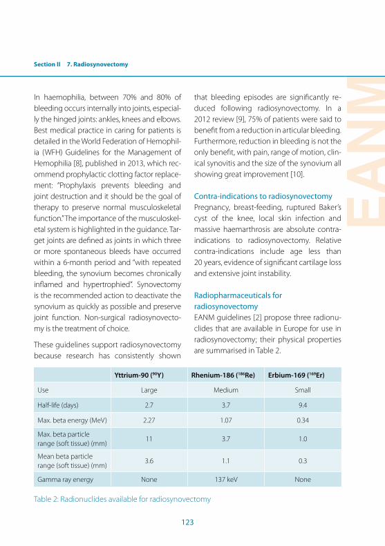
Section II 7. Radiosynovectomy
EANM
123
In haemophilia, between 70% and 80% of
bleeding occurs internally into joints, especial-
ly the hinged joints: ankles, knees and elbows.
Best medical practice in caring for patients is
detailed in the World Federation of Hemophil-
ia (WFH) Guidelines for the Management of
Hemophilia [8], published in 2013, which rec-
ommend prophylactic clotting factor replace-
ment: “Prophylaxis prevents bleeding and
joint destruction and it should be the goal of
therapy to preserve normal musculoskeletal
function.” The importance of the musculoskel-
etal system is highlighted in the guidance. Tar-
get joints are deined as joints in which three
or more spontaneous bleeds have occurred
within a 6-month period and “with repeated
bleeding, the synovium becomes chronically
inlamed and hypertrophied”. Synovectomy
is the recommended action to deactivate the
synovium as quickly as possible and preserve
joint function. Non-surgical radiosynovecto-
my is the treatment of choice.
These guidelines support radiosynovectomy
because research has consistently shown
that bleeding episodes are signiicantly re-
duced following radiosynovectomy. In a
2012 review [9], 75% of patients were said to
beneit from a reduction in articular bleeding.
Furthermore, reduction in bleeding is not the
only beneit, with pain, range of motion, clin-
ical synovitis and the size of the synovium all
showing great improvement [10].
Contra-indications to radiosynovectomy
Pregnancy, breast-feeding, ruptured Baker’s
cyst of the knee, local skin infection and
massive haemarthrosis are absolute contra-
indications to radiosynovectomy. Relative
contra-indications include age less than
20 years, evidence of signiicant cartilage loss
and extensive joint instability.
Radiopharmaceuticals for
radiosynovectomy
EANM guidelines [2] propose three radionu-
clides that are available in Europe for use in
radiosynovectomy; their physical properties
are summarised in Table 2.
Yttrium-90 (90Y) Rhenium-186 (186Re) Erbium-169 (169Er)
Use Large Medium Small
Half-life (days) 2.7 3.7 9.4
Max. beta energy (MeV) 2.27 1.07 0.34
Max. beta particle range (soft tissue) (mm)
11 3.7 1.0
Mean beta particle range (soft tissue) (mm)
3.6 1.1 0.3
Gamma ray energy None 137 keV None
Table 2: Radionuclides available for radiosynovectomy
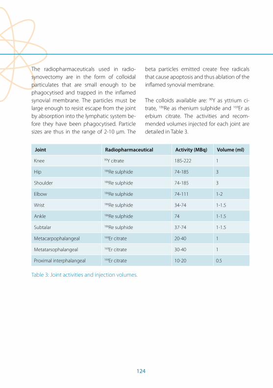
124
The radiopharmaceuticals used in radio-
synovectomy are in the form of colloidal
particulates that are small enough to be
phagocytised and trapped in the inlamed
synovial membrane. The particles must be
large enough to resist escape from the joint
by absorption into the lymphatic system be-
fore they have been phagocytised. Particle
sizes are thus in the range of 2-10 µm. The
beta particles emitted create free radicals
that cause apoptosis and thus ablation of the
inlamed synovial membrane.
The colloids available are: 90Y as yttrium ci-
trate, 186Re as rhenium sulphide and 169Er as
erbium citrate. The activities and recom-
mended volumes injected for each joint are
detailed in Table 3.
Joint Radiopharmaceutical Activity (MBq) Volume (ml)
Knee 90Y citrate 185-222 1
Hip 186Re sulphide 74-185 3
Shoulder 186Re sulphide 74-185 3
Elbow 186Re sulphide 74-111 1-2
Wrist 186Re sulphide 34-74 1-1.5
Ankle 186Re sulphide 74 1-1.5
Subtalar 186Re sulphide 37-74 1-1.5
Metacarpophalangeal 169Er citrate 20-40 1
Metatarsophalangeal 169Er citrate 30-40 1
Proximal interphalangeal 169Er citrate 10-20 0.5
Table 3: Joint activities and injection volumes.
Section II 7. Radiosynovectomy
EANM
125
The radiosynovectomy procedure
National legislation requirements vary be-
tween countries for administration of radio-
active agents. In-patient treatment may be
required and although this may not be legal-
ly demanded, because immobilisation must
follow radiosynovectomy, procedures may
require that less able patients stay in hospital.
Appropriately shielded accommodation and
toilet facilities may therefore be necessary.
Radiosynovectomy demands an aseptic in-
jection so appropriate clean areas and sterile
protocols are needed during administration.
Knees are the joints most often treated and
these are the only joints that can be injected
without imaging. Conirmation of the correct
intra-articular placement of the needle is pos-
sible by aspiration of synovial luid. For other
joints, imaging, e.g. by mobile image intensi-
ier or ultrasound, is necessary to ensure safe
positioning of the needle for injection.
Staing requirements
Radiosynovectomy requires a multi-disciplin-
ary team to be run eiciently. In addition to
the nuclear medicine medical, technological,
nursing and physics staf, a rheumatologist or
orthopaedic surgeon will probably supply the
expertise in synovial puncture. In the case of
haemophilic patients, haematologists will have
ensured prophylactic clotting factor cover.
Patient preparation
Previous intra-articular glucocorticoid injec-
tion will have been unsuccessful in patients
referred for radiosynovectomy. Recent ra-
diographs of joints to be treated will be re-
viewed prior to the procedure. Other imag-
ing techniques may be useful: Scintigraphic
assessment of soft tissue and active inlam-
mation is often carried out using 99mTc-MDP/
HDP/HEDP. Ultrasound is useful to evaluate
synovial structure and thickness and exclude
ruptured Baker’s cyst. Magnetic resonance
imaging may also be useful.
Knees with Baker’s cysts can be treated by
skilled practitioners [11]. The cyst may be
reduced using external pressure or synovial
puncture and aspiration of the main joint.
The danger of rupture and escape of the ra-
diopharmaceutical must be assessed by the
clinician.
Arthroscopy or surgery must not have been
performed during the 2–6 weeks preceding
radiosynovectomy.
The minimum interval between repeat radio-
synovectomy is 6 months.
Consent
Informed written consent must be obtained
prior to radiosynovectomy. Written and ver-
bal information must be given to the patient
and this should include:
1. Between 60% and 80% of patients beneit
from the therapy.
2. Response is unlikely within 14 days of
injection and may not be noticeable for
1 month.
126
3. There is a risk of a temporary increase in
synovitis.
4. Potential complications of treatment are:
a) Due to joint puncture: local haemor-
rhage, bruising, infection (very rare) and
future malignancy.
b) Theoretical risk of exposure to beta-
emitting radiation, including radiation
necrosis (rare) and future malignancy.
c) Risk of post-injection pyrexia or radio-
pharmaceutical allergy (very rare).
Administration
Radiosynovectomy is an aseptic technique.
It must therefore be performed under full
aseptic conditions by suitably qualiied med-
ical, nursing and technical staf. Skin prepa-
ration may have included the removal of ex-
cess hair. After clinical examination a sterile
ield is created around the joint to be treated
using towels.
There follows a brief description of the pro-
cedure using the example of the knee as the
target joint: The exposed skin of the injection
site is disinfected. Local anaesthesia is advis-
able before the joint puncture. Intra-articular
puncture of the knee may be achieved using
a 20-gauge 7.6-cm needle on a slightly lexed
knee proximal and lateral to the superolateral
angle of the patella. Local anaesthetic can be
injected irst subcutaneously through this
needle (this also ensures that skin-punch cyl-
inders are not carried into the joint).
Once the needle has been advanced into the
joint, aspiration of synovial luid will conirm
the correct position of the needle. Radio-
pharmaceutical must not be injected unless
intra-articular placement has been ensured
by aspiration. As already mentioned, joints
other than the knee must have imaging con-
irmation of position, e.g. by mobile image
intensiier; this is particularly important as
the joint may be misshapen by the disease
process and small joints with little luid make
the aspiration test impracticable.
In a joint with efusion, aspiration of excess
luid is useful so long as enough luid is left
to distribute the radiopharmaceutical in the
joint space. If no efusion is present, 10 ml
or more of saline may be useful to achieve
good dispersion. The radiopharmaceutical
is then injected and it is recommended that
this be followed by use of long-acting gluco-
corticoids to reduce any acute synovitis and
improve treatment response.
The needle is inally lushed with saline, be-
fore and during withdrawal, to reduce the
chance of leakage of radioactivity down the
needle track.
Once the needle has been removed, a skin
dressing is applied to the puncture site
and this is followed by joint immobilisa-
tion (splinting) for 48 h. For a knee this may
consist of a thigh-leg plaster/ resin cast. Im-
mobilisation reduces the transport of par-
ticles through the lymphatics to the regional
lymph nodes.
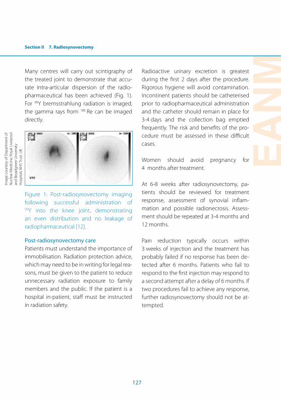
Section II 7. Radiosynovectomy
EANM
127
Many centres will carry out scintigraphy of
the treated joint to demonstrate that accu-
rate intra-articular dispersion of the radio-
pharmaceutical has been achieved (Fig. 1).
For 90Y bremsstrahlung radiation is imaged;
the gamma rays from 186 Re can be imaged
directly.
Figure 1: Post-radiosynovectomy imaging
following successful administration of 90Y into the knee joint, demonstrating
an even distribution and no leakage of
radiopharmaceutical [12].
Post-radiosynovectomy care
Patients must understand the importance of
immobilisation. Radiation protection advice,
which may need to be in writing for legal rea-
sons, must be given to the patient to reduce
unnecessary radiation exposure to family
members and the public. If the patient is a
hospital in-patient, staf must be instructed
in radiation safety.
Radioactive urinary excretion is greatest
during the irst 2 days after the procedure.
Rigorous hygiene will avoid contamination.
Incontinent patients should be catheterised
prior to radiopharmaceutical administration
and the catheter should remain in place for
3-4 days and the collection bag emptied
frequently. The risk and beneits of the pro-
cedure must be assessed in these diicult
cases.
Women should avoid pregnancy for
4 months after treatment.
At 6-8 weeks after radiosynovectomy, pa-
tients should be reviewed for treatment
response, assessment of synovial inlam-
mation and possible radionecrosis. Assess-
ment should be repeated at 3-4 months and
12 months.
Pain reduction typically occurs within
3 weeks of injection and the treatment has
probably failed if no response has been de-
tected after 6 months. Patients who fail to
respond to the irst injection may respond to
a second attempt after a delay of 6 months. If
two procedures fail to achieve any response,
further radiosynovectomy should not be at-
tempted.
Imag
e co
urte
sy o
f Dep
artm
ent o
f N
ucle
ar M
edic
ine,
Roy
al L
iver
poo
l an
d B
road
gree
n U
nive
rsity
H
osp
itals
NH
S Tr
ust.
UK.
128
References Section II, Chapter 7
References1. Fellinger K, Schmid L. Die locale Behandlung der rheu-
matischen Errkrankung (local therapy of RA). Wien Ztsch Inn Med. 1952;33:351.
2. Clunie G, Fischer M; European Association of Nuclear Medicine. EANM procedure guidelines for radiosyno-vectomy. Eur J Nucl Med Mol Imaging. 2003;30:BP12-6.
3. Momohara S, Tanaka S, Nakamura H, Mibe J, Iwamoto T, Ikari K, et al. Recent trends in orthopedic surgery per-formed in Japan for rheumatoid arthritis. Mod Rheuma-tol. 2011;21:337-42.
4. Bongartz T, Sutton AJ, Sweeting MJ, Buchan I, Matte-son EL, Montori V. Anti-TNF antibody therapy in rheu-matoid arthritis and the risk of serious infections and malignancies: systematic review and analysis of rare harmful efects in randomized controlled trials. JAMA. 2006;295:2275-85.
5. Ding T, Ledingham J, Luqmani R, Westlake S, Hyrich K, Lunt M, et al.; Standards, Audit and Guidelines Work-ing Group of BSR Clinical Afairs Committee and BHPR. BSR and BHPR rheumatoid arthritis guidelines on safety of anti-TNF therapies. Rheumatology (Oxford). 2010;49:2217-9.
6. Raaschou P, Simard J, Holmqvist M, Askling J. Rheuma-toid arthritis, anti-tumour necrosis factor therapy, and risk of malignant melanoma: nationwide population based cohort study from Sweden. BMJ. 2013;346:f1939.
7. Markou P, Chatzopoulos D. Yttrium-90 silicate radio-synovectomy treatment of painful synovitis in knee osteoarthritis. Results after 6 months. Hell J Nucl Med. 2009;12:33-6.
8. Srivasta A, Brewer AK, Mauser-Bunschoten EP, Key NS, Kitchen S, Llinas A, et al. WFH guidelines for the manage-ment of hemophilia. Haemophilia. 2013;19:e1-47.
9. Peyvandi F, Klamroth R, Carcao M, Federici AB, Di Minno G, Jimenez-Yiste V, Rodriguez Merchan EC. Management of bleeding disorders in adults. Haemophilia. 2012;18 (Suppl 2):24-36.
10. De La Corte-Rodriguez H, Rodriguez-Merchan EC, Jimenez-Yuste V. Radiosynovectomy in hemophilia: quantiication of its efectiveness through the assess-ment of 10 articular parameters. J Thromb Haemost. 2011;9:928-35.
11. Modder G. Radiosynoviorthesis: involvement of nuclear medicine in rheumatology and orthopaedics. Mecken-heim: Warlich, 1995. ISBN 3-930376-04-0.
EANM
129
Section II 8. Nursing Implications for Patients Undergoing Radionuclide Metabolic Therapy
Ana Gorgan, Nicoleta Lupu and Claudiu Peștean
General aspects of nursing
Concepts and models
Nursing has many deinitions, but may most
appropriately be considered to comprise the
diagnosis and treatment of human reactions
to actual or potential health problems [1].
There are many conceptual models of nurs-
ing, the diferences between them relating
to the deinition of nursing and its purpose,
the deinition of the patient, the deinition of
state of health and the deinition of the envi-
ronment which interacts with the patient [1].
Central to nursing conceptual models is the
role of the individual, the individual’s family
and the community in maintaining and/or
recovering the state of health [2].
Most concepts and models emphasise the
necessity of a holistic approach to ensure
that all the needs of patients and the con-
text in which their health and well-being are
grounded are taken into consideration.
Nursing, as an integrated part of the social
assistance system, includes health assis-
tance, disease prevention and health care
for all types of disease (physical or mental)
and disability at any age. Nurses focus on
individual, familial or group reactions against
the disease. They need to respond to the real
or potential health problems of individuals
or a community. Nursing interventions, as
a response to the reactions against disease,
range from individual interventions aimed
at recovering the individual’s health, to the
development of speciic policies designed
to preserve the health of the population in
the long term, as pointed out by the Inter-
national Council of Nurses. The World Health
Organisation and the International Council of
Nurses deine nursing as part of the health
system [3].
Nursing, as a profession, involves various ac-
tivities that can be complex and specialised
or simple, such as the gesture of holding a
patient’s hand. Nursing is a mix of science
and art: it requires theoretical knowledge
about health care, applied in daily practice
with ability by skilled personnel. Nursing
activities are aimed in four main directions:
health maintenance, disease prevention,
health recovery and assistance in the event
of disability or even death.
According to Virginia Henderson, author of
the irst scientiic theory about health care
needs and a key igure in laying the basis for
modern nursing, nursing means helping the
individual to ind his or her route to health or
recovery, taking into consideration that the
individual should have the strength, the will
and the knowledge to do this [4]. The con-
ceptual nursing model established by Virgin-
ia Henderson is based on 14 fundamental pa-
tient needs. According to this model, mainte-
nance of the individual’s independence, or at
least a certain level of independence, allows
that individual to satisfy his or her fundamen-
tal needs.
The individual should be considered ho-
listically, including the following dimen-
sions: bio-physiological, psychological,
130
sociocultural and spiritual. Environment fac-
tors should also be taken into consideration
because they may produce imbalances and
consequently adversely afect the indepen-
dence of the individual.
The state of disease represents a disruption
of the equilibrium between the organism
and the environment, which is an alarm sig-
nal expressed as physical and/or psychologi-
cal distress, a diiculty or maladjustment to a
new situation, temporary or permanent. This
imbalance represents a negative event or ex-
perience for the individual.
The conceptual nursing model, through its
perspective, ofers a systematised meth-
odology to identify the health problems of
the patient and to elaborate a nursing plan
which will reduce the patient’s dependency.
It also ofers modalities for the implementa-
tion of this plan and for the evaluation of its
eicacy upon completion.
The conceptual nursing model ofers several
advantages:
• It identiies the speciic problems of the
patient. In this way it is possible to estab-
lish general and speciic objectives that
will enable recovery from these problems.
To accomplish these objectives, speciic in-
terventions are applied according to nurs-
ing skills and knowledge. The patient and
his or her family are included in the health
care team.
• The identiication of the patient’s needs
improves communication between the
members of the health care team.
• The nursing process uses consistent and
orderly records relating to all aspects of
nursing. These records allows quantiica-
tion of all health care activities and, in this
way, evaluation of the quality of health
care provided.
The nursing process
The nursing process has ive phases:
The analysis. This entails the gathering of all
necessary information to identify the ac-
tual or potential health problems, and for
this purpose, an objective and impartial
attitude is required. The information ob-
tained can be subjective (when collected
from the patient or his family) or objective
(when it derives from measurable condi-
tions).
Nursing diagnostics (the analysis from a
clinical point of view of the individual’s
response to actual or potential health
problems). This consists in identifying the
patient’s health problem and formulat-
ing and validating a nursing diagnosis.
Formulation of a diagnosis consists in cor-
relating the health problem to its cause or
aetiology. One example concerns the im-
pact of restrictions on contact with other
persons on positive relations with others;
this is a potential nursing diagnosis for a
patient undergoing radioiodine therapy
for diferentiated thyroid carcinoma,
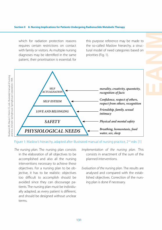
Section II 8. Nursing Implications for Patients Undergoing Radionuclide Metabolic Therapy
EANM
131
which for radiation protection reasons
requires certain restrictions on contact
with family or visitors. As multiple nursing
diagnoses may be identiied in the same
patient, their prioritisation is essential; for
this purpose reference may be made to
the so-called Maslow hierarchy, a struc-
tural model of need categories based on
priorities (Fig. 1).
SELF ACTUALISATION
SELF ESTEEM
LOVE AND BELONGING
SAFETY
phYSIOLOGICAL NEEDS
morality, creativity, spaonteity, recognition of facts
Conidence, respect of others, respect from others, recognition
Friendship, family, sexual intimacy
physical and mental safety
Breathing, homeostasis, food water, sex, sleep
Figure 1: Maslow’s hierarchy, adapted after Illustrated manual of nursing practice, 2nd edn. [1]
The nursing plan. The nursing plan consists
in the elaboration of all objectives to be
accomplished and also all the nursing
interventions necessary to achieve these
objectives. For a nursing plan to be ob-
jective, it has to be realistic: objectives
too diicult to accomplish should be
avoided since they can discourage pa-
tients. The nursing plan must be individu-
ally adapted, as every patient is diferent,
and should be designed without unclear
terms.
Implementation of the nursing plan. This
consists in enactment of the sum of the
planned interventions.
Evaluation of the nursing plan. The results are
analysed and compared with the estab-
lished objectives. Correction of the nurs-
ing plan is done if necessary.
Buxb
aum
BS,
Mau
ro E
, No
rris
CG
, ed
s. Il
lust
rate
d m
anu
al o
f nu
rsin
g
pra
ctic
e, 2
nd
ed
n. S
prin
gh
ou
se: S
prin
gh
ou
se C
orp
ora
tion
; 199
4.
132
The nursing applied to patients
undergoing radionuclide metabolic
therapy
Radionuclide metabolic therapy is a speciic
and complex therapeutic alternative. The
approach is multidisciplinary and nursing
care plays a well-deined role in the process
of healing. Besides carrying out the speciic
duties associated with health care, the nurse
has the role of teaching the patient about dif-
ferent aspects relating to his or her sufering,
thereby assisting the patient in accepting
and defeating the illness.
The pathologies in which radionuclide meta-
bolic therapy can be applied are diverse, but
most are oncological diseases. The disease,
its symptomatology and also its therapy may
afect the patient’s needs, from the vital ones
to those with low priority. Nursing interven-
tions are extremely important in recovering
or improving the patient’s state of health.
In the following we will approach radionu-
clide metabolic therapy not from the per-
spective of pathology, but from the perspec-
tive of the altered patient’s needs and the
nursing care to be applied.
Alteration of respiratory function
Example: patients with primary or secondary
lung tumours. Potential nursing interventions
include monitoring of breathing, conscious-
ness surveillance, prevention of complica-
tions, maintenance of an appropriate climate
and environmental conditions, maintenance
of an adequate position, administration of
medications and evaluation of their efects,
preparing and using instruments for oxygen
therapy, preparing the patient (physically
and psychologically) and the materials for
investigations (e.g. pleural puncture, bron-
choscopy, tomography), helping in mobilisa-
tion and transport and teaching the patient
how to cough.
Inadequate circulation/alteration of
cardiovascular function
Example: some cases of hyperthyroidism or
neroendocrine tumours secreting adrena-
line/noradrenaline. Potential nursing in-
terventions include monitoring of blood
pressure and heart rate, consciousness
surveillance, prevention of complications,
maintenance of appropriate climate and en-
vironmental conditions, teaching the patient
to ensure appropriate hydration, administra-
tion of medications and evaluation of their
efects, preparing the patient (physically and
psychologically) and the materials for inves-
tigations (e.g. ECG, SPECT-CT, PET-CT), and
helping in mobilisation and transport.
Inadequate nutrition
Example: some gastrointestinal tumours or
advanced stages of disease. Potential nursing
interventions include help/support in feed-
ing, assuring balanced nutrition, improve-
ment of nutritional status, maintenance of
an appropriate nutritional status, hydro-
electrolytic equilibrium, administration of
medications and evaluation of their efects,
dietary assessment, and patient training in
relation to diet, encompassing the purpose
Section II 8. Nursing Implications for Patients Undergoing Radionuclide Metabolic Therapy
EANM
133
of the diet, the type and quantity of food,
food to be avoided, how to replace forbid-
den nutriments and what substances to use
to improve taste [5].
Inadequate intestinal evacuation
Example: serotonin-induced diarrhoea in
patients with secreting neuroendocrine
tumours. Potential nursing interventions
include monitoring of vital and vegetative
signs, evaluation of hydro-electrolytic equi-
librium, hydro-electrolytic equilibration via
intravenous infusion of solutions, assess-
ment of signs of dehydration, maintenance
of hygiene, administration of medications
and evaluation of their efects, prevention of
complications, maintenance of an appropri-
ate climate and environmental conditions,
prevention of infection, and helping in mo-
bilisation and transport [6].
Alteration of body temperature; fever with or
without rigors
Example: radionuclide metabolic therapy
of lymphomas [7]. Potential nursing in-
terventions include monitoring of body
temperature, prevention of complications,
maintenance of an appropriate position,
maintenance of an appropriate climate and
environmental conditions, maintenance of
hygiene, and preparation of the patient for
investigations or speciic procedures.
Alteration of physical and psychological
comfort
Potential nursing interventions include pro-
viding psychological support for the patient
and his/her family, maintaining a support-
ive attitude in all phases through which the
patient passes in relation to the illness (de-
nial, confusion, resignation and acceptance),
helping the patient to accept the illness
according to his/her beliefs and traditions,
improving self-esteem, maintaining a safe
environment, and maintaining psychologi-
cal comfort. In addition, the patient must be
informed about the radionuclide metabolic
therapeutic procedure and its potential side-
efects [8].
Alteration of health status related to anxiety
Examples: the anxiety due to lack of knowl-
edge regarding disease prognosis or the
treatment, and anxiety due to alteration of
self-image. Potential nursing interventions
include eicient communication with the
patient and his/her family and training relat-
ing to the prescribed treatment, e.g. names
of medicines, doses, schedule of administra-
tion and path of administration. The patient
should be encouraged to communicate feel-
ings and thoughts, which will assist in accep-
tance of the disease [9].
134
Alteration of the need for movement
Example: due to increasing bone pain in the
irst days after bone pain palliation therapy or
due to the restrictions on movement after ra-
diosynovectomy). Potential nursing interven-
tions include maintenance of an appropriate
climate and environmental conditions, help-
ing the patient to adopt an adequate posi-
tion, and helping in mobilisation [10].
Alteration of self-image/self-esteem
Example: due to degradation of life, the main
causes being the symptoms speciic to each
disease, especially in advanced stages. Po-
tential nursing interventions include teach-
ing the patient to accept his or her condition,
helping the patient to improve self-percep-
tion and helping the patient to accept the
conditions associated with being ill.
Alteration of the need for communication
Example: due to the restrictions required by
radionuclide therapy regarding contact with
others, such as in the case of radioiodine
therapy for diferentiated thyroid carcinoma.
Potential nursing interventions include help-
ing patients to stimulate and exploit their
personal psychological resources, teaching
patients how to gain conidence, teaching
patients to express their feelings and con-
cerns about their condition, helping patients
to accept their situation and educating pa-
tients to respect the restrictions.
These are the patient’s needs that are most
frequently altered in the pathologies in
which radionuclide therapy may be applied.
Almost all nursing care needs arise due to
the pathology itself, but some also appear
as a consequence of therapy, as collateral ef-
fects. The nurse has an extremely important
role in the multidisciplinary team, being di-
rectly responsible for identifying the nursing
care required, establishing the nursing plan
and applying the plan in accordance with
established nursing principles, thereby con-
tributing to recovery or improvement of the
patient’s health status.
EANM
135
References Section II, Chapter 8
References1. Buxbaum BS, Mauro E, Norris CG, eds. Illustrated manual
of nursing practice, 2nd edn. Springhouse: Springhouse Corporation; 1994.
2. Taylor CR, Lillis C, LeMone P, Lynn P, eds. Fundamentals of nursing. The art and science of nursing care, 7th edn. Philadelphia: Wolters Kluwer Health / Lippincott Wil-liams & Wilkins; 2011.
3. Buta MG, Buta L. O istorie universală a nursingului. Cluj-Napoca: Dacia; 2000.
4. Teodorescu DL, Iorga A, Rista M. Teorii, concepte, diag-nostic în nursing, 2nd edn. Arad: “Vasile Goldiş” Univer-sity Press; 2009.
5. Titirca L. Ghid de nursing cu tehnici de evaluare si îngrijiri
corespunzãtoare nevoilor fundamentale. Bucharest: Viaţa Medicală Românească; 2008.
6. Titirca L. Ghid de nursing: tehnici de evaluare si îngri-
jiri acordate de asistentii medicali, vol II. Bucharest: Viaţa Medicală Românească; 2008.
7. Eary JF, Brenner W, eds. Nuclear medicine therapy. New York: Informa Healthcare; 2007.
8. Alfaro-Lefevre R. Applying nursing process: a step by step guide, 6th edn. Philadelphia: Lippincott Williams & Wilkins; 2005.
9. Walton Spradley B, Allender JA. Community health nurs-ing: concepts and practice, 4th edn. Philadelphia: Lip-pincott Williams & Wilkins; 1995.
10. Juall Carpenito L, Moyet L. Handbook of nursing di-agnosis, 11th edn. Philadelphia: J.B. Lippincott; 2005.
136
Section III1. Future Perspectives in Radionuclide Therapy
Cathy S. Cutler
Introduction
Radiotherapy has progressed signiicantly
since the days in which X-rays or radioac-
tive sources were used to ablate cells. Today
technology has advanced to methods that
selectively deliver radiation to the tumour,
thereby sparing normal tissue, leading to
efective ablative doses. Advances in imag-
ing and therapy have led to more efective
approaches to treatment with fewer side-
efects. This chapter presents methods still
in the early stages of development that have
the potential to change the way clinical prac-
tice in radiotherapy is performed.
Molecular imaging has shown its potential to
non-invasively image both normal and ab-
normal biochemical pathways in individual
patients. Molecular targets such as hypoxia,
angiogenesis, receptor expression and me-
tabolism are just a few of the biochemi-
cal pathways of interest to evaluate as end
points in clinical trials. Preclinical imaging is
becoming a key translational tool for proof
of mechanism and concept studies. Agents
currently being designed utilise radionu-
clides that emit photons that can be imaged
as well as particles that can be used for treat-
ment. Besides the obvious advantages, more
exact patient assessment and dosage, there
are signiicant savings of time and other re-
sources when developing one drug that can
serve two purposes rather than two separate
drugs. Theranostics for [dual] imaging and
therapy hold out promise for accelerated
drug development and translation into the
clinic. The aim of this chapter is to review
some recent advances in the development
of radiotherapeutic agents.
The fundamental challenge for therapy is to
deliver toxic doses selectively to cancer cells
while sparing all normal tissues. A variety of
platforms have been used to design radio-
therapy agents, from radiolabelling small
molecules, tracers that mimic the in vivo be-
haviour of the natural substance, to the use
of much more complex molecules such as
antibodies and nanoparticles. Conventional
agents have had low therapeutic indices
due to suboptimal biodistribution — large
doses are delivered to normal tissues while
only a minute portion of the intravenously
administered drug actually reaches the tar-
get. Targeted delivery techniques are being
developed to circumvent such problems by
resulting in selective localisation of drugs to
cancer cells [1–6].
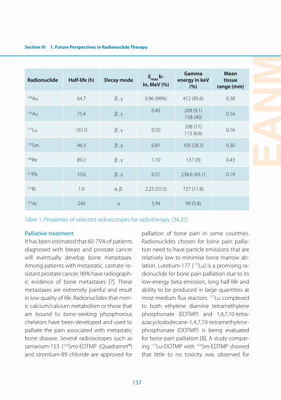
Section III 1. Future Perspectives in Radionuclide Therapy
EANM
137
Radionuclide Half-life (h) Decay modeE
max b-
In, MeV (%)
Gamma
energy in keV
(%)
Mean
tissue
range (mm)
198Au 64.7 β-, γ 0.96 (99%) 412 (95.6) 0.38
199Au 75.4 β-, γ0.45 208 (9.1)
158 (40)0.14
177Lu 161.0 β-, γ 0.50208 (11) 113 (6.6)
0.16
153Sm 46.3 β-, γ 0.81 103 (28.3) 0.30
186Re 89.2 β-, γ 1.10 137 (9) 0.43
212Pb 10.6 β-, γ 0.57 238.6 (43.1) 0.19
212Bi 1.0 α, β- 2.25 (55.5) 727 (11.8)
225Ac 240 α 5.94 99 (5.8)
Table 1: Properties of selected radioisotopes for radiotherapy [34,35]
Palliative treatment
It has been estimated that 60-75% of patients
diagnosed with breast and prostate cancer
will eventually develop bone metastases.
Among patients with metastatic, castrate-re-
sistant prostate cancer, 90% have radiograph-
ic evidence of bone metastases [7]. These
metastases are extremely painful and result
in low quality of life. Radionuclides that mim-
ic calcium/calcium metabolism or those that
are bound to bone-seeking phosphorous
chelators have been developed and used to
palliate the pain associated with metastatic
bone disease. Several radioisotopes such as
samarium-153 (153Sm)-EDTMP (Quadramet®)
and strontium-89 chloride are approved for
palliation of bone pain in some countries.
Radionuclides chosen for bone pain pallia-
tion need to have particle emissions that are
relatively low to minimise bone marrow ab-
lation. Lutetium-177 (177Lu) is a promising ra-
dionuclide for bone pain palliation due to its
low-energy beta emission, long half-life and
ability to be produced in large quantities at
most medium lux reactors. 177Lu complexed
to both ethylene diamine tetramethylene
phosphonate (EDTMP) and 1,4,7,10-tetra-
azacyclododecane-1,4,7,10-tetramethylene-
phosphonate (DOTMP) is being evaluated
for bone pain palliation [8]. A study compar-
ing 177Lu-DOTMP with 153Sm-EDTMP showed
that little to no toxicity was observed for
138
the 177Lu agent when administered to give
the same skeletal dose as the 153Sm agent,
indicating larger 177Lu doses could be given
that may result in longer remission times
[8]. Clinical trials are ongoing through the
International Atomic Energy Authority (IAEA)
coordination evaluating 177Lu-EDTMP. A scan
showing the bone uptake observed for 177Lu-
EDTMP giving a dose of 37 MBq/kg and im-
aged at 7 days is shown in Figure 1.
Figure 1: Planar image of a patient 7 days after receiving 37 MBq/kg of 177Lu-EDTMP for palliation
of pain due to metastatic bone cancer [35,37,38]
Radium-223 (223Ra; RaCl2) is an alpha-emit-
ting radioisotope that has been shown to
target areas of osteoblastic metastases. Un-
like the agents described above, which are
excreted very quickly (normally within 4 h)
via the kidneys, 223Ra is cleared primarily via
Section III 1. Future Perspectives in Radionuclide Therapy
EANM
139
the intestines [9]. This may be advantageous
in patients who have genitourinary cancers
or reduced kidney function. 223Ra, as an al-
pha emitter, delivers a very high dose to a
very small area. This may lead to a reduction
in some of the toxic side-efects observed
with the beta-minus emitters such as leuko-
penia, thrombocytopenia and bone marrow
suppression. In a large randomised phase III
trial, 223Ra has demonstrated improvements
in overall survival, a delay of skeletal-related
events (pathological fractures, spine cord
compression and requirements for surgery or
radiation to the bone) and biochemical pa-
rameters, with a remarkably tolerant adverse
event proile in men with castration-resistant
prostate cancer [9]. This has been the irst
agent to demonstrate improvement in over-
all survival, although it should be pointed out
this was never evaluated for the other agents.
Expanded trials are planned with this agent.
Peptides
The use of peptides and proteins such as
monoclonal antibodies to deliver cytotoxic
drugs has resulted in more selective treat-
ments and in huge advancements in treating
metastatic disease. CD20-targeted Bexxar®
labelled with iodine-131 (131I) and Zevalin®
labelled with yttrium-90 (90Y), routinely used
to treat follicular non-Hodgkin’s lymphoma,
have demonstrated the efectiveness of the
targeted approach. Large proteins such as
monoclonal antibodies demonstrate high
ainity, but due to their large sizes, sufer
from long residence times in the blood, slow
tumour penetration, dose-limiting toxicity to
the liver and bone and, in some patients, ad-
verse haematological efects and resistance.
It can take up to 7 days for these large pro-
teins to reach maximum uptake at the target
site, limiting the radionuclides that can be
used. A concerted focus has turned to evalu-
ating peptides for their smaller size and rapid
receptor targeting and blood clearance, abil-
ity to be easily synthesised and rapidly modi-
ied through solid phase and solution phase
peptide synthesis, and increased tumour
penetration and distribution; in addition,
they do not elicit immune responses. Thus
peptides are being developed as diagnostic
tools and biomarkers for cancer prognosis
and tumour targeting agents carrying ra-
dionuclides to cancer cells. Many are being
used for theranostic applications in which
they can be used both as an imaging char-
acterisation tool and as a delivery device for
radiotherapy radionuclides.
Peptide receptor radionuclide therapy (PRRT)
combines peptides with radionuclides to tar-
get receptors overexpressed on cancer cells.
Somatostatin analogues, the irst developed,
have been the most widely investigated.
Octreoscan was the irst radiolabelled pep-
tide approved for imaging neuroendocrine
tumours and is considered the gold stan-
dard for diagnosis of somatostatin receptor
positive tumours [10, 11]. Upon injection, the
radiolabelled octreotide attaches to soma-
tostatin receptors overexpressed on cancer
cells, thereby allowing an external SPECT
140
camera to image the location of the tumours
in the body. A number of somatostatin ana-
logues have been developed for imaging
and have been expanded to bind radionu-
clides for PRRT. These agents are composed
of a peptide conjugated to a bifunctional
chelator through a linker. The peptide acts as
the guiding system for selective delivery, the
bifunctional chelator serves to stably com-
plex the radioisotope, normally a metal, and
the linker serves not only for stable conjuga-
tion but also as a pharmacodynamic modii-
er. Changes to the linker can be made to ine
tune the uptake and clearance of the agent,
such as increasing the hydrophobicity if the
overall molecule proves to be too hydrophilic
and clears too quickly. The most commonly
used chelator has been DOTA (1,4,7,10-tetra-
azacyclotetradecane-N,N’,N”,N”’-tetra-acetic
acid) which forms kinetically and thermody-
namically stable complexes with a variety of
metals but sufers from having to be heated
at high temperatures for long periods of time
and promiscuous binding with all metals,
which can result in lowered speciic activ-
ity. Other chelators have been developed
that are more metal speciic and that can be
labelled at room temperature in high yields
and often negate the need for a terminal
puriication. The technetium-99m (99mTc) so-
matostatin analogue, NeoTect®, approved
in the United States, is used for the diferen-
tiation of malignant versus benign masses in
the lungs, often after initial disclosure with
CT or MRI. It difers from octreotide in that
the disulphide bond between the two Cys
is removed, enabling a bond between the
Phe and Cys of the cyclic hexapeptide tar-
geting vector. Hydrophobicity was added
by substituting a Tyr for a Val and linking to
a linear tetrapeptide chelator motif, which
serves to coordinate the Tc at the Dap-Lys-
Cys sequence (Dap= β-diaminopropionic
acid) shown in Fig. 2 [12, 13]. The chelation
involves three nitrogens (two amide nitrogen
atoms of Lys and Cys and one amine nitro-
gen of Dap) and one sulphur of Cys. 99mTc of-
fers better imaging quality than indium-111
(111In) and is more widely available. NeoTect®
itself has demonstrated higher ainity than
Octreoscan® for receptor subtypes 2 and 5
and slower clearance from normal tissues via
the kidneys, where it is excreted intact.
Section III 1. Future Perspectives in Radionuclide Therapy
EANM
141
Figure 2: Structure of the somatotastatin analogue for the drug NeoTect®
Signiicant work has gone into developing
analogues that could deliver therapeutic
doses. It was determined that replacement of
tyrosine with phenylalanine enhanced aini-
ty for the subtype 2 receptor, and that chang-
ing the chelate from DTPA to DOTA resulted
in a more stable complex that could be used
with 90Y, i.e. 90Y-DOTA-Tyr3-octreotide [14]. The
DOTA chelator is able to coordinate most +2
and +3 radiometals and remain stable in vivo.
This version is termed DOTATOC and, labelled
with 90Y, it has been used to treat hundreds
of patients. Toxicity in the kidneys led to the
development of DOTATATE, which has been
predominantly used with 177Lu. 177Lu has low-
er b- than 90Y and an imageable gamma emis-
sion at 208 keV that can be utilised to assess
post-therapy uptake and dosimetry. 177Lu has
also been used in a large number of patients
in Europe and is expected to enter clinical tri-
als in the United States within the next year.
Further studies have shown that using the
two radionuclides in combination results in a
more eicacious treatment than using either
alone. A small study in 50 patients in which
combined 90Y/177Lu-DOTATATE therapy was
evaluated in comparison to 90Y-DOTATATE
alone, applying 90Y/177Lu-DOTATATE in a ratio
of 1:1, demonstrated higher progression-
free survival in the tandem therapy group:
29.4 months vs 21.4 months [15]. Treatment
has also been enhanced by the use of PET
imaging with gallium-68 (68Ga) labelled to
DOTATOC, DOTATATE or the new analogues
NODAGATOC (DOTA has been replaced with
the NOTA chelator) and DOTANOC (NOC =
NaI3-octreotide, an sst2, sst3 and sst5 bind-
ing peptide), in which the peptide has been
further modiied. PET imaging with 68Ga al-
lows for the determination of the receptor
Cu
tler C
S. U
niv
ersi
ty o
f MO
142
density through the standardised uptake
value (SUV). Novel derivatives such as NODA-
GATOC made with the NOTA chelator have
allowed for room temperature synthesis in
high yields that have been converted to easy
kit formulations similar to those routinely
used for 99mTc.
PRRT and imaging have been extended to
looking at other peptides, such as bombesin,
that target the gastrin-releasing peptide re-
ceptor known to be over-expressed in breast,
prostate and lung tissues [16, 17]. A phase 1
clinical trial of 177Lu-DOTA- [4-aminobenzyl]-
BB (6–14) was undertaken [18]. Newer
bombesin versions have been developed,
such as the antagonists that demonstrate
less non-speciic uptake and higher receptor
ainity [19, 20]. Other peptides being evalu-
ated include those that target cholecystoki-
nin (CCK) receptors [21]. In addition to new
analogues, new radiometals have been eval-
uated, including scandium-44 (44Sc) for imag-
ing and bismuth-213 (213Bi) for therapy. The
longer half-life of 44Sc (4.4 h) allows for imag-
ing at later time points. 213Bi-DOTATOC was
evaluated and demonstrated higher thera-
peutic eicacy when compared to 177Lu-
DOTATOC in human adenocarcinoma cells.
Monoclonal antibodies
Targeted radioisotope therapy involves the
administration of radiolabelled compounds
to selectively damage and/or destroy dis-
eased tissue (e.g. cancer). The design of a
successful radiotherapeutic agent must
therefore involve selection of a compatible
combination of a targeting molecule, to de-
liver the radiolabelled compound with high
selectivity to site(s) of disease, and a radioiso-
tope, to cause damage/destruction to its lo-
cal environment through its energetic decay
properties. Radioimmunotherapy treatment
of cancer uses a class of compounds that
combine monoclonal antibodies (mAbs)
and particle-emitting radioisotopes. mAbs
target receptors either uniquely expressed
on cancer cells or expressed in signiicantly
higher numbers on cancer cells than on cells
of healthy tissue; this diference in receptor
expression allows for selective targeting of
diseased cells. Particle-emitting radioiso-
topes (i.e. Auger electron, beta and alpha
emitters) deposit their energy over short dis-
tances, thereby localising tissue damage. For
example, lymphomas have been successfully
treated with the RIT compounds Zevalin®
and Bexxar®, CD20 receptor-targeting mAbs
radiolabelled with beta-emitting radioiso-
topes. 90Y and 131I, respectively. Zevalin® and
Bexxar® exploit the long blood circulation
time of mAbs (large proteins) for targeting of
these blood-based cancers; however, the res-
idence time in blood (~7 days after injection)
limits the eicacy of RIT using mAbs for solid
tumour treatment. Prior to selective binding
of their receptor targets, the blood circula-
tion of conventional RIT compounds contrib-
utes to whole-body irradiation and results in
substantial radiation dose to the radiosensi-
tive bone marrow. Protecting the bone mar-
row from excessive radiation exposure limits
Section III 1. Future Perspectives in Radionuclide Therapy
EANM
143
the amount of radioactivity that can be safely
injected into a patient, consequently lower-
ing tumour doses and reducing therapeutic
eicacy.
To administer the highest possible radiation
dose to target tissue and the lowest possible
dose to non-target tissue, most of the decay
process should occur after the radiolabelled
compound has been delivered to the target
site and cleared from the blood and normal
tissues. Thus, RIT pretargeting methods have
been developed which decouple delivery
of the radioactivity from that of the mAb.
In brief, the mAb is conjugated with a tag
or artiicial receptor designed to bind with
high ainity to a small molecular probe. The
tagged mAb is injected and allowed sui-
cient time to reach maximum uptake at the
tumour and clearance from the blood. Then,
a small radiolabelled probe is injected that
binds rapidly and with high ainity to the
“receptor” on the pre-localised mAb, with the
remaining unbound fraction excreted rapidly
via the kidneys. The outcomes of pretarget-
ing methods are higher radiation dose deliv-
ery to the tumour (typically tenfold improve-
ment), increased therapeutic eicacy and
minimal dose and toxicity to normal tissues,
such as the bone marrow.
Most pretargeting strategies are based on
high-ainity biological recognition systems
(e.g. mAb/hapten and biotin/avidin), which
sufer from immunogenicity-inducing re-
sponses as they are recognised by the body’s
defence system. Such an immune response
precludes repeat or dose fractionation treat-
ments of solid tumours, which have demon-
strated maximum eicacy for some cancer
treatments, such as external beam therapy
and chemotherapy. A new pretargeting
method has been proposed that uses an
unusually rapid organic chemical reaction,
instead of biological components, to bind
the small radiolabelled probe to the tumour-
bound mAb with high ainity. Bertozzi and
others have developed biorthogonal reac-
tions using ring-strained cyclo-octene deriv-
atives for biological labelling. The approach is
based on the inverse electron demand Diels-
Alder reaction between a DOTA-tetrazine
analogue (Fig. 3), which demonstrates high
thermodynamic and chemical stability with
most metals, and a trans-cyclo-octene (TCO)
(Fig. 3) conjugated to the murine mAb CC49
[22]. This reaction was chosen based on its ex-
ceptionally high second-order rate constant
of 13,090 ± 80 M1s-1 at 37°C in phosphate-
bufered saline [22]. CC49 has high ainity to
TAG-72, an antigen with limited internalisa-
tion and shedding that is over-expressed in
a wide range of solid tumours, such as colon
cancer [23, 24]. It is hypothesised that, by
moving away from biological pretargeting
components and instead using an orthogo-
nal chemical reaction, it may be possible to
avoid an immune response to allow repeat
and/or fractionated treatments and thereby
improve therapeutic eicacy.

144
1
2: R=NHS
Figure 3: Structures of bio-orthogonal pretargeting molecules tetrazine DOTA (1) and trans-
cyclo-octene TCONHS (2) 36
Initial in vivo proof-of-principle imaging stud-
ies were performed in mice bearing colon
cancer xenografts that were administered
100 µg of TCO-CC49 followed one day later
with 3.4 equivalents of 111In-DOTA-tetrazine
(40.7 MBq) [22] . The animals were imaged
3 h later, and the tumour was shown to have
a 4.2% ID/gram uptake and a 13:1 tumour to
muscle ratio. Besides the tumour, the bulk of
the activity was found in the bladder, a small
amount in the kidneys and some residual in
the liver and blood that was attributed to
circulating TCO-CC49. Control studies with
unmodiied CC49 and TCO-modiied ritux-
imab, a mAb with no TAG-72 ainity, showed
no tumour uptake and supported the an-
tigen-speciic binding of TCO-CC49 with
111In-DOTA-tetrazine. Following such prom-
ising results, the TCO-CC49/DOTA-tetrazine
pretargeting method was further improved
(e.g. for dosing and timing of radiolabelled
probe) and evaluated against conventionally
labelled DOTA-CC49 with the therapeutic
radioisotope 177Lu [25]. Using the biodistribu-
tion data obtained, the estimated maximum
deliverable dose to the tumour without be-
ing lethal to bone marrow was nearly ive
times higher with the pretargeting method
at 80 Gy than the sub-therapeutic dose of
17 Gy for the conventional labelling meth-
od. If such an approach works out, it could
change the way we currently use antibodies
and also give greater eicacy.
Cu
tler C
S. U
niv
ersi
ty o
f MO
Section III 1. Future Perspectives in Radionuclide Therapy
EANM
145
Nanoparticles
A disadvantage of the conventional bi-
functional chelating system is that, at
most, only one radioactive molecule per
targeting moiety is delivered. This is highly
dependent on the specific activity of the
radioisotope and on any metal contami-
nants, as many chelators are not selective
but will bind to a number of common met-
als. A low specific activity and/or high pres-
ence of unwanted metal impurities can
result in low labelling yields for the desired
radioisotope and saturation of the receptor
sites if they are present in low quantities.
This has been recently demonstrated by
Asti et al. [26], who performed a system-
atic study evaluating the effect of specific
activity and incorporation of a variety of
metals on a typical DOTATOC clinical for-
mulation. Furthermore, it is often not pos-
sible to design a chelator that can stably
complex a metal such as an alpha emitter
and its daughters upon decay. The release
of the daughter can result in unwanted ra-
diation dose to normal tissues and further
decrease the effectiveness of the adminis-
tered dose. For example, DOTA is known to
form strong complexes with 213Bi and lead-
212 (212Pb); however, decay of 212Pb results
in more than a 35% release of 213Bi. Rose-
now was the first to investigate using lipo-
somes to stably encapsulate 212Pb and its
daughters [26]. It has been demonstrated
that 10-nm liposomes result in the reten-
tion of 213Bi after the decay of 212Pb [27].
Liposomes have been radiolabelled either
by incubating the liposome with the radio-
nuclide of interest, allowing it to incorporate
into the lipid bilayer, or by attachment of
chelators to the surface, such as DTPA ana-
logues and other familiar chelators. Both of
these methods sufer from signiicant loss
of the radionuclides and modiication to the
surface, adversely impacting targeting vec-
tors. To overcome this, another method has
been developed in which the radiometal is
irst chelated to a lipophilic chelator, which
then crosses the bilayer, resulting in entrap-
ment of the radiometal in the aqueous core.
Examples include the use of oxime to label 111In and hexamethylpropylene oxime to
incorporate 99mTc. Rhenium-186 (186Re) has
been incorporated using the chelator N,N-
bis(2-mercaptoethyl-N,N-diethylenediamine,
which reacts with encapsulated cysteine
to secure the 186Re within the core [28].
Fullerenes have been evaluated both in the
spherical coniguration commonly referred
to as Bucky balls and as carbon nanotubes.
Their surface allows for the conjugation of
targeting vectors, while the inside serves to
safeguard the radioisotope from interact-
ing with the biological milieu, thus ofering
the potential of stably retaining and deliv-
ering the radionuclide, something that has
not been possible through standard chela-
tion. Ultra-short single-walled carbon nano-
tubes (SWNT) were irst tested illed with
iodine-125 (125I). The 125I was loaded and then
oxidised to 125I2, which demonstrated long re-
tention. The carbon nanotubes were directed
146
to the lung and no uptake was observed in
the thyroid, which is where free iodine in the
body is known to collect. This method was
also shown to deliver a 10 times higher dose
per gram of tissue than is obtainable with
other methods.
There is considerable interest in using al-
pha emitters for treating micrometastases
or single cell leukaemias because the path
length of the typically used beta-minus par-
ticle emitters is hundreds of cell diameters,
resulting in damage to healthy normal tissue.
With their high LET, alpha emitters are more
lethal over a shorter range — it is estimated
that only one to three alpha emissions are re-
quired to kill a cell versus thousands for beta
emitters. Astatine-211 (211At) is a prospective
radionuclide for therapy owing to its 7.2-h
half-life and its ability to be imaged by SPECT
for post-therapy distribution and dosimetry;
furthermore, it can be produced on a cyclo-
tron. Astatine, however, has proven to be dif-
icult to complex as carbon–astatine bonds
are weak and result in dehalogenation in vivo.
Consequently, carboranes and nanoparticles
have been evaluated [9,10]. Two diferent
types of nanoparticle have been assessed:
silver nanoparticles covalently coated with
poly(ethylene oxide) and SWNT. Reducing
conditions with silver nanoparticles resulted
in high labelling yields. SWNT nanoparticles
were labelled with 211At- in the same man-
ner as the 125I-; initially the anion was loaded
and, as with the I, signiicant leakage was
reported; however, upon oxidation with
chloramine-T or N-chlorosuccinimide, the
AtCl molecules were more highly retained,
showing labelling yields of 78–93%; these
yields are much higher than those previously
reported for corborane derivatives, which
have demonstrated the highest incorpora-
tion, 40–60%. Incubation in water showed
retention of 72–85% of the At in the SWNT
and 85–93% retention in serum, indicating
that these may well serve as stable delivery
systems for At.
The alpha emitter actinium-225 (225Ac) holds
much promise for targeted alpha therapies
as an in vivo generator. The nuclear decay of 225Ac (10-day half-life with the emission of
four alpha particles) holds the potential to
become an ideal radiotherapy system. The
10-day half-life provides suicient time for
complex syntheses and attachment to tar-
geting biomolecules, while the rapid decay
and high LET of the daughters’ alpha emis-
sions ensure that the therapeutic payload is
delivered in the vicinity of the target tissue.
Despite the clear beneits of delivering mul-
tiple alpha particles from the decay of 225Ac
directly to tumour sites, the diiculty of se-
questering the alpha-emitting daughters in
traditional bifunctional-conjugated targeting
modalities leads to release of the daughters
and non-speciic uptake in non-target or-
gans. This non-target dose negates the ad-
vantages of targeted alpha therapy, namely
high tumour dose with minimal collateral
damage to healthy tissue. In order to harness
the potential of multiple alpha particles from
Section III 1. Future Perspectives in Radionuclide Therapy
EANM
147
225Ac, nanoparticles are being studied as a
way to retain the decay products for delivery
to the target site. Initially a multivesicular li-
posomal carrier was evaluated. The nanopar-
ticles were further tagged with a vector for
anti-HER2/neu and evaluated in an ovarian
cell line. While internalisation was at a high
rate, however, retention of the 225Ac was not
as high as needed. A lanthanum phosphate
nanoparticle (LaPO4 NP) evaluated in vivo
showed higher retention, and upon complex-
ation with an antibody to lung endothelium,
high retention in mouse lung tissue [29]. This
design has been further modiied to include
a gold coating and incorporation of gado-
linium into the lanthanum gadolinium phos-
phate nanoparticles (NPs) for the purpose of
retaining both 225Ac and its daughters in an
attempt to increase retention. The magnetic
gadolinium simpliied the separation and
puriication chemistry. Transmission electron
microscopy analysis of LaGdPO4-AuNPs par-
ticles showed monodisperse particles with
average diameters of 4–5 nm. Radiochemical
analysis indicated that LaGdPO4-AuNPs with-
out additional layers sequestered 60.2±3.0%
of the irst decay daughter of 225Ac, fran-
cium-221 (221Fr). The addition of two shells
of LaGdPO4 and one shell of Au increased
221Fr retention to 69.2±1.7%, while the addi-
tion of four shells of GdPO4 and one shell of
Au increased retention to 92±1.0%. Reten-
tion of the irst decay daughter is essential
to minimising normal tissue toxicity. In vivo
distribution of the nanoparticles conjugated
to a monoclonal antibody that targets lung
endothelium showed that the nanoparticles
exhibited high lung uptake (151%ID/g) [30].
Additionally, the uptake could be blocked by
administering cold unlabelled antibody: up-
take dropped to 16.8%ID/g. After 24 h the ac-
tivity was transferred to the liver and spleen
due to the reticuloendothelium system. This
can be overcome by using clodronate lipo-
somes to reduce reticuloendothelial func-
tioning. The properties of low toxicity and
favourable biodistributions make the LaGd-
PO4-AuNP system a promising platform for
targeted alpha therapy with 225Ac.
A major challenge in cancer therapy has been
delivery and retention. Due to their size and
ability to circumvent some of the hurdles en-
countered with traditional agents, nanopar-
ticles are promising alternatives. They have
unique properties that can be optimised to
allow for higher penetration and retention
in tumour cells. Unlike traditional medicine,
the ield of nanomedicine utilises particulate
matter with sizes that are the same or smaller
than those of cellular components, allowing
nanoparticles to cross multiple biological
barriers to enhance delivery and retention
for optimal treatment. The size similarity to
cellular components makes nanoparticles at-
tractive candidates for curing functional ab-
normalities that instigate disease at the cellu-
lar level. Current approaches result in serious
adverse side-efects that signiicantly limit
the number of therapeutic molecules that
can be delivered to the tumour site, resulting
in low eicacy. Efective tumour treatment
148
requires a high-impact therapeutic payload
to destroy cancer cells. Due to the enhanced
permeability of tumour cells, nanoparticles
can be sized such that they are selectively
taken up and retained in cancer cells, this
being called the enhanced permeability and
retention (EPR) efect [31]. Control over the
supply of therapeutic payload enables on-
cologists to deliver an optimised efective
treatment for cancer patients. In this regard,
it is important to note that nanoparticulates
containing radioactive isotopes provide an
opportunity to tune the radioactive thera-
peutic dose delivered to tumour cells.
The conventional method for delivering
radioisotopes has been either to complex
them to a targeting molecule for selective
delivery, or to form a chemical complex with
in vivo properties that will efect uptake at
the tumour site. Although the conventional
method has produced successful treatments,
not all metals can be stably complexed by
simple ligands. Some, such as gold and sil-
ver, have no stable chelation methods; in vivo
injection results in their release. Others, such
as many of the alpha emitters, are released
from the compound upon decay and there-
fore cause signiicant toxicity, limiting their
usefulness. The irst nanoparticles developed
and translated to the clinic were liposomes.
Comprising various platforms, they basi-
cally consist of a lipid outer sphere and an
aqueous core, which allows the trapping of
hydrophobic drugs in the lipid layer and of
hydrophilic drugs in the aqueous inner core.
Targeting vectors can be attached to the
surface to aid in localisation and recognition,
as well as ligands for complexing diagnostic
radionuclides.
Radioisotopes of gold and other coinage
metals have nuclear properties that make
them ideal for use in imaging and therapeu-
tic applications. However, their biologically
active redox chemistry and lack of stable
chelating agents have to date limited their
utilisation in vivo. They have had application
in the area of brachytherapy with the bare
metal being encapsulated, but otherwise
have been used sparingly, the major concern
being the inherent toxicities observed for the
free metals in vivo. A novel approach is the
development of metal nanoparticles that
can be derivatised on their surface, allowing
for optimisation of biodistribution in vivo.
Once a metal nanoparticle is fabricated, its
in vivo properties can be evaluated and al-
tered by varying such properties as its size,
charge, hydrodynamic size, organic surface
layer and shape. These can be optimised to
enable more favourable uptake and clear-
ance in vivo. Furthermore, owing to their
high surface area, they can be conjugated
with a plethora of agents that can be used
for selective targeting, such as peptides and
small molecules, to enhance retention in
the bloodstream or to enhance clearance.
These properties can be optimised through
in vivo studies to develop lead compounds
for imaging and therapy. A major advan-
tage of nanoparticles over the commonly
used bifunctional chelator approach is that
Section III 1. Future Perspectives in Radionuclide Therapy
EANM
149
the nanoparticle platform allows for a much
higher payload delivery, as more than one
radioisotope can be incorporated into the
nanoparticle, producing a much higher dose
delivery even with a low speciic activity ra-
dionuclide.
Radioisotopes of gold (Au) as shown in
Table 1 have attractive nuclear proper-
ties that make them ideal for imaging and
therapy. Two radioisotopes of gold stand
out. 198Au has a moderate enegry beta par-
ticle (0.96 keV β- max), making it a good
candidate for therapy, and more than 90%
emission of a 412-keV gamma emission that
allows for in vivo tracking and dosimetry cal-
culation. These properties prompted its use
as a brachytherapy agent for prostate cancer. 199Au has a low-energy beta max (0.46 MeV)
and emits a 158-keV gamma in 36% yield
that is easily detected by SPECT cameras.
Additionally, 199Au can be produced carrier-
free by neutron irradiation of platinum-198,
which produces platinum-199, which decays
in 3.2 min to 199Au.
FGold nanoparticles (AuNPs) have been
around for quite some time, but their harsh
production methods with toxic chemicals
have prevented them from being attached
easily to biomolecules for evaluation in vivo.
An improved synthesis has been developed
that uses THPAL, a trimeric phosphino ala-
nine, to reduce gold salts in water-containing
organics, which coat the surface of the gold
nanoparticles and form 12- to 15-nm-sized
gold nanoaprticles with a hydrodynamic
diameter of 60–85 nm [32]. These methods
have been shown to be robust and non-toxic
in the non-radioactive form. The methods
have been altered to allow for formation of
both 198Au and 199Au nanoparticles and also
have been shown to permit formation of
nanoparticles of other coin metals such as
palladium and silver.
Gum arabic-coated gold nanoparticles (GA-198AuNPs) have been the most studied to
date [33]. This formulation was initially evalu-
ated for therapeutic eicacy in SCID mice
bearing induced human prostate tumours.
Diferent modes of injection were tried,
with the intratumoral injection resulting in
the highest uptake and retention. The par-
ticles were then evaluated for their eicacy
in tumour stabilisation in the same tumour
models. Tumours received a 70 Gy dose by
administering 15 MBq. An overall 82% reduc-
tion in tumour volume was noted between
the mice receiving the GA-198AuNP com-
pared to controls receiving saline injections.
Biodistributions at the end showed a reduc-
tion of gold nanoparticles in the tumour,
from 70% at 24 h to 19.9%±4.2% ID at 30 days
post injection. Clearance was predominantly
through the urinary tract and minimum up-
take was observed in other organs. Based on
these results, these nanoparticles are now
being evaluated for their therapeutic eicacy
in dogs with spontaneous prostate cancer.
Prostate cancer in dogs has been shown to
mimic that observed in humans on the func-
tional level as well as in metastatic rate and
occurrence. A phase 1 study in dogs starting
150
at 50 Gy and increasing to 105 Gy has shown
no toxicity issues in the dogs and up to a 60%
reduction in tumour volume. Based on these
results, these nanoparticles are being evalu-
ated in other solid tumours and are being
considered for a phase 1 human study.
Conclusion
An expanding role for radiotherapy is unfold-
ing through the recent advances presented
here. These novel delivery methods are giv-
ing rise to even higher doses to cancer cells
while vastly minimising the dose to sur-
rounding normal tissues. These methods are
providing more lexibility in the use of radio-
nuclides on the one hand and increasingly
personalised treatments regimens on the
other. These novel delivery systems are going
to make radiotherapy a more exciting option
for patients and their physicians.
EANM
151
References1. Enback J, Laakkonen. Tumour-homing peptides: tools
for targeting, imaging and destruction. Biochem Soc Trans. 2007;35(4):780-3.
2. Dorsam RT. G-protein-coupled receptors and cancer. Nature Reviews Cancer. 2007;7(2):79-94.
3. Aina OT, Stroka TC, Chen ML, Lam KS. Therapeutic cancer targeting proteins. Biopolymers. 2002;66(3):184-99.
4. Meng L, Yang L, Zhao X, Zhang L, Zhang H, al. E. Tar-geted delivery of chemotherapy agents using a liver cancer-speciic apatamer. PLoS ONE. 2012;7(4):Article ID e33434.
5. Zhang XX, Eden HS, Chen X. Peptides in cancer nano-medicine: drug carriers, targeting ligands and protease substrates. J Control Release. 2012;159:2-13.
6. Thundimadathil J. Cancer treatment using peptides: cur-rent therapies and future prospects. Journal of Amino Acids. 2012:Article ID 967347, 13 pages.
7. Petrylak DP, Tangen CM, Hussain MH, al. E. Docetaxol and estramustine compared with mitroxanthine and prednisone for advanced refractory prostate cancer. N Engl J Med. 2004;351(15):1513-20.
8. Bryan JN, Bommarito D, Kim DY, Berent LM, Bryan ME, Lattimer JC, et al. Comparison of systemic toxicities of 177Lu-DOTMP and 153Sm-EDTMP administered intrave-nously at equivalent skeletal doses to normal doses. J Nucl Med Technol. 2009;37(1):45-52.
9. Harrison MR, Wong TZ, Armstrong AJ, George DJ. Radium-223 chloride: a potential new treatment for castration-resistant prostate cancer patients with meta-static bone disease. Cancer Management and Research. 2013;5:1-14.
10. Ruini V, Calcagni ML, Baum RP. Imaging of neuroendo-crine tumours. Semin Nucl Med. 2006;36(3):228-47.
11. Virgolini I, Traoub T, Novotny, al. E. Experince with In-111 and Ytrrium-90 labeled somatostatin analogs. Curr Pharm Des. 2002;8(20):1781-807.
12. Cantorias MC, Howell RW, Todaro L, al. E. MO tripeptide diasteriomers (M=99/99mTc,Re): models to identify the structure of 99mTc peptide targeted radiopharmaceu-ticals. Inorg Chem. 2007;46:7326-40.
13. Cyr J, Pearson D, Nelson C, al. E. Isolation, characteriza-tion, and biological evaluation of syn and anti diasteri-omers of [99mTc]Technetium depreotide: a somatostatin receptor binding tumor imaging agent. J Med Chem. 2007;50:4295-300.
14. Heppeler A, Froidevaux S, Macke HR, Jermann E, Behe M, Powell P, et al. Radiometal-labelled macrocyclic chelator-derivatized somatostatin analogue with su-perb tumour-targeting properties and potential for receptor-mediated internal radiotherapy. Chem--Eur J. 1999;5(7):1974-81.
15. Kunikowska J, Krolicki L, Hubalewska-Dydejczyk, Miko-lajczak R, Sowa-Staszccak A, Pawlak D. Clinical results of radionuclide therapy of neuroendocrine tumours with 90Y-DOTATE and tandem 90Y/177Lu-DOTATATE: which is a better therapy option. Eur J Nucl Med Mol Imaging. 2011;38:1788-97.
16. Van de Wiele C, Phonteyne P, Pauwels P, al. E. Gastrin-releasing peptide receptor imaging in human breast carcinoma versus immunohistochemistry. J Nucl Med. 2008;49(2):260-4.
17. Zhang H, Chen J, Waldherr C, al. E. Synthesis and evaluation of bombesin derivatives on the basis of pan-bombesin peptides labeled with Indium-111, Lutetium-177, and Yttrium-90 for targeting bombe-sin receptor-expressing tumours. Cancer Res. 2004;64(18):6707-15.
18. Schroeder RP, van Weerden WM, Bangma C, Krenning EP, De Jong M. Peptide receptor imaging of prostate can-cer with radiolabeled bombesin analoques. Methods. 2009;48:200-4.
19. Cescato R, Maina T, Nock B, Nikolopoulou A, Charalam-didis D, al. E. Bombesin receptor antagonists may be preferable to agonists for tumor targeting. J Nucl Med. 2008;49(2):318-26.
20. Mansi R, Wang X, Forrer F, Waser B, Cescato R, Graham K, et al. Development of a potent DOTA-conjugated bombesin antagoinst for targeting GRPr-positive tu-mors. Eur J Nucl Med Mol Imaging. 2011;38:97-107.
21. Mather SJ, McKenzie AJ, Sosabowski JK, Morris TM, El-lison D, Watson SA. Selection of radiolabeled gastrin analogs for peptide receptor-targeted radionuclide therapy. J Nucl Med. 2007;48:615-22.
References Section III, Chapter 1
152
22. Rossin R, Renart Verkerk P, van den Bosch SM, Vulders RCM, Verel I, Lub J, et al. In vivo chemistry for pretar-geted tumor imaging in live mice. Angew Chem, Int Ed. 2010;49(19):3375-8, S/1-S/22.
23. Muraro R, Kuroki M, Wunderlich D, Poole DJ, Colcher D, Thor A, et al. Generation and characterization of B72.3 second generation monoclonal antibodies reactive with the tumor-associated glycoprotein 72 antigen. Cancer Res. 1988;48(16):4588-96.
24. Schlom J, Eggensperger D, Colcher D, Molinolo A, Houchens D, Miller LS, et al. Therapeutic advantage of high-ainity anticarcinoma radioimmunoconjugates. Cancer Res. 1992;52(5):1067-72.
25. Rossin R, Sandra M, van den Bosch SM, Robillard MS, edi-tors. Tumor pretargeting with retro-Diels-Alder reaction: biodistribution and mouse dosimetry. World Molecualr Imaging Congress; 2011 September 7-10th, 2011; San Diego, CA.
26. Rosenow MK, Zucchini GL, Bridwell PM, Stuart FP, Fried-man AM. Properties of liposomes containing Lead-212. Int J Nucl Med Biol. 1983;10(4):189-97.
27. Le Du A, Mougin-Degraef M, Botosoa E, Rauscher A, Chauvet AF, Barbet J, et al. In vivo 212Pb/212Bi generator using indium-DTPA-tagged liposomes. Radiochim Acta. 2011;99(11):743-9.
28. Bao A, Goins B, al. E. 186Re-liposome labeling us-ing 186Re-SNS/S complexes in vitro stability, imag-ing, and biodistribution data in rats. J Nucl Med. 2003;44(12):1992-9.
29. Woodward J, Kennel SJ, Stuckey A, Osborne D, Wall J, Rondinone AJ, et al. LaPO4 nanoparticles doped with Actinium-225 that partially sequester daughter radio-nuclides. Bioconjug Chem. 2011;22(4):766-76.
30. McLaughlin M, Woodward J, Boll RA, Wall J, Rondinone SJ, Mirzadeh S, et al. Gold coated lanthanide phosphate nanoparticles for targeted alpha generator radiothera-py. PLoS ONE. 2013;8(1):e54531.
31. Noguchi Y, Wu J, al. E. Early phase tumor accumulation of macromolecules: a great diference in clearnace rate between tumor and normal tissues. Japanese Journal of Cancer Research: Gann. 1998;89(3):307-14.
32. Kannan R, Rahing V, Cutler C, Pandrapragada R, Katti KK, Kattumuri V, et al. Nanocompatible chemistry toward fabrication of target-speciic gold nanoparticles. J Am Chem Soc. 2006;128(35):11342-3.
33. Chanda N, Kan P, Watkinson LD, Shukla R, Zambre A, Carmack TL, et al. Radioactive gold nanoparticles in can-cer therapy: therapeutic eicacy studies of GA-198AuNP nanoconstruct in prostate tumor-bearing mice. Nano-medicine (Philadelphia, PA, U S). 2010;6(2):201-9.
34. Radioisotopes and radiochemicals. 2013 [cited 2012 October 1]; Available from: http://www.murr.missouri.edu/ps_radio.php.
35. Cutler CS, Hennkens HM, Sisay N, Huclier-Markai S, Ju-risson Silvia S. Radiometals for combined imaging and therapy. Chem Rev. 2013;113:858-83.
36. Cutler CS. Structures if bio-orthogonal pretargeting molecules tetrazine DOTA (1) and trans-cyclo-octene TCONHS (1). 2013.
37. Trindade V, Paolino A, Rodríguez G, Coppe F, Fernán-dez M, Lopez A, et al. 177Lu-EDTMP: Primera experiencia en Uruguay para terapia paliativa de metástasis óseas. Alasbimn Journal. 2009;46.
38. Trindade V, Rodríguez G, Cabral P, Fernández M, Paolino A, Gaudiano J, et al. 177Lu-EDTMP: Radiofármaco para terapia paliativa del dolor producido por Metástasis óseas. Alasbimn Journal. 2009;43.
39. Cutler CS. Structure of the somatostatin analogue for the drug Neotect. 2013.

EANM
153
Notes
154
Imprint
ISBN: 978-3-902785-08-4
Publisher:
European Association of Nuclear Medicinec/o vereint: Association & Conference Management Ltd.Hollandstrasse 14/Mezzanine, 1020 Vienna, AustriaPhone: +43-(0)1-212 80 30 | Fax: +43-(0)1-212 80 30-9Email: [email protected] | URL: www.eanm.org
Editors:
Claudiu Peştean, Vanessa Veloso Jerónimo, Peter Hogg
English Language Editing:
Rick Mills
Project Management:
Katharina Leissing
Content:
No responsibility is taken for the correctness of this information.Information as per date of printing September 2013. EANM Association and Congress Managementvereint: Association & Conference Management Ltd.Hollandstrasse 14/Mezzanine, 1020 Vienna, AustriaPhone: +43-(0)1-212 80 30 | Fax: +43-(0)1-212 80 30-9Email: [email protected] | URL: www.eanm.org
Layout and Design:
kreativ · Mag. Evelyne Sacher-ToporekLinzer Strasse 358a/1/7, 1140 Vienna, AustriaPhone: +43-(0)1-416 52 27 | Fax: +43-(0)1-416 85 26Email: [email protected] | URL: www.kreativ-sacher.at
Printing:
Print&Smile GmbHHubert JaudMichael-Rottmayr-Straße 46, 5110 Oberndorf, AustriaPhone: +43-(0)6272-7135-456 | Fax: +43-(0)6272-7135-499Email: [email protected]
European Association of Nuclear Medicine
®EANMEANM
vereint Ltd. is licensee of the Austrian Eco-label for
„Green Meetings and Green Events“
LizenznehmerUmweltzeichen
Green Meetings & Green Events
Österreichisches