Embed Size (px)
Citation preview
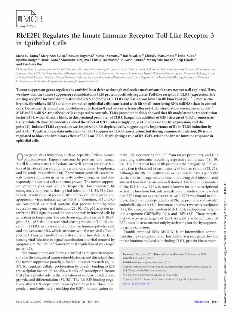
Rb/E2F1 Regulates the Innate Immune Receptor Toll-Like Receptor 3in Epithelial Cells
Manabu Taura,a Mary Ann Suico,b Kosuke Koyama,b Kensei Komatsu,b Rui Miyakita,b Chizuru Matsumoto,b Eriko Kudo,a
Ryusho Kariya,a Hiroki Goto,a Shunsuke Kitajima,c Chiaki Takahashi,c Tsuyoshi Shuto,b Mitsuyoshi Nakao,d Seiji Okada,a
and Hirofumi Kaib
Division of Hematopoiesis, Center for AIDS Research, Kumamoto University, Kumamoto, Japana; Department of Molecular Medicine, Graduate School of PharmaceuticalSciences, Global COE Cell Fate Regulation Research and Education Unit, Kumamoto University, Kumamoto, Japanb; Division of Oncology and Molecular Biology, Cancerand Stem Cell Research Program, Cancer Research Institute, Kanazawa University, Kanazawa, Japanc; and Department of Medical Cell Biology, Institute of MolecularEmbryology and Genetics, Kumamoto University, Kumamoto, Japand
Tumor suppressor genes regulate the antiviral host defense through molecular mechanisms that are not yet well explored. Here,we show that the tumor suppressor retinoblastoma (Rb) protein positively regulates Toll-like receptor 3 (TLR3) expression, thesensing receptor for viral double-stranded RNA and poly(I·C). TLR3 expression was lower in Rb knockout (Rb�/�) mouse em-bryonic fibroblasts (MEF) and in mammalian epithelial cells transfected with Rb small-interfering RNA (siRNA) than in controlcells. Consequently, induction of cytokines interleukin-8 and beta interferon after poly(I·C) stimulation was impaired in Rb�/�
MEF and Rb siRNA-transfected cells compared to controls. TLR3 promoter analysis showed that Rb modulates the transcriptionfactor E2F1, which directly binds to the proximal promoter of TLR3. Exogenous addition of E2F1 decreased TLR3 promoter ac-tivity, while Rb dose dependently curbed the effect of E2F1. Interestingly, poly(I·C) increased the Rb expression, and thepoly(I·C)-induced TLR3 expression was impaired in Rb-depleted cells, suggesting the importance of Rb in TLR3 induction bypoly(I·C). Together, these data indicated that E2F1 suppresses TLR3 transcription, but during immune stimulation, Rb is up-regulated to block the inhibitory effect of E2F1 on TLR3, highlighting a role of Rb-E2F1 axis in the innate immune response inepithelial cells.
Oncogenic virus infections, such as hepatitis C virus, humanpapillomavirus, Kaposi’s sarcoma herpesvirus, and human
T-cell leukemia virus 1 infections, are well-known causative fac-tors of hepatocellular carcinoma, cervical carcinoma, lymphoma,and leukemia, respectively (30). These tumorigenic viruses atten-uate tumor suppressor genes, activate proto-oncogenes, and con-sequently induce host cell abnormal growth. The tumor suppres-sor proteins p53 and Rb are frequently downregulated byoncogenic viral proteins during viral infection (11, 34, 51). Con-versely, reactivation of p53 and Rb induces cell cycle arrest andapoptosis in virus-induced cancer (43, 61). Therefore, p53 and Rbare considered as critical proteins that prevent tumorigenesiscaused by oncogenic viral infection (21, 30, 47). p53 activates in-terferon (IFN) signaling and induces apoptosis in infected cells byactivating its target gene, the interferon regulatory factor 9 (IRF9)gene (36). p53 also increases viral sensing molecule Toll-like re-ceptor 3 (TLR3) expression and function in human epithelial cellsand mouse tissues (48), which correlates with the antiviral effect ofp53 (35). Thus, p53 multiply regulates antiviral host defense, fromsensing viral infection to signal transduction and viral removal byapoptosis, at the level of transcriptional regulation of p53 targetgenes (42).
The tumor suppressor Rb was identified as the protein respon-sible for the congenital tumor retinoblastoma, and this establishedthe tumor suppressor paradigm for Rb in cancer research (8, 17,37). Rb regulates cellular proliferation by directly binding to E2Ftranscription factors (9, 16, 45), a family of transcription factorsthat play a pivotal role in the regulation of cellular proliferation,growth, and differentiation (39, 58). The Rb-E2F binding nega-tively affects E2F-dependent transcription by at least three inde-pendent mechanisms: (i) masking the E2F’s transactivation do-
main, (ii) sequestering the E2F from target promoters, and (iii)recruiting chromatin-modifying repressive complexes (18, 19,23). The functional loss of Rb promotes the deregulated E2F ac-tivity that is observed in vast majority of human tumors (22, 25).Although the Rb-E2F pathway is well known to have a generallycrucial role in oncogenesis, its functions during viral infection andantiviral host defense are not well studied. The founding memberof the E2F family, E2F1, is mostly known for its transcriptionalactivating functions but, intriguingly, recent studies have revealedthat E2F1 may act as a repressor as well. E2F1 was found to sup-press, directly and independently of Rb, the promoters of vascularendothelial factor A (31), human telomerase reverse transcriptase(12), the antiapoptotic protein Mcl-1 (13), endoplasmic reticu-lum chaperone GRP78/Bip (41), and IRF3 (55). These surpris-ingly diverse gene targets of E2F1 revealed a wide influence ofE2F1 on cellular events not only by activating but also by suppress-ing gene expression.
Double-stranded RNA (dsRNA) is an intermediate compo-nent during viral replication in host cells that is recognized by hostinnate immune molecules, including TLR3, protein kinase recep-
Received 19 October 2011 Returned for modification 22 November 2011Accepted 25 January 2012
Published ahead of print 6 February 2012
Address correspondence to Hirofumi Kai, [email protected], orSeiji Okada, [email protected].
M.T. and M.A.S. contributed equally to this article.
Copyright © 2012, American Society for Microbiology. All Rights Reserved.
doi:10.1128/MCB.06454-11
0270-7306/12/$12.00 Molecular and Cellular Biology p. 1581–1590 mcb.asm.org 1581
Dow
nloa
ded
from
http
s://j
ourn
als.
asm
.org
/jour
nal/m
cb o
n 09
Feb
ruar
y 20
22 b
y 21
3.10
8.16
1.35
.
tor (PKR), RIG-I, and MDA5 (32). These sensing molecules in-duce immune responses such as cytokine and chemokine produc-tion, which is an integral part in the initial action of antiviral hostdefense in both innate and adaptive immunity (1, 5). TLR3 andPKR transcription is induced by p53, which initiates the signalingcascade of antiviral host response (48, 59). Adversely, viral dsRNAsuppresses p53 expression (29). These reports highlighted the cor-relation between tumor suppressor genes and dsRNA sensing. Inthe present study, we found that Rb, via E2F1, also critically im-pacts on TLR3 expression and signaling in epithelial cells. Rbknockout mouse embryonic fibroblasts (MEF), as well as cellstransfected with Rb small interfering RNA (siRNA), showed de-creased TLR3 expression. On the other hand, E2F1 negatively reg-ulated TLR3 expression in epithelial cells. TLR3 ligand poly(I·C)-induced cytokine response was attenuated in the absence of Rb.Moreover, we found that poly(I·C) stimulation increased Rb, butnot p53 expression, and inhibited E2F1 recruitment to TLR3proximal promoter region, resulting in increased TLR3 transcrip-tion. Based on our findings, we identified Rb as a novel TLR3regulator that affects viral host defense.
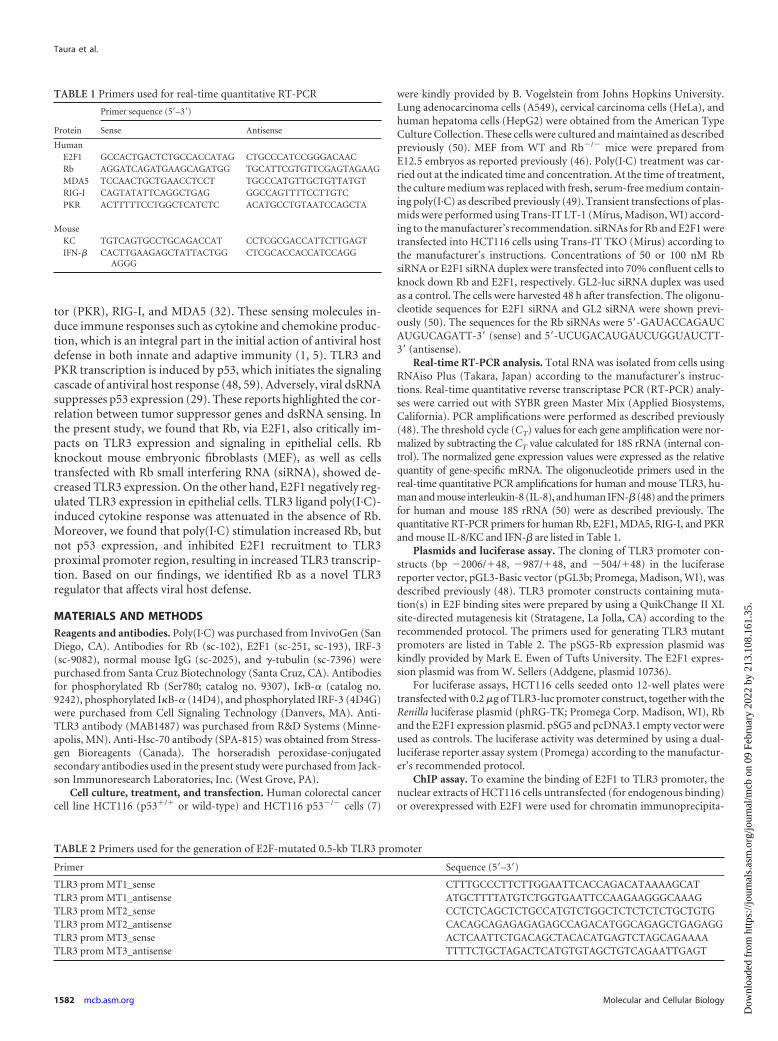
MATERIALS AND METHODSReagents and antibodies. Poly(I·C) was purchased from InvivoGen (SanDiego, CA). Antibodies for Rb (sc-102), E2F1 (sc-251, sc-193), IRF-3(sc-9082), normal mouse IgG (sc-2025), and �-tubulin (sc-7396) werepurchased from Santa Cruz Biotechnology (Santa Cruz, CA). Antibodiesfor phosphorylated Rb (Ser780; catalog no. 9307), I�B-� (catalog no.9242), phosphorylated I�B-� (14D4), and phosphorylated IRF-3 (4D4G)were purchased from Cell Signaling Technology (Danvers, MA). Anti-TLR3 antibody (MAB1487) was purchased from R&D Systems (Minne-apolis, MN). Anti-Hsc-70 antibody (SPA-815) was obtained from Stress-gen Bioreagents (Canada). The horseradish peroxidase-conjugatedsecondary antibodies used in the present study were purchased from Jack-son Immunoresearch Laboratories, Inc. (West Grove, PA).
Cell culture, treatment, and transfection. Human colorectal cancercell line HCT116 (p53�/� or wild-type) and HCT116 p53�/� cells (7)
were kindly provided by B. Vogelstein from Johns Hopkins University.Lung adenocarcinoma cells (A549), cervical carcinoma cells (HeLa), andhuman hepatoma cells (HepG2) were obtained from the American TypeCulture Collection. These cells were cultured and maintained as describedpreviously (50). MEF from WT and Rb�/� mice were prepared fromE12.5 embryos as reported previously (46). Poly(I·C) treatment was car-ried out at the indicated time and concentration. At the time of treatment,the culture medium was replaced with fresh, serum-free medium contain-ing poly(I·C) as described previously (49). Transient transfections of plas-mids were performed using Trans-IT LT-1 (Mirus, Madison, WI) accord-ing to the manufacturer’s recommendation. siRNAs for Rb and E2F1 weretransfected into HCT116 cells using Trans-IT TKO (Mirus) according tothe manufacturer’s instructions. Concentrations of 50 or 100 nM RbsiRNA or E2F1 siRNA duplex were transfected into 70% confluent cells toknock down Rb and E2F1, respectively. GL2-luc siRNA duplex was usedas a control. The cells were harvested 48 h after transfection. The oligonu-cleotide sequences for E2F1 siRNA and GL2 siRNA were shown previ-ously (50). The sequences for the Rb siRNAs were 5=-GAUACCAGAUCAUGUCAGATT-3= (sense) and 5=-UCUGACAUGAUCUGGUAUCTT-3= (antisense).
Real-time RT-PCR analysis. Total RNA was isolated from cells usingRNAiso Plus (Takara, Japan) according to the manufacturer’s instruc-tions. Real-time quantitative reverse transcriptase PCR (RT-PCR) analy-ses were carried out with SYBR green Master Mix (Applied Biosystems,California). PCR amplifications were performed as described previously(48). The threshold cycle (CT) values for each gene amplification were nor-malized by subtracting the CT value calculated for 18S rRNA (internal con-trol). The normalized gene expression values were expressed as the relativequantity of gene-specific mRNA. The oligonucleotide primers used in thereal-time quantitative PCR amplifications for human and mouse TLR3, hu-man and mouse interleukin-8 (IL-8), and human IFN-� (48) and the primersfor human and mouse 18S rRNA (50) were as described previously. Thequantitative RT-PCR primers for human Rb, E2F1, MDA5, RIG-I, and PKRand mouse IL-8/KC and IFN-� are listed in Table 1.
Plasmids and luciferase assay. The cloning of TLR3 promoter con-structs (bp �2006/�48, �987/�48, and �504/�48) in the luciferasereporter vector, pGL3-Basic vector (pGL3b; Promega, Madison, WI), wasdescribed previously (48). TLR3 promoter constructs containing muta-tion(s) in E2F binding sites were prepared by using a QuikChange II XLsite-directed mutagenesis kit (Stratagene, La Jolla, CA) according to therecommended protocol. The primers used for generating TLR3 mutantpromoters are listed in Table 2. The pSG5-Rb expression plasmid waskindly provided by Mark E. Ewen of Tufts University. The E2F1 expres-sion plasmid was from W. Sellers (Addgene, plasmid 10736).
For luciferase assays, HCT116 cells seeded onto 12-well plates weretransfected with 0.2 �g of TLR3-luc promoter construct, together with theRenilla luciferase plasmid (phRG-TK; Promega Corp. Madison, WI), Rband the E2F1 expression plasmid. pSG5 and pcDNA3.1 empty vector wereused as controls. The luciferase activity was determined by using a dual-luciferase reporter assay system (Promega) according to the manufactur-er’s recommended protocol.
ChIP assay. To examine the binding of E2F1 to TLR3 promoter, thenuclear extracts of HCT116 cells untransfected (for endogenous binding)or overexpressed with E2F1 were used for chromatin immunoprecipita-
TABLE 1 Primers used for real-time quantitative RT-PCR
Protein
Primer sequence (5=–3=)
Sense Antisense
HumanE2F1 GCCACTGACTCTGCCACCATAG CTGCCCATCCGGGACAACRb AGGATCAGATGAAGCAGATGG TGCATTCGTGTTCGAGTAGAAGMDA5 TCCAACTGCTGAACCTCCT TGCCCATGTTGCTGTTATGTRIG-I CAGTATATTCAGGCTGAG GGCCAGTTTTCCTTGTCPKR ACTTTTTCCTGGCTCATCTC ACATGCCTGTAATCCAGCTA
MouseKC TGTCAGTGCCTGCAGACCAT CCTCGCGACCATTCTTGAGTIFN-� CACTTGAAGAGCTATTACTGG
AGGGCTCGCACCACCATCCAGG
TABLE 2 Primers used for the generation of E2F-mutated 0.5-kb TLR3 promoter
Primer Sequence (5=–3=)TLR3 prom MT1_sense CTTTGCCCTTCTTGGAATTCACCAGACATAAAAGCATTLR3 prom MT1_antisense ATGCTTTTATGTCTGGTGAATTCCAAGAAGGGCAAAGTLR3 prom MT2_sense CCTCTCAGCTCTGCCATGTCTGGCTCTCTCTCTGCTGTGTLR3 prom MT2_antisense CACAGCAGAGAGAGAGCCAGACATGGCAGAGCTGAGAGGTLR3 prom MT3_sense ACTCAATTCTGACAGCTACACATGAGTCTAGCAGAAAATLR3 prom MT3_antisense TTTTCTGCTAGACTCATGTGTAGCTGTCAGAATTGAGT
Taura et al.
1582 mcb.asm.org Molecular and Cellular Biology
Dow
nloa
ded
from
http
s://j
ourn
als.
asm
.org
/jour
nal/m
cb o
n 09
Feb
ruar
y 20
22 b
y 21
3.10
8.16
1.35
.
tion (ChIP) assay according to the protocol described previously (48).First, 2 �g of anti-E2F1 antibody or normal rabbit IgG was incubated withprecleared chromatin. Then, PCR was performed using LA Taq polymer-ase (Takara). The primers used recognize the 500-bp fragment of TLR3promoter containing E2F1 consensus sites. The primer sequences were5=-CTCTCAGCTTTGCCATGTTTGGC-3= (sense) and 5=-CATTTCATCAGGGAAGTGTGTGGC-3= (antisense). Primers for the CDC6 promoterand the GAPDH (glyceraldehyde-3-phosphate dehydrogenase) promoter(50) were used for positive and negative controls, respectively.
Immunoblotting. To analyze the protein expression of TLR3, E2F1,I�B-�, phosphorylated I�B-�, IRF-3, and phosphorylated IRF-3, we per-formed immunoblotting essentially according to our previously reportedprotocol (48). For Western blotting of Rb and p53, nuclear extracts wereprepared from HCT116 cells according to a procedure we described pre-viously (28). Protein samples were fractionated on an SDS– 8% PAGE geland transferred to polyvinylidene difluoride membrane. After blocking,the membranes were probed with the appropriate antibodies, and blotswere visualized using SuperSignal (Pierce, Rockford, IL).
Statistical analysis. Data are presented as means � the standard errors(SE) or standard deviations (SD), as indicated in the figure legends. Thesignificance of the difference between the two groups was assessed usingthe Student unpaired two-tailed t test. For three or more group compar-isons, we used one-way analysis of variance (ANOVA) with Dunnett’smultiple comparison test or with the Tukey-Kramer test (JMP software;SAS Institute). A P value of �0.05 was considered statistically significant.
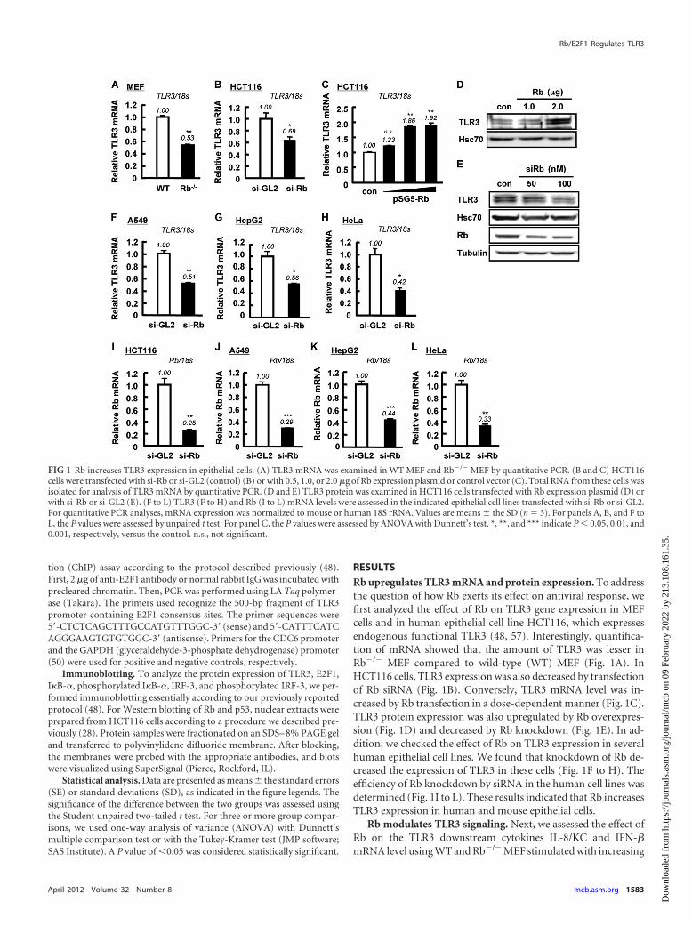
RESULTSRb upregulates TLR3 mRNA and protein expression. To addressthe question of how Rb exerts its effect on antiviral response, wefirst analyzed the effect of Rb on TLR3 gene expression in MEFcells and in human epithelial cell line HCT116, which expressesendogenous functional TLR3 (48, 57). Interestingly, quantifica-tion of mRNA showed that the amount of TLR3 was lesser inRb�/� MEF compared to wild-type (WT) MEF (Fig. 1A). InHCT116 cells, TLR3 expression was also decreased by transfectionof Rb siRNA (Fig. 1B). Conversely, TLR3 mRNA level was in-creased by Rb transfection in a dose-dependent manner (Fig. 1C).TLR3 protein expression was also upregulated by Rb overexpres-sion (Fig. 1D) and decreased by Rb knockdown (Fig. 1E). In ad-dition, we checked the effect of Rb on TLR3 expression in severalhuman epithelial cell lines. We found that knockdown of Rb de-creased the expression of TLR3 in these cells (Fig. 1F to H). Theefficiency of Rb knockdown by siRNA in the human cell lines wasdetermined (Fig. 1I to L). These results indicated that Rb increasesTLR3 expression in human and mouse epithelial cells.
Rb modulates TLR3 signaling. Next, we assessed the effect ofRb on the TLR3 downstream cytokines IL-8/KC and IFN-�mRNA level using WT and Rb�/� MEF stimulated with increasing
FIG 1 Rb increases TLR3 expression in epithelial cells. (A) TLR3 mRNA was examined in WT MEF and Rb�/� MEF by quantitative PCR. (B and C) HCT116cells were transfected with si-Rb or si-GL2 (control) (B) or with 0.5, 1.0, or 2.0 �g of Rb expression plasmid or control vector (C). Total RNA from these cells wasisolated for analysis of TLR3 mRNA by quantitative PCR. (D and E) TLR3 protein was examined in HCT116 cells transfected with Rb expression plasmid (D) orwith si-Rb or si-GL2 (E). (F to L) TLR3 (F to H) and Rb (I to L) mRNA levels were assessed in the indicated epithelial cell lines transfected with si-Rb or si-GL2.For quantitative PCR analyses, mRNA expression was normalized to mouse or human 18S rRNA. Values are means � the SD (n � 3). For panels A, B, and F toL, the P values were assessed by unpaired t test. For panel C, the P values were assessed by ANOVA with Dunnett’s test. *, **, and *** indicate P � 0.05, 0.01, and0.001, respectively, versus the control. n.s., not significant.
Rb/E2F1 Regulates TLR3
April 2012 Volume 32 Number 8 mcb.asm.org 1583
Dow
nloa
ded
from
http
s://j
ourn
als.
asm
.org
/jour
nal/m
cb o
n 09
Feb
ruar
y 20
22 b
y 21
3.10
8.16
1.35
.
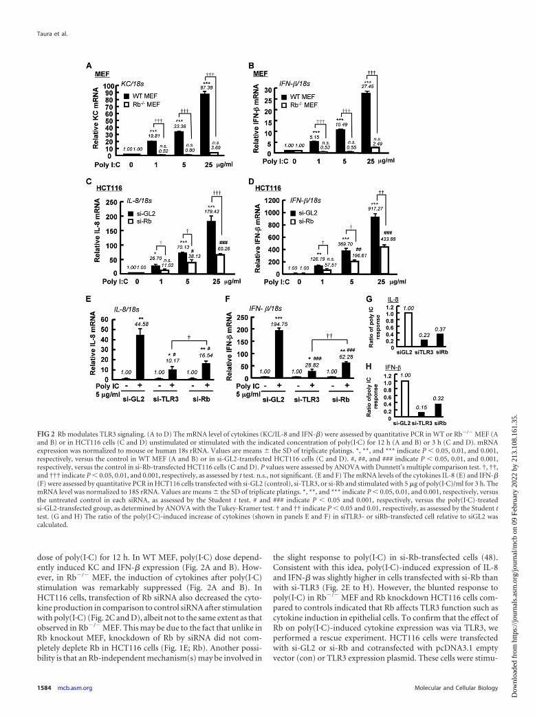
dose of poly(I·C) for 12 h. In WT MEF, poly(I·C) dose depend-ently induced KC and IFN-� expression (Fig. 2A and B). How-ever, in Rb�/� MEF, the induction of cytokines after poly(I·C)stimulation was remarkably suppressed (Fig. 2A and B). InHCT116 cells, transfection of Rb siRNA also decreased the cyto-kine production in comparison to control siRNA after stimulationwith poly(I·C) (Fig. 2C and D), albeit not to the same extent as thatobserved in Rb�/� MEF. This may be due to the fact that unlike inRb knockout MEF, knockdown of Rb by siRNA did not com-pletely deplete Rb in HCT116 cells (Fig. 1E; Rb). Another possi-bility is that an Rb-independent mechanism(s) may be involved in
the slight response to poly(I·C) in si-Rb-transfected cells (48).Consistent with this idea, poly(I·C)-induced expression of IL-8and IFN-� was slightly higher in cells transfected with si-Rb thanwith si-TLR3 (Fig. 2E to H). However, the blunted response topoly(I·C) in Rb�/� MEF and Rb knockdown HCT116 cells com-pared to controls indicated that Rb affects TLR3 function such ascytokine induction in epithelial cells. To confirm that the effect ofRb on poly(I·C)-induced cytokine expression was via TLR3, weperformed a rescue experiment. HCT116 cells were transfectedwith si-GL2 or si-Rb and cotransfected with pcDNA3.1 emptyvector (con) or TLR3 expression plasmid. These cells were stimu-
FIG 2 Rb modulates TLR3 signaling. (A to D) The mRNA level of cytokines (KC/IL-8 and IFN-�) were assessed by quantitative PCR in WT or Rb�/� MEF (Aand B) or in HCT116 cells (C and D) unstimulated or stimulated with the indicated concentration of poly(I·C) for 12 h (A and B) or 3 h (C and D). mRNAexpression was normalized to mouse or human 18s rRNA. Values are means � the SD of triplicate platings. *, **, and *** indicate P � 0.05, 0.01, and 0.001,respectively, versus the control in WT MEF (A and B) or in si-GL2-transfected HCT116 cells (C and D). #, ##, and ### indicate P � 0.05, 0.01, and 0.001,respectively, versus the control in si-Rb-transfected HCT116 cells (C and D). P values were assessed by ANOVA with Dunnett’s multiple comparison test. †, ††,and ††† indicate P � 0.05, 0.01, and 0.001, respectively, as assessed by t test. n.s., not significant. (E and F) The mRNA levels of the cytokines IL-8 (E) and IFN-�(F) were assessed by quantitative PCR in HCT116 cells transfected with si-GL2 (control), si-TLR3, or si-Rb and stimulated with 5 �g of poly(I·C)/ml for 3 h. ThemRNA level was normalized to 18S rRNA. Values are means � the SD of triplicate platings. *, **, and *** indicate P � 0.05, 0.01, and 0.001, respectively, versusthe untreated control in each siRNA, as assessed by the Student t test. # and ### indicate P � 0.05 and 0.001, respectively, versus the poly(I·C)-treatedsi-GL2-transfected group, as determined by ANOVA with the Tukey-Kramer test. † and †† indicate P � 0.05 and 0.01, respectively, as assessed by the Student ttest. (G and H) The ratio of the poly(I·C)-induced increase of cytokines (shown in panels E and F) in siTLR3- or siRb-transfected cell relative to siGL2 wascalculated.
Taura et al.
1584 mcb.asm.org Molecular and Cellular Biology
Dow
nloa
ded
from
http
s://j
ourn
als.
asm
.org
/jour
nal/m
cb o
n 09
Feb
ruar
y 20
22 b
y 21
3.10
8.16
1.35
.
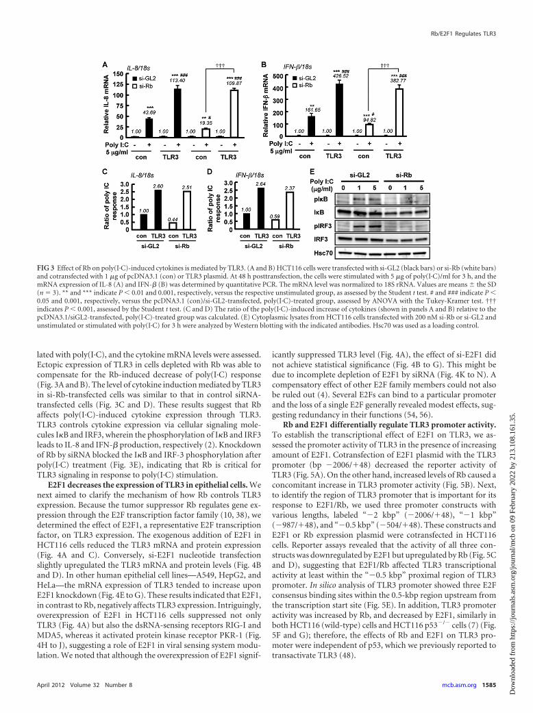
lated with poly(I·C), and the cytokine mRNA levels were assessed.Ectopic expression of TLR3 in cells depleted with Rb was able tocompensate for the Rb-induced decrease of poly(I·C) response(Fig. 3A and B). The level of cytokine induction mediated by TLR3in si-Rb-transfected cells was similar to that in control siRNA-transfected cells (Fig. 3C and D). These results suggest that Rbaffects poly(I·C)-induced cytokine expression through TLR3.TLR3 controls cytokine expression via cellular signaling mole-cules I�B and IRF3, wherein the phosphorylation of I�B and IRF3leads to IL-8 and IFN-� production, respectively (2). Knockdownof Rb by siRNA blocked the I�B and IRF-3 phosphorylation afterpoly(I·C) treatment (Fig. 3E), indicating that Rb is critical forTLR3 signaling in response to poly(I·C) stimulation.
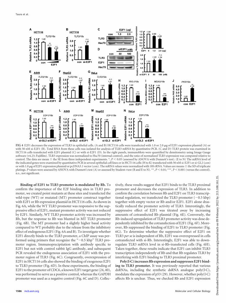
E2F1 decreases the expression of TLR3 in epithelial cells. Wenext aimed to clarify the mechanism of how Rb controls TLR3expression. Because the tumor suppressor Rb regulates gene ex-pression through the E2F transcription factor family (10, 38), wedetermined the effect of E2F1, a representative E2F transcriptionfactor, on TLR3 expression. The exogenous addition of E2F1 inHCT116 cells reduced the TLR3 mRNA and protein expression(Fig. 4A and C). Conversely, si-E2F1 nucleotide transfectionslightly upregulated the TLR3 mRNA and protein levels (Fig. 4Band D). In other human epithelial cell lines—A549, HepG2, andHeLa—the mRNA expression of TLR3 tended to increase uponE2F1 knockdown (Fig. 4E to G). These results indicated that E2F1,in contrast to Rb, negatively affects TLR3 expression. Intriguingly,overexpression of E2F1 in HCT116 cells suppressed not onlyTLR3 (Fig. 4A) but also the dsRNA-sensing receptors RIG-I andMDA5, whereas it activated protein kinase receptor PKR-1 (Fig.4H to J), suggesting a role of E2F1 in viral sensing system modu-lation. We noted that although the overexpression of E2F1 signif-
icantly suppressed TLR3 level (Fig. 4A), the effect of si-E2F1 didnot achieve statistical significance (Fig. 4B to G). This might bedue to incomplete depletion of E2F1 by siRNA (Fig. 4K to N). Acompensatory effect of other E2F family members could not alsobe ruled out (4). Several E2Fs can bind to a particular promoterand the loss of a single E2F generally revealed modest effects, sug-gesting redundancy in their functions (54, 56).
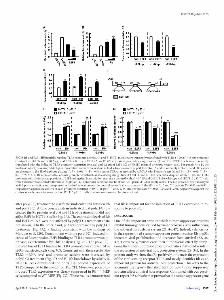
Rb and E2F1 differentially regulate TLR3 promoter activity.To establish the transcriptional effect of E2F1 on TLR3, we as-sessed the promoter activity of TLR3 in the presence of increasingamount of E2F1. Cotransfection of E2F1 plasmid with the TLR3promoter (bp �2006/�48) decreased the reporter activity ofTLR3 (Fig. 5A). On the other hand, increased levels of Rb caused aconcomitant increase in TLR3 promoter activity (Fig. 5B). Next,to identify the region of TLR3 promoter that is important for itsresponse to E2F1/Rb, we used three promoter constructs withvarious lengths, labeled “�2 kbp” (�2006/�48), “�1 kbp”(�987/�48), and “�0.5 kbp” (�504/�48). These constructs andE2F1 or Rb expression plasmid were cotransfected in HCT116cells. Reporter assays revealed that the activity of all three con-structs was downregulated by E2F1 but upregulated by Rb (Fig. 5Cand D), suggesting that E2F1/Rb affected TLR3 transcriptionalactivity at least within the “�0.5 kbp” proximal region of TLR3promoter. In silico analysis of TLR3 promoter showed three E2Fconsensus binding sites within the 0.5-kbp region upstream fromthe transcription start site (Fig. 5E). In addition, TLR3 promoteractivity was increased by Rb, and decreased by E2F1, similarly inboth HCT116 (wild-type) cells and HCT116 p53�/� cells (7) (Fig.5F and G); therefore, the effects of Rb and E2F1 on TLR3 pro-moter were independent of p53, which we previously reported totransactivate TLR3 (48).
FIG 3 Effect of Rb on poly(I·C)-induced cytokines is mediated by TLR3. (A and B) HCT116 cells were transfected with si-GL2 (black bars) or si-Rb (white bars)and cotransfected with 1 �g of pcDNA3.1 (con) or TLR3 plasmid. At 48 h posttransfection, the cells were stimulated with 5 �g of poly(I·C)/ml for 3 h, and themRNA expression of IL-8 (A) and IFN-� (B) was determined by quantitative PCR. The mRNA level was normalized to 18S rRNA. Values are means � the SD(n � 3). ** and *** indicate P � 0.01 and 0.001, respectively, versus the respective unstimulated group, as assessed by the Student t test. # and ### indicate P �0.05 and 0.001, respectively, versus the pcDNA3.1 (con)/si-GL2-transfected, poly(I·C)-treated group, assessed by ANOVA with the Tukey-Kramer test. †††indicates P � 0.001, assessed by the Student t test. (C and D) The ratio of the poly(I·C)-induced increase of cytokines (shown in panels A and B) relative to thepcDNA3.1/siGL2-transfected, poly(I·C)-treated group was calculated. (E) Cytoplasmic lysates from HCT116 cells transfected with 200 nM si-Rb or si-GL2 andunstimulated or stimulated with poly(I·C) for 3 h were analyzed by Western blotting with the indicated antibodies. Hsc70 was used as a loading control.
Rb/E2F1 Regulates TLR3
April 2012 Volume 32 Number 8 mcb.asm.org 1585
Dow
nloa
ded
from
http
s://j
ourn
als.
asm
.org
/jour
nal/m
cb o
n 09
Feb
ruar
y 20
22 b
y 21
3.10
8.16
1.35
.
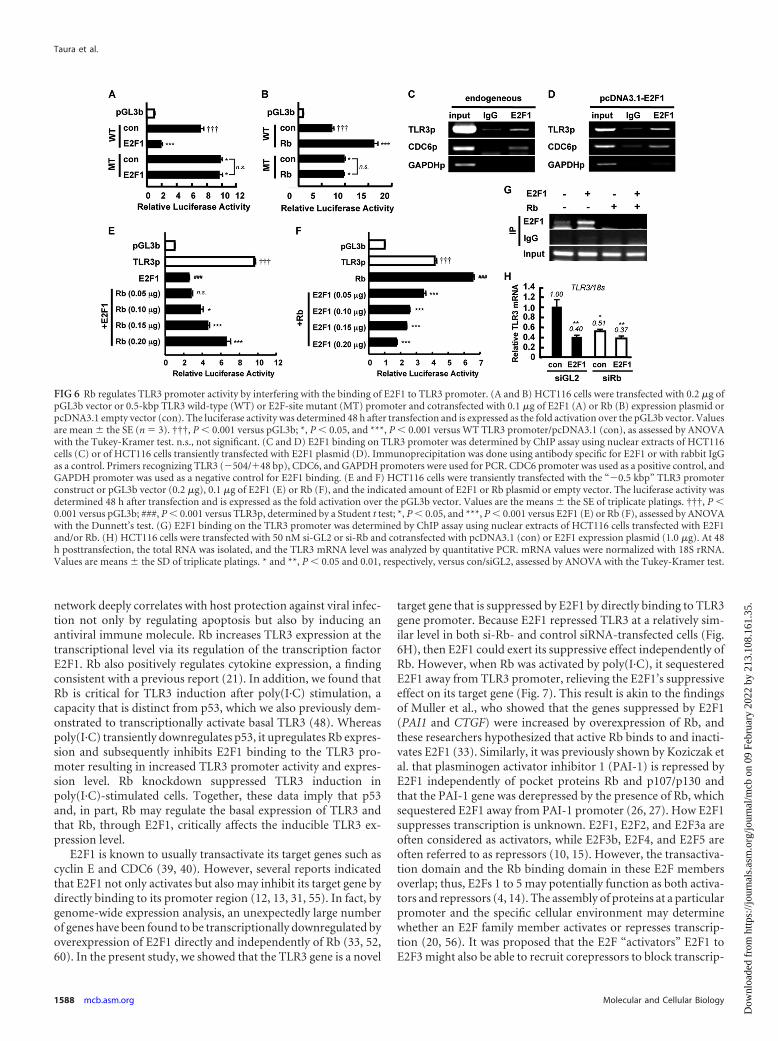
Binding of E2F1 to TLR3 promoter is modulated by Rb. Toconfirm the importance of the E2F binding sites in TLR3 pro-moter, we created point mutants at these sites and transfected thewild-type (WT) or mutated (MT) promoter construct togetherwith E2F1 or Rb expression plasmid in HCT116 cells. As shown inFig. 6A, while the WT TLR3 promoter was responsive to the sup-pressive effect of E2F1, mutant promoter activity was not reducedby E2F1. Similarly, WT TLR3 promoter activity was increased byRb, but the response to Rb was blunted in MT TLR3 promoter(Fig. 6B). The MT promoter had a slightly higher basal activitycompared to WT probably due to the release from the inhibitoryeffect of endogenous E2F1 (Fig. 6A and B). To investigate whetherE2F1 directly binds to the TLR3 promoter, a ChIP assay was per-formed using primers that recognize the “�0.5 kbp” TLR3 pro-moter region. Immunoprecipitation with antibody specific toE2F1 but not with control rabbit IgG antibody, and subsequentPCR revealed the endogenous association of E2F1 with the pro-moter region of TLR3 (Fig. 6C). Congruently, overexpression ofE2F1 in HCT116 cells also showed the binding of exogenous E2F1to TLR3 promoter (Fig. 6D). In these experiments, the binding ofE2F1 to the promoter of CDC6, a known E2F1 target gene (24, 40),was performed to serve as a positive control, whereas the GAPDHpromoter was used as a negative control (Fig. 6C and D). Collec-
tively, these results suggest that E2F1 binds to the TLR3 proximalpromoter and decreases the expression of TLR3. In addition toconfirm the correlation between Rb and E2F1 on TLR3 transcrip-tional regulation, we transfected the TLR3 promoter (�0.5 kbp)together with empty vector or Rb and/or E2F1. E2F1 alone dras-tically reduced the promoter activity of TLR3. Interestingly, thesuppressive effect of E2F1 was titrated away by increasingamounts of cotransfected Rb plasmid (Fig. 6E). Conversely, theRb-induced upregulation of TLR3 promoter activity was dose de-pendently inhibited by the cotransfection of E2F1 (Fig. 6F). More-over, Rb suppressed the binding of E2F1 to TLR3 promoter (Fig.6G). To determine whether the suppressive effect of E2F1 onTLR3 per se is independent of Rb, E2F1 was overexpressed in cellscotransfected with si-Rb. Interestingly, E2F1 was able to down-regulate TLR3 mRNA level in si-Rb-transfected cells (Fig. 6H).Taken together, these results indicate that E2F1 can inhibit TLR3transcription independently of Rb and that Rb regulates TLR3 byinterfering with E2F1 binding to TLR3 proximal promoter.
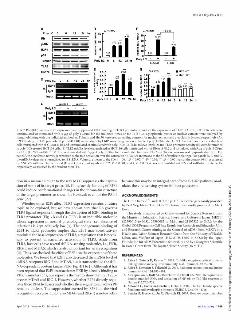
Poly(I·C) increases Rb expression and suppresses E2F1 bind-ing to TLR3 promoter. It was previously reported that variousdsRNAs, including the synthetic dsRNA analogue poly(I·C),modulate the expression of p53 (29). However, whether poly(I·C)affects Rb is unclear. Thus, we checked Rb and E2F1 expression
FIG 4 E2F1 decreases the expression of TLR3 in epithelial cells. (A and B) HCT116 cells were transfected with 1.0 or 2.0 �g of E2F1 expression plasmid (A) orwith 50 nM si-E2F1 (B). Total RNA from these cells was isolated for analysis of TLR3 mRNA by quantitative PCR. (C and D) TLR3 protein was examined inHCT116 cells transfected with E2F1 plasmid (C) or with si-E2F1 (D). In the right panels, immunoblots were quantified by densitometry using Image Gaugesoftware (v4.23; Fujifilm). TLR3 expression was normalized to Hsc70 (internal control), and the ratio of normalized TLR3 expression was computed relative tocontrol. The data are mean � the SE from three independent experiments. *, P � 0.05 (assessed by ANOVA with Dunnett’s test). (E to N) The mRNA level ofthe indicated genes were examined by quantitative PCR in several epithelial cell lines or in HCT116 cells (H to K) transfected with 50 nM si-E2F1 or si-GL2 (con)or with 1.0 �g of E2F1 expression plasmid or pcDNA3.1 vector (con). The mRNA values were normalized with 18S rRNA. Values are means � the SD of triplicateplatings. P values were assessed by ANOVA with Dunnett’s test (A) or assessed by Student t test (B and E to N). **, P � 0.01; ***, P � 0.001 (versus the control).n.s., not significant.
Taura et al.
1586 mcb.asm.org Molecular and Cellular Biology
Dow
nloa
ded
from
http
s://j
ourn
als.
asm
.org
/jour
nal/m
cb o
n 09
Feb
ruar
y 20
22 b
y 21
3.10
8.16
1.35
.
after poly(I·C) treatment to clarify the molecular link between Rband poly(I·C). A time course analysis indicated that poly(I·C) in-creased the Rb protein level at 6 and 12 h of treatment but did notaffect E2F1 in HCT116 cells (Fig. 7A). The expression levels of Rband E2F1 mRNA were not affected by poly(I·C) treatment (datanot shown). On the other hand, p53 was decreased by poly(I·C)treatment (Fig. 7A), a finding consistent with the findings ofMarques et al. (29). Concomitant with the poly(I·C)-induced in-crease of Rb expression, E2F1 binding to TLR3 promoter was sup-pressed, as determined by ChIP analysis (Fig. 7B). The poly(I·C)-induced loss of E2F1 binding to TLR3 promoter was prevented insi-Rb-transfected cells (Fig. 7C). Consistent with these results, theTLR3 mRNA level and promoter activity were increased bypoly(I·C) treatment (Fig. 7D and E). Rb knockdown by siRNA inHCT116 cells diminished the poly(I·C)-induced expression ofTLR3 compared to the si-control (Fig. 7F). Moreover, poly(I·C)-induced TLR3 expression was clearly suppressed in Rb�/� MEFcells compared to WT MEF (Fig. 7G). These results demonstrated
that Rb is important for the induction of TLR3 expression in re-sponse to poly(I·C).
DISCUSSION
One of the important ways in which tumor suppressor proteinsinhibit tumorigenesis caused by viral oncogenes is by influencingthe antiviral host defense system (21, 44, 47). Indeed, a deficiencyin the expression of a tumor suppressor protein, such as Rb or p53,increases viral proliferation and decreases host survival (35, 36,47). Conversely, viruses exert their tumorigenic effect by damp-ening the tumor suppressor proteins’ activities that could result inthe repression of antiviral host protection system (29, 34). In thepresent study we show that Rb positively influences the expressionof the viral sensing receptor TLR3 and newly identifies Rb as animportant protein for antiviral host protection. This adds to theaccumulating reports that shed light on how tumor suppressorproteins affect antiviral host response. Combined with our previ-ous report (48), this further proves that the tumor suppressor gene
FIG 5 Rb and E2F1 differentially regulate TLR3 promoter activity. (A and B) HCT116 cells were transiently transfected with TLR3 (�2006/�48 bp) promoterconstruct or pGL3b vector (0.2 �g) and 0.05 or 0.1 �g of E2F1 (A) or Rb (B) expression plasmid or empty vector. (C and D) HCT116 cells were transientlytransfected with the indicated TLR3 promoter constructs (0.2 �g) and 0.1 �g of E2F1 (C) or Rb (D) plasmid or empty vector (con). For panels A to D, theluciferase activity was assayed 48 h posttransfection and is expressed as the fold activation over the pGL3b vector (A and B) or empty vector (C and D). Valuesare the mean � the SE of triplicate platings. *, P � 0.05; ***, P � 0.001 versus TLR3p, as assessed by ANOVA with Dunnett’s test (A and B). *, P � 0.05; **, P �0.01; ***, P � 0.001 versus control of each promoter construct, as assessed by using Student t test (C and D). (E) Schematic diagram of the “�0.5 kb” TLR3promoter with the indicated positions of E2F binding site. Transcription start site is denoted with a “�1”. (F and G) HCT116 wild-type and HCT116 p53�/� cellswere transiently transfected with various lengths of TLR3 promoter construct and Rb (F) or E2F1 plasmid (G) or empty vector. The luciferase activity was assayedat 48 h posttransfection and is expressed as the fold activation over the control vector. Values are means � the SE (n � 3). * and *** indicate P � 0.05 and 0.001,respectively, against the control of each promoter construct in HCT116 p53�/� cells. #, ##, and ### indicate P � 0.05, 0.01, and 0.001, respectively, against thecontrol of each promoter construct in HCT116 p53�/� cells. P values were assessed by Student t test.
Rb/E2F1 Regulates TLR3
April 2012 Volume 32 Number 8 mcb.asm.org 1587
Dow
nloa
ded
from
http
s://j
ourn
als.
asm
.org
/jour
nal/m
cb o
n 09
Feb
ruar
y 20
22 b
y 21
3.10
8.16
1.35
.
network deeply correlates with host protection against viral infec-tion not only by regulating apoptosis but also by inducing anantiviral immune molecule. Rb increases TLR3 expression at thetranscriptional level via its regulation of the transcription factorE2F1. Rb also positively regulates cytokine expression, a findingconsistent with a previous report (21). In addition, we found thatRb is critical for TLR3 induction after poly(I·C) stimulation, acapacity that is distinct from p53, which we also previously dem-onstrated to transcriptionally activate basal TLR3 (48). Whereaspoly(I·C) transiently downregulates p53, it upregulates Rb expres-sion and subsequently inhibits E2F1 binding to the TLR3 pro-moter resulting in increased TLR3 promoter activity and expres-sion level. Rb knockdown suppressed TLR3 induction inpoly(I·C)-stimulated cells. Together, these data imply that p53and, in part, Rb may regulate the basal expression of TLR3 andthat Rb, through E2F1, critically affects the inducible TLR3 ex-pression level.
E2F1 is known to usually transactivate its target genes such ascyclin E and CDC6 (39, 40). However, several reports indicatedthat E2F1 not only activates but also may inhibit its target gene bydirectly binding to its promoter region (12, 13, 31, 55). In fact, bygenome-wide expression analysis, an unexpectedly large numberof genes have been found to be transcriptionally downregulated byoverexpression of E2F1 directly and independently of Rb (33, 52,60). In the present study, we showed that the TLR3 gene is a novel
target gene that is suppressed by E2F1 by directly binding to TLR3gene promoter. Because E2F1 repressed TLR3 at a relatively sim-ilar level in both si-Rb- and control siRNA-transfected cells (Fig.6H), then E2F1 could exert its suppressive effect independently ofRb. However, when Rb was activated by poly(I·C), it sequesteredE2F1 away from TLR3 promoter, relieving the E2F1’s suppressiveeffect on its target gene (Fig. 7). This result is akin to the findingsof Muller et al., who showed that the genes suppressed by E2F1(PAI1 and CTGF) were increased by overexpression of Rb, andthese researchers hypothesized that active Rb binds to and inacti-vates E2F1 (33). Similarly, it was previously shown by Koziczak etal. that plasminogen activator inhibitor 1 (PAI-1) is repressed byE2F1 independently of pocket proteins Rb and p107/p130 andthat the PAI-1 gene was derepressed by the presence of Rb, whichsequestered E2F1 away from PAI-1 promoter (26, 27). How E2F1suppresses transcription is unknown. E2F1, E2F2, and E2F3a areoften considered as activators, while E2F3b, E2F4, and E2F5 areoften referred to as repressors (10, 15). However, the transactiva-tion domain and the Rb binding domain in these E2F membersoverlap; thus, E2Fs 1 to 5 may potentially function as both activa-tors and repressors (4, 14). The assembly of proteins at a particularpromoter and the specific cellular environment may determinewhether an E2F family member activates or represses transcrip-tion (20, 56). It was proposed that the E2F “activators” E2F1 toE2F3 might also be able to recruit corepressors to block transcrip-
FIG 6 Rb regulates TLR3 promoter activity by interfering with the binding of E2F1 to TLR3 promoter. (A and B) HCT116 cells were transfected with 0.2 �g ofpGL3b vector or 0.5-kbp TLR3 wild-type (WT) or E2F-site mutant (MT) promoter and cotransfected with 0.1 �g of E2F1 (A) or Rb (B) expression plasmid orpcDNA3.1 empty vector (con). The luciferase activity was determined 48 h after transfection and is expressed as the fold activation over the pGL3b vector. Valuesare mean � the SE (n � 3). †††, P � 0.001 versus pGL3b; *, P � 0.05, and ***, P � 0.001 versus WT TLR3 promoter/pcDNA3.1 (con), as assessed by ANOVAwith the Tukey-Kramer test. n.s., not significant. (C and D) E2F1 binding on TLR3 promoter was determined by ChIP assay using nuclear extracts of HCT116cells (C) or of HCT116 cells transiently transfected with E2F1 plasmid (D). Immunoprecipitation was done using antibody specific for E2F1 or with rabbit IgGas a control. Primers recognizing TLR3 (�504/�48 bp), CDC6, and GAPDH promoters were used for PCR. CDC6 promoter was used as a positive control, andGAPDH promoter was used as a negative control for E2F1 binding. (E and F) HCT116 cells were transiently transfected with the “�0.5 kbp” TLR3 promoterconstruct or pGL3b vector (0.2 �g), 0.1 �g of E2F1 (E) or Rb (F), and the indicated amount of E2F1 or Rb plasmid or empty vector. The luciferase activity wasdetermined 48 h after transfection and is expressed as the fold activation over the pGL3b vector. Values are the means � the SE of triplicate platings. †††, P �0.001 versus pGL3b; ###, P � 0.001 versus TLR3p, determined by a Student t test; *, P � 0.05, and ***, P � 0.001 versus E2F1 (E) or Rb (F), assessed by ANOVAwith the Dunnett’s test. (G) E2F1 binding on the TLR3 promoter was determined by ChIP assay using nuclear extracts of HCT116 cells transfected with E2F1and/or Rb. (H) HCT116 cells were transfected with 50 nM si-GL2 or si-Rb and cotransfected with pcDNA3.1 (con) or E2F1 expression plasmid (1.0 �g). At 48h posttransfection, the total RNA was isolated, and the TLR3 mRNA level was analyzed by quantitative PCR. mRNA values were normalized with 18S rRNA.Values are means � the SD of triplicate platings. * and **, P � 0.05 and 0.01, respectively, versus con/siGL2, assessed by ANOVA with the Tukey-Kramer test.
Taura et al.
1588 mcb.asm.org Molecular and Cellular Biology
Dow
nloa
ded
from
http
s://j
ourn
als.
asm
.org
/jour
nal/m
cb o
n 09
Feb
ruar
y 20
22 b
y 21
3.10
8.16
1.35
.
tion in a manner similar to the way MYC suppresses the expres-sion of some of its target genes (6). Congruently, binding of E2F1could induce conformational changes in the chromatin structureof the target promoter, as shown by Koziczak et al. for the PAI-1gene (27).
Whether other E2Fs affect TLR3 expression remains a futuretopic to be explored, but we have shown here that Rb governsTLR3 ligand response through the disruption of E2F1 binding toTLR3 promoter (Fig. 7B and C). TLR3 is an inducible moleculewhose expression in normal physiological state (i.e., absence ofinfection) is kept relatively low (3). The endogenous binding ofE2F1 to TLR3 promoter implies that E2F1 may constitutivelymodulate the basal expression of TLR3, a regulation that is neces-sary to prevent unwarranted activation of TLR3. Aside fromTLR3, host cells have several dsRNA-sensing molecules, i.e., PKR,RIG-I, and MDA5, which are also important for viral recognition(2). Thus, we checked the effect of E2F1 on the expression of thesemolecules. We found that E2F1 also decreased the mRNA level ofdsRNA receptors RIG-I and MDA5, but it transactivated the dsR-NA-dependent protein kinase PKR (Fig. 4H to J). Although it hasbeen reported that E2F1 transactivates PKR by directly binding toPKR promoter (53), our report is the first to show that E2F1 sup-presses MDA5 and RIG-I. However, whether E2F1 directly regu-lates these RNA helicases and whether their regulation involves Rbremains unclear. The suppression exerted by E2F1 on the viralrecognition receptor TLR3 (also MDA5 and RIG-I) is noteworthy
because this may be an integral part of how E2F-Rb pathway mod-ulates the viral sensing system for host protection.
ACKNOWLEDGMENTS
The HCT116 p53�/� and HCT116 p53�/� cells were generously providedby Bert Vogelstein. The pSG5-Rb plasmid was kindly provided by MarkEwen.
This study is supported by Grants-in-Aid for Science Research fromthe Ministry of Education, Science, Sports, and Culture of Japan (MEXT)(22390015 to H.K., 23590082 to M.S., and 21107522 to S.O.); by theGlobal COE Programs (Cell Fate Regulation Research and Education Unitand Research Center Aiming at the Control of AIDS) from MEXT; by aHealth and Labor Sciences Research Grant from the Ministry of Health,Labor, and Welfare of Japan (H22-AIDS-I-002 to S.O.); by the JapanFoundation for AIDS Prevention fellowship; and by a Sasagawa ScientificResearch Grant from The Japan Science Society (to M.T.).
REFERENCES1. Akira S, Takeda K, Kaisho T. 2001. Toll-like receptors: critical proteins
linking innate and acquired immunity. Nat. Immunol. 2:675– 680.2. Akira S, Uematsu S, Takeuchi O. 2006. Pathogen recognition and innate
immunity. Cell 124:783– 801.3. Alexopoulou L, Holt AC, Medzhitov R, Flavell RA. 2001. Recognition of
double-stranded RNA and activation of NF-�B by Toll-like receptor 3.Nature 413:732–738.
4. Attwooll C, Lazzerini-Denchi E, Helin K. 2004. The E2F family: specificfunctions and overlapping interests. EMBO J. 23:4709 – 4716.
5. Beutler B, Hoebe K, Du X, Ulevitch RJ. 2003. How we detect microbes
FIG 7 Poly(I·C) increased Rb expression and suppressed E2F1 binding to TLR3 promoter to induce the expression of TLR3. (A to E) HCT116 cells wereunstimulated or stimulated with 5 �g of poly(I·C)/ml for the indicated times or for 12 h (C). Cytoplasmic lysates or nuclear extracts were analyzed byimmunoblotting with the indicated antibodies. Tubulin and Hsc70 were used as loading controls for nuclear extracts and cytoplasmic lysates, respectively (A).E2F1 binding to TLR3 promoter (bp �504/�48) was analyzed by ChIP assay using nuclear extracts of poly(I·C)-treated HCT116 cells (B) or nuclear extracts ofcells transfected with si-GL2 or si-Rb and unstimulated or stimulated with poly(I·C) (C). TLR3 mRNA level (D) and TLR3 promoter activity (E) were determinedin poly(I·C)-treated HCT116 cells. (F) TLR3 mRNA level was analyzed in HCT116 cells transfected with si-Rb or si-GL2 and stimulated with 5 �g of poly(I·C)/mlfor 12 h. (G) WT and Rb�/� MEF were stimulated with 5 �g of poly(I·C)/ml for the indicated time, and TLR3 mRNA level was assessed by quantitative PCR. Forpanel E, the luciferase activity is expressed as the fold activation over the control (0 h). Values are means � the SE of triplicate platings. For panels D, F, and G,the mRNA values were normalized to 18S rRNA. Values are means � the SD (n � 3). *, P � 0.05; **, P � 0.01; ***, P � 0.001 versus the control (0 h), as assessedby ANOVA with the Dunnett’s test (D and G). n.s., not significant. ***, P � 0.001, and #, P � 0.05 versus unstimulated si-GL2- and si-Rb-transfected cells,respectively, as assessed by the Student t test (F).
Rb/E2F1 Regulates TLR3
April 2012 Volume 32 Number 8 mcb.asm.org 1589
Dow
nloa
ded
from
http
s://j
ourn
als.
asm
.org
/jour
nal/m
cb o
n 09
Feb
ruar
y 20
22 b
y 21
3.10
8.16
1.35
.
and respond to them: the Toll-like receptors and their transducers. J.Leukoc. Biol. 74:479 – 485.
6. Bracken AP, Ciro M, Cocito A, Helin K. 2004. E2F target genes: unrav-eling the biology. Trends Biochem. Sci. 29:409 – 417.
7. Bunz F, et al. 1998. Requirement for p53 and p21 to sustain G2 arrest afterDNA damage. Science 282:1497–1501.
8. Cavenee WK, et al. 1983. Expression of recessive alleles by chromosomalmechanisms in retinoblastoma. Nature 305:779 –784.
9. Chellappan SP, Hiebert S, Mudryj M, Horowitz JM, Nevins JR. 1991.The E2F transcription factor is a cellular target for the RB protein. Cell65:1053–1061.
10. Chen HZ, Tsai SY, Leone G. 2009. Emerging roles of E2Fs in cancer: anexit from cell cycle control. Nat. Rev. Cancer 9:785–797.
11. Cho J, et al. 2001. HCV core protein modulates Rb pathway through pRbdown-regulation and E2F-1 up-regulation. Biochim. Biophys. Acta 1538:59 – 66.
12. Crowe DL, Nguyen DC, Tsang KJ, Kyo S. 2001. E2F-1 represses tran-scription of the human telomerase reverse transcriptase gene. Nucleic Ac-ids Res. 29:2789 –2794.
13. Croxton R, Ma Y, Song L, Haura EB, Cress WD. 2002. Direct repressionof the Mcl-1 promoter by E2F1. Oncogene 21:1359 –1369.
14. DeGregori J. 2002. The genetics of the E2F family of transcription factors:shared functions and unique roles. Biochim. Biophys. Acta 1602:131–150.
15. Dimova DK, Dyson NJ. 2005. The E2F transcriptional network: oldacquaintances with new faces. Oncogene 24:2810 –2826.
16. Dyson N. 1998. The regulation of E2F by pRB-family proteins. Genes Dev.12:2245–2262.
17. Ewen ME. 1994. The cell cycle and the retinoblastoma protein family.Cancer Metastasis Rev. 13:45– 66.
18. Ferreira R, et al. 2001. Cell cycle-dependent recruitment of HDAC-1correlates with deacetylation of histone H4 on an Rb-E2F target promoter.EMBO Rep. 2:794 –799.
19. Frolov MV, Dyson NJ. 2004. Molecular mechanisms of E2F-dependentactivation and pRB-mediated repression. J. Cell Sci. 117:2173–2181.
20. Fry CJ, Farnham PJ. 1999. Context-dependent transcriptional regula-tion. J. Biol. Chem. 274:29583–29586.
21. Garcia MA, et al. 2009. Activation of NF-�B pathway by virus infectionrequires Rb expression. PLoS One 4:e6422.
22. Halaban R. 2005. Rb/E2F: a two-edged sword in the melanocytic system.Cancer Metastasis Rev. 24:339 –356.
23. Harbour JW, Dean DC. 2000. The Rb/E2F pathway: expanding roles andemerging paradigms. Genes Dev. 14:2393–2409.
24. Hateboer G, et al. 1998. Cell cycle-regulated expression of mammalianCDC6 is dependent on E2F. Mol. Cell. Biol. 18:6679 – 6697.
25. Iaquinta PJ, Lees JA. 2007. Life and death decisions by the E2F transcrip-tion factors. Curr. Opin. Cell Biol. 19:649 – 657.
26. Koziczak M, Krek W, Nagamine Y. 2000. Pocket protein-independentrepression of urokinase-type plasminogen activator and plasminogen ac-tivator inhibitor 1 gene expression by E2F1. Mol. Cell. Biol. 20:2014 –2022.
27. Koziczak M, Muller H, Helin K, Nagamine Y. 2001. E2F1-mediatedtranscriptional inhibition of the plasminogen activator inhibitor type 1gene. Eur. J. Biochem. 268:4969 – 4978.
28. Lu Z, et al. 2004. MEF up-regulates human beta-defensin 2 expression inepithelial cells. FEBS Lett. 561:117–121.
29. Marques JT, et al. 2005. Down-regulation of p53 by double-strandedRNA modulates the antiviral response. J. Virol. 79:11105–11114.
30. Martin D, Gutkind JS. 2008. Human tumor-associated viruses and newinsights into the molecular mechanisms of cancer. Oncogene 27(Suppl2):S31–S42.
31. Merdzhanova G, et al. 2010. The transcription factor E2F1 and the SRprotein SC35 control the ratio of pro-angiogenic versus antiangiogenicisoforms of vascular endothelial growth factor-A to inhibit neovascular-ization in vivo. Oncogene 29:5392–5403.
32. Meylan E, Tschopp J. 2006. Toll-like receptors and RNA helicases: twoparallel ways to trigger antiviral responses. Mol. Cell 22:561–569.
33. Muller H, et al. 2001. E2Fs regulate the expression of genes involved indifferentiation, development, proliferation, and apoptosis. Genes Dev. 15:267–285.
34. Munakata T, Nakamura M, Liang Y, Li K, Lemon SM. 2005. Down-
regulation of the retinoblastoma tumor suppressor by the hepatitis C virusNS5B RNA-dependent RNA polymerase. Proc. Natl. Acad. Sci. U. S. A.102:18159 –18164.
35. Munoz-Fontela C, et al. 2005. Resistance to viral infection of super p53mice. Oncogene 24:3059 –3062.
36. Munoz-Fontela C, et al. 2008. Transcriptional role of p53 in interferon-mediated antiviral immunity. J. Exp. Med. 205:1929 –1938.
37. Nevins JR. 2001. The Rb/E2F pathway and cancer. Hum. Mol. Genet.10:699 –703.
38. Nevins JR. 1998. Toward an understanding of the functional complexityof the E2F and retinoblastoma families. Cell Growth Differ. 9:585–593.
39. Ohtani K, DeGregori J, Nevins JR. 1995. Regulation of the cyclin E geneby transcription factor E2F1. Proc. Natl. Acad. Sci. U. S. A. 92:12146 –12150.
40. Ohtani K, Tsujimoto A, Ikeda M, Nakamura M. 1998. Regulation of cellgrowth-dependent expression of mammalian CDC6 gene by the cell cycletranscription factor E2F. Oncogene 17:1777–1785.
41. Racek T, et al. 2008. Transcriptional repression of the prosurvival endo-plasmic reticulum chaperone GRP78/BIP by E2F1. J. Biol. Chem. 283:34305–34314.
42. Rivas C, Aaronson SA, Munoz-Fontela C. 2010. Dual role of p53 ininnate antiviral immunity. Viruses 2:298 –313.
43. Sarek G, et al. 2007. Reactivation of the p53 pathway as a treatmentmodality for KSHV-induced lymphomas. J. Clin. Invest. 117:1019 –1028.
44. Sherr CJ, McCormick F. 2002. The Rb and p53 pathways in cancer.Cancer Cell 2:103–112.
45. Shirodkar S, et al. 1992. The transcription factor E2F interacts with theretinoblastoma product and a p107-cyclin A complex in a cell cycle-regulated manner. Cell 68:157–166.
46. Takahashi C, et al. 2003. Rb and N-ras function together to controldifferentiation in the mouse. Mol. Cell. Biol. 23:5256 –5268.
47. Takaoka A, et al. 2003. Integration of interferon-alpha/beta signalling top53 responses in tumour suppression and antiviral defense. Nature 424:516 –523.
48. Taura M, et al. 2008. p53 regulates Toll-like receptor 3 expression andfunction in human epithelial cell lines. Mol. Cell. Biol. 28:6557– 6567.
49. Taura M, et al. 2010. TLR3 induction by anticancer drugs potentiatespoly I:C-induced tumor cell apoptosis. Cancer Sci. 101:1610 –1617.
50. Taura M, et al. 2011. MEF/ELF4 transactivation by E2F1 is inhibited byp53. Nucleic Acids Res. 39:76 – 88.
51. Thomas M, Pim D, Banks L. 1999. The role of the E6 –p53 interaction inthe molecular pathogenesis of HPV. Oncogene 18:7690 –7700.
52. Vernell R, Helin K, Muller H. 2003. Identification of target genes of thep16INK4A-pRB-E2F pathway. J. Biol. Chem. 278:46124 – 46137.
53. Vorburger SA, et al. 2002. Role for the double-stranded RNA activatedprotein kinase PKR in E2F-1-induced apoptosis. Oncogene 21:6278 –6288.
54. Wu L, et al. 2001. The E2F1-3 transcription factors are essential forcellular proliferation. Nature 414:457– 462.
55. Xu HG, et al. 2011. Direct repression of the human IRF-3 promoter byE2F1. Immunogenetics 63:189 –196.
56. Xu X, et al. 2007. A comprehensive ChIP-chip analysis of E2F1, E2F4, andE2F6 in normal and tumor cells reveals interchangeable roles of E2F familymembers. Genome Res. 17:1550 –1561.
57. Yamamoto M, et al. 2003. Role of adaptor TRIF in the MyD88-independent Toll-like receptor signaling pathway. Science 301:640 – 643.
58. Yan Z, et al. 1998. Cdc6 is regulated by E2F and is essential for DNAreplication in mammalian cells. Proc. Natl. Acad. Sci. U. S. A. 95:3603–3608.
59. Yoon CH, Lee ES, Lim DS, Bae YS. 2009. PKR, a p53 target gene, playsa crucial role in the tumor-suppressor function of p53. Proc. Natl. Acad.Sci. U. S. A. 106:7852–7857.
60. Young AP, Nagarajan R, Longmore GD. 2003. Mechanisms of transcrip-tional regulation by Rb-E2F segregate by biological pathway. Oncogene22:7209 –7217.
61. Zhao CY, Szekely L, Bao W, Selivanova G. 2010. Rescue of p53 functionby small-molecule RITA in cervical carcinoma by blocking E6-mediateddegradation. Cancer Res. 70:3372–3381.
Taura et al.
1590 mcb.asm.org Molecular and Cellular Biology
Dow
nloa
ded
from
http
s://j
ourn
als.
asm
.org
/jour
nal/m
cb o
n 09
Feb
ruar
y 20
22 b
y 21
3.10
8.16
1.35
.