Embed Size (px)
Citation preview
Research ArticleCrotalus durissus ruruima Snake Venom and a PhospholipaseA2 Isolated from This Venom Elicit Macrophages to Form LipidDroplets and Synthesize Inflammatory Lipid Mediators
Ana Eduarda Zulim de Carvalho ,1 Karina Giannotti,1 Elbio Leiguez Junior,1
Márcio Matsubara,1 Maria Cristina Dos Santos,2 Consuelo Latorre Fortes-Dias,3
and Catarina Teixeira 1
1Butantan Institute, São Paulo, São Paulo, Brazil2Departamento de Ciências Fisiológicas, Universidade do Amazonas, Manaus, Brazil3Fundação Ezequiel Dias, Belo Horizonte, Minas Gerais, Brazil
Correspondence should be addressed to Catarina Teixeira; [email protected]
Received 28 December 2018; Revised 13 May 2019; Accepted 3 September 2019; Published 4 November 2019
Academic Editor: Benoit Stijlemans
Copyright © 2019 Ana Eduarda Zulim de Carvalho et al. This is an open access article distributed under the Creative CommonsAttribution License, which permits unrestricted use, distribution, and reproduction in any medium, provided the original workis properly cited.
Viper snake Crotalus durissus ruruima (Cdr) is a subspecies found in northern area of Brazil. Among the snakes of Crotalus genussubspecies, the venom of Cdr presents highest level of crotoxin, which is the major component of Crotalus snake venoms, formedby two subunits (crotapotin and a phospholipase A2 named CBr) and presents potent neurotoxic activity. Curiously, the venom ofC. d. ruruima (CdrV) is better neutralized by antibothropic than by anticrotalic serum, strongly suggesting that this venom hassimilarities with venom of Bothrops genus snakes with regard to the ability to induce inflammation. Macrophages are cells witha central role in inflammatory and immunological responses. Upon inflammatory stimuli, these cells exhibit increased numbersof lipid droplets, which are key organelles in the synthesis and release of inflammatory mediators. However, the effects of CdrVand CBr in macrophage functions are unknown. We herein investigated the ability of CdrV and CBr to activate macrophageswith focus on the formation of lipid droplets (LDs), synthesis of lipid mediators, and mechanisms involved in these effects. Theinvolvement of LDs in PGE2 biosynthesis was also assessed. Stimulation of murine macrophages with CdrV and CBr induced anincreased number of LDs and release of prostanoids (PGE2, PGD2, and TXB2). Neither CdrV nor CBr induced the expression ofCOX-1 and COX-2 by macrophages. LDs induced by both CdrV and CBr are associated to PLIN2 recruitment and expressionand were shown to be dependent on COX-1, but not COX-2 activity. Moreover, PGE2 colocalized to CdrV- and CBr-inducedLDs, revealing the role of these organelles as sites for the synthesis of prostanoids. These results evidence, for the first time,the ability of a whole snake venom to induce formation of LDs and the potential role of these organelles for the productionof inflammatory mediators during envenomation by Crotalus snakes.
1. Introduction
Snake venoms of the Viperidae family are largely recognizedto induce proinflammatory reactions during envenomation[1]. However, as an exception, venoms from Crotalus genussnakes exert potent neurotoxic effects, do not induce inflam-matory responses in their victims, and have been reported asnegative modulators of the inflammatory response bothin vivo and in vitro experimental conditions [2–5].
Crotalus durissus are venomous rattlesnakes found in theAmericas with many subspecies irregularly distributedthroughout the continent [6]. The major toxic and lethaleffects of Crotalus durissus ssp. venoms are associated to cro-toxin, a heterodimer toxin composed by the noncovalentassociation of a basic subunit (CB), comprising a phospholi-pase A2 (PLA2), and an acidic subunit that lacks enzymaticactivity known as CA or crotapotin [7–10]. The CB subunitfrom Crotalus durissus terrificus snake venom is responsible
HindawiJournal of Immunology ResearchVolume 2019, Article ID 2745286, 12 pageshttps://doi.org/10.1155/2019/2745286
for the myotoxic and neurotoxic actions induced by crotoxinwhereas the CA subunit is recognized as a chaperone of CB[11]. Besides neurotoxicity and myotoxicity, crotoxin fromC. d. terrificus has been reported to display antibactericidal,anti-inflammatory, immunomodulatory, and antitumoreffects [4]. Moreover, the PLA2 CB subunit was describedto negatively modulate the components of inflammatoryreaction, reducing the spreading and phagocytic activity ofmurine macrophages both in in vivo and in in vitro experi-mental models [12–15]. Accordingly, this subunit has beenshown to induce the release of resolutive lipid mediators,such as 15-d-PGJ2, from murine macrophages, and to elicitthe formation of lipid droplets (LDs) in these cells [16, 17].
Among the seven subspecies of C. durissus snakes recog-nized in Brazil, C. d. ruruima is the subspecies presenting thehighest level of crotoxin (82.7%) [18]. The subspecies isfound in the northern area of Brazil and south of Venezuelaand is responsible for most of the ophidic accidents in thestate of Roraima, Brazil [19], causing letal, neurotoxic, coag-ulant, and myotoxic effects in the victims [20]. Interestingly,the venom of C. d. ruruima snake is better neutralized byantibothropic than by anticrotalic serum, strongly suggestingthat this venom may have some similarities with the venomof Bothrops genus snakes, with regard to the ability to induceinflammatory reaction, which is the major characteristic ofBothrops venom actions. In spite of these particular features,the biological activities displayed by CdrV and their toxinsremain poorly explored specially those related to the innateimmune response.
Macrophages are central cells of the immune system bytheir ability to recognize antigens and to release a large arrayof inflammatory mediators, which regulate most of the eventsof inflammation [21]. Under inflammatory conditions, acti-vated macrophages present high levels of organelles termedlipid droplets (LDs) in their cytoplasm. Nowadays, these lipidinclusions are recognized as platforms for the synthesis ofinflammatory lipid mediators by compartmentalizing COX-1, COX-2, and 5-lipooxygenase enzymatic systems [22].Moreover, LDs were shown to contain proteins related tomembrane trafficking, cell signaling, lipid metabolism, andstructural proteins, such as perilipin 2 (PLIN2) [23].Although C. d. terrificus venom and its subunit CB have theiractivities uponmacrophages extensively explored, the actionsdisplayed by C. d. ruruima venom (CdrV) and its PLA2 (CBr)upon these cells remain to be explored. Therefore, we hereininvestigated the ability of CdrV and CBr to activate macro-phages with focus on (i) formation of LDs, (ii) synthesis oflipid mediators, and (iii) mechanisms involved in theseeffects. Implication of LDs in the synthesis of prostaglandinswas also assessed.
2. Materials and Methods
2.1. Animals. Male Swiss mice (18–20 g) were obtained fromButantan Institute (São Paulo, Brazil) facilities. The animalswere housed in a temperature-controlled room (22-24°C)with a 12 h light-dark cycle and received fresh water and foodad libitum until they were used. The present study wasapproved by Butantan Institute Animal Experimentation
Ethics Committee (reference no. 1019/13) in accordancewith the procedures laid down by the Universities Federationfor Animal Welfare.
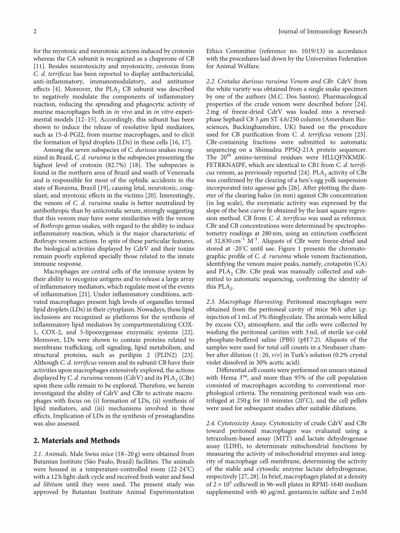
2.2. Crotalus durissus ruruima Venom and CBr. CdrV fromthe white variety was obtained from a single snake specimenby one of the authors (M.C. Dos Santos). Pharmacologicalproperties of the crude venom were described before [24].2mg of freeze-dried CdrV was loaded into a reversed-phase Sephasil C8 5 μm ST 4.6/250 column (Amersham Bio-sciences, Buckinghamshire, UK) based on the procedureused for CB purification from C. d. terrificus venom [25].CBr-containing fractions were submitted to automaticsequencing on a Shimadzu PPSQ-21A protein sequencer.The 20th amino-terminal residues were HLLQFNKMIK-FETRKNAIPF, which are identical to CB1 from C. d. terrifi-cus venom, as previously reported [24]. PLA2 activity of CBrwas confirmed by the clearing of a hen’s egg yolk suspensionincorporated into agarose gels [26]. After plotting the diam-eter of the clearing halos (in mm) against CBr concentration(in log scale), the enzymatic activity was expressed by theslope of the best curve fit obtained by the least square regres-sion method. CB from C. d. terrificus was used as reference.CBr and CB concentrations were determined by spectropho-tometry readings at 280 nm, using an extinction coefficientof 32,830 cm-1 M-1. Aliquots of CBr were freeze-dried andstored at -20°C until use. Figure 1 presents the chromato-graphic profile of C. d. ruruima whole venom fractionation,identifying the venom major peaks, namely, crotapotin (CA)and PLA2 CBr. CBr peak was manually collected and sub-mitted to automatic sequencing, confirming the identity ofthis PLA2.
2.3. Macrophage Harvesting. Peritoneal macrophages wereobtained from the peritoneal cavity of mice 96h after i.p.injection of 1mL of 3% thioglycolate. The animals were killedby excess CO2 atmosphere, and the cells were collected bywashing the peritoneal cavities with 3mL of sterile ice-coldphosphate-buffered saline (PBS) (pH7.2). Aliquots of thesamples were used for total cell counts in a Neubauer cham-ber after dilution (1 : 20, v/v) in Turk’s solution (0.2% crystalviolet dissolved in 30% acetic acid).
Differential cell counts were performed on smears stainedwith Hema 3™, and more than 95% of the cell populationconsisted of macrophages according to conventional mor-phological criteria. The remaining peritoneal wash was cen-trifuged at 250 g for 10 minutes (20°C), and the cell pelletswere used for subsequent studies after suitable dilutions.
2.4. Cytotoxicity Assay. Cytotoxicity of crude CdrV and CBrtoward peritoneal macrophages was evaluated using atetrazolium-based assay (MTT) and lactate dehydrogenaseassay (LDH), to determinate mitochondrial functions bymeasuring the activity of mitochondrial enzymes and integ-rity of macrophage cell membrane, determining the activityof the stable and cytosolic enzyme lactate dehydrogenase,respectively [27, 28]. In brief, macrophages plated at a densityof 2 × 10⁵ cells/well in 96-well plates in RPMI-1640 mediumsupplemented with 40 μg/mL gentamicin sulfate and 2mM
2 Journal of Immunology Research
L-glutamine were incubated with 100μL of selected concen-trations of crude venom or CBr diluted in medium or withthe same volume of medium alone (control), for 1, 3, 6, and12 h at 37°C in a humidified atmosphere of 5% CO2. MTT(5mg/mL) was dissolved in PBS and filtered to remove asmall amount of insoluble residue present in some batches.Stock MTT solution (10% in culture medium) was added toall wells, and the plates were incubated at 37°C for 3 h.Next, a volume of 100 μL of DMSO was added to the wellsand mixed thoroughly at room temperature for 30min.Absorbance at 540nm was then recorded in a microtiterplate reader. Results were expressed as the percentage ofviable cells, and the control cells (incubated with mediumalone) were considered 100% viable. To perform LDHassay, samples were incubated with culture medium orCBr or CdrV for 1, 3, 6, and 12h. The supernatant of the cellswas transferred to a 96-well plate. After adding the LDH sub-strate solution to each well, the plate was incubated for 30min.After incubation, the absorbance was read at 490nm on anELISA plate reader. Absorbance in wells without cells but con-taining the medium alone was used as the control. The per-centage of cytotoxicity was calculated by the followingequation: %cytotoxicity rate = ½ð LDH releaseÞ − ðcontrolÞ −ðtreated sample LDHactivityÞ� ÷ total of LDH activity.
2.5. Macrophage Culture and Stimulation. Peritoneal macro-phages were plated on sterile glass coverslips in 24-well poly-styrene culture plates and allowed to attach for 30min at37°C under a 5% CO2 atmosphere. Nonadherent cells wereremoved by washing plates with PBS. After that, cells werechallenged with selected concentrations of CdrV (1, 10, and100 μg/mL) or CBr (1.25, 3.25, 6.5, and 13 μg/mL) or cul-tured medium alone (control) for different periods of time.Whenever appropriate, the following inhibitors were used:
valeryl salicylate (30 μM)—an inhibitor of COX-1—or etori-coxib (1μM)—an inhibitor of COX-2. All the stock solutionswere prepared in DMSO and stored at -20°C. Aliquots werediluted in RPMI-1640 immediately before use to give therequired concentration. The final DMSO concentration wasalways lower than 1% and had no effect on the basal numberof LDs. Pharmacological inhibitors were added 60min beforethe stimulation of macrophages with CdrV or CBr or culturemedium (control). Cells treated with the inhibitors were ana-lyzed for viability with the MTT assay. No significant changesin cell viability were registered with any of the above agents orvehicle at the concentrations used (Supplementary Figure 1).
2.6. Lipid Droplet Staining and Quantification. The analysisof LD numbers was performed in OsO4-stained cells asdescribed by Leiguez et al. [17, 29]. In brief, macrophagesplated on sterile glass coverslips (2 × 10⁵ cells) were fixed in4% paraformaldehyde (PFA) in 0.1M phosphate buffer,pH7.2, for 15min and stained with OsO4. The coverslipswere washed in 0.1M phosphate buffer, stained in 1% OsO4(30min), washed in deionized H2O, immersed in 1.0% thio-carbohydrazide (5min), washed again in 0.1M phosphatebuffer, restained with 1% OsO4 (3min), washed with H2O,and then dried and mounted. The morphology of the fixedcells was examined, and LDs were identified as round osmio-philic structures, which were counted under phase-contrastmicroscopy using a 100x objective lens in 50 consecutivelyscanned macrophages per coverslip. The total number oflipid droplets obtained in 50 macrophages is summed anddivided by 50, as we can suggest an average of lipid dropletswe have per cell. For assays with fluorescent-labeled LDs,peritoneal macrophages (2 × 10⁵ cells) adhered to autoclavedglass coverslips were incubated with Nile Red staining solu-tion freshly prepared in 0.1M phosphate buffer (10 μg/mL)
100
80
60
40
20
0
250
200
150
100
50
0
0 20 40 60 80 100 min
mAU % BCBr
CA
Figure 1: Chromatography profile of crude C. d. ruruima venom on Sephasil C8 5 μm ST 4.6/250 (Amersham Biosciences). Flow rate:1.l mL/min. Fractions of 0.5ml were eluted under a gradient of 0.1% TFA in acetonitrile (0–25% in 0.4mL; 25–50% in 105mL; and 50-100%in 0.4mL). PLA2 activity of CBr was 1:459 ± 0:2392mm · mL/mg. Under the same experimental conditions, CB from C. d. terrificus gave2:171 ± 0:1870mm.
3Journal of Immunology Research
for 30min at room temperature and washed with phosphatebuffer. After several washes, the coverslips were mountedwith Fluoromount-G and examined under a fluorescencemicroscope (Zeiss LSM 510 Meta confocal microscope).
2.7. Immunocytochemistry Analysis. Detection of PLIN2 inCdrV- or CBr-stimulated macrophages was performed byPLIN2 immunostaining. Cells were fixed in 2% formalde-hyde at room temperature for 20min and permeabilized with0.2% Triton-X 100 in 0.1M phosphate buffer. The unspecificsites were blocked with 0.5% albumin in 0.1M phosphatebuffer for 60min. After that, macrophages were incubatedfor 1 h with a rabbit polyclonal anti-PLIN2 (1 : 2000) dilutedin 0.1M phosphate buffer with 0.2% Triton-X100. After sev-eral washes with PBS (10min each), the preparations wereincubated with secondary Alexa Fluor 488-conjugated anti-rabbit antibody (1,500) in the dark for 1 h. After new severalwashes, the slides were mounted with Fluoromount-G andexamined under a confocal laser scanning microscope (ZeissLSM 510 Meta). For immunodetection of PGE2 cells werefixed and permeabilized during 1 h at 37°C with 1% EDACin calcium- and magnesium-free Hank’s balanced salt solu-tion (HBSS¯/¯) [30].
Then, macrophages were washed with HBSS¯/¯ andblocked with 0.5% albumin in 0.1M phosphate buffer for60min. The cells were washed again with HBSS¯/¯ andincubated for 1 h with monoclonal antibodies againstPGE2 (1 : 100). After further washes, cells were incubatedwith goat anti-mouse Alexa Fluor 488 (1 : 250) and NileRed solution (1 : 250) for 1 h. The coverslips were thenwashed three times and mounted with Fluoromount-Gand examined under a confocal laser-scanning microscope(Zeiss LSM 510 Meta) [30].
2.8. Western Blotting. For the analysis of COX-1, COX-2,and PLIN2 protein content, whole cell extracts wereobtained by lysing the cell pellets with 80mL of samplebuffer (0.5M Tris-HCl, pH6.8, 20% SDS, 1% glycerol, 1Mβ-mercaptoethanol, 0.1% bromophenol blue) and thenboiled for 10min at 100°C. Samples were resolved by electro-phoresis (SDS-PAGE) on a 10% separation gel overlaid witha 5% stacking gel. After that, proteins were transferred to anitrocellulose membrane (GE Healthcare, Buckinghamshire,UK) using a Mini Trans-Blot® (Bio-Rad Laboratories, Rich-mond, CA, USA), and the membranes were blocked for 1 hwith 5% nonfat dry milk in TTBS (20mM Tris, 100mMNaCl and 0.5% Tween 20). Membranes were then incubatedwith a primary rabbit antibody against COX-1 or COX-2 orPLIN2 (Abcam, San Francisco, USA) overnight at 4°C orwith β-actin (Sigma, San Louis, USA) for 1 h at room tem-perature. They were then washed and incubated with theappropriate horseradish peroxidase-conjugated anti-rabbitIgG secondary antibody (1 : 1000 dilution, 1 h, at room tem-perature). Detectionwas by the enhanced chemiluminescence(ECL) method according to the manufacturer’s instructions(GE Healthcare, Buckinghamshire, UK).
Band densities were quantified with a GS 800 Densitom-eter (Bio-Rad Laboratories, Richmond, CA) using Molecular
Analyst® image analysis software (Bio-Rad Laboratories,Richmond, CA).
2.9. Quantification of PGE2, PGD2, and TXA2 Concentrations.PGE2, PGD2, and TXA2 concentrations were determined byenzyme immunoassay using a commercial kit (CaymanChemical Company, Ann Arbor, MI). In brief, 50μL aliquotsof each extracted sample were incubated with the eicosa-noids conjugated with acetylcholinesterase, and the specificrabbit antiserum in 96-well plates was coated with anti-rabbit IgG mouse monoclonal antibody. After the additionof the substrate, the absorbance of the samples was recordedat 405nm in a microplate reader (Labsystems Multiskan),and concentrations of PGE2, PGD2, and TXA2 concentra-tions were estimated from standard curves [31].
2.10. Statistical Analysis. Data are expressed as the mean ±standard error of mean (SEM) of at least three independentexperiments. Multiple comparisons among groups wereperformed with one-way ANOVA followed by Tukey’s test.Probabilities of less than 5% (p< 0.05) were considered statis-tically significant.
3. Results
3.1. CdrV and CBr Induce LD Formation in PeritonealMacrophages. Preliminary studies were performed to evalu-ate the effect of CdrV and CBr on the viability of isolatedmacrophages by using the tetrazolium-based (MTT) andLDH assays. To this purpose, macrophages in culture wereincubated with three distinct concentrations of CdrV (1, 10,and 100 μg/mL) or CBr (3.25, 6.5, and 13μg/mL) or mediumalone (control) for 12h. Obtained results showed that neitherCdrV nor CBr affected mitochondrial enzyme activity incomparison with control cells (data not shown). Similarly,neither CdrV nor CBr altered the release of LDH in compar-ison with control cells (data not shown). These results indi-cate that both CdrV and CBr did not affect cell viability atthe concentrations and time of incubation used.
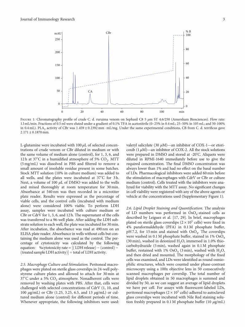
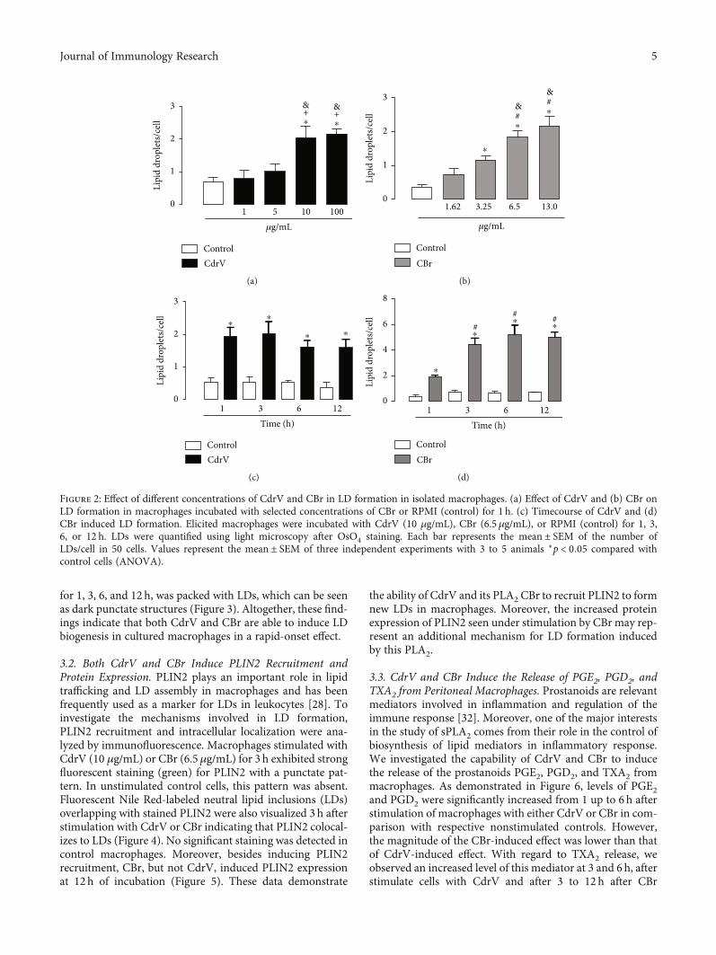
LD formation was analyzed after incubation of macro-phages with noncytotoxic concentrations of CdrV (1, 10,and 100 μg/mL) or CBr (1.62, 3.25, 6.5, and 13μg/mL) orRPMI (control) for 1 h. As shown in Figures 2(a) and 2(b),incubation of macrophages with CdrV at concentrations of10 to 100 μg/mL induced a significant increase in the numberof LDs compared with control cells incubated with culturemedium alone. Incubation of macrophages with CBr atconcentrations of 6.5 and 13 μg/mL, but not 1.25 and3.25μg/mL, induced a significant increase in the number ofLDs compared with control cells.
To determine the timecourse of CdrV- and CBr-inducedLD formation, submaximal concentrations of these agentswere used (10μg/mL for CdrV and 6.5μg/mL for CBr), andthe number of LDswas determined after 1–12 h of incubation.As shown in Figures 2(c) and 2(d), both CdrV andCBr causedan increase in LD number from 1 up to 12h of incubationcompared with control cells. Control macrophages stainedwith OsO4 showed very few LDs in the cytoplasm. In contrast,the cytoplasm of macrophages stimulated by CdrV or CBr,
4 Journal of Immunology Research
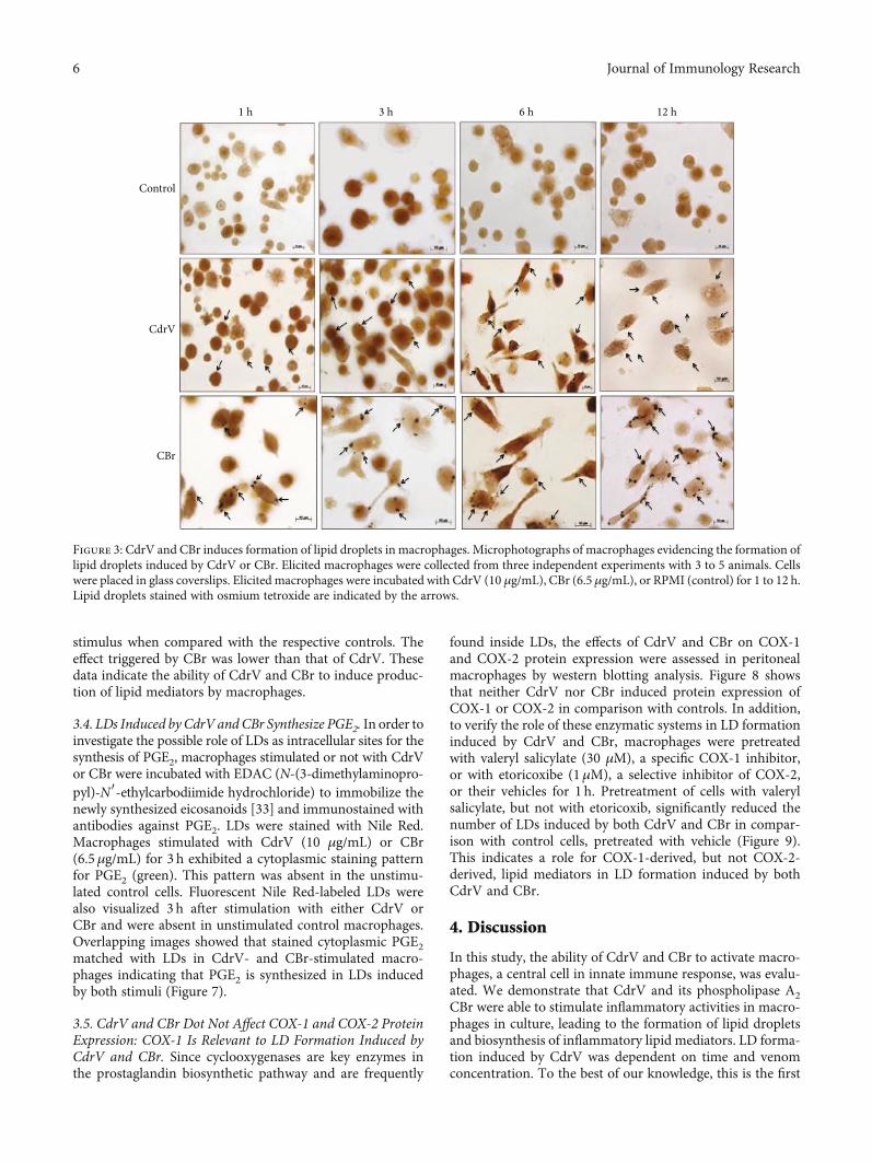
for 1, 3, 6, and 12 h, was packed with LDs, which can be seenas dark punctate structures (Figure 3). Altogether, these find-ings indicate that both CdrV and CBr are able to induce LDbiogenesis in cultured macrophages in a rapid-onset effect.
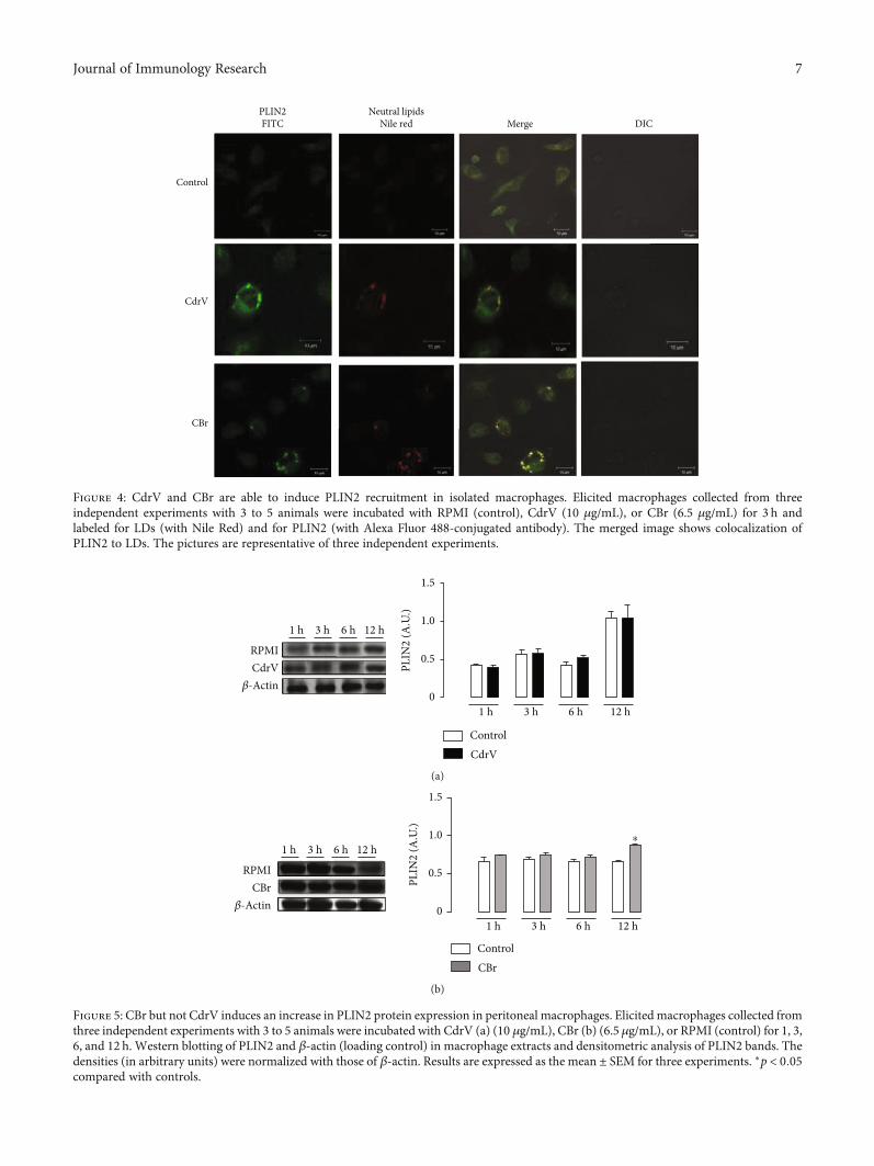
3.2. Both CdrV and CBr Induce PLIN2 Recruitment andProtein Expression. PLIN2 plays an important role in lipidtrafficking and LD assembly in macrophages and has beenfrequently used as a marker for LDs in leukocytes [28]. Toinvestigate the mechanisms involved in LD formation,PLIN2 recruitment and intracellular localization were ana-lyzed by immunofluorescence. Macrophages stimulated withCdrV (10 μg/mL) or CBr (6.5μg/mL) for 3 h exhibited strongfluorescent staining (green) for PLIN2 with a punctate pat-tern. In unstimulated control cells, this pattern was absent.Fluorescent Nile Red-labeled neutral lipid inclusions (LDs)overlapping with stained PLIN2 were also visualized 3 h afterstimulation with CdrV or CBr indicating that PLIN2 colocal-izes to LDs (Figure 4). No significant staining was detected incontrol macrophages. Moreover, besides inducing PLIN2recruitment, CBr, but not CdrV, induced PLIN2 expressionat 12h of incubation (Figure 5). These data demonstrate
the ability of CdrV and its PLA2 CBr to recruit PLIN2 to formnew LDs in macrophages. Moreover, the increased proteinexpression of PLIN2 seen under stimulation by CBr may rep-resent an additional mechanism for LD formation inducedby this PLA2.
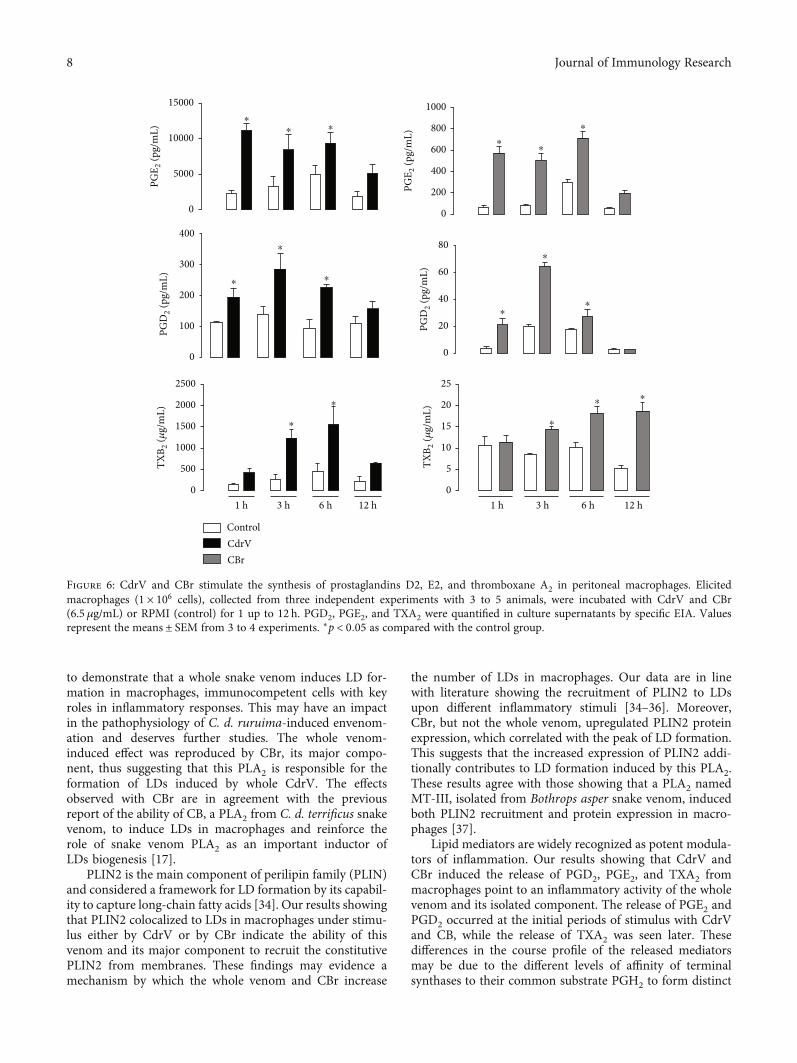
3.3. CdrV and CBr Induce the Release of PGE2, PGD2, andTXA2 from Peritoneal Macrophages. Prostanoids are relevantmediators involved in inflammation and regulation of theimmune response [32]. Moreover, one of the major interestsin the study of sPLA2 comes from their role in the control ofbiosynthesis of lipid mediators in inflammatory response.We investigated the capability of CdrV and CBr to inducethe release of the prostanoids PGE2, PGD2, and TXA2 frommacrophages. As demonstrated in Figure 6, levels of PGE2and PGD2 were significantly increased from 1 up to 6 h afterstimulation of macrophages with either CdrV or CBr in com-parison with respective nonstimulated controls. However,the magnitude of the CBr-induced effect was lower than thatof CdrV-induced effect. With regard to TXA2 release, weobserved an increased level of this mediator at 3 and 6h, afterstimulate cells with CdrV and after 3 to 12 h after CBr
3
2
1
0
&+⁎
&+⁎
1 5 10 100𝜇g/mL
Lipi
d dr
ople
ts/ce
ll
ControlCdrV
(a)
3
2
1
0
#⁎
1.62 3.25 6.5 13.0
𝜇g/mL
Lipi
d dr
ople
ts/ce
ll
⁎
& #⁎
&
ControlCBr
(b)
3
2
1
0
⁎⁎
⁎ ⁎
1 3 6 12Time (h)
Lipi
d dr
ople
ts/ce
ll
ControlCdrV
(c)
8
4
6
2
0
⁎
⁎
⁎⁎
1 3 6 12Time (h)
Lipi
d dr
ople
ts/ce
ll
## #
ControlCBr
(d)
Figure 2: Effect of different concentrations of CdrV and CBr in LD formation in isolated macrophages. (a) Effect of CdrV and (b) CBr onLD formation in macrophages incubated with selected concentrations of CBr or RPMI (control) for 1 h. (c) Timecourse of CdrV and (d)CBr induced LD formation. Elicited macrophages were incubated with CdrV (10 μg/mL), CBr (6.5μg/mL), or RPMI (control) for 1, 3,6, or 12 h. LDs were quantified using light microscopy after OsO4 staining. Each bar represents the mean ± SEM of the number ofLDs/cell in 50 cells. Values represent the mean ± SEM of three independent experiments with 3 to 5 animals ∗p < 0:05 compared withcontrol cells (ANOVA).
5Journal of Immunology Research
stimulus when compared with the respective controls. Theeffect triggered by CBr was lower than that of CdrV. Thesedata indicate the ability of CdrV and CBr to induce produc-tion of lipid mediators by macrophages.
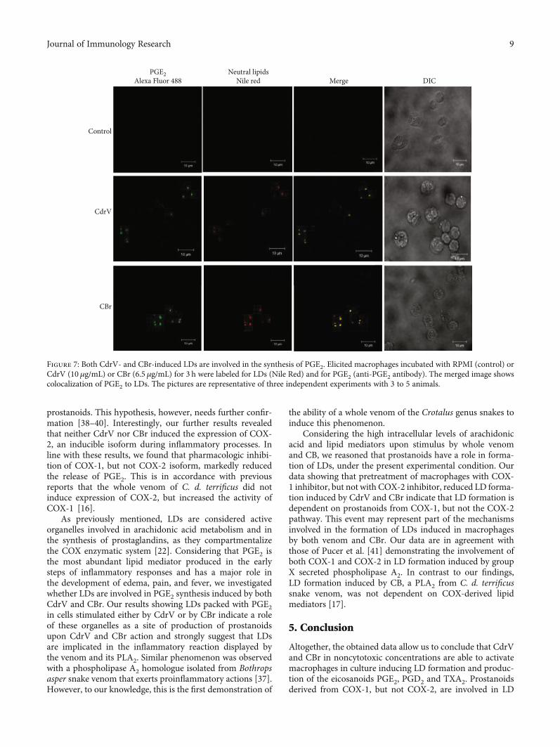
3.4. LDs Induced by CdrV and CBr Synthesize PGE2. In order toinvestigate the possible role of LDs as intracellular sites for thesynthesis of PGE2, macrophages stimulated or not with CdrVor CBr were incubated with EDAC (N-(3-dimethylaminopro-pyl)-N′-ethylcarbodiimide hydrochloride) to immobilize thenewly synthesized eicosanoids [33] and immunostained withantibodies against PGE2. LDs were stained with Nile Red.Macrophages stimulated with CdrV (10 μg/mL) or CBr(6.5μg/mL) for 3 h exhibited a cytoplasmic staining patternfor PGE2 (green). This pattern was absent in the unstimu-lated control cells. Fluorescent Nile Red-labeled LDs werealso visualized 3 h after stimulation with either CdrV orCBr and were absent in unstimulated control macrophages.Overlapping images showed that stained cytoplasmic PGE2matched with LDs in CdrV- and CBr-stimulated macro-phages indicating that PGE2 is synthesized in LDs inducedby both stimuli (Figure 7).
3.5. CdrV and CBr Dot Not Affect COX-1 and COX-2 ProteinExpression: COX-1 Is Relevant to LD Formation Induced byCdrV and CBr. Since cyclooxygenases are key enzymes inthe prostaglandin biosynthetic pathway and are frequently
found inside LDs, the effects of CdrV and CBr on COX-1and COX-2 protein expression were assessed in peritonealmacrophages by western blotting analysis. Figure 8 showsthat neither CdrV nor CBr induced protein expression ofCOX-1 or COX-2 in comparison with controls. In addition,to verify the role of these enzymatic systems in LD formationinduced by CdrV and CBr, macrophages were pretreatedwith valeryl salicylate (30 μM), a specific COX-1 inhibitor,or with etoricoxibe (1μM), a selective inhibitor of COX-2,or their vehicles for 1 h. Pretreatment of cells with valerylsalicylate, but not with etoricoxib, significantly reduced thenumber of LDs induced by both CdrV and CBr in compar-ison with control cells, pretreated with vehicle (Figure 9).This indicates a role for COX-1-derived, but not COX-2-derived, lipid mediators in LD formation induced by bothCdrV and CBr.
4. Discussion
In this study, the ability of CdrV and CBr to activate macro-phages, a central cell in innate immune response, was evalu-ated. We demonstrate that CdrV and its phospholipase A2CBr were able to stimulate inflammatory activities in macro-phages in culture, leading to the formation of lipid dropletsand biosynthesis of inflammatory lipid mediators. LD forma-tion induced by CdrV was dependent on time and venomconcentration. To the best of our knowledge, this is the first
Control
CdrV
CBr
1 h 3 h 6 h 12 h
Figure 3: CdrV and CBr induces formation of lipid droplets in macrophages. Microphotographs of macrophages evidencing the formation oflipid droplets induced by CdrV or CBr. Elicited macrophages were collected from three independent experiments with 3 to 5 animals. Cellswere placed in glass coverslips. Elicited macrophages were incubated with CdrV (10 μg/mL), CBr (6.5 μg/mL), or RPMI (control) for 1 to 12 h.Lipid droplets stained with osmium tetroxide are indicated by the arrows.
6 Journal of Immunology Research
Control
CdrV
CBr
PLIN2FITC
Neutral lipidsNile red Merge DIC
Figure 4: CdrV and CBr are able to induce PLIN2 recruitment in isolated macrophages. Elicited macrophages collected from threeindependent experiments with 3 to 5 animals were incubated with RPMI (control), CdrV (10 μg/mL), or CBr (6.5 μg/mL) for 3 h andlabeled for LDs (with Nile Red) and for PLIN2 (with Alexa Fluor 488-conjugated antibody). The merged image shows colocalization ofPLIN2 to LDs. The pictures are representative of three independent experiments.
1.5
1.0
0.5
01 h
PLIN
2 (A
.U.)
3 h 6 h 12 h
1 h 3 h 6 h 12 h
RPMICdrV
𝛽-Actin
ControlCdrV
(a)
1.5
1.0
0.5
0
⁎
1 h
PLIN
2 (A
.U.)
ControlCBr
3 h 6 h 12 h
1 h 3 h 6 h 12 h
RPMICBr
𝛽-Actin
(b)
Figure 5: CBr but not CdrV induces an increase in PLIN2 protein expression in peritoneal macrophages. Elicited macrophages collected fromthree independent experiments with 3 to 5 animals were incubated with CdrV (a) (10 μg/mL), CBr (b) (6.5 μg/mL), or RPMI (control) for 1, 3,6, and 12 h. Western blotting of PLIN2 and β-actin (loading control) in macrophage extracts and densitometric analysis of PLIN2 bands. Thedensities (in arbitrary units) were normalized with those of β-actin. Results are expressed as themean ± SEM for three experiments. ∗p < 0:05compared with controls.
7Journal of Immunology Research
to demonstrate that a whole snake venom induces LD for-mation in macrophages, immunocompetent cells with keyroles in inflammatory responses. This may have an impactin the pathophysiology of C. d. ruruima-induced envenom-ation and deserves further studies. The whole venom-induced effect was reproduced by CBr, its major compo-nent, thus suggesting that this PLA2 is responsible for theformation of LDs induced by whole CdrV. The effectsobserved with CBr are in agreement with the previousreport of the ability of CB, a PLA2 from C. d. terrificus snakevenom, to induce LDs in macrophages and reinforce therole of snake venom PLA2 as an important inductor ofLDs biogenesis [17].
PLIN2 is the main component of perilipin family (PLIN)and considered a framework for LD formation by its capabil-ity to capture long-chain fatty acids [34]. Our results showingthat PLIN2 colocalized to LDs in macrophages under stimu-lus either by CdrV or by CBr indicate the ability of thisvenom and its major component to recruit the constitutivePLIN2 from membranes. These findings may evidence amechanism by which the whole venom and CBr increase
the number of LDs in macrophages. Our data are in linewith literature showing the recruitment of PLIN2 to LDsupon different inflammatory stimuli [34–36]. Moreover,CBr, but not the whole venom, upregulated PLIN2 proteinexpression, which correlated with the peak of LD formation.This suggests that the increased expression of PLIN2 addi-tionally contributes to LD formation induced by this PLA2.These results agree with those showing that a PLA2 namedMT-III, isolated from Bothrops asper snake venom, inducedboth PLIN2 recruitment and protein expression in macro-phages [37].
Lipid mediators are widely recognized as potent modula-tors of inflammation. Our results showing that CdrV andCBr induced the release of PGD2, PGE2, and TXA2 frommacrophages point to an inflammatory activity of the wholevenom and its isolated component. The release of PGE2 andPGD2 occurred at the initial periods of stimulus with CdrVand CB, while the release of TXA2 was seen later. Thesedifferences in the course profile of the released mediatorsmay be due to the different levels of affinity of terminalsynthases to their common substrate PGH2 to form distinct
15000
10000
5000
0
⁎⁎ ⁎
PGE 2
(pg/
mL)
1000
800
400
600
200
0
⁎⁎
⁎
PGE 2
(pg/
mL)
400
200
300
100
0
⁎
⁎
⁎
PGD
2 (pg
/mL)
80
40
60
20
0
⁎
⁎
⁎
PGD
2 (pg
/mL)
25
15
20
10
5
0
⁎
⁎ ⁎
TXB 2
(𝜇g/
mL)
1 h 3 h 6 h 12 h
⁎
⁎
2500
1500
2000
1000
500
0
TXB 2
(𝜇g/
mL)
1 h 3 h 6 h 12 h
ControlCdrVCBr
Figure 6: CdrV and CBr stimulate the synthesis of prostaglandins D2, E2, and thromboxane A2 in peritoneal macrophages. Elicitedmacrophages (1 × 10⁶ cells), collected from three independent experiments with 3 to 5 animals, were incubated with CdrV and CBr(6.5 μg/mL) or RPMI (control) for 1 up to 12 h. PGD2, PGE2, and TXA2 were quantified in culture supernatants by specific EIA. Valuesrepresent the means ± SEM from 3 to 4 experiments. ∗p < 0:05 as compared with the control group.
8 Journal of Immunology Research
prostanoids. This hypothesis, however, needs further confir-mation [38–40]. Interestingly, our further results revealedthat neither CdrV nor CBr induced the expression of COX-2, an inducible isoform during inflammatory processes. Inline with these results, we found that pharmacologic inhibi-tion of COX-1, but not COX-2 isoform, markedly reducedthe release of PGE2. This is in accordance with previousreports that the whole venom of C. d. terrificus did notinduce expression of COX-2, but increased the activity ofCOX-1 [16].
As previously mentioned, LDs are considered activeorganelles involved in arachidonic acid metabolism and inthe synthesis of prostaglandins, as they compartmentalizethe COX enzymatic system [22]. Considering that PGE2 isthe most abundant lipid mediator produced in the earlysteps of inflammatory responses and has a major role inthe development of edema, pain, and fever, we investigatedwhether LDs are involved in PGE2 synthesis induced by bothCdrV and CBr. Our results showing LDs packed with PGE2in cells stimulated either by CdrV or by CBr indicate a roleof these organelles as a site of production of prostanoidsupon CdrV and CBr action and strongly suggest that LDsare implicated in the inflammatory reaction displayed bythe venom and its PLA2. Similar phenomenon was observedwith a phospholipase A2 homologue isolated from Bothropsasper snake venom that exerts proinflammatory actions [37].However, to our knowledge, this is the first demonstration of
the ability of a whole venom of the Crotalus genus snakes toinduce this phenomenon.
Considering the high intracellular levels of arachidonicacid and lipid mediators upon stimulus by whole venomand CB, we reasoned that prostanoids have a role in forma-tion of LDs, under the present experimental condition. Ourdata showing that pretreatment of macrophages with COX-1 inhibitor, but not with COX-2 inhibitor, reduced LD forma-tion induced by CdrV and CBr indicate that LD formation isdependent on prostanoids from COX-1, but not the COX-2pathway. This event may represent part of the mechanismsinvolved in the formation of LDs induced in macrophagesby both venom and CBr. Our data are in agreement withthose of Pucer et al. [41] demonstrating the involvement ofboth COX-1 and COX-2 in LD formation induced by groupX secreted phospholipase A2. In contrast to our findings,LD formation induced by CB, a PLA2 from C. d. terrificussnake venom, was not dependent on COX-derived lipidmediators [17].
5. Conclusion
Altogether, the obtained data allow us to conclude that CdrVand CBr in noncytotoxic concentrations are able to activatemacrophages in culture inducing LD formation and produc-tion of the eicosanoids PGE2, PGD2 and TXA2. Prostanoidsderived from COX-1, but not COX-2, are involved in LD
Control
CdrV
CBr
Neutral lipidsNile red Merge DIC
PGE2Alexa Fluor 488
Figure 7: Both CdrV- and CBr-induced LDs are involved in the synthesis of PGE2. Elicited macrophages incubated with RPMI (control) orCdrV (10 μg/mL) or CBr (6.5 μg/mL) for 3 h were labeled for LDs (Nile Red) and for PGE2 (anti-PGE2 antibody). The merged image showscolocalization of PGE2 to LDs. The pictures are representative of three independent experiments with 3 to 5 animals.
9Journal of Immunology Research

RPMICdrV
𝛽-Actin
1 h 3 h 6 h 12 hRPMI
CBr𝛽-Actin
1 h 3 h 6 h 12 h
RPMIVCdr
𝛽-Actin
1 h 3 h 6 h 12 h
RPMICBr
𝛽-Actin
1 h 3 h 6 h 12 h
ControlCdrVCBr
2.0
1.5
1.0
0.5
0.0
COX-
1 (A
.U.)
1 3 6 12
2.0
1.5
1.0
0.5
0.0
COX-
1 (A
.U.)
1 3 6 12
Time (h)Time (h)
Time (h)
2.0
1.5
1.0
0.5
0.0
COX-
2 (A
.U.)
1 3 6 12
0.4
0.3
0.2
0.1
0.0
COX-
2 (A
.U.)
1 3 6 12
Time (h)
Figure 8: COX-2 and COX-1 protein expression is not upregulated by CdrV and CBr in peritoneal macrophages. Neither Cdr venom nor CBraffected COX-1 and COX-2 protein expression in peritoneal macrophages. Elicited macrophages from three independent experiments with 3 to5 animals were incubated with CdrV (10μg/mL) and CBr (6.5μg/mL) or RPMI (control) for 1, 3, 6, and 12h. The figure shows western blottingand densitometric analysis of COX-1 and COX-2 and β-actin (loading control) bands in macrophage extracts. The densities (in arbitraryunits)were normalizedwith those ofβ-actin. Results are expressed as themean ± SEM for three experiments. ∗p < 0:05 comparedwith controls.
10
8
6
4
2
0
Lipi
d dr
ople
ts/ce
ll
Vehicle Valeryl Etoricoxib
Salicylate
8
6
4
2
0
Lipi
d dr
ople
ts/ce
ll
Vehicle Valeryl Etoricoxib
Salicylate
ControlCdrVCBr
⁎
⁎
⁎⁎
⁎#
⁎#
Figure 9: COX-1 but not COX-2 is involved in LD formation induced by CdrV and CBr in peritoneal macrophages. Elicited macrophagesfrom three independent experiments with 3 to 5 animals were treated with valeryl salicylate (30 μM) or etoricoxibe (1 μM) for 1 h beforestimulation with CdrV (10 μg/mL) or CBr (6.5 μg/mL) or RPMI (control) for 1 h. LDs were counted using light microscopy after osmiumstaining. Each bar represents the mean ± SEM of the number of LDs/cell in 50 cells. Values represent the means ± SEM for three to fiveanimals. ∗p < 0:05 compared with control cells; #p < 0:05.
10 Journal of Immunology Research
assembly. Conversely, LDs play an active role in productionof prostanoids induced by both CdrV and CBr. These datareveal a proinflammatory activity of both Crotalus durissusruruima whole venom and its sPLA2 by exerting stimulatoryactivities on macrophages, which are central cells in immu-nological responses. Therefore, new perspectives can beopened in the search of new therapeutic targets aimed atreducing the severity of the accidents caused by the Crotalusdurissus ruruima snake.
Data Availability
Readers may access the data underlying the findings ofthis study by contacting the contributing author, CatarinaTeixeira, at [email protected].
Conflicts of Interest
The authors declare that there is no conflict of interestregarding the publication of this paper.
Acknowledgments
This work was supported by research grants from the Funda-ção de Amparo à Pesquisa do Estado de São Paulo (FAPESP,Brazil; grant numbers 2014/18549-1 and 2015/50040-4).AEZC is a recipient of a PhD fellowship from CAPES(1667206); KCG is a recipient of a post-doc fellowshipfrom FAPESP (grant number 18/05637-0); EL is a recipi-ent of a post-doc fellowship from FAPESP (grant number2015/24701-3) and CT is a recipient of PQ-CNPq (grantnumber 307379/2016-7). CLFD is a recipient of a produc-tivity fellowship from FAPEMIG (grant number BIP-00182-18). The authors would like to thank Renata Hagedo Amaral Hernandez for providing technical assistanceand Dr. Henrique Krambeck Rofatto (FAPESP 00/11624-5,Butantan Institute) for helping with the confocal laser scan-ning microscopy analysis.
Supplementary Materials
Supplementary Figure 1: Effect of CdrV and CBr on cellviability. Cells were incubated with CdrV or CBr (3.25, 6.5,and 13.0μg/mL) or RPMI (control) from 1 to 12 h, andcytotoxicity was assessed by (A) LDH and (B) MTT assays.Values represent the mean ± SEM from four animals(ANOVA). (Supplementary Materials)
References
[1] J. M. Gutiérrez and H. W. Fan, “Improving the control ofsnakebite envenomation in Latin America and the Caribbean:a discussion on pending issues,” Transactions of the Royal Soci-ety of Tropical Medicine and Hygiene, vol. 112, no. 12, pp. 523–526, 2018.
[2] D. F. Cardoso and I. Mota, “Effect of Crotalus venom on thehumoral and cellular immune response,” Toxicon, vol. 35,no. 4, pp. 607–612, 1997.
[3] A. Rangel-Santos, C. Lima, M. Lopes-Ferreira, and D. F. Car-doso, “Immunosuppresive role of principal toxin (crotoxin)
of Crotalus durissus terrificus venom,” Toxicon, vol. 44,no. 6, pp. 609–616, 2004.
[4] S. C. Sampaio, S. Hyslop, M. R. Fontes et al., “Crotoxin: novelactivities for a classic β-neurotoxin,” Toxicon, vol. 55, no. 6,pp. 1045–1060, 2010.
[5] A. P. Freitas, B. C. Favoretto, P. B. Clissa, S. C. Sampaio, andE. L. Faquim-Mauro, “Crotalus durissus terrificus venommodulates the functional activity of dendritic cells via formylpeptide receptors,” Journal of Immunology Research, vol. 2018,Article ID 7873257, 15 pages, 2018.
[6] P. E. Vanzolini and M. E. Calleffo, “A taxonomic bibliographyof the South American snakes of the Crotalus durissus complex(Serpentes, Viperidae),” Anais da Academia Brasileira de Ciên-cias, vol. 74, no. 1, pp. 37–83, 2002.
[7] K. Rübsamen, H. Breithaupt, and E. Habermann, “Biochemis-try and pharmacology of the crotoxin complex. I. Subfractio-nation and recombination of the crotoxin complex,”Naunyn-Schmiedebergs Archiv für Pharmakologie, vol. 270,no. 3, pp. 274–288, 1971.
[8] R. A. Hendon and H. Fraenkel-Conrat, “Biological roles of thetwo components of crotoxin,” Proceedings of the NationalAcademy of Sciences, vol. 68, no. 7, pp. 1560–1563, 1971.
[9] G. Faure and C. Bon, “Several isoforms of crotoxin are presentin individual venoms from the South American rattlesnakeCrotalus durissus terrificus,” Toxicon, vol. 25, no. 2, pp. 229–234, 1987.
[10] H. Breithaupt, “Enzymatic characteristics of crotalus phospho-lipase A2 and the crotoxin complex,” Toxicon, vol. 14, no. 3,pp. 221–233, 1976.
[11] C. Bon, C. Bouchier, V. Choumet et al., “Crotoxin, half-century of investigations on a phospholipase A2 neurotoxin,”Acta Physiologica et Pharmacologica Latinoamericana, vol. 39,no. 4, pp. 439–448, 1989.
[12] S. C. Sampaio, P. Brigatte, M. C. C. Sousa-e-Silva et al., “Con-tribution of crotoxin for the inhibitory effect of Crotalus duris-sus terrificus snake venom on macrophage function,” Toxicon,vol. 41, no. 7, pp. 899–907, 2003.
[13] S. C. Sampaio, A. C. Rangel-Santos, C. M. Peres, R. Curi, andY. Cury, “Inhibitory effect of phospholipase A(2) isolated fromCrotalus durissus terrificus venom on macrophage function,”Toxicon, vol. 45, no. 5, pp. 671–676, 2005.
[14] S. C. Sampaio, T. C. Alba-Loureiro, P. Brigatte et al., “Lipoxy-genase-derived eicosanoids are involved in the inhibitory effectof Crotalus durissus terrificus venom or crotoxin on rat macro-phage phagocytosis,” Toxicon, vol. 47, no. 3, pp. 313–321,2006.
[15] V. O. Zambelli, S. C. Sampaio, L. S. Sudo-Hayashi et al., “Cro-toxin alters lymphocyte distribution in rats: involvement ofadhesion molecules and lipoxygenase-derived mediators,”Toxicon, vol. 51, no. 8, pp. 1357–1367, 2008.
[16] V. Moreira, J. M. Gutiérrez, A. M. Soares, S. R. Zamunér,E. Purgatto, and T. CeF, “Secretory phospholipases A(2)isolated from Bothrops asper and from Crotalus durissus terri-ficus snake venoms induce distinct mechanisms for biosynthe-sis of prostaglandins E2 and D2 and expression ofcyclooxygenases,” Toxicon, vol. 52, no. 3, pp. 428–439, 2008.
[17] K. C. Giannotti, E. Leiguez, A. E. Z. Carvalho et al., “Asnake venom group IIA PLA2 with immunomodulatoryactivity induces formation of lipid droplets containing 15-d-PGJ2 in macrophages,” Scientific Reports, vol. 7, no. 1, article4098, 2017.
11Journal of Immunology Research
[18] J. J. Calvete, L. Sanz, P. Cid et al., “Snake venomics of theCentral American rattlesnake Crotalus simus and the SouthAmerican Crotalus durissus complex points to neurotoxicityas an adaptive paedomorphic trend along Crotalus dispersalin South America,” Journal of Proteome Research, vol. 9,no. 1, pp. 528–544, 2010.
[19] S. P. Nascimento, “Epidemiological characteristics of snakebites in the state of Roraima, Brazil, 1992-1998,” Cadernos deSaúde Pública, vol. 16, no. 1, pp. 271–276, 2000.
[20] M. C. Dos Santos, L. C. L. Ferreira, W. D. Da Silva, and M. F.Furtado, “Characterization of the biological activities of the‘yellow’ and ‘white’ venoms from Crotalus durissus ruruimacompared with the Crotalus durissus terrificus venom. Neu-tralizing activity of Crotalus durissus ruruima antivenins,”Toxicon, vol. 31, no. 11, pp. 1459–1469, 1993.
[21] N. Stoy, “Macrophage biology and pathobiology in the evolu-tion of immune responses: a functional analysis,” Pathobiol-ogy, vol. 69, no. 4, pp. 179–211, 2001.
[22] P. T. Bozza, W. Yu, and P. F. Weller, “Mechanisms of forma-tion and function of eosinophil lipid bodies: inducible intracel-lular sites involved in arachidonic acid metabolism,”Memóriasdo Instituto Oswaldo Cruz, vol. 92, Suppl 2, pp. 135–140, 1997.
[23] C. Sztalryd and D. L. Brasaemle, “The perilipin family of lipiddroplet proteins: gatekeepers of intracellular lipolysis,” Biochi-mica et Biophysica Acta - Molecular and Cell Biology of Lipids,vol. 1862, no. 10, pp. 1221–1232, 2017.
[24] M. C. Dos-Santos, E. B. Assis, T. D. Moreira, J. Pinheiro,and C. L. Fortes-Dias, “Individual venom variability in Cro-talus durissus ruruima snakes, a subspecies of Crotalus dur-issus from the Amazonian region,” Toxicon, vol. 46, no. 8,pp. 958–961, 2005.
[25] C. L. Fortes-Dias, Y. Lin, J. Ewell, C. R. Diniz, and T. Y. Liu, “Aphospholipase A2 inhibitor from the plasma of the SouthAmerican rattlesnake (Crotalus durissus terrificus). Proteinstructure, genomic structure, and mechanism of action,” TheJournal of Biological Chemistry, vol. 269, no. 22, pp. 15646–15651, 1994.
[26] E. Habermann and K. L. Hardt, “A sensitive and specific platetest for the quantitation of phospholipases,” Analytical Bio-chemistry, vol. 50, no. 1, pp. 163–173, 1972.
[27] T. Mosmann, “Rapid colorimetric assay for cellular growthand survival: application to proliferation and cytotoxicityassays,” Journal of Immunological Methods, vol. 65, no. 1-2,pp. 55–63, 1983.
[28] C. L. Markert, “Lactate dehydrogenase. Biochemistry andfunction of lactate dehydrogenase,” Cell Biochemistry andFunction, vol. 2, no. 3, pp. 131–134, 1984.
[29] E. Leiguez, J. P. Zuliani, A. M. Cianciarullo, C. M. Fernandes,J. M. Gutiérrez, and C. Teixeira, “A group IIA‐secreted phos-pholipase A2 from snake venom induces lipid body formationin macrophages: the roles of intracellular phospholipases A2and distinct signaling pathways,” Journal of Leukocyte Biology,vol. 90, no. 1, pp. 155–166, 2011.
[30] C. Bandeira-Melo, P. F. Weller, and P. T. Bozza, “EicosaCell:an Immunofluorescent-based assay to localize newly synthe-sized eicosanoid lipid mediators at intracellular sites,”Methodsin Molecular Biology, vol. 689, pp. 163–181, 2011.
[31] V. Moreira, B. Lomonte, M. A. Vinolo, R. Curi, J. M. Gutiérrez,and C. Teixeira, “An Asp49 phospholipase A2from snakevenom induces cyclooxygenase-2 expression and prostaglan-din E2production via activation of NF-κB, p38MAPK, and
PKC in macrophages,” Mediators of Inflammation, vol. 2014,Article ID 105879, 10 pages, 2014.
[32] E. Ricciotti and G. FitzGerald, “Prostaglandins and inflamma-tion,” Arteriosclerosis, Thrombosis, and Vascular Biology,vol. 31, no. 5, pp. 986–1000, 2012.
[33] C. Bandeira-Melo, P. F. Weller, and P. T. Bozza, “Identifyingintracellular sites of eicosanoid lipid mediator synthesis withEicosaCell assays,” Methods in Molecular Biology, vol. 717,pp. 277–289, 2011.
[34] J. Gao, H. Ye, and G. Serrero, “Stimulation of adipose differen-tiation related protein (ADRP) expression in adipocyte precur-sors by long-chain fatty acids,” Journal of Cellular Physiology,vol. 182, no. 2, pp. 297–302, 2000.
[35] G. Larigauderie, C. Furman, M. Jaye et al., “Adipophilinenhances lipid accumulation and prevents lipid efflux fromTHP-1 macrophages,” Arteriosclerosis, Thrombosis, and Vas-cular Biology, vol. 24, no. 3, pp. 504–510, 2004.
[36] C. M. Maya-Monteiro, P. E. Almeida, H. D'Avila, A. S.Martins, and A. P. Rezende, “leptin induces macrophagelipid body formation by a phosphatidylinositol 3-kinase- andmammalian target of rapamycin-dependent mechanism,”The Journal of Biological Chemistry, vol. 283, no. 4, pp. 2203–2210, 2008.
[37] K. C. Giannotti, E. Leiguez, V. Moreira et al., “A Lys49 phos-pholipase, isolated from Bothrops asper snake venom, induceslipid droplet formation in macrophages which depends on dis-tinct signaling pathways and the C-terminal region,” BioMedResearch International, vol. 2013, Article ID 807982, 14 pages,2013.
[38] H. Matsumoto, H. Naraba, M. Murakami et al., “Concordantinduction of prostaglandin E2synthase with cyclooxygenase-2leads to preferred production of prostaglandin E2over throm-boxane and prostaglandin D2in lipopolysaccharide-stimulatedrat peritoneal macrophages,” Biochemical and BiophysicalResearch Communications, vol. 230, no. 1, pp. 110–114, 1997.
[39] T. G. Brock, R. W. McNish, and M. Peters-Golden, “Arachi-donic acid is preferentially metabolized by cyclooxygenase-2to prostacyclin and prostaglandin E2,” The Journal of Biologi-cal Chemistry, vol. 274, no. 17, pp. 11660–11666, 1999.
[40] P. S. Penglis, L. G. Cleland, M. Demasi, G. E. Caughey, andM. J. James, “Differential regulation of prostaglandin E2 andthromboxane A2 production in human monocytes: implica-tions for the use of cyclooxygenase inhibitors,” Journal ofImmunology, vol. 165, no. 3, pp. 1605–1611, 2000.
[41] A. Pucer, V. Brglez, C. Payré, J. Pungerčar, G. Lambeau, andT. Petan, “Group X secreted phospholipase A2 induces lipiddroplet formation and prolongs breast cancer cell survival,”Molecular Cancer, vol. 12, no. 1, p. 111, 2013.
12 Journal of Immunology Research

Stem Cells International
Hindawiwww.hindawi.com Volume 2018
Hindawiwww.hindawi.com Volume 2018
MEDIATORSINFLAMMATION
of
EndocrinologyInternational Journal of
Hindawiwww.hindawi.com Volume 2018
Hindawiwww.hindawi.com Volume 2018
Disease Markers
Hindawiwww.hindawi.com Volume 2018
BioMed Research International
OncologyJournal of
Hindawiwww.hindawi.com Volume 2013
Hindawiwww.hindawi.com Volume 2018
Oxidative Medicine and Cellular Longevity
Hindawiwww.hindawi.com Volume 2018
PPAR Research
Hindawi Publishing Corporation http://www.hindawi.com Volume 2013Hindawiwww.hindawi.com
The Scientific World Journal
Volume 2018
Immunology ResearchHindawiwww.hindawi.com Volume 2018
Journal of
ObesityJournal of
Hindawiwww.hindawi.com Volume 2018
Hindawiwww.hindawi.com Volume 2018
Computational and Mathematical Methods in Medicine
Hindawiwww.hindawi.com Volume 2018
Behavioural Neurology
OphthalmologyJournal of
Hindawiwww.hindawi.com Volume 2018
Diabetes ResearchJournal of
Hindawiwww.hindawi.com Volume 2018
Hindawiwww.hindawi.com Volume 2018
Research and TreatmentAIDS
Hindawiwww.hindawi.com Volume 2018
Gastroenterology Research and Practice
Hindawiwww.hindawi.com Volume 2018
Parkinson’s Disease
Evidence-Based Complementary andAlternative Medicine
Volume 2018Hindawiwww.hindawi.com
Submit your manuscripts atwww.hindawi.com