Embed Size (px)
Citation preview
Universidade Federal do Rio Grande do Sul
Programa de Pós-Graduação em Ciências da Saúde:
Cardiologia e Ciências Cardiovasculares
Respostas Vasculares Locais e Sistêmicas após Exercício Resistido na
Insuficiência Cardíaca Crônica
Grace Guindani Vidal
Orientador: Prof. Dr. Ricardo Stein
Co-orientador: Prof. Dr. Jorge Pinto Ribeiro
Porto Alegre
2010

Livros Grátis
http://www.livrosgratis.com.br
Milhares de livros grátis para download.

Universidade Federal do Rio Grande do Sul
Programa de Pós-Graduação em Ciências da Saúde:
Cardiologia e Ciências Cardiovasculares
Respostas Vasculares Locais e Sistêmicas após Exercício Resistido na
Insuficiência Cardíaca Crônica
Grace Guindani Vidal
Orientador: Prof. Dr. Ricardo Stein
Co-orientador: Prof. Dr. Jorge Pinto Ribeiro
Porto Alegre
2010
Dissertação de mestrado apresentada como Requisito parcial para obtenção do título de Mestre em Ciências Cardiovasculares, à Universidade Federal do Rio Grande do Sul, Programa de Pós-Graduação em Ciências da Saúde: Cardiologia e Ciências Cardiovasculares

Dedico toda superação e garra para realizar este trabalho
Ao meu avô Tito, Clemente Guindani,
pelo amor que a mim transmitiu e que a cada dia aprendo a
cultivar em minha vida.
“Pantera,
sem palavras para descrever a incontrolável saudade!”

AGRADECIMENTOS A conclusão deste trabalho e principalmente realização de um sonho não teria sido possível sem apoio de todos a quem expresso minha gratidão. Ao meu orientador professor Dr. Ricardo Stein, sempre disponível, de contagiante entusiasmo e profissionalismo, por sempre me apoiar e me guiar para meu desenvolvimento na área científica. Ao professor Dr. Jorge Pinto Ribeiro, por me receber com todo suporte e incentivo, inspirando segurança para o início de minha carreira acadêmica. Ao meu colega Daniel Umpierre, pela paciência e amizade. Obrigada por dividir seu conhecimento comigo, inspirando-me sempre a desenvolver minhas idéias. A todos meus queridos colegas do LaFiEx, especialmente Franciele Ramos, Paula Figueiredo, Cristiane Faria, Fernanda Balzan e Ana Paula Corrêa, pelo auxílio toda vez que necessário e pelos momentos inexplicáveis. Ao meu colega Ramon Monero e a minha colega Shana Grigoletti, pois sem eles jamais haveria conseguido realizar este estudo. Obrigada por fazerem parte desta etapa de minha vida. A toda equipe dos Métodos Não-Invasivos do HCPA, especialmente as secretárias Cleusa e Sandra, por serem disponíveis e prestativas desde o início. As todas as técnicas de enfermagem, em especial Madalena, Marta e Simone pela paciência e ensinamentos para o aprimoramento de meus conhecimentos. A querida secretária do curso Sirlei, por disponibilizar seu tempo para me escutar e ajudar. Ao querido secretário do CPC e amigo Everaldo, sempre simpático, solucionando problemas de maneira competente e rápida, essencial para a continuidade do projeto. Aos professores Nadine Clausell e Luís Eduardo Rohde, por sua ética e pela oportunidade de trabalhar em parceria com o grupo de Insuficiência Cardíaca. A todos os professores do curso, por fazerem parte da evolução de minha carreira científica. A costuraria do HCPA, pelo auxílio na construção de materias essenciais para a realização de meu projeto. Obrigada por sua disposição.

A toda equipe da Engenharia Mecânica do HCPA, especialmente Sandro, Elizandro e Simão, por realizarem incontáveis tentativas até chegarmos a um formato final dos equipamentos utilizados no teste de força. Obrigada por serem incansáveis. Ao professor Marco Vaz, colega Fernando Diefenthaeler, colega Fábio Lanferdini e toda a turma da sala 212 da ESEF – UFRGS, por tornarem possível a realização do teste de força, pelos ensinamentos, pela paciência, mas principalmente pela amizade e coleguismo. A minha amiga Shana Grigoletti, profissional exemplar, mas pricipalmente um dos motivos pelos quais também valeu a pena fazer o mestrado, amizade verdadeira. Amiga, sem palavras para descrever meu agradecimento para você. A minha amiga Lorena Mundstock, que mesmo a distância se faz tão presente, sempre com seu apoio e incentivo. Ao meu querido Ian, por sempre me lembrar de como é bom ser criança. A minha família, por ser minha família e totalmente louca. A minha vó Olga, por iluminar tanto meu caminho. Ao meu quarteto fantástico, mamis Bety, pela garra e incentivo, mano Feli, pelas brincadeiras sem comentários, paidrasto Odonico, pelo bom papo e ótimos conselhos e namo Beto, pelo carinho e companheirismo. Aos meus amores, por estarem ao meu lado incondicionalmente. A minha mãe coruja, que esteve sempre, constatamente junto comigo em todos os passos de minha vida. Obrigada por acreditar em mim e por ser minha melhor amiga. Ao meu amor Beto, por me fazer acreditar sempre no meu potencial, pelo imensurável carinho e apoio, pela paciência inacabável e pelas risadas incontroláveis. Especialmente pelo seu, pelo meu, pelo nosso amor que faz com que o meu coração bata loucamente desde o momento que te conheci.

“A força não provém da capacidade física e sim de uma vontade
indomável.” (Mahatma Gandhi)

SUMÁRIO LISTA DE ABREVIATURAS................................................................................................. 07
LISTA DE FIGURAS .............................................................................................................. 08
LISTA DE ANEXOS ............................................................................................................... 09
CAPÍTULO I .......................................................................................................................... 10
INTRODUÇÃO........................................................................................................................ 10
CAPÍTULO II ......................................................................................................................... 12
REVISÃO DE LITERATURA ................................................................................................ 12
Treinamento Físico na ICC............................................................................................. 12
Função Vascular e Treinamento Resistido na ICC ........................................................ 15
Estabilidade Hemodinâmica Durante o Exercício Resistido na ICC ............................. 17
Fluxo Sanguíneo Durante o Exercício na ICC ............................................................... 18
Fluxo Sanguíneo Pós-exercício na ICC .......................................................................... 20
Referências ...................................................................................................................... 23
CAPÍTULO III ....................................................................................................................... 32
OBJETIVOS............................................................................................................................. 32
ANEXO I ................................................................................................................................. 33
ARTIGO RESPOSTAS VASCULARES NA ICC PÓS EXERCÍCIO RESISTIDO.............. 33

LISTA DE ABREVIATURAS
Capítulos I
ICC: Insuficiência cardíaca crônica
TF: Treinamento Físico
Capítulo II
ICC: Insuficiência cardíaca crônica
FC: Frequência cardíaca
NO: Óxido nítrico
TF: Treinamento Físico
DAC: Doença Arterial Coronariana
FSA: Fluxo sanguíneo no antebraço
VO2pico: Consumo de oxigênio de pico
DC: Débito cardíaco
LISTAS DE FIGURAS
Capítulo II
Figura 1 ...................................................................................................... 22
ANEXO
Figura 1 ...................................................................................................... 43
Figura 2 ...................................................................................................... 44
Figura 3 ...................................................................................................... 45
Figura 4 ...................................................................................................... 46

LISTA DE ANEXOS
ANEXO I. Blunted Local but Preserved Systemic Vascular Responses 33 after Resistance Exercise in Chronic Heart Failure

10
Capítulo 1
INTRODUÇÃO
A Insuficiência Cardíaca Crônica (ICC) é uma síndrome complexa que se
caracteriza por um uma série de fenômenos multifatoriais. Tanto a função sistólica
quanto o relaxamento ventricular podem estar prejudicados e, desta forma, as
necessidades metabólicas do organismo acabam não sendo plenamente atendidas [1-
2].
A disfunção sistólica, por sua vez, promove uma resposta hemodinâmica
anormal e desencadeia alterações periféricas que parecem ter participação fundamental
na resposta funcional durante o exercício. Anormalidades metabólicas dos músculos
periféricos e mudanças da vasculatura periférica associam-se com modificações do
sistema respiratório (dispnéia), podendo promover fadiga precoce quando estes
pacientes são submetidos ao esforço físico [3-4].
Diante deste quadro patológico, o treinamento físico (TF) tem sido estudado
em profundidade, sendo considerado ferramenta importante no tratamento não-
medicamentoso [5]. Desta forma, o exercício físico regular pode ter um potencial
impacto sobre desfechos clínicos relevantes [6-8], auxiliando na reversão de diversas
alterações fisiopatológicas que limitam a capacidade funcional de pacientes com ICC
[3-4, 9].
Neste contexto, diversos pesquisadores têm destacado os efeitos crônicos do
exercício resistido na manutenção da força muscular, massa muscular, da densidade
mineral óssea, da capacidade funcional e da prevenção e/ou reabilitação de problemas
musculoesqueléticos nestes indivíduos [10-12].
Cabe salientar, que além dos potenciais benefícios crônicos promovidos pelo
TF, existem evidências de que uma única sessão de exercício possa desencadear
11
diferentes modificações subagudas (após o esforço) sobre aspectos metabólicos e
hemodinâmicos nestes pacientes [13]. Em pacientes com ICC, Umpierre e
colaboradores [14] demonstraram que mesmo apresentando respostas vasculares
atenuadas ao exercício aeróbico submáximo em cicloergômetro de membros
inferiores, foi observado um aumento na vasodilatação endotélio-dependente de
antebraço, achado este, que caracterizou um potencial efeito sistêmico benéfico sobre
os leitos não ativos [14].
Sendo assim, destacaremos nesta revisão aspectos clínicos e fisiopatológicos da
ICC, contextualizando o leitor sobre os efeitos agudos e crônicos do exercício físico
nesta população. Por fim, considerando a carência de informações sobre as respostas
pós-exercício na insuficiência cardíaca, conduzimos um estudo original que aborda o
efeito subagudo local e sistêmico do exercício resistido sobre a função vascular e
hemodinâmica em sujeitos com ICC estável.

12
Capítulo 2
REVISÃO DE LITERATURA
Treinamento Físico na Insuficiência Cardíaca Crônica
Na atualidade, a Insuficiência Cardíaca Crônica (ICC) vem sendo considerada
como uma doença não apenas do coração, mas também da circulação. Mecanismos
neuro-humorais são ativados com o objetivo de preservar a homeostase circulatória em
situações nas quais há redução do débito cardíaco. Apesar de primeiramente ser vista
como uma resposta compensatória benéfica, a liberação endógena de neuro-hormônios
vasoconstritores exerce papel deletério no desenvolvimento da ICC. Assim, a
progressão da insuficiência pré-existente se dá às custas da ativação dos sistemas
nervoso simpático e renina-angiotensina-aldosterona, aumentando a sobrecarga de
volume e a pós-carga do ventrículo esquerdo [15].
Assim sendo, a ICC é uma doença multifatorial que causa hiperatividade
simpática e atenuação parassimpática, fenômenos estes que têm influência direta sobre
o funcionamento hemodinâmico destes indivíduos [16]. Portanto, para portadores de
ICC a perda da capacidade funcional é conseqüência de alterações centrais e
periféricas. Sob a ótica central, ocorre incapacidade em se aumentar adequadamente o
volume sistólico e a freqüência cardíaca (FC), resultando em menor fração de ejeção e
menor débito cardíaco. Sob o prisma periférico, observa-se diminuição da capacidade
oxidativa do músculo esquelético, menor perfusão muscular, presença de disfunção
endotelial e favorecimento na incidência de acidose nas fases iniciais do exercício
[17].
Esta resposta vasomotora alterada (disfunção endotelial) é uma condição
fisiopatológica desreguladora, na qual a ativação endotelial acaba sendo prejudicada
[18-22]. Neste particular, os pacientes têm sua capacidade funcional diminuída, uma

13
vez que o endotélio durante o exercício desempenha um papel importante na perfusão
periférica [23-25]. Este comprometimento endotelial prejudica a vasodilatação, a qual
é majoritariamente mediada pelo fator de relaxamento derivado do endotélio, o óxido
nítrico (NO). A síntese do NO, por sua vez, está diretamente vinculada a estímulos
dependentes do endotélio, sendo ativada através de mediadores endócrinos, ou
decorrentes de estimulação mecânica. O estímulo mecânico ocorre devido ao aumento
da velocidade de fluxo sanguíneo, conhecido como estresse de cisalhamento.
Considerando que ocorre aumento de fluxo sanguíneo durante o exercício e, assim,
estimulação mecânica do endotélio, pacientes com ICC – ainda que tenham respostas
hemodinâmicas anormais durante o esforço físico – podem ter benefícios secundários
a prática do exercício crônico [26-27].
O treinamento físico (TF), por sua vez, exerce um papel fundamental na
promoção da síntese e liberação de NO, além do aumento da densidade capilar. Atua
também sobre a angiogênese e sobre a vasodilatação, reduzindo o estresse oxidativo e
a resistência vascular periférica em tecidos ativos, aumentando a capacidade
metabólica e o fluxo sanguíneo para os músculos esqueléticos [28-33]. Neste
particular, através da ação sobre todos estes fatores, o TF promove incremento na
capacidade funcional [9], o que impacta diretamente sobre a qualidade de vida destes
pacientes [9, 34]. Inclusive, observa-se redução da disfunção neurovascular e variados
graus de atenuação sobre a resposta da atividade nervosa simpática muscular, fator
este que está relacionado a um melhor ou pior prognóstico nestes pacientes [10, 35].
Neste cenário, um ensaio clínico randomizado conduzido por Hambrecht e
colaboradores [36], evidenciou que ademais das adaptações periféricas geradas pelo
exercício físico regular, este pode promover adaptações na função hemodinâmica

14
central, como melhora do volume de ejeção do ventrículo esquerdo e redução da
cardiomegalia [36].
A disfunção na musculatura esquelética em pacientes com ICC é outra
modificação fisiopatológica que decorre da combinação de fatores relacionados à
diminuição do fluxo sangüíneo associada às mudanças celulares induzidas pela
inatividade física [16, 37]. Além disso, modificações na composição das fibras
musculares são um achado clássico presente nesta síndrome. Pacientes com ICC
apresentam maior proporção de fibras do Tipo II (glicolíticas) às custas da redução das
fibras do Tipo I (oxidativas). Na microestrutura pode-se encontrar também redução no
número e no tamanho das mitocôndrias. Sob o ponto de vista metabólico, observa-se
redução em maior ou menor grau na ação de algumas enzimas oxidativas, como citrato
sintase e a succinil CoA desidrogenase. Em conjunto, ocorre diminuição na
capacidade oxidativa no músculo esquelético, o que induz a um predomínio do
metabolismo anaeróbico durante as fases iniciais do exercício, com acidificação
intracelular e fadiga precoce [38-39]. Inclusive, Drexler e colaboradores [40] destacam
que as alterações que ocorrem nos músculos esqueléticos se relacionam com uma
liberação precoce do lactato, diminuição do tamanho mitocondrial, redução das
enzimas oxidativas, atrofia das fibras tipo II e alteração das respostas musculares
metabólicas [40]. Em suma, tais achados vão ao encontro do conceito de que pacientes
com ICC desenvolvem de fato atrofia da musculatura esquelética, assim como
anormalidades metabólicas, sendo que a primeira contribui tanto para a redução da
capacidade funcional quanto para modificações deletérias no metabolismo muscular
[40-43].
A inflamação persistente que envolve estes pacientes também pode
contribuir para a miopatia do músculo esquelético, sendo o TF útil na reverção,

15
mesmo que parcial, desta resposta inflamatória [28, 44]. Neste contexto, tem sido
sugerido que o exercício regular possa funcionar como uma importante estratégia não
farmacológica para a melhora da inflamação, tanto em pacientes ICC, assim como em
portadores de doença arterial coronariana (DAC) [45]. O TF parece impactar de forma
benéfica sobre a modulação das respostas imunes de pacientes com ICC, expressa pela
elevada circulação de citocinas pró-inflamatórias [46], assim como atuar sobre o efeito
deletério promovido pela inflamação plaqueta-mediada [47]. Em suma, o aumento da
condutância vascular e a diminuição de citoquinas inflamatórias podem contribuir para
o aumento da capacidade oxidativa muscular, o que acaba promovendo um aumento
na capacidade funcional [48].
Resumindo, o TF pode trazer inúmeros benefícios para portadores de ICC:
melhora no balanço entre a atividade simpática e parassimpática, melhora no perfil
inflamatório, redução na hiperatividade de receptores metabólicos presentes na
musculatura esquelética, aumento na capacidade oxidativa do músculo esquelético e
incremento na capacidade para a atividades físicas diárias, assim como para o
exercício físico [12, 49].
Função Vascular e Treinamento Resistido na ICC
Ao longo dos últimos anos a literatura tem se mostrado rica no que diz respeito
ao impacto do exercício físico sobre a vasodilatação fluxo mediada em membros
ativos. O efeito local de adaptação nas extremidades exercitadas é mediado pelo
aumento na vasodilatação dependente do endotélio, a qual responde a um estímulo
regulador sobre a NO sintase endotelial produzido pelo aumento do estresse de
cisalhamento [32, 50-52]. Estudos com treinamento em cicloergômetro de membros
inferiores demonstraram que o exercício físico regular melhora a produção de óxido
nítrico endotelial no membro exercitado, bem como é um mediador da vasodilatação

16
endotélio-dependente na vasculatura músculo-esquelética em pacientes com ICC e
DAC. Estes resultados indicam que a correção da disfunção endotelial possa estar
associada com uma melhora significativa na capacidade funcional nesse subgrupo de
doentes [31, 53-54].
Em relação ao TF resistido, existem evidências de que o exercício de preensão
manual aumenta a resposta vasodilatadora no antebraço durante a hiperemia reativa,
adaptação atribuída a uma melhora no relaxamento vascular endotélio-dependente,
fato este que atenua a vasoconstrição excessiva apresentada por pacientes com ICC
[32, 55]. É digno de nota frisar que, existe uma lacuna na literatura em relação ao
papel do exercício resistido sobre os leitos vasculares não exercitados (efeito vascular
sistêmico). Nessa área do conhecimento, estudos com treinamento aeróbico, assim
como com o treinamento combinado já estão disponíveis há alguns anos [56-57].
Segundo Linke e colaboradores [56], em pacientes com ICC estável, o treinamento em
cicloergômetro de membros inferiores promove correção da disfunção endotelial da
extremidade superior, indicando um efeito sistêmico do treinamento, com atuação
tanto sobre fatores humorais quanto sobre os autonômicos [56]. Resultado semelhante
foi encontrado através do treinamento combinado (exercício aeróbico e resistido).
Cardiopatas isquêmicos, após 8 semanas de treinamento combinado
predominantemente de membros inferiores, aumentaram a vasodilatação mediada pelo
fluxo na artéria braquial, indicando adaptação endotelial sistêmica proporcionada pelo
exercício [57].
No cenário da ICC, um único estudo avaliou os efeitos sistêmicos do
treinamento resistido na artéria braquial. Após três meses de treinamento de
intensidade moderada, os indivíduos apresentaram melhora da força muscular e
resistência, da potência aeróbica, e obtiveram um incremento no fluxo sanguíneo em

17
antebraço (FSA) [58]. Apesar de estes pacientes terem sido submetidos ao treinamento
resistido exclusivamente, os autores incluíram no programa de exercícios intervalos de
recuperação em cicloergômetro para membros superiores/ inferiores. Esta recuperação
ativa com componente aeróbico pode ter influenciado o resultado final da pesquisa,
uma vez que está bem definido na literatura que tanto uma única sessão de exercício,
como o treinamento aeróbio provocam um relaxamento vascular sistêmico em
pacientes ICC [14, 56].
Em síntese, sobre a possível adaptação endotelial sistêmica vale destacar que
o tipo de exercício e a massa muscular envolvida são fatores determinantes para
provocar um estímulo suficientemente capaz de induzir uma adaptação funcional em
leitos não ativos [59-60]. No que diz respeito à influência do exercício resistido sobre
a função vascular, ainda existem muitas lacunas que necessitam ser preenchidas.
Estabilidade Hemodinâmica Durante o Exercício Resistido na ICC
No entanto, estudos com variadas técnicas de avaliação aprofundaram o
conhecimento da hemodinâmica durante o exercício resistido em pacientes ICC.
Meyer e colaboradores submeteram indivíduos com ICC a duas diferentes intensidades
de esforço (60% e 80%) no leg press e encontraram um aumento no índice cardíaco e
no volume sistólico durante sua execução. Nesse estudo, os autores observaram
estabilidade na função ventricular esquerda durante o exercício resistido em pacientes
compensados, todos em terapia medicamentosa otimizada [61].
Resultados similares ocorreram em outro estudo que arrolou pacientes com
insuficiência cardíaca grave, os quais não apresentaram instabilidade hemodinâmica
durante exercício resistido de membros superiores e inferiores. Nestes indivíduos com
IC classe III e IV (FE: 25±2%, VO2pico: 12,4±0,7ml.kg-1.min-1), ocorreu discreta
diminuição do volume sistólico em exercício de membros inferiores, e manutenção do

18
mesmo no exercício com membros superiores (em relação aos valores de repouso). O
débito cardíaco (DC), por sua vez, não se alterou [62].
Outro estudo demonstrou que função ventricular esquerda se mantém estável
durante o exercício resistido de intensidade moderada, em pacientes ICC [63]. Apesar
das elevações na pressão arterial diastólica e pressão arterial média durante exercícios
de resistência, não houve evidência de deterioração na função ventricular esquerda, do
exercício em relação ao repouso.
Por fim, mesmo não estando completamente esclarecidos os mecanismos que
propiciam a manutenção da função ventricular em diferentes classes de ICC, a
resposta cronotrópica parece ser a principal responsável pela manutenção do DC
durante o exercício resistido [49, 61-63].
Fluxo Sanguíneo Durante o Exercício na ICC
As mudanças que ocorrem durante a realização do esforço (respostas agudas),
sugerem estímulos fisiológicos importantes à adaptação crônica obtida após
determinado período de TF. Durante o exercício, terminações nervosas sensíveis em
relação às mudanças metabólicas (metaborreceptores musculares), são ativadas, o que
gera um aumento na atividade nervosa simpática eferente e, por conseqüência,
aumento na resistência vascular periférica em regiões inativas, pressão arterial
sistêmica e frequência cardíaca. Em portadores de ICC, a sensibilidade desses
metaborreceptores está aumentada, potencializando a atividade nervosa simpática
durante o exercício. Essa resposta pode, em parte, explicar a atenuada queda na
resistência vascular periférica e a reduzida vasodilatação observada durante o
exercício, tanto em humanos quanto em animais com ICC [5, 64].
19
A intolerância ao exercício e a consequente diminuição da capacidade
funcional na ICC têm como uma das causas a redução de fluxo sanguíneo periférico.
Este comprometimento pode ser revertido através da prática regular de exercícios, a
qual promove um estímulo à adaptação endotelial em consequência do aumento de
fluxo na região exercitada [65-69].
Um importante estudo [19] que avaliou a participação do NO na regulação do
fluxo em antebraço, tanto no repouso quanto durante o exercício de preensão manual,
demonstrou que o comportamento do fluxo sanguíneo durante a atividade motora de
pequenos grupos musculares causaria estimulação mecânica insuficiente para o
aumento da função do NO na regulação vascular em pacientes ICC ou ainda que, a
disfunção endotelial no repouso estaria associada a uma resposta vasodilatadora
atenuada também no exercício [19].
Diferentemente, o exercício dinâmico envolvendo grandes grupos musculares
requer um elevado aporte sanguíneo à musculatura ativa [70]. Em um estudo com
indivíduos saudáveis [71] o NO foi inibido no antebraço durante protocolos separados
de exercício de antebraço ou em cicloergômetro, demonstrando que a inibição ocorreu
somente quando o exercício foi realizado com os membros inferiores. Esta resposta,
que ocorreu em intensidades moderada e alta, indica aumento da função do NO em
membro inativo durante o exercício, sugerindo que a mesma ocorra devido ao padrão
de fluxo apresentado no exercício de membros inferiores. Na mesma linha, Tanaka e
colaboradores [72] encontraram durante exercício em cicloergômetro em membros
inferiores, aumentos no fluxo sanguíneo do antebraço e na taxa de estresse de
cisalhamento, efeitos que ocorreram à medida que a intensidade foi aumentada. Estas
evidências [71-72] apontam para um possível aumento do estresse de cisalhamento no

20
membro não-ativo no exercício, indicando que este pode ser um importante estímulo à
adaptação endotelial sistêmica induzida pelo exercício.
Porém na ICC, quanto maior a massa muscular envolvida [73] durante o
exercício maior será vasoconstrição e menor fluxo sanguíneo em membro inativo [74-
75]. Estes achados corroboram com o observado por Sullivan e colaboradores [52].
Após exercício em cicloergômetro de membros inferiores, foi observado maior
vasoconstrição e menor fluxo sanguíneo em segmentos não-exercitados em pacientes
ICC, confirmando uma atenuação no aumento do fluxo sanguíneo e na queda da
resistência vascular nos membros ativos durante o exercício na ICC.
Em suma, indivíduos com ICC apresentam marcadas alterações na
vasoconstrição e no fluxo sanguíneo durante o exercício em membros inativos, devido
à necessidade de redistribuição de fluxo para os músculos em atividade. Esta dinâmica
de fluxo sanguíneo anormal durante o esforço apresentada por pacientes ICC, parece
influenciar a adaptação vascular sistêmica após o TF.
Fluxo Sanguíneo Pós-exercício na ICC
No meio desta década, pesquisas começaram a investigar não apenas os
benefícios cardiovasculares do exercício regular, mas também os efeitos resultantes de
uma única sessão de exercício. Os efeitos subagudos se referem ao fenômeno
fisiológico que ocorre entre as sessões de esforço físico, e que envolve mecanismos
que transferem os sinais do estresse agudo às adaptações desenvolvidas durante o
período de treinamento [13]. As modificações subagudas podem exercer influências
excitatórias, como ocorre no aumento da FC [14], ou inibitória, conforme demonstrado
pela menor atividade simpática após o esforço [76]. Segundo Nobrega e colaboradores
[13], as respostas também se manifestam com padrões diferentes e podem ser
classificadas em 3 tipos, comparativamente aos efeitos agudos (Figura 1): resposta tipo

21
1 (linha contínua) - apresenta efeitos que são gradativamente diminuídos no período
pós-esforço, como ocorre com a frequência cardíaca e com o consumo de oxigênio;
resposta tipo 2 (linha tracejada) - representa o comportamento na qual as respostas
subagudas estão mais intensas do que quando manifestas durante o exercício:
reativação parassimpática - aumento nos seus valores quando do término do exercício
[57]; resposta tipo 3 (linha pontilhada) - refere-se aos efeitos que são desencadeados
somente após a sessão de exercício, como ocorre na redução marcada dos níveis de
pressão arterial [77].
Seguindo o racional de que a reativação parassimpática aumenta no período
pós-exercício, nosso grupo desenvolveu um estudo em pacientes com ICC e uma única
sessão em cicloergômetro, analisando a resposta vascular sistêmica (FSA) no período
pós-exercício. Os resultados evidenciaram que apesar de uma resposta vascular
atenuada (até 10 min pós-exercício) ao exercício, estes pacientes apresentaram
aumento na vasodilatação endotélio-dependente (até 30 min pós-exercício) em
antebraço, resposta que pode contribuir para a adaptação endotelial sistêmica
provocada pelo treinamento aeróbico [14].
Corroborando com este achado, um relaxamento endotelial subagudo ao
exercício também foi demonstrado em um estudo com ratos. Neste estudo, o exercício
resistido dinâmico foi realizado com carga a 50% da repetição máxima. É importante
frisar que foi observada uma importante diminuição da reatividade vascular em leito
não exercitado, bem como diminuição do valor basal de pressão arterial no período
pós-exercício. Apesar destes achados relevantes, o estudo apresenta como limitação
que o leito vascular estudado era específico aos ratos, fato este que pode reduzir a
validade externa da aplicação clínica das respostas obtidas aos seres humanos [78].

22
Em conjunto, esses estudos demonstram a importância de que se realizem
investigações mais direcionadas para os efeitos subagudos do exercício, isolando o
componente aeróbico ou o resistido, devido ao fato de que essas alterações podem
influenciar diretamente sobre as adaptações induzidas pelo TF. Além disso,
informações disponíveis na literatura indicam que o treinamento resistido, realizado de
forma exclusiva ou em combinação aos exercícios aeróbicos, pode impactar sobre a
função endotelial. As evidências também apontam para um importante efeito do
exercício resistido sobre o aumento de fluxo sangüíneo periférico, o que pode
contribuir para minimizar a limitação funcional presente na insuficiência cardíaca.
Figura 1. Magnitudes dos efeitos sub-agudos em relação aos agudos.
As respostas sub-agudas de uma sessão de esforço podem ser menores (linha contínua), maiores (linha
tracejada), ou muito maiores (linha tracejada) do que àquelas observadas durante o exercício. Adaptado
de Nóbrega (7), com permissão.

23
REFERÊNCIAS
1. Michielin, F., Doenças do Coração 2003, São Paulo: Robe.
2. Filho, J.M., RS; Barbosa, PR., Insuficiência Cardíaca. 1994, São Paulo:
Fundação Byck.
3. Dal Lago P, S.R., Ribeiro JP, Exercício em pacientes com insuficiência
cardíaca: Do dogma às evidências. Revista da Sociedade de Cardiologia do Rio
Grande do Sul, , 2005(4).
4. Negrão CE; Franco FGM, B.A., Roveda F. , Evidências atuais dos benefícios do
condicionamento físico no tratamento da insuficiência cardíaca congestiva.
Revista da Sociedade de Cardiologia do Estado de São Paulo 2004. 1: p. 147-
157.
5. Rondon MUPB, A.M., Braga AMFW, Negrão CE Exercício Físico e
Insuficiência Cardíaca. Revista da Sociedade de Cardiologia de São Paulo 2000.
10(1).
6. Belardinelli, R., et al., Randomized, controlled trial of long-term moderate
exercise training in chronic heart failure: effects on functional capacity, quality
of life, and clinical outcome. Circulation, 1999. 99(9): p. 1173-82.
7. Flynn, K.E., et al., Effects of exercise training on health status in patients with
chronic heart failure: HF-ACTION randomized controlled trial. JAMA, 2009.
301(14): p. 1451-9.
8. O'Connor, C.M., et al., Efficacy and safety of exercise training in patients with
chronic heart failure: HF-ACTION randomized controlled trial. JAMA, 2009.
301(14): p. 1439-50.

24
9. Bocalini, D.S., L. dos Santos, and A.J. Serra, Physical exercise improves the
functional capacity and quality of life in patients with heart failure. Clinics (Sao
Paulo), 2008. 63(4): p. 437-42.
10. Brum PC, F.C., Tinucci T, Negrão CE Adaptações agudas e crônicas do
exercício físico no sistema cardiovascular. Revista Paulista de Educação Física,
2004. 18: p. 21-31.
11. Da Silva MSV, B.E., Guimarães GV, Padovani CR, Da Silva MHGG, Pereira
SF, Fontes RD Benefício do Treinamento Físico no Tratamento da Insuficiência
Cardíaca: Estudo com Grupo Controle. Arquivos Brasileiros de Cardiologia,
2002. 4: p. 351-356.
12. Batlouni, M., Cardiologia: Princípios e Prática. 1999, Porto Alegre: ARTMED.
13. da Nobrega, A.C., The subacute effects of exercise: concept, characteristics, and
clinical implications. Exerc Sport Sci Rev, 2005. 33(2): p. 84-7.
14. Umpierre, D., et al., Blunted vascular responses but preserved endothelial
vasodilation after submaximal exercise in chronic heart failure. Eur J
Cardiovasc Prev Rehabil, 2009. 16(1): p. 53-9.
15. Monachini, Fisiopatologia da Insuficiência Cardíaca Congestiva – Alterações
Básicas e Mecanismos Adaptativos. Revista da sociedade de Cardiologia do
Estado de São Paulo, 1998. 2: p. 234-242.
16. Negrão, C.B., ACP. , Efeito do treinamento físico na insuficiência cardíaca:
implicações autonômicas, hemodinâmicas e metabólicas. . Revista da Sociedade
de Cardiologia do Estado de São Paulo, 1998. 2: p. 273-284.
17. Moraes, R., Diretriz de Reabilitação Cardíaca. Arquivos Brasileiros de
Cardiologia, 2005. 84(5).

25
18. Kubo, S.H., et al., Endothelium-dependent vasodilation is attenuated in patients
with heart failure. Circulation, 1991. 84(4): p. 1589-96.
19. Katz, S.D., et al., Exercise-induced vasodilation in forearm circulation of
normal subjects and patients with congestive heart failure: role of endothelium-
derived nitric oxide. J Am Coll Cardiol, 1996. 28(3): p. 585-90.
20. Ramsey, M.W., et al., Endothelial control of arterial distensibility is impaired in
chronic heart failure. Circulation, 1995. 92(11): p. 3212-9.
21. Ontkean, M., R. Gay, and B. Greenberg, Diminished endothelium-derived
relaxing factor activity in an experimental model of chronic heart failure. Circ
Res, 1991. 69(4): p. 1088-96.
22. Maxwell, A.J., et al., Limb blood flow during exercise is dependent on nitric
oxide. Circulation, 1998. 98(4): p. 369-74.
23. Gottschall, C.A., Dinâmica Cardiovascular: Do Miócito a Maratona 2005, São
Paulo: Atheneu.
24. Dixon, L.J., et al., Functional consequences of endothelial nitric oxide synthase
uncoupling in congestive cardiac failure. Circulation, 2003. 107(13): p. 1725-8.
25. Hambrecht, R., et al., Exercise intolerance in patients with chronic heart failure
and increased expression of inducible nitric oxide synthase in the skeletal
muscle. J Am Coll Cardiol, 1999. 33(1): p. 174-9.
26. Ignarro, L.J., et al., Endothelium-derived relaxing factor produced and released
from artery and vein is nitric oxide. Proc Natl Acad Sci U S A, 1987. 84(24): p.
9265-9.
27. Palmer, R.M., A.G. Ferrige, and S. Moncada, Nitric oxide release accounts for
the biological activity of endothelium-derived relaxing factor. Nature, 1987.
327(6122): p. 524-6.

26
28. Adamopoulos, S., J.T. Parissis, and D.T. Kremastinos, New aspects for the role
of physical training in the management of patients with chronic heart failure. Int
J Cardiol, 2003. 90(1): p. 1-14.
29. Duscha, B.D., et al., Implications of chronic heart failure on peripheral
vasculature and skeletal muscle before and after exercise training. Heart Fail
Rev, 2008. 13(1): p. 21-37.
30. Wisloff, U., et al., Superior cardiovascular effect of aerobic interval training
versus moderate continuous training in heart failure patients: a randomized
study. Circulation, 2007. 115(24): p. 3086-94.
31. Hambrecht, R., et al., Regular physical exercise corrects endothelial dysfunction
and improves exercise capacity in patients with chronic heart failure.
Circulation, 1998. 98(24): p. 2709-15.
32. Hornig, B., V. Maier, and H. Drexler, Physical training improves endothelial
function in patients with chronic heart failure. Circulation, 1996. 93(2): p. 210-
4.
33. Negrao, C.E. and H.R. Middlekauff, Adaptations in autonomic function during
exercise training in heart failure. Heart Fail Rev, 2008. 13(1): p. 51-60.
34. Collins, E., et al., Effects of exercise training on aerobic capacity and quality of
life in individuals with heart failure. Heart Lung, 2004. 33(3): p. 154-61.
35. Roveda, F., et al., The effects of exercise training on sympathetic neural
activation in advanced heart failure: a randomized controlled trial. J Am Coll
Cardiol, 2003. 42(5): p. 854-60.
36. Hambrecht, R., et al., Effects of exercise training on left ventricular function and
peripheral resistance in patients with chronic heart failure: A randomized trial.
JAMA, 2000. 283(23): p. 3095-101.
27
37. Harrington, D., et al., Skeletal muscle function and its relation to exercise
tolerance in chronic heart failure. J Am Coll Cardiol, 1997. 30(7): p. 1758-64.
38. Massie, B.M., Exercise tolerance in congestive heart failure. Role of cardiac
function, peripheral blood flow, and muscle metabolism and effect of treatment.
Am J Med, 1988. 84(3A): p. 75-82.
39. Mann, D.L. and M.B. Reid, Exercise training and skeletal muscle inflammation
in chronic heart failure: feeling better about fatigue. J Am Coll Cardiol, 2003.
42(5): p. 869-72.
40. Drexler, H., et al., Alterations of skeletal muscle in chronic heart failure.
Circulation, 1992. 85(5): p. 1751-9.
41. Okita, K., et al., Skeletal muscle metabolism limits exercise capacity in patients
with chronic heart failure. Circulation, 1998. 98(18): p. 1886-91.
42. Mancini, D.M., et al., Contribution of skeletal muscle atrophy to exercise
intolerance and altered muscle metabolism in heart failure. Circulation, 1992.
85(4): p. 1364-73.
43. Middlekauff, H.R., et al., Exaggerated muscle mechanoreflex control of reflex
renal vasoconstriction in heart failure. J Appl Physiol, 2001. 90(5): p. 1714-9.
44. Niebauer, J., Effects of exercise training on inflammatory markers in patients
with heart failure. Heart Fail Rev, 2008. 13(1): p. 39-49.
45. Lara Fernandes, J., et al., Acute and chronic effects of exercise on inflammatory
markers and B-type natriuretic peptide in patients with coronary artery disease.
Clin Res Cardiol, 2010.
46. Adamopoulos, S., et al., Physical training modulates proinflammatory cytokines
and the soluble Fas/soluble Fas ligand system in patients with chronic heart
failure. J Am Coll Cardiol, 2002. 39(4): p. 653-63.

28
47. Bjornstad, H.H., et al., Exercise training decreases plasma levels of soluble
CD40 ligand and P-selectin in patients with chronic heart failure. Eur J
Cardiovasc Prev Rehabil, 2008. 15(1): p. 43-8.
48. Gielen, S., et al., Anti-inflammatory effects of exercise training in the skeletal
muscle of patients with chronic heart failure. J Am Coll Cardiol, 2003. 42(5): p.
861-8.
49. McKelvie, R.S., Exercise training in patients with heart failure: clinical
outcomes, safety, and indications. Heart Fail Rev, 2008. 13(1): p. 3-11.
50. Demopoulos, L., et al., Exercise training in patients with severe congestive heart
failure: enhancing peak aerobic capacity while minimizing the increase in
ventricular wall stress. J Am Coll Cardiol, 1997. 29(3): p. 597-603.
51. Noris, M., et al., Nitric oxide synthesis by cultured endothelial cells is modulated
by flow conditions. Circ Res, 1995. 76(4): p. 536-43.
52. Sullivan, M.J., et al., Relation between central and peripheral hemodynamics
during exercise in patients with chronic heart failure. Muscle blood flow is
reduced with maintenance of arterial perfusion pressure. Circulation, 1989.
80(4): p. 769-81.
53. Gokce, N., et al., Effect of exercise on upper and lower extremity endothelial
function in patients with coronary artery disease. Am J Cardiol, 2002. 90(2): p.
124-7.
54. Kobayashi, N., et al., Exercise training in patients with chronic heart failure
improves endothelial function predominantly in the trained extremities. Circ J,
2003. 67(6): p. 505-10.
29
55. Katz, S.D., et al., Training improves endothelium-dependent vasodilation in
resistance vessels of patients with heart failure. J Appl Physiol, 1997. 82(5): p.
1488-92.
56. Linke, A., et al., Endothelial dysfunction in patients with chronic heart failure:
systemic effects of lower-limb exercise training. J Am Coll Cardiol, 2001. 37(2):
p. 392-7.
57. Maiorana, A., et al., Effect of aerobic and resistance exercise training on
vascular function in heart failure. Am J Physiol Heart Circ Physiol, 2000.
279(4): p. H1999-2005.
58. Selig, S.E., et al., Moderate-intensity resistance exercise training in patients
with chronic heart failure improves strength, endurance, heart rate variability,
and forearm blood flow. J Card Fail, 2004. 10(1): p. 21-30.
59. Green, D.J., A.J. Maiorana, and N.T. Cable, Point: exercise training does induce
vascular adaptations beyond the active muscle beds. J Appl Physiol, 2008.
105(3): p. 1002-4; discussion 1007.
60. Thijssen, D.H., et al., Last word on point: counterpoint: exercise training
does/does not induce vascular adaptations beyond the active muscle beds. J
Appl Physiol, 2008. 105(3): p. 1011.
61. Meyer, K., et al., Hemodynamic responses during leg press exercise in patients
with chronic congestive heart failure. Am J Cardiol, 1999. 83(11): p. 1537-43.
62. Cheetham, C., et al., Effect of aerobic and resistance exercise on central
hemodynamic responses in severe chronic heart failure. J Appl Physiol, 2002.
93(1): p. 175-80.
63. Karlsdottir, A.E., et al., Hemodynamic responses during aerobic and resistance
exercise. J Cardiopulm Rehabil, 2002. 22(3): p. 170-7.

30
64. Piepoli, M., et al., Contribution of muscle afferents to the hemodynamic,
autonomic, and ventilatory responses to exercise in patients with chronic heart
failure: effects of physical training. Circulation, 1996. 93(5): p. 940-52.
65. Sullivan, M.J. and F.R. Cobb, Dynamic regulation of leg vasomotor tone in
patients with chronic heart failure. J Appl Physiol, 1991. 71(3): p. 1070-5.
66. Wilson, J.R. and D.M. Mancini, Factors contributing to the exercise limitation
of heart failure. J Am Coll Cardiol, 1993. 22(4 Suppl A): p. 93A-98A.
67. Green, D.J., et al., Effect of exercise training on endothelium-derived nitric
oxide function in humans. J Physiol, 2004. 561(Pt 1): p. 1-25.
68. Joyner, M.J. and N.M. Dietz, Nitric oxide and vasodilation in human limbs. J
Appl Physiol, 1997. 83(6): p. 1785-96.
69. Niebauer, J. and J.P. Cooke, Cardiovascular effects of exercise: role of
endothelial shear stress. J Am Coll Cardiol, 1996. 28(7): p. 1652-60.
70. Saltin, B., et al., Skeletal muscle blood flow in humans and its regulation during
exercise. Acta Physiol Scand, 1998. 162(3): p. 421-36.
71. Green, D.J., et al., Comparison of forearm blood flow responses to incremental
handgrip and cycle ergometer exercise: relative contribution of nitric oxide. J
Physiol, 2005. 562(Pt 2): p. 617-28.
72. Tanaka, H., et al., Increases in blood flow and shear stress to nonworking limbs
during incremental exercise. Med Sci Sports Exerc, 2006. 38(1): p. 81-5.
73. Magnusson, G., et al., Peak skeletal muscle perfusion is maintained in patients
with chronic heart failure when only a small muscle mass is exercised.
Cardiovasc Res, 1997. 33(2): p. 297-306.
74. Zelis, R., D.T. Mason, and E. Braunwald, Partition of blood flow to the
cutaneous and muscular beds of the forearm at rest and during leg exercise in

31
normal subjects and in patients with heart failure. Circ Res, 1969. 24(6): p. 799-
806.
75. Chiba, Y., et al., Vasoconstrictive response in the vascular beds of the non-
exercising forearm during leg exercise in patients with mild chronic heart
failure. Circ J, 2007. 71(6): p. 922-8.
76. MacDonald, J., J. MacDougall, and C. Hogben, The effects of exercise intensity
on post exercise hypotension. J Hum Hypertens, 1999. 13(8): p. 527-31.
77. Melo, C.M., et al., Postexercise hypotension induced by low-intensity resistance
exercise in hypertensive women receiving captopril. Blood Press Monit, 2006.
11(4): p. 183-9.
78. Faria Tde, O., et al., Acute resistance exercise reduces blood pressure and
vascular reactivity, and increases endothelium-dependent relaxation in
spontaneously hypertensive rats. Eur J Appl Physiol, 2010. 110(2): p. 359-66.

32
Capítulo 3
OBJETIVOS
Com base no que está disponível na literatura, fica claro que ainda são pouco
explorados os efeitos subagudos do exercício resistido, os quais apresentam relevância
fisiológica e clínica. Neste contexto, conduzimos um experimento sobre as respostas
vasculares e hemodinâmicas de pacientes com insuficiência cardíaca e de indivíduos
saudáveis, tendo como objetivos:
1. Testar a hipótese de que uma sessão de exercício resistido realizado em membros
inferiores pode induzir aumento local no fluxo sanguíneo e na função vasomotora, bem
como redução na resistência vascular de pacientes portadores de insuficiência cardíaca.
2. Testar a hipótese de que uma sessão de exercício resistido realizado em membros
inferiores pode induzir aumento sistêmico no fluxo sanguíneo e na função vasomotora,
bem como redução na resistência vascular de pacientes portadores de insuficiência
cardíaca.

33
ANEXO
Blunted Local but Preserved Systemic Vascular Responses after Resistance
Exercise in Chronic Heart Failure
Grace Guindania, Ricardo Steina,b,c, Shana S. Grigolettia, Daniel Umpierrea, Marco Vazd, Jorge P. Ribeiroa,b,c aExercise Pathophysiology Research Laboratory,
bCardiology Division, Hospital de Clínicas de Porto
Alegre; cDepartment of Medicine, Faculty of Medicine, Federal University of Rio Grande Sul; Porto
Alegre, Brazil and dExercise Research Laboratory
Head title: Postexercise effect in heart failure Address for correspondence:
Ricardo Stein, MD, ScD
Hospital de Clínicas de Porto Alegre,
Rua Ramiro Barcelos 2350,
90035-007, Porto Alegre, RS, Brazil
Phone: +55 51 9806 2423
Fax: +55 51 2101 6857
E-mail: [email protected]

34
Abstract
Background: Exercise training improves endothelial function in patients with chronic
heart failure (CHF) despite their abnormal vascular responses to exercise. We
hypothesized that, because of their attenuated vascular responses, CHF patients would
present abnormal endothelium-dependent vasodilation after a single resistance exercise
session.
Methods: Ten CHF patients and 10 healthy controls participated in three experiments,
on different days: 1) 25 min rest lying down (Control); 2) 25 min of resistance exercises
with lower limbs with flow measurement in the lower limbs (RELL); 3) 25 min of
resistance exercises with lower limbs with flow measurement in the upper limbs
(REUL). Measurements of heart rate (HR), blood pressure (BP), forearm blood flow
(FBF) and/or calf blood flow (CBF), and reactive hyperemia (RH) in the forearm and
calf were made before and after exercise or control.
Results: CHF patients had no changes in the mean BP throughout the protocols,
whereas mean BP was reduced up to 30 min in RELL and up to 10 min in REUL after
exercise in healthy controls. In both groups, FBF and RH in forearm were increased and
forearm vascular resistance was reduced up to 30 min after exercise. Moreover, our
study demonstrated a local improvement of vascular function and a decrease in calf
vascular resistance after a single session of resistance exercise in both groups, an effect
that lasted up to 60 min in postexercise period.
Conclusion: Our findings demonstrate attenuated local vascular responses, but
preserved systemic endothelial function in patients with CHF, after resistance exercise,
which may contribute to the possible systemic endothelial adaptation post resistance
training.

35
Introduction
Increasingly the effects of postexercise have been investigated, since these
responses may help the understanding of chronic physiological adaptations caused by
long-term physical training [1]. Accordingly, it is well known the inhibitory effect after
exercise on blood pressure (BP) in normotensive individuals, but more evidenced in
hypertensive subjects as demonstrated in several studies [2-5].
Elegant studies have demonstrated improved systemic endothelial function
through aerobic [6] and resistance training [7]. More recently, researches analyzing the
systemic vascular responses on the postexercise period have been developed. As
showed in a study of our group, despite the attenuated vascular response in comparison
to healthy controls, chronic heart failure (CHF) patients had a systemically improved
endothelium-dependent vasodilation after a single submaximal cycle exercise session
[8]. As well, postexercise effects of resistance exercise have been shown in rats,
demonstrating decreased resting blood pressure and reactivity to phenylephrine and
increased endothelium-dependent relaxation, which may have been influenced by nitric
oxide (NO) and vasodilators prostanoids [9].
However, it remains unclear whether the systemic endothelial effects can also be
achieved by a single and local session of resistance exercise in patients with CHF, who
may have abnormal responses to exercise, with greater reduction of blood flow on
nonexercising limbs, reducing shear stress stimulus on the endothelium [10-12].
Therefore, the objective of the present study was to investigate the effect of lower limb
circuit resistance exercise on blood flow, vascular resistance, and endothelium-
dependent vasodilation, in both vasculatures, exercised (lower limb) and nonexercised
(upper limb) of stable CHF patients and healthy controls.

36
Participants and methods
Study participants
Twenty subjects (men, women) were selected to participate on this study. The
heart failure group included ten outpatients with history of chronic congestive heart
failure because of left ventricular systolic dysfunction, left ventricular ejection fraction
of less than or equal to 40% (determined by echocardiography), New York Heart
Association Class (NYHA) II or III, body mass index below 30 kg·m-2, nonsmokers,
sedentary, and clinically stable for at least six months before the study. Patients whose
present diabetes mellitus, uncontrolled hypertension, pulmonary, or renal disease,
musculoskeletal or respiratory problems or other comorbidity that contraindicate
exercise were not included. All medications in regular use were maintained throughout
the study period. The control group included 10 healthy individuals matched for age,
sex and race. The inclusion criteria were been normotensive, body mass index less than
30 kg·m-2, nonsmokers, not practicing regular exercise, not taking medications, and had
no evidence of chronic diseases or manifested risk factors for heart disease as identified
by medical history, physical examination, resting electrocardiography, cardiopulmonary
exercise testing, and blood chemistry. Female patients with CHF and healthy women
were postmenopausal and not taking hormone replacement therapy. The Institutional
Review Board approved the study protocol and all participants gave their written
informed consent before participation.
Study protocol
On separated days all individuals performed blood exams, a maximal
cardiopulmonary test on treadmill and the maximal voluntary contraction (MVC) test,
always respecting a minimum interval of seven days. After this initial physical
assessment, individuals participated in three randomized experimental conditions:
control and two identical resistance exercise sessions for lower limbs – one evaluating
local effect with flow measurement in the lower limbs (RELL) and the other analyzing
systemic effects with flow measurement in the upper limbs (REUL). These
experimental sessions were separated by 5-7 days, and all conditions were carried out in
the morning. Participants were 2h fasted, and procedures occurred on a quiet room, with
the temperature between 20 and 22°C. Each intervention lasted 25 min, with
participants resting in a litter during the control session, or performing resistance

37
exercise for lower limbs at the intensity of 50% of the MVC, as indentify by de strength
test.
In the first phase of the experiments, responses of blood flow and reactive
hyperemia (RH) were studied in nonexercised (upper limb - systemic) and/or exercised
(lower limb - local) vasculature. After an initial 20-min rest, baseline measurements of
forearm (FBF) and/or calf (CBF) blood flow, RH in both limbs, blood pressure (BP),
and heart rate (HR) were made, followed by the exercise or control condition. At the
end of each intervention, the participant immediately returned to the supine position and
the postintervention measurements were carried out in different time points
(immediately after, 10, 30 and 60 minutes post).
Control session
In the control session were measured local and systemic blood flow and RH in
both limbs in the same day. The order of measurement was randomized for each
individual. During the 25 min of resting, BP and HR were measured every 5 min.
Resistance exercise sessions
Both days of exercise were identical with regard to the lower limb exercise,
differing by the measurement, local or systemic. In one day of exercise were assessed
the local blood flow and RH on the exercised limb (RELL); and in the other exercise
day were evaluated the systemic blood flow and RH in the nonexercised limb (REUL).
In the exercise interventions were conducted four exercises with lower limbs, in which
the subject performed five minutes of warm-up without load in all exercises, 25-minute
of resistance exercise, on the intensity of 50% of MVC [13], making a total of 4 sets of
10 repetitions (concentric phase – 2s, eccentric – 2s) for each exercise. The execution
time of the exercises was controlled by a metronome and the interval between each
exercise was 1 min 30 sec. The resistance exercises included: knee flexion - prone
position, leggings fixed ankles, unilateral, individual flexing the knee at a maximal
angle of 90°; leg press - which was performed unilaterally by a fixed initial knee angle
of 90°, with participants seated, pushing the platform of the apparatus, performing
extension/ flexion of knee and hip; knee extension – with the patients seated, without
both feet on the ground and leggings positioned around both ankles, bilaterally; plantar
flexion - individuals seated with feet on the floor, knees at 90°, with leggings on their
knees, and raising their heels as much as possible, bilaterally. The exercises performed
38
bilaterally had a 30-second interval between sets, while that performed unilaterally had
no rest. All hemodynamic measurements were taken at the peak of each exercise, during
the last series for each unit (knee flexion, knee extension, plantar flexion and leg press).
At the end of the analysis subjects were passively stretched in all worked muscles
groups. The breathing patterns that were applied refer to the expiration in the concentric
phase and inspiration in the eccentric phase [13] and during all exercise participants
received verbal stimulation.
Cardiopulmonary exercise testing
The VO2peak was measured through a maximum cardiopulmonary exercise
testing with expired gas analysis, in a treadmill Inbramed ® KT 10200 (Porto Alegre,
Brazil), with speed 0-10 km/h (0-5 mph) and elevation of ramp between 0 and 4%. The
protocol began speed was of 2.4 km/h and an inclination of 0%. The increase in speed
occurred every 20 seconds (0.1 to 0.2 km/h) and increased the slope every 60 seconds
(0.5 to 0.7%), aiming to reach fatigue in an average time 8 minutes. In the pre-test, the
individual walked two to four minutes to adapt to the treadmill. The monitoring of the
heart was assessed by electrocardiographic tracing obtained every 3 minutes and
whenever deemed necessary by the cardiologist in charge (Software Elite-Micromed
GMT). The measurement of blood pressure (BP) was performed with a
sphygmomanometer every two minutes during the test. Oxygen consumption (VO2),
carbon dioxide production (VCO2), minute ventilation (VE), and respiratory exchange
ratio (RER) were measured every 20 seconds, using a commercial system (Metalyzer
3B, CPX system, Cortex, Leipzig, Germany). The VO2peak was considered the
maximum consumption achieved in the last seconds of exercise. Measurements were
obtained at rest for 3 minutes, during the entire period of progressive exercise, and
sitting along the first 3 minutes of recovery.
Strength exercise testing
A warm-up of 6-8 repetitions in each exercise without load was performed
before the test, amounting to 3-4 min. During warm-up adjustments were made in each
unit to the height of each participant. In the knee flexion test, the participant was
positioned in prone on a litter. The initial angle was 0°, which was maintained during
the implementation of the MVC [14]. In knee extension the individuals departed from
an initial angle of 90°, reaching a final angle of 60° [15]. In plantar flexion test the
participants sat on the chair so their knees stay released and remain at 90°.

39
Furthermore, they supported their feet in a box with adjustable height, keeping only the
tip of their feet fixed. The test began and was maintained with the ankle at 90° [16]. On
the leg press, the initial angle was 90° and the final knee angle was 135°. Participants
pressed their feet as forcefully as possible on the platform of the apparatus, while
breathing out [13]. In each period the participants performed 3 trials that lasted 5
seconds, with 2-min intervals observed between consecutive contractions. All tests were
carried out unilaterally, on the nondominant limb. The highest isometric peak strength
value was obtained among the three MVCs in each exercise considering the maximal
isometric voluntary contraction [17]. Maximal isometric muscle strength was measured
with a load cell (Model BTS 200 kg, MioTec Biomedical, Porto Alegre, Brazil),
connected to an electromyography Miograph system (MioTec Biomedical, Porto
Alegre, Brazil). Strength muscle was monitored with the software Miograph (MioTec
Biomedical, Porto Alegre, Brazil) with a sampling rate of 2,000 Hz per channel. The
breathing patterns that were applied refer to the expiration in the concentric phase.
Vascular analysis
Forearm and calf blood flow
Throughout the experimental sessions, brachial BP and HR were measured in
the dominant arm using a calibrated oscillometric automatic device (Dinamap 1846
SX/P; Critikon, Florida, USA). Baseline BP measurement was made in triplicate, and
the first value was excluded. During the FBF and CBF measurements, BP was measured
each minute. FBF and CBF were measured by venous occlusion pletysmography (D.E
Hokanson, Washington, USA), in the nondominant limb, as previously described [18].
In short, a rapid inflator cuff was used in the upper arm/ leg to occlude venous outflow
(50-60 mmHg), and three blood flow recordings were made each minute for 2 minutes.
Reactive hyperemia
Reactive hyperemia was measured using an occlusion at 250 mmHg for 5 min,
which was released by 10 s intervals for 1 min. The upper limb cuffs were positioned
around the wrist and above the elbow of the nondominant arm and inflated to 250
mmHg. Likewise, to measure reactive hyperemia in the nondominant leg cuffs were
placed around the ankle and upper thigh. Peak blood flow was measured in the femoral
artery upon deflation of the quadriceps cuff. Reactive hyperemia was calculated using
the peak blood flow after 5 min occlusion.

40
Vascular Resistance
Forearm and calf vascular resistance were calculated as the mean blood pressure
(MBP) divided by FBF/ CBF and are expressed as units. All flow recordings were
manually traced by an operator (G.G.) who was blinded to the groups, condition and
time. The reproducibility of blood flow measurements was determined as previously
described [8].
Statistical analysis
All values are expressed as mean ± SEM, unless indicated. Comparisons of the
CHF patients and healthy controls characteristics were carried out by unpaired t-test or
Mann-Whitney U test. To compare hemodynamic responses, a two-way repeated
measures analysis of variance (time and condition) was used, with the Newman-Keuls
method to identify significant differences. Absolute values and percent changes from
baseline were used for comparisons. A two-tailed P value of less than 0.05 was
considered as statistically significant.
Results
Subject characteristics
Table 1 shows that CHF patients and healthy controls were well matched for
age, sex distribution, and body mass index. Resting heart rate and diastolic blood
pressure (DBP) were significantly lower in patients CHF patients. Patients with CHF
had moderated reduction of left-ventricular systolic function and mild-to-moderated
impairment in functional capacity. Total cholesterol was lower in patients with CHF and
VO2peak was reduced in this group. All CHF patients were studied under currently
recommended medical therapy.
Hemodynamic responses during MCV test, control and exercise sessions
Table 2 shows that, in the strength test day CHF patients had statistically lower
values of MBP and HR when compared to healthy controls (both, P < 0.05 vs. healthy
controls). In both groups, the hemodynamic responses to each of the exercise sessions
were similar. In the control day there was no significant difference in HR at rest among
groups, unless MBP (P < 0.05 vs. healthy controls). MBP and HR were statistically
higher in both days of exercise in comparison to the control day in CHF patients, as well
as, in healthy controls (both, P < 0.05 vs. control session).

41
Table 1. Subject’s characteristics
CHF patients Healthy controls n 10 10
Men/ women 8/2 8/2
Age, yr 55 ± 2 55 ± 1
Body mass index, kg·m-2 27 ± 1 26 ± 1
Etiology of CHF
Ischemic/non-ischemic 2/8 --
LVEF, % 29 ± 2 --
VO2peak, mL·kg-1·min-1 20 ± 1** 29 ± 2
RER peak 1 ± 0,04 1 ± 0,02
Resting systolic BP, mmHg 111 ± 4 123 ± 5
Resting diastolic BP, mmHg 68 ± 3* 79 ± 4
Heart rate, bpm 68 ± 2* 77 ± 3
Total cholesterol, mg/dL 179 ± 9* 213 ± 10
HDL cholesterol, mg/dL 44 ± 5 50 ± 5
LDL cholesterol, mg/dL 104 ± 6 135 ± 11
Triglycerides, mg/dL 147 ± 8 139 ± 10
Blood glucose, mg/dL 88 ± 3 95 ± 3
Medications
β-Blockers 10 --
Diuretics 8 --
ACE-i or ARA 10 --
Digoxin 4 --
Anticoagulants 2 --
Strenght (N) / angle (degrees)
Leg press 851 ± 68/ 49 ± 1 967 ± 117/ 48 ± 1
Knee extension 419 ± 32/ 62 ± 1 442 ± 34/ 64 ± 1
Knee flexion 242 ± 19/ 1 ± 1 238 ± 24/ 1 ± 1
Plantar flexion 474 ± 44/ 93 ± 1 521 ± 41/ 91 ± 2
Data are mean ± SEM or number of subjects. ACE-i = angiotensin-converting enzyme inhibitors; ARA, angiotensin receptor antagonists; BP = blood pressure; CHF = chronic heart failure; LVEF = left ventricular ejection fraction; RER = respiratory exchange ratio; VO2peak, peak oxygen uptake; N, Newton. *P=0.001 vs. healthy controls; **P<0.05 vs. healthy controls.

42
Table 2. Hemodynamic responses during MVC test, exercise and control sessions
MVC test
Control session
Exercise (RELL)
Exercise (REUL)
CHF 95±1* 84±5* 103±4*† 102±3*‡ MBP, mmHg
HC 109±3 92±3 119±4† 117±3‡
CHF 71±1* 61±4 81±2† 79±2*‡ HR, bpm
HC 77±1 63±3 90±3† 93±4‡
Data are mean ± SEM. MBP = mean blood pressure; HR = heart rate; CHF = chronic heart failure; HC = healthy controls; MVC = maximal voluntary contraction; RELL = resistance exercise with blood flow measurement in lower limbs; REUL = resistance exercise with blood flow measurement in upper limbs. *P < 0.05 vs. healthy controls; † P < 0.05 vs. control session; ‡ P < 0.05 vs. control session.
Local vascular responses to resistance exercise
Figure 1 presents the local hemodynamic responses to exercise (RELL) and to
the control session in CHF patients and healthy controls. At baseline, all hemodynamic
values were similar between the two sessions for both groups, and did not differ
significantly between patients with CHF and healthy controls, except for the MBP in
both interventions (P < 0.05 vs. healthy controls). CBF was increased in CHF patients
and healthy controls (both, exercise vs. control session: P < 0.05) up to 60 min after
exercise. In addition, postexercise CVR was reduced in CHF patients and in healthy
controls (both, exercise vs. control session: P < 0.05) up to 60 min in the postexercise
period. Healthy controls showed greater magnitude changes than CHF patients of CBF
and CVR after exercise, demonstrating a significant difference between groups in these
variables (P < 0.05 vs. healthy controls). Patients with CHF had no significant reduction
in MBP after exercise, whereas healthy controls presented a significant reduction in the
MBP which lasted up to 60 min during the postexercise period (exercise vs. control: P <
0.05). HR remained unchanged in the control session in both groups, and was increased
after exercise up to 30 min in healthy controls (P < 0.05 vs. baseline), whereas remained
the same patients with CHF.
Systemic vascular responses to resistance exercise
Figure 2 presents the systemic hemodynamic responses to exercise (REUL) and
to the control session in CHF patients and healthy controls. At baseline, all
hemodynamic values were similar between the two sessions for both groups, and did
not differ significantly between patients with CHF and healthy controls.

43
Fig. 1. Local hemodynamic responses to exercise ( ) or control session ( ) in chronic heart failure
(CHF) patients (left panels) and healthy controls (right panels). Postexercise CBF increased up to 60 min
in CHF patients, as well in controls (both, exercise vs. control: P < 0.05). CVR was decreased in CHF
group and healthy controls (both, exercise vs. control session: P < 0.05) up to 60 min in the postexercise
period. **Statistically significant differences between the groups (P < 0.05 vs. healthy controls).

44
Fig. 2. Systemic hemodynamic responses to exercise ( ) and control session ( ) in CHF patients (left
panels) and healthy controls (right panels). Forearm blood flow increased and forearm vascular resistance
decreased up to 30 min after the exercise in CHF patients and in healthy controls (both, exercise vs.
control session: P < 0.05 ). Healthy subjects reduced the postexercise mean blood pressure up to 10 min
after exercise (P < 0.05 vs. control session).
45
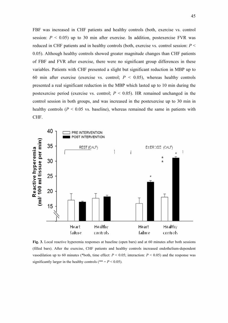
FBF was increased in CHF patients and healthy controls (both, exercise vs. control
session: P < 0.05) up to 30 min after exercise. In addition, postexercise FVR was
reduced in CHF patients and in healthy controls (both, exercise vs. control session: P <
0.05). Although healthy controls showed greater magnitude changes than CHF patients
of FBF and FVR after exercise, there were no significant group differences in these
variables. Patients with CHF presented a slight but significant reduction in MBP up to
60 min after exercise (exercise vs. control; P < 0.05), whereas healthy controls
presented a real significant reduction in the MBP which lasted up to 10 min during the
postexercise period (exercise vs. control; P < 0.05). HR remained unchanged in the
control session in both groups, and was increased in the postexercise up to 30 min in
healthy controls (P < 0.05 vs. baseline), whereas remained the same in patients with
CHF.
Fig. 3. Local reactive hyperemia responses at baseline (open bars) and at 60 minutes after both sessions
(filled bars). After the exercise, CHF patients and healthy controls increased endothelium-dependent
vasodilation up to 60 minutes (*both, time effect: P < 0.05; interaction: P < 0.05) and the response was
significantly larger in the healthy controls (** = P < 0.05).

46
Reactive hyperemia
Figure 3 shows the RH local response at 60 min after the interventions for both
groups in two experimental sessions (control session and exercise). A single resistance
exercise session for lower limbs produced a rise in endothelium-dependent vasodilation
of calf up to 60 min in the postexercise period in CHF patients as well in healthy
controls (both, time effect: P < 0.05; interaction: P < 0.05). After the exercise there was
a statistically difference between CHF patients and healthy controls (P < 0.05 vs.
healthy controls). Figure 4 shows the RH systemic response at 30 min after the
interventions for both groups in two experimental sessions (control session and
exercise). A single resistance exercise session for lower limbs produced a augment in
endothelium-dependent vasodilation of forearm at 10 and 30 min in the postexercise
period in patients with CHF as well in healthy controls (both, time effect: P < 0.05;
interaction: P < 0.05).
Fig. 4. Systemic reactive hyperemia responses at baseline (open bars) and at 30 minutes after both
sessions (filled bars). After the exercise, CHF patients and healthy controls increased endothelium-
dependent vasodilation up to 30 minutes after the exercise (*both, time effect: P < 0.05; interaction: P <
0.05)

47
Discussion
The major new finding of this study is that a single resistance exercise session
promotes a sustained increase in systemic endothelial function in patients with CHF, as
well as, in healthy controls. After a bout of resistance exercise performed exclusively
with the legs, our CHF patients presented increased FBF response, together with a
normal endothelium-dependent vasodilation in the nonworked limb. As expected, the
healthy control group presented simultaneous increases in FBF and RH up to 30 min
after exercise, a response that was also found in CHF patients. Moreover, our study
demonstrated a local improvement of vascular and endothelial function after a single
session of resistance exercise in both groups, an effect that lasted up to 60 min in
postexercise period. The local postexercise vascular responses in healthy controls were
statistically higher than in CHF patients, but the systemic vascular responses were not
different between groups after resistance exercise, suggesting attenuated local vascular
responses, but preserved systemic endothelial function in CHF patients after resistance
exercise. To the best of our knowledge, this is the first study that has evaluated local
and systemic endothelial vasodilator function in patients with CHF, as well in healthy
controls, after a single resistance exercise session.
We have recently shown that patients with CHF present a normal endothelium-
dependent function in the nonexercised limb, after a single bout of aerobic exercise,
despite the attenuated FBF response [8]. These findings corroborate with the results
shown in the study of Linke et al. [6] who showed that after 4 weeks of aerobic exercise
training in CHF patients, there was a correction of endothelial dysfunction in upper
extremity, indicating a systemic effect after a local aerobic training in endothelial
function [6]. Together, these findings confirm that acute aerobic exercise as well as
aerobic training can improve systemic endothelial function in patients with CHF [6, 8].
In addition to these systemic vascular relaxation induced by aerobic training, the effect
on local vasculature is also well described by several studies in this population [19-21].
Different from the present research, most studies involving resistance training
were designed to be of mixed (aerobic and resistance) mode [22-24] and, therefore, the
effects of the resistance training were difficult to differentiate from those of aerobic
training. Maioranna et al. [22] were the first to demonstrate that a relatively short
combined aerobic and resistance program is associated with structural as well as
functional changes and that the vascular benefit may be generalized to the circulation
48
rather than limited to the skeletal muscle bed involved in the training stimulus [22].
Some studies have demonstrated the isolated effect of resistance training but in small
muscle groups [25-26]. These experiments showed that handgrip training specifically
enhances endothelium-dependent vasodilation in the trained skeletal muscle circulation
of patients with CHF. This local adaptation at the exercised limb is mediated through an
augment in endothelium-dependent vasodilation, probably caused by improved
regulation of endothelial NO synthase as a consequence of the regular increase in shear
stress [25, 27-29]. This mechanism may explain in part the local blood flow and reactive
hyperemia demonstrated responses in the present study after resistance exercise by CHF
patients.
The improved systemic vascular response evidenced in our study after resistance
exercise in CHF patients indicates that the postexercise effects could participate in the
exercise-induce systemic endothelial adaptation. Consequently, patients with CHF may
be dependent on the postexercise vascular stimulus in nonexercised limbs, since it is
well established that CHF patients have abnormal hemodynamic response during
exercise [10-12], with possible reduced stimulus to inactive vascular beds. An elegant
study by Selig et al. [7] demonstrated the isolate effects of resistance training on the
peripheral manifestations of the CHF. After three months of resistance training, CHF
patients presented improvements in skeletal muscle strength and endurance as well as
aerobic power, increases in peak lactate, and augmented in FBF [7]. Despite the fact
that patients were subjected to resistance training exclusively, the authors included in
the training recovery intervals in arm/leg cycling, which might have influenced the final
result of this research, since it is already well described that aerobic exercise causes
systemic vascular relaxation [6, 8]. In the same line, a study with spontaneously
hypertensive rats demonstrated an important decrease of the vascular reactivity of the
caudal bed and in the resting BP after a single resistance exercise session [9].
Additionally, the evaluation of vascular responsiveness was performed in nonexercised
vessel, indicating a systemic effect after resistance exercise, despite the fact that the
vascular bed used is specific to rats, limiting the clinical application of these responses
[9].
The mechanisms responsible for the systemic vascular responses after resistance
exercise of our CHF patients are not readily apparent. During lower limb resistance
exercise involving large muscle groups, there was a significant increase in forearm

49
blood flow, which could result in increased shear stress throughout the vasculature [30-
31]. Moreover, the release of vasodilator agents from active muscle beds and their
subsequent circulation to inactive regions [30] might also contribute to the vascular
responses. Therefore, the nature of the exercise stimulus might determine whether shear
signals are sufficient to induce functional adaption in non-active beds [30].
In agreement with the findings of Meyer et al. [13], our CHF patients, as well as
the healthy controls, presented hemodynamic stability during strength test and in both
days of resistance exercise. Nevertheless, in that classic study, changes indicated an
enhanced contractile function of the left ventricle in well-compensated CHF patients
during resistance exercise [13]. In addition, our healthy controls presented reduced BP
levels in the postexercise period. Patients with CHF may present attenuated postexercise
hypotensive response due to a reduced attenuation in vascular resistance in the
postexercise period when compared to healthy subjects [8]. This may occur because, in
middle-aged individuals, postexercise hypotension is elicited by neural and vascular
components, leading to a reduction in regional and systemic vascular resistance [3].
Another recent study [32] demonstrated that low and high-intensity resistance exercises
promote systolic post-exercise hypotension in normotensive subjects, while only low-
intensity exercise decreases DBP. After both exercise intensities, post-exercise
hypotension was due to a reduction in cardiac output that is not completely
compensated by an increase in systemic vascular resistance. Reduction in cardiac output
is a consequence of a decrease in stroke volume besides the increase in HR promoted by
the activation of sympathetic and deactivation of parasympathetic modulation of the
heart [32].
This study has some limitations. All CHF patients included in the present study
had an optimal clinical status and were using guideline-recommended medications.
Moreover, the number of non-ischaemic CHF patients in the present research was
relative higher than ischaemic (8/2). According to a recent study, CHF patients with
ischaemic etiology exhibit greater impairment of endothelium-dependent dilation of the
brachial artery when compared with patients with non-ischaemic CHF [33]. As a result,
these factors could have influenced the magnitude of the responses of our CHF patients.
But, in contrast, our data reflect how the postexercise cardiovascular regulation really
occurs in the daily scenario of these patients. In addition, it has been previously
demonstrated that the use of beta-blockers has no influence in FBF and FVR at rest,

50
during isometric exercise, and during adrenergic stimulus [34]. Also, Levinger et al.
[35] have show that the improvement exercise capacity and skeletal muscle strength
after 8 weeks of resistance training was not affected by the use of beta-blockers in CHF.
In conclusion, our findings demonstrate attenuated local vascular responses, but
preserved systemic endothelial function in patients with CHF, after resistance exercise,
which may contribute to the possible systemic endothelial adaptation post resistance
training.
Acknowledgements
This work was supported in part by grants from CAPES and CNPq, Brasília,
Brazil, and FIPE-HCPA, Porto Alegre, Brazil.

51
REFERENCES
1. da Nobrega, A.C., The subacute effects of exercise: concept, characteristics, and
clinical implications. Exerc Sport Sci Rev, 2005. 33(2): p. 84-7. 2. Forjaz, C.L., et al., Factors affecting post-exercise hypotension in normotensive
and hypertensive humans. Blood Press Monit, 2000. 5(5-6): p. 255-62. 3. Halliwill, J.R., Mechanisms and clinical implications of post-exercise
hypotension in humans. Exerc Sport Sci Rev, 2001. 29(2): p. 65-70. 4. Kenney, M.J. and D.R. Seals, Postexercise hypotension. Key features,
mechanisms, and clinical significance. Hypertension, 1993. 22(5): p. 653-64. 5. Melo, C.M., et al., Postexercise hypotension induced by low-intensity resistance
exercise in hypertensive women receiving captopril. Blood Press Monit, 2006. 11(4): p. 183-9.
6. Linke, A., et al., Endothelial dysfunction in patients with chronic heart failure:
systemic effects of lower-limb exercise training. J Am Coll Cardiol, 2001. 37(2): p. 392-7.
7. Selig, S.E., et al., Moderate-intensity resistance exercise training in patients
with chronic heart failure improves strength, endurance, heart rate variability,
and forearm blood flow. J Card Fail, 2004. 10(1): p. 21-30. 8. Umpierre, D., et al., Blunted vascular responses but preserved endothelial
vasodilation after submaximal exercise in chronic heart failure. Eur J Cardiovasc Prev Rehabil, 2009. 16(1): p. 53-9.
9. Faria Tde, O., et al., Acute resistance exercise reduces blood pressure and
vascular reactivity, and increases endothelium-dependent relaxation in
spontaneously hypertensive rats. Eur J Appl Physiol, 2010. 110(2): p. 359-66. 10. LeJemtel, T.H., et al., Failure to augment maximal limb blood flow in response
to one-leg versus two-leg exercise in patients with severe heart failure. Circulation, 1986. 74(2): p. 245-51.
11. Magnusson, G., et al., Peak skeletal muscle perfusion is maintained in patients
with chronic heart failure when only a small muscle mass is exercised. Cardiovasc Res, 1997. 33(2): p. 297-306.
12. Zelis, R., D.T. Mason, and E. Braunwald, Partition of blood flow to the
cutaneous and muscular beds of the forearm at rest and during leg exercise in
normal subjects and in patients with heart failure. Circ Res, 1969. 24(6): p. 799-806.
13. Meyer, K., et al., Hemodynamic responses during leg press exercise in patients
with chronic congestive heart failure. Am J Cardiol, 1999. 83(11): p. 1537-43. 14. Mohamed, O., J. Perry, and H. Hislop, Relationship between wire EMG activity,
muscle length, and torque of the hamstrings. Clin Biomech (Bristol, Avon), 2002. 17(8): p. 569-79.
15. ZHANG, Q., Influence of Knee Joint Position on Co-contractions of Agonist
and Antagonist Muscles during Maximal Voluntary Contraction:
Electromyography and Cybex Measurement J. Phys. Ther. Sci, 2007. 19: p. 125-130.
16. Urlando, A. and D. Hawkins, Achilles tendon adaptation during strength
training in young adults. Med Sci Sports Exerc, 2007. 39(7): p. 1147-52.

52
17. Baroni, B.M., Effect of Light-Emitting Diodes Therapy (LEDT) on Knee
Extensor Muscle Fatigue: LEDT Prevents Muscle Fatigue in Humans. Photomedicine and Laser Surgery, 2010.
18. Chiappa, G.R., et al., Inspiratory muscle training improves blood flow to resting
and exercising limbs in patients with chronic heart failure. J Am Coll Cardiol, 2008. 51(17): p. 1663-71.
19. Kobayashi, N., et al., Exercise training in patients with chronic heart failure
improves endothelial function predominantly in the trained extremities. Circ J, 2003. 67(6): p. 505-10.
20. Hambrecht, R., et al., Regular physical exercise corrects endothelial dysfunction
and improves exercise capacity in patients with chronic heart failure. Circulation, 1998. 98(24): p. 2709-15.
21. Gokce, N., et al., Effect of exercise on upper and lower extremity endothelial
function in patients with coronary artery disease. Am J Cardiol, 2002. 90(2): p. 124-7.
22. Maiorana, A., et al., Effect of aerobic and resistance exercise training on
vascular function in heart failure. Am J Physiol Heart Circ Physiol, 2000. 279(4): p. H1999-2005.
23. Maiorana, A., et al., Combined aerobic and resistance exercise training
improves functional capacity and strength in CHF. J Appl Physiol, 2000. 88(5): p. 1565-70.
24. Tyni-Lenne, R., et al., Comprehensive local muscle training increases aerobic
working capacity and quality of life and decreases neurohormonal activation in
patients with chronic heart failure. Eur J Heart Fail, 2001. 3(1): p. 47-52. 25. Hornig, B., V. Maier, and H. Drexler, Physical training improves endothelial
function in patients with chronic heart failure. Circulation, 1996. 93(2): p. 210-4.
26. Katz, S.D., et al., Training improves endothelium-dependent vasodilation in
resistance vessels of patients with heart failure. J Appl Physiol, 1997. 82(5): p. 1488-92.
27. Demopoulos, L., et al., Exercise training in patients with severe congestive heart
failure: enhancing peak aerobic capacity while minimizing the increase in
ventricular wall stress. J Am Coll Cardiol, 1997. 29(3): p. 597-603. 28. Noris, M., et al., Nitric oxide synthesis by cultured endothelial cells is modulated
by flow conditions. Circ Res, 1995. 76(4): p. 536-43. 29. Sullivan, M.J., et al., Relation between central and peripheral hemodynamics
during exercise in patients with chronic heart failure. Muscle blood flow is
reduced with maintenance of arterial perfusion pressure. Circulation, 1989. 80(4): p. 769-81.
30. Green, D.J., A.J. Maiorana, and N.T. Cable, Point: exercise training does induce
vascular adaptations beyond the active muscle beds. J Appl Physiol, 2008. 105(3): p. 1002-4; discussion 1007.
31. Thijssen, D.H., et al., Last word on point: counterpoint: exercise training
does/does not induce vascular adaptations beyond the active muscle beds. J Appl Physiol, 2008. 105(3): p. 1011.
32. Rezk, C.C., et al., Post-resistance exercise hypotension, hemodynamics, and
heart rate variability: influence of exercise intensity. Eur J Appl Physiol, 2006. 98(1): p. 105-12.

53
33. Klosinska, M., et al., Endothelium-dependent and -independent vasodilation is
more attenuated in ischaemic than in non-ischaemic heart failure. Eur J Heart Fail, 2009. 11(8): p. 765-70.
34. Hryniewicz, K., et al., Comparative effects of carvedilol and metoprolol on
regional vascular responses to adrenergic stimuli in normal subjects and
patients with chronic heart failure. Circulation, 2003. 108(8): p. 971-6. 35. Levinger, I., et al., The effect of resistance training on left ventricular function
and structure of patients with chronic heart failure. Int J Cardiol, 2005. 105(2): p. 159-63.

Livros Grátis( http://www.livrosgratis.com.br )
Milhares de Livros para Download: Baixar livros de AdministraçãoBaixar livros de AgronomiaBaixar livros de ArquiteturaBaixar livros de ArtesBaixar livros de AstronomiaBaixar livros de Biologia GeralBaixar livros de Ciência da ComputaçãoBaixar livros de Ciência da InformaçãoBaixar livros de Ciência PolíticaBaixar livros de Ciências da SaúdeBaixar livros de ComunicaçãoBaixar livros do Conselho Nacional de Educação - CNEBaixar livros de Defesa civilBaixar livros de DireitoBaixar livros de Direitos humanosBaixar livros de EconomiaBaixar livros de Economia DomésticaBaixar livros de EducaçãoBaixar livros de Educação - TrânsitoBaixar livros de Educação FísicaBaixar livros de Engenharia AeroespacialBaixar livros de FarmáciaBaixar livros de FilosofiaBaixar livros de FísicaBaixar livros de GeociênciasBaixar livros de GeografiaBaixar livros de HistóriaBaixar livros de Línguas

Baixar livros de LiteraturaBaixar livros de Literatura de CordelBaixar livros de Literatura InfantilBaixar livros de MatemáticaBaixar livros de MedicinaBaixar livros de Medicina VeterináriaBaixar livros de Meio AmbienteBaixar livros de MeteorologiaBaixar Monografias e TCCBaixar livros MultidisciplinarBaixar livros de MúsicaBaixar livros de PsicologiaBaixar livros de QuímicaBaixar livros de Saúde ColetivaBaixar livros de Serviço SocialBaixar livros de SociologiaBaixar livros de TeologiaBaixar livros de TrabalhoBaixar livros de Turismo