Embed Size (px)
Citation preview
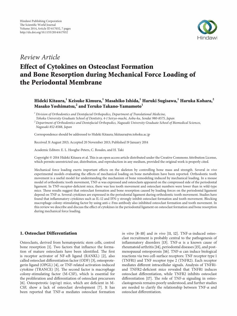
Review ArticleEffect of Cytokines on Osteoclast Formationand Bone Resorption during Mechanical Force Loading ofthe Periodontal Membrane
Hideki Kitaura,1 Keisuke Kimura,1 Masahiko Ishida,1 Haruki Sugisawa,1 Haruka Kohara,2
Masako Yoshimatsu,2 and Teruko Takano-Yamamoto1
1 Division of Orthodontics and Dentofacial Orthopedics, Department of Translational Medicine,Tohoku University Graduate School of Dentistry, 4-1 Seiryo-machi, Aoba-ku, Sendai 980-8575, Japan
2Department of Orthodontics and Dentofacial Orthopedics, Nagasaki University Graduate School of Biomedical Sciences,Nagasaki 852-8588, Japan
Correspondence should be addressed to Hideki Kitaura; [email protected]
Received 31 August 2013; Accepted 20 November 2013; Published 19 January 2014
Academic Editors: E. L. Hooghe-Peters, C. Rosales, and H. Taki
Copyright © 2014 Hideki Kitaura et al.This is an open access article distributed under the Creative Commons Attribution License,which permits unrestricted use, distribution, and reproduction in any medium, provided the original work is properly cited.
Mechanical force loading exerts important effects on the skeleton by controlling bone mass and strength. Several in vivoexperimental models evaluating the effects of mechanical loading on bone metabolism have been reported. Orthodontic toothmovement is a useful model for understanding the mechanism of bone remodeling induced by mechanical loading. In a mousemodel of orthodontic tooth movement, TNF-𝛼 was expressed and osteoclasts appeared on the compressed side of the periodontalligament. In TNF-receptor-deficient mice, there was less tooth movement and osteoclast numbers were lower than in wild-typemice. These results suggest that osteoclast formation and bone resorption caused by loading forces on the periodontal ligamentdepend on TNF-𝛼. Several cytokines are expressed in the periodontal ligament during orthodontic tooth movement. Studies havefound that inflammatory cytokines such as IL-12 and IFN-𝛾 strongly inhibit osteoclast formation and tooth movement. Blockingmacrophage colony-stimulating factor by using anti-c-Fms antibody also inhibited osteoclast formation and tooth movement. Inthis review we describe and discuss the effect of cytokines in the periodontal ligament on osteoclast formation and bone resorptionduring mechanical force loading.
1. Osteoclast Differentiation
Osteoclasts, derived from hematopoietic stem cells, controlbone resorption [1]. Two factors that influence the forma-tion of mature osteoclasts have been identified. The firstis receptor activator of NF-𝜅B ligand (RANKL) [2], alsocalled osteoclast differentiation factor (ODF) [3], osteoprote-gerin ligand (OPGL) [4], or TNF-related activation-inducedcytokine (TRANCE) [5]. The second factor is macrophagecolony-stimulating factor (M-CSF), which is essential forthe proliferation and differentiation of osteoclast precursors[6]. Osteopetrotic (op/op) mice, which are deficient in M-CSF, show a lack of osteoclast development [7]. It hasbeen reported that TNF-𝛼 mediates osteoclast formation
in vitro [8–10] and in vivo [11, 12]. TNF-𝛼-induced osteo-clast recruitment is probably central to the pathogenesis ofinflammatory disorders [13]. TNF-𝛼 is a known cause ofrheumatoid arthritis [14], periodontal diseases [15], and post-menopausal osteoporosis [16]. TNF-𝛼 can induce biologicalreactions via two cell-surface receptors: TNF receptor type 1(TNFR1) and TNF receptor type 2 (TNFR2). Each receptormediates different intracellular signals. Analysis of TNFR1-and TNFR2-deficient mice revealed that TNFR1 inducesosteoclast differentiation, while TNFR2 inhibits osteoclastdifferentiation [17]. The role of TNF-𝛼 signaling in osteo-clastogenesis remains poorly understood, and further studiesare needed to clarify the relationship between TNF-𝛼 andosteoclast differentiation.
Hindawi Publishing Corporatione Scientific World JournalVolume 2014, Article ID 617032, 7 pageshttp://dx.doi.org/10.1155/2014/617032
2 The Scientific World Journal
Direction of tooth movement
Ni-Ti coil spring
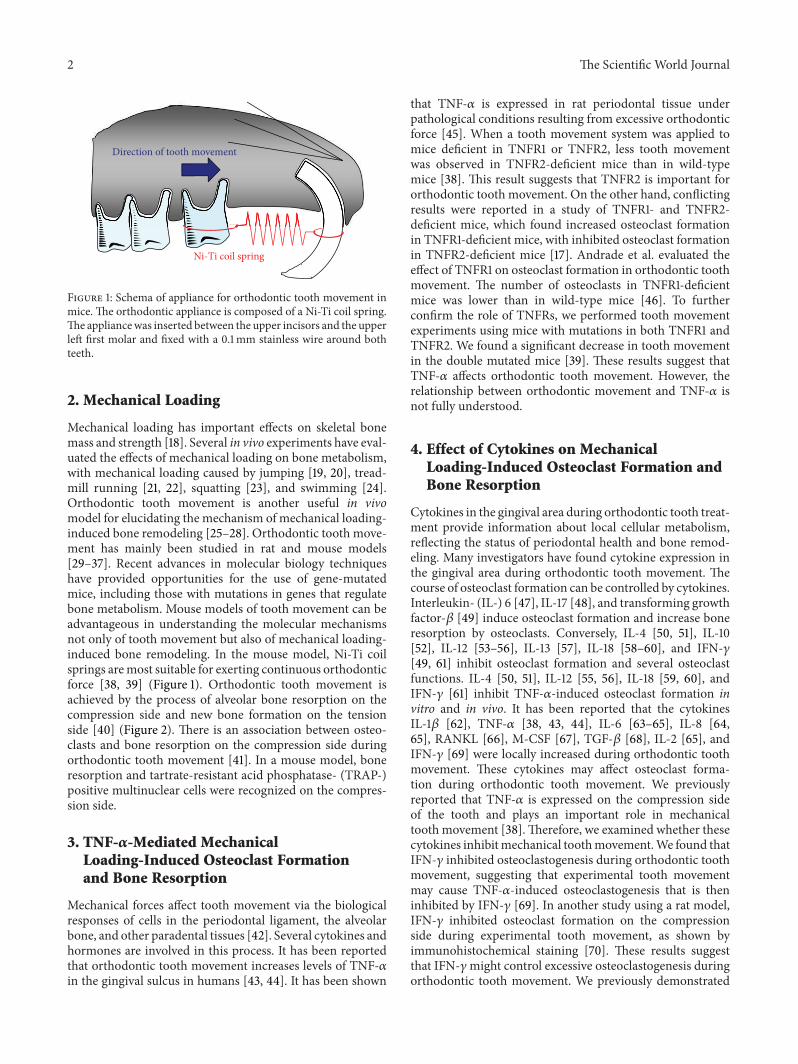
Figure 1: Schema of appliance for orthodontic tooth movement inmice. The orthodontic appliance is composed of a Ni-Ti coil spring.The appliancewas inserted between the upper incisors and the upperleft first molar and fixed with a 0.1mm stainless wire around bothteeth.
2. Mechanical Loading
Mechanical loading has important effects on skeletal bonemass and strength [18]. Several in vivo experiments have eval-uated the effects of mechanical loading on bone metabolism,with mechanical loading caused by jumping [19, 20], tread-mill running [21, 22], squatting [23], and swimming [24].Orthodontic tooth movement is another useful in vivomodel for elucidating the mechanism of mechanical loading-induced bone remodeling [25–28]. Orthodontic tooth move-ment has mainly been studied in rat and mouse models[29–37]. Recent advances in molecular biology techniqueshave provided opportunities for the use of gene-mutatedmice, including those with mutations in genes that regulatebone metabolism. Mouse models of tooth movement can beadvantageous in understanding the molecular mechanismsnot only of tooth movement but also of mechanical loading-induced bone remodeling. In the mouse model, Ni-Ti coilsprings aremost suitable for exerting continuous orthodonticforce [38, 39] (Figure 1). Orthodontic tooth movement isachieved by the process of alveolar bone resorption on thecompression side and new bone formation on the tensionside [40] (Figure 2). There is an association between osteo-clasts and bone resorption on the compression side duringorthodontic tooth movement [41]. In a mouse model, boneresorption and tartrate-resistant acid phosphatase- (TRAP-)positive multinuclear cells were recognized on the compres-sion side.
3. TNF-𝛼-Mediated MechanicalLoading-Induced Osteoclast Formationand Bone Resorption
Mechanical forces affect tooth movement via the biologicalresponses of cells in the periodontal ligament, the alveolarbone, and other paradental tissues [42]. Several cytokines andhormones are involved in this process. It has been reportedthat orthodontic tooth movement increases levels of TNF-𝛼in the gingival sulcus in humans [43, 44]. It has been shown
that TNF-𝛼 is expressed in rat periodontal tissue underpathological conditions resulting from excessive orthodonticforce [45]. When a tooth movement system was applied tomice deficient in TNFR1 or TNFR2, less tooth movementwas observed in TNFR2-deficient mice than in wild-typemice [38]. This result suggests that TNFR2 is important fororthodontic tooth movement. On the other hand, conflictingresults were reported in a study of TNFR1- and TNFR2-deficient mice, which found increased osteoclast formationin TNFR1-deficient mice, with inhibited osteoclast formationin TNFR2-deficient mice [17]. Andrade et al. evaluated theeffect of TNFR1 on osteoclast formation in orthodontic toothmovement. The number of osteoclasts in TNFR1-deficientmice was lower than in wild-type mice [46]. To furtherconfirm the role of TNFRs, we performed tooth movementexperiments using mice with mutations in both TNFR1 andTNFR2. We found a significant decrease in tooth movementin the double mutated mice [39]. These results suggest thatTNF-𝛼 affects orthodontic tooth movement. However, therelationship between orthodontic movement and TNF-𝛼 isnot fully understood.
4. Effect of Cytokines on MechanicalLoading-Induced Osteoclast Formation andBone Resorption
Cytokines in the gingival area during orthodontic tooth treat-ment provide information about local cellular metabolism,reflecting the status of periodontal health and bone remod-eling. Many investigators have found cytokine expression inthe gingival area during orthodontic tooth movement. Thecourse of osteoclast formation can be controlled by cytokines.Interleukin- (IL-) 6 [47], IL-17 [48], and transforming growthfactor-𝛽 [49] induce osteoclast formation and increase boneresorption by osteoclasts. Conversely, IL-4 [50, 51], IL-10[52], IL-12 [53–56], IL-13 [57], IL-18 [58–60], and IFN-𝛾[49, 61] inhibit osteoclast formation and several osteoclastfunctions. IL-4 [50, 51], IL-12 [55, 56], IL-18 [59, 60], andIFN-𝛾 [61] inhibit TNF-𝛼-induced osteoclast formation invitro and in vivo. It has been reported that the cytokinesIL-1𝛽 [62], TNF-𝛼 [38, 43, 44], IL-6 [63–65], IL-8 [64,65], RANKL [66], M-CSF [67], TGF-𝛽 [68], IL-2 [65], andIFN-𝛾 [69] were locally increased during orthodontic toothmovement. These cytokines may affect osteoclast forma-tion during orthodontic tooth movement. We previouslyreported that TNF-𝛼 is expressed on the compression sideof the tooth and plays an important role in mechanicaltooth movement [38].Therefore, we examined whether thesecytokines inhibitmechanical toothmovement.We found thatIFN-𝛾 inhibited osteoclastogenesis during orthodontic toothmovement, suggesting that experimental tooth movementmay cause TNF-𝛼-induced osteoclastogenesis that is theninhibited by IFN-𝛾 [69]. In another study using a rat model,IFN-𝛾 inhibited osteoclast formation on the compressionside during experimental tooth movement, as shown byimmunohistochemical staining [70]. These results suggestthat IFN-𝛾might control excessive osteoclastogenesis duringorthodontic tooth movement. We previously demonstrated
The Scientific World Journal 3
Orthodontic force Orthodontic force Orthodontic force
Periodontal ligament Compression sideTension side Compression side Tension side
Osteoblast Osteoclast
Bone resorptionBone formation
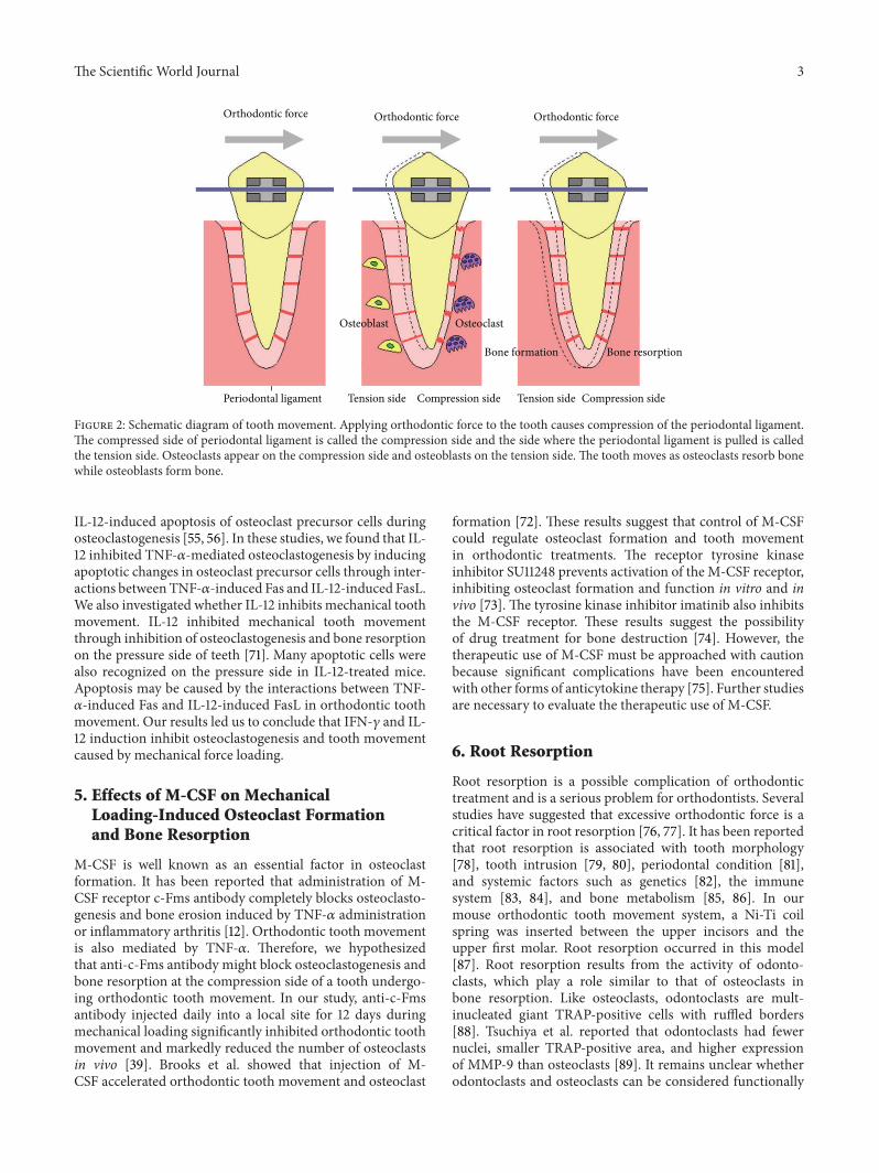
Figure 2: Schematic diagram of tooth movement. Applying orthodontic force to the tooth causes compression of the periodontal ligament.The compressed side of periodontal ligament is called the compression side and the side where the periodontal ligament is pulled is calledthe tension side. Osteoclasts appear on the compression side and osteoblasts on the tension side. The tooth moves as osteoclasts resorb bonewhile osteoblasts form bone.
IL-12-induced apoptosis of osteoclast precursor cells duringosteoclastogenesis [55, 56]. In these studies, we found that IL-12 inhibited TNF-𝛼-mediated osteoclastogenesis by inducingapoptotic changes in osteoclast precursor cells through inter-actions between TNF-𝛼-induced Fas and IL-12-induced FasL.We also investigated whether IL-12 inhibits mechanical toothmovement. IL-12 inhibited mechanical tooth movementthrough inhibition of osteoclastogenesis and bone resorptionon the pressure side of teeth [71]. Many apoptotic cells werealso recognized on the pressure side in IL-12-treated mice.Apoptosis may be caused by the interactions between TNF-𝛼-induced Fas and IL-12-induced FasL in orthodontic toothmovement. Our results led us to conclude that IFN-𝛾 and IL-12 induction inhibit osteoclastogenesis and tooth movementcaused by mechanical force loading.
5. Effects of M-CSF on MechanicalLoading-Induced Osteoclast Formationand Bone Resorption
M-CSF is well known as an essential factor in osteoclastformation. It has been reported that administration of M-CSF receptor c-Fms antibody completely blocks osteoclasto-genesis and bone erosion induced by TNF-𝛼 administrationor inflammatory arthritis [12]. Orthodontic tooth movementis also mediated by TNF-𝛼. Therefore, we hypothesizedthat anti-c-Fms antibody might block osteoclastogenesis andbone resorption at the compression side of a tooth undergo-ing orthodontic tooth movement. In our study, anti-c-Fmsantibody injected daily into a local site for 12 days duringmechanical loading significantly inhibited orthodontic toothmovement and markedly reduced the number of osteoclastsin vivo [39]. Brooks et al. showed that injection of M-CSF accelerated orthodontic tooth movement and osteoclast
formation [72]. These results suggest that control of M-CSFcould regulate osteoclast formation and tooth movementin orthodontic treatments. The receptor tyrosine kinaseinhibitor SU11248 prevents activation of the M-CSF receptor,inhibiting osteoclast formation and function in vitro and invivo [73]. The tyrosine kinase inhibitor imatinib also inhibitsthe M-CSF receptor. These results suggest the possibilityof drug treatment for bone destruction [74]. However, thetherapeutic use of M-CSF must be approached with cautionbecause significant complications have been encounteredwith other forms of anticytokine therapy [75]. Further studiesare necessary to evaluate the therapeutic use of M-CSF.
6. Root Resorption
Root resorption is a possible complication of orthodontictreatment and is a serious problem for orthodontists. Severalstudies have suggested that excessive orthodontic force is acritical factor in root resorption [76, 77]. It has been reportedthat root resorption is associated with tooth morphology[78], tooth intrusion [79, 80], periodontal condition [81],and systemic factors such as genetics [82], the immunesystem [83, 84], and bone metabolism [85, 86]. In ourmouse orthodontic tooth movement system, a Ni-Ti coilspring was inserted between the upper incisors and theupper first molar. Root resorption occurred in this model[87]. Root resorption results from the activity of odonto-clasts, which play a role similar to that of osteoclasts inbone resorption. Like osteoclasts, odontoclasts are mult-inucleated giant TRAP-positive cells with ruffled borders[88]. Tsuchiya et al. reported that odontoclasts had fewernuclei, smaller TRAP-positive area, and higher expressionof MMP-9 than osteoclasts [89]. It remains unclear whetherodontoclasts and osteoclasts can be considered functionally
4 The Scientific World Journal
identical. We tested our hypothesis that IL-12 and anti-c-Fms antibody might inhibit odontoclastogenesis and rootresorption during orthodontic tooth movement by injectingIL-12 locally adjacent to the firstmolar every other day duringthe experimental period. We found that IL-12 inhibitedodontoclastogenesis and root resorption during orthodontictooth movement [71]. Anti-c-Fms antibody also significantlyinhibited odontoclastogenesis and root resorption duringorthodontic tooth movement [87]. M-CSF and its receptorare potential therapeutic targets inmechanical stress-inducedodontoclastogenesis, and injection of an anti-c-Fms antibodymight be useful to prevent mechanical stress-induced rootresorption during orthodontic tooth movement.
7. Conclusion
Many studies have reported the expression of variouscytokines during mechanical loading of the periodontalligament. Several studies using gene-mutated mice haveshown that TNF-𝛼 plays a key role in mechanical forceloading-induced osteoclast formation in the periodontalligament. Therefore, it is important to study the relationshipbetween TNF-𝛼-induced osteoclast formation and cytokinesexpressed during mechanical loading. Further studies areneeded to fully understand the effect of cytokines onmechan-ical loading-induced osteoclast formation.
Conflict of Interests
The authors declare that there is no conflict of interestsregarding the publication of this paper.
References
[1] T. Suda, N. Takahashi, and T. J. Martin, “Modulation ofosteoclast differentiation,” Endocrine Reviews, vol. 13, no. 1, pp.66–80, 1992.
[2] D. M. Anderson, E. Maraskovsky, W. L. Billingsley et al., “Ahomologue of the TNF receptor and its ligand enhance T-cellgrowth and dendritic-cell function,” Nature, vol. 390, no. 6656,pp. 175–179, 1997.
[3] H. Yasuda, N. Shima, N. Nakagawa et al., “Osteoclast differenti-ation factor is a ligand for osteoprotegerin/ osteoclastogenesis-inhibitory factor and is identical to TRANCE/RANKL,” Pro-ceedings of the National Academy of Sciences of the United Statesof America, vol. 95, no. 7, pp. 3597–3602, 1998.
[4] D. L. Lacey, E. Timms, H.-L. Tan et al., “Osteoprotegerinligand is a cytokine that regulates osteoclast differentiation andactivation,” Cell, vol. 93, no. 2, pp. 165–176, 1998.
[5] B. R.Wong, J. Rho, J. Arron et al., “TRANCE is a novel ligand ofthe tumor necrosis factor receptor family that activates c-JunN-terminal kinase in T cells,”The Journal of Biological Chemistry,vol. 272, no. 40, pp. 25190–25194, 1997.
[6] H. Kodama, M. Nose, S. Niida, and A. Yamasaki, “Essentialrole of macrophage colony-stimulating factor in the osteoclastdifferentiation supported by stromal cells,” The Journal ofExperimental Medicine, vol. 173, no. 5, pp. 1291–1294, 1991.
[7] J. W. Wiktor, A. Bartocci, A. W. Ferrante Jr. et al., “Totalabsence of colony-stimulating factor 1 in the macrophage-deficient osteopetrotic (op/op) mouse,” Proceedings of the
National Academy of Sciences of the United States of America,vol. 87, no. 12, pp. 4828–4832, 1990.
[8] Y. Azuma, K. Kaji, R. Katogi, S. Takeshita, and A. Kudo, “Tumornecrosis factor-𝛼 induces differentiation of and bone resorptionby osteoclasts,”The Journal of Biological Chemistry, vol. 275, no.7, pp. 4858–4864, 2000.
[9] K. Kobayashi, N. Takahashi, E. Jimi et al., “Tumor necrosisfactor 𝛼 stimulates osteoclast differentiation by a mechanismindependent of theODF/RANKL-RANK interaction,”The Jour-nal of Experimental Medicine, vol. 191, no. 2, pp. 275–286, 2000.
[10] K. Fuller, C. Murphy, B. Kirstein, S. W. Fox, and T. J. Cham-bers, “TNF𝛼 potently activates osteoclasts, through a directaction independent of and strongly synergistic with RANKL,”Endocrinology, vol. 143, no. 3, pp. 1108–1118, 2002.
[11] H. Kitaura, M. S. Sands, K. Aya et al., “Marrow stromal cellsand osteoclast precursors differentially contribute to TNF-𝛼-induced osteoclastogenesis in vivo,”The Journal of Immunology,vol. 173, no. 8, pp. 4838–4846, 2004.
[12] H. Kitaura, P. Zhou, H.-J. Kim, D. V. Novack, F. P. Ross, andS. L. Teitelbaum, “M-CSFmediates TNF-induced inflammatoryosteolysis,” The Journal of Clinical Investigation, vol. 115, no. 12,pp. 3418–3427, 2005.
[13] M. Wong, D. Ziring, Y. Korin et al., “TNF𝛼 blockade inhuman diseases: mechanisms and future directions,” ClinicalImmunology, vol. 126, no. 2, pp. 121–136, 2008.
[14] K. Redlich, S. Hayer, R. Ricci et al., “Osteoclasts are essentialfor TNF-𝛼-mediated joint destruction,” The Journal of ClinicalInvestigation, vol. 110, no. 10, pp. 1419–1427, 2002.
[15] Y. Abu-Amer, F. P. Ross, J. Edwards, and S. L. Teitelbaum,“Lipopolysaccharide-stimulated osteoclastogenesis is mediatedby tumor necrosis factor via its P55 receptor,” The Journal ofClinical Investigation, vol. 100, no. 6, pp. 1557–1565, 1997.
[16] R. B. Kimble, S. Srivastava, F. P. Ross, A. Matayoshi, and R.Pacifici, “Estrogen deficiency increases the ability of stromalcells to support murine osteoclastogenesis via an interleukin-1-and tumor necrosis factor-mediated stimulation ofmacrophagecolony-stimulating factor production,”The Journal of BiologicalChemistry, vol. 271, no. 46, pp. 28890–28897, 1996.
[17] Y. Abu-Amer, J. Erdmann, L. Alexopoulou, G. Kollias, F. PatrickRoss, and S. L. Teitelbaum, “Tumor necrosis factor receptorstypes 1 and 2 differentially regulate osteoclastogenesis,” TheJournal of Biological Chemistry, vol. 275, no. 35, pp. 27307–27310,2000.
[18] H. M. Frost, “On our age-related bone loss: insights from a newparadigm,” Journal of Bone andMineral Research, vol. 12, no. 10,pp. 1539–1546, 1997.
[19] T. Notomi, S. J. Lee, N. Okimoto et al., “Effects of resistanceexercise training on mass, strength, and turnover of bone ingrowing rats,” European Journal of Applied Physiology, vol. 82,no. 4, pp. 268–274, 2000.
[20] Y. Kodama, Y. Umemura, S. Nagasawa et al., “Exercise andmechanical loading increase periosteal bone formation andwhole bone strength in C57BL/6J mice but not in C3H/Hejmice,” Calcified Tissue International, vol. 66, no. 4, pp. 298–306,2000.
[21] J. Iwamoto, J. K. Yeh, and J. F. Aloia, “Differential effect oftreadmill exercise on three cancellous bone sites in the younggrowing rat,” Bone, vol. 24, no. 3, pp. 163–169, 1999.
[22] T. Notomi, Y. Okazaki, N. Okimoto, S. Saitoh, T. Nakamura, andM. Suzuki, “A comparison of resistance and aerobic training formass, strength and turnover of bone in growing rats,” EuropeanJournal of Applied Physiology, vol. 83, no. 6, pp. 469–474, 2000.
The Scientific World Journal 5
[23] K. C.Westerlind, J. D. Fluckey, S. E. Gordon,W. J. Kraemer, P. A.Farrell, and R. T. Turner, “Effect of resistance exercise trainingon cortical and cancellous bone in mature male rats,” Journal ofApplied Physiology, vol. 84, no. 2, pp. 459–464, 1998.
[24] K. J. Hart, J. M. Shaw, E. Vajda, M. Hegsted, and S. C. Miller,“Swim-trained rats have greater bone mass, density, strength,and dynamics,” Journal of Applied Physiology, vol. 91, no. 4, pp.1663–1668, 2001.
[25] E. Storey, “The nature of tooth movement,” The AmericanJournal of Orthodontics, vol. 63, no. 3, pp. 292–314, 1973.
[26] T. Takano-Yamamoto, T. Takemura, Y. Kitamura, and S.Nomura, “Site-specific expression of mRNAs for osteonectin,osteocalcin, and osteopontin revealed by in situ hybridizationin rat periodontal ligament during physiological tooth move-ment,”The Journal of Histochemistry and Cytochemistry, vol. 42,no. 7, pp. 885–896, 1994.
[27] Y. Ohba, T. Ohba, K. Terai, and K. Moriyama, “Expression ofcathepsin KmRNAduring experimental toothmovement in ratas revealed by in situ hybridization,” Archives of Oral Biology,vol. 45, no. 1, pp. 63–69, 2000.
[28] Y. Kobayashi, F. Hashimoto, H.Miyamoto et al., “Force-inducedosteoclast apoptosis in vivo is accompanied by elevation intransforming growth factor 𝛽 and osteoprotegerin expression,”Journal of Bone and Mineral Research, vol. 15, no. 10, pp. 1924–1934, 2000.
[29] F. Hashimoto, Y. Kobayashi, S. Mataki, K. Kobayashi, Y.Kato, and H. Sakai, “Administration of osteocalcin acceleratesorthodontic tooth movement induced by a closed coil spring inrats,” European Journal of Orthodontics, vol. 23, no. 5, pp. 535–545, 2001.
[30] P. Brudvik and P. Rygh, “The initial phase of orthodontic rootresorption incident to local compression of the periodontalligament,” European Journal of Orthodontics, vol. 15, no. 4, pp.249–263, 1993.
[31] D. Pavlin, S. B. Dove, R. Zadro, and J. Gluhak-Heinrich,“Mechanical loading stimulates differentiation of periodontalosteoblasts in a mouse osteoinduction model: effect on typeI collagen and alkaline phosphatase genes,” Calcified TissueInternational, vol. 67, no. 2, pp. 163–172, 2000.
[32] M. Kaku, S. Kohno, T. Kawata et al., “Effects of vascularendothelial growth factor on osteoclast induction during toothmovement in mice,” Journal of Dental Research, vol. 80, no. 10,pp. 1880–1883, 2001.
[33] Y. Tsuji, T. Yamaza, M. A. Kido et al., “Expression of cathepsinK mRNA and protein in odontoclasts after experimental toothmovement in the mouse maxilla by in situ hybridization andimmunoelectronmicroscopy,”Cell andTissue Research, vol. 303,no. 3, pp. 359–369, 2001.
[34] J. Gluhak-Heinrich, L. Ye, L. F. Bonewald et al., “Mechanicalloading stimulates dentin matrix protein 1 (DMP1) expressionin osteocytes in vivo,” Journal of Bone andMineral Research, vol.18, no. 5, pp. 807–817, 2003.
[35] S. Kohno, M. Kaku, K. Tsutsui et al., “Expression of vascularendothelial growth factor and the effects on bone remodel-ing during experimental tooth movement,” Journal of DentalResearch, vol. 82, no. 3, pp. 177–182, 2003.
[36] C. R. Chung, K. Tsuji, A. Nifuji, T. Komori, K. Soma, and M.Noda, “Micro-CT evaluation of tooth, calvaria and mechanicalstress-induced toothmovement in adult Runx2/Cbfa1 heterozy-gous knock-out mice,” Journal of Medical and Dental Sciences,vol. 51, no. 1, pp. 105–113, 2004.
[37] S. Kuroda, T. A. Balam, Y. Sakai, N. Tamamura, and T. Takano-Yamamoto, “Expression of osteopontin mRNA in odontoclastsrevealed by in situ hybridization during experimental toothmovement in mice,” Journal of Bone and Mineral Metabolism,vol. 23, no. 2, pp. 110–113, 2005.
[38] M. Yoshimatsu, Y. Shibata, H. Kitaura et al., “Experimentalmodel of tooth movement by orthodontic force in mice andits application to tumor necrosis factor receptor-deficientmice,”Journal of Bone andMineralMetabolism, vol. 24, no. 1, pp. 20–27,2006.
[39] H. Kitaura, M. Yoshimatsu, Y. Fujimura et al., “An anti-c-Fms antibody inhibits orthodontic tooth movement,” Journal ofDental Research, vol. 87, no. 4, pp. 396–400, 2008.
[40] E. Storey, “Tissue response to the movement of bones,” TheAmerican Journal of Orthodontics, vol. 64, no. 3, pp. 229–247,1973.
[41] K. Yokoya, T. Sasaki, and Y. Shibasaki, “Distributional changesof osteoclasts and pre-osteoclastic cells in periodontal tissuesduring experimental tooth movement as revealed by quantita-tive immunohistochemistry of H(+)-ATPase,” Journal of DentalResearch, vol. 76, no. 1, pp. 580–587, 1997.
[42] V. Krishnan and Z. Davidovitch, “On a path to unfoldingthe biological mechanisms of orthodontic tooth movement,”Journal of Dental Research, vol. 88, no. 7, pp. 597–608, 2009.
[43] J. J. Lowney, L. A. Norton, D. M. Shafer, and E. F. Rossomando,“Orthodontic forces increase tumor necrosis factor 𝛼 in thehuman gingival sulcus,” The American Journal of Orthodonticsand Dentofacial Orthopedics, vol. 108, no. 5, pp. 519–524, 1995.
[44] S. Uematsu, M. Mogi, and T. Deguchi, “Interleukin (IL)-1𝛽, IL-6, tumor necrosis factor-𝛼, epidermal growth factor, and 𝛽2-microglobulin levels are elevated in gingival crevicular fluidduring human orthodontic tooth movement,” Journal of DentalResearch, vol. 75, no. 1, pp. 562–567, 1996.
[45] T. Ogasawara, Y. Yoshimine, T. Kiyoshima et al., “In situexpression of RANKL, RANK, osteoprotegerin and cytokinesin osteoclasts of rat periodontal tissue,” Journal of PeriodontalResearch, vol. 39, no. 1, pp. 42–49, 2004.
[46] I. Andrade Jr., T. A. Silva, G. A. B. Silva, A. L. Teixeira, and M.M. Teixeira, “The role of tumor necrosis factor receptor type 1 inorthodontic tooth movement,” Journal of Dental Research, vol.86, no. 11, pp. 1089–1094, 2007.
[47] Y. Gao, I. Morita, N. Maruo, T. Kubota, S. Murota, and T. Aso,“Expression of IL-6 receptor and GP130 in mouse bonemarrowcells during osteoclast differentiation,” Bone, vol. 22, no. 5, pp.487–493, 1998.
[48] S. Kotake, N. Udagawa, N. Takahashi et al., “IL-17 in synovialfluids from patients with rheumatoid arthritis is a potent stimu-lator of osteoclastogenesis,”The Journal of Clinical Investigation,vol. 103, no. 9, pp. 1345–1352, 1999.
[49] S. W. Fox, K. Fuller, K. E. Bayley, J. M. Lean, and T. J.Chambers, “TGF-𝛽1 and IFN-𝛾 direct macrophage activationby TNF-𝛼 to osteoclastic or cytocidal phenotype,” The Journalof Immunology, vol. 165, no. 9, pp. 4957–4963, 2000.
[50] H. Kitaura, N. Nagata, Y. Fujimura et al., “Interleukin-4 directlyinhibits tumor necrosis factor-𝛼-mediated osteoclast formationin mouse bone marrowmacrophages,” Immunology Letters, vol.88, no. 3, pp. 193–198, 2003.
6 The Scientific World Journal
[51] T. Fujii, H. Kitaura, K. Kimura, Z. W. Hakami, and T. Takano-Yamamoto, “IL-4 inhibits TNF-alpha-mediated osteoclast for-mation by inhibition of RANKL expression in TNF-alpha-activated stromal cells and direct inhibition of TNF-alpha-activated osteoclast precursors via a T-cell-independent mech-anism in vivo,” Bone, vol. 51, no. 4, pp. 771–780, 2012.
[52] J. M. Owens, A. C. Gallagher, and T. J. Chambers, “IL-10modulates formation of osteoclasts in murine hemopoieticcultures,” The Journal of Immunology, vol. 157, no. 2, pp. 936–940, 1996.
[53] N. J. Horwood, J. Elliott, T. J. Martin, andM. T. Gillespie, “IL-12alone and in synergy with IL-18 inhibits osteoclast formation invitro,”The Journal of Immunology, vol. 166, no. 8, pp. 4915–4921,2001.
[54] N. Nagata, H. Kitaura, N. Yoshida, and K. Nakayama, “Inhibi-tion of RANKL-induced osteoclast formation in mouse bonemarrow cells by IL-12: involvement of IFN-𝛾 possibly inducedfrom non-T cell population,” Bone, vol. 33, no. 4, pp. 721–732,2003.
[55] H. Kitaura, N. Nagata, Y. Fujimura, H. Hotokezaka, N. Yoshida,and K. Nakayama, “Effect of IL-12 on TNF-𝛼-mediated osteo-clast formation in bone marrow cells: apoptosis mediated byFas/Fas ligand interaction,”The Journal of Immunology, vol. 169,no. 9, pp. 4732–4738, 2002.
[56] M. Yoshimatsu, H. Kitaura, Y. Fujimura et al., “IL-12 inhibitsTNF-𝛼 induced osteoclastogenesis via a T cell-independentmechanism in vivo,” Bone, vol. 45, no. 5, pp. 1010–1016, 2009.
[57] Y. Onoe, C. Miyaura, T. Kaminakayashiki et al., “IL-13 andIL-4 inhibit bone resorption by suppressing cyclooxygenase-2-dependent prostaglandin synthesis in osteoblasts,” The Journalof Immunology, vol. 156, no. 2, pp. 758–764, 1996.
[58] N. Udagawa, N. J. Horwood, J. Elliott et al., “Interleukin-18(interferon-𝛾-inducing factor) is produced by osteoblasts andacts via granulocyte/macrophage colony-stimulating factor andnot via interferon-𝛾 to inhibit osteoclast formation,”The Journalof Experimental Medicine, vol. 185, no. 6, pp. 1005–1012, 1997.
[59] H.Kitaura,M.Tatamiya,N.Nagata et al., “IL-18 induces apopto-sis of adherent bonemarrow cells in TNF-𝛼mediated osteoclastformation in synergy with IL-12,” Immunology Letters, vol. 107,no. 1, pp. 22–31, 2006.
[60] Y.Morita, H. Kitaura,M. Yoshimatsu et al., “IL-18 inhibits TNF-𝛼-induced osteoclastogenesis possibly via a T cell-independentmechanism in synergy with IL-12 in vivo,” Calcified TissueInternational, vol. 86, no. 3, pp. 242–248, 2010.
[61] H. Kohara, H. Kitaura, Y. Fujimura et al., “IFN-𝛾 directlyinhibits TNF-𝛼-induced osteoclastogenesis in vitro and in vivoand induces apoptosis mediated by Fas/Fas ligand interactions,”Immunology Letters, vol. 137, no. 1-2, pp. 53–61, 2011.
[62] A. A. Celebi, S. Demirer, B. Catalbas, and S. Arikan, “Effect ofovarian activity on orthodontic tooth movement and gingivalcrevicular fluid levels of interleukin-1𝛽 and prostaglandin E
2in
cats,”The Angle Orthodntist, vol. 83, no. 1, pp. 70–75, 2013.[63] D. F. Madureira, A. Taddei Sde, M. H. Abreu et al., “Kinetics
of interleukin-6 and chemokine ligands 2 and 3 expression ofperiodontal tissues during orthodontic tooth movement,” TheAmerican Journal of Orthodontics and Dentofacial Orthopedics,vol. 142, no. 4, pp. 494–500, 2012.
[64] N. Hamamcı, F. Acun Kaya, E. Uysal, and B. Yokus, “Iden-tification of interleukin 2, 6, and 8 levels around miniscrewsduring orthodontic tooth movement,” European Journal ofOrthodontics, vol. 34, no. 3, pp. 357–361, 2012.
[65] G. Basaran, T. Ozer, F. A. Kaya, and O. Hamamci, “Interleukins2, 6, and 8 levels in human gingival sulcus during orthodontictreatment,”The American Journal of Orthodontics and Dentofa-cial Orthopedics, vol. 130, no. 1, pp. 7.e1–7.e6, 2006.
[66] M. Yamaguchi, “RANK/RANKL/OPG during orthodontictooth movement,” Orthodontics and Craniofacial Research, vol.12, no. 2, pp. 113–119, 2009.
[67] M. Kaku, M. Motokawa, Y. Tohma et al., “VEGF and M-CSFlevels in periodontal tissue during toothmovement,”BiomedicalResearch, vol. 29, no. 4, pp. 181–187, 2008.
[68] T. P. Garlet, U. Coelho, J. S. Silva, and G. P. Garlet, “Cytokineexpression pattern in compression and tension sides of theperiodontal ligament during orthodontic tooth movement inhumans,” European Journal of Oral Sciences, vol. 115, no. 5, pp.355–362, 2007.
[69] H.Kohara,H. Kitaura,M. Yoshimatsu et al., “Inhibitory effect ofinterferon-𝛾 on experimental toothmovement inmice,” Journalof Interferon & Cytokine Research, vol. 32, no. 9, pp. 426–431,2007.
[70] N. Alhashimi, L. Frithiof, P. Brudvik, and M. Bakhiet,“Orthodonticmovement induces high numbers of cells express-ing IFN-𝛾 atmRNAandprotein levels,” Journal of Interferon andCytokine Research, vol. 20, no. 1, pp. 7–12, 2000.
[71] M. Yoshimatsu, H. Kitaura, Y. Fujimura et al., “Inhibitory effectsof IL-12 on experimental tooth movement and root resorptionin mice,” Archives of Oral Biology, vol. 57, no. 1, pp. 36–43, 2012.
[72] P. J. Brooks, A. F. Heckler, K. Wei, and S.-G. Gong, “M-CSF accelerates orthodontic tooth movement by targetingpreosteoclasts in mice,” Angle Orthodontist, vol. 81, no. 2, pp.277–283, 2011.
[73] L. J. Murray, T. J. Abrams, K. R. Long et al., “SU11248 inhibitstumor growth and CSF-1R-dependent osteolysis in an exper-imental breast cancer bone metastasis model,” Clinical andExperimental Metastasis, vol. 20, no. 8, pp. 757–766, 2003.
[74] A. L. Dewar, A. C.W. Zannettino, T. P. Hughes, and A. B. Lyons,“Inhibition of c-fms by imatinib: expanding the spectrum oftreatment,” Cell Cycle, vol. 4, no. 7, pp. 851–853, 2005.
[75] M. C. Genovese, S. Cohen, L. Moreland et al., “Combinationtherapy with etanercept and anakinra in the treatment ofpatients with rheumatoid arthritis who have been treatedunsuccessfully with methotrexate,” Arthritis and Rheumatism,vol. 50, no. 5, pp. 1412–1419, 2004.
[76] E. Chan and M. A. Darendeliler, “Physical properties of rootcementum. Part 5: volumetric analysis of root resorption cratersafter application of light and heavy orthodontic forces,” TheAmerican Journal of Orthodontics and Dentofacial Orthopedics,vol. 127, no. 2, pp. 186–195, 2005.
[77] D. A. Harris, A. S. Jones, and M. A. Darendeliler, “Physicalproperties of root cementum. Part 8: volumetric analysis of rootresorption craters after application of controlled intrusive lightand heavy orthodontic forces: a microcomputed tomographyscan study,” The American Journal of Orthodontics and Dento-facial Orthopedics, vol. 132, no. 3, p. 277, 2006.
[78] G. T. Sameshima and K. O. Asgarifar, “Assessment of rootresorption and root shape: periapical vs panoramic films,”AngleOrthodontist, vol. 71, no. 3, pp. 185–189, 2001.
[79] G. T. Sameshima and P. M. Sinclair, “Predicting and preventingroot resorption. Part II: treatment factors,” The AmericanJournal of Orthodontics andDentofacial Orthopedics, vol. 119, no.5, pp. 511–515, 2001.
[80] G. Han, S. Huang, J. W. von den Hoff, X. Zeng, and A. M.Kuijpers-Jagtman, “Root resorption after orthodontic intrusion
The Scientific World Journal 7
and extrusion: an intraindividual study,” Angle Orthodontist,vol. 75, no. 6, pp. 912–918, 2005.
[81] S. Sringkarnboriboon, Y. Matsumoto, and K. Soma, “Rootresorption related to hypofunctional periodontium in experi-mental toothmovement,” Journal of Dental Research, vol. 82, no.6, pp. 486–490, 2003.
[82] R. A. Al-Qawasmi, J. K. Hartsfield Jr., E. T. Everett et al.,“Genetic predisposition to external apical root resorption inorthodontic patients: linkage of chromosome-18 marker,” Jour-nal of Dental Research, vol. 82, no. 5, pp. 356–360, 2003.
[83] N. Alhashimi, L. Frithiof, P. Brudvik, and M. Bakhiet, “CD40-CD40L expression during orthodontic toothmovement in rats,”Angle Orthodontist, vol. 74, no. 1, pp. 100–105, 2004.
[84] M. Nishioka, H. Ioi, S. Nakata, A. Nakasima, and A. Counts,“Root resorption and immune system factors in the Japanese,”Angle Orthodontist, vol. 76, no. 1, pp. 103–108, 2006.
[85] K. Takada,H. Kajiya,H. Fukushima, F.Okamoto,W.Motokawa,andK.Okabe, “Calcitonin in humanodontoclasts regulates rootresorption activity via protein kinase A,” Journal of Bone andMineral Metabolism, vol. 22, no. 1, pp. 12–18, 2004.
[86] C. Verna, L. E. Hartig, S. Kalia, and B. Melsen, “Influenceof steroid drugs on orthodontically induced root resorption,”Orthodontics & Craniofacial Research, vol. 9, no. 1, pp. 57–62,2006.
[87] H. Kitaura, Y. Fujimura, M. Yoshimatsu et al., “An M-CSFreceptor c-Fms antibody inhibits mechanical stress-inducedroot resorption during orthodontic tooth movement in mice,”Angle Orthodontist, vol. 79, no. 5, pp. 835–841, 2009.
[88] T. Sasaki, “Differentiation and functions of osteoclasts andodontoclasts in mineralized tissue resorption,” MicroscopyResearch and Technique, vol. 61, no. 6, pp. 483–495, 2003.
[89] M. Tsuchiya, Y. Akiba, I. Takahashi et al., “Comparison ofexpression patterns of cathepsin K and MMP-9 in odontoclastsand osteoclasts in physiological root resorption in the ratmolar,” Archives of Histology and Cytology, vol. 71, no. 2, pp. 89–100, 2008.

Submit your manuscripts athttp://www.hindawi.com
Stem CellsInternational
Hindawi Publishing Corporationhttp://www.hindawi.com Volume 2014
Hindawi Publishing Corporationhttp://www.hindawi.com Volume 2014
MEDIATORSINFLAMMATION
of
Hindawi Publishing Corporationhttp://www.hindawi.com Volume 2014
Behavioural Neurology
EndocrinologyInternational Journal of
Hindawi Publishing Corporationhttp://www.hindawi.com Volume 2014
Hindawi Publishing Corporationhttp://www.hindawi.com Volume 2014
Disease Markers
Hindawi Publishing Corporationhttp://www.hindawi.com Volume 2014
BioMed Research International
OncologyJournal of
Hindawi Publishing Corporationhttp://www.hindawi.com Volume 2014
Hindawi Publishing Corporationhttp://www.hindawi.com Volume 2014
Oxidative Medicine and Cellular Longevity
Hindawi Publishing Corporationhttp://www.hindawi.com Volume 2014
PPAR Research
The Scientific World JournalHindawi Publishing Corporation http://www.hindawi.com Volume 2014
Immunology ResearchHindawi Publishing Corporationhttp://www.hindawi.com Volume 2014
Journal of
ObesityJournal of
Hindawi Publishing Corporationhttp://www.hindawi.com Volume 2014
Hindawi Publishing Corporationhttp://www.hindawi.com Volume 2014
Computational and Mathematical Methods in Medicine
OphthalmologyJournal of
Hindawi Publishing Corporationhttp://www.hindawi.com Volume 2014
Diabetes ResearchJournal of
Hindawi Publishing Corporationhttp://www.hindawi.com Volume 2014
Hindawi Publishing Corporationhttp://www.hindawi.com Volume 2014
Research and TreatmentAIDS
Hindawi Publishing Corporationhttp://www.hindawi.com Volume 2014
Gastroenterology Research and Practice
Hindawi Publishing Corporationhttp://www.hindawi.com Volume 2014
Parkinson’s Disease
Evidence-Based Complementary and Alternative Medicine
Volume 2014Hindawi Publishing Corporationhttp://www.hindawi.com


![VE1-3.PPT [Schreibgeschützt] [Kompatibilitätsmodus] · Toxikokinetik • Resorption (Aufnahme) • Verteilung • Metabolismus (Biotransformation) • Elimination (Ausscheidung)](https://img.pdfslide.tips/doc/110x75/5d4f197688c9932e758b9f6e/ve1-3ppt-schreibgeschuetzt-kompatibilitaetsmodus-toxikokinetik-resorption.jpg)