Embed Size (px)
Citation preview
Revista de Biología Marina y Oceanografía
ISSN: 0717-3326
Universidad de Valparaíso
Chile
Delgadillo-Nuño, Manuel A.; Liñán-Cabello, Marco A.; Reyes-Gómez, Juan; Soriano-Santiago, Olinda
Response to pH stress in the reef-building coral Pocillopora capitata (Anthozoa: Scleractinia)
Revista de Biología Marina y Oceanografía, vol. 49, núm. 3, diciembre, 2014, pp. 449-459
Universidad de Valparaíso
Viña del Mar, Chile
Available in: http://www.redalyc.org/articulo.oa?id=47932687004
How to cite
Complete issue
More information about this article
Journal's homepage in redalyc.org
Scientific Information System
Network of Scientific Journals from Latin America, the Caribbean, Spain and Portugal
Non-profit academic project, developed under the open access initiative
449Vol. 49, Nº 3, 2014Revista de Biología Marina y Oceanografía
Revista de Biología Marina y OceanografíaVol. 49, Nº3: 449-459, diciembre 2014
ARTICLE
Response to pH stress in the reef-building coralPocillopora capitata (Anthozoa: Scleractinia)
Respuestas al estrés de pH en el coral petreo Pocillopora capitata (Anthozoa: Scleractinia)
Manuel A. Delgadillo-Nuño1, Marco A. Liñán-Cabello1,Juan Reyes-Gómez2 and Olinda Soriano-Santiago1
1Acuacultura/Biotecnología, FACIMAR, Universidad de Colima, Km 19.5, Carretera Manzanillo-Barra de Navidad, Manzanillo,Colima, México. [email protected] de Ciencias, Universidad de Colima, Bernal Díaz del Castillo 340, Villa San Sebastián, Colima, México
Resumen.- Para evaluar la respuesta metabólica del coral simbiótico Pocillopora capitata a la reducción del pH del aguade mar en un sistema in vitro, 112 ramas de P. capitata se obtuvieron de la comunidad de corales La Boquita (LB) sin exhibiralgún daño aparente de blanqueamiento. Se evaluaron 2 tratamientos de pH: a) 7,85/7,95 pH (Tratamiento C1), b) 7,60/7,70(Tratamiento C2 ) y 8,00/8,40 pH tratamiento de control (CC). Las ramas de coral fueron asignadas aleatoriamente a unidadesexperimentales (n= 38 por tratamiento). Las muestras fueron tomadas en 2 tiempos para el análisis bi oquímico y paraevaluar cualitativamente los aspectos microestructurales por microscopía electrónica de barrido: tiempo inicial, muestratomada en 12 h (T1); tiempo final, muestras del último día de la exposición (Tf, día 7 del experimento). A diferencia del tejidosimbionte, el análisis bioquímico del tejido del coral reveló que P. capitata muestra una respuesta inmediata reflejadaen los coeficientes de ARN/ADN y proteína/ADN, así como en las concentraciones de ARN y proteína, particularmente enel tratamiento de C2 en las horas iniciales del experimento. El análisis microestructural cualitativo identificó principalmenteefectos en el tratamiento C2, que fue influenciado por la presencia de disgregaciones micro-superficiales en las regionesterminales de las fibras esqueléticas.
Palabras clave: Pocillopora capitata, coral, ARN/ADN, Proteina/ADN, fibras esqueléticas, estrés
Abstract.- To evaluate the metabolic response of the symbiotic coral Pocillopora capitata to reduced seawater pH in an invitro system, 112 branches of P. capitata with no apparent damage from bleaching were collected from the La Boquita (LB)coral community. Two pH treatments were evaluated: a) pH 7.85/7.95 (Treatment C1), b) 7.60/7.70 (Treatment C2), and 8.00/8.40 pH control treatment (CC). The coral branches were randomly assigned to experimental units (n= 38 per treatment).Samples were taken at 2 separate times for biochemical analysis and qualitative assessment of microstructural aspectswith scanning electron microscopy: initial time, sample taken at 12 h (T1); end time, samples from the final day of exposure(Tf, day 7 of the experiment). Unlike the symbiont tissue, the biochemical analysis of the host tissue revealed that P. capitatadisplayed an immediate response as reflected in the RNA/DNA and protein/DNA ratios as well as in the concentrations ofRNA and protein, particularly in the initial hours of the experiment in treatment C2. Qualitative microstructural analysisprimarily identified effects in treatment C2 which was influenced by the presence of micro surface detachments in theterminal regions of the skeletal fibers.
Key words: Pocillopora capitata, coral, RNA/DNA, Protein/DNA, skeletal fibers, stress
INTRODUCTION
Coral reefs currently face the challenge of adaptation/acclimatization due to increased ocean temperatures andchanges in the physical chemistry of the oceans, includingan increase in the dissolution of CO2 (Hoegh-Guldberg etal. 2007, Baker et al. 2008, Veron et al. 2009). Increases inatmospheric CO2 concentrations that are predicted tooccur over the next 100 years will be a major challenge tothe growth of coral reefs (Hoegh-Guldberg et al. 2007).Of the CO2 emitted into the atmosphere by humanactivities, 30% is absorbed by the ocean (Kleypas &
Langdon 2006), causing a decrease in surface water pH,which is a major threat to reefs. The increasing oceansurface temperature associated with increasing CO2concentrations will lead to an increase in the frequencyand severity of coral bleaching with negativeconsequences for their survival, growth, and reproduction(Hoegh-Guldberg 1999). Similarly, the acidification of theoceans could cause coral bleaching, a result that wouldbe fatal to coral ecosystems.
450 Delgadillo-Nuño et al.Signs of stress by pH in Pocillopora capitata
In corals, transport of dissolved inorganic carbon fromthe marine environment to the dinoflagellate involves alarge number of metabolic pathways that allow thecompound to be used by the anthozoa and dinoflagellatesymbionts (Yellowlees et al. 2008). Moreover, variationsin the dissolved carbon concentration can affect coralphysiology (Harrould-Kolieb & Savitz 2009). DNA andRNA content and the RNA/DNA ratio have been usedfor nearly 40 years to determine cellular activities such asincreased cell size, increased cell number, and RNA andprotein synthesis activity (Sulkin et al. 1975). Specifically,the RNA/DNA ratio evaluates the rate of RNA synthesis,which is related to the ability of an organism to produceproteins; however, in some cases, the increase in proteinsynthesis may reflect stress responses (Buckley &Szmant 2004). In this instance, the RNA/DNA ratio canalso be a biomarker of physiological stress.
According to Buckley & Szmant (2004), the ability ofcorals to survive global environmental change dependson the physiological mechanisms of acclimatization andadaptation. Gates & Edmunds (1999) used the RNA/DNAin coral in the coral Madracis mirabilis were able toobserve that this ratio varies by a factor of 5 to 10 betweenclonal genotypes, suggesting that there are large intrinsicmetabolic differences across individuals even of the samespecies; this is most likely due to the dynamic capacityfor the exchange of different endosymbiotic dinoflagellateclades of the coral
In the massive corals Porites lobata and P. lutea, theRNA/DNA ratios may be higher under conditions of highturbidity and sedimentation; however, the ratio decreaseswith depth in the reef. These behaviors may indicategenetic variations (in metabolic function) to permitadaptation to the environment or may reflect marginalacclimation of metabolism in response to theenvironmental situation (Bak & Meesters 2000, Meesterset al. 2002). It is clear that responses to environmentalfactors are highly similar between different species andgenera of hermatypic corals. By contrast, in 2 species ofcoral affected by bleaching, Porites lutea and Acroporaformosa, the physiological response to stress resulted inchanges in the pigmentation composition, zooxanthellaedensity, mitotic index (MI) of zooxanthellae, RNA/DNAratios, and protein profiles in healthy corals. In semi-milledand bleached corals, the MI-zooxanthellae, the RNA/DNAratio and protein profile in both species increased asbleaching increased; high concentrations of nucleic acidsand proteins were attributed to the increased expressionof new stress responses proteins (Harithsa et al. 2005).
Another study acknowledged that the influence ofmacroalgal cover on Acropora millepora, was influencedby decreases in pH, changes in dissolved oxygensaturation, and increased concentrations of dissolvedinorganic and organic carbon, exhibited a steady increasein the RNA/DNA ratio during the experiment, which,according to Hauri et al. (2010), may represent thesynthesis of proteins in response to stress.
Based on the above-mentioned studies, the RNA/DNAratio is considered a suitable marker for evaluating themetabolism and physiology of a wide variety of coralspecies and may represent a valuable tool for thediagnosis of the physiological effects and as an indicatorof the response time to several effectors of stress(including those associated with climate change) to studyhow the decline in ocean pH could affect coralpopulations.
The objective of this investigation was to study theeffect of the decrease in pH as an indicator of increasedCO2 concentration; in symbiont and P. capitata, wedetermined the RNA/DNA and protein/DNA ratios, RNAcontent and total protein content. The physiologicalresponse capacity may differ between host and symbiont,and this could lead to bleaching as well as short-termeffects in calcification patterns.
MATERIALS AND METHODS
STUDY SITE AND SAMPLE COLLECTION
Samples of the coral Pocillopora capitata (Verrill, 1864),were collected from La Boquita reef (LB) in the Bay ofSantiago in Manzanillo, Colima, México.
A jetty was constructed in 2003, resulting in theexchange of water masses between the sea and the lagoonadjacent to the northwest margin of the reef (19o06’13.85"-19o06’12.08"N, and 104o23’49.180"-104o23’45.810"W). Asa result, the reef is subjected to intense and constantstressors including input of sediment and warm waterwith high salinity.
In July 2010, 112 individual branches obtained from adifferent coral colonies of P. capitata that showed noapparent bleaching damage were collected from LB at 2.2m depth within 100 m of the jetty. The branches (~ 8 cm)were collected and transported to the Facultad de CienciasMarinas (FACIMAR, Universidad de Colima, México)laboratory in 10 L buckets containing ambient seawater,immediately transferred to the aquarium and kept at 25ºC
451Vol. 49, Nº 3, 2014Revista de Biología Marina y Oceanografía
and 35 of salinity for 3 days under an intensive system ofsea water exchange in order to minimize the influence ofcollection stress prior to experimentation.
EXPERIMENTAL DESIGN
The experimental system consisted of 3 experimental units(50 x 35 x 10 cm). Each container had a volume of 17.5 L ofseawater, and each unit was installed in a system tocontrol the pH via Venturi reactor-diffuser powered bypressurized CO2. Each unit was installed with a waterfiltration device (RESUN®, HF-2001 340 L h-1, mechanicalfiltration, chemical and biological) and was maintainedwith artificial light (210 mol m-2 s1) at an averagetemperature of 29°C. The pH levels were monitored 5 timesa day using a senION Hatch® potentiometer model 378.Two criteria that correspond to the acidification scenariospredicted for 50 and 100 years by the IntergovernmentalPanel on Climate Change (Anthony et al. 2008) wereconsidered: a) pH 7.85-7.95 (Treatment C1); b) pH of 7.60-7.70 (Treatment C2); and c) pH of 8.00-8.40 (controlTreatment CC). According to the IPCC (2007), these valuesrepresent the current levels of CO2 in atmosphere of 380ppm of CO2 and projected intermediate (high category IV,520-700 ppm), and high-end (above category VI, 1000-1300 ppm) values.
The coral branches were randomly assigned toexperimental units (n= 38 per treatment). For biochemicaland scanning electron microscopy analysis, samples weretaken in triplicate for each treatment at 2 separate timepoints: initial time, sample taken at 12 h (T1); and endtime, samples from the final day of exposure (Tf, day 7experiment).
PURIFICATION OF NUCLEIC ACIDS AND TOTAL PROTEIN
Nucleic acids were purified and quantified using a matrixof nucleic acid binding conditions (Karp 2010). Inparticular, a commercial kit, AllPrep™ DNA/RNA/ProteinKit (QIAGEN Inc., Valencia, CA, USA), that permitsselective binding of genomic DNA was used, likewiseseparation of total RNA under the RNeasy® principle andthe chemical precipitation of total protein was done ofsame tissue.
The coral tissue from each sample was removed fromthe skeleton with compressed air and weighted (wettissue) in analytical balance. According to the modificationproposed by Buckley & Szmant (2004), the removed wettissues was homogenized at 4 to 8°C with 1 ml of filteredseawater in a sterile 0.45 m matrix and the homogenatewas centrifuged at 1,537 g for 5 min at 4°C to precipitate
the Symbiodinium cells. The aqueous phase (host) wastransferred to a fresh tube and centrifuged at 8,854 g for15 min at 4°C to precipitate the host cells. Both precipitateswere stabilized in AllProtect® DNA/RNA/Protein solution(QIAGEN Inc., Valencia, CA, USA). The concentration ofRNA, DNA and protein was determined for each precipitateaccording to the AllPrep™ DNA/RNA/Protein protocol(QIAGEN Inc., Valencia, CA, USA). Absorbance wasmeasured at 260-nm, and RNA and DNA dilution bufferswere used in the measurement. To quantify total proteinin corals according to Gates & Edmunds (1999), we usedthe method of Bradford (1976) and the Coomassie®Protein Assay Kit Reagent (Pierce, Thermo FisherScientific Inc., Rockford, IL, USA). The absorbance wasmeasured at 595-nm, and concentrations were determinedfrom a standard calibration curve. The concentration oftotal RNA/DNA is expressed in g mg-1 tissue and mgmg-1 of wet tissue in the case of protein.
SKELETAL MICROMORPHOLOGY
The microstructure of the freshly deposited skeletoncollected at T1 and Tf from different treatments wasexamined by scanning electron microscopy (SEM) usinga low vacuum (JEOL JSM-6390LV). The samplepreparation and analysis were performed according toMarubini et al. (2003). Briefly, after washing the bareskeletons several times with distilled water, the skeletonswere dried at room temperature. The microstructure ofthe coral skeleton was observed by SEM scans at lowvacuum (30 Pa). Three nubbins were randomly selectedfrom each treatment for 3D-observations with SEM (morethan 50 per nubbin) focused on the distal tips of crystalfibers across septa. Only the study of microstructuresconsidered additional control treatment (CL) in which thespecimens of P. capitata were from a region located about7.5 km; this region is not subjected to intense andconstant stressors.
STATISTICAL ANALYSIS
To test for normality, independence, and homogeneity ofthe data, Bartlett and Kolmogorov–Smirnov tests wereapplied (Zar 1999). A one-way ANOVA was used to assessthe significant differences. Treatment and initial-final timeof the experiment were fixed factors and with interaction(treatment x time effects) followed by Tukey tests. A t-test was used to assess the significant differencesbetween treatment and time. Significance was determinedat P = 0.05. All the tests were performed using R SoftwareV 3.0.1 (The R Foundation for Statistical Computing 2013).
452 Delgadillo-Nuño et al.Signs of stress by pH in Pocillopora capitata
RESULTS
ANTHOZOA
RNA and total protein concentration were used asindicators of physiological response in the anthozoanspecies Pocillopora capitata to exposure to pH < 8. TheRNA/DNA ratio was also used as an indicator ofmetabolism, and the protein/DNA ratio was used as anindicator of protein metabolism.
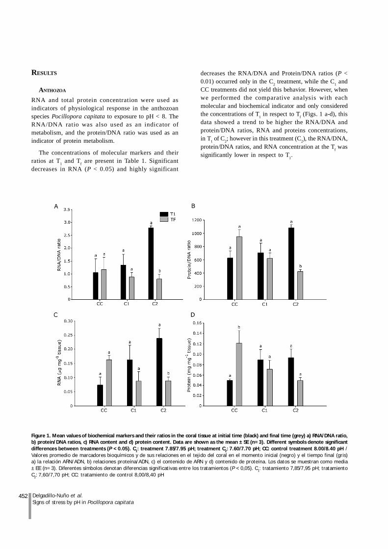
The concentrations of molecular markers and theirratios at T1 and Tf are present in Table 1. Significantdecreases in RNA (P < 0.05) and highly significant
decreases the RNA/DNA and Protein/DNA ratios (P <0.01) occurred only in the C2 treatment, while the C1 andCC treatments did not yield this behavior. However, whenwe performed the comparative analysis with eachmolecular and biochemical indicator and only consideredthe concentrations of T1 in respect to Tf (Figs. 1 a-d), thisdata showed a trend to be higher the RNA/DNA andprotein/DNA ratios, RNA and proteins concentrations,in T1 of C2; however in this treatment (C2), the RNA/DNA,protein/DNA ratios, and RNA concentration at the Tf wassignificantly lower in respect to T1.
Figure 1. Mean values of biochemical markers and their ratios in the coral tissue at initial time (black) and final time (grey) a) RNA/DNA ratio,b) protein/DNA ratios, c) RNA content and d) protein content. Data are shown as the mean ± SE (n= 3). Different symbols denote significantdifferences between treatments (P < 0.05). C1: treatment 7.85/7.95 pH; treatment C2: 7.60/7.70 pH; CC: control treatment 8.00/8.40 pH /Valores promedio de marcadores bioquímicos y de sus relaciones en el tejido del coral en el momento inicial (negro) y el tiempo final (gris)a) la relación ARN/ADN, b) relaciones proteína/ADN, c) el contenido de ARN y d) contenido de proteína. Los datos se muestran como media± EE (n= 3). Diferentes símbolos denotan diferencias significativas entre los tratamientos (P < 0,05). C1: tratamiento 7,85/7,95 pH; tratamientoC2: 7,60/7,70 pH; CC: tratamiento de control 8,00/8,40 pH

453Vol. 49, Nº 3, 2014Revista de Biología Marina y Oceanografía
DINOFLAGELLATE ENDOSYMBIONTS
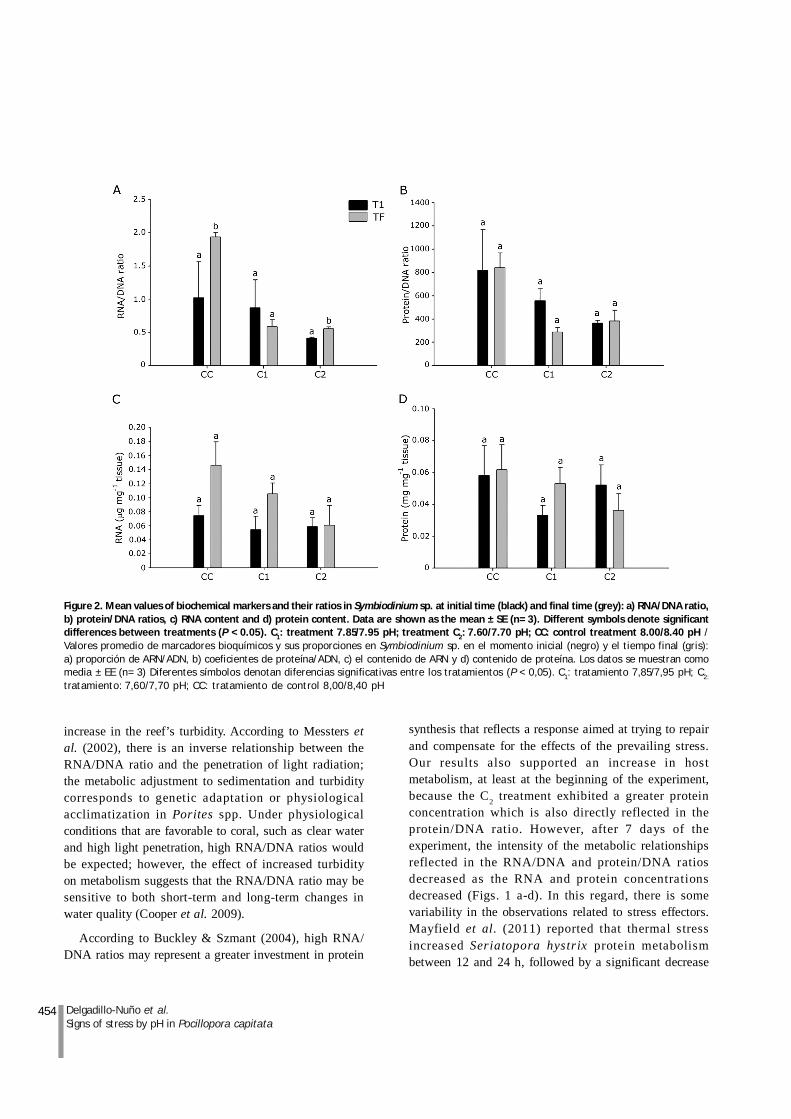
Regarding the behavior of the symbiont, Table 1 showsthat significant differences between the initial and finaltime were apparent only in the C2 and C1 treatments in theRNA/DNA ratio. A decreasing trend in the protein/DNAratio was observed in treatment C2, but this trend was notsignificant.
Comparison of each relationship and molecularindicator of T1 in respect to Tf of symbiont in eachtreatment are presented in Figures 2 a-d. Generally, thereis a tendency to decrease the values in the RNA/DNA,protein/DNA ratios and the concentration of RNA andprotein in CI and C2 treatments compared to the controltreatment without being significantly different; however,the RNA/DNA ratio in Tf -CC was the only that showed asignificantly higher ratios compared to other treatmentsat different times (P < 0.05). This differs from previouslydescribed responses for the host in the initial phase ofthe experiment, in which higher concentrations wereobserved in the treatment C2 time T1.
SKELETAL MICROMORPHOLOGY
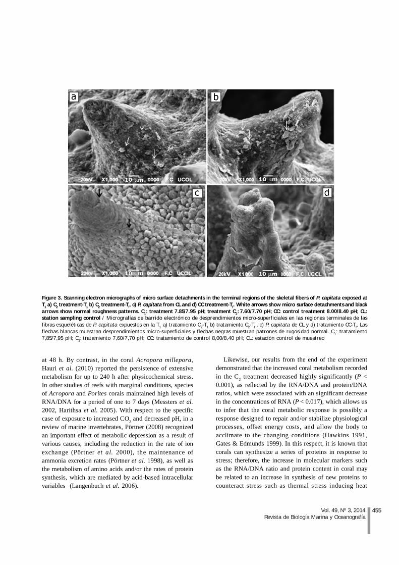
Skeletal micromorphology was observed by SEM as aqualitative marker of the physiological effects ofdecreased pH on P. capitata (Figs. 3 a-d). The effects ofC2 and C1 on the surface relief pattern could be observed
(Figs. 3 a-b). At T1, effects of C2 could only be detected atisolated sites, characterized mainly by micro surfacedetachments in the superficial regions of the skeletal, atthe end of the experiment we observe more detachmentsin treatment C2 in contrast with areas without detachmentsand increased rugosity of coral control treatmentcollected from distant location. The Figure 3 c shows theobservation of a greater abundance of ridges distributedin the spines and in the basis of those same.
DISCUSSION
Increased CO2 and the consequent decrease in pH mayaffect the physiology and health of various species ofcoral, rapidly leading to alterations in metabolic processes,productivity, the balance of dinoflagellate endosymbionts,and calcification, which could result in bleaching and,finally, cessation of growth (Anthony et al. 2008).
When Pocillopora capitata was exposed to the lowestpH level (C2), the RNA/DNA ratio increased significantly,reflecting mainly an increase in the concentration of RNAwith respect to the variability of DNA that is consideredless variable (Fig. 1c). Bak & Meesters (2000) characterizedthe behavior of the RNA/DNA ratio in Porites spp. andrecognized that this increase in the metabolic ratiorepresents an intensification that was influenced by an
Table 1. Average values of RNA/DNA and protein/DNA ratios, total amounts of RNA and protein, in Pocillopora capitata (Cnidaria) and Symbiodiniumsp. (Zooxanthellae), during pH-low levels experiment at initial time (T1) and final time (Tf). C1: treatment 7.85/7.95 pH; treatment C2: 7.60/7.70 pH;CC: control treatment 8.00/8.40 pH. Means with different superscript are significantly different (P < 0.05) / Valores promedio de las relacionesARN/ADN y proteína/ADN, concentración total de ARN y proteínas, en Pocillopora capitata (Cnidaria) and Symbiodinium sp. (Zooxanthellae),durante niveles experimentales bajos de pH a tiempo inicial (T1) y tiempo final (Tf). C1: tratamiento 7,85/7,95 pH; tratamiento C2: 7,60/7,70 pH;CC: tratamiento de control 8,00/8,40 pH. Valores medios con diferente literal son significativamente diferentes (P < 0,05)
454 Delgadillo-Nuño et al.Signs of stress by pH in Pocillopora capitata
increase in the reef’s turbidity. According to Messters etal. (2002), there is an inverse relationship between theRNA/DNA ratio and the penetration of light radiation;the metabolic adjustment to sedimentation and turbiditycorresponds to genetic adaptation or physiologicalacclimatization in Porites spp. Under physiologicalconditions that are favorable to coral, such as clear waterand high light penetration, high RNA/DNA ratios wouldbe expected; however, the effect of increased turbidityon metabolism suggests that the RNA/DNA ratio may besensitive to both short-term and long-term changes inwater quality (Cooper et al. 2009).
According to Buckley & Szmant (2004), high RNA/DNA ratios may represent a greater investment in protein
synthesis that reflects a response aimed at trying to repairand compensate for the effects of the prevailing stress.Our results also supported an increase in hostmetabolism, at least at the beginning of the experiment,because the C2 treatment exhibited a greater proteinconcentration which is also directly reflected in theprotein/DNA ratio. However, after 7 days of theexperiment, the intensity of the metabolic relationshipsreflected in the RNA/DNA and protein/DNA ratiosdecreased as the RNA and protein concentrationsdecreased (Figs. 1 a-d). In this regard, there is somevariability in the observations related to stress effectors.Mayfield et al. (2011) reported that thermal stressincreased Seriatopora hystrix protein metabolismbetween 12 and 24 h, followed by a significant decrease
Figure 2. Mean values of biochemical markers and their ratios in Symbiodinium sp. at initial time (black) and final time (grey): a) RNA/DNA ratio,b) protein/DNA ratios, c) RNA content and d) protein content. Data are shown as the mean ± SE (n= 3). Different symbols denote significantdifferences between treatments (P < 0.05). C1: treatment 7.85/7.95 pH; treatment C2: 7.60/7.70 pH; CC: control treatment 8.00/8.40 pH /Valores promedio de marcadores bioquímicos y sus proporciones en Symbiodinium sp. en el momento inicial (negro) y el tiempo final (gris):a) proporción de ARN/ADN, b) coeficientes de proteína/ADN, c) el contenido de ARN y d) contenido de proteína. Los datos se muestran comomedia ± EE (n= 3) Diferentes símbolos denotan diferencias significativas entre los tratamientos (P < 0,05). C1: tratamiento 7,85/7,95 pH; C2:
tratamiento: 7,60/7,70 pH; CC: tratamiento de control 8,00/8,40 pH
455Vol. 49, Nº 3, 2014Revista de Biología Marina y Oceanografía
at 48 h. By contrast, in the coral Acropora millepora,Hauri et al. (2010) reported the persistence of extensivemetabolism for up to 240 h after physicochemical stress.In other studies of reefs with marginal conditions, speciesof Acropora and Porites corals maintained high levels ofRNA/DNA for a period of one to 7 days (Messters et al.2002, Harithsa et al. 2005). With respect to the specificcase of exposure to increased CO2 and decreased pH, in areview of marine invertebrates, Pörtner (2008) recognizedan important effect of metabolic depression as a result ofvarious causes, including the reduction in the rate of ionexchange (Pörtner et al. 2000), the maintenance ofammonia excretion rates (Pörtner et al. 1998), as well asthe metabolism of amino acids and/or the rates of proteinsynthesis, which are mediated by acid-based intracellularvariables (Langenbuch et al. 2006).
Likewise, our results from the end of the experimentdemonstrated that the increased coral metabolism recordedin the C2 treatment decreased highly significantly (P <0.001), as reflected by the RNA/DNA and protein/DNAratios, which were associated with an significant decreasein the concentrations of RNA (P < 0.017), which allows usto infer that the coral metabolic response is possibly aresponse designed to repair and/or stabilize physiologicalprocesses, offset energy costs, and allow the body toacclimate to the changing conditions (Hawkins 1991,Gates & Edmunds 1999). In this respect, it is known thatcorals can synthesize a series of proteins in response tostress; therefore, the increase in molecular markers suchas the RNA/DNA ratio and protein content in coral maybe related to an increase in synthesis of new proteins tocounteract stress such as thermal stress inducing heat
Figure 3. Scanning electron micrographs of micro surface detachments in the terminal regions of the skeletal fibers of P. capitata exposed atT1 a) C2 treatment-T1 b) C2 treatment-Tf, c) P. capitata from CL and d) CC treatment-Tf. White arrows show micro surface detachments and blackarrows show normal roughness patterns. C1: treatment 7.85/7.95 pH; treatment C2: 7.60/7.70 pH; CC: control treatment 8.00/8.40 pH; CL:station sampling control / Micrografías de barrido electrónico de desprendimientos micro-superficiales en las regiones terminales de lasfibras esqueléticas de P. capitata expuestos en la T1 a) tratamiento C2-T1 b) tratamiento C2-Tf , c) P. capitata de CL y d) tratamiento CC-Tf. Lasflechas blancas muestran desprendimientos micro-superficiales y flechas negras muestran patrones de rugosidad normal. C1: tratamiento7,85/7,95 pH; C2: tratamiento 7,60/7,70 pH; CC: tratamiento de control 8,00/8,40 pH; CL: estación control de muestreo
456 Delgadillo-Nuño et al.Signs of stress by pH in Pocillopora capitata
shock proteins or HSPs (Harithsa et al. 2005, Hauri et al.2010). In addition to their role in overall cellular protection,HSPs have been reported to enhance ‘thermotolerance’,or the ability to recover from stress and the ability tocope with subsequent stress processes in variousorganisms (Tomanek 2010, Sokolova et al. 2011), includingthe octocoral Dendronephthya klunzingeri (Wiens et al.(2000). These stress factors have occurred in the coralcommunity LB, as documented by Liñan-Cabello et al (2008,2010b) and will be subsequently discussed in this study.
Moreover, the initial dinoflagellate stress responsewas different from that observed in host; instead of anincrease in symbiont metabolism at T1, a significantdecrease in the RNA/DNA and protein/DNA ratios wereobserved in treatment C2 compared to treatments C1 andCC (P < 0.05, Figs. 2 a-b) without significant changes inRNA and protein. This apparent lack of metabolicresponse is in agreement with other studies that havesuggested that symbiont photosynthetic metabolism isnot favored by the enrichment of dissolved inorganiccarbon, and the photosynthetic capacity is unaffectedby changes in CO2 and pH in the short and medium term(Langdon et al. 2003, Reynaud et al. 2003). In this regard,Muehllehner & Edmunds (2008) note that, while thedecrease in pH can negatively affect the calcification ofthe coral Pocillopora meandrina, no deficiency inendosymbiotic photosynthetic capacity occurs uponexposure for 14 days to pH 7.8 at low temperatures of29°C and lower. In this regard, Marubini et al. (2008)recognized that the lack of a response in Symbiodiniumspp. may be associated with low permeability to CO2 inthe membrane of Symbiodinium and/or the peri-symbioticmembrane, coupled with considerable control of thesupply of carbon by the host. This confirms the possibledeficiencies previously established in relation to themetabolic response of Symbiodinium to increasingdissolved CO2 related to a short-term decrease in pH andCO3
2- (Marubini et al. 2003, Langdon et al. 2003, Reynaudet al. 2003). Despite the identification of the metabolicresponse signs of Symbiodinium referred by Kaniewskaet al. (2012) to ocean acidification, the present studyconfirms deficiencies of a specific short and medium-termmetabolic response of Symbiodinium sp. to increasedCO2. These differences in the responsiveness betweenhost and symbiont were recently confirmed by Soriano-Santiago et al. (2013) in an in vitro experiment using lowpH, they observed a change in oxidative stress markerswithin the first few hours, but this response did notprevent cellular damage.
At the Tf. time point, the metabolism of Symbiodiniumwas characterized by significantly lower RNA/DNA andprotein/DNA ratios in C1 and C2 treatments (Fig. 2 a-b).For its part, the slight increase in the RNA/DNA ratio inthe C2 treatment at the end of the experiment compared tothe initial time, similar to that observed in the controltreatment, could be due to the different responsecapabilities of the endosymbiont community. Differentclades of Symbiodinium sp. could be present in differenthost species and in a single host that inhabits variousenvironments, leading to differences in symbiontphotosynthetic characteristics (Leggat et al. 1999). In thisrespect, recently Cunning et al. (2013) recognized thatPocillopora are the corals typically associated withSymbiodinium in clade C and/or D, with clade Dassociations having greater thermal tolerance andresistance to bleaching. Thus, the acclimatization responseof the specimens from LB may be influenced by theactivity of a specific symbiont clade that has allowed it torespond to the stress environment prevailing in thelocality. In turn, this could permit specimens todemonstrate a greater responsiveness to pH treatments,giving rise to the highest survival in days for specimensfrom LB (10) compared with the survival of 7 days recentlyreported by Delgadillo-Nuño (2012) for specimens of P.capitata from another locality of Colima coast, in anexperimental system in vitro, similar to conditions ofacidification used in this experiment. In the presence ofmedium pH conditions (7.85-7.95), host tissue exhibited asignificant increase in the RNA and protein content similarto that presented in the C2 treatment of LB of our study.Similarly, the symbiont did not exhibit any significantmetabolic changes upon treatment. Thus, there wereclearly differences in the metabolic responses of hostand symbionts in specimens from LB and corals fromother areas with less history of environmental and/oranthropogenic stress. Notwithstanding the differentresponses to low pH treatments, the specimens exposedto low pH treatment showed signs of bleaching and diedat 10 days. Anthony et al. (2008) reported that low pHcould disrupt the major route of CO2 accumulation orelectron transport, destabilizing the intracellular protongradient and directly affecting the ability of symbiont tofix carbon, in addition to disrupting photosyntheticprocesses.
It has previously been reported that specimens of P.capitata from LB, in addition to exhibiting short-termadjustments such as the amount of photosyntheticpigment and/or sensitivity to the photosynthetic
457Vol. 49, Nº 3, 2014Revista de Biología Marina y Oceanografía
response reported in other coral species, exhibit a seriesof short-term enzymatic responses aimed at counteringthe effects of ROS, thereby promoting adaptation toshallow coastal environments which typically exhibit highlevels of UVR and thermal variability (Flores-Ramirez &Liñán-Cabello 2007, Liñán-Cabello et al. 2010a). Accordingto Freire et al. (2011), changes in HSP (and other proteins)expression can be detected within a few hours (1-6 h),corresponding to the duration of most physiologicalevents that occur in tidal cycles. Recently, different signsof stress in Pocillopora spp. corals have been reportedas a result of the inadequate location of the artificialstructure intercommunication of Juluapan Lagoon in theBay of Santiago where the LB coral community is located;this exposes the coral community to tidal effects, rapidlyinfluencing turbidity, sediment, nutrients, temperature andosmolarity (Liñán-Cabello et al. 2008, 2010b). Theenvironmental history of the LB coral community mayhave generated a physiological alertness that promotes arapid metabolic response, but, at the prevailing low pHconditions, may have interfered with carbonconcentration mechanisms in the photorespirationmachinery and/or the direct impact of metabolic acidosisas a consequence of the decrease of seawater pH (Leggatet al. 1999, Gattuso et al. 1999, Kim et al. 2004), which ledto the bleaching and deaths of our specimens.
According to our microscopic observations, theabundance of thorns bordering the LB control treatment(Fig. 3 d) contrasts sharply with the submitted bounds ofcontrol P. capitata obtained from the locality CL (Fig. 3c). In this regard, Brown et al. (1997) study the effect ofthe dynamic action on the layout and arrangement of thesurface ultrastructure. According to these authors,specimens of P. damicornis that were located on a largerdynamic envelope wave-influenced side and shape ofthe skeletal spines. Thereby, spines are more present incorallites on branch tips exposed to wave action. In thissense, we can considerer the lack of LB’s roughness couldbe a sign of microstructural deterioration associated withcontinuous exposure to environmental variability, as thispopulation is also in a shallow location (0.5-2.5 m). Thearea is highly exposed to ocean dynamics; therefore, itshould exhibit a greater roughness pattern.
Differences in the patterns of abundance of ridgesdistributed in the spines and in the basis of thoseobserved under high magnification were a consequenceof the micro surface detachments in the terminal regionsof C2 with respect to C1 and CC (Figs. 3 a, b and d). This
coincides with reports by Marubini et al. (2003), whoused SEM to study the suppression of the growth ofscleractinian coral environments induced by lowcarbonate ion concentrations and recognized differencesin the size of available microcrystalline units; themagnitude of the effects varied depending on the species:the fibers were most reactive in Acropora verweyi, lessin Turbinaria reniformis, and intermediate in Galaxeafascicularis and Pavona cactus. In corals, aragonitesaturation state control via ion supply for calcificationand skeletal growth involves a large number of metabolicpathways; however, temperature and pCO2 affect growthphysiology and calcification (Tambutté et al. 2011).Therefore, the present results of skeletal surface andmetabolic responses suggest this effect in P. capitata,but further experiments are needed to better estimate theeffects and adaptation capacity of Pocillopora spp. fromLB.
The RNA/DNA ratio was a useful tool to observe rapidchanges in active host metabolism. Of the two treatmentsused, the treatment at the lower pH resulted in increasedmetabolic activity, primarily the host, which was directlyrelated to the protein/DNA ratio and RNA and proteinconcentrations. This response was identified in the earlyhours of the experiment but its incidence subsequentlydecreased. In the case of Symbiodinium, a metabolicresponse similar to the host response was not apparent.Qualitative microstructural analysis primarily revealedeffects from the C2 treatment in the first hours of testingwith an influence on the micro surface detachments interminal regions of the skeletal fibers.
Observed pH changes may directly affect coralcalcification or indirectly affect calcification through coralmetabolism, they may affect symbionts by inducing adecrease in zooxanthellae photorespiration andproductivity or bleaching, as both of these processescan affect coral calcification.
Further studies using other quantitative indicators areneeded to confirm that decreases in pH may be associatedwith skeletogenesis in scleractinian corals.
ACKNOWLEDGMENTS
This project was supported by the Consejo Nacional deCiencia y Tecnología of Mexico (CONACYT 2009-118024).We appreciate the thorough and thoughtful comments oftwo anonymous reviewers.
458 Delgadillo-Nuño et al.Signs of stress by pH in Pocillopora capitata
LITERATURE CITED
Anthony KRN, DI Kline, G Diaz-Pulido, S Dove & OHoegh-Guldberg. 2008 . Ocean acidification causesbleaching and productivity loss in coral reef builders.Proceedings of the National Academy of Sciences of theUnited States of America 105: 17442-17446.
Bak RPM & EH Meesters. 2000. Acclimatization/adaptationof coral reefs in a marginal environment. Proceedings 9thInternational Coral Reef Symposium, Bali, Indonesia,October, pp. 265-272.
Baker AC, PW Glynn & B Riegl. 2008. Climate change andcoral reef bleaching: An ecological assessment of long-termimpacts, recovery trends and future outlook. EstuarineCoastal and Shelf Science 80(4): 435-471.
Bradford MM. 1976. A rapid and sensitive method for thequantitation of microgram quantities of protein utilizingthe principle of protein-dye binding. Analytical Biochemistry72: 248-254.
Brown BE. 1997 . Adaptations of reef corals to physicalenvironmental stress. Advances in Marine Biology 31: 222-299.
Buckley BA & AM Szmant. 2004 . RNA/DNA ratios asindicators of metabolic activity in four species of Caribbeanreef-building corals. Marine Ecology Progress Series 282:143-149.
Cooper T, J Gilmour & K Fabricius. 2009. Bioindicators ofchanges in water quality on coral reefs: review andrecommendations for monitoring programmers. Coral Reefs28(3): 589-606.
Cunning R, PW Glynn & AC Baker. 2013 . Flexibleassociations between Pocillopora corals and Symbiodiniumlimit utility of symbiosis ecology in defining species. CoralReefs 32(3): 795-801.
Delgadillo-Nuño MA. 2012. Relaciones de ácidos nucleicoscomo trazadores de estrés fisiológico en Pocillopora capitataasociado a la disminución de pH. Tesis de Licenciatura,Universidad de Colima, México.
Flores-Ramírez LA & MA Linán-Cabello. 2007 .Relationships among thermal stress, bleaching and oxidativedamage in the hermatypic coral, Pocillopora capitata.Comparative Biochemistry and Physiology C 146: 194-202.
Freire CA, AF Welker, JM Storey, KB Storey & M Hermes-Lima. 2011. Oxidative stress in estuarine and intertidalenvironments (temperate and tropical). In: Abele D, TZenteno-Savín & J Vázquez-Medina (eds). Oxidative stressin aquatic ecosystems, pp. 41-57. John Wiley & Sons,Chichester.
Gates RD & PJ Edmunds. 1999 . The physiologicalmechanisms of acclimatization in tropical reef corals.American Zoologist 39: 30-43.
Gattuso JP, D Allemand & M Frankignoullej. 1999 .Photosynthesis and calcification at cellular, organismal andcommunity levels in coral reefs: A review on interactionsand control by carbonate. The American Zoologist 183:160-183.
Harithsa S, C Raghukumar & SG Dalal. 2005 . Stressresponse of two coral species in the Kavaratti atoll of theLakshadweep Archipelago, India. Coral Reefs 24: 463-474.
Harrould-Kolieb E & J Savitz. 2009. Acidificación: ¿Cómoafecta el CO2 a los océanos?, 29 pp. Oceana, Madrid. <http://oceana.org/sites/default/files/reports/Acidification_Report_2009_Spa.pdf>
Hauri C, KE Fabricius, B Schaffelke & C Humphrey. 2010.Chemical and physical environmental conditions underneathmat- and canopy-forming macroalgae, and their effects onunderstorey corals public library of science. PLOS ONE5(9): e12685, <doi:101371/journalpone0012685>
Hawkins AJS. 1991. Protein turnover: A functional appraisal.Functional Ecology 5: 222-233.
Hoegh-Guldberg O. 1999. Climate change, coral bleachingand the future of the world’s coral reefs. Marine andFreshwater Research 50: 839-866.
Hoegh-Guldberg O, PJ Mumby, AJ Hooten, RS Steneck PGreenfield, E Gomez, CD Harvell, PF Sale, AJ Edwards,K Caldeira, N Knowlton CM Eakin, R Iglesias-Prieto,N Muthiga, RH Bradbur, A Dubi & ME Hatziolos. 2007.Coral reefs under rapid climate change and oceanacidification. Science 318: 1737.
IPCC. 2007. Cambio climático 2007. En: Pachauri RK & AReisinger (dir). Informe de Síntesis. Contribución de losGrupos de Trabajo I, II y III al Cuarto Informe de Evaluacióndel Grupo Intergubernamental de Expertos sobre el CambioClimático, pp. 1-104. IPCC, Ginebra.
Kaniewska P, PR Campbell, DI Kline, M Rodriguez-Lanetty, DJ Miller, S Dove & O Hoegh-Guldberg. 2012.Major cellular and physiological impacts of oceanacidification on a reef building coral. PLOS ONE 7(4):e34659, <doi:101371/journalpone0034659>
Karp GC. 2010. Cell and molecular biology: Concepts andexperiments, 776 pp. John Wiley & Sons, Gainesville.
Kim Y, AF Yakunin, E Kuznetsova, X Xu, M Pennycooke,J Gu, F Cheung, M Proudfoot, CH Arrowsmith, AJoachimiak, AM Edwards & D Christendat. 2004 .Structure and function-based characterization of a newphosphoglycolate phosphatase from Thermoplasmaacidophilum. Journal Biology Chemical 279: 517-526.
Kleypas JA & C Langdon. 2006. Coral reefs and changingseawater carbonate chemistry. In: Phinney JT, O Hoegh-Guldberg, J Kleypas, W Skirving & A Strong (eds). Coralreefs and climate change: science and management. Coastaland Estuarine Series 61: 73-110. American GeophysicalUnion, Washington.
Langdon C, WS Broecker, DE Hammond, E Glenn KFitzsimmons, SG Nelson, T-S Peng, I Hajdas & GBonani. 2003. Effect of elevated CO2 on the communitymetabolism of an experimental coral reef. GlobalBiogeochemistry C 17: 1011.
Langenbuch M, C Bock, D Leibfritz & HO Pörtner. 2006.Effects of environmental hypercapnia on animal physiology:a 13C-NMR study of protein synthesis rates in the marineinvertebrate Sipunculus nudus. Comparative BiochemicalPhysiology A 144: 479-484.
459Vol. 49, Nº 3, 2014Revista de Biología Marina y Oceanografía
Leggat W, MR Badger & D Yellowlees. 1999. Evidence foran inorganic carbon-concentrating mechanism in thesymbiotic dinoflagellate Symbiodinium sp. Plant Physiology121: 1247-1255.
Liñán-Cabello MA, D Hernández-Medina, P Florián-Álvarez & A Mena-Herrera. 2008. Estado actual delarrecife coralino ‘La Boquita’, Colima. IRIDIA 5: 10-23.
Liñán-Cabello MA, LA Flores-Ramírez, JF Cobo-Díaz, TZenteno-Savin, NO Olguín-Monroy, A Olivos-Ortiz &A Tintos-Gómez. 2010a. Response to short term ultravioletstress in the reef-building coral Pocillopora capitata(Anthozoa: Scleractinia). Revista de Biología Tropical 58(1):103-118.
Liñán-Cabello MA, LA Flores-Ramírez, T Zenteno-Savin,NO Olguín-Monroy, R Sosa-Avalos, M Patiño-Barragan& A Olivos-Ortiz. 2010b. Seasonal changes of antioxidantand oxidative parameters in the coral Pocillopora capitataon the Pacific coast of Mexico. Marine Ecology 31(3): 407-417.
Marubini F, C Ferrier-Pages & JP Cuif. 2003. Suppressionof skeletal growth in scleractinian corals by decreasingambient carbonate-ion concentration: a cross-familycomparison. Proceeding of the Royal Society of London B270: 179-184.
Marubini F, C Ferrier-Pagès, P Furla & D Allemand. 2008.Coral calcification responds to seawater acidification: aworking hypothesis towards a physiological mechanism.Coral Reefs 27: 491-499.
Mayfield AB, LH Wang, PC Tang, TY Fan, Y-YHsiao, C-LTsai & C-S Chen. 2011 . Assessing the impacts ofexperimentally elevated temperature on the biologicalcomposition and molecular chaperone gene expression of areef coral. PLOS ONE 6 (10), e26529, <doi:101371/journalpone0026529>
Meesters EH, G Nieuwland, GCA Duineveld, A Kok &RPM Bak. 2002. RNA/DNA ratios of scleractinian coralssuggest acclimatization /adaptation in relation to lightgradients and turbidity regimes. Marine Ecology ProgressSeries 227: 233-239.
Muehllehner N & PJ Edmunds. 2008. Effects of oceanacidification and increased temperature on skeletal growthof two scleractinian corals, Pocillopora meandrina andPorites rus. Proceedings of the 11th International CoralReef Symposium, Ft Lauderdale, Florida, pp. 7-11.
Pörtner H. 2008. Ecosystem effects of ocean acidification intimes of ocean warming: a physiologist’s view. MarineEcology Progress Series 373: 203-217.
Pörtner HO, A Reipschläger & N Heisler. 1998. Metabolismand acid–base regulation in Sipunculus nudus as a functionof ambient carbon dioxide level. Journal of ExperimentalBiology 201: 43-55.
Pörtner HO, C Bock & A Reipschläger. 2000. Modulation ofthe cost of pH regulation during metabolic depression: a31PNMR study in invertebrate (Sipunculus nudus) isolatedmuscle. Journal of Experimental Biology 203: 2417-2428.
Reynaud S, N Leclercq, S Romaine-Lioud, C Ferrier-Pages,C Ferrier-Page, J Jaubert & JP Gattuso. 2003. Interactingeffects of CO2 partial pressure and temperature onphotosynthesis and calcification in a scleractinian coral.Global Change Biology 9: 1660-1668.
Sokolova IM, AA Sukhotin & G Lannig. 2011. Stress effectson metabolism and energy budgets in mollusks. In: AbeleD, T Zenteno-Savín & J Vazquez-Medina (eds). Oxidativestress in aquatic ecosystems, pp. 261-280. John Wiley &Sons, Chichester.
Soriano-Santiago OS, MA Liñán-Cabello, MA Delgadillo-Nuño, C Ortega-Ortiz & S Cuevas-Venegas. 2013 .Physiological responses to oxidative stress associated withpH variations in host tissue and zooxanthellae ofhermatypic coral Pocillopora capitata. Marine FreshwaterBehavior Physiology 46(5): 275-286.
Sulkin SD, RP Morgan & LL Minasian. 1975. Biochemicalchanges during larval development of the Xanthid CrabRhithropanopeus harrisii. II. Nucleic acids. Marine Biology32: 113-117.
Tambutté S, M Holcomb, C Ferrier-Pagès, S Reynaud, ETambutté, D Zoccola & D Allemand. 2011 . Coralbiomineralization: from the gene to the environment. Journalof Experimental Marine Biology and Ecology 408(1): 58-78.
Tomanek L. 2010. Variation in the heat shock response and itsimplication for predicting the effect of global climate changeon species’ bioegographic distribution ranges and metaboliccosts. Journal Experimental Biology 213: 971-979.
Veron JEN, O Hoegh-Guldberg, TM Lenton, JM Lough,DO Obura, P Pearce-Kelly, CR Sheppard, M Spalding,MG Stafford-Smith & AD Rogers. 2009. The coral reefcrisis: the critical importance of < 350 ppm CO2. MarinePollution Bulletin 58: 1428-1436.
Wiens M, MS Ammar, AH Nawar, C Koziol, HM Hassanein,M Eisinger, IM Muller & WE Muller. 2000. Inductionof heat-shock (stress) protein gene expression by selectednatural and anthropogenic disturbances in the octocoralDendronephthya klunzingeri. Journal Experimental MarineBiology Ecology 245: 265-276.
Yellowlees D, TA Rees & W Leggat. 2008 . Metabolicinteractions between algal symbionts and invertebrate hosts.Plant Cell Environment 31: 679-694.
Zar JH. 1999. Biostatistical analysis, 662 pp. Prentice-Hall,Englewood Cliffs.
Received 14 January 2014 and accepted 12 August 2014
Editor: Claudia Bustos D.