Embed Size (px)
Citation preview

Alternative Techniques to Obtain Monoclonal Antibodiesat a Small Scale: Current State and Future Goals
Rodolfo Valdés Véliz
Monoclonal Antibodies Division. Center for Genetic Engineering and Biotechnology.Ave 31 e/ 158 y 190, Playa, Ciudad de La Habana, Cuba. Phone: (53-7) 271 8008;
Fax (53-7) 271 3208; E-mail: [email protected]
ABSTRACTThe ascites method has been one of the most popular means for producing large quantities of highly concentratedmonoclonal antibodies since its inception in 1972. Here we summarize some alternative methods to producemonoclonal antibodies and we compare them with the traditional method (ascites). The advantages and disadvan-tages of these techniques will be considered in order to investigate the practicality of using in vitro techniques togenerate large quantities of MAbs. We also present the regulatory restrictions for MAbs production in differentcountries. Finally, a brief overview of the emerging technologies is described.
Key words: in vitro, in vivo, monoclonal antibodies
Biotecnología Aplicada 2002;19:119-131
RESUMENTécnicas alternativas para la obtención de anticuerpos monoclonales a pequeña escala: Estado actualy perspectivas. Desde su introducción en 1972 el método de producción de ascitis ha sido uno de los máspopulares para producir grandes cantidades de anticuerpos monoclonales (AcMs) altamente concentrados. En elpresente trabajo resumimos los métodos alternativos de producción de AcMs y los comparamos con el métodotradicional de producción. Para investigar la factibilidad práctica del empleo de técnicas de producción in vitro en laobtención de grandes cantidades de AcMs consideramos las ventajas y desventajas de estas técnicas. Tambiénevaluamos las restricciones regulatorias para producir AcMs en diferentes países. Finalmente se describe una breverevisión de las tecnologías emergentes para producir AcMs.
Palabras claves: anticuerpos monoclonales, in vitro, in vivo
IntroductionThe continuous production of monoclonal antibodies(MAbs) from mouse splenic lymphocytes after im-mortalization by fusion with a plasmocytoma cell linewas first reported by Köhler and Milstein in 1975[1]. At that time, the technique was just another toolfor their research, but the impact was so enormous,that a Nobel prize was awarded to Köhler and Milstein,along with Jerne, in 1984.
This gave research a valuable, powerful and versa-tile tool, since the specificity of antibodies is exquis-ite and extremely sensitive. Since then, the productionof monoclonal antibodies has reached multibillion-dollar markets in diagnostics, therapeutics and for pu-rification of proteins and chemicals [2].
The challenge for many laboratories growing hy-bridoma cell lines is to produce adequate amounts ofMAbs. The main limitating aspects are space, timeand money. Media amounts of MAbs typically re-quire an investment in expensive bioreactors, spaceallocated to many of tissue culture flasks stacked inincubators, or a very large number of mice.
The applications of MAbs are numerous and di-verse. At present four user groups can be identifiedaccording to the amount of antibody required(Table 1). Currently there are more than 100 000 dif-ferent MAbs available. Most of them are producedat a small scale (<0.1 g) solely for bench-related pur-poses. Larger amounts are often required for diag-nostic kits and reagents (0.1–0.5 g), for routinediagnostic procedures and in pre-clinical evaluationstudies (0.5–10 g). Finally, larger amounts are re-quired for prophylactic, therapeutic purposes and
to immunopurify antigens (>10 g). This outlineclearly shows that the application of MAbs is al-most exclusively limited to research and human medi-cine. Although the application of MAbs for otherpurposes is easily envisaged, this has not come ma-terialized because the production costs MAbs on alarge scale are extremely high, regardless of the pro-duction system used.
Here we compare the classical way of producingMAbs (ascites) with the in vitro methods and finallywe present some emerging technologies.
Ascites Production of MAbsAntibodies can be obtained at high concentrationsby injecting the hybridoma cells into the peritonealcavity of genetically compatible mice or rats and ob-taining the antibody in the ascitic fluid (Figure 1).
Table 1. Monoclonal antibodies: user groups.
Group MAb required Proportion Most widespread
method Applications
A <0.1 g ~60% In vivo Fundamental and applied research Commercial production of diagnostic kits for research and analytical purposes
B 0.1-0.5 g ~30% In vivo
Development and production of diagnostic kits For evaluating the usefulness of novel therapeutic MAbs in animal experiments
C 0.5-10 g ~10% In vitro Routine diagnostic procedures In pre-clinical evaluation studies
D >10 g <1% In vitro Prophylactic, therapeutic and for purification purposes
Figure 1. Production of monoclonalantibodies by the ascites method.
REVIS
IÓN

Rodolfo Valdés Véliz In vivo vs. in vitro MAbs production
Biotecnología Aplicada 2002; Vol.19, Nos. 3 y 4120
While culture supernatants yield on average 1–10 µg/mL, the ascitic fluid contains 1–20 mg/mL.
Several mechanisms have been reported [3, 4] toaccount for the production of ascites. To produceMAbs as ascites fluid, mice are primed with an intra-peritoneal injection of pristane (2,6,10,14-tetramethylpentadecane). The effect of the primer is two-fold:it suppresses the immune system so that the growthof the hybridoma cells in the abdomen is not stronglyimpaired, and it causes toxic irritation which leadsto peritonitis and the secretion of serous fluid. Af-ter 7–21 days, the abdomen of the inoculated animalsswells, indicating ascites production and tumor growth.The cells multiply and produce antibodies, whereasthe animal responds with the production of fluid inthe peritoneal cavity The antibody-rich ascitic fluid(about 5 mL from a mouse and 10–40 mL from a rat)is harvested by paracentesis, processed and animalsare sometimes re-tapped after two days to increasethe total volume of ascites obtained per animal. Vari-ous parameters affect the yield of ascites from inocu-lated mice or the concentration of MAbs [5].
Numerous parameters have been identified that mayaffect MAb production and/ or produce abnormali-ties, pain distress, or death in the animals as a resultof the procedures used. These parameters include thehybridoma cell line used [6]; the stock or strain [7, 8];sex and age [9] of the mouse selected; volume of pris-tane [10–12] or their ascitogenic priming agent admin-istered [13–15] and the timing of pristane, or anyother ascitogenic priming agent, administration in re-lation to hybridoma cell inoculation [6, 9, 13–16]; thehybridoma cell inoculum used [6, 9, 17]; frequencyand total number of abdominal taps [6, 18–20]; themethods used for the abdominal taps [6, 7, 13]; andthe frequency of clinical observations and criteria foreuthanizing animals [20].
The ascites method to obtain MAbs can be ana-lyzed from two different points of view: Manufac-turing feasibility and regulatory issues.
Manufacturing feasibilityMurine ascites production has been the time-honoredtechnique for producing small-scale, research labora-tory quantities of MAbs [6, 21–24].
Since the introduction of the hybridoma technol-ogy, MAbs produced in mice, the so-called ascitesinduction method, has been the method of choice. Thismethod was preferred for a number of reasons: 1) Sim-plicity of the procedure; 2) No specific skills are re-quired; 3) No capital investments are needed to set upthe facilities for laboratory animals since they are gen-erally available; 4) High concentration of MAbs arefound in the ascites; 5) Ascites can be obtained in arelatively short period of time; 6) In the peritoneumthe cell densities are similar to those of solid tissues,about 109 to 1010 cells/mL; 7) Purity expressed as thespecific antibody content is high; 8) Low cost; 9) Nointensive use of labor or equipment is needed unlessspecial (i.e., SCID, nude) animals are used; and 10)Many clones can be grown at the same time.
However, one of the main manufacturing advan-tages of the ascites method is the extremely highyield and productivity of MAb, which generally liesin the range of 1–20 mg/mL [25].
These points are particularly relevant to the smallresearch laboratory with limited funds and time, yethaving the ability to develop many MAbs of interest.These are, therefore, advantages which cannot be eas-ily ignored.
The weak points are the low concentrations ob-tained in some of the wasted tissue culture superna-tants (100–1000-fold lower than ascites); the factthat the use of serum can create problems similar tothose of ascites in regard to contamination and down-stream processing; the high volume of the mediumand serum used in certain in vitro systems requiresdownstream concentration and purification; some invitro systems have high initial equipment start-up costs;the high degree of contamination with animal serumproteins that is essential for the in vitro culture of hy-bridoma cells and the fact that sometimes (3–5%), celllines do not adapt to in vitro conditions, limit the useof MAbs prepared by the conventional in vitro tech-nique and have made the ascites procedure favorablefor a long time, specially for small-scale production asneeded for bench-related activities.
Regulatory remarksAlthough the original research of Köhler and Milsteinwas principally an in vitro technique, it was also ap-parent that MAbs could be produced by injecting thehybridoma cells into the abdominal cavities of differentspecies of rodents. This was the initial use of the as-cites method. Since these in vivo MAbs were easilymade in any laboratory and the ascites process, widelyviewed as both simple and inexpensive, had long beenintroduced, its use rapidly expanded. In the decadesfollowing the original discovery, tens of millions of ani-mals suffered and died. Milstein noted, “in later years,both on practical and humane grounds, I became con-cerned with the use of ascitic fluids”. The wide advan-tages of the ascites method are outweighed by a numberof disadvantages. It is accepted that ascites productionis extremely painful and causes suffering by peritoni-tis, abdominal distention and invasive malignancy andsurvival is of approximately 14 days [7, 26, 27]. Anoverview of clinical and pathological effects was pub-lished in 1999 [20]. Analysing the overall picture ofclinical, pathophysiological and pathological changesshown in Table 2, it can be postulated that the produc-tion of ascites and the growth of tumors in rodents alsolead to a substantial impairment of animal welfare.
Table 2. Clinical, pathophysiological and pathologicaleffects of ascites production.
Clinical Pathophysiological PathologicalAbdominal distension Anorexia Peritonitis
Decreased activityand body mass
AnaemiaInfiltrative tumorgrowth
Shrunken eyes DehydrationAdhesionsin the abdomen
Difficultyin walking
Tachypnea andDyspnea
Enlargedabdominal organs
Hunchedposture
Circulatory shock Blood in theabdominal cavity
Respiratory distressDecreased venous,arterial, and renalblood flow
Apathy Ascites production
Death Immunosuppression

Rodolfo Valdés Véliz In vivo vs. in vitro MAbs production
Biotecnología Aplicada 2002; Vol.19, Nos. 3 y 4121
These limitations generate disadvantages, whichmakes the in vivo method a less attractive method.Some of these disadvantages are: it requires animal fa-cilities, support services, trained personnel and dailymonitoring to minimize pain and distress; it requiresverification and approval of the Institutional AnimalUse and Care Committee (IACUC); the use of pristanemay produce residual contamination, reactivate endog-enous murine retroviruses and promote production ofIgG autoantigens; it requires manipulative skills duringrepeated tapping to locate remaining ascitic fluid; as-citic fluid is a more complex mixture than blood plasma,having the same difficulties associated with the use ofserum for in vitro cultures and mice may not produceascites due to premature death, development of solidtumors or failure to establish hybridoma growth.
There are other non-regulatory disadvantages thatalso limit the use of ascites: a greater variability (withinanimals) in some cases; the reduced MAb immunoreac-tivity generally observed, and the higher contaminationlevels obtained (biochemically identical immunoglobu-lins, growth factors, rodent plasma proteins, withbioreactive cytokines, bacteria and viruses).
“Ascites” means “bag” in Greek, and many labora-tories have continued to use animals as if they werebags to fill with antibody-containing fluid. At the sametime that the ascites method became widespread, theappropriateness of its use was increasingly questionedin Europe, particularly in certain countries of the Eu-ropean Union.
Thus the replacement, reduction and refinement (3R),the 3R concept had been used first 46 years before byCharles Hume to provide a framework for improvingthe conduct and ethical acceptability of experimentaltechniques on animals. Given that animals used in re-search may experience pain, suffering or lasting harm,the first step must be to consider whether less harmfulor harmless alternatives could be used (replacement).Where this is not possible, care should be taken tominimize pain [28, 29], either before and during theexperiment or after the experiment. Refinement is of-ten achieved, for example, by providing the animalswith an environment in which they can feel secure andcomfortable, ensuring that they are free from infectiousdiseases, and by using appropriate anesthetics and an-algesics if surgical techniques are to be used [30]. Fi-nally, the number of animals used in a given projectneeds to be minimized (reduction), while ensuring thatthe objectives of the study are still achieved; typically,this will also reduce the total number of animal suffer-ing. The 50’s were described as the age of renaissance,the 60’s, as the age of darkness, the 70’s, the age ofreason, the 80’s the age of reformation and the 90’shave been classified by some researchers as the age ofrevolution.
There are two general laws in Europe to protectlaboratory animals: 1) The Council Directive 86/609/EU [31] and 2) The European Convention for theProtection of Vertebrate Animals Used for Experi-mental and other Scientific Purposes, ETS 123[32]..The directive came into force in 1986 and theconvention in 1991. The convention has been signedand ratified by Belgium, Cyprus, Finland, Germany,Greece, The Netherlands, Norway, Spain, Swedenand Switzerland.
However, specific national policies have establishedguidelines or regulations which restrict or prohibitascites production in rodents in Switzerland, Sweden,The Netherlands [33], the UK [34] and in Germanspeaking countries (Table 3).
The Center for the Validation of Alternative Meth-ods (ECVAM) has played one of the main roles inthese regulatory steps. In 1993 the Scientific Advi-sory Committee of the ECVAM defined as its maingoal, the promotion of scientific and regulatory ac-ceptance of alternative methods which are of impor-tance to the biosciences and which reduce, refine orreplace the use of laboratory animals.
By 1996, in vitro production of MAbs was themethod of choice in Europe for commercial concernsand other needs of individual researchers. As a re-sult, a group of experts in immunology and the invitro science representatives from many memberstates of the European Union met at the ECVAM todiscuss the current status of in vivo and in vitromethods of MAb production [35]. After careful con-siderations of all uses for MAbs and all available invivo and in vitro production options, the panel con-cluded that “for all levels of MAb production thereare one or more in vitro methods which are not onlyscientifically acceptable, but are also reasonably andpractically available; and as a consequence in vivoproduction can no longer be justified and should
Table 3. Some examples of national and institutional regulations and guidelines on the production of MAbs.
Country/ Organization Year Regulation/Guidelines
CCAC 1989 Guidelines on acceptable immunological methods. Guidelines regarding the collection of ascites and the use of humane endpoints.
Germany 1989 In vivo production of MAbs is only permitted: • If the MAbs concerned are to be used for diagnostic and therapeutic purposes
in case of emergencies; • To reserve single hybridomas that are infected or do n ot grow in vitro; • If the MAbs concerned are needed to investigate new scientific problems.
1989 Code of practice for the production of MAbs: • In vivo production only permitted when < 10 animals are needed; • Specific justification of the protocol ; • Consultation of the animal welfare officer with regard to moment and
frequency of collecting ascites, moment of euthanasia.
Netherlands
1996 In vivo production of MAbs is prohibited. Requests for exemptions are granted or rejected by the Inspectorate after a grou p of experts is consulted.
SCAW 1989 Guidelines with regard to collection of ascites and the use of humane endpoints. Sweden 1990 Recommendation of the National Board of Agriculture.
Existing alternatives should normally be used. In vivo production needs specific justification.
1989 Rules for the judgment of approval requests for animal experiments for the production of MAbs.
Switzerland
1994 In vivo production of MAbs is prohibited. Exceptions are only permitted: • When the MAbs concerned are to be use d for diagnostic and therapeutic
purposes in case of emergencies; • To rescue single hybridomas that are infected or do not grow in vitro. In case of exceptions: only 1 tap; increase in body weight may not exceed 20%.
1987 United Kingdom Coordinating Com mittee on Cancer Research (UKCCCR) guidelines: 1 tap only, the amount of ascites may not exceed 20% of the body weight.
1991 Home Office: antibody production -advice on protocols for minimal severity. Only < 20 animals per hybridoma. Repeated tapping is n ot allowed. Humane endpoints according to UKCCCR guidelines.
UK
1999 Project licenses for the ascites method are not granted unless under exceptional circumstances
CCAC, Canadian Council on Animal Care; SCAW, Scientists Center for Animal Welfare. Source: R esearch in Immunology, 74th Forum in Immunology. Vol. 149(6):529�620.

Rodolfo Valdés Véliz In vivo vs. in vitro MAbs production
Biotecnología Aplicada 2002; Vol.19, Nos. 3 y 4122
cease”. The conclusion continues to state that therewas “no fundamental reason why hybridoma cells,which have been generated and cultivated in vitro,should suddenly be able to produce antibodies onlyin an animal’s peritoneal cavity” [35].
The attention from animal-welfare organizations,the scientific community, and regulatory agencies hasincreased in the United States as well [36, 37]. Thusbased on the results of the meeting and considering thesimilarities in the arguments used by both Europeanand United States researchers to support the contin-ued reliance on the ascites methods, the AlternativeResearch and Development Foundation (ARDF) andthe American Anti-Vivisection Society (AAVS) de-cided it was time for an ascites prohibition to be en-acted in the United States. Thus after a series ofcontradictions on August 29, 1999 [38] the ARDFsponsored an international panel of experts to partici-pate in a workshop on the production of monoclonalantibodies in Bologna, Italy, and the panel concludedthat “The National Institutes of Health and the Na-tional Center for Research Resources should aggres-sively promote and encourage the use of its (and other)Institutional Monoclonal Antibody Core ProductionFacilities. The NIH should also place greater empha-sis on providing more opportunities for in vitro MAbproduction training for individual researchers/techni-cians at Core Monoclonal Antibody Production Fa-cilities”; “ In vitro production should be the accepted,required choice for MAbs; with ascites consideredthe alternative”; and “If the ascites methods are ap-proved, every effort should be made to reduce andrefine the procedures (e.g., limiting the number of tapsor increasing body weight) to minimize the pain anddistress experienced by the animals”.
However, in vitro replacement alternatives for MAbproduction in vivo seem unlikely to be optimal else-where in Europe, since, under most circumstances, theproduction of MAbs can be undertaken in vitro [39]by methods which are reasonably and practicablyavailable.
In Vitro MAbs Production
Should we use the in vitro MAbs productionsystems?As expressed above, the advantages of the ascitesmethod are widely minimized by the animal welfareconcerns and also by certain production limitations.Hence, in recent years the in vivo method has beeneventually substituted by in vitro production. Im-provements in cell culture techniques and cell cultureequipment have led to in vitro methods becoming avail-able which are able to compete with the in vivo method,both in capacity as well as in cost-effectiveness.
The in vitro method offers the following advan-tages: 1) It does not use animals for MAb production;2) No animal care issues or the IACUC approval arerequired; 3) It avoids or significantly decreases theneed for experienced laboratory personnel in animalrearing and use; 4) A wide variety of production op-tions is available; 5) Large scale production is pos-sible at a lower cost than in vivo method; 6) Many invitro systems produce MAbs with equal or betterconcentration and quality than those of ascites; 7) No
host contributed immunoglobulin or typical ascitic fluidcontaminants are found; 8) Immunoreactivity is some-times higher than with ascites (90–95%); 9) The use ofserum or a protein-free medium make downstream pu-rification easier; 10) Most hybridomas can be adaptedto serum/protein-free conditions; and 11) Under mostcircumstances it eliminates the need for Mouse Anti-body Production (MAP) testing.
The demand for animal cell derived products andthe needs to eliminate the in vivo methods have stimu-lated the development of bioreactors in the last de-cade. Most of those developments allow for a highercell concentration in the bioreactor than that found inthe past, primarily through the continuous flow ofculture medium.
Much effort has been required to reproduce the bio-logical conditions in terms of the medium and equip-ment. Therefore the question is not whether hybridomascan be grown in vitro, but whether they can be grown ina culture medium for long enough to yield the amountof MAbs needed, at a reasonable cost. Hybridomas areanchorage-independent cells, and can therefore be grownand maintained in either stationary or suspension cul-ture. The amount of antibody produced depends onthe concentration of cells attained and the time theyremain viable and secrete MAbs. The viability and pro-ductivity of hybridomas depend on if they meet theirnutritional requirements, removing metabolic wasteproducts, and providing a stable pH, temperature, anddissolved oxygen for metabolism.
Culture mediaThere are various basal media formulations suitable forgrowing hybridomas. These formulations frequentlyhave a mixture of carbohydrates, amino acids, salts,vitamins, hormones and growth factors [40–42] andthey are usually supplemented with glutamine as asupplementary energy; they are also buffered with so-dium bicarbonate or organic buffers such as HEPES.Foetal calf serum (FCS) is frequently added to the me-dia to provide growth factors and hormones. However,as a result of the disadvantages of the serum (it is ex-pensive, undefined, there are batch to batch variations,the purity of the MAb preparation decreases, there isa potential contamination with germs, as well as animalwelfare concerns) a variety of serum-free and more re-cently protein-free media formulations [43–45] andsupplements are available for hybridoma growth. Incontrast, FCS introduces a moral problem, because it isobtained by cardiac puncture from a bovine foetus onthe slaughter line. Time between the slaughter of thecow and the death of the fetus by exsanguinations canbe of 5 to 30 min. It cannot be excluded that fetusesused for FCS harvest experience pain or suffer from thebleeding procedure.
Culture methodsResearch should consider a number of factors whenselecting an in vitro method or methods for MAb pro-duction to ensure the quality of the final product:1. How many and how much MAb(s) do you need?2. How concentrated do you need the sample?3. What level of purity do you need?4. How much space do you have in your facilities?5. How much time and money do you have?

Rodolfo Valdés Véliz In vivo vs. in vitro MAbs production
Biotecnología Aplicada 2002; Vol.19, Nos. 3 y 4123
6. Is your personnel well trained?7. Can you make in-house MAb production or a con-
tract?The amount of MAb produced depends on the con-
centration of the cells attained and the time theyremain viable and secrete MAbs. The viability of hy-bridomas depends on whether they meet their nutri-tional requirements, removing metabolic wasteproducts, and providing a stable pH, temperature,and dissolved oxygen for metabolism.
The in vitro systems can be classified in low andhigh cell density culture systems. The differences be-tween them seem to be caused by the supply of gas-eous (O2) and of water-soluble non-gaseous nutrients(glucose, amino acids etc.) and the elimination of gas-eous (CO2) and non-gaseous water soluble products(lactic acid, ammonium ions). The degree to whichboth needs are covered determines the limits and ef-fectiveness of the culture method:• Nutrients consumed can be replaced by adding them
to the culture;• The demand oxygen can be met by agitating the
cultures or by aerating them through gassing;• The gaseous metabolite CO2 can be removed in the
same way.Thus, in low-density systems the water-soluble
non-gaseous metabolites can only be removed bychanging the whole culture medium. Therefore the celland MAb concentrations are low. If the culture vesselincorporates a continuous removal of the water-soluble non-gaseous metabolic products, higher den-sities and MAb concentrations can be obtained.
Once you know details on your true status you canstart producing MAbs using one of the following pro-duction methods:
I. Low cell density culture systems
I.I. Static suspension culturesIt is the simplest cell culture system. The cell require-ments for nutrients and O2 as well as the need toeliminate the gaseous and non-gaseous metabolic prod-ucts from the direct environment of the cells areachieved by diffusion.
Tissue culture flasks. This system is used for sta-tionary culture (Figure 2). Here, cells and media areplaced in the flask, kept in a CO2 incubator, and handledas a batch culture. The initial seeding density requiredfor the reproducible, rapid proliferation of cells variesamong cell lines, and the time between the end of pro-liferation, the start of cell viability decline, and the peakantibody level is also hybridoma-dependent [46]. In-cubation periods are typically 7–10 days before har-vesting. Little or no monitoring is required, and theflasks can be harvested when the medium turns yellow(become acid) and cell viability drops to approximately5–10% [47]. The surface area for CO2 and oxygen dif-fusion is low in these systems. The main disadvantageis that MAb concentrations are very low therefore flaskstake up most of the incubator space and large volumesof media must be processed.
I.II. Agitated suspension culture(Figures 3 and 4)
Agitation solves the limitations of the static prod-ucts, because the cells receive a supply of nutrients
and CO2 is removed more efficiently. As a conse-quence, cell and MAb concentration are higher than inthe static suspension culture. Cell growth is howeverlimited by the accumulation of water-soluble non-gas-eous metabolic products, such as lactic acid. How-ever, this system is superior to the stationary culture,only to a certain, but not very impressive, degree.
The low purity of the MAbs, the expenses in cellculture media as well as the costs of processing largevolumes of the harvest and the tedious purificationprocedures, result in high costs and limit the applica-bility of these in vitro culture procedures.
Roller bottles and stirrer flasks. Among the majordrawbacks of the roller bottle system is its labor-intensive nature and the lack of a pH and dissolvedoxygen control. On the other hand, the roller bottles area very flexible system [48]. For products whose mar-ket size may fluctuate drastically in a very short timeperiod, the use of a roller bottles system allows therapid expansion or reduction of production capacity.This flexibility in capacity with a minimum capital in-vestment probably contributed to the slow adoptionof the new technology by the vaccine industry. MAbconcentration ranges from 10 to 220 µg/mL and theaverage culture period is of approximately 12 days.
Advantages and disadvantages are similar to thosedescribed for T-Flasks, but roller and spinner flasksrequire more incubator space and are more expensivethan T-Flasks.
Figure 2. In vitro production ofmonoclonal antibodies in stationarysystems. T-Flasks.
A B C
Figure 3. In vitro production of monoclonal antibodies in suspension cultures. A, spinner flasks; B, stirrertanks; C, roller bottles.
Figure 4. In vitro production of monoclonal antibodies influidized bed bioreactors.

Rodolfo Valdés Véliz In vivo vs. in vitro MAbs production
Biotecnología Aplicada 2002; Vol.19, Nos. 3 y 4124
Among various bioreactors, the stirred tank hasfound a wider acceptance in cell culture processing,especially for suspension cells. Microcarrier cultiva-tion was first introduced by van Wezel [49] in 1967; itthen matured and found wider acceptance in the late70’s and early 80’s [50, 51] The original microcarrierswere based on cross-linked dextran. Subsequent de-velopments have concentrated on other materials suchas polystyrene [52, 53], cellulose [54], gelatin [55,56], and glass [57]. The microcarrier culture techniquehas become a good way of increasing the efficiency ofstirred systems.
The trend in the development of the stirredbioreactor in the 80’s was to look for non mechanicalagitation. Thus, the bubble column or airlift bioreac-tors [58, 59] were designed to avoid an excess damageof the cells caused by direct sparging [60, 61]. Stirredtanks, airlift [62] or bubble column fermentors havebeen successfully used for antibody production at alarge scale [63]. A microcarrier culture is typicallyinitiated by inoculating with cells that have beentrypsinized and detached from a surface. Three com-peting processes occur after inoculation: the cell at-tachment to the microcarrier surface, the clustering ofcells to form clumps, or it is kept in suspension overan extended period, with a loss of viability. A rela-tively fast attachment rate is thus essential for a suc-cessful microcarrier culture.
I.III. Membrane-based and matrix-basedculture systems (Figure 5)
Wave bioreactorsTM. The wave bioreactorTM, whichwas recently introduced, is a pre-sterilized and flex-ible bag. The bag is partially filled with the culturemedium and the cells and the system remain fixedand in a CO2 atmosphere. The waves facilitate thegas diffusion to the culture. The 2 L bags and 20 Lbags are suitable for small-scale production; the scal-ing up to a 500 L culture volume has been demon-strated using this technology. Cell densities reach≥7 x 10⁶ cells/mL.
Gas-permeable cell culture bags. This system fea-tures attached ports, and tubing with roller clamps,which are used for the inoculation of cells and media,sampling during production and bags. The bag is in-oculated with cells and media, placed in a CO2 incu-bator, and generally handled as a batch culture. Themedium is harvested when MAb concentrationreaches a plateau or cell viability drops to approxi-mately 10%.
Compared to standard T-Flasks, bags have moresurface area for CO2 and oxygen diffusion, thus im-proving cell oxygenation. This has the following ad-vantages: the MAb concentration per cell is higherthan in T-Flasks, bags are a completely closed systemthat reduces microbial contamination, the space re-quired in the incubator is reduced, it is technicallysimple, and it may require less media than T-Flasks.
miniPERM® systems. Falkenberg et al. developedit in 1993 [64]. This modular system consists of adisposable 40 mL production module and a nutrientreservoir, which holds up to 550 mL of the medium, asemi-permeable membrane separates both modules.The cells and the secreted MAbs are retained in theproduction module. Nutrients, metabolic wastes, and
dissolved gases are exchanged across the dialysis mem-brane between the production and nutrient modules.By rolling the vessel at high speed (5 rpm), the cellsare kept in a stable suspension. The reported advan-tages of this system include: the growth of cells athigh cell densities (≥107cells/mL), high MAb concen-tration (10–30-fold higher than in stationary cultures)high product purity related to serum reduction or re-moval from the nutrient compartment, it is relativelyeasy to use, it is able to maintain the cultures for arelatively long period of time, and some componentsmay be re-used.
CELLine culture systems. They are based on a mem-brane compartmentalization technology. Cells andsecreted MAbs are retained in a small-volume cellcompartment, which is separated by a semi-perme-able membrane from a larger volume of the basal me-dium contained in the nutrient medium compartment.
It is a very special flask, that has the followingadvantages over other classical stationary systems: itrequires 70% less time, the reduction in total cost is of40%, less material is needed, 50–100X more MAbconcentration is obtained. The system has other addi-tional advantages: a good optical quality, reduced costsand concentration steps, a self-contained cell cultiva-tion-without the need for pumps and gassing sys-tems, the long term use in the incubator is possible,cost is substantially lower for storage and logistics,considerably less waste is produced, etc [65].
II. High cell density culture systems (Figure 6)These systems make it difficult to maintain the oxy-gen and nutrient levels. They are, however, more ad-vantageous than the low cell density culture systems:they require less serum, they give higher MAb con-centration and the culture may be maintained for alonger period of time.
Hollow fiber bioreactors (HFB). These bioreactorsshare the advantages of the perfusion systems: they aregood for both stable and unstable products, they offeran optimal culture control, there is no product degrada-tion, there is a low variability, they also offer high cellviability and the system is scalable (from g to kg).
The first work on the hollow fiber bioreactor sys-tems was reported twenty nine years ago by Knazeket al. [66], who had the intention of reproducing the invivo capillary system to provide a more physiologi-cal environment for cells with regard to nutrient sup-ply, metabolic waste removal, and pH, while providinga stable pericellular microenvironment [67]. Manyresearches have used HFB [68–71].
A B C
Figure 5. In vitro production of monoclonal antibodies in membrane-based and matrix-based culturesystems. A, wave biorreactor; B, gas-permeable bags; C, CELLine culture system.
Rodolfo Valdés Véliz In vivo vs. in vitro MAbs production
Biotecnología Aplicada 2002; Vol.19, Nos. 3 y 4125
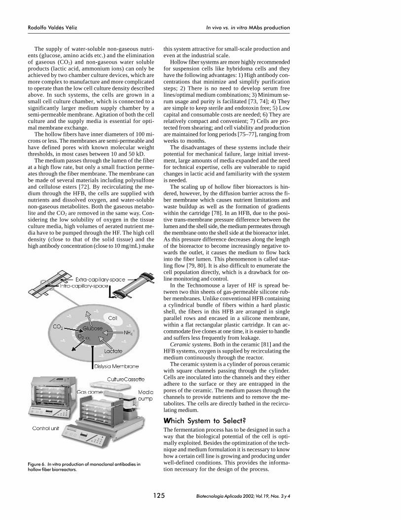
The supply of water-soluble non-gaseous nutri-ents (glucose, amino acids etc.) and the eliminationof gaseous (CO2) and non-gaseous water solubleproducts (lactic acid, ammonium ions) can only beachieved by two chamber culture devices, which aremore complex to manufacture and more complicatedto operate than the low cell culture density describedabove. In such systems, the cells are grown in asmall cell culture chamber, which is connected to asignificantly larger medium supply chamber by asemi-permeable membrane. Agitation of both the cellculture and the supply media is essential for opti-mal membrane exchange.
The hollow fibers have inner diameters of 100 mi-crons or less. The membranes are semi-permeable andhave defined pores with known molecular weightthresholds, in most cases between 10 and 50 kD.
The medium passes through the lumen of the fiberat a high flow rate, but only a small fraction perme-ates through the fiber membrane. The membrane canbe made of several materials including polysulfoneand cellulose esters [72]. By recirculating the me-dium through the HFB, the cells are supplied withnutrients and dissolved oxygen, and water-solublenon-gaseous metabolites. Both the gaseous metabo-lite and the CO2 are removed in the same way. Con-sidering the low solubility of oxygen in the tissueculture media, high volumes of aerated nutrient me-dia have to be pumped through the HF. The high celldensity (close to that of the solid tissue) and thehigh antibody concentration (close to 10 mg/mL) make
this system attractive for small-scale production andeven at the industrial scale.
Hollow fiber systems are more highly recommendedfor suspension cells like hybridoma cells and theyhave the following advantages: 1) High antibody con-centrations that minimize and simplify purificationsteps; 2) There is no need to develop serum freelines/optimal medium combinations; 3) Minimum se-rum usage and purity is facilitated [73, 74]; 4) Theyare simple to keep sterile and endotoxin free; 5) Lowcapital and consumable costs are needed; 6) They arerelatively compact and convenient; 7) Cells are pro-tected from shearing; and cell viability and productionare maintained for long periods [75–77], ranging fromweeks to months.
The disadvantages of these systems include theirpotential for mechanical failure, large initial invest-ment, large amounts of media expanded and the needfor technical expertise, cells are vulnerable to rapidchanges in lactic acid and familiarity with the systemis needed.
The scaling up of hollow fiber bioreactors is hin-dered, however, by the diffusion barrier across the fi-ber membrane which causes nutrient limitations andwaste buildup as well as the formation of gradientswithin the cartridge [78]. In an HFB, due to the posi-tive trans-membrane pressure difference between thelumen and the shell side, the medium permeates throughthe membrane onto the shell side at the bioreactor inlet.As this pressure difference decreases along the lengthof the bioreactor to become increasingly negative to-wards the outlet, it causes the medium to flow backinto the fiber lumen. This phenomenon is called star-ling flow [79, 80]. It is also difficult to enumerate thecell population directly, which is a drawback for on-line monitoring and control.
In the Technomouse a layer of HF is spread be-tween two thin sheets of gas-permeable silicone rub-ber membranes. Unlike conventional HFB containinga cylindrical bundle of fibers within a hard plasticshell, the fibers in this HFB are arranged in singleparallel rows and encased in a silicone membrane,within a flat rectangular plastic cartridge. It can ac-commodate five clones at one time, it is easier to handleand suffers less frequently from leakage.
Ceramic systems. Both in the ceramic [81] and theHFB systems, oxygen is supplied by recirculating themedium continuously through the reactor.
The ceramic system is a cylinder of porous ceramicwith square channels passing through the cylinder.Cells are inoculated into the channels and they eitheradhere to the surface or they are entrapped in thepores of the ceramic. The medium passes through thechannels to provide nutrients and to remove the me-tabolites. The cells are directly bathed in the recircu-lating medium.
Which System to Select?The fermentation process has to be designed in such away that the biological potential of the cell is opti-mally exploited. Besides the optimization of the tech-nique and medium formulation it is necessary to knowhow a certain cell line is growing and producing underwell-defined conditions. This provides the informa-tion necessary for the design of the process.
Figure 6. In vitro production of monoclonal antibodies inhollow fiber biorreactors.
Rodolfo Valdés Véliz In vivo vs. in vitro MAbs production
Biotecnología Aplicada 2002; Vol.19, Nos. 3 y 4126
According to results reported by Merten in 1988[82] different hybridoma cell lines express differentproduction kinetics and these production patterns canbe classified into three groups. The first group (pat-tern type I) shows the best release of monoclonalantibodies at the beginning of a batch culture duringthe lag phase and the onset of the exponential growthphase and with no increase during the death phase[83–85]. Production pattern II is characterized by arelatively high initial specific production, which de-creases during maximum growth, and, after havingreached the stationary and death phases it increasesagain [86–88]. Production pattern number III is char-acterized by a relatively stable production during thegrowth of the cells, and almost no production whenthe cells enter the stationary and death phases [89].
The three specific production kinetic patterns mayhave implications on the construction of a productionprocess. If the product released is more or less growthassociated (pattern III), a continuous culture systemmight be the best choice because it provides for thepermanent growth of cells. Cells, showing productionpatterns II or I, might preferentially be cultivated in adiscontinuous mode, because growth and specific pro-duction are more or less inversely related. Pattern IIimplies that the culture system has to provide a growthphase independently from a production phase. Thiscan be achieved by the use of the following processmodes: batch, fed-batch, repeated batch with or with-out a second stage, or a continuous culture systemwith a second stage. Cells showing production pat-tern I should have the highest production in repeatedbatch systems.
Assuming that the differences between high andlow density cell cultures are not too great, the abovementioned implications of batch production kineticsmight be extended for high cell density systems, also.In any case, the cells have to be held in a physiologicalstate, which is optimal for overall production.
The growth and production kinetics of cell lines arewithin the most important parameters for MAb pro-duction on an industrial scale.
For researchers working in the academic field, theuse of the available in vitro techniques indicated aboveis often problematic.
For in vitro production the following points shouldbe considered: high yields obtained at high concentra-tions, the reproduction of the in vivo condition, andthe cells need to be grown at high cell densities. Thereare many types of equipment on the market. In rollerbottles, which are suitable for adherent cells and stirredbottles, the cells are grown until the medium is ex-hausted. The concentration of the MAb is not muchgreater than that grown in normal tissue culture flasks,with the additional problem that proteases and toxinsare released by the dying cells. Also, there are numer-ous types of bioreactor of all sizes, but they are aimedat commercial production. To obtain tissue-like den-sities, the dialysis tubing technology, miniPERM, andthe hollow fiber technology can be used. There is anatural reluctance for researchers to invest so muchand to face the high costs of consumable products andmaintenance. The miniPERM system seems to be verypromising, it takes up little space, it may be run atonce and the initial and maintenance costs are low.
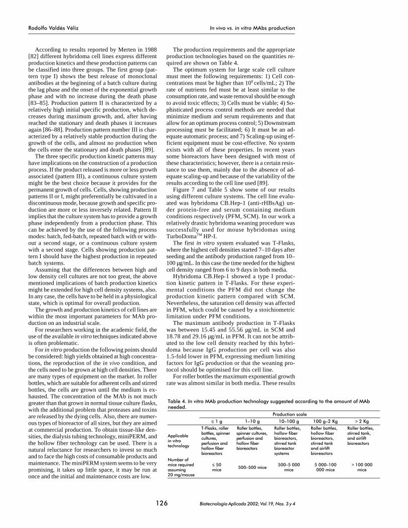
The production requirements and the appropriateproduction technologies based on the quantities re-quired are shown on Table 4.
The optimum system for large scale cell culturemust meet the following requirements: 1) Cell con-centrations must be higher than 108 cells/mL; 2) Therate of nutrients fed must be at least similar to theconsumption rate, and waste removal should be enoughto avoid toxic effects; 3) Cells must be viable; 4) So-phisticated process control methods are needed thatminimize medium and serum requirements and thatallow for an optimum process control; 5) Downstreamprocessing must be facilitated; 6) It must be an ad-equate automatic process; and 7) Scaling-up using ef-ficient equipment must be cost-effective. No systemexists with all of these properties. In recent yearssome bioreactors have been designed with most ofthese characteristics; however, there is a certain resis-tance to use them, mainly due to the absence of ad-equate scaling-up and because of the variability of theresults according to the cell line used [89].
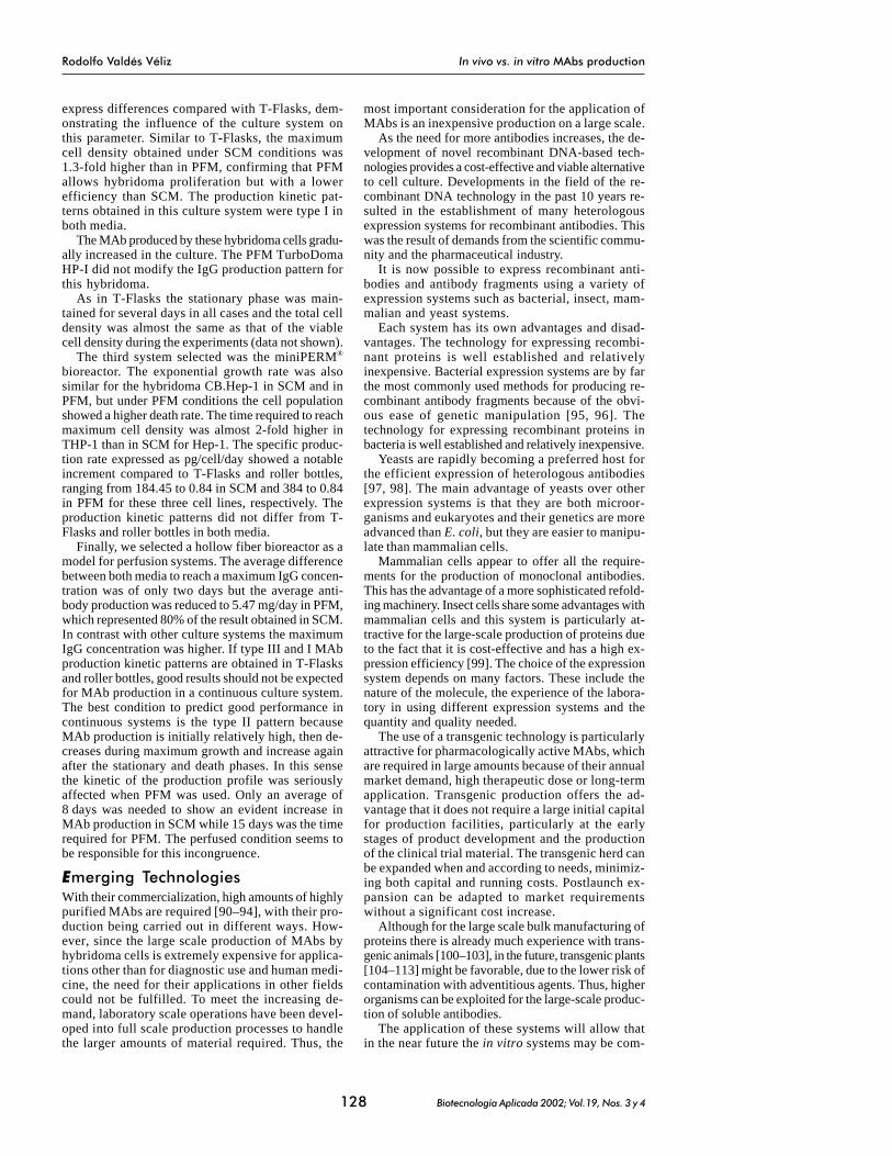
Figure 7 and Table 5 show some of our resultsusing different culture systems. The cell line evalu-ated was hybridoma CB.Hep-1 (anti-rHBsAg) un-der protein-free and serum containing mediumconditions respectively (PFM, SCM). In our work arelatively drastic hybridoma weaning procedure wassuccessfully used for mouse hybridomas usingTurboDomaTM HP-I.
The first in vitro system evaluated was T-Flasks,where the highest cell densities started 7–10 days afterseeding and the antibody production ranged from 10–100 µg/mL. In this case the time needed for the highestcell density ranged from 6 to 9 days in both media.
Hybridoma CB.Hep-1 showed a type I produc-tion kinetic pattern in T-Flasks. For these experi-mental conditions the PFM did not change theproduction kinetic pattern compared with SCM.Nevertheless, the saturation cell density was affectedin PFM, which could be caused by a stoichiometriclimitation under PFM conditions.
The maximum antibody production in T-Flaskswas between 15.45 and 55.56 µg/mL in SCM and18.78 and 29.16 µg/mL in PFM. It can not be attrib-uted to the low cell density reached by this hybri-doma because IgG production per cell was also1.5-fold lower in PFM, expressing medium limitingfactors for IgG production or that the weaning pro-tocol should be optimised for this cell line.
For roller bottles the maximum exponential growthrate was almost similar in both media. These results
Table 4. In vitro MAb production technology suggested according to the amount of MAb needed.
Production scale ≤ 1 g 1�10 g 10�100 g 100 g�2 Kg > 2 Kg
Applicable in vitro technology
T-Flasks, roller bottles, spinner cultures, perfusion and hollow fiber bioreactors
Roller bottles, spinner cultures, perfusion and hollow fiber bioreactors
Roller bottles, hollow fiber bioreactors, stirred tank bioreactor systems
Roller bottles, hollow fiber bioreactors, stirred tank and airlift bioreactors
Roller bottles, stirred tank, and airlift bioreactors
Number of mice required assuming 20 mg/mouse
≤ 50 mice 500�500 mice
500�5 000 mice
5 000�100 000 mice
> 100 000 mice

Rodolfo Valdés Véliz In vivo vs. in vitro MAbs production
Biotecnología Aplicada 2002; Vol.19, Nos. 3 y 4127
Figure 7. Cell proliferation and MAb CB.Hep-1 production using different in vitro systems. A, T-Flasks; B, roller bottles; C, miniPERM; D, hollow fiber biorreactors.
Table 5. CB.Hep-1 hybridoma performance in serum containing a protein-free medium.
Parameters T-Flasks Roller bottles miniPERM bioreactors
Hollow fiber bioreactor
SCM PFM SCM PFM SCM PFM SCM PFM
Production time (days) 9 9 10 10 20 28 32 37
Exponential growth rate (h-1) -0.008�0.032 -0.0012�0.024 0.005�0.024 -0.0008�0.034 0.063�0.044 0.007�0.035 - -
Specific production rate (pg/cell/day) 12.2�2.22 9.598�1.04 28.78�1.40 27.69�0.59 184.45�0.84 384�0.84 - -
Total IgG (mg/run) 2.31 2.33 20.09 13.17 51.33 54.29 100.72 90.28
Scale factor 1 1 8.69 5.62 2.5 4.12 1.96 1.66
Medium consumed (L/run) 0.25 1.02 1.65 1.39 7.28 10.16
Production kinetic pattern I I I I I I - -
IIMDM5%FCS cells/mLIMDM5%FCS pg/cellxd
THP-l cells/mLTHP-l pg/cellxd
A B
pg/c
ellx
d
Cel
ls/m
L
3.00E+06
2.50E+06
2.00E+06
1.50E+06
1.00E+06
5.00E+05
0.00E+001 3 5 76 842 9
Days
14
12
10
8
6
2
0
-2
-4
-6
4 Cel
ls/m
L
2.50E+06
2.00E+06
1.50E+06
1.00E+06
5.00E+05
0.00E+001 3 5 76 842 9 10
Days
pg/c
ellx
d
35
30
25
20
15
5
0
-5
c
10
C D
THP-l cells/mLTHP-l pg/cellxd
IIMDM5%FCS cells/mLIMDM5%FCS pg/cellxd
IMDM5%FCS pg/cellxdTHP-l pg/cellxd
pg/c
ellx
d
Cel
ls/m
L
12000000
10000000
8000000
6000000
4000000
2000000
0
800
600
400
200
0
-200
-400
4 12 24 28208
Days
16
µg/m
L1600
0
400
800
1200
Days
3 6 12 2715 3024 3633 39219 18
Rodolfo Valdés Véliz In vivo vs. in vitro MAbs production
Biotecnología Aplicada 2002; Vol.19, Nos. 3 y 4128
express differences compared with T-Flasks, dem-onstrating the influence of the culture system onthis parameter. Similar to T-Flasks, the maximumcell density obtained under SCM conditions was1.3-fold higher than in PFM, confirming that PFMallows hybridoma proliferation but with a lowerefficiency than SCM. The production kinetic pat-terns obtained in this culture system were type I inboth media.
The MAb produced by these hybridoma cells gradu-ally increased in the culture. The PFM TurboDomaHP-I did not modify the IgG production pattern forthis hybridoma.
As in T-Flasks the stationary phase was main-tained for several days in all cases and the total celldensity was almost the same as that of the viablecell density during the experiments (data not shown).
The third system selected was the miniPERM®
bioreactor. The exponential growth rate was alsosimilar for the hybridoma CB.Hep-1 in SCM and inPFM, but under PFM conditions the cell populationshowed a higher death rate. The time required to reachmaximum cell density was almost 2-fold higher inTHP-1 than in SCM for Hep-1. The specific produc-tion rate expressed as pg/cell/day showed a notableincrement compared to T-Flasks and roller bottles,ranging from 184.45 to 0.84 in SCM and 384 to 0.84in PFM for these three cell lines, respectively. Theproduction kinetic patterns did not differ from T-Flasks and roller bottles in both media.
Finally, we selected a hollow fiber bioreactor as amodel for perfusion systems. The average differencebetween both media to reach a maximum IgG concen-tration was of only two days but the average anti-body production was reduced to 5.47 mg/day in PFM,which represented 80% of the result obtained in SCM.In contrast with other culture systems the maximumIgG concentration was higher. If type III and I MAbproduction kinetic patterns are obtained in T-Flasksand roller bottles, good results should not be expectedfor MAb production in a continuous culture system.The best condition to predict good performance incontinuous systems is the type II pattern becauseMAb production is initially relatively high, then de-creases during maximum growth and increase againafter the stationary and death phases. In this sensethe kinetic of the production profile was seriouslyaffected when PFM was used. Only an average of8 days was needed to show an evident increase inMAb production in SCM while 15 days was the timerequired for PFM. The perfused condition seems tobe responsible for this incongruence.
Emerging TechnologiesWith their commercialization, high amounts of highlypurified MAbs are required [90–94], with their pro-duction being carried out in different ways. How-ever, since the large scale production of MAbs byhybridoma cells is extremely expensive for applica-tions other than for diagnostic use and human medi-cine, the need for their applications in other fieldscould not be fulfilled. To meet the increasing de-mand, laboratory scale operations have been devel-oped into full scale production processes to handlethe larger amounts of material required. Thus, the
most important consideration for the application ofMAbs is an inexpensive production on a large scale.
As the need for more antibodies increases, the de-velopment of novel recombinant DNA-based tech-nologies provides a cost-effective and viable alternativeto cell culture. Developments in the field of the re-combinant DNA technology in the past 10 years re-sulted in the establishment of many heterologousexpression systems for recombinant antibodies. Thiswas the result of demands from the scientific commu-nity and the pharmaceutical industry.
It is now possible to express recombinant anti-bodies and antibody fragments using a variety ofexpression systems such as bacterial, insect, mam-malian and yeast systems.
Each system has its own advantages and disad-vantages. The technology for expressing recombi-nant proteins is well established and relativelyinexpensive. Bacterial expression systems are by farthe most commonly used methods for producing re-combinant antibody fragments because of the obvi-ous ease of genetic manipulation [95, 96]. Thetechnology for expressing recombinant proteins inbacteria is well established and relatively inexpensive.
Yeasts are rapidly becoming a preferred host forthe efficient expression of heterologous antibodies[97, 98]. The main advantage of yeasts over otherexpression systems is that they are both microor-ganisms and eukaryotes and their genetics are moreadvanced than E. coli, but they are easier to manipu-late than mammalian cells.
Mammalian cells appear to offer all the require-ments for the production of monoclonal antibodies.This has the advantage of a more sophisticated refold-ing machinery. Insect cells share some advantages withmammalian cells and this system is particularly at-tractive for the large-scale production of proteins dueto the fact that it is cost-effective and has a high ex-pression efficiency [99]. The choice of the expressionsystem depends on many factors. These include thenature of the molecule, the experience of the labora-tory in using different expression systems and thequantity and quality needed.
The use of a transgenic technology is particularlyattractive for pharmacologically active MAbs, whichare required in large amounts because of their annualmarket demand, high therapeutic dose or long-termapplication. Transgenic production offers the ad-vantage that it does not require a large initial capitalfor production facilities, particularly at the earlystages of product development and the productionof the clinical trial material. The transgenic herd canbe expanded when and according to needs, minimiz-ing both capital and running costs. Postlaunch ex-pansion can be adapted to market requirementswithout a significant cost increase.
Although for the large scale bulk manufacturing ofproteins there is already much experience with trans-genic animals [100–103], in the future, transgenic plants[104–113] might be favorable, due to the lower risk ofcontamination with adventitious agents. Thus, higherorganisms can be exploited for the large-scale produc-tion of soluble antibodies.
The application of these systems will allow thatin the near future the in vitro systems may be com-

Rodolfo Valdés Véliz In vitro vs. in vivo MAbs production
129 Biotecnología Aplicada 2002; Vol.19, Nos. 3 y 4
1. Köhler G, Milstein C. Continuous culture offused cells secreting antibodies of predefinedspecificity. Nature 1975;256:485�97.
2. Cooney CL. Are we prepared for animalcell technology in the 21st century? In: BeuveryEC, Griffiths JB, Zeijlemaker WP, editors. Ani-mal cell technology: developments towardsthe 21st century. ESACT/JAACT proceedings.Veldhoven: Kluwer Academic; 1995; xxv-xxx.
3. Garrison RN, Kaelin LD, Galloway RH, HeuserLS. Malignant ascites. Clinical and experimen-tal observations. Annals of Surgery 1986;203:644�51.
4. Nagy JA. Lymphatic and nonlymphaticpathways of peritoneal absorption in mice:physiology versus pathology. Blood Purifica-tion 1992;10:149�62.
5. Hendriksen CFM, de Leeuw W. Productionof monoclonal antibodies by the ascitesmethod in laboratory animals. 74th Forum inImmunology. Res in Immunology 1998;149:535�42.
6. Chandler JP. Factors influencing monoclonalantibody production in mouse ascites fluid. In:Commercial production of monoclonal anti-bodies: a guide for scale-up: SS. Seaver NewYork: Marcel Dekker Inc; 1987. p.75�92.
7. Brodeur BR, Tsang PS. High yield mono-clonal antibody production in ascites. JImmunol Methods 1986;86:239�41.
8. Stewart F, Callander A, Garwes DJ. Com-parison of ascites production for monoclonalantibodies in BALB/c and BALB/c-derivedcross-bred mice. J Immunol Methods 1989;119:269�75.
9. Brodeur BR, Tsang P, Larose Y. Parametersaffecting ascites tumor formation in mice andmonoclonal antibody production. J ImmunolMethods 1984;71:265�72.
10. Hoogenraad NJ. The effect of pristane onthe ascites tumor formation and monoclonalantibody production. Methods Enzymol1986;121:375�81.
11. Amyx HL. Control of animal pain anddistress in antibody production and infectiondisease studies. J Am Vet Med Assoc 1987;191:1287�9.
12. Silverman J. Protocol review. Monoclonalantibody production: in vivo vs. in vitro. LabAnim 1987;16:19�20.
13. Gillette RW. Alternatives to pristane prim-ing for ascitic fluid and monoclonal antibodyproduction. J Immunol Methods 1987;99:21�3.
14. Mueller UW, Hawes CS, Jones WR. Mono-clonal antibody production by hybridomagrowth in Freund�s adjuvant primed mice. JImmunol Methods 1986;87:193�6.
15. Jones SL, Cox JC, Pearson JE. Increasedmonoclonal antibody ascites production inmice primed with Freund�s incomplete adju-vant. J Immunol Methods 1990;129:227�31.
16. Hoogenraad N, Helman T, HoogenraadJ. The effect of pre-injection of mice with pris-tine on ascites tumor formation and mono-clonal antibody production. J Immunol Meth-ods 1983;61:317�20.
17. Zola H. Conventional mouse monoclonalantibodies. In: Zola H, editor. Monoclonalantibodies: the second generation. BIOS,Oxford, England; 1995. p.29.
18. Galfre G, Milstein C. Preparation of mono-clonal antibodies: strategies and procedures.Methods Enzymol 1981;73:1�46.
19. Epstein N, Epstein M. The hybridoma tech-nology: production of monoclonal antibodies.Adv Biotechnol Processes 1986;6:179�218.
20. Jackson LR, Trudel LJ, Fox JG, LipmanNS. Monoclonal Antibody Production inMurine Ascites I. Clinical and Pathologicfeatures. Laboratory Animal Science 1999;49:70�80.
21. Goding JW. Antibody production by hy-bridomas. J. Immunol Methods 1980;39:285�308.
22. McGuill MW Rowan AN. Refinement ofmonoclonal antibody production and ani-mal well-being. ILAR News 1989;31:7�11.
23. Jackson LR, Trudel LJ, Fox JG, Lipman NS.Monoclonal antibody production in murineascites II. Production characteristics. Labora-tory Animal Science 1999;49:80�6.
24. Mahana W, Paraf A. Mice ascites as asource of polyclonal and monoclonal anti-bodies. Journal of Immunol Methods 1993;161:187�92.
25. Hendriksen CFM. A call for a Europeanprohibition of monoclonal antibody (MAb)production by the ascites procedure in labo-ratory animals ATLA 1998;26:523�40.
26. Kuhlmann I, Kurth W, Ruhdel I. Mono-clonal antibodies: in vivo and in vitro pro-duction on a laboratory scale, with consider-ation of the legal aspects of animal protection.ATLA 1985;17:73�82.
27. Amyx HL. Control of animal pain anddistress in antibody production and infectiondisease studies. J Am Vet Med Assoc 1987;191:1287�9.
28. Rowan AN. The third R: Refinement. Alter-native to Laboratory Animals. ATLA 1995;23:332�46.
29. McGuill MW, Rowan AN. Refinement ofmonoclonal antibody production and ani-mal well-being. ILAR News 1989;31:7�10.
30. Flecknell PA. Refinement of animal use:assessment and alleviation of pain and dis-tress. Laboratory Animals 1994;28:222�31.
31. Council Directive 86/609/EEC on theapproximation of laws, regulations and ad-ministrative provisions of the member statesregarding the protection of animals used forexperimental and other scientific purposes.Official Journal of the European Communi-ties L; 1986. p.1.
32. European Convention for the protectionof vertebrate animals used for experimentaland other scientific purposes. Strasburg:Council of Europe 1986;51.
33. Code of Practice for the production ofmonoclonal antibodies. Rijswijk, The Nether-lands; 1993. p.6.
34. Antibody production advice on protocolsfor minimal severity. In: Report of the AnimalProcedures Committee for 1991. Appendix II.London II: HMSO Cmmnd; 1992. p.2048.
35. Marx U, Embleton MJ, Fischer R, Gruber FP,Hansson U, Heuer J de Leeuw WA, et al. Mono-clonal antibody production: the report andrecommendations of ECVAM workshop 23.ATLA 1997;25:121�37.
36. Workshop sponsored by the John�s HopkinsCenter for alternatives to animal testing andthe office for protection from research risks,National Institutes of Health, 1997. Alternativesin monoclonal antibody production.
37. National Institutes of Health. 1997. Pro-duction of monoclonal antibodies usingmouse ascites method. OPRR Reports, Animalwelfare number 98�01.
38. McArdle JE, Colleen JL. Production ofmonoclonal antibodies Workshop. Alterna-tive Research and Development Foundation1999. August 29.
39. Hendriksen C, Rozing J, van der Kamp M,de Leeuw W. The production of monoclonalantibodies: are animal still needed. ATLA1996;24:109�10.
40. Ham RG. Importance of the basal nutrientmedium in the design of hormonally definedmedia. In: Sato GH, Pardee AB, DA Sirbasku,editors. Growth of cells in hormonally definedmedia. New York: Cold Spring Harbor Lab;1982. p.39�60.
41. Maurer HR. Towards chemically defined,serum free media for mammalian cell culture.In: Freshney RI, editor. Animal cell culture: apractical approach. Oxford: IRL Press Ltd;1986. p.13�31.
42. Mizrahi A, Lazar A. Media for cultivationof animal cells: an overview. Cytotechnology1988;1:199�214.
43. Tarleton R, Beyer A. Medium-scale produc-
pleted with all of these alternative methods and atthat moment the in vivo method to produce MAbswill only be a filed image.
ConclusionsAt present, we believe that there are sufficient in vitromethodologies and technologies available which can sup-plant the use of mice in the routine production of MAbsfrom hybridomas for most applications. The best choicedepends on the line, media and purposes. The substitu-tion by in vitro methods must come about by persua-sion by those who have had “hands on” experience and
are familiar with the problems and can therefore recom-mend suitable alternatives without being beholden toany particular commercial organization. Although thetotal exclusion of mice for the generation or productionof MAbs will not likely occur soon, there are manyforces at work, now and in the future, that will greatlydecrease the need for in vivo production.
It is clear that there are now many ways to ex-press high quantities of soluble and functional re-combinant antibodies in both research and industrialsettings. The obvious key is to find the system thatbest suits your needs.

Rodolfo Valdés Véliz In vitro vs. in vivo MAbs production
130 Biotecnología Aplicada 2002; Vol.19, Nos. 3 y 4
tion and purification of monoclonal antibod-ies in protein-free medium. Biotechniques1991;11:590�3.
44. Franek F, Dolnikova J. Hybridoma growthand monoclonal antibody production in iron-rich protein-free medium: effect of nutrient con-centration. Cytotechnology 1991;7:33�8.
45. Bertheussen K. Growth of cells in a newdefined protein-free medium. Cytotechnology1993;11:219�31.
46. Seaver S. Culture methods affects antibodysecretion of hybridoma cells. In: Seaver S,editor. Commercial production of mono-clonal antibodies. A guide for scale up. NewYork: Marcel Dekker Inc; 1987. p.49�71.
47. Peterson NC. Considerations for in vitromonoclonal antibody production. Res Immu-nol 1998;149:553�7.
48. Ruhdel I, Kulhlmann I. Production ofmonoclonal antibodies in roller bottles withreduced serum concentrations and serumsubstitutes. Bioengineering 1994;10:15�8.
49. van Wezel AL. Growth of cell strains andprimary cells on microcarriers in homogenousculture. Nature 1967;216:64�5.
50. Meinger B. Cell culture on beads used forthe industrial production of foot and mouthdisease virus. Dev Biol Stand 1978;42:141�5.
51. van Wezel AL, van Herwaarden JAM, vande Heuvel-de Rijk EW. Large-scale concentra-tion and purification of virus suspension frommicrocarrier culture for the preparation of in-activated virus vaccines. Dev Biol Standard1979;42:65�9.
52. Kuo MJC, Lewis C, Martin RA, Miller RE,Schoenfeld RA, Scheck JM, et al. Growth of an-chorage dependent mammalian cells on gly-cine derivatized polystyrene in suspensionculture. In vitro 1981;17:901�6.
53. Johansson A, Nielsen V. A new Microcarrier.Dev Biol Stand 1980;46:125�9.
54. Reuveny S, Siberstein L, Shahar A, Free-man E, Mizrahi S. Cell and virus propagationon cylindrical cellulose based microcarriers.Dev Biol Stand 1982;50:115�23.
55. Varani J, Dame M, Fediske T, Beals F,Hillegas W. Substrate-dependent differencesin growth and biological properties of fibro-blasts and epithelial cells grown in microcar-rier culture. J Biol Stand 1985;1:67�76.
56. Nilsson K, Birnbaum S, Mosbach K. Mi-crocarrier culture of recombinant ChineseHamster Ovary Cells for production of humaninterferon and human tissue type plasmino-gen activator. Appl Microbiol Biotechnol1988;27:366�71.
57. Varani J, Dame M, Beals TF, Wass JA. Growthof three established cell lines on glass micro-carriers. Biotechn Bioeng 1983;25: 1359�72.
58. KatingerJWD, Scheirer W, Kromer E. Bubblecolumn reactor for mass propagation of ani-mal cell in suspension culture. Ger Chem Eng1979;2:31�8.
59. Broad DF, Brown MR, Grant AP, Wood LA.Scale up of mammalian cell culture. In: SpierR, editor. Advances in animal cell biology andtechnology for bioprocesses. Butterworths,Kent 1989. p.412�7.
60. Handa A, Emery AN, Spier RE. On the evalu-ation of gas-liquid interfacial effects on hybri-doma viability in bubble column bioreactors.Dev Biol Stand 1987;66:241�53.
61. Tramper J, Vlak M. Bioreactor design forgrowth of shear-sensitive mammalian and in-sect cells. In: Mizrahi A, editor. Adv Biotechnol
Proc. New York: Alan. R. Liss; 1988. p.199�208.
62. Petrossian A, Cortessis GP. Large-scale pro-duction of monoclonal antibodies in definedserum-free media in airlift bioreactors.Biotechniques 1990;8:414�422.
63. Birch JR, Lambert K, Thompson PW, KennyAC, Wood LA. Antibody production with air-lift fermentors. In: Lyderson BK editor. LargeScale cell culture technology. New York:Hanser Publications; 1987. p.1�20.
64. Falkenberg FW, Hengelage T, Krane M,Bartels I, Albrecht A, Holtmeier N, et al. A simpleand inexpensive high density dyalisis tubingcell culture system for the in vitro productionof monoclonal antibodies in high concentra-tion. J Immunol Methods 1993;165:193�206.
65. THE SCIENTIST®/LAB Consumer. Buildinga better mouse trap. The news journal for thelife scientist. 1998;12:15�24.
66. Knazek RA, Gullino PM, Kohler PO, DedrickRL. Cell culture on artificial capillaries: Anapproach to tissue in vitro. Science 1972;178:65�6.
67. Knazek RA. Solid tissue masses formed invitro from cells cultured on artificial capillar-ies. Federation Proc 1974;33:1978�81.
68. Hirschel MD, Gruenberg ML. An auto-mated hollow fiber system for large-scalemanufacture of mammalian cell secretedproduct. In: Lyderson BK, editor. Large scalecell culture technology. New York: Publica-tions Hanser; 1987. p.113�44.
69. Tharakan JP, Chau PC. A radial flow hol-low fiber bioreactor for the large-scale cul-ture of mammalian cells. Biotechn Bioeng1986;28:329�42.
70. Evans TL, Miller RA. Large-scale produc-tion of murine monoclonal antibodies usinghollow fiber bioreactors. Biotechniques1988;6:762�7.
71. Jackson LR, Trudel LJ, Fox JG, Lipman NS.Evaluation of hollow fiber bioreactors as analternative to murine ascites production forsmall-scale monoclonal antibody produc-tion. Journal of Immunol Methods 1996;189:217�31.
72. Heath C, Belfort G. Membrane andbioreactors. J Biochem 1990;22:823�935.
73. Hopkinson J. Hollow fiber cell culture sys-tems for economical cell-product manufac-turing. Bio/Technology 1985;3:225�30.
74. Tiebout RF. Tissue culture in hollow fibersystems: implications for downstream process-ing and stability analysis. Dev Biol Stand1989;71:65�71.
75. Reuveny S, Lazar A. Equipment and proce-dures for production of monoclonal antibod-ies in culture. In: Mizrahi A, editor. Monoclonalantibodies: production and application. NewYork: Alan R. Liss; 1989. p.45�80.
76. Andersen BG, Gruenberg ML. Optimiza-tion techniques for the production of mono-clonal antibodies utilizing hollow fiber tech-nology. In: Seaver S, editor. Commercialproduction of monoclonal antibodies: aguide for scale up. New York: Marcel Dekker;1987. p.175�95.
77. von Wedel RJ. Mass culture of mouse andhuman hybridoma cells in hollow fiber cul-ture. In: Seaver S, editor. Commercial produc-tion of monoclonal antibodies: a guide forscale-up. New York: Marcel Dekker; 1987.p.150�73.
78. Piret JM. Cooney CL. Immobilized mam-malian cell cultivation in hollow fiberbioreactors. Biotechnol Adv 1990;8:763�83.
79. Starling EH. On the adsorption of fluidsfrom the connective tissue spaces. J Physiol1986;19:312�26.
80. Hammer BF, Heath CA, Mirer SD, Belfort G.Quantitative flow measurement in bioreactorsby nuclear magnetic resonance imaging. Bio/Technology 1990;8:327�30.
81. Lyderson BK. Perfusion cell culture systembased on ceramic matrices, in large-scale cellculture technology. Lyderson BK, editor. NewYork: Hanser Publications; 1987. p.169�92.
82. Merten O-W. Batch production andgrowth kinetics of hybridomas. Cytotechnol-ogy 1988;1:113�21.
83. Williams JA. Effects of medium concentra-tion on antibody production. J Tissue CultureMethods 1984;8:115�8.
84. Lavery M, Kearns MJ, Price DG, Emery AN,Jefferis and Nienow AW. Physical conditionsduring batch culture of hybridomas in labo-ratory scale stirred tank reactor. Develp BiolStandard 1985;60:199�206.
85. Merten O-W, Reiter S, Himmler G, ScheirerW, Kapager H. Production kinetics of mono-clonal antibodies. Develop Biol Standard1985;60:219�27.
86. Boraston R, Thompson PW, Garland S,Birch JR. Growth and oxygen requirements ofantibody production mouse hybridoma cellsin suspensión culture. Develop Biol Standard1984;55:1033�111.
87. Merten O-W, Reiter S, Katinger H. Stabili-zation effect of reduced cultivation tempera-ture of human-mouse hybridomas. DevelopBiol Standard. 1985b;60:509�12.
88. Altshuler GL, Dziewulski DM, Sowek JA,Belfort G. Continuous hybridoma growth andmonoclonal antibody production in hollowfiber reactors-separators. Biotechnol Bioeng1986;646�58.
89. Spalding BJ. A growth market for bioreac-tors. Biotechnology 1991;9:338�41.
90. Kundu PK, Prasad NS, Datta D. Mono-clonal antibody: high density culture of hy-bridoma cells and downstream processingfor IgG recovery. Indian J Exp Biol 1998;36:125�35.
91. Yang JD, Angelillo Y, Chaudhry M, Golden-berg C, Goldenberg DM. Achievement of highcell density and high antibody productivity bya controlled-fed perfusion bioreactor process.Biotechnol Bioeng 2000; 69:74�82.
92. Myers DE, Sicheneder A, Clementson D,Dvorak N, Venkatachalam T, Sev AR, et al.Large scale manufacturing of B43 (anti-CD19)-genistein for clinical trials in leukemiaand lymphoma. Leuk Lymphoma 1998;29:329�38.
93. Frenken LG, Hessing JG, Van den HondelCA, Verrips CT. Recent advances in the large-scale production of antibody fragments us-ing lower eukaryotic microorganisms. ResImmunol 1998;149:589�99.
94. Werner RG. Innovative and economicpotential of mammalian cell culture. Arznei-mittelforschung 1998;48:423�6.
95. Maeda F, Nagatsuka Y, Ihara S, Aotsuka S,Ono Y, Inoko H, Takekoshi M. Bacterial expres-sion of a human recombinant monoclonal an-tibody fab fragment against hepatitis B surfaceantigen. J Med Virol 1999; 58:338�45.
96. Burioni R, Plaisant P, Bugli F, Delli Carri V,Clementi M, Fadda G. A vector for the expres-sion of recombinant monoclonal Fab frag-ments in bacteria. J Immunol Methods 1998;217:195�9.

Rodolfo Valdés Véliz In vitro vs. in vivo MAbs production
131 Biotecnología Aplicada 2002; Vol.19, Nos. 3 y 4
97. Pennell CA, Eldin P. In vitro production ofrecombinant antibody fragments in Pichiapastoris. Research in Immunology. 74th Fo-rum in Immunology 1998;149:529�620.
98. Hollenberg CP, Gellissen. Production ofrecombinant proteins by methylotropic yeasts.Curr Opin Biotechnol 1997;8:554�60.
99. Hasemann CA, Capra JD. High-levelproduction of a functional immunoglobu-lin heterodimer in a baculovirus expresiónsystem. Proc Natl Acad Sci USA 1990;87:3942�50.
100. Bebbigton C. Expression of antibodygenes in mammalian cells. In: Monoclonalantibodies. The second generation. Zola H,editor. Bios Scientific Publishers; Oxford.
101. Davis CG, Gallo ML, Corvalan JR.Transgenic mice as a source of fully humanantibodies for the treatment of cancer. CancerMetastasis Rev 1999;18:421�5.
102. Kolb AF, Pewe L, Webster J, Perlman S,Whitelaw CB, Siddell SG. Virus-neutralizingmonoclonal antibody expressed in milk oftransgenic mice provides full protectionagainst virus-induced encephalitis. J Virol2001;75:2803�9.
103. van Kuik Romeijn P, de Groot N, Hoo-ijberg E, de Boer HA. Expression of a functionalmouse-human chimeric anti-CD19 antibodyin the milk of transgenic mice. Transgenic Res2000;9:155�9.
104. Chargelegue D, Vine ND, van Dollewe-erd CJ, Drake PM, Ma JK. A murine mono-clonal antibody produced in transgenicplants with plant-specific glycans is not im-munogenic in mice. Transgenic Res 2000;9:187�94.
105. Stevens LH, Stoopen GM, Elbers IJ,Molthoff JW, Bakker HA, Lommen A, Bosch D,Jordi W. Effect of climate conditions and plantdevelopmental stage on the stability of anti-bodies expressed in transgenic tobacco.Plant Physiol 2000;124:173�82.
106. Ma JK, Hikmat BY, Wycoff K, Vine ND,Chargelegue D, Yu L, Hein MB, Lehner T. Char-acterization of a recombinant plant mono-clonal secretory antibody and preventive im-munotherapy in humans. Nat Med 1998;4:601�6.
107. Khoudi H, Laberge S, Ferullo JM, Bazin R,Darveau A, Castonguay Y, Allard G, LemieuxR, Vezina LP. Production of a diagnostic mono-clonal antibody in perennial alfalfa plants.
Biotechnol Bioeng 1999;64: 135�43.
108. Macheteau M, Laine AC, Bourhis C,Lange C, Vine ND, Ma JK, Lerouge P, Faye L. N-Glycosylation of a mouse IgG expressed intransgenic tobacco plants. Glycobiology1999;9:365�72.
109. Zeitlin L, Olmsted SS, Moench TR, Co MS,Martinell BJ, Paradkar VM, Russell DR, Queen C,Cone RA, Whaley KJ. A humanized monoclonalantibody produced in transgenic plants forimmunoprotection of the vagina against geni-tal herpes. Nat Biotechnol 1998;16:1361�4.
110. Larrick JW, Yu L, Chen J, Jaiswal S, WycoffK. Production of antibodies in transgenicplants. Res Immunol 1998;149:603�8.
111. Okada Y. Transgenic plants as a medi-cine production system. TanpakushitsuKakusan Koso 2000;45:607�13.
112. Wynn RL, Meiller TF, Crossley HL. Tobacco�plantibodies� for caries prevention. Gen Dent1999;47:450�4.
113. Stein KE, Webber KO. The regulation ofbiologic products derived from bioengi-neered plants. Curr Opin Biotechnol 2001;12:308�11.
Received in April, 2001. Acceptedfor publication in October, 2001.