Embed Size (px)
Citation preview

Role of PKA signaling in D2 receptor-expressingneurons in the core of the nucleus accumbensin aversive learningTakashi Yamaguchia, Akihiro Gotob, Ichiro Nakaharaa,c, Satoshi Yawataa, Takatoshi Hikidaa,d, Michiyuki Matsudae,Kazuo Funabikia,c,1, and Shigetada Nakanishia,1
aDepartment of Systems Biology, Osaka Bioscience Institute, Osaka 565-0874, Japan; bLaboratory for Memory Mechanisms, Brain Science Institute, RIKEN,Saitama 351-0198, Japan; cLaboratory of Molecular Cell Biology and Development, Graduate School of Biostudies, Kyoto University, Kyoto 606-8315,Japan; dDepartment of Research and Drug Discovery, Medical Innovation Center, Graduate School of Medicine, Kyoto University, Kyoto 606-8501, Japan;and eLaboratory of Bioimaging and Cell Signaling, Graduate School of Biostudies, Kyoto University, Kyoto 606-8315, Japan
Contributed by Shigetada Nakanishi, July 30, 2015 (sent for review June 14, 2015; reviewed by Kazuto Kobayashi and Masayuki Masu).
The nucleus accumbens (NAc) serves as a key neural substrate foraversive learning and consists of two distinct subpopulations ofmedium-sized spiny neurons (MSNs). The MSNs of the direct path-way (dMSNs) and the indirect pathway (iMSNs) predominantlyexpress dopamine (DA) D1 and D2 receptors, respectively, andare positively and negatively modulated by DA transmitters viaGs- and Gi-coupled cAMP-dependent protein kinase A (PKA) sig-naling cascades, respectively. In this investigation, we addressedhow intracellular PKA signaling is involved in aversive learning in acell type-specific manner. When the transmission of either dMSNsor iMSNs was unilaterally blocked by pathway-specific expressionof transmission-blocking tetanus toxin, infusion of PKA inhibitorsinto the intact side of the NAc core abolished passive avoidancelearning toward an electric shock in the indirect pathway-blockedmice, but not in the direct pathway-blocked mice. We then exam-ined temporal changes in PKA activity in dMSNs and iMSNs inbehaving mice by monitoring Förster resonance energy transferresponses of the PKA biosensor with the aid of microendoscopy.PKA activity was increased in iMSNs and decreased in dMSNs inboth aversive memory formation and retrieval. Importantly, theincreased PKA activity in iMSNs disappeared when aversive mem-ory was prevented by keeping mice in the conditioning apparatus.Furthermore, the increase in PKA activity in iMSNs by aversivestimuli reflected facilitation of aversive memory retention. Theseresults indicate that PKA signaling in iMSNs plays a critical role inboth aversive memory formation and retention.
basal ganglia | in vivo FRET imaging | transmission blockade |cAMP-PKA signal | aversive behavior
Aversive stimuli induce not only rapid avoidance behavior,but also memory formation to escape from uncomfortable
environments, and thus strongly influence animal behavior (1–3).The mesolimbic dopaminergic (DA) system plays a critical rolein both rapid aversive reaction and memory formation (3–5). Thenucleus accumbens (NAc) receives DA inputs from the ventraltegmental area (VTA) and serves as a key neural substrate forthe control of aversive learning (6–8). The NAc consists of twosubpopulations of medium-sized spiny neurons (MSNs) (9–11).The MSNs of the direct pathway (dMSNs) send their axons tothe substantia nigra pars reticulata (SNr) and VTA, and selec-tively express dopamine D1 receptors, whereas the MSNs of theindirect pathway (iMSNs) indirectly project to the SNr and VTAvia the ventral pallidum (VP) and predominantly express D2receptors (12, 13). D1 receptors stimulate the cAMP-dependentprotein kinase A (PKA) signaling cascade via Gs and exhibit alow affinity for DA (14–16). Conversely, D2 receptors inhibit thecAMP-PKA cascade via Gi and show a high affinity for DA (14–16). Thus, these two distinct types of MSNs, constituting two par-allel pathways, contribute to the dynamic modulation of neuronalcell excitability and synaptic plasticity in the NAc circuitry (14–16).
Although accumulated evidence indicates that DA modulationof the NAc is critical for both reward-based and aversive re-actions (3, 5, 6, 17), the response of DA neurons in the VTA toaversive stimuli is not uniform; that is, some DA neurons arestimulated in response to aversive stimuli, whereas most othersreact by transiently suppressing their firing (18–22). Recentoptogenetic studies have revealed that not only activation ofiMSNs, but also inactivation of the VTA neurons, which down-regulates DA levels in the NAc, evoke an aversive reaction andlearning (23–26); however, how intracellular cAMP-PKA sig-naling is involved in the induction and retention of aversivememory in a cell type-dependent manner in the NAc circuitremains largely elusive.In the present investigation, we addressed this issue using two
approaches. We first used asymmetric reversible neurotransmissionblocking (aRNB) techniques (27, 28), in which either the direct orindirect pathway at one side of the NAc was selectively blocked bythe pathway-specific expression of transmission-blocking tetanustoxin and the other intact side was manipulated by injection of PKAinhibitors. In the second approach, we examined temporal changesin PKA activities of these two pathways in the formation of aversivememory by monitoring Förster resonance energy transfer (FRET)responses of PKA selective for either dMSNs or iMSNs with the aidof in vivo microendoscopic analysis (29, 30). These two differentapproaches explicitly demonstrated that the activation of PKA in
Significance
The nucleus accumbens (NAc) is a key neural substrate thatcontrols aversive learning through D1 receptor-expressing di-rect pathway neurons and D2 receptor-expressing indirectpathway neurons. We explored how aversive learning is con-trolled by intracellular PKA signaling in these two types ofneurons in the NAc. We approached this issue not only bypathway-specific inhibition of PKA in either direct or indirectpathway neurons, but also by microendoscopic analysis ofFRET responses of the PKA biosensor distinctly expressed in thetwo types of neurons. We obtained consistent findings fromthese two different approaches, and demonstrate that activa-tion of PKA in the indirect pathway neurons plays a pivotal rolein both the formation and the retention of aversive memory.
Author contributions: T.Y., K.F., and S.N. designed research; T.Y. performed research; A.G., I.N.,S.Y., T.H., and M.M. contributed new reagents/analytic tools; I.N. wrote software to analyzedata; T.Y. and K.F. analyzed data; and T.Y., K.F., and S.N. wrote the paper.
Reviewers: K.K., Fukushima Medical University; and M.M., University of Tsukuba.
The authors declare no conflict of interest.1To whom correspondence may be addressed. Email: [email protected] [email protected].
This article contains supporting information online at www.pnas.org/lookup/suppl/doi:10.1073/pnas.1514731112/-/DCSupplemental.
www.pnas.org/cgi/doi/10.1073/pnas.1514731112 PNAS | September 8, 2015 | vol. 112 | no. 36 | 11383–11388
NEU
ROSC
IENCE
iMSNs plays a key role in both the formation and the retention ofaversive memory.
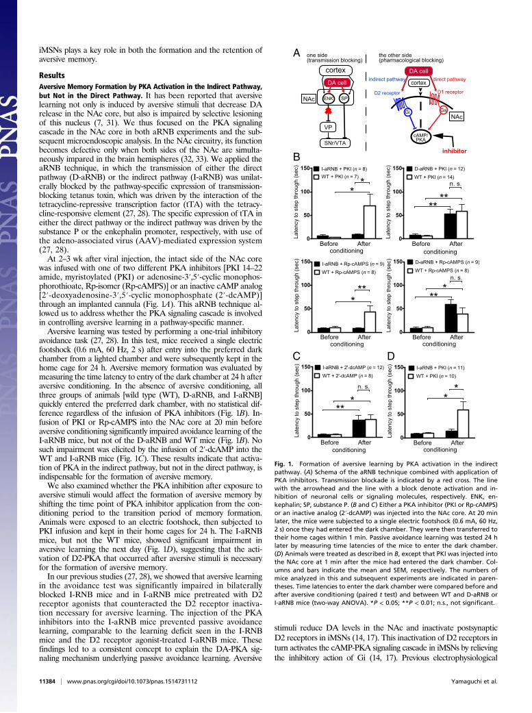
ResultsAversive Memory Formation by PKA Activation in the Indirect Pathway,but Not in the Direct Pathway. It has been reported that aversivelearning not only is induced by aversive stimuli that decrease DArelease in the NAc core, but also is impaired by selective lesioningof this nucleus (7, 31). We thus focused on the PKA signalingcascade in the NAc core in both aRNB experiments and the sub-sequent microendoscopic analysis. In the NAc circuitry, its functionbecomes defective only when both sides of the NAc are simulta-neously impared in the brain hemispheres (32, 33). We applied theaRNB technique, in which the transmission of either the directpathway (D-aRNB) or the indirect pathway (I-aRNB) was unilat-erally blocked by the pathway-specific expression of transmission-blocking tetanus toxin, which was driven by the interaction of thetetracycline-repressive transcription factor (tTA) with the tetracy-cline-responsive element (27, 28). The specific expression of tTA ineither the direct pathway or the indirect pathway was driven by thesubstance P or the enkephalin promoter, respectively, with use ofthe adeno-associated virus (AAV)-mediated expression system(27, 28).At 2–3 wk after viral injection, the intact side of the NAc core
was infused with one of two different PKA inhibitors [PKI 14–22amide, myristoylated (PKI) or adenosine-3′,5′-cyclic monophos-phorothioate, Rp-isomer (Rp-cAMPS)] or an inactive cAMP analog[2′-deoxyadenosine-3′,5′-cyclic monophosphate (2′-dcAMP)]through an implanted cannula (Fig. 1A). This aRNB technique al-lowed us to address whether the PKA signaling cascade is involvedin controlling aversive learning in a pathway-specific manner.Aversive learning was tested by performing a one-trial inhibitory
avoidance task (27, 28). In this test, mice received a single electricfootshock (0.6 mA, 60 Hz, 2 s) after entry into the preferred darkchamber from a lighted chamber and were subsequently kept in thehome cage for 24 h. Aversive memory formation was evaluated bymeasuring the time latency to entry of the dark chamber at 24 h afteraversive conditioning. In the absence of aversive conditioning, allthree groups of animals [wild type (WT), D-aRNB, and I-aRNB]quickly entered the preferred dark chamber, with no statistical dif-ference regardless of the infusion of PKA inhibitors (Fig. 1B). In-fusion of PKI or Rp-cAMPS into the NAc core at 20 min beforeaversive conditioning significantly impaired avoidance learning of theI-aRNB mice, but not of the D-aRNB and WT mice (Fig. 1B). Nosuch impairment was elicited by the infusion of 2′-dcAMP into theWT and I-aRNB mice (Fig. 1C). These results indicate that activa-tion of PKA in the indirect pathway, but not in the direct pathway, isindispensable for the formation of aversive memory.We also examined whether the PKA inhibition after exposure to
aversive stimuli would affect the formation of aversive memory byshifting the time point of PKA inhibitor application from the con-ditioning period to the transition period of memory formation.Animals were exposed to an electric footshock, then subjected toPKI infusion and kept in their home cages for 24 h. The I-aRNBmice, but not the WT mice, showed significant impairment inaversive learning the next day (Fig. 1D), suggesting that the acti-vation of D2-PKA that occurred after aversive stimuli is necessaryfor the formation of aversive memory.In our previous studies (27, 28), we showed that aversive learning
in the avoidance test was significantly impaired in bilaterallyblocked I-RNB mice and in I-aRNB mice pretreated with D2receptor agonists that counteracted the D2 receptor inactiva-tion necessary for aversive learning. The injection of the PKAinhibitors into the I-aRNB mice prevented passive avoidancelearning, comparable to the learning deficit seen in the I-RNBmice and the D2 receptor agonist-treated I-aRNB mice. Thesefindings led to a consistent concept to explain the DA-PKA sig-naling mechanism underlying passive avoidance learning. Aversive
stimuli reduce DA levels in the NAc and inactivate postsynapticD2 receptors in iMSNs (14, 17). This inactivation of D2 receptors inturn activates the cAMP-PKA signaling cascade in iMSNs by relievingthe inhibitory action of Gi (14, 17). Previous electrophysiological
B
A the other side(pharmacological blocking)
DA cell
cortex
Gi
D2 receptor D1 receptor
Gs
cAMP/PKA
inhibitor
indirect pathway direct pathway
cortex
ENK SP
VP
SNr/VTA
DA cell
one side (transmission blocking)
C D
NAc
NAc
Fig. 1. Formation of aversive learning by PKA activation in the indirectpathway. (A) Schema of the aRNB technique combined with application ofPKA inhibitors. Transmission blockade is indicated by a red cross. The linewith the arrowhead and the line with a block denote activation and in-hibition of neuronal cells or signaling molecules, respectively. ENK, en-kephalin; SP, substance P. (B and C) Either a PKA inhibitor (PKI or Rp-cAMPS)or an inactive analog (2′-dcAMP) was injected into the NAc core. At 20 minlater, the mice were subjected to a single electric footshock (0.6 mA, 60 Hz,2 s) once they had entered the dark chamber. They were then transferred totheir home cages within 1 min. Passive avoidance learning was tested 24 hlater by measuring time latencies of the mice to enter the dark chamber.(D) Animals were treated as described in B, except that PKI was injected intothe NAc core at 1 min after the mice had entered the dark chamber. Col-umns and bars indicate the mean and SEM, respectively. The numbers ofmice analyzed in this and subsequent experiments are indicated in paren-theses. Time latencies to enter the dark chamber were compared before andafter aversive conditioning (paired t test) and between WT and D-aRNB orI-aRNB mice (two-way ANOVA). *P < 0.05; **P < 0.01; n.s., not significant.
11384 | www.pnas.org/cgi/doi/10.1073/pnas.1514731112 Yamaguchi et al.
studies found that when the D2 receptors in iMSNs were inhibited,elaborate synaptic modulation via NMDA receptors, A2a adeno-sine receptors, and endocannabinoid CB1 receptors was criticallyinvolved in inducing long-term potentiation in glutamatergic trans-mission of iMSNs (34, 35). Furthermore, our behavioral studiesusing aRNB techniques revealed that all of these LTP-inducing keyreceptors, when pharmacologically manipulated, prevented passiveavoidance learning specific for the I-aRNB mice (17, 28). Thus, thePKA signaling cascade via D2 receptors and the sequential mech-anistic events mediated by the key receptors play a pivotal role inthe induction of aversive learning in an indirect pathway-dependentmanner of the NAc circuit.
Temporal Dynamics of PKA Activity in dMSNs and iMSNs in AversiveLearning. To explore how the cell type-specific PKA signaling cas-cade is involved in the process of aversive learning, we appliedin vivo microendoscopic analysis, in which time-lapse changes inPKA activities were pursued by measuring FRET responses ofthe PKA biosensor in behaving animals (30). In this methodology,the genetically encoded FRET biosensor of PKA was specificallyexpressed in either dMSNs or iMSNs by crossing floxed PKAbiosensor-expressing transgenic mice with D1-Cre or D2-Cre bac-terial artificial chromosome (BAC) transgenic mice, respectively(30, 36). Cell-specific fluorescence changes between the active andinactive forms of PKA in dMSNs (D1-PKA) and iMSNs (D2-PKA)were monitored by microendoscopic analysis of freely moving mice.Aversive learning was examined by performing a one-trial in-
hibitory avoidance test (27). In this test, FRET responses were
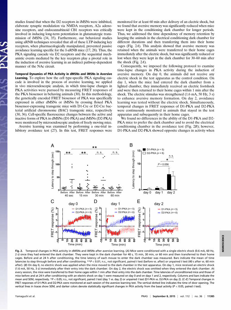
monitored for at least 60 min after delivery of an electric shock, butwe found that aversive memory was significantly reduced when micewere kept in the conditioning dark chamber for longer periods.Thus, we addressed the time dependency of memory retention bykeeping the animals in the electrical conditioning dark chamber fordifferent durations and then transferring them into their homecages (Fig. 2A). This analysis showed that aversive memory wasretained when the animals were transferred to their home cagesimmediately after the electric shock, but was significantly reduced orlost when they were kept in the dark chamber for 30–60 min afterthe shock (Fig. 2A).Consequently, we imposed the following protocol to examine
time-lapse changes in PKA activity during the induction ofaversive memory. On day 0, the animals did not receive anyelectric shock in the test apparatus as the control condition. Onday 1, when the mice had entered the dark chamber from alighted chamber, they immediately received an electric footshockand were then returned to their home cages within 1 min after theshock. The electric stimulus was strengthened (1.6 mA, 50 Hz, 3 s)to enhance aversive memory formation. On day 2, avoidancelearning was tested without the electric shock. Simultaneously,temporal changes in FRET responses of D1-PKA and D2-PKAwere continuously monitored in animals that stayed in the testapparatus and subsequently in their home cages.We found no differences in the ability of the D1-PKA and D2-
PKA mice to prefer the dark chamber and to avoid the electricalconditioning chamber in the avoidance test (Fig. 2B); however,D1-PKA and D2-PKA showed opposite changes in activity when
day 0 day 2E
B
C D day 1
A
shock
Fig. 2. Temporal changes in PKA activity in dMSNs and iMSNs after aversive learning. (A) Mice were conditioned with a single electric shock (0.6 mA, 60 Hz,2 s) once they had entered the dark chamber. They were kept in the dark chamber for 40 s, 15 min, 30 min, or 60 min and then transferred to their homecages. Before and at 24 h after conditioning, the time latency of each mouse to enter the dark chamber was measured. Bars indicate the mean of timelatencies to step through before and after conditioning. **P < 0.01; n.s., not significant, paired t test (before vs. after) or unpaired t test (40 s after vs. 60 minafter). (B) On day 0, no electric shock was applied when the mice moved to the dark chamber in the test apparatus. On day 1, mice received an electric shock(1.6 mA, 50 Hz, 3 s) immediately after their entry into the dark chamber. On day 2, the electric shock was omitted when they entered the dark chamber. Atevery session, the mice were transferred to their home cages within 1 min after their entry into the dark chamber. Time latencies of unconditionedmice and those ofmice before and at 24 h after conditioning with an electric shock on day 1 were measured on day 0 and on days 1 and 2, respectively. Columns and bars indicate themean and SEM, respectively. *P < 0.05; n.s., not significant, paired t test (day 1 vs. day 2) or unpaired t test (D1-PKA vs. D2-PKA on day 2). (C–E) Temporal changes inFRET responses of D1-PKA and D2-PKA were monitored at each session of the aversive learning test. The vertical dotted line indicates the time of door opening. Thevertical lines in traces show SEM, and darker colors denote statistically significant changes in PKA activity from the basal activity (P < 0.05, paired t test).
Yamaguchi et al. PNAS | September 8, 2015 | vol. 112 | no. 36 | 11385
NEU
ROSC
IENCE
the mice were handled and placed in the test apparatus on day0 (Fig. 2C). D1-PKA and D2-PKA slowly and continuously in-creased and decreased, respectively, during the stay of theseanimals in their home cages. Because the similar change in ac-tivity of D2-PKA was observed in animals that remained in thetest apparatus (see Fig. 4B), the changes in D1-PKA and D2-PKA on day 0 might have resulted from handling of the mice toinitiate the avoidance test; this finding was not further exploredin this investigation, however. Importantly, changes in the ac-tivity of D1-PKA and D2-PKA after the electric shock on day 1were in marked contrast to those observed without the electricshock on day 0 (Fig. 2D). The electric shock caused a progressiveincrease in D2-PKA activity but resulted in a rapid decrease inD1-PKA activity, which remained low thereafter (Fig. 2D). Asimilar temporal change in the activity of both D1-PKA and D2-PKA was observed on day 2, when the conditioned mice en-countered the aversive context of the test apparatus (Fig. 2E).Thus, the synergistic and reciprocal changes in PKA activity indMSNs and iMSNs were evoked not only in the induction, butalso in the retrieval, of aversive memory.Mice retained the ability to avoid the conditioned chamber for
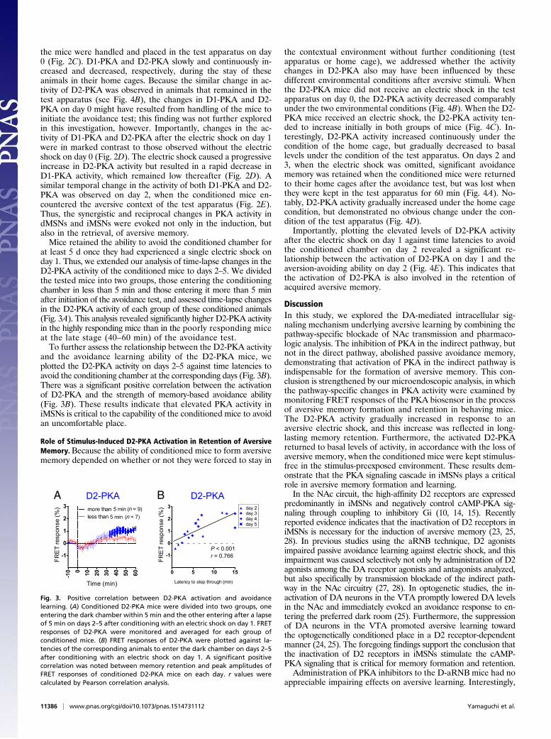
at least 5 d once they had experienced a single electric shock onday 1. Thus, we extended our analysis of time-lapse changes in theD2-PKA activity of the conditioned mice to days 2–5. We dividedthe tested mice into two groups, those entering the conditioningchamber in less than 5 min and those entering it more than 5 minafter initiation of the avoidance test, and assessed time-lapse changesin the D2-PKA activity of each group of these conditioned animals(Fig. 3A). This analysis revealed significantly higher D2-PKA activityin the highly responding mice than in the poorly responding miceat the late stage (40–60 min) of the avoidance test.To further assess the relationship between the D2-PKA activity
and the avoidance learning ability of the D2-PKA mice, weplotted the D2-PKA activity on days 2–5 against time latencies toavoid the conditioning chamber at the corresponding days (Fig. 3B).There was a significant positive correlation between the activationof D2-PKA and the strength of memory-based avoidance ability(Fig. 3B). These results indicate that elevated PKA activity iniMSNs is critical to the capability of the conditioned mice to avoidan uncomfortable place.
Role of Stimulus-Induced D2-PKA Activation in Retention of AversiveMemory. Because the ability of conditioned mice to form aversivememory depended on whether or not they were forced to stay in
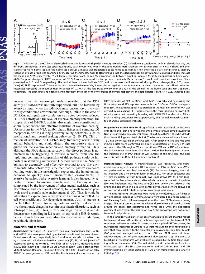
the contextual environment without further conditioning (testapparatus or home cage), we addressed whether the activitychanges in D2-PKA also may have been influenced by thesedifferent environmental conditions after aversive stimuli. Whenthe D2-PKA mice did not receive an electric shock in the testapparatus on day 0, the D2-PKA activity decreased comparablyunder the two environmental conditions (Fig. 4B). When the D2-PKA mice received an electric shock, the D2-PKA activity ten-ded to increase initially in both groups of mice (Fig. 4C). In-terestingly, D2-PKA activity increased continuously under thecondition of the home cage, but gradually decreased to basallevels under the condition of the test apparatus. On days 2 and3, when the electric shock was omitted, significant avoidancememory was retained when the conditioned mice were returnedto their home cages after the avoidance test, but was lost whenthey were kept in the test apparatus for 60 min (Fig. 4A). No-tably, D2-PKA activity gradually increased under the home cagecondition, but demonstrated no obvious change under the con-dition of the test apparatus (Fig. 4D).Importantly, plotting the elevated levels of D2-PKA activity
after the electric shock on day 1 against time latencies to avoidthe conditioned chamber on day 2 revealed a significant re-lationship between the activation of D2-PKA on day 1 and theaversion-avoiding ability on day 2 (Fig. 4E). This indicates thatthe activation of D2-PKA is also involved in the retention ofacquired aversive memory.
DiscussionIn this study, we explored the DA-mediated intracellular sig-naling mechanism underlying aversive learning by combining thepathway-specific blockade of NAc transmission and pharmaco-logic analysis. The inhibition of PKA in the indirect pathway, butnot in the direct pathway, abolished passive avoidance memory,demonstrating that activation of PKA in the indirect pathway isindispensable for the formation of aversive memory. This con-clusion is strengthened by our microendoscopic analysis, in whichthe pathway-specific changes in PKA activity were examined bymonitoring FRET responses of the PKA biosensor in the processof aversive memory formation and retention in behaving mice.The D2-PKA activity gradually increased in response to anaversive electric shock, and this increase was reflected in long-lasting memory retention. Furthermore, the activated D2-PKAreturned to basal levels of activity, in accordance with the loss ofaversive memory, when the conditioned mice were kept stimulus-free in the stimulus-preexposed environment. These results dem-onstrate that the PKA signaling cascade in iMSNs plays a criticalrole in aversive memory formation and learning.In the NAc circuit, the high-affinity D2 receptors are expressed
predominantly in iMSNs and negatively control cAMP-PKA sig-naling through coupling to inhibitory Gi (10, 14, 15). Recentlyreported evidence indicates that the inactivation of D2 receptors iniMSNs is necessary for the induction of aversive memory (23, 25,28). In previous studies using the aRNB technique, D2 agonistsimpaired passive avoidance learning against electric shock, and thisimpairment was caused selectively not only by administration of D2agonists among the DA receptor agonists and antagonists analyzed,but also specifically by transmission blockade of the indirect path-way in the NAc circuitry (27, 28). In optogenetic studies, the in-activation of DA neurons in the VTA promptly lowered DA levelsin the NAc and immediately evoked an avoidance response to en-tering the preferred dark room (25). Furthermore, the suppressionof DA neurons in the VTA promoted aversive learning towardthe optogenetically conditioned place in a D2 receptor-dependentmanner (24, 25). The foregoing findings support the conclusion thatthe inactivation of D2 receptors in iMSNs stimulate the cAMP-PKA signaling that is critical for memory formation and retention.Administration of PKA inhibitors to the D-aRNB mice had no
appreciable impairing effects on aversive learning. Interestingly,
BD2-PKAA D2-PKA
P < 0.001r = 0.766
Fig. 3. Positive correlation between D2-PKA activation and avoidancelearning. (A) Conditioned D2-PKA mice were divided into two groups, oneentering the dark chamber within 5 min and the other entering after a lapseof 5 min on days 2–5 after conditioning with an electric shock on day 1. FRETresponses of D2-PKA were monitored and averaged for each group ofconditioned mice. (B) FRET responses of D2-PKA were plotted against la-tencies of the corresponding animals to enter the dark chamber on days 2–5after conditioning with an electric shock on day 1. A significant positivecorrelation was noted between memory retention and peak amplitudes ofFRET responses of conditioned D2-PKA mice on each day. r values werecalculated by Pearson correlation analysis.
11386 | www.pnas.org/cgi/doi/10.1073/pnas.1514731112 Yamaguchi et al.
however, our microendoscopic analysis revealed that the PKAactivity of dMSNs was not only suppressed, but also lowered, byaversive stimuli when the D1-PKA mice encountered the elec-trically conditioned environment. Although, unlike in the case ofD2-PKA, no significant correlation was noted between reducedD1-PKA activity and the level of aversive memory retention, thesuppression of D1-PKA activity also might have contributed tostimulus-dependent and effective induction of aversive learning.DA neurons in the VTA exhibit phasic firings and stimulate D1receptors in dMSNs during positively acting behaviors, such asmotivational and reward-seeking behaviors (3, 10, 17). This di-rection of activation of D1-PKA may frequently occur duringanimal behaviors and could disturb the suppressive state re-quired for the aversive reaction and memory formation. Thus,although the PKA signaling cascade in the direct pathway is notinvolved in the triggering of aversive memory formation, therapid and continuous suppression of this pathway could be im-portant in stabilizing suppressive DA modulation in the NAc foranimals to accurately and effectively respond to aversive envi-ronments. Also noteworthy is the fact that the passive avoidancelearning tested in this investigation represents the innate animalbehavior to quickly avoid uncomfortable environments. Inaversive behavior, active aversive learning is also induced by re-peated exposure to aversive stimuli, and this learning is morecomplicated by the involvement of other mental activities, such asmotivational and intentional activities, for animals to more posi-tively avoid uncomfortable environments (8, 37). Thus, it would beinteresting to explore how active aversive learning is controlled in acell type-specific and DA-dependent manner. Also of interest isthe fact that D2 receptor antagonists are widely used as effec-tive therapeutic drugs for certain psychiatric disorders, such asschizophrenia (38). Thus, further investigation of PKA and itsdownstream signaling in D2 receptor-expressing iMSNs wouldbe useful in better understanding the mechanisms underlyingpsychiatric disorders.
Materials and MethodsAnimals. Male mice aged ∼2–3 mo were used in all experiments. The D-aRNBand I-aRNB mice were generated by unilateral injection of the recombinantD- and I-AAV viruses, respectively, into four sites at one side of the NAc coreby a stereotaxic technique as described previously (27, 28). Virus-injected WTlittermates served as controls. Two lines of D1-Cre BAC transgenic mice(EY262 and FK150) and 1 line of D2-Cre BAC mice (ER44) were obtained fromMutant Mouse Regional Resource Centers. The FRET biosensor of PKA,AKAR3EV, was generated (29), and the Cre-dependent expression of the
FRET biosensor of PKA in dMSNs and iMSNs was achieved by crossing thefloxed-stop AKAR3EV reporter mice with the D1-Cre or D2-Cre transgenicmice (30). The pathway-specific expression of the FRET biosensor of PKA wasverified by visualizing FRET fluorescence in the corresponding pathway (30).All mice were maintained by breeding with C57BL/6J founder mice. All an-imal handling procedures were approved by the Animal Research Commit-tee of Osaka Bioscience Institute.
Drug Infusion in aRNB Mice. For drug infusion, the intact side of the NAc coreof D-aRNB and I-aRNB mice was implanted with a cannula aimed toward theNAc, as described previously (28). Then 100 nM Rp-cAMPS, 100 nM 2′-dcAMP(both from Biolog), and 0.82 μM PKI (Tocris) were delivered in a volume of1 μL into the intact side of the NAc core. After behavioral analysis, the druginjection sites were confirmed by direct visualization of a series of slicesections of the NAc region. When conditioned WT and aRNB mice enteredthe dark chamber more than 200 s after the door had been opened or whenthe injection site of PKA inhibitor was outside of the NAc core, the datawere discarded (∼10% of the animals analyzed).
Microendoscopic Analysis. A microendoscope was fabricated, and micro-endoscopic analysis to monitor FRET responses of PKA in dMSNs and iMSNswas performed as described previously (30). For in vivo imaging, the scalpwas opened, and a hole was drilled in the skull (1.2 mm anterioposterior and1.1 mm mediolateral from bregma). Two skull screws (M1.0; 4 mm long)were first implanted as anchors, after which the endoscope with its μ-drive(30) was implanted into the NAc core (3.5 mm below the surface of thebrain) and cemented in place with dental acrylic. Animals were allowed torecover for at least 4 d before optical recordings were made.
The time-lapse FRET recordings were made by monitoring 20–25 frames ofraw endoscope images at 15 frames/s. Data were stored as a noncompressedAVI file every 1 min, offline-averaged, smoothed, and FRET-calculated usingImageJ. The mice connected to the microendoscope were allowed to movearound the entire test apparatus and home cage. Once the endoscopeplacement was fixed at the session on day 0, the endoscope was not movedfrom its fixed position.
In the inhibitory avoidance test, care was taken to ensure that the mousehad calmed down sufficiently in the home cage and that the trace of FRETresponses had become stable. For quantitative analysis of FRET responses, thefluorescence intensities of CFP and FRETweremeasured in the entire field of aview that corresponded to the diameter of a microendoscopic fiber bundle(300 μm), and averaged values of both CFP and FRET were calculatedwithout subtraction of their background fluorescence (30). The effect ofbleaching of fluorophores was evaluated as negligible by time-lapse imag-ing without stimulation (30). The cell viability and the location of a micro-endoscopic tip in the NAc core was confirmed by DAPI staining and GFPimmunostaining of serial sections of NAc after microendoscopic analysis(30) (Fig. S1).
day 0 day 1 days 2 & 3A B C D
shock
E
Fig. 4. Activation of D2-PKA by an electrical stimulus and its relationship with memory retention. (A) Animals were conditioned with an electric shock by twodifferent procedures. In the test apparatus group, each mouse was kept in the conditioning dark chamber for 60 min after an electric shock and thentransferred to its home cage. In the home cage group, a mouse was transferred to its home cage within 1 min after the electric conditioning. Memoryretention of each group was examined by measuring the time latencies to step through into the dark chamber on days 2 and 3. Columns and bars indicatethe mean and SEM, respectively. *P < 0.05; n.s., not significant, paired t test (comparison between days) or unpaired t test (test apparatus vs. home cage).(B–D) Temporal changes in FRET responses of D2-PKA were monitored for two groups of animals. Data for day 0, day 1, and combined days 2 and 3 arepresented in B, C, and D, respectively. The vertical lines in traces indicate SEM, and darker colors indicate statistically significant changes (P < 0.05, pairedt test). (E) FRET responses of D2-PKA at the late stage (40–60 min) of day 1 were plotted against latencies to enter the dark chamber on day 2. Filled circles andrectangles represent the mean of FRET responses of D2-PKA at the late stage (40–60 min) of day 1 in the animals in the home cage and test apparatus,respectively. The open circle and open rectangle represent the mean of the two groups of animals, respectively. The bars indicate ± SEM. *P < 0.05, unpaired t test.
Yamaguchi et al. PNAS | September 8, 2015 | vol. 112 | no. 36 | 11387
NEU
ROSC
IENCE
Avoidance Test of aRNB Mice with Application of PKA Inhibitors. The behavioralapparatus used in theexperimentswith theaRNBmicewas composedof a lightedchamber (10 × 13 × 13 cm) and a dark chamber (18 × 13 × 13 cm). The lightedchamber, which was illuminated by a lamp, had a plastic floor, clear walls, andan entrance (5 cm wide) to the dark chamber. The dark chamber had a metalgrid floor for electric shock, walls, and a roof, which were all black in color.
At 20 min after drug infusion, the animals were subjected to an electricshock (0.6 mA, 60 Hz, 2 s), when they had stepped with all four paws into thedark chamber. At 1 min after the shock, the animals were returned to theirhome cages, except when subjected to experiments to determine the timedependency of memory formation, in which case they were kept in theconditioning dark chamber for 40 s, 15 min, 30 min, or 60 min and thenreturned to their home cages. In some experiments, drug infusion was donewithin 1min after delivery of the electric shock. Memory retentionwas tested24 h later by measuring the latency to enter the dark chamber.
Avoidance Test with Microendoscopic Recording. The test apparatus used in thisexperiment was composed of a dark chamber (20 × 17 × 21 cm) and a lightedchamber (10 × 17 × 21 cm). The lighted chamber had a plastic floor and whitewalls without a roof and was illuminated by a lamp. The dark chamber had ametal grid floor, a roof, and walls, all of which were colored black. The lightedchamber was linked to the dark chamber through a sliding door.
The avoidance test consisted of three sessions. On day 0, the mouse wasplaced in a lighted chamber, and the door leading to the dark chamber was
then opened. Once the mouse had stepped with all four paws into the darkchamber, the door was closed. Themouse was then returned to its home cagewithin 1 min after it had entered the dark chamber (home cage condition) orkept in the dark chamber for 60 min (test apparatus condition). On day 1, themouse was placed in the lighted chamber of the test apparatus. Once themouse had entered the dark chamber, the door was closed, and an electricfootshock was delivered (1.6 mA, 50 Hz, 3 s). The mouse was returned to itshome cage 1 min after receiving the footshock in the dark chamber (homecage condition) or kept in the dark chamber for 60 min (test apparatuscondition). On days 2–5, memory retention was tested every 24 h by re-peating the foregoing procedures without electric shock and measuring thetime latencies for the mice to step into the dark chamber in which they hadreceived an electric shock on day 1. When a mouse did not enter the darkchamber within 15 min, it was returned to its home cage.
Statistical Analysis. Statistical analyses, including paired and unpaired t tests,two-way ANOVA, and Pearson correlation analysis, were conducted usingGraphPad Prism 5.0 and are described in the figure legends.
ACKNOWLEDGMENTS. This work was supported by Research Grants-in-Aid2222005 (to S.N.), 24111552 (to K.F.), 22300136 (to K.F.), 26560470 (to K.F.),23120011 (to T.H., S.Y., and S.N.) and 26830022 (to T.Y.) from the Ministry ofEducation Culture, Sports, Science and Technology of Japan; by the TakedaScience Foundation (S.N.); and by the Uehara Memorial Foundation (K.F.).
1. LeDoux JE (2000) Emotion circuits in the brain. Annu Rev Neurosci 23:155–184.2. Cardinal RN, Parkinson JA, Hall J, Everitt BJ (2002) Emotion and motivation: The role
of the amygdala, ventral striatum, and prefrontal cortex. Neurosci Biobehav Rev
26(3):321–352.3. Bromberg-Martin ES, Matsumoto M, Hikosaka O (2010) Dopamine in motivational
control: Rewarding, aversive, and alerting. Neuron 68(5):815–834.4. Pezze MA, Feldon J (2004) Mesolimbic dopaminergic pathways in fear conditioning.
Prog Neurobiol 74(5):301–320.5. McCutcheon JE, Ebner SR, Loriaux AL, Roitman MF (2012) Encoding of aversion by
dopamine and the nucleus accumbens. Front Neurosci 6:137.6. Roitman MF, Wheeler RA, Wightman RM, Carelli RM (2008) Real-time chemical re-
sponses in the nucleus accumbens differentiate rewarding and aversive stimuli. Nat
Neurosci 11(12):1376–1377.7. Badrinarayan A, et al. (2012) Aversive stimuli differentially modulate real-time dopa-
mine transmission dynamics within the nucleus accumbens core and shell. J Neurosci
32(45):15779–15790.8. Oleson EB, Gentry RN, Chioma VC, Cheer JF (2012) Subsecond dopamine release in the
nucleus accumbens predicts conditioned punishment and its successful avoidance.
J Neurosci 32(42):14804–14808.9. Alexander GE, Crutcher MD (1990) Functional architecture of basal ganglia circuits:
Neural substrates of parallel processing. Trends Neurosci 13(7):266–271.10. Gerfen CR, Surmeier DJ (2011) Modulation of striatal projection systems by dopamine.
Annu Rev Neurosci 34:441–466.11. Gerfen CR, et al. (1990) D1 and D2 dopamine receptor-regulated gene expression of
striatonigral and striatopallidal neurons. Science 250(4986):1429–1432.12. Humphries MD, Prescott TJ (2010) The ventral basal ganglia, a selection mechanism at
the crossroads of space, strategy, and reward. Prog Neurobiol 90(4):385–417.13. Smith RJ, Lobo MK, Spencer S, Kalivas PW (2013) Cocaine-induced adaptations in
D1 and D2 accumbens projection neurons (a dichotomy not necessarily synonymous
with direct and indirect pathways). Curr Opin Neurobiol 23(4):546–552.14. Surmeier DJ, Ding J, Day M, Wang Z, Shen W (2007) D1 and D2 dopamine-receptor
modulation of striatal glutamatergic signaling in striatal medium spiny neurons.
Trends Neurosci 30(5):228–235.15. Shiflett MW, Balleine BW (2011) Molecular substrates of action control in cortico-
striatal circuits. Prog Neurobiol 95(1):1–13.16. Tritsch NX, Sabatini BL (2012) Dopaminergic modulation of synaptic transmission in
cortex and striatum. Neuron 76(1):33–50.17. Nakanishi S, Hikida T, Yawata S (2014) Distinct dopaminergic control of the direct and
indirect pathways in reward-based and avoidance learning behaviors. Neuroscience
282C:49–59.18. Schultz W, Dayan P, Montague PR (1997) A neural substrate of prediction and reward.
Science 275(5306):1593–1599.19. Ungless MA, Magill PJ, Bolam JP (2004) Uniform inhibition of dopamine neurons in
the ventral tegmental area by aversive stimuli. Science 303(5666):2040–2042.
20. Brischoux F, Chakraborty S, Brierley DI, Ungless MA (2009) Phasic excitation of do-pamine neurons in ventral VTA by noxious stimuli. Proc Natl Acad Sci USA 106(12):4894–4899.
21. Matsumoto M, Hikosaka O (2009) Two types of dopamine neuron distinctly conveypositive and negative motivational signals. Nature 459(7248):837–841.
22. Cohen JY, Haesler S, Vong L, Lowell BB, Uchida N (2012) Neuron type-specific signalsfor reward and punishment in the ventral tegmental area. Nature 482(7383):85–88.
23. Kravitz AV, Tye LD, Kreitzer AC (2012) Distinct roles for direct and indirect pathwaystriatal neurons in reinforcement. Nat Neurosci 15(6):816–818.
24. Tan KR, et al. (2012) GABA neurons of the VTA drive conditioned place aversion.Neuron 73(6):1173–1183.
25. Danjo T, Yoshimi K, Funabiki K, Yawata S, Nakanishi S (2014) Aversive behavior in-duced by optogenetic inactivation of ventral tegmental area dopamine neurons ismediated by dopamine D2 receptors in the nucleus accumbens. Proc Natl Acad SciUSA 111(17):6455–6460.
26. Ilango A, et al. (2014) Similar roles of substantia nigra and ventral tegmental dopa-mine neurons in reward and aversion. J Neurosci 34(3):817–822.
27. Hikida T, Kimura K, Wada N, Funabiki K, Nakanishi S (2010) Distinct roles of synaptictransmission in direct and indirect striatal pathways to reward and aversive behavior.Neuron 66(6):896–907.
28. Hikida T, et al. (2013) Pathway-specific modulation of nucleus accumbens in rewardand aversive behavior via selective transmitter receptors. Proc Natl Acad Sci USA110(1):342–347.
29. Komatsu N, et al. (2011) Development of an optimized backbone of FRET biosensorsfor kinases and GTPases. Mol Biol Cell 22(23):4647–4656.
30. Goto A, et al. (2015) Circuit-dependent striatal PKA and ERK signaling underlies rapidbehavioral shift in mating reaction of male mice. Proc Natl Acad Sci USA 112(21):6718–6723.
31. Kelley AE, Berridge KC (2002) The neuroscience of natural rewards: Relevance toaddictive drugs. J Neurosci 22(9):3306–3311.
32. Goto Y, Grace AA (2005) Dopaminergic modulation of limbic and cortical drive ofnucleus accumbens in goal-directed behavior. Nat Neurosci 8(6):805–812.
33. Block AE, Dhanji H, Thompson-Tardif SF, Floresco SB (2007) Thalamic-prefrontal cor-tical-ventral striatal circuitry mediates dissociable components of strategy set shifting.Cereb Cortex 17(7):1625–1636.
34. Shen W, Flajolet M, Greengard P, Surmeier DJ (2008) Dichotomous dopaminergiccontrol of striatal synaptic plasticity. Science 321(5890):848–851.
35. Higley MJ, Sabatini BL (2010) Competitive regulation of synaptic Ca2+ influx by D2dopamine and A2A adenosine receptors. Nat Neurosci 13(8):958–966.
36. Gong S, et al. (2007) Targeting Cre recombinase to specific neuron populations withbacterial artificial chromosome constructs. J Neurosci 27(37):9817–9823.
37. Ilango A, Shumake J, Wetzel W, Scheich H, Ohl FW (2012) The role of dopamine in thecontext of aversive stimuli with particular reference to acoustically signaled avoid-ance learning. Front Neurosci 6:132.
38. Seeman P (2010) Dopamine D2 receptors as treatment targets in schizophrenia. ClinSchizophr Relat Psychoses 4(1):56–73.
11388 | www.pnas.org/cgi/doi/10.1073/pnas.1514731112 Yamaguchi et al.