Embed Size (px)
Citation preview

CLEYTON ROBERTO SOBRINHO
MECANISMOS PURINÉRGICOS NO BULBO VENTROLATERAL
ROSTRAL MODULAM RESPOSTAS CARDIOVASCULARES E
RESPIRATÓRIAS PROMOVIDAS PELA ATIVAÇÃO DOS
QUIMIORRECEPTORES CENTRAIS E PERIFÉRICOS
Tese apresentada ao Programa de Pós-Graduação
em Fisiologia Humana do Instituto de Ciências
Biomédicas da Universidade de São Paulo, para
obtenção do Título de Doutor em Ciências.
São Paulo
2015

CLEYTON ROBERTO SOBRINHO
MECANISMOS PURINÉRGICOS NO BULBO VENTROLATERAL
ROSTRAL MODULAM RESPOSTAS CARDIOVASCULARES E
RESPIRATÓRIAS PROMOVIDAS PELA ATIVAÇÃO DOS
QUIMIORRECEPTORES CENTRAIS E PERIFÉRICOS
Tese apresentada ao Programa de Pós-Graduação
em Fisiologia Humana do Instituto de Ciências
Biomédicas da Universidade de São Paulo, para
obtenção do Título de Doutor em Ciências.
Área de Concentração: Fisiologia Humana
Orientador: Prof. Dr. Thiago dos Santos Moreira
Versão original
São Paulo
2015


Aos amigos e familiares, ou seja, todos
aqueles que participaram direta
indeiretamente do desenvolvimento não
apenas deste trabalho, mas de meu caráter.

AGRADECIMENTOS
Ao meu orientador, Prof. Dr. Thiago S. Moreira, pela oportunidade, estimulação e apoio
no desenvolvimento deste trabalho, mas também pela compreensão e pela ótima convivência.
A Prof.a Dra. Ana Carolina Takakura, que juntamente ao Prof. Thiago desde o ínicio
oferecem suporte para meu crecimento cientifico, mas também pela inspiração pessoal, pois
ambos representam exemplos de dedicação.
Aos Professores Dr. Vagner Antunes, Dra. Renata Frazão e a Dra. Mirian Bassi,
examinadores no exame de qualificação, pela discussão e contribuição.
Aos professores presentes na banca examinadora de defesa, pela avaliação, leitura e
atenção dispensada.
Aos membros do Laboratório de Controle Cardiorrespiratório e do Laboratório de
Controle Neural Cardiorrespiratório, Leonardo, Thales, Milene, Rosélia, Bárbara, Janayna,
Elvis, Talita, Thais, Josiane, Fabiana, Luiz, Silvio, Marina e Karen, pelas horas de
convivência, pelos conhecimentos transferidos, pelas discussões e horas de descontração.
Ao Instituto de Ciências Biomédicas e aos funcionários dos mais diversificados setores,
sempre solícitos e de crucial importância para o andamento e finalização deste trabalho, em
especial para Adilson Alves, Ana Maria Campos, Julieta Scialfa e Mônica Amaral, pela
amizade e suporte técnico.
Aos professores Fernando Abdukader, Luiz R. G. Britto, Sara J. Shammah Lagnado e
Martin A. Metzger que me prestaram total apoio sempre que requisitados.
Ao meu pai José Miguel Sobrinho, minha irmã Denise, meu cunhado José Fernando e
especialmente ao meu sobrinho Estevão, pelo apoio e carinho nos momentos mais difíceis.
A minha querida Helena Panizza, pelo carinho, compreensão e apoio, e a sua família Sr.
Cezar, Sra. Ana, Julia e meu amigo Theo pela companhia e acolhimento.

Aos amigos Adilson, Eduardo, Fernando, Julieta, Milena, Miguel, Rodrigo e Tereza,
que mesmo disfarçados em momentos de descontração, acredito que cada encontro nosso me
transforma em uma pessoa melhor.
Aos Amigos Rafa, Cecilia, Kalene, André, Milene, Anselmo, Taisa, Andréa, Leila,
Rosana, Raquel, Márcio, Leo, Marilia, Eduardo, Gisele, Flávia, Hildebrando, Livia, Verônica,
Ibrahim, Yigran e muitos outros, pela amizade e longas e agradáveis conversas.
Ao Dr. Daniel Mulkey, pela supervisão durante o estágio realizado na Universidade de
Connecticut, e juntamente aos membros de seu laboratório, Ian Wenker, Ginny, Dawey e
Fushan e suas famílias, me receberam de braços abertos em suas casas e me proporcionaram
momentos muito felizes. Em especial, a Ian e sua esposa Jelena, com quem estabeleci fortes
laços de amizade e tenho enorme gratidão, além claro de nossa colaboração científica.
A Fundação de Amparo à Pesquisa do Estado de São Paulo (FAPESP) pelo
indispensável auxílio financeiro (Processo 2011/13462-7 e 2013/02350-9), e ao acessores
científicos desses projetos que mesmo anonimamente prestaram colaborações imensuráveis.

Ostra feliz não faz pérola.
Rubem Alves

RESUMO
SOBRINHO, C. R. Mecanismos purinérgicos no bulbo ventrolateral rostral modulam
respostas cardiovasculares e respiratórias promovidas pela ativação dos
quimiorreceptores centrais e periféricos. 2015. 168 f. Tese (Doutorado em Fisiologia
Humana) - Instituto de Ciências Biomédicas, Universidade de São Paulo, São Paulo, 2015.
Quimiorrecepção é o mecanismo pelo qual células especializadas detectam variações nos
níveis de CO2, O2 e H+ presentes no sangue, liquor e parênquima, onde dois sistemas
principais são reconhecidos: quimiorreceptores periféricos, localizados na bifurcação da aorta
e no corpúsculo carotídeo e quimiorreceptores centrais, representados por grupamentos de
neurônios especializados em detectar apenas as variações de CO2/H+. Em comum, estas
estruturas informam centros respiratórios e simpáticos no sistema nervoso central (SNC)
gerando ajustes da atividade cardiorrespiratória. A região ventrolateral do bulbo contém
neurônios bulbo-espinais que modulam atividade simpática (grupamento C1) e neurônios
quimiossensíveis localizados no núcleo retrotrapezóide (RTN). Adicionalmente a
quimiorrecepção intrínseca dos neurônios do RTN, tem sido demonstrado que outras células
nesta região (possivelmente astrócitos) atuam como sensores de CO2/H+, liberando ATP para
promover a ativação de neurônios via receptores purinérgicos P2, na tentativa de promover
ajustes cardiorrespiratórios. No presente trabalho, ultilizamos ratos Wistar adultos,
anestesiados e ventilados artificialmente para avaliar a ação da sinalização purinérgica em
regiões encefálicas com características quimiossensíveis bem como o envolvimento dos
receptores P2 durante as respostas cardiorrespiratórias promovidas pela ativação dos
quimiorreceptores centrais e periféricos. Avaliamos também a participação de astrócitos neste
processo. Os receptores P2, expressos em neurônios do RTN, modulam parcialmente a
resposta quimiorreceptora central, enquanto que receptores P2Y1, expressos em neurônios
C1, modulam parcialmente a resposta quimiorreceptora periférica, juntamente com receptores
glutamatérgicos ionotrópicos. Adicionalmente, encontramos evidências convincentes de que a
sinalização purinérgica na região do núcleo do trato solitário comissural (NTScom) ou na
região da rafe pálido (RPa) não contribui para resposta quimiorreceptora central. Além disso,
encontramos que varicosidades na região ventrolateral do bulbo rostral provenientes no
NTScom expressam VNUT e VGLUT2, indicando a presença de vesículas de núcleotideos e
glutamatérgicas, respectivamente. Finalmente, nossos experimentos revelaram que a
manipulação farmacológica de astrócitos no RTN com a injeção da gliotoxina fluorocitrato,
mas não da região do NTScom e RPa, produz alterações respiratórias mediadas por receptores
P2. Nossos achados evidenciam a importância e contribuem para descriminação dos
mecanismos de ação da sinalização purinérgica na região bulbo ventrolateral rostral durante a
ativação de quimiorreceptores centrais e periféricos.
Palavras-Chave: Núcleo retrotrapezóide. Grupamento C1. Quimiorrecepção central.
Quimiorrecepção periférica. Sinalização purinérgica. Receptores purinérgicos P2.

ABSTRACT
SOBRINHO, C. R. Purinergic mechanism in rostroventrolateral medulla modulate
cardiovascular and respiratory responses promoted by central and peripheral
chemoreceptors activation. 2015. 168 p. Ph. D. Thesis (Human Phisiology) - Instituto de
Ciências Biomédicas, Universidade de São Paulo, São Paulo, 2015.
Chemoreception is the mechanism by which specialized cells detect changes in the levels of
CO2, O2 and H+ in the blood, cerebrospinal fluid and brain, where two main systems are
recognized: peripheral chemoreceptors are located at the the aorta and carotid body, and the
central chemoreceptors groups represented by specialized neurons detect only changes in
CO2/H+. In common, these structures inform respiratory and sympathetic centers in the central
nervous system (CNS), generating adjustments of cardiorrespiratory activity. The rostral
ventrolateral medulla contains neurons that project to spinal cord to modulate sympathetic
activity (C1 group) and intrinsic chemoreceptors neurons in the RTN neurons. In addition, it
has been shown that other cells in the RTN region (possibly astrocytes) act as CO2 sensors by
releasing ATP to produce neuronal activation via P2 purinergic receptors. In this study, we
used male adult Wistar rats, anesthetized and artificially ventilated to evaluate the effect of
purinergic signaling in regions with chemosensitive characteristics and the involvement of P2
receptors in cardiorespiratory responses elicited by activation of central and peripheral
chemoreceptors. We also evaluated the role of astrocytes in this process. The P2 receptors are
expressed in RTN neurons and partially modulate the central chemoreceptors
cardiorespiratory responses, whereas P2Y1 receptors are expressed in C1 neurons, and
partially modulate peripheral chemoreceptor cardiorespiratory responses, together with
ionotropic glutamate receptors. Additionally, we found convinced evidences that the
purinergic signaling in the commissural aspect of the nucleus of the solitary tract (NTScom)
or in the raphe pallidus (RPA/PPy) region did not contribute to central chemoreceptor
response. Furthermore, we fond that varicosidades in the rostral ventrolateral medulla from
the commNTS express VNUT and VGLUT2, indicating the presence of nucleotides and
glutamate vesicles, respectively. Finally, our experiments have shown that pharmacological
manipulation of astrocytes within the RTN with the gliotoxin fluorocitrate injection produces
respiratory changes mediated by P2 receptors. Our findings show the importance and
contribute to discrimination of the mechanisms of purinergic signaling in the rostral
ventrolateral medulla during activation of central and peripheral chemoreceptors.
Key words: Retrotrapezoid nucleus. C1 group. Central chemoreception. Peripheral
chemoreception. Purinergic signaling. P2 Purinergic receptors.

LISTA DE FIGURAS
Figura 1: Bulbo ventrolateral rostral – RTN, C1 e a organização dos grupamentos
respiratórios...............................................................................................................................21
Figura 2: Injeção bilateral de PPADS no núcleo retrotrapezóide atenua a resposta
cardiorrespiratória produzida pela estimulação dos quimiorreceptores centrais......................43
Figura 3: Injeção bilateral de MRS 2179 no núcleo retrotrapezóide não altera a resposta
cardiorrespiratória produzida pela estimulação dos quimiorreceptores centrais......................45
Figura 4: Efeitos cardiorrespiratórios promovidos pela injeção unilateral do agonista
purinérgico P2Y1 na região do núcleo retrotrapezóide............................................................46
Figura 5: Efeitos cardiorrespiratórios produzidos pelas injeções bilaterais de PPADS no
bulbo ventrolateral rostral nas respostas cardiovasculares e respiratórias produzidas pela
estimulação dos quimiorreceptores periféricos.........................................................................48
Figura 6: Efeitos produzidos pelas injeções bilaterais de MRS2179 e ácido quinurênico no
bulbo ventrolateral rostral nas respostas cardiovasculares, simpáticas e respiratórias
produzidas pela estimulação dos quimiorreceptores periféricos...............................................51
Figura 7: Lesão seletiva de neurônios catecoláminérgicos C1 promovida pela injeção da
toxina saporina conjulgada com Dopamina-β-Hidroxilase.......................................................54
Figura 8: Alterações cardiorrespiratórias promovidas pela ativação dos receptores P2Y1 no
bulbo ventrolateral rostral em animais submetidos a lesão do grupamento catecolaminérgicos
C1..............................................................................................................................................55
Figura 9: Projeções da parte comissural do núcleo do trato solitário para o bulbo ventrolateral
rostral são imunorreativas para VGLUT2 e VNUT..................................................................57
Figura 10: Aplicação de ATP na parte comissural do núcleo do trato solitário promove
alterações cardiorrespiratórias mediadas por receptores P2......................................................59

Figura 11: Injeção de PPADS na parte comissural do núcleo do trato solitário não altera a
resposta cardiorrespiratória produzida pela estimulação dos quimiorreceptores
centrais......................................................................................................................................60
Figura 12: Aplicação de ATP no núcleo pálido da rafe/ região parapiramidal não produz
alterações cardiorrespiratórias...................................................................................................62
Figura 13: Injeção bilteral de PPADS no núcleo pálido da rafe/ região parapiramidal não
altera a resposta cardiorrespiratória produzida pela estimulação dos quimiorreceptores
centrais......................................................................................................................................63
Figure 14: Aplicação de ATP no núcleo retrotrapezóide promove alterações
cardiorrespiratórias....................................................................................................................65
Figura 15: Local das injeções de fluorocitrato no núcleo retrotrapezóide...............................68
Figura 16: Efeitos cardiorrespiratórios promovidos pela injeção unilateral de fluorocitrato na
região do núcleo retrotrapezóide...............................................................................................69
Figura 17: Efeitos cardiorrespiratórios promovidos pela injeção de fluorocitrato na parte
comissural do núcleo do trato solitário ou do núcleo pálido da rafe/ região
parapiramidal............................................................................................................................71
Figura 18: Injeção bilateral de PPADS no núcleo retrotrapezóide atenua a resposta
respiratória produzida pela injeção unilateral de fluorocitrato no núcleo
retrotrapezóide..........................................................................................................................73
Figura 19: Injeção bilateral de MRS 2179 no núcleo retrotrapezóide não altera a resposta
respiratória produzida pela injeção unilateral de fluorocitrato no núcleo retrotrapezóide........75
Figura 20: Conclusão...............................................................................................................92

LISTA DE ABREVIATURAS E SIGLAS
7 ou VII - núcleo motor do nervo facial
7n - nervo facial
XII - núcleo hipoglosso
A5 - grupamento noradrenérgico A5
A6 - grupamento noradrenérgico A6 ou locus coeruleus
ADP - difosfato de adenosina
ADO - adenosina
AMP - monofosfato de adenosina
anti-DβH-SAP - Neurotoxina Saporina conjulgada com dopamina β-hidroxilase
AP - Anteroposterior
ATP - Trifosfato de Adenosina
BDA - Biotina Dextrana Amina
BötC - Complexo de Bötzinger
C1 - Grupamento adrenérgico C1
cc - canal central;
CO2 - Dióxido de Carbono
DV - Dorsoventral
E-NTPDases - ectonucleotidases trifosfato de difosfohidrolase
FCt - Fluorocitrato
GPR4 - receptores ativados por prótons ligados a proteína G
Gr - Núcleo Grácil
GRD - Grupamento Respiratório Dorsal
GRV - Grupamento Respiratório Ventral
GRVc - Grupamento Respiratório Ventrolateral caudal
GRVr- Grupamento Respiratório Ventrolateral rostral
H+ - Íons de Hidrogênio
IO, núcleo olivar inferior
ISN, núcleo salivatório inferior
KCN - Cianeto de potássio
KF - Kolliker-Fuse
Kyn – ácido quinurênico
ML – Médio lateral

mmHg - milímetros de mercúrio
nA, núcleo ambíguo;
NSO, núcleo olivar superior;
NTS - Núcleo do trato solitário
NTScom - porção comissural do núcleo do trato solitário
O2 - Oxigênio
PAM - pressão arterial média
PCO2 – Pressão Parcial de Oxigênio
PO2 - Pressão Parcial de Dióxido de Carbono
PNA - Atividade do nervo frênico
pré-BötC - complexo de pré-Bötzinger
py - trato piramidal
RPa- núcleo pálido da rafe
RPa/PPy - núcleo pálido da rafe/ região parapiramidal
RTN - núcleo retrotrapezóide
RVL – Bulbo ventrolateral rostral
SNC - Sistema Nervoso Central
sSNA - Atividade do nervo simpático esplâncnico
SO, oliva superior
Sp5, trato espinal do trigêmeo
SSN, núcleo salivatório superior
tz - corpo trapezoide
TASK – canais de potássio sensíveis a pH
TH – Tirosina Hidroxilase
VNUT - transportador vesicular de nucleotídeos
VGLUT2 - transportador vesicular de glutamato

SUMÁRIO
1 INTRODUÇÃO ................................................................................................................... 19
1.1 Função e controle neural da respiração ......................................................................... 19
1.2 Bulbo ventrolateral rostral (RVL) .................................................................................. 20
1.2.1 Caracterização do RTN e do grupamento catecolaminérgico C1 .................................. 20
1.3 Quimiorrecepção central e periférica ............................................................................. 23
1.3.1 Quimiorrecepção periférica ............................................................................................ 24
1.3.2 Quimiorrecepção central ................................................................................................. 25
1.3.3 Integração do quimiorreflexo central e periférico: participação do bulbo ventrolateral
rostral ....................................................................................................................................... 27
1.4 Sinalização Purinérgica .................................................................................................... 28
1.5 Glia, sinalização purinérgica e quimiorrecepção central.............................................. 29
2 OBJETIVOS ........................................................................................................................ 31
2.1 Objetivos específicos ......................................................................................................... 32
3 MATERIAIS E MÉTODOS ............................................................................................... 33
3.1 Animais .............................................................................................................................. 34
3.2 Procedimentos cirúrgicos e anestesia .............................................................................. 34
3.3 Farmacos utilizados .......................................................................................................... 35
3.4 Injeções encefálicas ........................................................................................................... 36
3.5 Registro da pressão arterial ............................................................................................. 37
3.6 Medida da atividade do nervo frênico ............................................................................ 37
3.7 Medida da atividade da ativida simpática ...................................................................... 38
3.8 Lesão de neurônios catecolaminérgicos do grupamento C1 ......................................... 38
3.9 Injeção de traçador anterógrado no NTScom ............................................................... 39
3.10 Perfusão, histologia e imunoistoquímica ...................................................................... 39
3.11 Análise dos resultados .................................................................................................... 40
4 RESULTADOS ................................................................................................................... 41
4.1 Alterações cardiorrespiratórias frente ao estímulo hipercapnico são moduladas pela
pela sinalização purinérgica na região do RTN ................................................................... 42
4.1.1 Receptores purinérgicos P2 no RTN participam nas alterações cardiorrespiratórias
promovidas pela ativação dos quimiorreceptores centrais ...................................................... 42

4.1.2 Receptores purinérgicos P2Y1 no RTN não contribuem para as alterações
cardiorrespiratórias promovidas pela ativação dos quimiorreceptores centrais .................... 44
4.2 Receptores purinérgicos P2Y1 em neurônios C1 modulam a resposta respiratória,
simpática e pressora à ativação dos quimiorreceptores periféricos................................... 47
4.2.1 Receptores purinérgicos P2 na região do bulbo ventrolateral rostral participam das
alterações cardiorrespiratórias promovidas pela ativação dos quimiorreceptores periféricos
.................................................................................................................................................. 47
4.2.2 Participação de receptores purinérgicos P2Y1 e glutamatérgicos inotrópicos do bulbo
ventrolateral rostral nas alterações cardiorrespiratórias promovidas pela ativação dos
quimiorreceptores periféricos .................................................................................................. 49
4.2.3 Alterações cardiorrespiratórias promovidas pela ativação dosreceptores P2Y1 no bulbo
ventrolateral rostral são mediadas por neurônios catecolaminérgicos C1 ............................. 52
4.2.4 Varicosidades presentes no bulbo ventroalteral rostral oriundos do NTS comissural
expressam VNUT e VGLUT2 .................................................................................................... 56
4.3 Participação da sinalização purinérgica no controle da atividade cardiorrespiratória
no núcleo retrotrapezóide, núcleo do trato solitário e região do núcleo pálido da rafe
/região parapiramidal: possível envolvimento na quimiossensíbilidade central ............. 58
4.3.1 Aplicação de ATP no NTScom promove alterações cardiorrespiratórias, mas o bloqueio
de receptores P2 não altera as respostas cardiorrespiratórias promovidas pela ativação do
quimiorreflexo central .............................................................................................................. 58
4.3.2 Aplicação de ATP na RPa/PPy não promove alterações cardiorrespiratórias e o
bloqueio de receptores P2 também não foi efetivo em alterar as respostas
cardiorrespiratórias promovidas pela ativação do quimiorreflexo central ............................. 61
4.3.3 Aplicação de ATP no RTN promove alterações cardiorrespiratórias mediadas pelos de
receptores P2 ............................................................................................................................ 64
4.4 Efeitos cardiorrespiratórios produzidos pela injeção de fluorocitrato em diferentes
áreas encefálicas com caracteristicas quimiossensíveis: interação entre as células gliais e
neurônios e a possível participação da sinalização purinérgica ......................................... 66
4.4.1 Injeção unilateral de fluorocitrato no núcleo retrotrapezóide promove alterações
respiratórias ............................................................................................................................. 66
4.4.2 Efeitos cardiorrespiratórios promovidos pela injeção unilateral de fluorocitrato no
núcleo do trato solitário comissural ou na região do núcleo pálido da rafe/região
parapiramidal ........................................................................................................................... 70

4.4.2 Participação de receptores purinérgicos P2 nas alterações respiratórias promovidos
pela injeção unilateral de fluorocitrato no núcleo retrotrapezóide ......................................... 72
4.4.3 Receptores purinérgicos P2Y1 não estão envolvidos nas alterações respiratórias
promovidos pela injeção unilateral de fluorocitrato no núcleo retrotrapezóide ..................... 74
5 DISCUSSÃO ........................................................................................................................ 76
5.1 Contribuição da sinalização purinérgica na região do RTN no controle
cardiorrespiratório promovido pela ativação dos quimiorreceptores centrais ................ 78
5.2 Contribuição dos mecanismos purinérgicos na região do bulbo ventrolateral rostral
no controle cardiorrespiratório promovido pela ativação dos quimiorreceptores
periféricos ................................................................................................................................ 82
5.3 Sinalização purinérgica não contribui para quimiorrecepção central na região
comissural do núcleo do trato solitário e na região do núcleo pálido da rafe/região
parapiramidal ......................................................................................................................... 85
5.4 Interação glia-neurônio na região do RTN no controle cardiorrespiratório .............. 87
6 CONCLUSÃO ...................................................................................................................... 90
REFERÊNCIAS ..................................................................................................................... 94
APÊNDICES ......................................................................................................................... 106
APÊNDICE A - Lista de artigos publicados referentes a tese de doutorado ........................ 107
APÊNDICE B- Lista de artigos submetidos e em preparação .............................................. 108
APÊNDICE C- Artigos publicados ....................................................................................... 109

1 INTRODUÇÃO

19
1.1 Função e controle neural da respiração
O sistema respiratório possui diversas funções, dentre as quais podemos destacar sua
participação em processos mastigatórios e de deglutição, na fonação e principalmente no
processo de trocas gasosas pulmonares, de forma a garantir o fornecimento adequado de
oxigênio (O2) para os tecidos assim como a remoção de dióxido de carbono (CO2). De
particular interesse, esta última função promove ainda a regulação do pH plasmático,
mantendo-o dentro dos limites de normalidade fisiológica.
A musculatura respiratória trabalha de forma coordenada para assegurar o fluxo de ar
adequado para os pulmões. Para tanto, o sistema nervoso central (SNC) participa ativamente
no controle da musculatura respiratória, podendo exercer sua atividade de forma voluntária
por comandos gerados em áreas corticais superiores. Em geral esses comandos podem ser
recrutados de forma consciente durante manobras específicas como, por exemplo, durante
uma apnéia voluntária, porém, o controle motor é essencialmente gerado de forma
involuntária e, assim como a maioria dos comandos fundamentais para assegurar a
sobrevivência ou integridade do individuo, são gerados ou modulados por estruturas
subcorticais, localizadas no diencéfalo e/ou tronco encefálico (BARNA; TAKAKURA;
MOREIRA, 2012; CEZARIO et al., 2008; GUYENET; BAYLISS, 2015; GUYENET;
STORNETTA; BAYLISS, 2010; MOTTA et al., 2009; SCHREIHOFER; GUYENET, 1997).
Dentre os grupamentos neurais que controlam a atividade respiratória podemos destacar
regiões dorsais e ventrais distribuídas ao longo do eixo rostrocaudal do tronco encefálico.
O sub-núcleo ventrolateral dos núcleos do trato solitário (NTS) contém neurônios
predominantemente inspiratórios e corresponde à subdivisão chamada de Grupamento
Respiratório Dorsal (GRD) (CASTRO; LIPSKI; KANJHAN, 1994). O Grupamento
Respiratório Ventral (GRV) contém importantes grupamentos neurais envolvidos no controle
respiratório. A região caudal à área postrema contém neurônios pré-motores expiratórios e
recebe o nome de Grupamento Respiratório Ventrolateral caudal (GRVc) (FORTUNA et al.,
2008). O Grupamento Respiratório Ventrolateral rostral (GRVr), localizado ao nível da área
postrema, apresenta neurônios pré-motores inspiratórios que projetam para neurônios motores
que dão origem ao nervo frênico e inervam o principal músculo respiratório, o diafragma
(STORNETTA; SEVIGNY; GUYENET, 2003). Imediatamente adjacente ao GRVr está
localizado o grupamento pré-Bötzinger (pré-BötC), onde evidências experimentais apontam
para atividade marcapasso destes neurônios e, desta forma, seriam responsáveis por gerar o
ritmo inspiratório (FELDMAN; DEL NEGRO; GRAY, 2012; FELDMAN; DEL NEGRO,

20
2006; SMITH et al., 1991). O grupamento denominado de complexo Bötzinger (BötC),
localizado imediatamente rostral ao pré-BötC, contém interneurônios inibitórios, envolvidos
no processo expiratório (SCHREIHOFER; GUYENET, 1997). Ainda na região ventrolateral
do bulbo, existe um conjunto de neurônios localizados ventralmente ao núcleo motor do nervo
facial chamado de núcleo retrotrapezóide (RTN) (CONNELLY; ELLENBERGER;
FELDMAN, 1990). A particularidade mais relevante apresentada pelos neurônios do RTN é a
capacidade de detectar o aumento da pressão parcial de CO2 (PCO2) plasmática e do
parênquima encefálico assim como a consequente redução do pH, gerando rapidamente o
aumento da atividade respiratória (GUYENET; BAYLISS, 2015; KUMAR et al., 2015;
MOREIRA et al., 2006; MULKEY et al., 2004; TAKAKURA et al., 2006). Mais
recentemente também tem sido proposto que este núcleo poderia modular a expiração ativa
auxiliando na eliminação do CO2 (ABDALA et al., 2009; HUCKSTEPP et al., 2015;
JANCZEWSKI; FELDMAN, 2006b).
Além desses grupamentos, existem evidências bastante convincentes na literatura sobre
a participação de outros núcleos na modulação da atividade respiratória como, por exemplo,
os núcleos serotonérgicos da rafe, localizados na linha mediana do bulbo (DEPUY et al.,
2011; MESSIER; LI; NATTIE, 2004; RICHERSON, 2004), os grupamentos
catecolaminérgicos pontinos A5 e A6 (BIANCARDI et al., 2008; TAXINI et al., 2011), o
núcleo fastigial do cerebelo (MARTINO et al., 2006a, 2006b) e os neurônios orexinérgicos do
hipotálamo (DENG et al., 2007; WILLIAMS et al., 2007).
1.2 Bulbo ventrolateral rostral (RVL)
1.2.1 Caracterização do RTN e do grupamento catecolaminérgico C1
Como mencionado anteriormente, o RTN está localizado muito próximo da superfície
ventrolateral do bulbo (CONNELLY; ELLENBERGER; FELDMAN, 1990; MULKEY et al.,
2004). Este núcleo é constituído por aproximadamente 2100 neurônios no rato, os quais se
estendem ventralmente ao núcleo motor do facial desde sua porção mais caudal até a porção
caudal do corpo trapezóide, englobando uma distância de aproximadamente 2,0 mm no
sentido rostro-caudal (Figura 1) (TAKAKURA et al., 2008, 2014).

21
Figura 1: Bulbo ventrolateral rostral – RTN, C1 e a organização dos grupamentos
respiratórios
A: Fotomicrografia ilustrando a imununorreatividade nuclear para o gene PHOX2B (Alexa
488, verde) no núcleo retrotrapezóide (RTN) em uma secção coronal do bulbo de um rato. A
localização do RTN está no mesmo nível do neurônio representado em verde na figura B.
Imunorreatividade para Tirosina Hidroxilase (Cy3, vermelho), indicando a localização do
grupamento C1. Imunorreatividade dos neurônios motores donúcleo facial (7) após a injeção
do traçador retrógrado Fluoro-Gold intraperitonialmente (Stornetta et al.; 2006) Escala: 100
µm. B: Figura esquemática ilustrando uma secção parassagital de um encéfalo de rato desde a
ponte até o bulbo (modificado de Feldman e Del Negro, 2006; Guyenet e Bayliss, 2015). O
RTN está marcado em verde e as outras regiões da coluna respiratória estão em cinza.
Abreviaturas: 7, núcleo facial; 7n nervo facial; nA, núcleo ambíguo; BötC, complexo
Bötzinger; pre- BötC complexo pré-Bötzinger, rVRG, coluna respiratória ventral rostral;
cVRG, coluna respiratória ventral caudal; NTS, núcleo do trato solitário, ISN, núcleo
salivatório inferior; SSN, núcleo salivatório superior; SO, oliva superior; tz, corpo trapezóide.

22
Até o presente momento, sabe-se que os neurônios do RTN são neurônios não
colinérgicos e não aminérgicos, altamente responsivos à substância P e predominantemente
glutamatérgicos (GUYENET; BAYLISS, 2015; GUYENET; STORNETTA; BAYLISS,
2010; GUYENET et al., 2008; LAZARENKO et al., 2011; MULKEY et al., 2004; ROSIN;
CHANG; GUYENET, 2006). Essas assinaturas bioquímicas do RTN podem ser identificadas
por imunoistoquímica e utilizadas como referência para identificação e delimitação do núcleo.
O fator de transcrição Phox2b constitui outra importante característica dos neurônios do RTN,
e também pode ser identificado por imunoistoquímica para identificação dos neurônios
quimiossensíveis (ABBOTT et al., 2009a; AMIEL et al., 2003; BURKE et al., 2015;
MULKEY et al., 2004; TAKAKURA et al., 2008). Esse fator de transcrição é responsável por
modular a diferenciação celular e a sobrevida de restritos grupamentos neuronais localizados
na ponte e no bulbo, incluindo o RTN (AMIEL et al., 2003). Parece estar bem estabelecido na
literatura de que os neurônios do RTN estão envolvidos em basicamente três processos
neurais relacionados ao controle respiratório, como: a) controle do movimento inspiratório, já
que eles se projetam para regiões mais caudais da coluna respiratória ventral e para os
neurônios motores que controlam a inspiração (DOBBINS; FELDMAN, 1994); b) estão
envolvidos na quimiorrecepção central, pois respondem frente uma situação de hipercapnia
(aumento da concentração de CO2) (FELDMAN; MITCHELL; NATTIE, 2003; KUMAR et
al., 2015; NATTIE; LI; ST JOHN, 1991; NATTIE; LI, 1994, 2002a; OKADA et al., 2002;
PUTNAM; FILOSA; RITUCCI, 2004; SATO; SEVERINGHAUS; BASBAUM, 1992) e c)
por fim, estudos mais recentes têm procurado relacionar o RTN também à geração da
expiração ativa (ABDALA et al., 2009; JANCZEWSKI; FELDMAN, 2006a; PAGLIARDINI
et al., 2011).
Dados da literatura demonstraram que uma subpopulação de células do RTN em ratos
adultos estão inativas em situações basais e tornam-se ritmicamente ativas durante
hipercapnia (MARINA et al., 2010) ou desinibições locais (MOLKOV et al., 2010;
PAGLIARDINI et al., 2011). Experimentos realizados por Pagliardini e colaboradores (2011)
sugerem que o RTN contém um oscilador condicional que gera uma expiração ativa;
entretanto, existe uma inibição sináptica para o RTN que suprime a expiração ativa, e pode ser
desinibida durante uma situação de hipercapnia, hipóxia ou qualquer situação em que existe a
necessidade da expiração ativa. Entretanto, até o presente momento, não se sabe a
procedência desta inibição para o RTN. A literatura sugere algumas possíveis regiões como o
NTS e a região do Kolliker-Fuse (KF) (MOREIRA et al., 2007; ROSIN; CHANG;

23
GUYENET, 2006; TAKAKURA et al., 2007), e o complexo de Botzinger (CREAM; LI;
NATTIE, 2002; ROSIN; CHANG; GUYENET, 2006).
Imediatamente adjacente ao RTN, encontra-se um grupamento de neurônios que atuam
no controle do tônus simpático mediante sua influência sobre neurônios pré-ganglionares da
coluna intermédio-lateral, localizados na medula espinal (DAMPNEY, 1994; GUYENET,
2006). Em condições de repouso, os neurônios pré-motores simpáticos do bulbo ventrolateral
são a fonte primária do tônus simpático, sendo considerados os geradores da atividade
simpática (DAMPNEY et al., 2003; FELDBERG; GUERTZENSTEIN, 1972). Acredita-se
que todos os neurônios bulbo-espinhais pré-simpáticos do bulbo ventrolateral liberem
glutamato na coluna intermédio-lateral da medula espinhal, mas sabe-se que eles também
sintetizam outros neurotransmissores, incluindo adrenalina, encefalina, serotonina entre outros
(SUN, 1996). Os neurônios que sintetizam adrenalina correspondem a mais de 70% dos
neurônios pré-simpáticos do bulbo ventrolateral e pertencem, por definição, ao grupamento
C1 (ROSS et al., 1984; SCHREIHOFER; GUYENET, 1997). Estudos neuroanatômicos e
funcionais descreveram que esses neurônios do bulbo ventrolateral rostral estão localizados
imediatamente caudais ao limite posterior do núcleo motor do nervo facial, ventralmente ao
núcleo ambíguo e não somam mais do que algumas poucas centenas de corpos celulares
(SUN, 1996).
Resumidamente, a área C1 constitui parte da região rostroventrolateral do bulbo onde
estão localizados os neurônios vasomotores simpáticos e contém neurônios
catecolaminérgicos, mas também é capaz de liberar o glutamato como neurotransmissor, além
de outros neuropeptídeos (GUYENET et al., 2004; SCHREIHOFER; GUYENET, 1997;
STORNETTA; MCQUISTON; GUYENET, 2004). Sabe-se que além da clássica
contribuição dos neurônios C1 no controle da pressão arterial e atividade simpática, esse
importante grupamento neuronal pode influenciar diversas funções fisiológicas, incluindo
respostas neuroendócrinas devido um processo inflamatório, homeostase de glicose, processo
reprodutivo, respiração, termorregulação, ativação do eixo hipotalâmico e ingestão de
alimentos (GUYENET et al., 2013).
1.3 Quimiorrecepção central e periférica
Para que o ritmo e a amplitude respiratória sejam ajustados de forma a assegurar a
homeostase gasométrica, é necessário que o SNC receba informações refinadas e precisas dos
valores arteriais de O2 e CO2. Esse papel é atribuído às células conhecidas como

24
quimiorreceptores, que são estruturas especializadas, sensíveis as alterações químicas no
sangue (FELDMAN et al., 2003; GUYENET et al., 2010). Em condições normais, essas
células realizam o monitoramento contínuo, informando ao SNC sobre a pressão parcial de
oxigênio (PO2), PCO2 e pH plasmático, possibilitando que o mesmo promova os ajustes
adequados (GUYENET; STORNETTA; BAYLISS, 2010), e adicionalmente, em situações
onde ocorrem alterações drásticas desses parâmetros, como por exemplo, na hipóxia (redução
na PO2) ou na hipercapnia (redução na PCO2), as células quimiossensíveis sofrem alterações
equivalentes na atividade, produzindo vigorosas alterações na atividade respiratória,
frequentemente acompanhados por alterações na pressão arterial (BASTING et al., 2015;
BURKE et al., 2015; GUYENET; BAYLISS, 2015; MULKEY et al., 2004; PRABHAKAR,
2013; WENKER et al., 2012).
1.3.1 Quimiorrecepção periférica
Todas as células do organismo possuem uma capacidade intrínseca de detectar
variações na concentração extracelular de O2, CO2 e pH e, de certo modo, responder a tais
alterações (SEMENZA, 2011). Entretanto, um conjunto de células neuroepiteliais derivadas
da crista neural localizadas principalmente nos corpúsculos carotídeos apresentam a
peculiaridade de se despolarizar em condições de hipóxia, hipercapnia ou acidose (BISCOE;
DUCHEN, 1990a, 1990b; FITZGERALD; SHIRAHATA; ISHIZAWA, 1996; LAHIRI et al.,
2006), sensibilizando terminais nervosos aferentes que com elas fazem contato, os quais
enviam potenciais de ação para o sistema nervoso central, promovendo, assim, a ativação de
ajustes cardiovasculares e respiratórios com o objetivo de restaurar a PO2 para valores
adequados. Esse processo constitui o quimirreflexo periférico, o qual consiste num dos
principais elementos na manuntenção da homeostase cardiorrespiratória (MACHADO, 2001;
PRABHAKAR, 2013). Os ajustes promovidos pela ativação desse reflexo se caracterizam por
aumento da pressão arterial, decorrente de um incremento na atividade simpática vascular e o
aumento da ventilação (BARROS et al., 2002; BRAGA et al., 2007; ZOCCAL et al., 2008),
as quais ocorrem de forma sincronizada com o objetivo de otimizar os processos de troca
gasosa pulmonar e do débito cardíaco, melhorando a eficiência da captação de O2 e da
perfusão tecidual. Em situações de hipóxia, as células glomus encontradas na bifurcação
carotídea e na curvatura da aorta ativam as vias aferentes, e veiculam esta informação via
nervos vago e glossofaríngeo até o SNC (FELDMAN; MITCHELL; NATTIE, 2003;
KUMAR; PRABHAKAR, 2012; PRABHAKAR, 2013; TAKAKURA et al., 2006). Suas

25
fibras entram no tronco cerebral na fissura póstero-lateral do bulbo, incorporando-se ao trato
solitário, terminando predominantemente nas porções comissural medial e lateral dos núcleos
do trato solitário (NTScom) (FINLEY; KATZ, 1992; KUMAR; PRABHAKAR, 2012;
TAKAKURA et al., 2006). Abordagens funcionais e neuroanatômicas revelaram que o NTS
envia projeções à superfície ventrolateral do bulbo (AICHER et al., 1996; GRANATA, 1994;
KOSHIYA; GUYENET, 1996b; URBANSKI; SAPRU, 1988), uma região que abriga
populações neuronais fisiologicamente distintas, como o grupamento C1 mencionado acima e
os complexos neuronais responsáveis pela geração e manutenção do padrão inspiratório e
ritmo respiratório normal (FELDMAN; DEL NEGRO, 2006). Há evidências de que essas
projeções do NTS para o bulbo ventrolateral rostral e adjacências representem o substrato
neuroanatômico correspondente às vias funcionais simpato-excitatória e ventilatórias do
quimiorreflexo periférico, as quais fazem sinapses com neurônios C1 ou ventilatórios,
respectivamente (GUYENET; KOSHIYA, 1995; KOSHIYA; GUYENET, 1996a; MORAES
et al., 2011).
1.3.2 Quimiorrecepção central
Quimiorreceptores centrais são altamente sensíveis ao aumento na PCO2 e à acidose.
Embora seja crescente o número de trabalhos postulando que núcleos de células
quimiossensíveis estejam distribuídos ao longo do encéfalo (NATTIE, 2011), é comumente
aceito que o núcleo retrotrapezóide (RTN), localizado na porção ventral ao núcleo motor do
núcleo facial e muito próximo da superfície ventrolateral do bulbo (CONNELLY;
ELLENBERGER; FELDMAN, 1990; HUCKSTEPP et al., 2015; MULKEY et al., 2004;
WANG et al., 2013a) concentra o principal grupamento de células quimiossensíveis, uma vez
que este núcleo atente a todas as exigências para ser considerado um grupamento neural
quimiossensível, como: (a) sensibilidade ao CO2/H+ in vivo e in vitro, (b) fenótipo
neuroquímico excitatório e (c) projetar para o centro gerador do padrão respiratório
(MOREIRA et al., 2015).
Dados da literatura têm oferecido várias evidências sobre os mecanismos moleculares
responsáveis pela quimiorrecepção central no RTN (BURKE et al., 2015; GOURINE et al.,
2010; GUYENET; BAYLISS, 2015; HUCKSTEPP et al., 2010a; KUMAR et al., 2015;
MULKEY et al., 2004; WANG et al., 2013b) e dentre esses mecanismos, podemos destacar a
participação de canais de K+ (MULKEY et al., 2004, 2007). Como a corrente de sensibilidade
ao pH, identificada nos neurônios do RTN, mostrou uma pequena retificação e persistiu

26
mesmo quando o cálcio intracelular foi neutralizado, sugeriu-se que a sensibilidade desses
neurônios seja mediada pela inibição de canais de K+ (MULKEY et al., 2004, 2007), muito
provavelmente canais do tipo TASK da família K2P. Constatou-se, no entanto, que a
sensibilidade não depende de canais do tipo TASK-1 ou TASK-3 (canais de K+ da família
K2P sensíveis a acidose) (MULKEY et al., 2007) mas que dependia parcialmente de canais
TASK-2 (canais sensíveis a alcalose) (WANG et al., 2013b), já que o RTN de camundongos
"knock-out" para canais TASK-1 e TASK-3 continuavam apresentando sensibilidade ao pH
(MULKEY et al., 2007), enquanto que a depleção seletiva de TASK-2 em neurônios Phox2b
promoveu a redução da sensibilidade ao pH em muitos neurônios do RTN in vitro e
atenuação da resposta a alcalose in situ (WANG et al., 2013b).
Como sugerido acima, a sensibilidade ao CO2/H+ é apenas parcialmente alterada pela
depleção de TASK-2, indicando que esta propriedade não é mediada apenas por estes canais.
Sabemos agora no entanto, que a quimiossensibilidade desses neurônios depende fortemente
de uma ação em conjunto entre canais TASK-2 e receptores ativados por prótons ligados a
proteína G do tipo GPR4, onde a depleção deste último produz alterações muito mais
expressivas (KUMAR et al., 2015). A depleção ou silenciamento de GPR4 reproduz padrões
respiratórios caracteristicamente observados em animais com depleção de neurônios Phox2b ,
apresentando aumento no número de episódios de apneias espontâneas e redução da resposta a
hipercapnia (KUMAR et al., 2015), além de reduzir expressivamente o número de neurônios
ativados por CO2/pH in vivo e in vitro. Finalmente, depleção simultânea de GPR4 e TASK2
no mesmo animal promoveu alta taxa de mortalidade e massiva redução da resposta a
hipercapnia nos animais sobreviventes (~86%) (KUMAR et al., 2015).
Os mecanismos de quimiorrecepção central em outros grupamentos são menos
conhecidos, e provavelmente por esta razão, mais controversos. Por exemplo, acidificação
local do NTScom produz alterações respiratórias mais expressivas em níveis caudais deste
núcleo, em comparação aos níveis mais rostrais do NTS (NATTIE; LI, 2002b). Neurônios
quimiossensíveis da porção caudal do NTScom também foram identificados in vitro
(NICHOLS et al., 2009; SOBRINHO et al., 2014), mas a inibição com muscimol desta região
não alterou a resposta quimiorreceptora durante a hipercapnia in vivo (FAVERO et al., 2011).
Por outro lado, a aplicação de muscimol em porções mais rostrais do NTS parece prejudicar a
resposta quimiorreceptora central (NATTIE; LI, 2008).
Núcleos da rafe encontrados no bulbo (núcleos pálido, magno e obscuro da rafe, além
de neurônios da região parapiramidal) também são postulados como quimiorreceptores
centrais (RICHERSON, 2004), uma vez que em experimentos in vitro os neurônios

27
serotoninérgicos dessas regiões foram estimulados ou inibidos por alterações na concentração
de CO2/pH (RICHERSON, 2004; SOBRINHO et al., 2014). Adicionalmente, uma pequena
porcentagem de neurônios serotoninérgicos mesencéfalicos (núcleo dorsal da rafe) também
apresentaram alterações na atividade em resposta a acidose (SEVERSON et al., 2003;
VEASEY et al., 1997). Experimentos in vivo, foi detectado aumento da atividade respiratória
em resposta a acidose local induzida por microdiálise em grupamentos bulbares (NATTIE; LI,
2001), enquanto que a inibição com muscimol na rafe bulbar em porcos promove redução da
resposta respiratória a hipercapnia (MESSIER; LI; NATTIE, 2002).
Como mencionado anteriormente, uma série de outros grupamentos apresentam
características quimiossensíveis (BIANCARDI et al., 2008; DENG et al., 2007; MARTINO et
al., 2006a; TAXINI et al., 2011; WILLIAMS et al., 2007), porém não serão retratados aqui
por não constituírem parte do foco deste estudo.
1.3.3 Integração do quimiorreflexo central e periférico: participação do bulbo ventrolateral
rostral
Ao longo dos últimos anos, diferentes estudos comprovaram a existência de uma
importante interação entre os quimiorreceptores periféricos e os quimiorreceptores centrais
localizados no RTN (MULKEY et al., 2004; SMITH et al., 2006; TAKAKURA et al., 2006).
A estimulação dos quimiorreceptores carotídeos promove um aumento na frequência de
disparos dos neurônios do RTN, sendo abolida pela desnervação periférica dos
quimiorreceptores, sem afetar a propriedade intrínseca desses neurônios em resposta à
hipercapnia (MULKEY et al., 2004). Ademais, projeções diretas, predominantemente
excitatórias, do NTScom para os neurônios do RTN também já foram descritas
(TAKAKURA et al., 2006). Além de receber densas projeções do NTScom, o RTN estabelece
ainda conexões recíprocas com outras áreas cardiorrespiratórias como o complexo
parabraquial/Kolliker-Fuse, a coluna respiratória ventral, o grupamento noradrenérgico A5,
bem como áreas hipotalâmicas (ROSIN; CHANG; GUYENET, 2006).
Mais recentemente tem sido proposto que neurônios C1 também podem influenciar a
respiração (BURKE et al., 2014; WENKER et al., 2013). Como já mencionado anteriormente,
neurônios C1 recebem projeções do NTScom que veiculam informações de quimiorreceptores
periféricos (KOSHIYA; GUYENET, 1996b) e além das projeções bulbo-espinais, os
neurônios C1 também apresentam projeções para o locus coeruleus, região A5, núcleo motor

28
dorsal do vago e hipotálamo, ou seja, áreas em potencial para influenciar a ventilação
(GUYENET et al., 2013).
Esses dados sugerem que a região RVL (RTN/C1) e o NTScom representam duas
importantes regiões onde ocorrem integrações cardiorrespiratórias. Esta organização anátomo-
funcional sugere uma integração entre o quimiorreflexo central, a regulação central da
respiração e funções autônomas (MOREIRA et al., 2011; ROSIN; CHANG; GUYENET,
2006). Dado a importância desse processo, o presente trabalho tem como um dos seus
objetivos ampliar o nosso conhecimento a respeito da integração entre o quimiorreflexo
central e periférico na região do RVL.
1.4 Sinalização Purinérgica
O ATP, produzido durante a glicólise, é incontestavelmente mais estudado e mais
popular devido sua atuação como fonte de energia celular. No entanto, após um consideravél
número de evidências experimentais que datam desde o final dos anos 20 até os dias atuais,
sua importância também é reconhecida como molécula sinalizadora intracelular e extracelular
(paracrina e exócrina), inclusive atuando na comunicão central e periférica, na co-transmissão
com outros neurotransmissores e mais recentemente, como molécula sinalizadora na interação
glia-neurônio (ABBRACCHIO et al., 2009; BURNSTOCK, 2006a, 2007; FUNK, 2013;
GOURINE; WOOD; BURNSTOCK, 2009). O sistema que integra a sinalização purinérgica,
conta ainda com grande variedade de ectonucleotidases, enzimas que degradam o ATP, e de
diferentes subtipos de receptores que podem ser responsivos ao ATP ou ao produto da sua
degradação (FUNK, 2013).
Muitas teorias ainda são postuladas sobre o possível mecanimo pelo qual o ATP,
presente no citosol, é liberado ao espaço extracelular. Neste sentido, já foi demonstrado que o
ATP pode ser armazenado em vesiculas, por ação de transportadores vesiculares de
nucleotídeos dependentes de Cl- (SAWADA et al., 2008) e liberado via exocitose como um
neurotransmissor clássico, via aumento nas concentrações de Ca2+ intracelular (ANGELOVA
et al., 2015; GOURINE et al., 2010). Mais recentemente, tem sido proposto que o ATP,
também pode ser liberado via hemicanais de gap-junctions, como por exemplo do subtipo
denominado conexinas 26 (HUCKSTEPP et al., 2010a, 2010b; WENKER et al., 2012).
Uma vez na fenda sinaptica, o ATP pode atuar diretamente em receptores específicos,
ou pode ser degradado, via ectonucleotidades, em difosfato de adenosina (ADP), monofosfato
de adenosina (AMP) e por fim em adenosina (ADO). As ectonucleotidases são divididas em 4

29
famílias, algumas delas com diversos membros. A ectonucleotidase trifosfato de
difosfohidrolase (E-NTPDases 1, 2 , 3 e 8), a fosfatase alcalina e a ectonucleotidase ecto-5’, já
foram bem descritas no SNC em diferentes tipos de células (glias e neurônios)
(ABBRACCHIO et al., 2009; BURNSTOCK, 2006b, 2007; FUNK, 2013).
Os receptores purinérgicos são divididos em 3 grandes grupos. Os receptores P2X
apresentam sete subtipos, sendo todos eles canais iônicos, e, portanto, receptores ionotrópicos
sensiveis ao ATP. Os receptores P2Y respondem ao ATP e ao ADP, e são receptores
metabotrópicos acoplados a proteina G. Oito subtipos são encontrados no SNC, sendo o P2Y1
o subtipo mais abundante. Os subtipos P2Y1, 2, 4, 6 e 11 são receptores ligados a proteina
Gq/11 ou Gs, e portanto excitatórios, enquanto que os subtipos P2Y12 e 13 são ligados a
proteina Gi, e portanto inibitórios. O terceiro grupo é formado por uma familia de receptores
também acoplados a proteina G com 4 membros, os receptores P1, sendo eles P1 A1 e A3
acoplados a Gi e Gαi, respectivamente, enquanto os receptores P1 A2a e A2b são acoplados a
Gs. Receptores P1 são mais sensiveis a ADO (FUNK, 2013). É interessante mencionar que
uma ectonucleotidase pode ter mais afinidade pelo ATP ou ser seletiva a um subproduto da
via de degradação, pois desta forma, se houver maior expressão de um subtipo em
determinada região, isto pode ser determinante na quantidade e do tipo de purina presente na
fenda. Vinculado a distribuição de seus respectivos receptores, determinam o tipo, a potência
e a duração da resposta pré e pós sinaptica exercida pela purina.
1.5 Glia, sinalização purinérgica e quimiorrecepção central
Adicionalmente a quimiorrecepção intrínseca dos neurônios do RTN, tem sido
demonstrado experimentalmente que outras células do SNC (possivelmente células da glia,
mais precisamente os astrócitos) poderiam estar atuando também como sensores de CO2
(ERLICHMAN; LEITER; GOURINE, 2010; FUKUDA; LOESCHCKE, 1977; GOURINE et
al., 2010; MULKEY; WENKER, 2011; WENKER et al., 2010). Estudos anteriores
mostraram a existência na região do RTN, de um grupamento de células sensíveis ao CO2 e
com baixa atividade elétrica, sugerindo que astrócitos nessa região também poderiam
apresentar quimiosenssíbilidade. Sendo assim, outra possível forma desses neurônios serem
ativados pelo aumento na PCO2 seria uma forma indireta, ou seja, via astrócitos. As células da
glia também são sensíveis ao CO2 (WENKER et al., 2010), mas o entendimento a respeito do
mecanismo pelo qual essa ativação gera a ativação dos neurônios do RTN ainda está

30
evoluindo. Dessa maneira, estamos propondo que tanto neurônios como as células da glia da
região do RTN estariam envolvidos na detecção dos níveis de CO2/H+.
Originalmente, acreditava-se que a glia contribuía para o mecanismo de
quimiorrecepção por potencializar uma acidose extracelular produzida pelo CO2
(ERLICHMAN; LI; NATTIE, 1998; HOLLERAN; BABBIE; ERLICHMAN, 2001).
Astrócitos e oligodendrócitos ajudam a regular o K+ extracelular por captar o excesso de K+
durante períodos de aumento de atividade neuronal. Esse processo neutraliza alterações no K+
extracelular, mas pode também aumentar o co-transporte de Na+/HCO3- para a glia,
aumentando a queda no pH extracelular induzido pelo CO2 e potencializar a ativação dos
quimiorreceptores do RTN (CHESLER, 2003; ERLICHMAN et al., 2004).
Mais recentemente, evidências têm demonstrado uma segunda participação das células
da glia na detecção do CO2. Em outras regiões do SNC, já está bem estabelecido que a
ativação de astrócitos é capaz de promover a liberação de neurotransmissores, entre eles o
ATP (FELLIN, 2009). Além do mais, acredita-se que as células gliais sejam a principal fonte
de liberação de ATP durante uma situação de hipercapnia (GOURINE et al., 2010;
HUCKSTEPP et al., 2010a; MULKEY; WENKER, 2011). Níveis elevados de CO2
promovem a liberação de ATP no RTN promovendo um aumento na atividade respiratória
(GOURINE et al., 2005a). As alterações respiratórias promovidas pelo aumento dos níveis de
CO2 são atenuados pelo uso do antagonista de receptores purinérgicos P2, PPADS, sugerindo
que esses receptores estariam envolvidos nas respostas ventilatórias ao ATP (GOURINE et
al., 2005a). Além disso, a aplicação de ATP no RTN promove um aumento na respiração,
mimetizando os resultados observados pelos níveis elevados de CO2 (GOURINE et al., 2005a;
HUCKSTEPP et al., 2010a; WENKER et al., 2010, 2012). Dessa forma, fica claro que o ATP
exerce um papel de importante mediador da quimiorrecepção central via células da glia
(GOURINE et al., 2010).
Dessa maneira, a sinalização purinérgica emergiu como um sistema de grande
relevância para a importância da atividade funcional integrativa entre neurônios, glia e células
vasculares no SNC, facilitando um sistema de sinalização intracelular que permite uma
funcionalidade ideal entre neurônio-glia (BURNSTOCK; FREDHOLM; VERKHRATSKY,
2011). Considerando que um dos pontos chave para o entendimento da quimiorrecepção
central consiste no esclarecimento de mecanismos neuro-moleculares envolvidos nesse
processo, o presente trabalho teve como um dos objetivos avaliar os possíveis mecanismos de
interação glia-neurônio na região do RTN durante uma situação de hipercapnia, bem como a
participação dos receptores purinérgicos neste processo.

2 OBJETIVOS

32
Diante das informações que foram expostas acima, tivemos como objetivo central do
presente estudo ampliar o conhecimento sobre como o SNC, mais precisamente a regiões
quimiorreceptoras processam e integram informações, gerando respostas cardiorrespiratórias
adequadas durante a hipóxia e hipercapnia.
2.1 Objetivos específicos
1) Avaliar a contribuição da sinalização purinérgica no RTN, bem como de receptores
purinérgicos P2 para a resposta cardiorrespiratória a ativação de quimiorreceptores centrais
(hipercapnia).
2) Avaliar a contribuição da sinalização purinérgica na região RVL (RTN/C1) para a
resposta cardiorrespiratória a ativação de quimiorreceptores periféricos (hipóxia).
3) Avaliar a contribuição da sinalização purinérgica para resposta cardiorrepiratória
central a hipercapnia no NTScom e no RPa/PPy.
4) Avaliar se manipulação farmacológica de células da glia no RTN, no NTScom e no
RPa/PPy promove alterações da atividade cardiorrespiratória e a contribuição da sinalização
purinérgica local para estas respostas.

3 MATERIAIS E MÉTODOS
34
3.1 Animais
Foram utilizados um total de 98 ratos adultos (Rattus norvegicus, linhagem Wistar)
provenientes do biotério central do Instituto de Ciências Biomédicas da Universidade de São
Paulo (ICB - USP), com idade entre 60 e 90 dias e pesando entre 250 e 350 g. Os animais
foram acondicionados no biotério com ciclo claro/escuro de 12h, temperatura de 23 °C e
acesso livre a ração e água. Os procedimentos foram conduzidos com base no protocolo de
ética em experimentação animal adotado pelo Instituto de Ciências Biomédicas da
Universidade de São Paulo, (protocolo do Comitê de Ética no Uso de Animais: número
118/11- fl. 102).
3.2 Procedimentos cirúrgicos e anestesia
Inicialmente, os animais foram anestesiados com halotano 5% em 100% de oxigênio.
Os animais foram posteriormente traqueostomizados e colocados em ventilação artificial com
1,4 - 1,5% de halotano em 100% de oxigênio para continuação dos procedimentos cirúrgicos.
Em todos experimentos foram realizados os seguintes procedimentos cirúrgicos:
1) canulação da artéria femoral para registro de pressão arterial média (PAM) e
canulação da veia femoral para administração de farmacos;
2) todos os animais foram vagotomizados bilateralmente a fim de prevenir uma
influência da ventilação na atividade do nervo frênico.
4) colocados em um aparelho estereotáxico (modelo Kopf 1760, David Kopf
Instruments, Tujunga, CA, USA);
3) localização e exposição do nervo frênico via posição dorsolateral (HAWRYLUK et
al., 2012; MOREIRA et al., 2006; SOBRINHO et al., 2014; TAKAKURA et al., 2008;
TAXINI et al., 2011; WENKER et al., 2012, 2013).
Em uma série de experimentos foi realizado também o registro da atividade simpática,
obtida através do registro do ramo esplâcnico do plexo simpático via acesso retroperitonial
(MOREIRA et al., 2006; TAXINI et al., 2011; TOTOLA et al., 2013; WENKER et al., 2013).
Após a finalização dos procedimentos cirúrgicos, o anestésico halotano foi substituído
pelo anestésico endovenoso uretano (1,2 g/kg). Os animais foram ventilados com 100% de
oxigênio durante todo o período experimental, exceto durante os testes de quimioreflexo
central, quando CO2 foi adicionado a ventilação. Os animais receberam uma sonda retal para
monitorização da temperatura corpórea e sua temperatura foi mantida em 37 ºC, utilizando-se
35
um colchão com resistência interna para aquecimento. O índice de CO2-expirado foi
monitorado durante todo o experimento por meio de um capnômetro (Columbus Instruments,
Ohio, USA). O nível da anestesia foi sempre monitorado testando-se a ausência de efeitos no
reflexo de retirada, ausência de mudanças na pressão arterial e na atividade do nervo frênico
após o pinçamento da pata do animal. Satisfeitos esses critérios, o relaxante muscular
(pancurônio) foi administrado endovenosamente com uma dose inicial de 1 mg/kg.
3.3 Farmacos utilizados
Fluorocitrato (FCt) (sal de bário): toxina glial (Sigma-Aldrich, USA): 1 mM. O preparo
do FCt foi realizado conforme descrito em trabalhos anteriores (COSTA; MORAES;
MACHADO, 2013; ERLICHMAN; LI; NATTIE, 1998; HOLLERAN; BABBIE;
ERLICHMAN, 2001). Primeiramente, o FCt foi dissolvido em 0,1 M HCl. Após essa
dissolução, o sal de bário foi precipitado com a adição de 0,1 M Na2SO4. Essa solução foi
tamponada com 0,1 M NaPO4 e centrifugada a 800 rpm durante 10 minutos. O sobrenadante,
contendo o sal de bário, foi removido e recebeu adição de solução salina até atingir a
concentração final de 1 mM, então o pH foi corrigido para 7.4.
PPADS (pyridoxal-phosphate-6-azophenyl-2',4'-disulfonic acid): antagonista de
receptores purinérgicos P2 (Sigma-Aldrich, USA): 3 mM.
MRS2179 (2′-deoxy-N6-methyladenosine-3′,5′-bisphosphate): antagonista de receptores
purinérgicos P2Y1 (Tocris, USA): 100 μM.
Ácido quinurênico: antagonista de receptores glutamatérgicos ionotrópicos (Sigma-
Aldrich, USA): 100 mM
Trifosfato de adenosina (ATP) (Sigma-Aldrich, USA): 10 mM, pH ajustado para 7.4
Cianeto de potássio (KCN): 40 µg/0,1 ml por animal.Cianeto de potássio foi injetado i.v
para testar a funcionalidade do quimiorreflexo periférico.
Neurotoxina Saporina (anti-dopamina β-hidroxilase: anti-DH-SAP) (Advanced
Targeting Systems, San Diego, CA): toxina saporina seletiva para neurônios
catecolaminérgicos: 2,4 ng em 100 nl.
As doses utilizadas foram baseadas em trabalhos anteriores que mostraram os efeitos
dessas drogas no tronco encefálico ou perifericamente (ALVARES et al., 2014; GOURINE et
al., 2010; TAKAKURA et al., 2008, 2011; TAXINI et al., 2011; WANG et al., 2001;

36
WENKER et al., 2012). Microsesferas de latex fluorescentes (microbeads 2%, Lumafluor)
foram adicionados à diluição dos farmacos para facilitar a visualização dos sítios de injeção.
3.4 Injeções encefálicas
As injeções de farmacos no SNC foram realizadas sob pressão com nitrogênio,
utilizando-se pipetas de vidro (diâmetro interno 25-30 µm, Sutter Instrument Co, CA)
acopladas ao aparelho PicoSpritzer III (General Valve Corporation, NJ). O volume das
injeções foi de 50 nl. Nos experimentos que realizamos injeções bilaterais, o volume de 50 nl
foi injetado em cada lado.
Duas diferentes métodologias foram empregadas para realização de injeções no RTN:
1) Baseadas no potencial antidrômico do núcleo facial: Quando esta metodologia foi
empregada, após a trepanação do osso occipital, foram realizadas incisões laterais na pele da
face do animal e um eletrodo bipolar foi utilizado para estimular o ramo mandibular do nervo
facial (100 μsec; 0.5-2 mA; 1 Hz). Um eletrodo foi inserido dentro da pipeta de vidro
contendo o farmaco e conectado ao amplificador, enquanto o monitoramento era realizado
peloosciloscoópio. Como a origem dos neurônios deste nervo estão no núcleo facial,
avisualização do sinal elétrico propagado desaparece com o distanciamento da pipeta núcleo
facial. Injeções foram realizadas 200 μm rostrais à porção caudal do núcleo facial, 1,8 mm
lateral à linha média e 250 μm ventral ao limite ventral do potencial antidrômico do núcleo
facial.Os registros eletrofisiológicos foram realizados em um lado e a segunda injeção foi
realizada simetricamente do outro lado baseado nas coordenadas estereotáxicas da primeira
injeção.
2) O lambda e a sutura sagital também foram frequentemente utilizados para obtenção
das coordenadas estereotáxicas. Quando utilizadas, as coordenadas estreotáxicas foram:
Antero-posterior (AP) 1,8 mm caudal ao lambda; médio-lateral (ML) 2,3 mm lateral a linha
média e dorso-ventral (DV) 8,4 mm abaixo da dura-máter.
Para as injeções na região do núcleo palido da rafe/parapiramidal (RPa/PPy), as
coordenadas utilizadas também foram baseadas a partir do potencial antidrômico do núcleo
facial ou lambda. As injeções foram realizadas 200 μm rostral à porção caudal do núcleo
facial, 0-1,0 mm lateral à linha média e 250 μm ventral ao limite ventral do potencial
antidrômico do núcleo facial ou AP 1,8 mm caudal ao lambda; ML 0-1,0 mm lateral à linha
média e DV 8,3 mm ventral a dura-máter.

37
As coordenadas utilizadas para atingir o núcleo do trato solitário comissural (NTScom)
foram baseadas em relação ao calamus scriptorius (MOREIRA et al., 2005; TOTOLA et al.,
2013). As injeções foram realizadas 300-400 μm caudal ao calamus scriptorius, na linha
média e 500 μm ventral em relação à superfície do bulbo.
3.5 Registro da pressão arterial
Para o registro das variáveis cardiovasculares, os animais foram submetidos à canulação
da artéria femoral com tubo de polietileno (PE-10 conectado a um PE-50) para registro da
pressão arterial pulsátil (PAP) e pressão arterial média (PAM). A cânula da artéria femoral foi
conectada a um transdutor de pressão (Physiological Pressure Transducer mod. MLT844,
ADInstruments) acoplado a um pré-amplificador (Bridge Bio Amplifier mod. ML221,
ADInstruments) e ao sistema de registro computadorizado Cambridge Electronic Design
(CED-1401) de 8 canais. Foram registradas, simultaneamente, a PAP e a PAM.
Adicionalmente, os animais tiveram a veia femoral canulada com tubo de polietileno
(PE-10 conectado a um PE-50) para a infusão de farmacos de forma sistêmica.
3.6 Medida da atividade do nervo frênico
O nervo frênico direito foi exposto e isolado da divisão ventral do quinto ramo doplexo
cervical via acesso dorsolateral da região do pescoço. O nervo foi cortado distalmente e
colocado num eletrodo bipolar em forma de gancho. A atividade do nervo frênico foi filtrada
de 100 a 3000 Hz.O nervo e o eletrodo de registro foram cobertos com uma pasta de
moldagem dental (Kwik-CastTM). O eletrodo bipolar em que o nervo foi colocado estava
conectado a um conversor analógico-digital (modelo CED-1401) da Cambridge Electronics
Design (CED, Cambridge, UK) de 8canais. Este aparelho possui filtro passa-baixo, ligação
AC-DC (corrente direta-alternada), filtro de corte, permite variação do ganho e possibilita
correção da linha de base. A partir deste aparelho, o sinal foi copiado para um sistema de
aquisição de dados versão 6 do Spike2 software (CED). Os resultados foram gravados em
DVD e posteriormente analisados.

38
3.7 Medida da atividade da ativida simpática
Através de um acesso retroperitonial na lateral do abdomem o ramo esplâcnico do plexo
simpático foi dissecado e o segmento distal do nervo esplâncnico foi colocado sobre um
eletrodo bipolar em forma de gancho, conforme descrito préviamente (FAVERO et al., 2011;
MOREIRA et al., 2005; TAKAKURA et al., 2011; TOTOLA et al., 2013). Os passos
seguintes da preparação (isolamento, filtragem, registro e equipamentos utilizados) seguiram
os mesmos parâmetros utilizados para registro do nervo frênico.
3.8 Lesão de neurônios catecolaminérgicos do grupamento C1
Após a cirurgia cerebral, os ratos receberam uma injeção intramuscular (0,2 ml/rato) de
Pentabiótico Veterinário - Pequeno Porte (Fort Dodge Saúde Animal Ltda.) e do
analgésico/anti-inflamatório Ketoflex (cetoprofeno 1%, 0,03 ml/rato). Os animais receberam
água e ração ad libitum e foram mantidos no biotério de experimentação com temperatura e
umidade controladas. Os animais permaneceram por um período de 1 semana antes do início
dos experimentos.
As cirurgias para lesão dos neurônios catecalaminérgicos do grupamento C1 foram
realizadas 15 dias antes da realização dos experimentos. Os animais foram inicialmente
anestesiados intraperitonealmente com coquetel composto por cetamina (80 mg/kg) e xilazina
(7 mg/kg), e foram posicionados em aparelho estereotáxico (Kopf 1760, Davi Kopf
Instruments, Tujunga, CA, USA). Após uma incisão longitudinal na pele e no tecido
subcutâneo para a exposição da calota craniana, o lambda e o bregma foram utilizados como
referência para nivelar as cabeças dos animais. Injeções bilaterais (volume de 100 nl) de
saporina conjugada anti-dopamina β-hidroxilase (anti-DβH-SAP) (Advanced Targeting
Systems, San Diego, CA) ou salina (controle) na região C1 foram realizadas bilateralmente
sob pressão com nitrogênio, utilizando-se pipetas de vidro (diâmetro interno 25-30 µm, Sutter
Instrument Co, CA) acopladas ao aparelho PicoSpritzer III (General Valve Corporation, NJ).
As coordenas estereotaxicas utilizadas foram: 2,7 mm caudais ao lambda; 1,8 mm laterais a
sutura sagital e 8,2 mm ventral a dura-mater. Durante a cirurgia foram tomadas todas as
precauções para assepsia para reduzir o risco a infecções e após término, os ratos receberam
uma injeção intramuscular (0,2 ml/rato) de Pentabiótico Veterinário para animais de pequeno
porte (Fort Dodge Saúde Animal Ltda.) e de subcultânea de analgésico/anti-inflamatório
Ketoflex (cetoprofeno 1%, 0,03 ml/rato).

39
3.9 Injeção de traçador anterógrado no NTScom
A injeçãodo traçador anterógrado Biotina Dextrana Amina (BDA, MW 10000; 10%
w/v in 10 mM tampão fosfato, pH 7.4; Molecular Probes, Eugene, OR, USA) foi realizada em
animais anestesiados intraperitonealmente com uma mistura anestésica de cetamina (80
mg/kg) e xilasina (7 mg/kg). Posteriormente, os animais foram adaptados a um aparelho
estereotáxico e recebeu injeções, sob pressão com nitrogênio, utilizando-se pipetas de vidro
(diâmetro interno 25-30 µm, Sutter Instrument Co, CA) acopladas ao aparelho PicoSpritzer III
(General Valve Corporation, NJ) do traçador anterógrado BDA. As coordenas estereotaxicas
utilizadas foram: 0,4 mm caudal ao calamus scriptorius, na linha média e 0,3-0,5 mm abaixo
da superfie dorsal do tronco encefálico. Durante a cirurgia foram tomadas todas as precauções
para assepsia para reduzir o risco a infecções e após término, os ratos receberam uma injeção
intramuscular (0,2 ml/rato) de Pentabiótico Veterinário para animais de pequeno porte (Fort
Dodge Saúde Animal Ltda.) e do analgésico/anti-inflamatório Ketoflex via subcutânea
(cetoprofeno 1%, 0,03 ml/rato). Sete a dez dias após a cirurgia, os animais foram
profundamente anestesiados com pentobarbital de sódio (60 mg/kg) e perfundidos por via
intracardíaca. Os encéfalos foram removidos e processados para análise imunoistoquímica
para identificação de BDA, da vesicula transportadora para glutamato (VGLUT-2) e da
transportador vesicular de nucleotídeos (VNUT).
3.10 Perfusão, histologia e imunoistoquímica
Ao término dos experimentos, os animais ainda sob efeito de anestesia foram
perfundidos através do ventrículo cardíaco esquerdo com solução salina 0,9 % seguido de
formaldeído (4% em 0,1 M de fosfato, pH 7,4). Os encéfalos foram removidos e armazenados
durante 2 h nesse fixador a 4 °C para pós-fixação, e, então, transferidos para solução de
sacarose a 20% diluída em tampão fosfato de potássio (0,2M), onde permaneceram durante
aproximadamente 12h também a 4 °C.
Em seguida, cortes coronais de 40 µm foram obtidos através de micrótomo de
congelamento e armazenados em solução anti-congelante(crioprotetora: 20% de glicerol, 30%
de etileno glicol em 50 mM de fostato, pH 7.4) que preserva as qualidades do tecido
encefálico para posterior tratamento imunoistoquímico. Uma série de cortes histológicos foi

40
montada em lâminas gelatinizadas, corada pela técnica de Níssl e utilizada como controle
citoarquitetônico.
A imunorreatividade para BDA foi revelada utilizando estreptavidina Alexa 488
(1:2000, Jackson Immunoresearch, USA). Transportador vesicular de nucleotídeo (VNUT) foi
detectado utilizando-se um anticorpo primário coelho anti-VNUT (1:200, MBL, Japan)
seguido de Cy3 burro anti-coelho IgG (1:200; Invitrogen Carlsbad, CA, USA). Transportador
vesicular de glutamato (VGLUT2) foi detectado utilizando-se um anticorpo primário porco-
da-india anti-VGLUT2 (AB 5907; 1:2000; Chemicon International, Temecula, CA, USA).
Todas as reações foram realizadas pelo método de fluorescência.
Para avaliar a extensão da lesão pela injeção da toxina anti-DH-saporina, os cortes
encefálicos foram processados para visualização do marcador tirosina hidroxilase (TH). A
detecção da TH foi realizada utilizando-se o anticorpo primário camundongo anti-TH
(1:1000; Millipore, USA), seguido de cabra anti-camundongo biotinilado (1:500; Jackson,
West Grove, PA, USA). Para observar os neurônios Phox2b intactos do RTN foi utilizado o
anticorpo primário coelho anti-Phox2b (1:800), seguido de burro anti-coelho biotinilado
(1:500; Jackson, West Grove, PA, USA). Ambas reações foram realizadas pelo método de
peroxidase (BARNA; TAKAKURA; MOREIRA, 2012, 2014).
Finalmente, os cortes encefálicos foram montados em lâminas em sequência rostro-
caudal. Quando processadas por peroxidase, as lâminas foram desidratadas com álcool e xilol
e cobertas com Krystalon (EMD Chemicals Inc, NJ).
Depois de finalizados os tratamentos imunoistoquímicos, os cortes encefálicos foram
analisados num microscópio de fluorescência ou campo claro (Zeiss Axioskop 2), conforme
tratamento, para conferir a localização dos grupamentos neuronais marcados. Toda a
nomenclatura anatômica foi baseada no Atlas de Paxinos e Watson (Paxinos e Watson, 1998)
e em trabalhos anteriores (BARNA; TAKAKURA; MOREIRA, 2012, 2014; NATTIE;
GDOVIN; LI, 1993; TAKAKURA et al., 2008, 2006; WENKER et al., 2012, 2013).
3.11 Análise dos resultados
Os resultados foram tabelados. A média e o erro padrão da média foram representados
em gráficos. Teste T de student não pareado ou análise de variância de uma via seguido do
teste de Newman Keuls foram utilizados para as comparações entre diferentes tratamentos.
Diferenças foram consideradas significantes para p<0,05.

4 RESULTADOS

42
4.1 Alterações cardiorrespiratórias frente ao estímulo hipercapnico são moduladas pela
pela sinalização purinérgica na região do RTN
4.1.1 Receptores purinérgicos P2 no RTN participam nas alterações cardiorrespiratórias
promovidas pela ativação dos quimiorreceptores centrais
Para avaliar o envolvimento da sinalização purinérgica nas respostas
cardiorrespiratórias promovidas pela ativação dos quimiorreceptores centrais, o protocolo
experimental a seguir foi delineado para promover uma situação de hipercapnia antes e após o
bloqueio bilateral de receptores purinérgicos na região do RTN (Figs. 2B-C).
Como esperado, a hipercapnia produzida pelo aumento nos de níveis de CO2 de 5%
para 10% produziu alterações imediatas nas atividades cardiorrespiratórias em animais que
receberam injeções controle (salina). O rapido aumento nos níveis de CO2 produziu queda da
pressão arterial (-11 7 mmHg, p<0,05), seguida por um aumento gradual da pressão arterial
para valores basais. Imediatamente após o retorno dos níveis de CO2 para os valores basais
(5%), ocorreu um aumento da pressão arterial (21 ± 8 mmHg, p<0,05) retornando ao valor
basal após 5 minutos (Figs. 2A e 2D). A atividade do nervo frênico aumentou em amplitude
(100 ± 2%) e frequência (99 ± 2%) (Figs. 2A, D e E).
Injeções bilaterais de PPADS (3 mM - 50 nl) na região do RTN não promoveram
alterações na pressão arterial basal (124 ± 6 mmHg, vs. salina: 126 ± 5 mmHg, p>0,05) e na
amplitude e frequência do nervo frênico basal (99 ± 7% em relação ao controle, p>0,05) e não
alteraram a resposta hipotensora produzida pela hipercapnia (Fig. 2D). No entanto, o bloqueio
dos receptores purinérgicos P2 do RTN aboliu a resposta pressora produzida no final da
hipercapnia de 21 ± 6 para 8 ± 4 mmHg (p<0,05) (Figs. 2A e 2D) e reduziu o aumento da
amplitude (62 ± 9%, p<0,05) e da frequência (77 ± 3%, p<0,05) do nervo frênico produzido
pela hipercapnia (Figs. 2A, 2E-F).
As injeções bilaterais de PPADS estavam localizadas na região que contém a grande
população de neurônios quimiossensíveis na região do RTN (Fig. 2B-C). A figura
esquematica representada em 2C ilustra injeções de PPADS centradas no RTN ou em regiões
próximas, mais especificamente localizadas na região da rafe parapiramidal (n= 4), no núcleo
motor do facial (n= 3). O bloqueio dos receptores purinérgicos fora do RTN não foi capaz de
reduzir as respostas cardiorrespiratórias promovidas pela ativação dos quimiorreceptores
centrais.

43
Figura 2: Injeção bilateral de PPADS no núcleo retrotrapezóide atenua a resposta
cardiorrespiratória produzida pela estimulação dos quimiorreceptores centrais.
A) Traçado representativo demonstrando os efeitos produzidos pelo aumento nos níveis de
CO2 de 5 para 10% na pressão arterial, e na atividade do nervo frênico 10 minutos após a
aplicação bilateral de salina ou PPADS (3 mM - 50 nl) no núcleo retrotrapezóide. B-C) Local
das injeções no núcleo retrotrapezóide. O corte coronal representa Bregma = -11.6 mm,
(Paxinos e Watson, 2007). Escala = 1 mm. D) Efeitos produzidos pelo aumento nos níveis de
CO2 de 5 to 10% na amplitude do nervo frênico após a injeção de salina ou PPADS no RTN.
E) Efeitos produzidos pelo aumento nos níveis de CO2 de 5 to 10% na frequência do nervo
frênico após a injeção de salina ou PPADS no RTN. F) Efeitos produzidos pelo aumento nos
níveis de CO2 de 5 to 10% na pressão arterial média após a injeção de salina ou PPADS no
RTN. *diferente da injeção de de salina. Abreviações: py, trato pirâmidal; VII, núcleo motor
do nervo facial; RPa, núcleo pálido da rafe, Sp5, trato espinal do trigêmeo (p<0,05); n = 6
ratos.

44
4.1.2 Receptores purinérgicos P2Y1 no RTN não contribuem para as alterações
cardiorrespiratórias promovidas pela ativação dos quimiorreceptores centrais
Para avaliar o envolvimento de receptores purinérgicos P2Y1 nas respostas
cardiorrespiratórias promovidas pela ativação dos quimiorreceptores centrais, avaliamos as
alterações cardiorrespiratórias promovidas pela hipercapnia antes e após o bloqueio de
receptores purinérgicos P2Y1 na região do RTN mediante a aplicação bilateral de antagonista
MRS 2179. Afim de confirmar os resultados encontrados, realizamos também injeções de
agonista P2Y1 MRS 2365 unilateral na região do RTN (Figs. 3 e 4)
Como descrito anteriormente, em animais do grupo controle (salina), a hipercapnia (de
5% para 10% nos níveis de CO2) produziu uma imediata queda da pressão arterial (-16 6
mmHg, p<0,05) seguida por um aumento gradual da pressão arterial para valores basais.
Imediatamente após o retorno dos níveis de CO2 para os valores basais (5%), ocorreu um
aumento da pressão arterial (24 5 mmHg, p>0,05), retornando ao valor basal após 5 minutos
(Figs. 3A e 3C). A atividade do nervo frênico apresentou aumento em amplitude (98 3%) e
frequência (97 2%) (Figs. 3A, 3D-E).
Injeções bilaterais de MRS 2179 (100 M - 50 nl) na região do RTN não promoveram
alterações na pressão arterial basal (119 ± 7 mmHg vs. salina: 121 ± 3 mmHg, p>0.05) ou
atividade basal do nervo frênico (102 ± 5% do controle, p>0,05). As injeções bilaterais de
MRS 2179 no RTN foram inefetivas em promover alterações na resposta pressora produzida
no final da hipercapnia (245 mmHg vs salina: 194, p>0,05) (Figs. 3A e C). MRS 2179 no
RTN também não alterou o aumento da amplitude (86 15% do controle, p>0,05) e da
frequência (108 5%, do controle, p>0,05) do nervo frênico produzido pela hipercapnia
(Figs. 3A, 3D-E). A figura esquematica 2B ilustra a distibuição das injeções de MRS 2179
localizadas no RTN.
Injeções unilaterais de agonista P2Y1, MRS 2365 (100 µM -50 nl) na região do RTN
promoveu aumento da PAM (23 ± 1 mmHg vs controle 4 ± 1 mmHg, p<0,05) e na atividade
do nervo frênico em amplitude e frequência (59 ± 6% e 53 ± 5%, respectivamente).
Adicionalmente, a aplicação prévia de antagonista P2Y1 MRS 2179 bloqueou a resposta ao
agonista, sugerindo que a dose que estávamos utilizando do antagonista era efetiva em
bloquear os efeitos do agonista P2Y1 (Figs. 4A-E).

45
Figura 3: Injeção bilateral de MRS 2179 no núcleo retrotrapezóide não altera a resposta
cardiorrespiratória produzida pela estimulação dos quimiorreceptores centrais.
A) Traçado representativo demonstrando os efeitos produzidos pelo aumento nos níveis de
CO2 de 5 to 10% na pressão arterial, e atividade do nervo frênico 10 minutos após a aplicação
de injeções bilaterais de salina ou MRS 2179 (100 M - 50 nl) no núcleo retrotrapezóide
(RTN). B) Local das injeções no RTN. O corte coronal representa Bregma = -11.6 mm
(Paxinos e Watson, 2007). Efeitos produzidos pelo aumento nos níveis de CO2 de 5 to 10% na
C) pressão arterial média, D) amplitude do nervo frênico e E) frequência do nervo
frênicoapós a injeção de salina ou MRS 2179 no RTN. Abreviações: py, trato piramidal; VII,
núcleo motor do nervo facial; RPa, núcleo pálido da rafe, Sp5, trato espinal do trigemeo
(p<0,05); *diferente da injeção de de salina. n = 5 ratos.

46
Figura 4: Efeitos cardiorrespiratórios promovidos pela injeção unilateral do agonista
purinérgico P2Y1 na região do núcleo retrotrapezóide.
A) Traçado representativo demonstrando os efeitos cardiorespiratórios produzidos pela
injeção unilateral do agonista purinérgico P2Y1 MRS2365 (100 M - 50 nl) antes e após o
bloqueio dos receptores P2Y1 com MRS 2179 (100 M - 50 nl) na região do RTN. B) Local
das injeções unilaterais de MRS2365 no RTN. O corte coronal representa Bregma = -11.6 mm
(Paxinos e Watson, 2007). Escala = 1 mm.Alterações na C) pressão arterial média, D)
amplitude do nervo frênico e E) frequência do nervo frênicoproduzidos pela injeção unilateral
do agonista purinérgico P2Y1 MRS2365 antes e após o bloqueio bilateral dos receptores
P2Y1 com MRS 2179 na região do RTN. * Diferente da injeção de salina + MRS2365.
Abreviações: py, trato piramidal; VII, núcleo motor do nervo facial; RPa, núcleo pálido da
rafe, Sp5, trato espinal do trigemeo (p<0,05); n = 6 ratos.

47
4.2 Receptores purinérgicos P2Y1 em neurônios C1 modulam a resposta respiratória,
simpática e pressora à ativação dos quimiorreceptores periféricos
4.2.1 Receptores purinérgicos P2 na região do bulbo ventrolateral rostral participam das
alterações cardiorrespiratórias promovidas pela ativação dos quimiorreceptores
periféricos
Para avaliar o envolvimento da sinalização purinérgica na região RVL nas respostas
cardiorrespiratórias e simpática promovidas pela ativação dos quimiorreceptores periféricos, o
próximo protocolo experimental foi delineado para promover a exposição a uma hipóxia
citotóxica, promovido pela injeção de KCN, antes e após o bloqueio de receptores
purinérgicos na região do RTN.
A injeção intravenosa de KCN (40 μg/0,1 ml) promoveu um aumento na PAM (25 ± 6
mmHg, vs salina: 2 ± 2 mmHg, p<0,01), bem como aumento na atividade simpática (228 ±
13%, p<0,001) e na respiração, observados na amplitude (121 ± 4%, vs salina: 5 ± 4 %,
p<0,001) e na frequência (118 ± 8%, vs salina: 4 ± 4%, p<0,001) da atividade do nervo
frênico (Figs. 5A, 5C-F). Injeções bilaterais de PPADS (3 mM - 50 nl) na região do bulbo
ventrolateral rostral (Fig. 4B) não promoveram alterações na pressão arterial basal (119 ± 5
mmHg, vs. salina: 121 ± 6 mmHg, p>0,05), na atividade simpática (101 11% do controle,
p>0,05) ou na atividade do nervo frênico basal (97 ± 4% em relação ao controle, p>0,05) (Fig.
5A).
Tal como a resposta a ativação de quimiorreceptores centrais, a resposta a ativação de
quimiorreceptores periféricos também foi atenuada pela administração prévia de antagonista
purinérgico PPADS. Assim, a administração bilateral de PPADS no bulbo ventrolateral
rostral de animais anestesiados, reduziu o aumento da PAM (12 ± 3 mmHg, vs salina: 25 ± 6
mmHg, p<0,05), atenuou o aumento da atividade simpática (78 ± 4% vs salina 211 ± 13%,
p<0,05) e o aumento da amplitude (84 ± 4%, vs salina: 121 ± 14%, p<0,05) e frequência (45 ±
7%, vs salina: 118 ± 8%, p<0,05) do nervo frênico em resposta a injeção endovenosa de KCN
(Figs. 5A e 5C-F).

48
Figura 5: Efeitos cardiorrespiratórios produzidos pelas injeções bilaterais de PPADS no
bulbo ventrolateral rostral nas respostas cardiovasculares e respiratórias produzidas
pela estimulação dos quimiorreceptores periféricos.
A) Traçado representativo demonstrando os efeitos produzidos pela injeção endovenosa de
cianeto de potássio (KCN - 40 g/0,1 ml) sobre a pressão arterial, atividade simpática (sSNA)
e atividade do nervo frênico (PNA) após 10 minutos das injeções bilaterais de salina ou
PPADS (3 mM - 50 nl) no bulbo ventrolateral rostral, próximo ao núcleo retrotrapezóide. B)
local das injeções no bulbo ventrolateral rostral. O corte coronal representa Bregma = -11.6 a
-11.8 mm (Paxinos e Watson, 2007). Escala = 1 mm. Efeitos produzidos pela injeção
endovenosa de KCN na C) pressão arterial média, D) atividade do ramo esplâcnico do nervo
simpático, E) amplitude do nervo frênico e F) frequência do nervo frênico após a injeção de
salina ou PPADS no RTN. Abreviações: py, trato piramidal; VII, núcleo motor do nervo
facial; RPa, núcleo pálido da rafe, Sp5, trato espinal do trigemeo (p<0,05); * Diferente da
injeção de salina. n = 6.

49
4.2.2 Participação de receptores purinérgicos P2Y1 e glutamatérgicos inotrópicos do bulbo
ventrolateral rostral nas alterações cardiorrespiratórias promovidas pela ativação dos
quimiorreceptores periféricos
Neste experimento testamos inicialmente se a aplicação bilateral de antagonista seletivo
P2Y1 MRS 2179 no bulbo ventrolateral rostral seria capaz de atenuar as respostas
cardiorrespiratórias à ativação de quimiorreceptores periféricos pela injeção endovenosa de
KCN. Adicionalmente, avaliamos a resposta quimiorreceptora periférica após o bloqueio da
neurotrasmissão glutamatérgica na mesma região pela administração do antagonista de
receptores glutamatérgicos ionotrópicos, ácido quinurênico (Kyn). Finalmente, testamos a
resposta a hipóxia citotóxica induzida na presença de ambos os antagonistas (MRS 21798 e
Kyn).
A injeção bilateral do antagonista purinérgico seletivo para receptores P2Y1 (MRS
2179: 100 M - 50 nl) no bulbo ventrolateral rostral reduziu as respostas cardiovasculares,
simpáticas e respiratórias ao KCN (Fig. 6). O antagonista foi efetivo em atenuar o aumento da
PAM (22 ± 3 mmHg, vs salina: 15 ± 2 mmHg, p<0,05), a resposta simpatoexcitatória (218 ±
17% vs salina: 126 ± 8%, p<0,05) e o aumento da amplitude (25 ± 6%, p>0,05) e frequência
(77 ± 6%, p<0,05) do nervo frênico em resposta a injeção de KCN (Figs. 6A e 5C-F). Apenas
a injeção de MRS 2179 no bulbo ventrolateral rostral não foi capaz de promover alterações na
pressão arterial basal (123 ± 6 mmHg, vs. basal: 122 ± 8 mmHg, p>0,05) e na amplitude e
frequência do nervo frênico basal (102 ± 5% em relação ao basal, p>0,05) (Fig. 6A-B).
Consistente com resultados prévios da literatura, a injeção bilateral de ácido
quinurênico na região do bulbo ventrolateral rostral foi capaz de promover uma redução da
amplitude (59 ± 5% em relação ao basal, p<0,05) e um aumento da frequência basal (46 ± 4%
em relação ao basal, p<0,05) do nervo frênico, mas não alterou atividade simpática ou PAM
basal. A injeção de ácido quinurênico no RTN foi capaz de reduzir as respostas
cardiorrespiratórias promovidas pela ativação dos quimiorreceptores periféricos com injeções
endovenosas de KCN (Fig. 6).
A combinação das injeções de MRS 2179 e kyn no bulbo ventrolateral rostral reduziu
ainda mais o aumento da PAM (8 ± 2 mmHg, vs salina: 22 ± 3 mmHg, p<0,01), da atividade
simpática (75 ± 9 mmHg, vs salina: 218 ± 17 mmHg, p<0,001) e da amplitude (39 ± 6%, vs
salina: 124 ± 11%, p<0,05) do nervo frênico após a injeção de KCN. O aumento da frequência
do nervo frênico mediado pela injeção de kyn no bulbo ventrolateral rostral não foi alterado

50
após a combinação com a injeção do MRS2179 (137 ± 6 vs: Kyn: 169 ± 169 ± 4%, p>0,05)
(Fig. 6F).

51
Figura 6: Efeitos produzidos pelas injeções bilaterais de MRS2179 e ácido quinurênico
no bulbo venrolateral rostral nas respostas cardiovasculares, simpáticas e respiratórias
produzidas pela estimulação dos quimiorreceptores periféricos.
A) Traçado representativo demonstrando os efeitos produzidos pela injeção endovenosa de
cianeto de potássio (KCN - 40 g/0,1 ml) sobre a pressão arterial, atividade simpática do
ramo esplâcnico (sSNA) e atividade do nervo frênico (PNA) após 10 minutos das injeções
bilaterais de salina ou MRS2179 (100 M - 50 nl) ou ácido quinurênico (Kyn - 100 mM - 50
nl) ou a combinação de MRS2179 + ácido quinurênico na proção mais rostral do bulbo
ventrolateral rostral, próximo aonúcleo retrotrapezóide. B) Local das injeções no bulbo
ventrolateral rostral. O corte coronal representa Bregma = -11.6 a -11.8 mm (Paxinos e
Watson, 2007). Escala = 1 mm. Efeitos produzidos pela injeção intravenosa de KCN naC)
pressão arterial média, D) atividade simpática, E) amplitude do nervo frênico e F) frequência
do nervo frênico após a injeção de salina ou MRS2179 ou Kyn ou Kyn+MRS2179 no bulbo
ventrolateral rostral. Abreviações: py, trato piramidal; VII, núcleo motor do nervo facial; NA,
núcleo ambíguo; RPa, núcleo pálido da rafe, Sp5, trato espinal do trigêmeo. p>0,05. Diferente
da injeção de salina. + Diferente da injeção de MRS2179. n = 6.

52
4.2.3 Alterações cardiorrespiratórias promovidas pela ativação dosreceptores P2Y1 no bulbo
ventrolateral rostral são mediadas por neurônios catecolaminérgicos C1
O registro de neurônios em fatias de tronco encefálico contendo a região do bulbo
ventrolateral rostral revelou que os neurônios que respondem ao agonista MRS 2365 com
aumento da frequência de potenciais de ação não são os neurônios quimiosenssíveis
localizados na porção mais rostral do bulbo ventrolateral rostral, mais precisamente na região
do núcleo retrotrapezóide (RTN), mas sim os neurônios pré-motores simpáticos do
grupamento catecolaminérgico C1 (WENKER et al., 2013). Para confirmar se contribuição de
receptores P2Y1 na região do bulbo ventrolateral rostral, nas respostas cardiorrespiratórias é
mediada por neurônios do grupamento C1, realizamos injeções intravenosas de KCN (KCN -
40 g/0,1 ml) e injeções no bulbo ventrolateral rostral de agonista P2Y1 MRS 2365 de
animais que préviamente (15 dias) foram submetidos a lesão seletiva de neurônios
catecoláminérgicos pela toxina saporina conjulgada com Dopamina-β-Hidroxilase (anti-DH-
SAP) ou lesão fictícia (salina).
A avaliação imunoistoquímica revelou que a aplicação de anti-DH-SAP no bulbo
ventrolateral rostral foi efetiva em promover uma redução no número dos neurônios
catecolaminérgicos (imunorreativos para tirosina hidroxilase - TH) na região do bulbo
ventrolateral rostral (redução de 86 ± 4%, p<0,001) (Figs.7A-B; 7E-F). Os animais que
receberam injeções de anti-DβH-SAP não tiveram uma redução dos neurônios
catecolaminérgicos em níveis mais caudais do bulbo ventrolateral (Fig. 7F) ou em outras
regiões catecolaminérgicas do tronco encefálico (grupamento A2 no NTS ou grupamento A5
na ponte ventrolateral) (Figs. 7C-D), indicando que a lesão dos neurônios C1, neste modelo,
foi específica para o bulbo ventrolateral rostral. Não houve diferença no número de neurônios
quimiossensíveis do RTN (Phox2b+/TH-) nos animais controle ou com injeção da anti-DβH-
SAP (486 ± 45, vs. controle: 472 ± 34; p>0,05) (Figs. 7G).
Animais com lesão de neurônios C1 exibiram redução da atividade simpatica basal (72
± 6% do grupo controle, p<0,05), mas não apresentaram alterações na PAM (116 ± 4 mmHg
vs controle: 119 ± 8 mmHg, p>0,05) ou da atividade do nervo frênico (98 ± 2 do grupo
controle, p>0,05) (Fig. 8A).
O aumento da atividade simpática (2 ± 3% vs controle: 131 ± 7%, p<0,001) e da PAM
(1 ± 3 mmHg vs. controle: 25 ± 2 mmHg, p<0,01) produzidos pela injeção unilateral de MRS
2365 no bulbo ventrolateral rostral (100 µm - 50 nl) foi bloqueado nos animais que tiveram a

53
lesão do grupamento C1 (Figs. 8H-I). As respostas de aumento da atividade respiratória
(amplitude e frequência do nervo frênico) produzidas pela injeção do MRS 2365 foram
atenuadas nos animais com lesão do grupamento C1 (Figs. 8J-K). As injeções de MRS2365
foram localizadas na região rostral do bulbo ventrolateral, região que contem a grande
população de neurônios pré-motores simpáticos (Figs. 8B-C)
Nesses mesmos animais, lesão do grupamento C1, observamos também uma redução do
aumento da atividade simpática (53 ± 6% vs controle: 299 ± 11%, p<0,01) e da PAM (2 ± 2
mmHg vs. controle:18 ± 6 mmHg, p<0,05) produzidos pela injeção unilateral de MRS 2365
no bulbo ventrolateral rostral (Figs. 8D-E). O aumento da amplitude e frequência do nervo
frênico produzido pela injeção endovenosa de KCN não foi alterada nos animais com lesão do
grupamento C1 (Figs. 8F-G).

54
Figura 7: Lesão seletiva de neurônios catecoláminérgicos C1 promovida pela injeção da
toxina saporina conjulgada com Dopamina-β-Hidroxilase
A-B) Fotomicrografias ao nível da região catecolaminérgica C1 (bregma -11.80 mm)
ilustrando a imunorreatividade para tirosina hidroxilase (TH) a integridade dos neurônios
catecolaminérgicos em animais do grupo controle (A) e a depleção de neurônios
catecoláminérgicos do grupamento C1 em animais que receberam injeções de anti-DβH-SAP
(B). C-D) Fotomicrográfias da região A5 (C) e da região A2 (D) ilustrando a
imunorreatividade para TH a integridade dos neurônios catecolaminérgicos em outros
grupamentos catecoláminérgicos do tronco encefálico em animais que receberam injeções de
anti-DβH-SAP no grupamento C1. E) Figura esquemática ilustrando a distribuição de
neurônios TH+ (●) e de neurônios phox2b+(□) na região do bulbo ventrolateral rostral em
diversos planos coronais (bregma de -11.6 a 12.2 mm). F) número de neurônios TH+ por
secção ao longo do bregma em animais controle e lesados. A contagem foi feita em secções
de 40 µm e intervalo de 240 µm entre elas. G) Número neurônios Phox2b+ em animais
submetidos a lesão com anti-DβH-SAPou controle. Abreviações: VII, núcleo motor do facial;
Amb, núcleo ambiguo; cc, canal central; IO, núcleo olivar inferior; NSO, núcleo olivar
superior; py, trato piramidal; Sp5, trato espinal do trigêmeo; setas pretas representam
neurônios Phox2b+ e setas brancas representam neurônios TH+.

55
Figura 8: Alterações cardiorrespiratórias promovidas pela ativaçãodos receptores P2Y1
no bulbo ventrolateral rostral em animais submetidos a lesão do grupamento
catecolaminérgicos C1
A) Traçado representativo demonstrando os efeitos produzidos pela lesão fictícia (controle)
ou seletiva dos neurônios C1 na região do bulbo ventrolateral rostral sobre a PAM, atividade
simpática (sSNA) e atividade do nervo frênico (PNA) em uma situação basal, em resposta a
estimulação dos quimiorreceptores periféricosoua injeção unilateral do agonista P2Y1 MRS
2365 B) Fotomicrografia e C) Figura esquemática demonstrando o local de injeções de MRS
2365 na região do bulbo ventrolateral rostral. D-G) Efeitos produzidos pela injeção
endovenosa de cianeto de potássio (KCN - 40 g/0,1 ml) sobre a pressão arterial, atividade
simpática (sSNA) e atividade do nervo frênico (PNA) em animais controle ou com depleção
de neurônios C1. H-K) Efeitos produzidos pela injeçãode agonista P2Y1 MRS2365 no bulbo
ventrolateral rostralsobre a pressão arterial, atividade simpática (sSNA) e atividade do nervo
frênico (PNA) em animais controle ou com depleção de neurônios C1.Abreviações: py,
pirâmides; VII, núcleo motor do nervo facial; NA, núcleo ambíguo; RPa, núcleo pálido da
rafe, Sp5, trato espinal do trigêmeo, n = 6/grupo.*diferente do grupo controle.

56
4.2.4 Varicosidades presentes no bulbo ventroalteral rostral oriundos do NTS comissural
expressam VNUT e VGLUT2
O NTScom é o primeiro núcleo no SNC que recebe informações provenientes dos
quimiorreceptores periféricos (FINLEY; KATZ, 1992), e envia projeções excitatórias
(glutamatérgicas) para o bulbo ventrolateral rostral (AICHER et al., 1996; TAKAKURA et
al., 2006). Nossos achados sugerem que a sinalização purinérgica poderia ser co-liberada com
glutamato no bulbo ventrolateral rostral durante a ativação de quimiorreceptores periféricos,
como já sugerido em outras áreas (LARSSON et al., 2012). Na tentativa de confirmar nossa
hipótese, realizamos injeções do traçador anterogrado Biotina Dextrana Amina (BDA) no
NTScom (Figs. 9A-B) combinado a avaliação imunoistoquímica para determinar se os
terminais imunorreativos para BDA no bulbo ventrolateral rostral (portanto, provenientes do
NTScom) expressam o transportador vesicular de nucleotídeos (VNUT) e se são co-
localizados com vesiculas glutamatérgicas (VGLUT2) (Figs. 9C-G).
Nossas injeções de BDA foram centradas no NTScom, como ilustrado na
fotomicrografia 9A e pela figura esquemática 9B, representando a distribuição do centro das
injeções (n = 4). Como esperado, varicosidades imunorreativas para BDA (Alexa488 - verde)
foram encontradas na região do bulbo ventrolateral rostral (Fig. 9C), e foram colocalizadas
para VGLUT-2 (Fig. 8D, CY-3 - vermelho) e VNUT (Fig. 9E, azul). Em um total de 126
terminais contados em 31 secções contendo o bulbo ventrolateral rostral (n = 4), 72 delas
(representando 57%) foram possitivas também para VGLUT2 e VNUT (Figs. 9F e G).
Também foram encontrados 25 terminais colocalizados apenas com VGLUT2 (20%) e 8
apenas com VNUT (6%), além de 21 terminais (17%) onde não foi possivel identificar a
expressão de ambos (Fig.9G).

57
Figura 9: Projeções do NTScom para o bulbo ventrolateral rostral são imunorreativas
para VGLUT2 e VNUT
A) Fotomicrografia ilustrando a injeção de traçador anterógrado Biotina Dextran Amina
(BDA - 10 KD) na região do NTScom. B) Figura esquemática ilustrando a distribuição das
injeções de BDA no NTScom. C) Terminais positivos para BDA na região RVL na região do
RTN (Alexa488 - verde). D) Fotomicrografia demonstrando a imunorreatividade de VGLUT2
(CY-3 - vermelho). E) Fotomicrografia demonstrando a imunorreatividade de VNUT (azul).
F) Sobrepossição das 3 imagens anteriores demonstrando que VGLUT e VNUT são expressos
em varicosidades imunorreativas para BDA. G) Sumarização dos dados demonstrando a
porcentagem de terminais imunorreativos para BDA que apresentaram colocalização para
VGLUT E VNUT (branco), que expressam VGLUT2 (cinza claro), que expressam VNUT
(cinza escuro) ou que não apresentaram imunorreatividade para VGLUT ou VNUT (preto).
Abreviaçoes: XII, núcleo hipoglosso; cc, canal central; Gr, Núcleo Grácil; py, Trato
piramidal; Sp5, trato espinal do trigêmeo.

58
4.3 Participação da sinalização purinérgica no controle da atividade cardiorrespiratória
no núcleo retrotrapezóide, núcleo do trato solitário e região do núcleo pálido da
rafe /região parapiramidal: possível envolvimento na quimiossensíbilidade central
4.3.1 Aplicação de ATP no NTScom promove alterações cardiorrespiratórias, mas o bloqueio
de receptores P2 não altera as respostas cardiorrespiratórias promovidas pela
ativação do quimiorreflexo central
Experimentos in vitro revelaram que embora os neurônios quimiossensíveis da porção
comissural do núcleo do trato solitário (NTScom) sejam altamente responsivos para aplicação
local de ATP, o bloqueio de receptores P2 não foi efetivo em alterar a responsividade desses
neurônios ao CO2/H+ (SOBRINHO et al., 2014). Para avaliar se a sinalização purinérgica no
NTScom participa das respostas cardiorrespiratórias devido a ativação do quimiorreflexo
central e confirmar se ausência de respostas in vitro é devido a resposta funcional e não
devido a perturbações provocadas pela preparação, realizamos injeções de ATP (10 mM - 50
nl) no NTScom de animais anestesiados antes e após a injeção de antagonistas P2, PPADS (3
mM -50 nl). Avaliamos também se a injeção de PPADS seria capaz de atenuar as alterações
cardiorrespiratórias promovidas pela ativação do quimiorreflexo central.
A injeção de ATP no NTScom de animais anestesiados, vagotomizados, e ventilados
artificialmente produziu um aumento da PAM (21 ± 4 mmHg vs salina: 3 ± 1 mmHg, p<0,05
e um aumento da atividade do nervo frênico em amplitude (46 ± 6% vs salina: 6 ± 4%;
p<0,01) e frequência (103 ± 11% vs salina: 5 ± 5%, p<0,0001) (Figs. 10A; 10C-E).
A injeção do antagonista de receptores purinérgicos P2 PPADS no NTScom não
produziu alterações mensuraveis nos parâmetros cardiorrespiratórios basais avaliados, mas foi
efetiva em bloquear a resposta para aplicação de ATP (Figs 10A, C-E). Apesar da
responsividade do NTScom, mediada por P2, para aplicação exógena de ATP (Fig.10), a
injeção do antagonista purinérgico PPADS no NTScom não foi efetiva em alterar as respostas
cardiorrespiratórias ao CO2. O aumento da atividade do nervo frênico em amplitude (103 ±
9% vs. salina: 99 ± 13%, p>0,05), frequência (107 ± 6% vs salina: 105 ± 4%, p>0,05) e o
aumento da PAM (18 ± 7 mmHg vs salina: 21 ± 4 mmHg, p> 0,05) não foram alterados após
a injeção de PPADS no NTScom durante a ativação dos quimiorreceptores centrais (aumento
dos níveis de CO2 de 3-4% para 9-10%) (Figs. 10A, 10C-E).
As injeções no NTScom foram realizadas na linha média, 400 µm caudais ao calamus
scriptorius e 500 µm ventrais a supericie dorsal, região descrita na literatura que contém a
grande população de neurônios quimiossensíveis localizados no NTScom (Figs. 10B e 11B).

59
Figura 10: Aplicação de ATP no NTScomissural promove alteraçõescardiorrespiratórias
mediadas por receptores P2
A) Traçado representativo demonstrando os efeitos produzidos pela injeção de ATP (10 mM -
50 nl) na porção comissural do núcleo do trato solitário sobre a pressão arterial média (PAM)
e atividade do nervo frênico (PNA) antes e após a aplicação de antagonista P2 PPADS (3 mM
- 50 nl) no NTScom. B) Fotomicrografia ilustrando o local tipico de injeções de ATP ou
PPADS no NTScom. Efeitos produzidos pela aplicação de ATP antes e após a aplicação de
PPADS no NTScom sobre a C) PAM; D) amplitude de PNA e E) frequência de PNA.
Abreviações: cc, canal central; Gr, núcleo grácil; n = 5 ratos. *diferente de salina (p<0,05).

60
Figura 11: Injeção de PPADS na porção comissural do núcleo do trato solitárionão
altera a resposta cardiorrespiratória produzida pela estimulação dos quimiorreceptores
centrais.
A) Traçado Representativo demonstrando os efeitos produzidos pelo aumento nos níveis de
CO2 de 3-4 para 9-10% na pressão arterial, e na atividade do nervo frênico (PNA) 10 minutos
após a aplicação de salina ou PPADS (3 mM - 50 nl) no NTScom. B) figura esquemática
ilustrando o local das injeções no NTScom. Escala = 1 mm. Efeitos produzidos pelo aumento
nos níveis de CO2 de 3-4 para 9-10% na C) pressão arterial média (PAM), D) amplitude do
nervo frênico e E) frequência do nervo frênico após a injeção de salina ou PPADS no
NTScom.Abreviações: XII, núcleo hipoglosso; cc, canal central; Gr, núcleo grácil; py, trato
piramidal; Sp5, núcleo espinal do trigêmeo. n = 6 ratos.

61
4.3.2 Aplicação de ATP na RPa/PPy não promove alterações cardiorrespiratórias e o
bloqueio de receptores P2 também não foi efetivo em alterar as respostas
cardiorrespiratórias promovidas pela ativação do quimiorreflexo central
Bem como o NTScom, a literatura delimita que a região que engloba o núcleo pálido da
rafe (RPa) e a região parapiramidal (PPy), localizada na linha média do bulbo, no mesmo
bregma onde encontramos os neurônios quimiossensíveis do RTN, também possui neurônios
que atuam como quimiorreceptores centrais (NATTIE; LI, 2001; RICHERSON, 2004).
Através de experimentos realizados in vitro, demonstramos que os neurônios serotoninérgicos
da região da RPa/PPy não são sensíveis a aplicação exógena de ATP (SOBRINHO et al.,
2014). Assim, na tentativa de elucidar se a sinalização purinérgica no RPa/PPy participa na
resposta quimiorreceptora central e confirmar se ausência de respostas in vitro seja devido a
resposta funcional e não devido a perturbações provocadas pela preparação, realizamos
injeções de ATP (10 mM - 50 nl) na região da RPa/PPy de animais anestesiados, e avaliamos
a resposta para hipercapnia antes e após a aplicação local de antagonistas P2, PPADS (3 mM -
50 nl).
Injeções de ATP (10 mM - 50 nl) direcionadas a região RPa/PPy (n = 7) foram
centradas 1mm da linha média, no mesmo nível coronal do RTN (Fig. 11B). Injeções de ATP
nesta região não produziram alterações nos parâmetros cardiorrespiratórios avaliados (Figs.
12A, 12C-E).
A injeção bilateral do antagonista PPADS na região RPa/PPy (n = 5) não afetou a
atividade cardiorrespiratória basal e foi ineficaz em atenuar as respostas cardiorrespiratórias
promovidas pela hipercapnia (aumento dos níveis de CO2 de 3-4% para 9-10%). Mais
especificamente, o aumento da atividade do nervo frênico em amplitude (106 ± 11% vs salina:
105 ± 14%, p>0,05) e frequência (102 ± 8% vs salina: 101 ± 5%, p>0,05) e o aumento da
PAM (22 ± 4 mmHg vs salina: 22 ± 6 mmHg, p>0,05) promovido pela hipercapnia não foi
diferente após o bloqueio dos receptores purinérgicos na região da RPa/PPy (Figs. 13A, 13 C-
E).

62
Figura 12: Aplicação de ATP na região da RPa/PPy não produz alterações
cardiorrespiratórias
A) Traçado representativo demonstrando que a injeção de ATP (10 mM - 50 nl) na região do
núcleo pálido da rafe (RPa)/região parapiramidal (PPy) não produz alterações sobre a pressão
arterial média (PAM) e na atividade do nervo frênico (PNA). B) Fotomicrografia ilustrando o
local típico de injeções de ATP no RPa/PPy. C-E) Sumarização dos dados demonstrando que
a aplicação de ATP no RPA/PPy não altera a PAM ou PNA. Abreviação: py, trato piramidal.
n = 7 ratos.

63
Figura 13: Injeção bilteral de PPADS na região RPa/PPy não altera a resposta
cardiorrespiratória produzida pela estimulação dos quimiorreceptores centrais.
A) Traçado representativo demonstrando os efeitos produzidos pelo aumento nos níveis de
CO2 de 3-4 para 9-10% na pressão arterial, e na atividade do nervo frênico (PNA) 10 minutos
após a aplicação bilateral de salina ou PPADS (3 mM - 50 nl) no RPa/PPy. B) figura
esquemática ilustrando o local das injeções no RPa/PPy. Escala = 1 mm. Efeitos produzidos
pelo aumento nos níveis de CO2 de 3-4 para 9-10% na C) pressão arterial média (PAM), D)
na amplitude do nervo frênico e E) na frequência do nervo frênico após a injeção de salina ou
PPADS no Rpa/PPy. Abreviações: py, pirâmides; VII, núcleo motor do nervo facial; RPa,
núcleo pálido da rafe. n = 5 ratos.

64
4.3.3 Aplicação de ATP no RTN promove alterações cardiorrespiratórias mediadas pelos de
receptores P2
Injeções unilaterais de ATP (10 mM -50 nl, n = 6) no RTN foram realizadas como um
controle positivo dos demias dados apresentados acima. Como esperado, a aplicação de ATP
no RTN de animais anestesiados, vagotomizados e ventilados artificialmente produziu
aumento na PAM (14 ± 3 mmHg vs salina: 2 ± 3 mmHg, p<0,05) e da atividade do nervo
frênico, observada pelo aumento da amplitude (26 ± 5%, p<0,05) e frequência (21 ± 4%,
p<0,05) (Figs. 14A, 14C-E). As alterações cardiorrespiratórias promovidas pela injeção de
ATP no RTN foram bloqueadas pela aplicação prévia do antagonista de receptores
purinérgicos P2, PPADS (3 mM - 50 nl) no RTN (Figs 14A, 14C-E). As injeções de ATP e
PPADS no RTN estavam localizadas na região que contém a maior população de neurônios
quimiossensíveis do bulbo ventrolateral rostral e já descrito em resultados anteriores nessa
tese (Figs. 14B).

65
Figure 14: Aplicação de ATP no RTN promove alterações cardiorrespiratórias
A) Traçado representativo demonstrando os efeitos produzidos pela injeção de ATP (10 mM -
50 nl) no núcleo retrotrapezoide sobre a pressão arterial média (PAM) e atividade do nervo
frênico (PNA) antes e após a aplicação de antagonista P2 PPADS (3 mM - 50 nl) no RTN. B)
Fotomicrografia ilustrando o local tipico de injeções de ATP ou PPADS no RTN. Efeitos
produzidos pela aplicação de ATP antes e após a aplicação de PPADS no RTN sobre a C)
PAM, D) a amplitude do PNA e E) a frequência do PNA. Abreviações: py, pirâmides; VII,
núcleo motor do nervo facial. Escala = 1 mm., n= 6 ratos. * diferente de salina (p<0,05).

66
4.4 Efeitos cardiorrespiratórios produzidos pela injeção de fluorocitrato em diferentes
áreas encefálicas com caracteristicas quimiossensíveis: interação entre as células
gliais e neurônios e a possível participação da sinalização purinérgica
4.4.1 Injeção unilateral de fluorocitrato no núcleo retrotrapezóide promove alterações
respiratórias
Os resultados acima corroboram com trabalhos prévios da literatura que sugerem que a
sinalização purinérgica na região do bulbo ventrolateral rostral (RTN ou grupamento C1)
contribui para respostas de ativação dos quimiorreceptores centrais e periféricos (GOURINE
et al., 2005a, 2010; HUCKSTEPP et al., 2010a; SOBRINHO et al., 2014; WENKER et al.,
2012, 2013). Os mesmos trabalhos que oferecem suporte ao presente estudo, oferecem ainda
evidências que os astrócitos localizados na região do RTN podem atuar como células
quimiossensíveis e promover aumento na ventilação durante a ativação do quimioreflexo
central, seja pela liberação ATP na região do RTN (GOURINE et al., 2010; HUCKSTEPP et
al., 2010; WENKER et al., 2010, 2012) ou pela regulação do equilíbrio dos íons K+ou H+
entre os meios extracelular e intracelular (ERLICHMAN; LI; NATTIE, 1998; HOLLERAN;
BABBIE; ERLICHMAN, 2001).
Assim, o protocolo experimental a seguir foi realizado para avaliar as alterações
cardiorrespiratórias mediadas pela ativação das células da glia (astrócitos) na região do RTN
em animais anestesiados. Para este fim, utilizamos a gliotoxina fluorocitrato (1 mM - 50 nl)
que foi injetada unilateralmente no RTN de animais sob duas condições experimentais: a) em
uma situação de hiperóxia (quando os animais estão respirando 100% de O2 e apresentam
nível de CO2 expirado < 5%) e b) durante uma situação de hipercapnia (através da adição de
CO2 a ventilação e os animais apresentam nível de CO2 no ar expirado > 5%).
A figura 15 está ilustrando o local típico das injeções unilaterais de FCt no núcleo
retrotrapezóide (RTN). O centro das nossas injeções estavam localizadas abaixo do núcleo
motor do facial, região que contém grande concentração de neurônios quimiossensíveis do
RTN como já demonstrado anteriormente (ilustrado pela imunorreatividade para o fator de
transcrição Phox2b - núcleos marcados em vermelho - Cy3).
Em uma situação em que os animais anestesiados são mantidos abaixo do limiar
apneico (< 5% CO2 expirado), o nervo frênico apresenta baixa atividade e frequentemente é
possível observar seu silenciamento (Fig. 16A). A injeção unilateral de FCt no RTN, sob
essas condições (abaixo do limiar apneico), promove em alguns minutos (Δ = 5-7 min) a

67
geração de atividade respiratória, mais precisamente observamos um aumento da amplitude
do nervo frênico (42 ± 5%, p<0,01) (Fig. 16A). Sob as mesmas condições experimentais, mas
em animais que já apresentam atividade de nervo frênico (CO2 expirado > 5%, acima do
limiar apneico), a injeção unilateral de FCt no RTN promove em alguns minutos (Δ = 5-7
min) o aumento na amplitude (54±5% do controle, p<0,05) e na frequência respiratória (38 ±
2% do controle, p<0,05), (Figs. 16A-C). A injeção de FCt na região do RTN não promoveu
alterações significantes na PAM (115 ± 6, vs. salina: 118 ± 7 mmHg, p>0,05) (Fig. 16A).

68
Figura 15: Local das injeções de fluorocitrato no núcleo retrotrapezóide.
A) Fotomicrografia de uma injeção típica na região do núcleo retrotrapezóide (RTN). Em
vermelho estão representados os neurônios Phox2b+ (núcleos marcados - Cy3) na região do
RTN e em verde (Alexa 488) as microesferas de látex, ilustrando o local da injeção. B)
Ilustração mostrando o local das injeções no RTN (n = 6). Abreviações: py, pirâmides; VII,
núcleo motor do nervo facial. Escala = 1 mm

69
Figura 16: Efeitos cardiorrespiratórios promovidos pela injeção unilateral de
fluorocitrato na região do núcleo retrotrapezóide.
A) Traçado representativo demonstrando os efeitos cardiorespiratórios produzidos pela
injeção unilateral de fluorocitrato (1 mM - 50 nl) na região do RTN em animais mantidos
abaixo do limiar apneico (CO2 < 5%) e acima do limiar apneico (CO2 > 5%), respectivamente.
B) alterações na amplitude do nervo frênico produzidos pela injeção unilateral de fluorocitrato
na região do RTN. C) alterações na frequência do nervo frênico produzidos pela injeção
unilateral de fluorocitrato na região do RTN. * Diferente da injeção de salina (p<0,05); n = 6
ratos. As setas no traçado representam respirações com amplitude aumentadaapresentados
pelo animal. Esses eventos são comuns em preparações de animais anestesiados com uretano.

70
4.4.2 Efeitos cardiorrespiratórios promovidos pela injeção unilateral de fluorocitrato no
núcleo do trato solitário comissural ou na região do núcleo pálido da rafe/região
parapiramidal
Para avaliar se astrócitos de outras regiões do SNC descritas com características
quimiossensiveis podem influenciar a atividade cardiorrespiratória, injeções unilaterais de FCt
(1 mM - 50 nl) foram realizadas no NTScom ou na região RPa/PPy de animais anestesiados
(Figs. 17D e H).
Não foram observadas alterações significantes na PAM, na amplitude e frequência do
nervo frênico após injeções de FCt no NTScom ou na RPa/PPy (Figs. 17A-C e 17E-G).
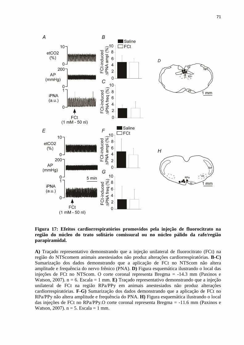
71
Figura 17: Efeitos cardiorrespiratórios promovidos pela injeção de fluorocitrato na
região do núcleo do trato solitário comissural ou no núcleo pálido da rafe/região
parapiramidal.
A) Traçado representativo demonstrando que a injeção unilateral de fluorocitrato (FCt) na
região do NTScomem animais anestesiados não produz alterações cardiorrespiratórias. B-C)
Sumarização dos dados demonstrando que a aplicação de FCt no NTScom não altera
amplitude e frequência do nervo frênico (PNA). D) Figura esquemática ilustrando o local das
injeções de FCt no NTScom. O corte coronal representa Bregma = -14.3 mm (Paxinos e
Watson, 2007). n = 6. Escala = 1 mm. E) Traçado representativo demonstrando que a injeção
unilateral de FCt na região RPa/PPy em animais anestesiados não produz alterações
cardiorrespiratórias. F-G) Sumarização dos dados demonstrando que a aplicação de FCt no
RPa/PPy não altera amplitude e frequência do PNA. H) Figura esquemática ilustrando o local
das injeções de FCt no RPa/PPy.O corte coronal representa Bregma = -11.6 mm (Paxinos e
Watson, 2007). n = 5. Escala = 1 mm.

72
4.4.2 Participação de receptores purinérgicos P2 nas alterações respiratórias promovidos
pela injeção unilateral de fluorocitrato no núcleo retrotrapezóide
O aumento na ventilação promovido pela injeção unilateral de FCt (1 mM - 50 nl) na
região do RTN já havia sido demonstrado préviamente (ERLICHMAN; LI; NATTIE, 1998;
HOLLERAN; BABBIE; ERLICHMAN, 2001). Os autores dos referidos trabalhos
demonstraram que a injeção de FCt em doses sub-tóxicas na região do RTN promove
alterações no pH extracelular mediado por seus efeitos em astrócitos. No entanto, Wenker e
colaboradores (WENKER et al., 2010) demonstraram em preparações in vitro que o FCt é
capaz de promover despolarização da membrana de astrócitos do RTN e aumento da atividade
de neurônios quimiossensíveis vizinhos por um mecanismo dependende da sinalização
purinérgica. Diante do exposto acima, o próximo protocolo experimental foi realizado para
tentar avaliar a participação de receptores purinérgicos P2 na região do RTN nas respostas
respiratórias promovidas pela manipulação farmacológica das células da glia.
Injeções bilaterais de PPADS (3 mM - 50 nl) no RTN não promoveram alterações na
PAM (113 ± 7, vs. salina: 115 ± 4 mmHg, p>0,05) e na amplitude (55 ± 3 vs. salina: 58 ± 6%,
p>0,05) e frequência do nervo frênico (37 ± 4 vs. salina: 42 ± 4 %, p>0,05) basais (Fig. 18A).
A injeção bilateral de PPADS no RTN foi efetiva em reduzir as respostas de aumento de
amplitude (37 ± 3%, vs FCt: 54 ± 5%, p<0,05) e frequência (30 ± 1%, vs FCt: 38 ± 2%,
p<0,05) do nervo frênico induzido pela injeção unilateral de FCt no RTN (Figs. 18A-C).
Importante retratar que a injeção bilateral de PPADS no RTN não modificou o tempo (Δ = 4,2
± 3, vs FCt: 5,5 ± 2 min, p>0,05) que o FCt é capaz de aumentar a atividade respiratória.

73
Figura 18: Injeção bilateral de PPADS no núcleo retrotrapezóide atenua a resposta
respiratória produzida pela injeção unilateral de fluorocitrato no núcleo retrotrapezóide
A) Traçado representativo demonstrando efeitos respiratórios produzidos pela injeção
unilateral de fluorocitrato (FCt: 1 mM - 50 nl) na região do RTN antes e após a injeção
bilateral do antagonista de receptores P2 PPADS (3 mM - 50 nl). Alterações na B) amplitude
e C) frequência do nervo frênico produzidos pela injeção unilateral de FCt na região do RTN
antes e após a injeção bilateral de PPADS no RTN. * Diferente da injeção de salina (p<0,05);
n = 6 ratos.
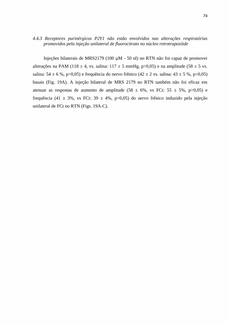
74
4.4.3 Receptores purinérgicos P2Y1 não estão envolvidos nas alterações respiratórias
promovidos pela injeção unilateral de fluorocitrato no núcleo retrotrapezóide
Injeções bilaterais de MRS2179 (100 µM - 50 nl) no RTN não foi capaz de promover
alterações na PAM (118 ± 4, vs. salina: 117 ± 5 mmHg, p>0,05) e na amplitude (58 ± 5 vs.
salina: 54 ± 6 %, p>0,05) e frequência do nervo frênico (42 ± 2 vs. salina: 43 ± 5 %, p>0,05)
basais (Fig. 19A). A injeção bilateral de MRS 2179 no RTN também não foi eficaz em
atenuar as respostas de aumento de amplitude (58 ± 6%, vs FCt: 55 ± 5%, p>0,05) e
frequência (41 ± 3%, vs FCt: 39 ± 4%, p>0,05) do nervo frênico induzido pela injeção
unilateral de FCt no RTN (Figs. 19A-C).

75
Figura 19: Injeção bilateral de MRS 2179 no núcleo retrotrapezóide não altera a
resposta respiratória produzida pela injeção unilateral de fluorocitrato no núcleo
retrotrapezóide.
A) Traçado representativo demonstrando que a injeção bilateral de antagonista P2Y1 MRS
2179 (100 M - 50 nl) não altera a resposta respiratória promovida injeção unilateral de
fluorocitrato (FCt: 1 mM - 50 nl) na região do RTN. Sumário dos dados demonstrando
alterações na B) amplitude e C) frequência do nervo frênico produzidos pela injeção unilateral
de FCt na região do RTN antes e após a injeção bilateral de MRS 2179 no RTN. n = 6 ratos.

5 DISCUSSÃO

77
“Falhar não é uma opção” (FELDMAN; DEL NEGRO; GRAY, 2012). Esta frase
retrata muito bem a importância crucial do sistema respiratório na manutenção da homeostase,
cuja função primordial é a manutenção dos níveis de gases no organismo com o objetivo de
evitar alterações no equilíbrio ácido-base. Neste sentido, os quimiorreceptores (centrais e
periféricos) frequentemente são os principais alvos de grupos de pesquisa que buscam
desvendar os mecanismos dessa complexa organização que controla o sistema
cardiorrespiratório.
O presente trabalho consiste em uma série de 4 estudos, onde buscamos elucidar os
mecanismos de participação da sinalização purinérgica no bulbo ventrolateral rostral (o RTN
e o grupamento catecolaminérgico C1) na geração ou modulação de respostas
cardiorrespiratórias durante a ativação de quimiorreceptores centrais e periféricos, bem como
a participação de células da glia neste processo. Os resultados obtidos são embasados por
evidências prévias da literatura e a nossa contribuição demonstrou que:
1- Alterações cardiorrespiratórias induzidas pela ativação de quimiorreceptores centrais
(hipercapnia) são atenuadas pelo bloqueio de receptores purinérgicos P2 com PPADS no
RTN. Possivelmente, essas respostas devem ser mediadas via receptores purinérgicos P2X,
uma vez que o bloqueio seletivo de receptores purinérgicos P2Y1 no RTN não foi capaz de
alterar as respostas cardiorrespiratórias promovidas pela ativação dos quimiorreceptores
centrais. No entanto, a aplicação de agonista P2Y1 nesta região promoveu aumento da
atividade cardiorrespiratória (WENKER et al., 2012);
2: Por outro lado, observamos que ambos antagonistas purinérgicos, não seletivo
(PPADS) e seletivo para receptores P2Y1 (MRS2179) injetados bilateralmente no bulbo
ventrolateral rostral, foram efetivos em atenuar as respostas cardiorrespiratórias e simpato-
excitatórias produzidas pela estimulação dos quimiorreceptores periféricos (hipóxia
citotóxica). O bloqueio associado entre os receptores purinérgicos e os receptores
glutamatérgicos ionotrópicos foi capaz de reduzir ainda mais o aumento da PAM, atividade
simpática e respiratória promovidas pela estimulação dos quimiorreceptores periféricos,
sugerindo uma possível interação glutamatérgica e purinérgica na região do bulbo
ventrolateral rostral durante a ativação dos quimiorreceptores periféricos. Tratando-se de
sinalização purinérgica no bulbo ventrolateral rostral, experimentos realizados em animais
com depleção de neurônios catecoláminérgicos do grupamento C1 foi possível demontrar que
a resposta simpato-excitatória e pressórica parece ser mediada pelos receptores P2Y1,
localizados exclusivamente nos neurônios C1. Além disso, observamos que terminais axonais
no bulbo vetrolateral rostral provenientes do NTScom expressam VGLUT2 e VNUT,

78
indicando que estes terminais liberam glutamato e, possivelmente purinas (WENKER et al.,
2013);
3- Muito embora o NTScom e o RTN, mas não a região RPa/PPy, responda a aplicação
exógena de ATP com aumento na atividade cardiorrespiratória, a aplicação de antagonista
purinérgico P2 não seletivo no NTScom ou RPa/PPy não foi capaz de alterar a resposta de
ativação dos quimiorreceptores centrais. Essas respostas foram diferentes do observado
previamente com injeções dos antagonistas P2 no RTN, sugerindo que a sinalização
purinérgica parece não modular a quimiossensibilidade ao CO2/pH nestes grupamentos
neurais (SOBRINHO et al., 2014).
4- A injeção da gliotoxina FCt no RTN, de animais anestesiados e vagotomizados, foi
capaz de gerar ou aumentar o padrão inspiratório na atividade do nervo frênico, mediada em
grande parte pela sinalização purinérgica nos receptores P2X, uma vez que a aplicação de
antagonista não seletivo P2, PPADS, mas não o antagonista seletivo P2Y1, MRS2179,
atenuou as respostas respiratórias promovidas pela aplicação de FCt. A injeção de FCt no
NTScom ou na RPa/PPy não foi capaz de promover alterações cardiorrespiratórias
(SOBRINHO et al., 2015, manuscrito em preparação).
5.1 Contribuição da sinalização purinérgica na região do RTN no controle
cardiorrespiratório promovido pela ativação dos quimiorreceptores centrais
O RTN representa o principal grupamento neural com propriedades quimiossensíveis
(CONNELLY; ELLENBERGER; FELDMAN, 1990; GUYENET; BAYLISS, 2015;
GUYENET; STORNETTA; BAYLISS, 2010; MULKEY et al., 2004; NATTIE; GDOVIN;
LI, 1993). Este reconhecimento pode ser atribuído ao fato que, dentre os núcleos encefálicos
que apresentam propriedades quimiossensíveis, este é o grupamento que melhor preenche
todos os critérios para ser considerado uma área quimiorreceptora. As unidades funcionais
presentes na região do RTN apresentam: a) sensibilidade intrínseca ao CO2/H+ in vivo e in
vitro, b) fenótipo neuroquímico excitatório (VGLUT2+), c) os neurônios dessa região
projetam-se para o centro gerador do padrão respiratório, d) recebem influências dos
quimiorreeptores periféricos e dos receptores de distensão pulmonar, e) geneticamente
expressam o fator de transcrição Phox2b e mutações seletivas desse fator de transcrição, na
região do RTN, promove uma síndrome extremamente rara chamada de Síndrome da
Hipoventilação Congênita Central, f) as células gliais da região do RTN parecem apresentar
também uma propriedade quimiossensível, e essa propriedade parece ser exclusiva dessa

79
região do encéfalo (GOURINE et al., 2005a; GUYENET; BAYLISS, 2015; HUCKSTEPP et
al., 2010a; MOREIRA et al., 2015; MULKEY et al., 2004; SOBRINHO et al., 2014;
WENKER et al., 2012).
Variações nas concentrações de CO2/H+ promovem oscilações proporcionais na
atividade de neurônios do RTN in vitro (MULKEY et al., 2004; KUMAR et al., 2015) e in
vivo (MULKEY et al., 2004; STORNETTA et al., 2006; TAKAKURA et al., 2006;
WENKER et al., 2012), nesta última, acompanhada pelo reflexo de aumento da atividade
ventilatória. Por exemplo, em animais anestesiados e ventilados artificialmente, tanto os
neurônios do RTN quanto a atividade do nervo frênico foram silentes abaixo de 4.1% de CO2
(também observado no presente estudo, Fig. 15), apresentaram aumento gradual na frequência
de disparos e da ventilação de acordo com o aumento da PCO2, e atingem em média 14
potenciais de ação por segundo quando estes animais exibem 10% de CO2 no ar expirado
(MULKEY et al., 2004). Lesões ou inativação seletiva do RTN resultam em redução na
ventilação basal, aumento na PCO2 em animais não anestesiados (NATTIE; LI, 2002b;
TAKAKURA et al., 2013, 2014) e atenuação da resposta ventilatória durante a hipercapnia
em animais anestesiados e não anestesiados (NATTIE; LI, 2002b; TAKAKURA et al., 2008,
2014).
O mecanismo pelo qual os neurônios do RTN detectam as variações de CO2/H+
finalmente começa a ser elucidado. Recentemente, foi identificado que receptores de
membrana do tipo GPR4, ligados a proteína G, atuando em sinergia com um canal de K+
sensível a alcalinização (TASK2), são expressos em neurônios do RTN e parecem ser os
mecanismos principais pelo qual os neurônios do RTN percebem variações de CO2 e pH
(KUMAR et al., 2015). Resumidamente, camundongos com depleção ou silenciamento
genético das proteínas GPR4 exibem aumentos consideráveis no número de episódios
espontâneos de apneia e redução da resposta ventilatória para hipercapnia. Esses animais
apresentam ainda redução no número de neurônios que expressam Fos após exposição ao
CO2, indicando redução no número de neurônios ativados pela hipercapnia, bem como
redução em número de neurônios que respondem a acidificação in vitro, e da sensibilidade ao
pH dos neurônios que ainda apresentam resposta residual à acidose. As alterações neurais e
respiratórias são revertidas após a reinserção do receptor GPR4, enquanto que a depleção
simultânea de GPR4 e TASK2 no mesmo animal promoveu alta taxa de mortalidade e a
eliminação quase completa das respostas ventilatórias e neuronais a hipercapnia nos animais
sobreviventes (KUMAR et al., 2015).

80
Somando-se a propriedade intrínseca, os neurônios do RTN respondem a múltiplos
neurotransmissores e neuromoduladores. Muitos estudos tem demonstrado que o RTN é
altamente responsivo a substância P, glutamato, GABA, ATP, orexina, serotonina (5-HT),
acetilcolina, noradrenalina e outros (ERLICHMAN; LEITER; GOURINE, 2010; FUKUDA;
LOESCHCKE, 1979; GOURINE et al., 2010; LAZARENKO et al., 2011; MULKEY et al.,
2004; TAKAKURA et al., 2008; WENKER et al., 2012). Apesar da importante influência
desses neurotransmissores/neuromoduladores na atividade dos neurônios do RTN, a
sensibilidade ao CO2 dessas células quimiossensíveis tende a não ser alterada na presença de
seus antagonistas.
No presente estudo, demonstramos que alterações cardiorrespiratórias induzidas pela
hipercapnia são atenuadas pela aplicação prévia de antagonista de receptores purinérgicos P2,
PPADS, no RTN. Em nossos experimentos, a hipercapnia produziu inicialmente uma
hipotensão, que seria resultado de um efeito direto do CO2 nos vasos sanguíneos e também,
possivelmente, na contratilidade cardíaca. Após a hipotensão, a pressão arterial começa a
voltar aos valores normais e, no fim do episódio da hipercapnia, a pressão arterial apresenta
uma tendência de aumento. Assume-se que esse aumento seria resultante de um aumento na
atividade simpática eferente, quando o efeito local do CO2 nos vasos sanguíneos já tenha sido
removido. A hipercapnia induziu também um aumento na PAM e na atividade respiratória
(MOREIRA et al., 2006; MULKEY et al., 2004; WENKER et al., 2012). Em nossos
resultados, constatamos que o bloqueio de receptores purinérgicos P2 pela injeção bilateral de
PPADS na região RTN reduziu o aumento na PAM e na atividade respiratória (amplitude e
frequência) em animais anestesiados submetidos à hipercapnia, sem alterar os valores basais
(WENKER et al., 2012).
Embora não tenhamos testado toda a diversidade de receptores P2Y, ou mesmo
utilizado um antagonista seletivo para receptores P2X, sugerimos que o receptor atuante neste
contexto pertence ao grupo de receptores purinérgicos ionotrópicos (P2X1-7), uma vez que a
aplicação de antagonista seletivo de receptores purinérgicos P2Y1 no RTN não foi capaz de
alterar as respostas cardiorrespiratórias promovidas pela ativação dos quimiorreceptores
centrais. Não podemos descartar a participação de outros receptores P2Y, pois resultados
recentes mostraram que os receptores P2Y são os receptores purinérgicos mais abundantes na
região do Complexo de Pré-Botzinger e medeiam as respostas respiratórias produzidas pelo
ATP (FUNK, 2013; LORIER et al., 2007, 2008).
O aumento nos níveis de CO2 promove a liberação de ATP na superfície ventral do
bulbo, promovendo um aumento na atividade respiratória (GOURINE et al., 2005a). Através

81
de micro-sensores colocados na superfície ventral do bulbo, foi observado que as áreas onde
ocorre maior concentração de ATP situam-se próximas as áreas classicamente descritas com
propriedades quimiossensíveis, como o RTN e os neurônios serotoninérgicos da Rafe
(GOURINE et al., 2005a). O nosso trabalho confirma e extende os dados da literatura
mostrando que a injeção de ATP diretamente na região do RTN é capaz de promover aumento
da atividade cardiorrespiratória (GOURINE et al., 2005a; SOBRINHO et al., 2014). Ademias
mostramos que tanto o efeito da aplicação exógena de ATP quanto as alterações promovidas
pela hipercapnia podem ser atenuadas pela aplicação prévia de PPADS (GOURINE et al.,
2005a; WENKER et al., 2012; SOBRINHO et al., 2014).
O uso de diferentes modelos experimentais pode ser importante para a completa
elucidação dos processos fisiológicos, uma vez que os resultados obtidos entre eles podem
apresentar algumas divergências. Por exemplo, em trabalhos prévios realizados in vitro,
nossos colaboradores já se contrapuseram em relação a participação da sinalização
purinérgica na resposta a ativação dos quimiorreceptores centrais localizados no RTN
(MULKEY et al., 2006), enquanto que Gourine e colaboradores utilizam tanto modelos de
animais anestesiados quanto experimentos in vitro, defendiam que a que a sinalização
purinérgica, dependente de receptors P2Y na superfície ventrolateral do bulbo, seria
indispensável para as resposta de ativação dos quimiorreceptores centrais e periféricos
(GOURINE et al., 2005a, 2005b, 2010). Onimaru e colaboradores, por outro lado, usaram um
modelo de isolamento do tronco encefálicos de ratos com idade de 1 a 4 dias de vida e foram
taxativos na afirmação que os receptores purinérgicos não são capazes de alterar as respostas
de atividade elétrica dos neurônios da região do RTN promovidas pela hipercapnia
(ONIMARU; IKEDA; KAWAKAMI, 2012). Dessa maneira, parece existir algumas
controvérsias na literatura em um passado muito recente no que diz respeito à atuação da
sinalização purinérgica no controle respiratório.
Nossos dados foram obtidos em animais anestesiados, vagotomizados e ventilados
artificialmente, e nossos achados foram fortemente corroborados pelos resultados obtidos em
outros modelos experimentais utilizados por nosso grupo. In vitro, através do registro
eletrofisiológico da atividade elétrica de neurônios em fatias coronais do bulbo contendo o
RTN (patch-clamp em fatias de encéfalo na configuração “loose-patch”), encontramos que na
presença de antagonistas P2 não seletivos, PPADS ou suramim, mas não os receptores P2Y1
(MRS 2179), o aumento na taxa de disparos dos neurônios do RTN promovido pelo CO2/H+ é
atenuado (WENKER et al., 2012). Da mesma forma, em animais não anestesiados e
submetidos a experimentos de pletismografia de corpo inteiro, a aplicação de PPADS, mas

82
não MRS 2179 no RTN, promoveu redução da resposta ventilatória a hipercapnia (BARNA et
al., 2015, submetido). É interessante mencionar que as porcentagens de inibição obtidas entre
os modelos foram muito semelhantes (~30%).
5.2 Contribuição dos mecanismos purinérgicos na região do bulbo ventrolateral rostral
no controle cardiorrespiratório promovido pela ativação dos quimiorreceptores
periféricos
Hipercapnia induz liberação de ATP na superfície ventral do bulbo (GOURINE et al.,
2005a, 2010), e contribui para a resposta quimiorreceptora através de receptores P2 na região
do RTN (BARNA et al., 2015, submetido; GOURINE et al., 2005a; SOBRINHO et al., 2014;
WENKER et al., 2012). Níveis elevados de ATP nesta região também foram detectados em
resposta a hipóxia (redução na PO2), mesmo em animais com os quimiorreceptores periféricos
desnervados (GOURINE et al., 2005b). Em nossos experimentos, encontramos que o
bloqueio dos receptores P2Y1 com a injeção do antagonista MRS 2179, o aumento na
frequência de disparos de neurônios do RTN in vitro, bem como a resposta cardiorrespiratória
in vivo para CO2/H+ permanecem inalteradas, sugerindo que esta classe de receptores não está
envolvida na quimiorrecepção central, pelo menos na região do RTN (BARNA et al., 2015,
submetido; WENKER et al., 2012).
Na tentativa de reforçar nossa hipótese de que a participação da sinalização purinérgica
nas respostas quimiorreceptoras centrais no RTN são inteiramente mediadas por receptores
P2X e que não há participação de receptores P2Y1, realizamos a injeção do agonista P2Y1,
MRS 2365 no RTN, enquanto registrávamos a atividade de neurônios do RTN in vitro ou a
atividade cardiorrespiratória de animais anestesiados (WENKER et al., 2012, 2013). Como
esperado, a injeção do antagonista de receptores P2Y1, MRS 2179, no RTN, não produziu
efeitos na frequência de potenciais de ação de neurônios quimiossensíveis. No entanto, a
aplicação do agonista P2Y1, MRS2365, no RTN de animais anestesiados promoveu aumento
da PAM e da atividade do nervo frênico, sugerindo que receptores P2Y parecem não
participar das respostas cardiorrespiratórias promovidas pela ativação do quimiorreflexo
central, mas os receptores P2Y1 estão presentes no bulbo ventrolateral e estariam modulando
a atividade cardiorrespiratória em algum outro contexto fisiológico, como o quimioreflexo
periférico (WENKER et al., 2012, 2013). Outra possibilidade que não podemos descartar é
que nossas injeções de agonistas e antagonistas purinérgicos tenham atingido os neurônios
catecolaminérgicos do grupamento C1. Evidências do nosso laboratório apontam para tal

83
evidência, pois experimentos realizados em animais não anestesiados e com lesão do
grupamento catecolaminérgico C1, a injeção de fármacos que atuam em receptores P2Y na
região do RTN não foram eficazes em produzir efeitos cardiorrespiratórios (BARNA et al.,
2015, submetido). Em nossos estudos direcionados ao RTN, frequentemente focamos na
região que circunda o terço caudal do núcleo motor do facial (ventral e caudal), pois nesta
região ocorre a maior densidade de células quimiossensíveis (MULKEY et al., 2004). No
entanto, os neurônios quimiossensíveis do RTN encontrados mais caudais estão localizados
muito próximos de neurônios catecolaminérgicos do grupamento C1 ou mesmo distribuídos
entre os neurônios da porção mais rostral desse grupamento (STORNETTA et al., 2006).
Está bem estabelecido na literatura que os neurônios localizados na região do bulbo
ventrolateral (neurônios C1 bulbo-espinais e neurônios quimiossensíveis do RTN) são
determinantes no controle e na integração de reflexos cardiorrespiratórios, regularmente
atribuindo-se as respostas respiratórias aos neurônios do RTN e a resposta pressora e
simpática aos neurônios C1 (ABBOTT et al., 2012, 2009a; BURKE et al., 2014, 2015;
GUYENET, 2006; ROSIN; CHANG; GUYENET, 2006; STORNETTA et al., 2006;
TAKAKURA et al., 2006). A destruição seletiva dos neurônios bulbo-espinais da região C1
ou dos neurônios quimiosenssíveis do RTN promovem a diminuição das respostas de ativação
dos quimiorreceptores periféricos e centrais, respectivamente (SCHREIHOFER; GUYENET,
2000; TAKAKURA et al., 2014). Ademais, a ativação seletiva dos neurônios
catecolaminérgicos da região C1 (bulbo-espinais) ou dos neurônios Phox2b+
(quimiosenssíveis) com a técnica da optogenética promove aumento da pressão arterial e da
respiração (ABBOTT et al., 2009a, 2009b; BURKE et al., 2015).
No presente trabalho mostramos que a sinalização purinérgica na região do bulbo
ventrolateral rostral tem importante papel para as respostas cardiorrespiratórias produzidas
pela hipóxia. Os nossos achados também são consistentes com o fato de que o ATP é co-
liberado com o glutamato em algumas sinapses, em especial nas sinapses da via do
quimiorreflexo periférico, uma vez que o bloqueio simultâneo de receptores glutamatérgicos
ionotrópicos e receptores P2Y1 produziram alterações mais acentuadas. Além do mais, foram
detectadas varicosidades no bulbo ventrolateral rostral provenientes do NTScom expressam
VGLUT e VNUT, corroborando com trabalhos anteriores que demonstram que a co-
transmissão purinas/glutamato na região do bulbo ventrolateral rostral estaria envolvida na
neurotransmissão do quimiorreflexo periférico (BRAGA et al., 2007; MORAES et al., 2011).

84
Os neurônios bulbo-espinais da região bulbo ventrolateral rostral são ativados pela
aplicação de ATP (SUN; WAHLESTEDT; REIS, 1992) e a injeção de agonistas purinérgicos
nessa região do encéfalo é capaz de promover alterações cardiorrespiratórias em animais
anestesiados (GOURINE et al., 2005a; WENKER et al., 2012; SOBRINHO et al., 2014).
Adicionalmente, a inibição de receptores purinérgicos P2 em regiões mais caudais do bulbo
ventrolateral, mais especificamente nas regiões que englobam o complexo de Bötzinger e pré-
Bötzinger, foram capazes de diminuir a resposta ventilatória, mas não a resposta pressora, da
ativação dos quimiorreceptores periféricos em animais não anestesiados (MORAES et al.,
2011). Em situações de hipóxia, as células glomus do corpúsculo carotídeo liberam ATP para
promover a ativação de receptores purinérgicos P2X2 e P2X3 nas vias aferentes para o SNC,
nas quais fazem a primeira sinapse na região do NTScom (FORD et al., 2015; MACHADO,
2001; PATON et al., 2001). Na região do NTScom, a injeção de ATP reproduz as respostas
cardiorrespiratórias (hipertensão, bradicardia e taquipnéia) encontradas durante a ativação do
quimiorreflexo periférico (DE PAULA et al., 2004). Consistente com a importância da
neurotransmissão purinérgica na via do quimiorreflexo, a injeção do antagonista de receptores
glutamatérgicos, ácido quinurênico, na região do NTS não foi capaz de alterar as respostas
cardiovasculares promovidas pela ativação do quimiorreflexo periférico em animais
acordados ou na preparação in situ (BRAGA et al., 2007), no entanto, o bloqueio simultâneo
de receptores glutamatérgicos e purinérgicos no NTScom foi capaz de reduzir as respostas
cardiorrespiratórias promovidas pela ativação dos quimiorreceptores periféricos (BRAGA et
al., 2007).
Os resultados apresentados sugerem que as respostas cardiorrespiratórias promovidas
pela ativação dos quimiorreceptores periféricos são mediadas em parte na região do bulbo
ventrolateral rostral, por mecanismos dependentes de receptores purinérgicos P2Y1 presentes
nos neurônios C1. Esta hipótese é fundamentada pelos experimentos realizados in vivo em
animais que foram submetidos a lesão seletiva de neurônios C1 com a toxina saporina
conjugada com dopamina-beta-hidroxilase (anti-DH-SAP), em que o aumento da PAM, da
atividade simpática e do nervo frênico produzido pela aplicação de agonista P2Y1 no bulbo
ventrolateral rostral foram bloqueadas. Resultados obtidos in vitro também suportam esta
hipótese, uma vez que neurônios imunorreativos para TH (possivelmente neurônios do
grupamento C1) e insensíveis as variações de CO2/H+, são capazes de despolarizar após a
aplicação do agonista P2Y1. Por outro lado, neurônios não catecolaminérgicos e
imunorreativos para Phox2b (TH-/Phox2b+) e sensíveis as variações de CO2/H+ não alteram
sua atividade após a aplicação do agonista purinérgico P2Y1 (WENKER et al., 2012). A

85
resposta pressora e simpato-excitatória ao KCN também foram atenuadas em animais com
lesão de neurônios C1, mas a resposta respiratória foi inalterada (WENKER et al., 2013).
As observações descritas acima parecem bem consistentes, pois sabe-se que o NTScom
não se projeta exclusivamente para o grupamento C1. Varicosidades oriundas do NTScom
também são encontrados no RTN (TAKAKURA et al., 2006; WENKER et al., 2013) ou em
grupamentos da coluna respiratória ventral (ALHEID; JIAO; MCCRIMMON, 2011). Dessa
maneira, acreditamos que os neurônios bulbo-espinais do grupamento C1 e os neurônios
quimiossensíveis do RTN apresentam comunicações recíprocas como parte da rede neural que
integra informações respiratórias e simpáticas (BURKE et al., 2014; TAKAKURA;
MOREIRA, 2011; TAKAKURA et al., 2011; WENKER et al., 2013).
5.3 Sinalização purinérgica não contribui para quimiorrecepção central na região
comissural do núcleo do trato solitário e na região do núcleo pálido da rafe/região
parapiramidal
Esta bem estabelecido que além do RTN, outras regiões do SNC podem atuar como
quimiorreceptores centrais, como por exemplo o NTScom (NATTIE; LI, 2002b), locus
coeruleus (BIANCARDI et al., 2008) e os núcleos da rafe bulbar (núcleos Magno, Obscuro, e
a região que engloba o núcleo pálido da rafe/região parapiramidal (CORCORAN et al., 2009;
MESSIER; LI; NATTIE, 2002; RICHERSON, 2004).
Como já descrito acima, os receptores purinérgicos do tipo P2 são amplamente
distribuídos no SNC (FUNK, 2013; YAO et al., 2000). Neste sentido, já está bem
estabelecido que a sinalização purinérgica pode atuar ao longo de toda a cadeia neural da via
do quimioreflexo periférico (ALVARES et al., 2014; ANTUNES; BRAGA; MACHADO,
2006; BRAGA et al., 2007; DE PAULA et al., 2004; FUNK, 2013; MORAES et al., 2011;
MOREIRA et al., 2015; PATON et al., 2001; WENKER et al., 2012, 2013), incluindo o
NTScom, onde injeções de agonistas purinérgicos produz alterações cardiorrespiratórias que
mimetizam a ativação dos quimiorreceptores periféricos (ANTUNES; BRAGA; MACHADO,
2006; DE PAULA et al., 2004). As alterações cardiorrespiratórias promovidas pela injeção
de ATP no NTS parecem ser dependentes da ativação de receptores P2 (ANTUNES et al.,
2001; SOBRINHO et al., 2014). No entanto, a aplicação do antagonista P2 PPADS no
NTScom não foi capaz de alterar as respostas cardiorrespiratórias promovidas pela ativação
do quimiorreflexo central (SOBRINHO et al., 2014). Em concordância com nossos achados
in vivo, os neurônios quimiossensíveis do NTScom respondem a aplicação de ATP com
aumento na frequência de potenciais de ação, mas a aplicação de antagonistas P2, PPADS ou

86
suramim, não produziram alterações na resposta ao CO2/H+, sugerindo que a sinalização
purinérgica, na região do NTS, parece não participar da mobilização do quimiorreflexo
central (SOBRINHO et al., 2014).
A sinalização purinérgica pode atuar na região do bulbo ventrolateral rostral como
moduladora de respostas quimiorreceptoras centrais e periféricas (GOURINE et al., 2005a,
2010; WENKER et al., 2012, 2013). Neste contexto, já foi sugerido que a sinalização
purinérgica também pode atuar em outros núcleos quimiossensíveis modulando respostas
ventilatórias. Por exemplo, já foi demonstrado em trabalhos anteriores que antagonistas
purinérgicos atenuam a resposta quimiorreceptora central em porções rostrais dos núcleos da
rafe encontrados no bulbo, mas não nos grupamentos caudais (como o núcleo pálido da rafe)
(DA SILVA et al., 2012). Além disso, injeções de ATP no núcleo pálido da rafe aumentam a
atividade respiratória, enquanto que aplicações no núcleo magno da rafe levaram a redução
(CAO; SONG, 2007).
Utilizando-se uma abordagem muito semelhante, nós não encontramos evidências em
nossos experimentos in vivo e in vitro que a sinalização purinérgica na região da RPa/PPy
possa atuar como moduladora da resposta quimiorreceptora central, como já demonstrado na
região do RTN (SOBRINHO et al., 2014).
A região que engloba o núcleo pálido da rafe e a região parapiramidal, previamente
reportada com propriedades quimiossensíveis está localizada ao mesmo nível rostro-caudal do
RTN (DA SILVA et al., 2012; DEPUY et al., 2011; TAKAKURA; MOREIRA, 2013). Uma
vez que esta estrutura encefálica esta localizada ventralmente no bulbo, é plausível que o ATP
liberado durante a hipercapnia também promova alterações neste sítio. Apesar disso, injeções
de ATP localizadas aproximadamente 1 mm mediais ao RTN não foram capazes de produzir
alterações cardiorrespiratórias. Da mesma maneira, injeções bilaterais de PPADS na RPa/PPy
não alteram as respostas a hipercapnia (SOBRINHO et al., 2014). Experimentos in vitro,
mostraram que neurônios serotoninérgicos da RPa/PPy podem responder de forma distinta a
variações de CO2/H+ (inibidos ou estimulados), no entanto, a atividade de ambas populações
neuronais se mantiveram inalteradas na presença de ATP ou de antagonista purinérgico P2
(SOBRINHO et al., 2014).

87
5.4 Interação glia-neurônio na região do RTN no controle cardiorrespiratório
Neurônios do RTN possuem propriedades intrínsecas para detectar variações de
CO2/H+, as quais envolvem receptores GPR4 e canais do tipo TASK-2 (GUYENET;
BAYLISS, 2015; KUMAR et al., 2015; MULKEY et al., 2004; WANG et al., 2013a, 2013b).
No entanto, como demonstrado no presente estudo, existem várias evidências que a
sinalização purinérgica pode modular parcialmente a quimiorrecepção central nesta região
(GOURINE et al., 2005a, 2010; MOREIRA et al., 2015; SOBRINHO et al., 2014; WENKER
et al., 2012).
Evidências experimentais apontam que o aumento da concentração de ATP observado
na superfície ventral em resposta hipercapnia é mediado por astrócitos (GOURINE et al.,
2005a, 2010; HUCKSTEPP et al., 2010a). Gourine e colaboradores relataram que durante a
hipercapnia, astrócitos da superfície ventral do bulbo apresentam aumento nas concentrações
de Ca2+ intracelular, previamente a liberação de ATP (GOURINE et al., 2010).
O envolvimento de astrócitos com a sinalização purinérgica na região do RTN também
já foi demonstrado em experimentos in vitro, através da adição de CO2 ao líquido cérebro-
espinal artificial (HUCKSTEPP et al., 2010a, 2010b; WENKER et al., 2010). Nesse sentido,
demonstrou-se que 20% das células da glia, localizadas na região do RTN, são capazes de
perceber alterações de pH mediante mecanismos de inibição de canais do tipo Kir 4.1-Kir 5.1
(WENKER et al., 2010). Adicionalmente, evidências experimentais mostraram que a
liberação de ATP, em resposta ao aumento das concentrações de CO2, possivelmente envolve
a ativação de hemicanais de conexinas 26, e esse seria um mecanismo independente da
mobilização de Ca2+ (HUCKSTEPP et al., 2010a). Corroborando com estes resultados, o
bloqueio de hemicanais de conexinas através da aplicação de carbonexolona na região do
RTN foi capaz de reduzir a atividade de neurônios quimiossensíveis nessa região (WENKER
et al., 2012). Adicionalmente, trabalhos recentes reforçaram dados prévios demonstrando que
aumento nas concentrações de ATP na superfície ventral do bulbo não ocorre apenas durante
a hipercapnia, mas também durante hipóxia, e sugerem que os astrócitos dessa região podem
atuar como células quimiossensíveis e estimular a atividade neuronal de neurônios C1,
mediante liberação de ATP e lactato (ANGELOVA et al., 2015; MARINA et al., 2015).
O fluorocitratro é uma toxina que atua seletivamente e reversivelmente em células da
glia. Esta toxina atua inibindo a aconitase, e bloqueia em até 95% a formação de glutamina,
sem afetar a síntese de neurotransmissores como glutamato ou acetilcolina, o que reforça a
hipótese de glio-seletividade (FONNUM; JOHNSEN; HASSEL, 1997; HASSEL et al., 1992).

88
Aqui nós reportamos que a injeção da gliotoxina FCt no RTN de animais anestesiados e
vagotomizados, foi capaz de gerar ou aumentar o padrão inspiratório na atividade do nervo
frênico. Além disso, demonstramos que o aumento na amplitude e frequência respiratória
devido a injeção de FCt no RTN foi significativamente atenuada em animais que receberam
injeções prévias do antagonista de receptores purinérgico P2 no RTN.
Um processo de acidificação encefálica é capaz de promover a despolarização de
células identificadas como astrócitos localizados na surpefície ventrolateral do bulbo
(FUKUDA; HONDA, 1975; WENKER et al., 2010). Erlichman e colaboradores (1998)
observaram, através da microdiálise, que a infusão de FCt na superfície ventrolateral do bulbo
promove queda no pH do parênquima, sugerindo que esta acidose tecidual poderia
despolarizar neurônios quimiossensíveis na região, o que promoveria um aumento da
atividade respiratória. Os autores também acreditam que a infusão de FCt estaria promovendo
um desequilíbrio de íons K+ mediante uma inativação de astrócitos.
Até o presente momento, não podemos propor nenhum mecanismo molecular de como
a injeção de FCt, na região ventrolateral do bulbo, mais especificamente na região do RTN,
estaria promovendo o aumento da atividade respiratória. A acidose local promovida pela
injeção de FCt poderia ser a responsável pelo aumento na atividade neuronal ou mesmo pela
liberação de ATP por astrócitos. No entanto, encontramos que o bloqueio de receptores P2 foi
capaz de atenuar a resposta respiratória ao FCt, corroborando com resultados in vitro e
eliminando que o efeito observado fosse apenas mediado pelo efeito da acidose em neurônios
(WENKER et al., 2010). Também já foi demonstrado que a liberação de ATP por astrócitos é
dependente de CO2 e não de H+, eliminando assim que o efeito observado seja devido aos
efeitos da acidose promovida pelo FCt sobre os astrócitos (HUCKSTEPP et al., 2010a,
2010b). Propomos então que os efeitos observados em resposta a aplicação de FCt no RTN é,
ao menos em parte, devido sua ação direta sobre astrócitos, que leva a despolarização da
membrana dessas células gliais e consequente liberação de ATP.
Em nossos experimentos, a injeção de fluorocitrato em áreas relatadas por exibir
propriedades quimiorreceptoras como o NTScom ou a região RPa/PPy falharam em promover
alterações cardiorrespiratórias, enfatizando a hipótese que mecanismos intrínsecos de
quimiorrecepção central envolvendo células da glia possam ser restritos a superfície
ventrolateral do bulbo, em especial no RTN (SOBRINHO et al., 2015, em preparação).
Suportando estes resultados, diferentemente da supericie ventral do bulbo, a acidificação no
líquido cerebroespinal, utilizada em experimentos in vitro, não promoveu oscilações de Ca2+
em astrócitos de outras áreas do SNC, como por exemplo o córtex cerebral ou de fatias de

89
regiões mais dorsais do tronco encefálico (GOURINE et al., 2010). Recentemente, foi
demonstrado através da técnica conhecida como preparação coração-tronco cerebral isolado
(preparação in situ), que permite o registro simultâneo da atividade de diferentes nervos em
animais livres de anestesia, que aplicação de FCt em diferentes regiões do NTS não
promoveram alterações na atividade do nervo frênico (COSTA; MORAES; MACHADO,
2013). No entanto, a aplicação de FCt em sítios da coluna respiratória ventral localizados
próximos ao RTN (PreBötzinger/Bötzinger) promoveram aumento da frequência, o qual os
autores correlacionaram com a redução no tempo da fase expiratória (COSTA; MORAES;
MACHADO, 2013). Em ambos os casos, não foram observadas alterações na atividade
simpática (COSTA; MORAES; MACHADO, 2013), que auxiliam a entender a ausência de
alterações nos registros de PAM de nossos experimentos.

6 CONCLUSÃO

91
A região do bulbo ventrolateral rostral contém duas populações distintas de neurônios
que são importantes na modulação de reflexos cardiorrespiratórios. Neurônios com
propriedades quimiossensíveis do RTN, os quais possuem propriedades intrínsecas para
detecção de CO2/H+ e neurônios bulbo-espinais catecolaminérgicos do grupamento C1 que
modulam o tônus simpático. Esses grupamentos apresentam projeções reciprocas e integram
reflexos diversos, pois recebem informação de quimiorreceptores periféricos via projeções do
NTScom (MOREIRA et al., 2015).
Essa mesma região contém uma população de células gliais (astrócitos) que também são
sensíveis as variações de CO2 e possivelmente de O2, e ao que tudo indica podem modular a
atividade neuronal de neurônios vizinhos através da liberação de ATP que ativaria os
receptores P2 expressos em neurônios do RTN. Por outro lado, a via glutamatérgica clássica
entre o NTScom e o bulbo ventrolateral rostral, responsável por grande parte da interação de
respostas reflexas durante a ativação de quimiorreceptores periféricos e centrais, também
utiliza de purinas como molécula sinalizadora, promovendo co-transmissão com o glutamato
através de receptores P2Y1 expressos em neurônios C1 e receptores P2 no RTN. Este
processo aparentemente se restringe a estes grupamentos localizados próximos a superfície
ventral do bulbo (Figs. 20A-B).

92

93
Figura 20: Conclusão
A: Figura esquemática ilustrando a organização dos grupamentos respiratórios, suas conexões
e os quimiorreceptores centrais e periféricos. A ativação de quimiorreceptores centrais ou
periféricos promove massiva ativação da coluna respiratória ventral, tendo como
consequência o aumento na ventilação e da atividade simpática causado pela estimulação do
grupamento C1. B: Figura esquemática de uma ampliação da área delimitada pelo quadrado
na figura A, ilustrando conexões reciprocas entre os neurônios do RTN e do grupamento C1.
Note que a porção caudal do RTN (em vermelho) se sobrepõe parcialmente (em laranja) com
o grupamento C1 (em amarelo). Os neurônios Phox2b do RTN (representados em verde)
possuem mecanismos intrínsecos para detecção de ao CO2/H+ (GPR4 e TASK-2), mas
também são estimulados por ATP, liberado por astrócitos durante a hipercapnia,
provavelmente via receptores P2X. A ativação dos neurônios do RTN leva a ativação de
neurônios catecolaminérgicos do grupamento C1 (representados em cinza), possivelmente
através da ação de receptores glutamatérgicos metabotrópicos. Por outro lado, a ativação de
quimiorreceptores periféricos promove a ativação de neurônios do RTN via projeções
oriundas de neurônios do NTScom, mas também via projeções de neurônios C1, o qual é
ativado pela ação do glutamato e do ATP, liberado pelos terminais axonais provenientes do
NTScom em receptores iGLU e P2Y1, respectivamente.

94
REFERÊNCIAS*
ABBOTT, S. B. et al. C1 neurons excite locus coeruleus and A5 noradrenergic neurons along
with sympathetic outflow in rats. The Journal of Physiology, v. 590, p. 2897–2915, 2012.
ABBOTT, S. B. G. et al. Photostimulation of retrotrapezoid nucleus phox2b-expressing
neurons in vivo produces long-lasting activation of breathing in rats. The Journal of
Neuroscience, v. 29, n. 18, p. 5806–5819, 2009a.
ABBOTT, S. B. G. et al. Photostimulation of channelrhodopsin-2 expressing ventrolateral
medullary neurons increases sympathetic nerve activity and blood pressure in rats. The
Journal of Physiology, v. 587, n. 23, p. 5613–5631, 2009b.
ABBRACCHIO, M. P. et al. Purinergic signalling in the nervous system: an overview.
Trends in Neurosciences, v. 32, n. 1, p. 19–29, 2009.
ABDALA, A. P. L. et al. Abdominal expiratory activity in the rat brainstem-spinal cord in
situ: patterns, origins and implications for respiratory rhythm generation. The Journal of
Physiology, v. 587, n. 14, p. 3539–3559, 2009.
AICHER, S. A. et al. Monosynaptic projections from the nucleus tractus solitarii to C1
adrenergic neurons in the rostral ventrolateral medulla: Comparison with input from the
caudal ventrolateral medulla. Journal of Comparative Neurology, v. 373, n. 1, p. 62–75,
1996.
ALHEID, G. F.; JIAO, W.; MCCRIMMON, D. R. Caudal nuclei of the rat nucleus of the
solitary tract differentially innervate respiratory compartments within the ventrolateral
medulla. Neuroscience, v. 190, p. 207–27, 2011.
ALVARES, T. S. et al. P2Y1 receptor-mediated potentiation of inspiratory motor output in
neonatal rat in vitro. The Journal of Physiology, v. 592, n. 14, p. 3089–3111, 2014.
AMIEL, J. et al. Polyalanine expansion and frameshift mutations of the paired-like homeobox
gene PHOX2B in congenital central hypoventilation syndrome. Nature Genetics, v. 33, n. 4,
p. 459–461, 2003.
ANGELOVA, P. R. et al. Functional Oxygen Sensitivity of Astrocytes. Journal of
Neuroscience, v. 35, n. 29, p. 10460–10473, 2015.
ANTUNES, V. R.; BRAGA, V. A.; MACHADO, B. H. Autonomic and respiratory responses
to microinjection of ATP into the intermediate or caudal nucleus tractus solitarius in the
working heart-brainstem preparation of the rat. Clinical and Experimental Pharmacology
& Physiology, v. 32, n. 5-6, p. 467–472, 2006.
* De acordo com:
ASSOCIAÇÃO BRASILEIRA DE NORMAS TÉCNICAS. NBR 6023: informação e documentação:
referências: elaboração. Rio de Janeiro, 2002.

95
BARNA, B. F.; TAKAKURA, A. C.; MOREIRA, T. S. Pontomedullary and hypothalamic
distribution of Fos-like immunoreactive neurons after acute exercise in rats. Neuroscience, v.
212, p. 120–130, 2012.
BARNA, B. F.; TAKAKURA, A. C.; MOREIRA, T. S. Acute exercise-induced activation of
Phox2b-expressing neurons of the retrotrapezoid nucleus in rats may involve the
hypothalamus. Neuroscience, v. 258, p. 355–363, 2014.
BARROS, R. C. H. et al. Cardiovascular responses to chemoreflex activation with potassium
cyanide or hypoxic hypoxia in awake rats. Autonomic Neuroscience : Basic & Clinical, v.
97, n. 2, p. 110–115, 2002.
BASTING, T. M. et al. Hypoxia Silences Retrotrapezoid Nucleus Respiratory
Chemoreceptors via Alkalosis. The Journal of Neuroscience, v. 35, n. 2, p. 527–543, 2015.
BIANCARDI, V. et al. Locus coeruleus noradrenergic neurons and CO2 drive to breathing.
Pflugers Archiv European Journal of Physiology, v. 455, n. 6, p. 1119–1128, 2008.
BISCOE, T. J.; DUCHEN, M. R. Cellular basis of transduction in carotid chemoreceptors.
The American Journal of Physiology, v. 258, n. 6, pt. 1, p. 271–278, 1990a.
BISCOE, T. J.; DUCHEN, M. R. Responses of type I cells dissociated from the rabbit carotid
body to hypoxia. The Journal of Physiology, v. 428, p. 39–59, 1990b.
BRAGA, V. A et al. Involvement of L-glutamate and ATP in the neurotransmission of the
sympathoexcitatory component of the chemoreflex in the commissural nucleus tractus solitarii
of awake rats and in the working heart-brainstem preparation. The Journal of Physiology, v.
581, n. 3, p. 1129–1145, 2007.
BURKE, P. G. R. et al. Optogenetic Stimulation of Adrenergic C1 Neurons Causes Sleep
State–Dependent Cardiorespiratory Stimulation and Arousal with Sighs in Rats. American
Journal of Respiratory and Critical Care Medicine, v. 190, n. 11, p. 1301–1310, 2014.
BURKE, P. G. R. et al. State-dependent control of breathing by the retrotrapezoid nucleus.
The Journal of Physiology, v. 13, p. 2909–2926, 2015.
BURNSTOCK, G. Purinergic signalling. British Journal of Pharmacology, p. 172–181,
2006a.
BURNSTOCK, G. Historical review: ATP as a neurotransmitter. Trends in
Pharmacological Sciences, v. 27, n. 3, p. 166–176, 2006b.
BURNSTOCK, G. Physiology and pathophysiology of purinergic neurotransmission.
Physiological Reviews, v. 87, n. 2, p. 659–797, 2007.
BURNSTOCK, G.; FREDHOLM, B. B.; VERKHRATSKY, A. Adenosine and ATP
receptors in the brain. Current Topics in Medicinal Chemistry, v. 11, n. 8, p. 973–1011,
2011.

96
CAO, Y.; SONG, G. Purinergic modulation of respiration via medullary raphe nuclei in rats.
Respiratory Physiology & Neurobiology, v. 155, n. 2, p. 114–120, 2007.
CASTRO, D. D.; LIPSKI, J.; KANJHAN, R. Electrophysiological study of dorsal respiratory
neurons in the medulla oblongata of the rat. Brain Research, v. 639, n. 1, p. 49–56, 1994.
CEZARIO, A. F. et al. Hypothalamic sites responding to predator threats - The role of the
dorsal premammillary nucleus in unconditioned and conditioned antipredatory defensive
behavior. European Journal of Neuroscience, v. 28, n. 5, p. 1003–1015, 2008.
CHESLER, M. Regulation and modulation of pH in the brain. Physiological Reviews, v. 83,
n. 4, p. 1183–1221, 2003.
CONNELLY, C. A.; ELLENBERGER, H. H.; FELDMAN, J. L. Respiratory activity in
retrotrapezoid nucleus in cat. The American Journal of Physiology, v. 258, n. 2, pt. 1, p.
33–44, 1990.
CORCORAN, A. E. et al. Medullary serotonin neurons and central CO2 chemoreception.
Respiratory Physiology and Neurobiology, v. 168, n. 1-2, p. 49–58, 2009.
COSTA, K. M.; MORAES, D. J. A.; MACHADO, B. H. Acute inhibition of glial cells in the
NTS does not affect respiratory and sympathetic activities in rats exposed to chronic
intermittent hypoxia. Brain Research, v. 1496, p. 36–48, 2013.
CREAM, C.; LI, A.; NATTIE, E. The retrotrapezoid nucleus (RTN): local cytoarchitecture
and afferent connections. Respiratory physiology & neurobiology, v. 130, n. 2, p. 121–137,
2002.
DA SILVA, G. S. F. et al. Purinergic transmission in the rostral but not caudal medullary
raphe contributes to the hypercapnia-induced ventilatory response in unanesthetized rats.
Respiratory Physiology and Neurobiology, v. 184, n. 1, p. 41–47, 2012.
DAMPNEY, R. A. Functional organization of central pathways regulating the cardiovascular
system. Physiological Reviews, v. 74, n. 2, p. 323–364, 1994.
DAMPNEY, R. A. L. et al. Medullary and supramedullary mechanisms regulating
sympathetic vasomotor tone. Acta Physiologica Scandinavica, v. 177, n. 3, p. 209–218,
2003.
DE PAULA, P. M. et al. Cardiovascular responses to microinjection of ATP into the nucleus
tractus solitarii of awake rats. American Journal of Physiology, v. 287, n. 5, p. R1164–
R1171, 2004.
DENG, B.-S. et al. Contribution of orexin in hypercapnic chemoreflex: evidence from genetic
and pharmacological disruption and supplementation studies in mice. Journal of Applied
Physiology, v. 103, n. 5, p. 1772–1779, 2007.
DEPUY, S. D. et al. Control of breathing by raphe obscurus serotonergic neurons in mice.
The Journal of Neuroscience, v. 31, p. 1981–1990, 2011.

97
DOBBINS, E. G.; FELDMAN, J. L. Brainstem network controlling descending drive to
phrenic motoneurons in rat. The Journal of Comparative Neurology, v. 347, n. 1, p. 64–86,
1994.
ERLICHMAN, J. S. et al. Heterogeneous patterns of pH regulation in glial cells in the dorsal
and ventral medulla. American Journal of Physiology, v. 286, n. 2, p. 289–302, 2004.
ERLICHMAN, J. S.; LEITER, J. C.; GOURINE, A. V. ATP, glia and central respiratory
control. Respiratory Physiology and Neurobiology, v. 173, n. 3, p. 305–311, 2010.
ERLICHMAN, J. S.; LI, A.; NATTIE, E. E. Ventilatory effects of glial dysfunction in a rat
brain stem chemoreceptor region. Journal Of Applied Physiology, v. 85, n. 5, p. 1599–1604,
1998.
FAVERO, M. T. et al. Chemosensory control by commissural nucleus of the solitary tract in
rats. Respiratory Physiology and Neurobiology, v. 179, n. 2-3, p. 227–234, 2011.
FELDBERG, W.; GUERTZENSTEIN, P. G. A vasodepressor effect of pentobarbitone
sodium. The Journal of Physiology, v. 224, n. 1, p. 83–103, 1972.
FELDMAN, J. L.; DEL NEGRO, C. A. Looking for inspiration: new perspectives on
respiratory rhythm. Nature Reviews, v. 7, n. 3, p. 232–242, 2006.
FELDMAN, J. L.; DEL NEGRO, C. A.; GRAY, P. A. Understanding the Rhythm of
Breathing: So Near, Yet So Far. Annual Review of Physiology, v. 75, n. 1, p. 423-452, 2012.
FELDMAN, J. L.; MITCHELL, G. S.; NATTIE, E. E. Breathing: rhythmicity, plasticity,
chemosensitivity. Annual Review of Neuroscience, v. 26, p. 239–266, 2003.
FELLIN, T. Communication between neurons and astrocytes: Relevance to the modulation of
synaptic and network activity, Journal of Neurochemistry, v. 108, p. 533-544, 2009.
FINLEY, J. C.; KATZ, D. M. The central organization of carotid body afferent projections to
the brainstem of the rat. Brain Research, v. 572, p. 108–116, 1992.
FITZGERALD, R. S.; SHIRAHATA, M.; ISHIZAWA, Y. The presynaptic component of a
cholinergic mechanism in the carotid body chemotransduction of hypoxia in the cat.
Advances in Experimental Medicine and Biology, v. 410, p. 245–252, 1996.
FONNUM, F.; JOHNSEN, A.; HASSEL, B. Use of fluorocitrate and fluoroacetate in the
study of brain metabolism. Glia, v. 21, n. 1, p. 106–113, 1997.
FORD, A. P. et al. P2X3 receptors and sensitization of autonomic reflexes. Autonomic
Neuroscience : Basic & Clinical, v. 191, p. 16–24, 2015.
FORTUNA, M. G. et al. Botzinger expiratory-augmenting neurons and the parafacial
respiratory group. The Journal of Neuroscience, v. 28, n. 10, p. 2506–2515, 2008.
FUKUDA, Y.; HONDA, Y. pH-sensitive cells at ventro--lateral surface of rat medulla
oblongata. Nature, v. 256, n. 5515, p. 317–318, 1975.

98
FUKUDA, Y.; LOESCHCKE, H. H. Effect of H+ on spontaneous neuronal activity in the
surface layer of the rat medulla oblongata in vitro. Pflügers Archiv European Journal of
Physiology, v. 371, p. 125–134, 1977.
FUKUDA, Y.; LOESCHCKE, H. H. A cholinergic mechanism involved in the neuronal
excitation by H+ in the respiratory chemosensitive structures of the ventral medulla oblongata
of rats in vitro. Pflügers Archiv European Journal of Physiology, v. 379, p. 125–135, 1979.
FUNK, G. D. Neuromodulation: Purinergic signaling in respiratory control. Comprehensive
Physiology, v. 3, n. 1, p. 331–363, 2013.
GOURINE, A. V et al. ATP is a mediator of chemosensory transduction in the central
nervous system. Nature, v. 436, p. 108–111, 2005a.
GOURINE, A. V et al. Release of ATP in the ventral medulla during hypoxia in rats: role in
hypoxic ventilatory response. The Journal of Neuroscience , v. 25, n. 5, p. 1211–1218,
2005b.
GOURINE, A. V et al. Astrocytes control breathing through pH-dependent release of ATP.
Science, v. 329, n. 5991, p. 571–575, 2010.
GOURINE, A. V.; WOOD, J. D.; BURNSTOCK, G. Purinergic signalling in autonomic
control. Trends in Neurosciences, v. 32, n. 5, p. 241–248, 2009.
GRANATA, A. R. Rostral ventrolateral medulla descending neurons excited by nucleus
tractus solitarii inputs. Brain Research, v. 648, n. 2, p. 299–305, 1994.
GUYENET, P. G.; Stornetta, R. L.; Weston, M. C.; McQuiston, T.; Simmons, J. R.;
Detection of amino acid and peptide transmitters in physiologically identified brainstem
cardiorespiratory neurons. Autonomic Neuroscience : Basic & Clinical, v. 114, n. 1-2, p. 1–
10, 2004.
GUYENET, P. G. The sympathetic control of blood pressure. Nature Reviews.
Neuroscience, v. 7, n. 5, p. 335–346, 2006.
GUYENET, P. G.; BAYLISS, D. A.; MULKEY, D. K.; STORNETTA, R. L.; MOREIRA T.
S.; TAKAKURA, A. T. The retrotrapezoid nucleus and central chemoreception. Advances in
Experimental Medicine and Biology, v. 605, p. 327-332, 2008.
GUYENET, P. G.; STORNETTA, R. L.; BOCHORISHVILI. G.; DEPUY, S. D.; BURKE, P.
G.; ABBOTT, S. B. C1 neurons: the body’s EMTs. American Journal of Physiology, v. 305,
n. 3, p. 187–204, 2013.
GUYENET, P. G.; BAYLISS, D. A. Neural Control of Breathing and CO2 Homeostasis.
Neuron, v. 87, n. 5, p. 946–961, 2015.
GUYENET, P. G.; KOSHIYA, N. Working model of the sympathetic chemoreflex in rats.
Clinical and Experimental Hypertension, n. 1-2, p. 167–179, 1995.
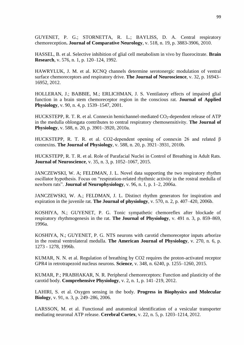
99
GUYENET, P. G.; STORNETTA, R. L.; BAYLISS, D. A. Central respiratory
chemoreception. Journal of Comparative Neurology, v. 518, n. 19, p. 3883-3906, 2010.
HASSEL, B. et al. Selective inhibition of glial cell metabolism in vivo by fluorocitrate. Brain
Research, v. 576, n. 1, p. 120–124, 1992.
HAWRYLUK, J. M. et al. KCNQ channels determine serotonergic modulation of ventral
surface chemoreceptors and respiratory drive. The Journal of Neuroscience, v. 32, p. 16943–
16952, 2012.
HOLLERAN, J.; BABBIE, M.; ERLICHMAN, J. S. Ventilatory effects of impaired glial
function in a brain stem chemoreceptor region in the conscious rat. Journal of Applied
Physiology, v. 90, n. 4, p. 1539–1547, 2001.
HUCKSTEPP, R. T. R. et al. Connexin hemichannel-mediated CO2-dependent release of ATP
in the medulla oblongata contributes to central respiratory chemosensitivity. The Journal of
Physiology, v. 588, n. 20, p. 3901–3920, 2010a.
HUCKSTEPP, R. T. R. et al. CO2-dependent opening of connexin 26 and related β
connexins. The Journal of Physiology, v. 588, n. 20, p. 3921–3931, 2010b.
HUCKSTEPP, R. T. R. et al. Role of Parafacial Nuclei in Control of Breathing in Adult Rats.
Journal of Neuroscience, v. 35, n. 3, p. 1052–1067, 2015.
JANCZEWSKI, W. A; FELDMAN, J. L. Novel data supporting the two respiratory rhythm
oscillator hypothesis. Focus on “respiration-related rhythmic activity in the rostral medulla of
newborn rats”. Journal of Neurophysiology, v. 96, n. 1, p. 1–2, 2006a.
JANCZEWSKI, W. A.; FELDMAN, J. L. Distinct rhythm generators for inspiration and
expiration in the juvenile rat. The Journal of physiology, v. 570, n. 2, p. 407–420, 2006b.
KOSHIYA, N.; GUYENET, P. G. Tonic sympathetic chemoreflex after blockade of
respiratory rhythmogenesis in the rat. The Journal of Physiology, v. 491 n. 3, p. 859–869,
1996a.
KOSHIYA, N.; GUYENET, P. G. NTS neurons with carotid chemoreceptor inputs arborize
in the rostral ventrolateral medulla. The American Journal of Physiology, v. 270, n. 6, p.
1273 - 1278, 1996b.
KUMAR, N. N. et al. Regulation of breathing by CO2 requires the proton-activated receptor
GPR4 in retrotrapezoid nucleus neurons. Science, v. 348, n. 6240, p. 1255–1260, 2015.
KUMAR, P.; PRABHAKAR, N. R. Peripheral chemoreceptors: Function and plasticity of the
carotid body. Comprehensive Physiology, v. 2, n. 1, p. 141–219, 2012.
LAHIRI, S. et al. Oxygen sensing in the body. Progress in Biophysics and Molecular
Biology, v. 91, n. 3, p. 249–286, 2006.
LARSSON, M. et al. Functional and anatomical identification of a vesicular transporter
mediating neuronal ATP release. Cerebral Cortex, v. 22, n. 5, p. 1203–1214, 2012.

100
LAZARENKO, R. M. et al. Orexin A activates retrotrapezoid neurons in mice. Respiratory
Physiology and Neurobiology, v. 175, p. 283–287, 2011.
LORIER, A. R. et al. P2Y1 receptor modulation of the pre-Bötzinger complex inspiratory
rhythm generating network in vitro. The Journal of Neuroscience, v. 27, n. 5, p. 993–1005,
2007.
LORIER, A. R. et al. ATP sensitivity of preBötzinger complex neurones in neonatal rat in
vitro: mechanism underlying a P2 receptor-mediated increase in inspiratory frequency. The
Journal of Physiology, v. 586, n. 5, p. 1429–1446, 2008.
MACHADO, B. H. Neurotransmission of the cardiovascular reflexes in the nucleus tractus
solitarii of awake rats. Annals of the New York Academy of Sciences, v. 940, p. 179–196,
2001.
MARINA, N. et al. Essential role of Phox2b-expressing ventrolateral brainstem neurons in
the chemosensory control of inspiration and expiration. The Journal of neuroscience : the
official journal of the Society for Neuroscience, v. 30, n. 37, p. 12466–12473, 2010.
MARINA, N. et al. Brainstem hypoxia contributes to the development of hypertension in the
spontaneously hypertensive rat. Hypertension, v. 65, n. 4, p. 775–783, 2015.
MARTINO, P. F. et al. Lesions in the cerebellar fastigial nucleus have a small effect on the
hyperpnea needed to meet the gas exchange requirements of submaximal exercise. Journal of
Applied Physiology, v. 101, n. 4, p. 1199–1206, 2006a.
MARTINO, P. F. et al. CO2/H+ chemoreceptors in the cerebellar fastigial nucleus do not
uniformly affect breathing of awake goats. Journal of Applied Physiology, v. 101, n. 1, p.
241–248, 2006b.
MESSIER, M. L.; LI, A.; NATTIE, E. E. Muscimol inhibition of medullary raphe neurons
decreases the CO2 response and alters sleep in newborn piglets. Respiratory Physiology and
Neurobiology, v. 133, n. 3, p. 197–214, 2002.
MESSIER, M. L.; LI, A.; NATTIE, E. E. Inhibition of medullary raphe serotonergic neurons
has age-dependent effects on the CO2 response in newborn piglets. Journal of Applied
Physiology, v. 96, n. 5, p. 1909–1919, 2004.
MOLKOV, Y. I. et al. Late-expiratory activity: emergence and interactions with the
respiratory CpG. Journal of Neurophysiology, v. 104, n. 5, p. 2713–2729, 2010.
MORAES, D. J. A. et al. Modulation of respiratory responses to chemoreflex activation by L-
glutamate and ATP in the rostral ventrolateral medulla of awake rats. American Journal of
Physiology, v. 300, n. 6, p. R1476–R1486, 2011.
MOREIRA, T. S. et al. Role of pressor mechanisms from the NTS and CVLM in control of
arterial pressure. American Journal of Physiology., v. 289, n. 5, p. 1416–1425, 2005.

101
MOREIRA, T. S. et al. Central chemoreceptors and sympathetic vasomotor outflow. The
Journal of Physiology, v. 577, n. 1, p. 369–386, 2006.
MOREIRA, T. S. et al. Inhibitory input from slowly adapting lung stretch receptors to
retrotrapezoid nucleus chemoreceptors. The Journal of Physiology, v. 580, n. 1, p. 285–300,
2007.
MOREIRA, T. S. et al. Central chemoreceptors and neural mechanisms of cardiorespiratory
control. Brazilian Journal of Medical and Biological Research, v. 44, n. 9, p. 883–889,
2011.
MOREIRA, T. S. et al. Independent purinergic mechanisms of central and peripheral
chemoreception in the rostral ventrolateral medulla. The Journal of Physiology, v. 593, n. 5,
p. 1067–1074, 2015.
MOTTA, S. C. et al. Dissecting the brain’s fear system reveals the hypothalamus is critical
for responding in subordinate conspecific intruders. Proceedings of the National Academy
of Sciences of the United States of America, v. 106, n. 12, p. 4870–4875, 2009.
MULKEY, D. K. et al. Respiratory control by ventral surface chemoreceptor neurons in rats.
Nature Neuroscience, v. 7, p. 1360–1369, 2004.
MULKEY, D. K. et al. Purinergic P2 receptors modulate excitability but do not mediate pH
sensitivity of RTN respiratory chemoreceptors. The Journal of Neuroscience, v. 26, n. 27, p.
7230–7233, 2006.
MULKEY, D. K. et al. TASK channels determine pH sensitivity in select respiratory neurons
but do not contribute to central respiratory chemosensitivity. The Journal of Neuroscience,
v. 27, p. 14049–14058, 2007.
MULKEY, D. K.; WENKER, I. C. Astrocyte chemoreceptors: mechanisms of H+ sensing by
astrocytes in the retrotrapezoid nucleus and their possible contribution to respiratory drive.
Experimental Physiology, v. 96, n. 4, p. 400–406, 2011.
NATTIE, E. Julius H. Comroe, Jr., distinguished lecture: central chemoreception: then ... and
now. Journal of Applied Physiology, v.110, n. 1, p. 1-8, 2011.
NATTIE, E. E.; GDOVIN, M.; LI, A. Retrotrapezoid nucleus glutamate receptors: control of
CO2-sensitive phrenic and sympathetic output. Journal of Applied Physiology, v. 74, p.
2958–2968, 1993.
NATTIE, E. E.; LI, A. Retrotrapezoid nucleus lesions decrease phrenic activity and CO2
sensitivity in rats. Respiration Physiology, v. 97, n. 1, p. 63–77, 1994.
NATTIE, E. E.; LI, A. CO2 dialysis in the medullary raphe of the rat increases ventilation in
sleep. Journal of Applied Physiology, v. 90, p. 1247–1257, 2001.
NATTIE, E. E.; LI, A. Substance P-saporin lesion of neurons with NK1 receptors in one
chemoreceptor site in rats decreases ventilation and chemosensitivity. The Journal of
Physiology, v. 544, n. 2, p. 603–616, 2002a.

102
NATTIE, E. E.; LI, A. CO2 dialysis in nucleus tractus solitarius region of rat increases
ventilation in sleep and wakefulness. Journal of Applied Physiology, v. 92, n. 5, p. 2119–
2130, 2002b.
NATTIE, E. E.; LI, A. H.; ST JOHN, W. M. Lesions in retrotrapezoid nucleus decrease
ventilatory output in anesthetized or decerebrate cats. Journal of Applied Physiology, v. 71,
n. 4, p. 1364–1375, 1991.
NATTIE, E.; LI, A. Muscimol dialysis into the caudal aspect of the Nucleus tractus solitarii
of conscious rats inhibits chemoreception. Respiratory Physiology and Neurobiology, v.
164, n. 3, p. 394–400, 2008.
NICHOLS, N. L. et al. Characterization of the chemosensitive response of individual solitary
complex neurons from adult rats. American Journal of Physiology, v. 296, n. 3, p. 763–773,
2009.
OKADA, Y. et al. Anatomical arrangement of hypercapnia-activated cells in the superficial
ventral medulla of rats. Journal of Applied Physiology, v. 93, n. 2, p. 427–439, 2002.
ONIMARU, H.; IKEDA, K.; KAWAKAMI, K. Postsynaptic mechanisms of CO2 responses
in parafacial respiratory neurons of newborn rats. The Journal of Physiology, v. 590, pt. 7, p.
1615–1624, 2012.
PAGLIARDINI, S. et al. Active expiration induced by excitation of ventral medulla in adult
anesthetized rats. The Journal of Neuroscience, v. 31, n. 8, p. 2895–2905, 2011.
PATON, J. F. et al. Properties of solitary tract neurones responding to peripheral arterial
chemoreceptors. Neuroscience, v. 105, n. 1, p. 231–48, 2001.
PRABHAKAR, N. R. Sensing hypoxia: physiology, genetics and epigenetics. The Journal of
Physiology, v. 591, n. 9, p. 2245–2257, 2013.
PUTNAM, R. W.; FILOSA, J. A.; RITUCCI, N. A. Cellular mechanisms involved in CO2
and acid signaling in chemosensitive neurons. American journal of Physiology, v. 287, n. 6,
p. 1493–1526, 2004.
RICHERSON, G. B. Serotonergic neurons as carbon dioxide sensors that maintain pH
homeostasis. Nature Reviews, v. 5, p. 449–461, 2004.
ROSIN, D. L.; CHANG, D. A.; GUYENET, P. G. Afferent and efferent connections of the rat
retrotrapezoid nucleus. Journal of Comparative Neurology, v. 499, n. 1, p. 64–89, 2006.
ROSS, C. A. et al. Rostral ventrolateral medulla: selective projections to the thoracic
autonomic cell column from the region containing C1 adrenaline neurons. The Journal of
Comparative Neurology, v. 228, n. 2, p. 168–185, 1984.
SATO, M.; SEVERINGHAUS, J. W.; BASBAUM, A. I. Medullary CO2 chemoreceptor
neuron identification by c-fos immunocytochemistry. Journal of Applied Physiology, v. 73,
n. 1, p. 96–100, 1992.

103
SAWADA, K. et al. Identification of a vesicular nucleotide transporter. Proceedings of the
National Academy of Sciences of the United States of America, v. 105, n. 15, p. 5683–
5686, 2008.
SCHREIHOFER, A. M.; GUYENET, P. G. Identification of C1 presympathetic neurons in rat
rostral ventrolateral medulla by juxtacellular labeling in vivo. Journal of Comparative
Neurology, v. 387, n. 4, p. 524–536, 1997.
SCHREIHOFER, A. M.; GUYENET, P. G. Sympathetic reflexes after depletion of
bulbospinal catecholaminergic neurons with anti-DbetaH-saporin. American Journal Of
Physiology, v. 279, n. 2, p. 729–742, 2000.
SEMENZA, G. L. Oxygen sensing, homeostasis, and disease. The New England Journal of
Medicine, v. 365, n. 6, p. 537–547, 2011.
SEVERSON, C. A et al. Midbrain serotonergic neurons are central pH chemoreceptors.
Nature Neuroscience, v. 6, n. 11, p. 1139–1140, 2003.
SMITH, C. A. et al. Response time and sensitivity of the ventilatory response to CO2 in
unanesthetized intact dogs: central vs. peripheral chemoreceptors. Journal of Applied
Physiology, v. 100, n. 1, p. 13–19, 2006.
SMITH, J. C. et al. Pre-Bötzinger complex: a brainstem region that may generate respiratory
rhythm in mammals. Science, v. 254, n. 5032, p. 726–729, 1991.
SOBRINHO, C. R. et al. Purinergic signalling contributes to chemoreception in the
retrotrapezoid nucleus but not the nucleus of the solitary tract or medullary raphe. The
Journal of Physiology, v. 592, pt. 6, p. 1309–1323, 2014.
STORNETTA, R. L. et al. Expression of Phox2b by brainstem neurons involved in
chemosensory integration in the adult rat. The Journal of Neuroscience, v. 26, n. 40, p.
10305–10314, 2006.
STORNETTA, R. L.; MCQUISTON, T. J.; GUYENET, P. G. GABAergic and glycinergic
presympathetic neurons of rat medulla oblongata identified by retrograde transport of
pseudorabies virus and in situ hybridization. The Journal of Comparative Neurology, v.
479, n. 3, p. 257–270, 2004.
STORNETTA, R. L.; SEVIGNY, C. P.; GUYENET, P. G. Inspiratory augmenting
bulbospinal neurons express both glutamatergic and enkephalinergic phenotypes. Journal of
Comparative Neurology, v. 455, n. 1, p. 113–124, 2003.
SUN, M. K. Pharmacology of reticulospinal vasomotor neurons in cardiovascular regulation.
Pharmacological Reviews, v. 48, n. 4, p. 465–494, 1996.
SUN, M. K.; WAHLESTEDT, C.; REIS, D. J. Action of externally applied ATP on rat
reticulospinal vasomotor neurons. European Journal of Pharmacology, v. 224, n. 1, p. 93–
96, 1992.

104
TAKAKURA, A. C. et al. GABAergic pump cells of solitary tract nucleus innervate
retrotrapezoid nucleus chemoreceptors. Journal of Neurophysiology, v. 98, n. 1, p. 374–381,
2007.
TAKAKURA, A. C. et al. Selective lesion of retrotrapezoid Phox2b-expressing neurons
raises the apnoeic threshold in rats. The Journal of Physiology, v. 586, p. 2975–2991, 2008.
TAKAKURA, A. C. et al. Ventrolateral medulla mechanisms involved in cardiorespiratory
responses to central chemoreceptor activation in rats. American Journal of Physiology, v.
300, n. 2, p. 501–510, 2011.
TAKAKURA, A. C. et al. Control of breathing and blood pressure by parafacial neurons in
conscious rats. Experimental Physiology, v. 98, n. 1, p. 304–315, 2013.
TAKAKURA, A. C. et al. Phox2b-expressing retrotrapezoid neurons and the integration of
central and peripheral chemosensory control of breathing in conscious rats. Experimental
Physiology, v. 99, n. 3, p. 571–585, 2014.
TAKAKURA, A. C.; MOREIRA, T. S. Contribution of excitatory amino acid receptors of the
retrotrapezoid nucleus to the sympathetic chemoreflex in rats. Experimental Physiology, v.
96, n. 10, p. 989–999, 2011.
TAKAKURA, A. C.; MOREIRA, T. S. Arterial chemoreceptor activation reduces the activity
of parapyramidal serotonergic neurons in rats. Neuroscience, v. 237, p. 199–207, 2013.
TAKAKURA, A. C. T. et al. Peripheral chemoreceptor inputs to retrotrapezoid nucleus
(RTN) CO2-sensitive neurons in rats. The Journal of Physiology, v. 572, p. 503–523, 2006.
TAXINI, C. L. et al. Control of the central chemoreflex by A5 noradrenergic neurons in rats.
Neuroscience, v. 199, p. 177–186, 2011.
TOTOLA, L. T. et al. Commissural nucleus of the solitary tract regulates the antihypertensive
effects elicited by moxonidine. Neuroscience, v. 250, p. 80–91, 2013.
URBANSKI, R. W.; SAPRU, H. N. Evidence for a sympathoexcitatory pathway from the
nucleus tractus solitarii to the ventrolateral medullary pressor area. Journal of the
Autonomic Nervous System, v. 23, n. 2, p. 161–174, 1988.
VEASEY, S. C. et al. Single-unit responses of serotonergic dorsal raphe neurons to specific
motor challenges in freely moving cats. Neuroscience, v. 79, n. 1, p. 161–169, 1997.
WANG, H. et al. Neurokinin-1 receptor-immunoreactive neurons of the ventral respiratory
group in the rat. Journal of Comparative Neurology, v. 434, n. 2, p. 128–146, 2001.
WANG, S. et al. Phox2b-expressing retrotrapezoid neurons are intrinsically responsive to H+
and CO2. The Journal of Neuroscience, v. 33, p. 7756–7761, 2013a.
WANG, S. et al. TASK-2 channels contribute to pH sensitivity of retrotrapezoid nucleus
chemoreceptor neurons. The Journal of Neuroscience, v. 33, n. 41, p. 16033–16044, 2013b.

105
WENKER, I. C. et al. Astrocytes in the retrotrapezoid nucleus sense H+ by inhibition of a
Kir4.1-Kir5.1-like current and may contribute to chemoreception by a purinergic mechanism.
Journal Of Neurophysiology, v. 104, n. 6, p. 3042–3052, 2010.
WENKER, I. C. et al. Regulation of ventral surface CO2/H+-sensitive neurons by purinergic
signalling. The Journal of Physiology, v. 590, p. 2137–2150, 2012.
WENKER, I. C. et al. P2Y1 receptors expressed by C1 neurons determine peripheral
chemoreceptor modulation of breathing, sympathetic activity, and blood pressure.
Hypertension, v. 62, n. 2, p. 263–273, 2013.
WILLIAMS, R. H. et al. Control of hypothalamic orexin neurons by acid and CO2.
Proceedings of the National Academy of Sciences of the United States of America, v. 104,
n. 25, p. 10685–10690, 2007.
YAO, S. T. et al. Comparative study on the distribution patterns of P2X(1)-P2X(6) receptor
immunoreactivity in the brainstem of the rat and the common marmoset (Callithrix jacchus):
association with catecholamine cell groups. The Journal Of Comparative Neurology, v.
427, n. 4, p. 485–507, 2000.
ZOCCAL, D. B. et al. Increased sympathetic outflow in juvenile rats submitted to chronic
intermittent hypoxia correlates with enhanced expiratory activity. The Journal Of
Physiology, v. 586, n. 13, p. 3253–3265, 2008.

APÊNDICES

107
APÊNDICE A - Lista de artigos publicados referentes a tese de doutorado
1- Regulation of ventral surface CO2/H+-sensitive neurons by purinergic signalling.
WENKER, I. C.; SOBRINHO, C. R.; TAKAKURA, A. C.; MOREIRA, T. S.; MULKEY, D.
K.; The Journal of physiology, v. 590, n. Pt 9, p. 2137–50, 2012.
2- P2Y1 receptors expressed by C1 neurons determine peripheral chemoreceptor modulation
of breathing, sympathetic activity, and blood pressure. WENKER, I. C*.; SOBRINHO, C.
R*.; TAKAKURA, A. C.; MULKEY, D. K.; MOREIRA, T. S.; Hypertension, v. 62, n. 2, p.
263–273, 2013.
*ambos autores contribuiram igualmente com a primeira autoria
3- Purinergic signalling contributes to chemoreception in the retrotrapezoid nucleus but not
the nucleus of the solitary tract or medullary raphe. SOBRINHO, C. R.*; WENKER, I. C.*;
POSS, E. M.; TAKAKURA, A. C., MOREIRA, T. S., MULKEY, D. K.; The Journal of
physiology, v. 592, n. Pt 6, p. 1309–23, 2014.
*ambos autores contribuiram igualmente com a primeira autoria
4- Independent purinergic mechanisms of central and peripheral chemoreception in the rostral
ventrolateral medulla. MOREIRA, T. S.; WENKER, I. C.; SOBRINHO, C. R.; BARNA BF,
TAKAKURA AC, MULKEY DK. The Journal of Physiology, v. 1, n. 593 August 2014, p.
1067–74, 2015.

108
APÊNDICE B- Lista de artigos submetidos e em preparação
1- Cleyton R Sobrinho, Bárbara Falquetto Barna, Ana C Takakura, Thiago S Moreira, and
Daniel K. Mulkey- Cholinergic control of ventral surface chemoreceptors involves Gq/IP3-
mediated inhibition of KCNQ channels. Submetido para Journal of Physiology. 2015 (artigo
referente ao periodo de estágio no exterior - submetido).
2- Cleyton R. Sobrinho, Ana C. Takakura, Thiago S. Moreria - Fluorocitrate injections on
RTN, but not in NTScom or RPa/PPy increased breathing by a purinergic mechanism: Glia to
neurons interactions. 2015 (artigo referente a tese - em preparação).

109
APÊNDICE C- Artigos publicados

J Physiol 590.9 (2012) pp 2137–2150 2137
The
Jou
rnal
of
Phys
iolo
gy
Regulation of ventral surface CO2/H+-sensitive neurons bypurinergic signalling
Ian C. Wenker1, Cleyton R. Sobrinho2, Ana C. Takakura3, Thiago S. Moreira2 and Daniel K. Mulkey1
1Department of Physiology and Neurobiology, University of Connecticut, Storrs, CT 06269, USADepartments of 2Pharmacology and 3Department of Physiology and Biophysics, University of Sao Paulo, Sao Paulo, SP 05508, Brazil
Key points
• The retrotrapezoid nucleus (RTN) is an important site of chemoreception, i.e. the mechanismby which the brain regulates breathing in response to changes in tissue CO2/H+.
• Mechanisms underlying RTN chemoreception appear to involve direct CO2/H+-mediatedneuronal activation and indirect neuronal activation by CO2-evoked ATP release (i.e. purinergicsignalling) from astrocytes.
• Here, we show in vitro and in vivo that purinergic signalling in the RTN contributes to ∼30%of RTN chemoreception.
• Purinergic drive in the RTN involves gap junction hemichannels but not P2Y1 receptors.• These results clearly indicate that purinergic signalling contributes to integrated output of the
RTN during hypercapnia and thus is an important determinant of respiratory drive.
Abstract Central chemoreception is the mechanism by which the brain regulates breathing inresponse to changes in tissue CO2/H+. A brainstem region called the retrotrapezoid nucleus (RTN)contains a population of CO2/H+-sensitive neurons that appears to function as an importantchemoreceptor. Evidence also indicates that CO2-evoked ATP release from RTN astrocytesmodulates activity of CO2/H+-sensitive neurons; however, the extent to which purinergicsignalling contributes to chemoreception by RTN neurons is not clear and the mechanism(s)underlying CO2/H+-evoked ATP release is not fully elucidated. The goals of this study are todetermine the extent to which ATP contributes to RTN chemoreception both in vivo and in vitro,and whether purinergic drive to chemoreceptors relies on extracellular Ca2+ or gap junction hemi-channels. We also examine the possible contribution of P2Y1 receptors expressed in the RTN to thepurinergic drive to breathe. We show that purinergic signalling contributes, in part, to the CO2/H+
sensitivity of RTN neurons. In vivo, phrenic nerve recordings of respiratory activity in adult ratsshow that bilateral injections of pyridoxal-phosphate-6-azophenyl-2′,4′-disulfonate (PPADS, aP2 receptor blocker) decreased the ventilatory response to CO2 by 30%. In vitro, loose-patchrecordings from RTN neurons show that P2 receptor blockers decreased responsiveness toboth 10% and 15% CO2 also by 30%. In the slice, the contribution of purinergic signallingto RTN chemoreception did not increase with temperature (22–35◦C) and was retained inlow extracellular Ca2+ medium. Conversely, the gap junction blockers carbenoxolone andcobalt decreased neuronal CO2/H+ sensitivity by an amount similar to P2 receptor antagonists.Inhibition of the P2Y1 receptor in the RTN had no effect on CO2 responsivness in vitro or in vivo;thus, the identity of P2 receptors underlying the purinergic component of RTN chemoreception
C© 2012 The Authors. The Journal of Physiology C© 2012 The Physiological Society DOI: 10.1113/jphysiol.2012.229666

2138 I. C. Wenker and others J Physiol 590.9
remains unknown. These results support the possibility that CO2/H+-evoked ATP release ismediated by a mechanism involving gap junction hemichannels.
(Resubmitted 3 February 2012; accepted after revision 9 March 2012; first published online 12 March 2012)Corresponding author I. C. Wenker: University of Connecticut, Department of Physiology and Neurobiology, 75 NorthEagleville Rd, Storrs, CT 06269, USA. Email: [email protected]
Abbreviations etCO2, end-expiratory CO2; MAP, mean arterial pressure; pFRG, parafacial respiratory group; NA,phrenic nerve activity; RTN, retrotrapezoid nucleus.
Introduction
Carbon dioxide provides the primary stimulus to breathe.Central respiratory chemoreceptors sense changes in tissueCO2, H+ and/or HCO3
− to regulate the rate and depthof breathing (for review see Huckstepp & Dale, 2011).It is now well established that the retrotrapezoid nucleus(RTN) is an important site of chemoreception. Specifically,the RTN contains a subset of neurons that is highlyCO2/H+ sensitive in vivo (Nattie et al. 1993; Mulkey et al.2004; Takakura et al. 2008) and in vitro (Mulkey et al.2004; Ritucci et al. 2005), is glutamatergic (Mulkey et al.2004; Weston et al. 2004) and projects to respiratorycentres to directly influence breathing (Mulkey et al.2004; Abbott et al. 2009). The mechanism by which RTNneurons sense pH involves inhibition of an unidentifiedvoltage-independent K+ conductance (Mulkey et al. 2004,2007). Evidence also indicates that purinergic signallingcontributes to the ventilatory response to CO2 (Thomaset al. 1999; Thomas & Spyer, 2000; Gourine et al. 2003;Gourine, 2005). At the level of the RTN, hypercapniahas been shown to evoke the discrete release of ATPnear the ventral medullary surface (Gourine et al. 2005,2010; Huckstepp et al. 2010b), and ATP has been shownto contribute to respiratory drive by increasing activityof CO2/H+-sensitive RTN neurons (Mulkey et al. 2006;Gourine et al. 2010; Wenker et al. 2010).
There is increasing evidence that astrocytes are thesource of purinergic drive to RTN chemoreceptors(Gourine et al. 2010; Huckstepp et al. 2010b; Wenkeret al. 2010); however, the extent to which ATPcontributes to RTN chemoreception is questionable andthe mechanisms underlying purinergic modulation ofRTN chemoreceptors are not clear (Mulkey & Wenker,2011). For example, a recent study reported that H+
sensitivity of RTN neurons could be blocked with aP2 receptor antagonist (Gourine et al. 2010), suggestingthat RTN chemoreception is entirely dependent onpurinergic signalling. The same study also suggestedthat the purinergic drive to RTN neurons is dependenton extracellular Ca2+; it showed that acidificationtriggered a Ca2+ wave that propagated between ventralsurface astrocytes, possibly by activating Ca2+-permeable
channels (e.g. P2X1 or P2X3), to potentiate additional ATPrelease and subsequently activate Phox2b-expressing RTNneurons in vitro and increased respiratory activity in vivo.Conversely, we have shown in vitro that RTN neuronsare highly pH sensitive in the presence of P2 receptorblockers (Mulkey et al. 2004, 2006; Wenker et al. 2010), andthat purinergic signalling contributes to only a portionof neuronal CO2/H+ sensitivity (Wenker et al. 2010).Still further evidence obtained using the brainstem–spinalcord preparation reported that purinergic signalling doesnot contribute to CO2 responsiveness of neurons in theparafacial respiratory group (pFRG)/RTN of newbornrats (Onimaru et al. 2012). In addition, the mechanismunderlying CO2-evoked ATP release in the RTN appearsto be mediated by direct gating of gap junction hemi-channels in a Ca2+-independent manner (Huckstepp et al.2010b).
The objectives of this study are to establish the extent towhich purinergic signalling contributes to RTN chemo-reception both in vivo and in vitro, and to gain insightinto the mechanisms underlying CO2-evoked ATP releaseand downstream activation of RTN neurons. To makethese determinations, we recorded phrenic nerve activityin vivo or the firing rate response of CO2/H+-sensitive RTNneurons in the brain slice preparation during exposureto hypercapnia alone and in the presence of P2 receptorblockers. In addition, we used a pharmacological approachto manipulate potential mechanisms of ATP releaseand activity of downstream P2 receptors. We find thatpurinergic signalling is an important component of RTNchemoreception; application of P2 receptor antagonistsinto the RTN reduced the ventilatory response to CO2
by ∼30% in vivo, and blunted the firing rate responseof RTN neurons to both 10% and 15% CO2 also by30%. This purinergic drive to breathe was independentof temperature, stimulus strength and changes in theextracellular Ca2+ gradient, but could be blocked withgap junction hemichannel antagonists. These resultsare most consistent with a recent study by Hucksteppand colleagues (Huckstepp et al. 2010b), and suggestthat astrocytes but not neurons mediate CO2-evokedATP release by a mechanism involving gap junctionhemichannels.
C© 2012 The Authors. The Journal of Physiology C© 2012 The Physiological Society

J Physiol 590.9 CO2-evoked ATP release by a hemichannel-dependent mechanism 2139
Methods
In vivo preparation
Animal use was in accordance with guidelines approvedby the University of Sao Paulo Animal Care and UseCommittee and conforms to the principles of UKregulations, as described in Drummond (2009). All effortswere made to minimize the number of animals used andtheir suffering. All in vivo experiments were performed inmale Wistar rats weighing 250–280 g; a total of 38 ratswere used for these in vivo experiments. The surgicalprocedures and experimental protocols were similar tothose previously described (Takakura & Moreira, 2011;Takakura et al. 2011). Briefly, general anaesthesia wasinduced with 5% halothane in 100% O2. A tracheostomywas made and the halothane concentration was reducedto 1.4–1.5% until the end of surgery. The femoralartery was cannulated (polyethylene tubing, 0.6 mm o.d.,0.3 mm i.d., Scientific Commodities, Lake Havasu City,AZ, USA) for measurement of arterial pressure (AP).The femoral vein was cannulated for administration offluids and drugs. The occipital plate was removed, anda micropipette was placed in the medulla oblongata viaa dorsal transcerebellar approach for microinjection ofdrugs. A skin incision was made over the lower jawfor placement of a bipolar stimulating electrode, nextto the mandibular branch of the facial nerve, as pre-viously described (Moreira et al. 2006; Takakura et al.2011). The phrenic nerve was accessed by a dorsolateralapproach after retraction of the right shoulder blade. Toprevent any influence of artificial ventilation on phrenicnerve activity (PNA), the vagus nerve was cut bilaterally.Additionally, a complete baro- and peripheral chemo-receptor deafferentation was performed by sectioning thevagosympathetic trunks, the superior laryngeal nerves andthe glossopharyngeal nerves (proximal to the junctionwith the carotid sinus nerves). Upon completion ofthe surgical procedures, halothane was replaced withurethane (1.2 g kg−1) administered slowly I.V. All rats wereventilated with 100% O2 throughout the experiment.Rectal temperature was maintained at 37◦C. End-tidalCO2 was monitored throughout each experiment witha capnometer (CWE, Inc., Ardmore, PA, USA) thatwas calibrated twice per experiment with a calibratedCO2–N2 mix. This instrument provided a reading of<0.1% CO2 during inspiration in animals breathing 100%O2, and an asymptotic, nearly horizontal reading duringexpiration. The adequacy of anaesthesia was monitoredduring a 20 min stabilization period by testing for theabsence of withdrawal responses, pressor responses andchanges in PNA to a firm toe pinch. After these criteriawere satisfied, the muscle relaxant pancuronium wasadministered at an initial dose of 1 mg kg−1 I.V. and theadequacy of the anaesthesia was thereafter gauged solely
by the lack of increase in AP and PNA rate or amplitudeto a firm toe pinch. Approximately hourly supplementsof one-third of the initial dose of urethane wereneeded to satisfy these criteria throughout the recordingperiod (2 h).
In vivo recordings of physiological variables
As previously described (Mulkey et al. 2004; Moreiraet al. 2006; Takakura et al. 2011), mean arterialpressure (MAP), phrenic nerve activity (PNA) andend-expiratory CO2 (etCO2) were digitized with amicro1401 (Cambridge Electronic Design), stored ona computer and processed off-line with version 6of Spike 2 software (Cambridge Electronic Design).Integrated phrenic nerve activity (iPNA) was obtainedafter rectification and smoothing (τ = 0.015 s) of theoriginal signal, which was acquired with a 30–300 Hzbandpass filter. PNA amplitude (PNA amp) and PNAfrequency (PNA freq) were expressed for each rat on a scalefrom 0 (value during apnoea) to 100 (value while breathing10% CO2).
Hypercapnia was produced by addition of pure CO2
to the 100% O2 supplied by artificial ventilation toincrease the maximum end-expiratory CO2 to 9.5–10%.End-expiratory CO2 was maintained for 5 min, and CO2
was removed. Each rat was submitted to three sessionsof hypercapnia: one session 10 min after bilateral salineinjections into the RTN, and two additional sessions, 10and 60 min after bilateral injections of saline, PPDAS orMRS2179 into the RTN.
Histology
At the end of each in vivo experiment, rats were deeplyanaesthetized with halothane and perfused through theheart with PBS (pH 7.4) followed by paraformaldehyde(4% in 0.1 M phosphate buffer, pH 7.4). The brainswere removed and stored in fixative for 24 h at 4◦C.The medulla was cut in 40-μm-thick coronal sectionswith a vibrating microtome (Vibratome 1000S Plus,USA). Sections were stored at −20◦C in a cryoprotectantsolution. The injection sites were confirmed with anAxioskop 2 microscope (Zeiss, Oberkochen, Germany).Sections from different brains were aligned with respectto a reference section, which was the most caudalsection containing an identifiable cluster of facial motorneurons. To this reference section was assigned a valueof 11.6 mm caudal to bregma (bregma −11.6 mm,Paxinos & Watson, 1998). Levels rostral or caudalto this reference section were determined by addingor subtracting the number of intervening sections ×40 μm.
C© 2012 The Authors. The Journal of Physiology C© 2012 The Physiological Society

2140 I. C. Wenker and others J Physiol 590.9
Brain slice preparation
All procedures were performed in accordance withNational Institutes of Health and University ofConnecticut Animal Care and Use Guidelines andconformed to the principles of UK regulations, asdescribed in Drummond (2009). A total of 64 rat pupswere used for these in vitro experiments. Slices containingthe RTN were prepared as previously described (Mulkeyet al. 2004). Briefly, neonatal rats (7–12 days postnatal)were decapitated under ketamine–xylazine anaesthesiaand transverse brainstem slices (300 μm) were cut usinga microslicer (DSK 1500E; Dosaka, Kyoto, Japan) inice-cold substituted Ringer solution containing (in mM):260 sucrose, 3 KCl, 5 MgCl2, 1 CaCl2, 1.25 NaH2PO4, 26NaHCO3, 10 glucose and 1 kynurenic acid. Slices wereincubated for ∼30 min at 37◦C and subsequently at roomtemperature in normal Ringer solution (in mM): 130 NaCl,3 KCl, 2 MgCl2, 2 CaCl2, 1.25 NaH2PO4, 26 NaHCO3 and10 glucose. Both substituted and normal Ringer solutionswere bubbled with 95% O2–5% CO2 (extracellular pH(pHo) 7.35).
Slice-patch electrophysiology
Individual slices were transferred to a recording chambermounted on a fixed-stage microscope (Zeiss Axioskop FS)and perfused continuously (∼2 ml min−1) with a bathsolution of normal Ringer solution (same as incubationRinger solution above) bubbled with 95% O2–5% CO2
(pHo 7.35). The pH of the bicarbonate-based bath solutionwas decreased to 6.90 by bubbling with 15% CO2 or 7.10by bubbling with 10% CO2. All recordings were madewith an Axopatch 200B patch-clamp amplifier, digitizedwith a Digidata 1322A A/D converter, and recorded usingpCLAMP 10.0 software (Molecular Devices). Recordingswere obtained at room temperature (∼22◦C) withpatch electrodes pulled from borosilicate glass capillaries(Warner Instruments) on a two-stage puller (P89; SutterInstrument) to a DC resistance of 4–6 M� when filled withan internal solution containing the following (in mM):120 KCH3SO3, 4 NaCl, 1 MgCl2, 0.5 CaCl2, 10 Hepes,10 EGTA, 3 Mg-ATP and 0.3 GTP-Tris (pH 7.2) plus0.2% biocytin; electrode tips were coated with Sylgard184 (Dow Corning). All recordings of neuronal firingrate were performed in the loose-patch configurationto ensure minimal alteration of the intracellular milieu.Firing rate histograms were generated by integratingaction potential discharge in 10 s bins and plottedusing Spike 5.0 software. Excitatory postsynaptic currents(EPSCs) were measured from chemosensitive neurons(identified in loose-patch) in whole-cell voltage clamp(Ihold = −60 mV) and analysed off-line with AxoGraphX using the following parameters: amplitude = −25 pA,rise constant = 0.7 ms and decay constant = 1.6 ms, witha threshold of detection set to −7 SD.
Drugs
All drugs were purchased from Sigma. For in vivoexperiments, the non-specific P2 receptor antagonistpyridoxalphosphate-6-azophenyl-2′,4′-disulfonic acid(PPADS), the P2Y1-receptor antagonist MRS2179 and theP2Y1-receptor agonist MRS2365 were diluted to 100 μM
in sterile saline (pH 7.4) and injected into the RTNusing single-barrel glass pipettes (tip diameter of 20 μm)connected to a pressure injector (Picospritzer III, ParkerHannifin Corp, Cleveland, OH, USA). For each injectionwe delivered a volume of 50 nl over a period of 5 s. Theseglass pipettes also allowed recordings of field potentialproperties that were used to help direct the electrode tipto the desired site. Injections in the RTN region wereguided by recordings of the facial field potential (Brown& Guyenet, 1985), and were placed 250 μm below thelower edge of the field, 1.7 mm lateral to the midline and200 μm rostral to the caudal end of the field. Recordingswere made on one side only; the second injection wasmade 1–2 min later at the same level on the contralateralside. We included a 5% dilution of fluorescent latexmicrobeads (Lumafluor, New City, NY, USA) with alldrug applications to mark the injection sites and verifythe spread of the injections (Takakura & Moreira, 2011;Takakura et al. 2011). For the in vitro experiments, webath applied PPADS (100 μM) or suramin (100 μM)to block P2 receptors, MRS2179 (3 μM) to block P2Y1receptors, and carbenoxolone (CBX, 100 μM) or cobalt(500 μM) to block gap junction hemichannels. A lowCa2+, high Mg2+ synaptic block solution was used toblock potential ATP release from neurons and to limitCa2+ influx into astrocytes from the extracellular space.The composition of the synaptic block medium used inthis study is similar to normal Ringer solution except thatMgCl2 was increased to 11.4 mM, CaCl2 was decreased to0.2 mM, and to maintain osmolality, NaCl was decreasedto 124 mM. The efficacy of this synaptic blocking mediumis well established (Richards & Sercombe, 1970; Mason,1980; Hatton, 1982) and confirmed here by recordingits effects on spontaneous EPSCs. In addition, thespecific P2Y1 receptor agonist (MRS2365; 100 μM inHepes-buffered medium, pH 7.3) was delivered focallyusing low-resistance pipettes connected to a PicospritzerIII (Parker Instrumentation, Cleveland, OH, USA) andmanoeuvered into close proximity of the target neurons.Application times were 600 ms, and vehicle controlexperiments were performed to ensure agonist responseswere not attributable to pressure artifacts.
Statistics
Data are reported as mean ± standard error of the mean.Statistical analysis was performed using Sigma Stat version3.0 software (Jandel Corporation, Point Richmond, CA,USA). t test, paired t test or one-way ANOVA followed by
C© 2012 The Authors. The Journal of Physiology C© 2012 The Physiological Society

J Physiol 590.9 CO2-evoked ATP release by a hemichannel-dependent mechanism 2141
the Newman–Keul multiple comparisons test were used asappropriate (P < 0.05).
Results
This study consists of three sets of experiments. First,to determine the extent to which purinergic signallingcontributes to central chemoreception, we tested theeffects of P2 receptor antagonists (PPADS or suramin)on the hypercapnic ventilatory response in vivo, and onthe firing rate response of RTN neurons to 10% and 15%CO2 at room or body temperature in vitro. Second, todetermine if purinergic drive to RTN neurons is dependenton synaptic input or requires extracellular Ca2+, as pre-viously reported, we compared effects of P2 receptorantagonists on neuronal CO2/H+ responsiveness undercontrol conditions and in low Ca2+–high Mg2+ synapticblock medium. Third, to determine if purinergic driveto CO2/H+-sensitive RTN neurons is mediated by hemi-channels, we tested neuronal CO2/H+ sensitivity in thepresence of the gap junction blockers CBX or cobalt. Notethat chemosensitive RTN neurons have been shown toexpress the transcription factor Phox2b (Stornetta et al.2006; Lazarenko et al. 2009); however, Phox2b has alsobeen shown to be expressed by other cells in relativelyclose proximity to the RTN, including catecholaminergicneurons (C1 and A5), facial motor neurons and thesuperior salivatory nucleus (Kang et al. 2007). Therefore,we chose to functionally identify CO2/H+-sensitive RTNneurons based on their characteristic firing rate responseto CO2/H+. Neurons were considered chemosensitive ifthey were spontaneously active in 5% CO2 (pHo 7.35) andresponded to 15% CO2 (pHo ∼6.90) with at least 1.5 Hzincrease in firing rate. This level of CO2/H+ responsivenessis similar to what we, and others, have reported for RTNchemosensitive neurons (Ritucci et al. 2005; Mulkey et al.2006; Wenker et al. 2010). RTN neurons that did notexhibit this minimum firing rate response to 15% CO2
were considered non-chemosensitive and excluded fromthis study.
Purinergic drive to RTN chemoreceptors in vivo
As expected, when urethane-anaesthetized, sino-aorticdenervated and vagotomized rats (n = 6 per group) areexposure to hypercapnia (10% CO2) PNA amplitudeand PNA frequency increased by 100 ± 2% and 99 ± 2%(Fig. 1A, D and E). Injections of PPADS (3 mM in 50 nl)were placed bilaterally in the RTN in these rats (Fig. 1Band C). The injection centre was 250 μm below the facialmotor nucleus and 200 μm rostral to the caudal endof this nucleus, targeting the region that contains thehighest density of CO2-sensitive RTN neurons (Mulkeyet al. 2004; Takakura & Moreira, 2011; Takakura et al.
2011). Application of PPADS into the RTN did not changeresting PNA activity (99 ± 7% of control value; P > 0.05),but did reduce the hypercapnia-induced increase in PNAamplitude (62 ± 9% of the control) and PNA frequency(77 ± 3% of the control) (Fig. 1D and E).
As shown in Fig. 1A, exposure to hypercapnia alsocaused hypotension (−11 ± 7 mmHg) followed by agradual return of mean arterial pressure (MAP) to controllevel, 30 to 40 s later. Immediately after hypercapnia ended,the MAP increased by 21 ± 6 mmHg, and returned tocontrol values within 5 min (Fig. 1A and F). Injectionof PPADS into the RTN did not change resting MAP(124 ± 6 mmHg compared with saline 126 ± 5 mmHg),but did reduce the hypercapnia-induced pressor responsefrom 21 ± 6 to 8 ± 4 mmHg (Fig. 1A and F).
Injections located outside the RTN region often (3 outof 4) reached the facial motor nucleus or dorsal to it andone injection (1 out of 4) was located in the parapyramidalregion (data not shown). Bilateral injections of PPADS inthe facial motor nucleus or in the parapyramidal regiondid not change the hypercapnic-induced respiratory orpressor responses in these animals (data not shown).
Purinergic drive to RTN chemoreceptors in vitro
Previous evidence indicates that temperature greatlyincreased pH sensitivity of RTN neurons in vitro (Guyenetet al. 2005). Therefore, we wanted to determine if thecontribution of purinergic drive to CO2/H+-sensitive RTNneurons also increased with temperature. We found thatincreasing temperature from ∼22◦C to 35◦C increasedbaseline activity by 2.01 ± 0.18 Hz (Fig. 2A) and increasedthe firing rate response to 15% CO2 from 1.9 ± 0.09 Hzto 3.3 ± 0.23 Hz (n = 4) (Fig. 2B). The temperaturecoefficient (Q10) for the CO2/H+ sensitivity of RTNneurons was estimated to be ∼1.5 ± 0.1 (n = 9); thisvalue is very similar to the pH-sensitive Q10 of RTNneurons over a similar pH range (∼1.55, extrapolatedfrom Fig. 8D; Guyenet et al. 2005]. At 35◦C, exposureto the P2 receptor antagonists PPADS (100 μM) orsuramin (100 μM) decreased firing rate by 0.73 ± 0.18 Hz,consistent with the hypothesis that CO2/H+-sensitive RTNneurons receive tonic purinergic input (Wenker et al.2010). Prior to testing CO2/H+ sensitivity at 35◦C, a hyper-polarizing current (∼5 pA) was delivered to approximatethe control level of neuronal activity. In the continuedpresence of PPADS or suramin at 35◦C (with baselineactivity adjusted by DC current injection to near-controllevels), exposure to 15% CO2 increased neuronal activityby 2.38 ± 0.23 Hz (n = 8); this response was significantlyless than the CO2/H+ response at 35◦C in the absence ofthe P2 receptor antagonist (initial response or after washout) (Fig. 2A–B). This purinergic-sensitive componentof RTN chemoreception has an estimated Q10 of ∼1.3.
C© 2012 The Authors. The Journal of Physiology C© 2012 The Physiological Society

2142 I. C. Wenker and others J Physiol 590.9
It is worth noting that this value is similar to thatdescribed for connexin-mediated ATP release or currentflux (Q10 = 1.2–1.4; Bukauskas et al. 1995; Valiunas et al.1999; Leybaert et al. 2003). In addition, heating from 22◦Cto 35◦C did not significantly increase effects of P2 receptorantagonists on CO2/H+ sensitivity of RTN neurons;PPADS and suramin decreased chemoreceptor activity by27.3 ± 4.8% (n = 12) and 27.9 ± 4.5% (n = 8) at 22◦Cand 35◦C, respectively (Fig. 2C). These results indicatethat purinergic drive to CO2/H+-sensitive RTN neuronsdoes not vary over this temperature range; therefore, allsubsequent in vitro experiments were performed at roomtemperature.
To address the possibility that purinergic drive to RTNneurons is stimulus dependent, we compared effects ofsuramin on the firing rate response of RTN neurons to 10%and 15% CO2. As shown in Fig. 3A and B, exposure to 10%(pHo 7.10) and 15% CO2 (pHo 6.90) increased chemo-receptor activity by 1.83 ± 0.17 Hz and 2.48 ± 0.20 Hz(n = 3), respectively. In addition, suramin had similareffects on the firing rate response of RTN neurons to 10%and 15% CO2. Previous (Wenker et al. 2010) and currentresults (Fig. 3C and D) show that ∼10 min incubation insuramin (100 μM) decreased the response of RTN neuronsto 10% and 15% CO2 by 33.5 ± 3.3% and 31.9 ± 7.2%,respectively (Fig. 3C and D), suggesting that purinergic
Figure 1. P2 receptor antagonist injections into the RTN attenuate effects of hypercapnia on arterialpressure and PNA in anaesthetized vago-sino-aortic denervated ratsA, recordings from one rat showing the effect of injection of PPADS into the RTN on changes in arterial pressure(AP) and phrenic nerve activity (PNA) elicited by an increase of end-expiratory CO2 from 5 to 10%. Responseswere recorded 10 min after bilateral injection of saline into the RTN, and 10 min after bilateral injection of PPADS(3 mM, 50 nl each side) in the RTN. B, photomicrograph of a coronal section showing bilateral injections in the RTN.Scale bar is 500 μm. C, computer-assisted plot of the centre of injection sites revealed by presence of fluorescentmicrobeads included in the injectate (coronal plane at Bregma −11.6; Paxinos & Watson, 1998). Scale bar is 1 mm.D–F, summary data (n = 6) show changes in PNA amplitude (�PNA ampl) (D), PNA frequency (�PNA freq) (E)and mean arterial pressure (�MAP) (F) elicited by stepping the end-expiratory CO2 from 5 to 10% during salineor PPADS injections into the RTN. Differences expressed as a percentage of the response to the CO2 challengeelicited during saline injection. ∗Different from saline (P < 0.05). Abbreviations: py, pyramid; RPa, raphe pallidus;Sp5, spinal trigeminal tract; VII, facial motor nucleus.
C© 2012 The Authors. The Journal of Physiology C© 2012 The Physiological Society

J Physiol 590.9 CO2-evoked ATP release by a hemichannel-dependent mechanism 2143
drive to these cells is relatively constant over this CO2
range.
Changes in the extracellular Ca2+ gradient do notaffect purinergic drive to chemoreceptors
Evidence suggests that H+-evoked ATP release triggersCa2+ influx into ventral surface astrocytes, possibly viaP2X receptors, and this leads to bulk ATP release andsubsequent activation of CO2/H+-sensitive RTN neurons(Gourine et al. 2010). To explore this possibility further,we tested the effects of a high MgCl2 and low CaCl2
Figure 2 Purinergic contribution to CO2/H+ sensitivity of RTNneurons is not temperature dependentA, traces of firing rate and bath temperature show CO2/H+responsiveness at room temperature (∼22◦C) and 35◦C with andwithout suramin (Sur, 100 μM), a P2 receptor antagonist. At roomtemperature, increasing bath CO2 from 5 to 15% increased firingrate ∼2 Hz. Heating to 35◦C increased firing rate ∼2 Hz. Prior totesting CO2/H+ sensitivity at 35◦C we delivered a hyperpolarizingcurrent (−5 nA; left arrow) into the cell to match firing rate tocontrol activity. Under these conditions exposure to 15% CO2
increased firing rate ∼3.5 Hz. A 10 min incubation in suramin (∗)decreased the firing rate response to 15% CO2 by 40%. CO2/H+sensitivity returned to control levels in wash. B, average data showthe CO2/H+-induced firing rate response at room temperature (aportion of these results were published previously Wenker et al.2010) and at 35◦C with or without the P2 receptor antagonistPPADS (100 μM, n = 4) or suramin (100 μM, n = 4). Based on theseresults we calculated the Q10 of CO2/H+ sensitivity and purinergicdrive to RTN neurons to be ∼1.5 and ∼1.3, respectively. C,purinergic drive to chemosensitive RTN neurons shown as %inhibition of CO2/H+ sensitivity by PPADS (n = 11) or suramin(n = 11) at 22◦C and 35◦C. Note that increasing temperature didnot increase the purinergic drive to chemosensitive RTN neurons.
medium on the CO2/H+ sensitivity of RTN neurons.We used low (200 μM) rather than zero Ca2+ in orderto minimize the effects of low Ca2+ on gap junctions.In addition, this medium has been shown to effectivelyblock Ca2+-dependent exocytosis with little disruption ofintrinsic membrane characteristics (Richards & Sercombe,1970). As before, CO2/H+-sensitive RTN neurons wereidentified based on their robust firing rate response to15% CO2 in normal Ringer solution. Exposure to lowCa2+–high Mg2+ medium alone did not consistentlyincrease baseline activity, suggesting that this low level ofextracellular Ca2+ did not facilitate gap junction-mediatedATP release. However, as noted below low Ca2+–highMg2+ medium can inhibit excitatory input to RTN chemo-receptors (Fig. 4C), and so potentially offset excitatoryeffects of connexin-mediated ATP release. A secondexposure to 15% CO2, this time after ∼10 min incubationin low Ca2+–high Mg2+ solution, also elicited a strongfiring rate response (Fig. 4A and B), indicating thata 10-fold decrease in the extracellular Ca2+ gradientdid not affect CO2/H+ sensitivity (Fig. 4B). To confirm
A
B C D
Figure 3. Purinergic drive does not increase with increasedCO2 intensityA, firing rate trace shows that exposure to 10 and 15% CO2
increased neuronal activity by 2.9 and 2.1 Hz, respectively. In thepresence of suramin (100 μM), exposure to 10% CO2 increasedneuronal activity only 1.6 Hz. Responsiveness to 10 and 15% CO2
was fully recovered when suramin was washed out. The asterisksdenote 10 min time breaks. B, average data (n = 3) show thatexposure to 10 and 15% CO2 increased neuronal activity in arelatively linear manner. C, summary data (n = 3) show that suramindecreased the firing rate response to 10% CO2. D, summary data(n = 3) show that suramin decreased the responsiveness to 10 and15% CO2 by similar amounts, indicating that purinergic drive tochemosensitive RTN neurons does not increase in response to thisrange of stimulus intensities.
C© 2012 The Authors. The Journal of Physiology C© 2012 The Physiological Society

2144 I. C. Wenker and others J Physiol 590.9
that Ca2+-dependent exocytosis was effectively attenuatedby these incubation conditions, we recorded excitatorypost-synaptic currents (EPSCs) from RTN chemoreceptorneurons during exposure to the low Ca2+–high Mg2+
solution. As expected, ∼5 min perfusion with lowCa2+–high Mg2+ solution caused a robust and reversibledecrease in EPSC frequency with only a modest effecton amplitude (Fig. 4C–E). These results suggest thatthe CO2/H+ sensitivity of RTN neurons, including thepurinergic component, is not dependent on neuronalvesicle release.
Gap junction hemichannels contribute toCO2/H+-evoked ATP release in the RTN
There is evidence that connexin 26 hemichannels aredirectly gated by CO2 (Huckstepp et al. 2010a)and contribute to CO2-evoked ATP release in theRTN (Huckstepp et al. 2010b). To test whetherconnexin-mediated transmitter release contributes to theCO2/H+ sensitivity of RTN neurons, we used CBX andcobalt to block hemichannels. We found that ∼10 minincubation in CBX (100 μM) decreased neuronal CO2/H+
Figure 4. Purinergic drive to RTN neurons is not dependent onextracellular Ca2+A, firing rate trace shows that the CO2/H+ sensitivity of an RTNneuron was unaffected by low Ca2+–high Mg2+ synaptic block (SB)medium. The asterisks denote 8 min time breaks. B, average data(n = 5) show that SB medium did not significantly affect CO2/H+sensitivity (P = 0.982). C, traces of holding current (at a potential of−60 mV) show that exposure to SB medium blocked spontaneousexcitatory post-synaptic currents (EPSCs) in chemosensitive RTNneurons. D and E, as expected, SB medium inhibited EPSC frequency(D, n = 3) but not EPSC amplitude (E, n = 3). Average decay timeconstant was 4.8 ± 0.2 ms (not shown). These results suggest thatextracellular Ca2+ is not required for the purinergic drive to breathe.
sensitivity by 0.74 ± 0.18 Hz (n = 6) or 27.7 ± 5.5%(Fig. 5A and B). Likewise, incubation in cobalt (500 μM)decreased neuronal CO2/H+ sensitivity by 0.64 ± 0.08Hz (n = 5) or 26.36 ± 2.73% (Fig. 5C and D). Note thatboth gap junction blockers decreased neuronal CO2/H+
sensitivity by an amount similar to the previously reportedeffects of P2 receptor antagonists (PPADS and suramin);at room temperature P2 receptor antagonists decreasedCO2/H+ sensitivity of RTN neurons by 0.7 ± 0.18 Hzor 27.3 ± 4.8% (Wenker et al. 2010). Furthermore, incombination suramin and cobalt decreased CO2/H+
sensitivity of RTN neurons by 28 ± 3.0% (Fig. 6A andB). The per cent inhibition by suramin and cobalt actingtogether was not different from that of either blockeralone (Fig. 6C), further suggesting that the majority ofCO2-evoked ATP release is mediated by gap junctionhemichannels. Together, these results strongly suggestthat gap junction hemichannels mediate CO2-evoked ATPrelease in the RTN.
Figure 5. Gap junction blockers decrease purinergic drive toRTN neuronsA, trace of firing rate shows that under control conditions, exposureto 15% CO2 increased the neuronal activity ∼3 Hz. A secondexposure to 15% CO2, this time after 10 min incubation withcarbenoxolone (CBX, 100 μM), increased firing rate only ∼2 Hz. Theasterisk indicates a gap in recording during the CBX wash. B,average data (n = 6) show that CBX decreased CO2/H+ sensitivity by∼30%. C, trace of firing rate shows that exposure to 15% CO2
increased neuronal activity 2.1 Hz. After returning to controlconditions, exposure to cobalt (500 μM) inhibited neuronal activity;therefore a depolarizing current (5 nA; arrow) was injected into thecell to approximate baseline activity under control conditions. In thecontinued presence of cobalt (with a holding current of 5.0 nA), asecond exposure to 15% CO2 this time only increased firing rate∼1.5 Hz. CO2 responsivness was fully recovered after washing outcobalt for ∼10 min. D, average data (n = 5) show that cobaltdecreased CO2/H+ sensitivity by ∼30%.
C© 2012 The Authors. The Journal of Physiology C© 2012 The Physiological Society

J Physiol 590.9 CO2-evoked ATP release by a hemichannel-dependent mechanism 2145
Figure 6. In combination, P2 receptor antagonist and gapjunction blockers decrease CO2/H+ sensitivity by an amountsimilar to each blocker aloneA, trace of firing rate shows that under control conditions exposureto 15% CO2 increased neuronal activity ∼2 Hz. A second exposureto 15% CO2, this time in the presence of suramin to block P2receptors and cobalt to block gap junction hemichannels, increasedfiring rate only ∼1 Hz or 72% of control. The asterisk indicates an 8min time break. CO2 sensitivity fully recovered after washing outsuramin and cobalt. B, summary data (n = 3) show that incombination suramin and cobalt decreased CO2/H+ sensitivity ofRTN neurons by 28 ± 3.0%. C, the per cent inhibition by suraminand cobalt acting together is not different from that of eitherblocker alone.
P2Y1 receptors in the RTN do not contribute tocentral chemoreception in vitro or in vivo
Evidence suggests that P2Y receptors mediate theexcitatory effects of ATP on CO2/H+-sensitive RTNneurons (Mulkey et al. 2007). Of these, P2Y1 receptorshave been shown to function as the primary substratefor ATP-mediated activation of inspiratory neurons inthe preBotzinger complex (Lorier et al. 2007, 2008),a region critically involved in inspiratory rhythmgeneration (Smith et al. 1991). Therefore, we consideredthe possibility that P2Y1 receptors also contribute toup-stream purinergic modulation of CO2/H+-sensitiveRTN neurons. To make this determination, we testedeffects of MRS2179 (a potent and specific P2Y1 receptorblocker) on CO2 sensitivity of RTN neurons in vitroand in vivo. In the brain slice preparation, incubation(∼10 min) in MRS2179 (3 μM) had negligible effects onbaseline activity and CO2/H+ sensitivity of RTN neurons(Fig. 7A and B). In addition, focal application of a specificP2Y1 receptor agonist (MRS2365, 100 μM) had no effecton activity of CO2/H+-sensitive RTN neurons (Fig. 7A).These results suggest P2Y1 receptors are not expressed byCO2/H+-sensitive RTN neurons and do not contribute totheir CO2 sensitivity.
To determine if P2Y1 receptors contribute toCO2-induced changes in breathing or blood pressure invivo, we measured CO2 responsivness after bilateral RTNinjections of saline or MRS2179. As described above, undercontrol conditions (i.e. after saline injections), exposure tohypercapnia (10% CO2) increases PNA and causes hypo-tension (−16 ± 6 mmHg) followed by an increase in MAP(24 ± 5 mmHg) (Figs 1 and 8). Bilateral RTN injections ofMRS2179 (100 μM in 50 nl) did not change resting MAP
Figure 7. Purinergic drive to RTN neurons does not depend on P2Y1 receptorsA, trace of firing rate shows that under control conditions exposure to 15% CO2 increased neuronal activity ∼3 Hz.A second exposure to 15% CO2, this time in the presence of the P2Y1 receptor-specific antagonist MRS2179(3 μM), also increased firing rate by ∼3 Hz. The asterisk indicates a 10 min time break during which the tissue wasincubated in MRS2179. Arrows indicate when the P2Y1-specific agonist MRS2365 (100 μM in 7.3 Hepes buffer)was focally applied. B, summary data (n = 6) show that MRS2179 had no effect on the CO2/H+ sensitivity of RTNneurons.
C© 2012 The Authors. The Journal of Physiology C© 2012 The Physiological Society

2146 I. C. Wenker and others J Physiol 590.9
Figure 8. P2Y1 receptors do not contributeto CO2-induced changes in breathing orblood pressureA, traces of end-expiratory CO2 (etCO2),arterial pressure (AP) and integrated PNA(iPNA) show CO2 responses 10 min after RTNinjections of saline or MRS2179 (100 μM, 50 nleach side). B, computer-assisted plot of thecentre of injection sites (Bregma −11.6;Paxinos & Watson, 1998). Scale bar in B, 1 mm.C–E, summary data (n = 5) showing changes inmean arterial pressure (�MAP) (C), PNAamplitude (�PNA ampl) (D) and PNA frequency(�PNA freq) (E) elicited by stepping theend-expiratory CO2 from 5 to 10% after RTNinjections of saline or MRS2179. Differencesare expressed as a percentage of the responseto CO2 after saline injection. Abbreviations: py,pyramid; RPa, raphe pallidus; Sp5, spinaltrigeminal tract; VII, facial motor nucleus.
(119 ± 5 mmHg compared with saline 121 ± 3 mmHg) orbaseline PNA activity (102 ± 5% of control). In addition,injections of MRS2179 did not affect the CO2-inducedpressor response (24 ± 5 mmHg vs. saline 19 ± 4 mmHg)or the CO2-induced increase in PNA amplitude (86 ± 15%of control) and PNA frequency (108 ± 5% of thecontrol) (Fig. 8A and C–E). However, unilateral RTNinjections of MRS2365 (100 μM in 50 nl) increasedMAP by 23 ± 1 mmHg (compared with 4 ± 1 mmHg
in control experiments) and increased PNA amplitudeand frequency by 59 ± 6% and 53 ± 5%, respectively(Fig. 9C–E). Further, bilateral injections of MRS2179entirely blocked the effects of MRS2365 (Fig. 9A–E),thus confirming effectiveness of the antagonist. Theseresults are consistent with our in vitro evidence thatP2Y1 receptors are not expressed by CO2/H+-sensitiveRTN neurons and do not contribute to central CO2
sensitivity.
Figure 9. RTN injections of a specific P2Y1agonist increased arterial pressure and PNA inanaesthetized vago-sino-aortic-denervated ratsA, traces of arterial pressure (AP) and integratedphrenic nerve activity (iPNA) show that unilateral RTNinjections of the specific P2Y1 receptor agonistMRS2365 (100 μM in 50 nl) increased AP and PNA. B,computer-assisted plot of injection sites (coronal planeat Bregma −11.6 (Paxinos & Watson, 1998). Scale barin B, 1 mm. C–E, summary data (n = 6) showMRS2365-induced �MAP (B), �PNA amp (C) and�PNA freq (D) after RTN injections of saline orMRS2179. Differences expressed as a percentage ofthe response to the CO2 challenge elicited duringsaline injection. Abbreviations: py, pyramid; Sp5,spinal trigeminal tract; VII, facial motor nucleus.
C© 2012 The Authors. The Journal of Physiology C© 2012 The Physiological Society

J Physiol 590.9 CO2-evoked ATP release by a hemichannel-dependent mechanism 2147
Discussion
There is compelling evidence that CO2-evoked ATPrelease from ventral surface astrocytes contributes torespiratory drive by activating chemosensitive RTNneurons (Gourine, 2005; Gourine et al. 2010; Wenkeret al. 2010). Here, we demonstrate in vivo that blockingP2 receptors in the RTN lessened the changes in PNAamplitude, PNA frequency and pressor responses elicitedby central chemoreflex activation. Additionally, we showin vitro that bath application of P2 receptor antagonists(PPADS and suramin) blunted the firing rate responseof RTN neurons to 10% and 15% CO2 by 30%. Weshow that the contribution of purinergic signalling tochemosensitive RTN neuronal activity is independent oftemperature and stimulus strength. These results clearlyindicate that purinergic signalling contributes in part tothe integrated output of the RTN during hypercapnia.We also found that the purinergic drive to chemo-sensitive RTN neurons was wholly retained in low Ca2+
synaptic block medium but was significantly attenuated bygap junction blockers (CBX and cobalt), suggesting thatastrocytes release ATP via gap junction hemichannels. Ourresults also show that P2Y1 receptors do not contribute toCO2/H+ sensitivity of the RTN; however, P2Y1 receptorsare expressed in the region and their activation caninfluence respiratory and sympathetic outflow of theRTN. The identity and function of these P2Y1-expressingneurons remain unknown. We conclude that RTN chemo-reception is determined primarily by CO2/H+-sensitiveneurons but is augmented by gap junction-mediated,CO2-evoked ATP release most probably from astrocytes.
Experimental limitations
Our in vivo experiments were performed is anaesthetizedrats and it is well known that anaesthetics can modifycentrally mediated cardio-respiratory reflexes. Therefore,it will be important for future studies to confirm therole of purinergic signalling in central chemoreceptionusing conscious animals. In addition, we did not measureATP directly but rather used the activity of RTN chemo-receptors as a functional measure of ATP release. Thisapproach is reasonable considering that CO2-evoked ATPrelease in the region of the RTN is well established(Gourine, 2005; Gourine et al. 2010; Huckstepp et al.2010b) and the focus of the current study is on thepurinergic drive to RTN neurons, which has receivedonly limited attention. This study was also limited bythe lack of pharmacological tools that allow for thespecific manipulation of astrocytes without confoundingeffects on neurons. Nevertheless, the results presentedhere provide important direction for future experimentsthat take advantage of astrocyte-specific transgenic animalmodels, e.g. IP3-receptor type 2 (IP3R2) knockout
mice (Petravicz et al. 2008) or the dominant negativesynaptobrevin 2 model (Pascual et al. 2005), to furtherdissect neuron–astrocyte interactions in the context ofchemoreception.
Contribution of purinergic signalling to RTNchemoreception
Bilateral injection of PPADS into the RTN of adult ratsdecreased the ventilatory response to CO2 by ∼30%.These results are entirely consistent with previous evidenceshowing that direct application of PPADS onto the exposedventral surface also decreased the hypercapnic ventilatoryresponse in vivo by up to ∼30% (Gourine et al. 2005). Inaddition, we show by using brain slices from neonatal ratpups that bath application of PPADS or suramin decreasedthe response of functionally identified CO2/H+-sensitiveRTN neurons by ∼30% regardless of stimulus strength orbath temperature. Considering that P2 receptor blockershad similar effects on chemosensitivity in neonatal (post-natal days 7–12) and adult rats suggests that the purinergicdrive to RTN neurons does not increase over this agerange. However, astrocyte are known to undergo dramaticchanges during the first postnatal week, and in other brainregions it is not until ∼day 4 that characteristically matureastrocytes first appear (i.e. electrically passive) (Zhou et al.2006). In the RTN, CO2/H+-sensitive astrocytes exhibit anelectrically passive signature and presumably these cellsare the source of ATP released in this region during hyper-capnia (Wenker et al. 2010). Therefore, if mature astrocytesare absent from the RTN in newborn animals, than weexpect CO2/H+ sensitivity to be determined exclusivelyby intrinsic neuronal properties. This is exactly whatOnimaru et al. (2012) demonstrated using the in vitrobrainstem–spinal cord preparation from newborn rats;bath application of P2 receptor blockers (MRS2179 orPPADS) had no effect on CO2 responsivness of pFRG/RTNneurons. Our results clearly show in neonatal and adultrats that CO2-evoked ATP release contributes to a portionof the hypercapnic ventilatory response by increasingbaseline activity of chemosensitive RTN neurons at anygiven level of CO2. However, it remains questionablewhether purinergic signalling directly contributes to theH+-sensing mechanism of RTN neurons. In addition,the purinergic-independent component of RTN chemo-reception (∼70%) was retained when synaptic activitywas blocked which strengthens the possibility that RTNneurons are CO2/H+ sensitive (Mulkey et al. 2004, 2006).
It is well established that hypercapnia produces biphasicchanges in vascular tone, i.e hypotension followed byhypertension. The initial hypotension probably resultsfrom a direct effect of CO2/H+ on vasculature smoothmuscle cells. Compensatory activation of sympatheticactivity allows arterial pressure to recover to near control
C© 2012 The Authors. The Journal of Physiology C© 2012 The Physiological Society

2148 I. C. Wenker and others J Physiol 590.9
levels during hypercapnia, and accounts for the increasein arterial pressure observed at the end of the hypercapniaepisode (Moreira et al. 2006; Takakura & Moreira, 2011).Application of PPADS into the RTN region decreasedthe sympathetic-mediated pressure response to hyper-capnia, suggesting a novel role of purinergic signallingin regulation of vascular tone during hypercapnia. Forexample, it is possible that CO2-evoked ATP releaseprovides excitatory drive to the nearby RVLM/C1 pre-sympathetic neurons. In the presence of PPADS thisexcitatory drive would be reduced and so potentiallydecrease sympathetic output. However, this possibilityremains speculative and in need of further investigation.
The identity of P2 receptors expressed byCO2/H+-sensitive RTN neurons remains unknown.It was shown previously that P2Y1 receptors mediate theexcitatory effects of ATP on neurons in the preBotzingercomplex (Lorier et al. 2007, 2008). However, we found thatP2Y1 receptors are not expressed by chemosensitive RTNneurons and do not contribute to CO2/H+ sensitivity ofthese cells in vitro or to CO2-induced changes in breathingor blood pressure in vivo. Therefore, P2Y1 receptors do notcontribute to RTN chemoreception. However, activationof P2Y1 receptors in the RTN increased breathing andMAP, suggesting that P2Y1 receptors are expressed in theregion and, when activated, can influence respiratory andsympathetic outflow. Considering that ADP not ATP isthe preferential ligand for P2Y1 receptors, we speculatethat ATP released during hypercapnia is not readilybroken down to ADP and adenosine, thus limiting thecontribution of these unknown P2Y1-expressing neuronsto regulation of breathing or blood pressure. A focus offuture work will be to determine the identity and functionof these P2Y1-expressing RTN neurons. Nevertheless, theabsence of an identifiable G-protein-coupled purinergicreceptor on chemosensitive RTN neurons suggests thatone or more P2X receptors may contribute to purinergicmodulation of chemosensitive RTN neurons, possiblyP2X1 and/or P2X3 (Gourine et al. 2010).
Requisite role of gap junction hemichannels inpurinergic drive to RTN chemoreceptors
Our evidence that gap junction blockers (CBX andcobalt), but not synaptic block medium, decreasedchemosensitive RTN neuronal activity by an amountproportional to inhibition by P2 receptor antagonists,suggests that the purinergic drive to RTN neurons isdependent on hemichannels. We consider it unlikely thatcommunication between astrocytes via gap junctions is amajor determinant of purinergic drive to neurons in thisregion for two reasons. First, RTN astrocytes are unusualin that they exhibit limited dye-coupling compared withastrocytes in other brain regions (Wenker et al. 2010),
suggesting that these cells are not extensively gap junctioncoupled. In addition, previous evidence showed thatcobalt preferentially blocks hemichannel current with littleeffect on transjunctional current through gap junctions inembryonic stem cells (Huettner et al. 2006). Our resultsare consistent with a recent in vivo study that showed thatapplication of connexin hemichannel antagonists near theRTN inhibited CO2-evoked ATP release and decreasedthe whole animal ventilatory response to CO2 by ∼25%(Huckstepp et al. 2010b).
There is convincing evidence that CO2-evoked ATPrelease in the RTN is dependent on connexin 26 (Cx26)(Huckstepp et al. 2010a). The mechanism by which CO2
influences the gating of Cx26 hemichannels is not fullyunderstood. Evidence suggests that molecular CO2 bindsdirectly to Cx26 to increase the open probability of thehemichannel (Huckstepp et al. 2010a). However, it has alsobeen shown that CO2/H+ sensitivity of RTN astrocytesis determined, in part, by inhibition of Kir4.1–Kir5.1channels (Wenker et al. 2010), suggesting that membranedepolarization could trigger ATP release. This mechanismcould be parsimonious with the current data consideringthat certain hemichannels exhibit voltage-dependentgating (Saez et al. 2005). However, rat Cx26 does notdemonstrate voltage dependence (Gonzalez et al. 2006);thus, if Cx26 is the main conduit for ATP releasefrom RTN astrocytes then acid-induced changes inmembrane potential might not contribute. Consistentwith this possibility, a recent in vivo study reportedthat Kir5.1 knockout mice exhibit normal ventilatoryresponse to CO2 in vivo (Trapp et al. 2011), suggestingthat astrocytic membrane depolarization is not requiredfor CO2-evoked ATP release. However, it should berecognized that Kir5.1 channels are expressed in otherorgans, including the kidneys, and as described byTrapp et al. these conventional knockout mice haveprofound metabolic acidosis which could conceivablyinterfere with this acid-sensing mechanism. Thus, thelink between CO2/H+-induced changes in astrocyticmembrane potential and ATP release remains to bedefinitively established.
Role of astrocytes in RTN purinergic signalling
There is general agreement that astrocytes (or more pre-cisely glial-like cells that express astrocytic markers) arethe most likely source of ATP release (Gourine et al.2010, Huckstepp et al. 2010b; Wenker et al. 2010). Forexample, Gourine and colleagues demonstrated, usingastrocyte-specific expression of channel rhodopsin 2, alight-sensitive cation channel, that generating calciumwaves in astrocytes results in ATP release that can activateRTN neurons in vitro and increase breathing in vivo(Gourine et al. 2010). Likewise, we have shown that
C© 2012 The Authors. The Journal of Physiology C© 2012 The Physiological Society

J Physiol 590.9 CO2-evoked ATP release by a hemichannel-dependent mechanism 2149
fluorocitrate-mediated activation of astrocytes resulted inincreased activity of chemosensitive RTN neurons by apurinergic-dependent mechanism (Wenker et al. 2010).In addition, Huckstepp and colleagues showed that hyper-capnia opened connexin hemichannels on glial fibrillaryacidic protein (GFAP)-expressing cells (Huckstepp et al.2010a), thus suggesting that direct CO2 gating of connexinhemichannels results in ATP release from astrocytes. Weshow here that CO2 sensitivity of RTN neurons is fullyretained in low Ca2+–high Mg2+ solution, thus furthersuggesting that that purinergic drive to RTN neurons isnot dependent on excitatory synaptic transmission.
In summary, the RTN contains a populationof CO2/H+-sensitive neurons that sends excitatoryglutamatergic projections to respiratory centres tostimulate breathing (Mulkey et al. 2004) and modulatesympathetic drive to the heart and vasculature (Moreiraet al. 2006; Takakura & Moreira, 2011). In addition toactivation by CO2/H+, RTN neurons are also modulatedby various neurotransmitters, including ATP (Mulkeyet al. 2006; Gourine et al. 2010; Huckstepp et al.2010b). Based on previous and present results, astrocytes(but not neurons) most probably mediate CO2-evokedATP release by a mechanism involving gap junctionhemichannels.
References
Abbott SB, Stornetta RL, Fortuna MG, Depuy SD, West GH,Harris TE & Guyenet PG (2009). Photostimulation ofretrotrapezoid nucleus phox2b-expressing neurons in vivoproduces long-lasting activation of breathing in rats.J Neurosci 29, 5806–5819.
Brown DL & Guyenet PG (1985). Electrophysiological study ofcardiovascular neurons in the rostral ventrolateral medullain rats. Circ Res 56, 359–369.
Bukauskas FF, Elfgang C, Willecke K & Weingart R (1995).Biophysical properties of gap junction channels formed bymouse connexin40 in induced pairs of transfected humanHeLa cells. Biophys J 68, 2289–2298.
Drummond GB (2009). Reporting ethical matters in TheJournal of Physiology: standards and advice. J Physiol 587,713–719.
Gonzalez D, Gomez-Hernandez JM & Barrio LC (2006).Species specificity of mammalian connexin-26 to form openvoltage-gated hemichannels. FASEB J 20, 2329–2338.
Gourine AV (2005). On the peripheral and centralchemoreception and control of breathing: an emerging roleof ATP. J Physiol 568, 715–724.
Gourine AV, Atkinson L, Deuchars J & Spyer KM (2003).Purinergic signaling in the medullary mechanisms ofrespiratory control in the rat: respiratory neurons express theP2X2 receptor subunit. J Physiol 552, 197–211.
Gourine AV, Kasymov V, Marina N, Tang F, Figueiredo MF,Lane S, Teschemacher AG, Spyer KM, Deisseroth K &Kasparov S (2010). Astrocytes control breathing throughpH-dependent release of ATP. Science 329, 571–575.
Gourine AV, Llaudet E, Dale N & Spyer KM (2005). ATP is amediator of chemosensory transduction in the centralnervous system. Nature 436, 108–111.
Guyenet PG, Mulkey DK, Stornetta RL & Bayliss DA (2005).Regulation of ventral surface chemoreceptors by the centralrespiratory pattern generator. J Neurosci 25, 8938–8947.
Hatton GI (1982). Phasic bursting activity of ratparaventricular neurones in the absence of synaptictransmission. J Physiol 327, 273–284.
Huckstepp RT & Dale N (2011). Redefining the components ofcentral CO2 chemosensitivity – towards a betterunderstanding of mechanism. J Physiol 589, 5561–5579.
Huckstepp RT, Eason R, Sachdev A & Dale N (2010a).CO2-dependent opening of connexin 26 and related betaconnexins. J Physiol 588, 3921–3931.
Huckstepp RT, id Bihi R, Eason R, Spyer KM, Dicke N,Willecke K, Marina N, Gourine AV & Dale N (2010b).Connexin hemichannel-mediated CO2-dependent release ofATP in the medulla oblongata contributes to centralrespiratory chemosensitivity. J Physiol 588, 3901–3920.
Huettner JE, Lu A, Qu Y, Wu Y, Kim M & McDonald JW(2006). Gap junctions and connexon hemichannels inhuman embryonic stem cells. Stem Cells 24, 1654–1667.
Kang BJ, Chang DA, Mackay DD, West GH, Moreira TS,Takakura AC, Gwilt JM, Guyenet PG & Stornetta RL (2007).Central nervous system distribution of the transcriptionfactor Phox2b in the adult rat. J Comp Neurol 503, 627–641.
Lazarenko RM, Milner TA, Depuy SD, Stornetta RL, West GH,Kievits JA, Bayliss DA & Guyenet PG (2009). Acid sensitivityand ultrastructure of the retrotrapezoid nucleus inPhox2b-EGFP transgenic mice. J Comp Neurol 517,69–86.
Leybaert L, Braet K, Vandamme W, Cabooter L, Martin PE &Evans WH (2003). Connexin channels, connexin mimeticpeptides and ATP release. Cell Commun Adhes 10, 251–257.
Lorier AR, Huxtable AG, Robinson DM, Lipski J, Housley GD& Funk GD (2007). P2Y1 receptor modulation of thepre-Botzinger complex inspiratory rhythm generatingnetwork in vitro. J Neurosci 27, 993–1005.
Lorier AR, Lipski J, Housley GD, Greer JJ & Funk GD (2008).ATP sensitivity of preBotzinger complex neurones inneonatal rat in vitro: mechanism underlying a P2receptor-mediated increase in inspiratory frequency.J Physiol 586, 1429–1446.
Mason WT (1980). Supraoptic neurones of rat hypothalamusare osmosensitive. Nature 287, 154–157.
Moreira TS, Takakura AC, Colombari E & Guyenet PG (2006).Central chemoreceptors and sympathetic vasomotoroutflow. J Physiol 577, 369–386.
Mulkey DK, Mistry AM, Guyenet PG & Bayliss DA (2006).Purinergic P2 receptors modulate excitability but do notmediate pH sensitivity of RTN respiratory chemoreceptors.J Neurosci 26, 7230–7233.
Mulkey DK, Stornetta RL, Weston MC, Simmons JR, Parker A,Bayliss DA & Guyenet PG (2004). Respiratory control byventral surface chemoreceptor neurons in rats. Nat Neurosci7, 1360–1369.
C© 2012 The Authors. The Journal of Physiology C© 2012 The Physiological Society

2150 I. C. Wenker and others J Physiol 590.9
Mulkey DK, Talley EM, Stornetta RL, Siegel AR, West GH,Chen X, Sen N, Mistry AM, Guyenet PG & Bayliss DA(2007). TASK channels determine pH sensitivity in selectrespiratory neurons but do not contribute to centralrespiratory chemosensitivity. J Neurosci 27, 14049–14058.
Mulkey DK & Wenker IC (2011). Astrocyte chemoreceptors:mechanisms of H+ sensing by astrocytes in theretrotrapezoid nucleus and their possible contribution torespiratory drive. Exp Physiol 96, 400–406.
Nattie EE, Fung ML, Li A & St John WM (1993). Responses ofrespiratory modulated and tonic units in the retrotrapezoidnucleus to CO2. Respir Physiol 94, 35–50.
Onimaru H, Ikeda K & Kawakami K (2012). Postsynpaticmechanisms of CO2 responses in parafacial respiratoryneurons of newborn rats. J Physiol 590, 1615–1624.
Parpura V, Basarsky TA, Liu F, Jeftinija K, Jeftinija S & HaydonPG (1994). Glutamate-mediated astrocyte-neuron signaling.Nature 369, 744–747.
Pascual O, Casper KB, Kubera C, Zhang J, Revilla-Sanchez R,Sul JY, Takano H, Moss SJ, McCarthy K & Haydon PG(2005). Astrocytic purinergic signaling coordinates synapticnetworks. Science 310, 113–116.
Paxinos G & Watson C (1998). The Rat Brain in StereotaxicCoordinates. Academic Press, San Diego.
Petravicz J, Fiacco TA & McCarthy KD (2008). Loss of IP3receptor-dependent Ca2+ increases in hippocampalastrocytes does not affect baseline CA1 pyramidal neuronsynaptic activity. J Neurosci 28, 4967–4973.
Richards CD & Sercombe R (1970). Calcium, magnesium andthe electrical activity of guinea-pig olfactory coex in vitro. JPhysiol 211, 571–584.
Ritucci NA, Erlichman JS, Leiter JC & Putnam RW (2005).Response of membrane potential and intracellular pH tohypercapnia in neurons and astrocytes from ratretrotrapezoid nucleus. Am J Physiol Regul Integr CompPhysiol 289, R851–R861.
Saez JC, Retamal MA, Basilio D, Bukauskas FF, Bennett MV(2005). Connexin-based gap junction hemichannels: gatingmechanisms. Biochim Biophys Acta 1711, 215–224.
Smith JC, Ellenberger HH, Ballanyi K, Richter DW & FeldmanJL (1991). Pre-Botzinger complex: a brainstem region thatmay generate respiratory rhythm in mammals. Science 254,726–729.
Stornetta RL, Moreira TS, Takakura AC, Kang BJ, Chang DA,West GH, Brunet JF, Mulkey DK, Bayliss DA & Guyenet PG(2006). Expression of Phox2b by brainstem neurons involvedin chemosensory integration in the adult rat. J Neurosci 26,10305–10314.
Takakura AC, Colombari E, Menani JV & Moreira TS (2011).Ventrolateral medulla mechanisms involved incardiorespiratory responses to central chemoreceptoractivation in rats. Am J Physiol Regul Integr Comp Physiol300, R501–R510.
Takakura AC & Moreira TS (2011). Contribution of excitatoryamino acid receptors of the retrotrapezoid nucleus to thesympathetic chemoreflex in rats. Exp Physiol 96, 989–999.
Takakura AC, Moreira TS, Stornetta RL, West GH, Gwilt JM &Guyenet PG (2008). Selective lesion of retrotrapezoidPhox2b-expressing neurons raises the apnoeic threshold inrats. J Physiol 586, 2975–2991.
Thomas T, Ralevic CA, Gadd CA & Spyer KM (1999). CentralCO2 chemoreception: a mechansim involving P2purinoceptors localized in the ventrolateral medulla of theanaesthetized rat. J Physiol 517, 899–905.
Thomas T & Spyer KM (2000). ATP as a mediator ofmammalian central CO2 chemoreception. J Physiol 523,441–447.
Trapp S, Tucker SJ & Gourine AV (2011). Respiratory responsesto hypercapnia and hypoxia in mice with genetic ablation ofKir5.1 (Kcnj16). Exp Physiol 96, 451–459.
Valiunas V, Manthey D, Vogel R, Willecke K & Weingart R(1999). Biophysical properties of mouse connexin30 gapjunction channels studied in transfected human HeLa cells.J Physiol 519, 631–644.
Wenker IC, Kreneisz O, Nishiyama A & Mulkey DK (2010).Astrocytes in the retrotrapezoid nucleus sense H+ byinhibition of a Kir4.1-Kir5.1-like current and may contributeto chemoreception by a purinergic mechanism.J Neurophysiol 104, 3042–3052.
Weston MC, Stornetta RL & Guyenet PG (2004). Glutamatergicneuronal projections from the marginal layer of the rostralventral medulla to the respiratory centers in rats. J CompNeurol 473, 73–85.
Zhou M, Schools GP & Kimelberg HK (2006). Development ofGLAST(+) astrocytes and NG2(+) glia in rat hippocampusCA1: mature astrocytes are electrophysiologically passive.J Neurophysiol 95, 134–143.
Author contributions
I.C.W.: experimental design; collection and analysis ofin vitro data; revising the manuscript. C.R.S.: collectionand analysis of in vivo data; revising the manuscript.A.C.T.: experimental design; collection and analysis of invivo data; revising the manuscript. T.S.M.: experimentaldesign; collection, analysis and interpretation of in vivodata; revising the manuscript. D.K.M.: experimentaldesign; data analysis; revising the manuscript; draftingthe manuscript. All authors approved the finalversion.
Acknowledgements
This work was supported by the National Institutes of HealthGrant HL104101 (D.K.M.), American Heart Association grant11PRE7580037 (I.C.W.) and The University of ConnecticutOutstanding Graduate Student Fellowship (I.C.W.). Thisresearch was also supported by public funding from Fundacaode Amparo a Pesquisa do Estado de Sao Paulo (FAPESP)grants 10/19336-0 (T.S.M.), 10/09776-3 (A.C.T.) and 11/13462-7(C.R.S.).
C© 2012 The Authors. The Journal of Physiology C© 2012 The Physiological Society

MoreiraIan C. Wenker, Cleyton R. Sobrinho, Ana C. Takakura, Daniel K. Mulkey and Thiago S.
Modulation of Breathing, Sympathetic Activity, and Blood PressureP2Y1 Receptors Expressed by C1 Neurons Determine Peripheral Chemoreceptor
Print ISSN: 0194-911X. Online ISSN: 1524-4563 Copyright © 2013 American Heart Association, Inc. All rights reserved.
is published by the American Heart Association, 7272 Greenville Avenue, Dallas, TX 75231Hypertension doi: 10.1161/HYPERTENSIONAHA.113.01487
2013;62:263-273; originally published online June 10, 2013;Hypertension.
http://hyper.ahajournals.org/content/62/2/263World Wide Web at:
The online version of this article, along with updated information and services, is located on the
http://hyper.ahajournals.org/content/suppl/2013/06/09/HYPERTENSIONAHA.113.01487.DC1.htmlData Supplement (unedited) at:
http://hyper.ahajournals.org//subscriptions/
is online at: Hypertension Information about subscribing to Subscriptions:
http://www.lww.com/reprints Information about reprints can be found online at: Reprints:
document. Permissions and Rights Question and Answer this process is available in the
click Request Permissions in the middle column of the Web page under Services. Further information aboutOffice. Once the online version of the published article for which permission is being requested is located,
can be obtained via RightsLink, a service of the Copyright Clearance Center, not the EditorialHypertensionin Requests for permissions to reproduce figures, tables, or portions of articles originally publishedPermissions:
by guest on August 12, 2014http://hyper.ahajournals.org/Downloaded from by guest on August 12, 2014http://hyper.ahajournals.org/Downloaded from by guest on August 12, 2014http://hyper.ahajournals.org/Downloaded from by guest on August 12, 2014http://hyper.ahajournals.org/Downloaded from by guest on August 12, 2014http://hyper.ahajournals.org/Downloaded from by guest on August 12, 2014http://hyper.ahajournals.org/Downloaded from by guest on August 12, 2014http://hyper.ahajournals.org/Downloaded from by guest on August 12, 2014http://hyper.ahajournals.org/Downloaded from by guest on August 12, 2014http://hyper.ahajournals.org/Downloaded from by guest on August 12, 2014http://hyper.ahajournals.org/Downloaded from by guest on August 12, 2014http://hyper.ahajournals.org/Downloaded from by guest on August 12, 2014http://hyper.ahajournals.org/Downloaded from

263
Peripheral chemoreceptors located in the carotid sinus sense changes in arterial CO
2 and O
2 and communicate this infor
mation to cardiorespiratory centers to regulate breathing and sympathetic outflow to ensure adequate ventilation–perfusion matching in tissues. There is strong evidence that overactivation of the peripheral chemoreflex by repeated bouts of hypoxia contributes to hypertension and cardiovascular mortality associated with obstructive sleep apnea.1–4 However, despite intense investigation, molecular mechanisms linking peripheral chemoreceptor drive to hypertension remain poorly understood.
It is well known that peripheral chemoreceptor afferents first synapse in the caudal portion of the nucleus tractus solitarii (cNTS)5,6 before relaying this information primarily to the rostral ventrolateral medulla (RVLM), including at the level of the retrotrapezoid nucleus (RTN).7–9 This brain stem region contains ≥2 functionally discrete but anatomically overlapping populations of neurons: CO
2/H+sensitive RTN neurons that
seem to function as respiratory chemoreceptors10 and C1 and nonC1 cells that control sympathetic vasomotor tone and arterial pressure.11–14 Evidence suggests that both RTN chemoreceptors9 and C1 cells15 receive peripheral chemoreceptor drive. The transmitter basis for this drive is thought to depend largely on glutamate because NTS terminals in this region are immunoreactive for vesicular glutamate transporter 2 (VGLUT2, a marker of glutamatergic cells)16,17, and bilateral injections of kynurenic acid (glutamate receptor blocker) blunted peripheral chemoreceptor–mediated activation of breathing and blood pressure.9,18 However, purinergic signaling has also been implicated in the peripheral chemoreflex,19–22 including at the level of the RVLM.23 For example, chemosensitive RTN neurons24,25 and presympathetic RVLM neurons26 are activated by purinergic agonists, application of purinergic agonists into the RVLM increased breathing27,28 and blood pressure29,30 in anesthetized rats, and inhibition of P2 receptors in the nearby Bötzinger and
Abstract—Catecholaminergic C1 cells of the rostral ventrolateral medulla (RVLM) are key determinants of the sympathoexcitatory response to peripheral chemoreceptor activation. Overactivation of this reflex is thought to contribute to increased sympathetic activity and hypertension; however, molecular mechanisms linking peripheral chemoreceptor drive to hypertension remain poorly understood. We have recently determined that activation of P2Y1 receptors in the RVLM mimicked effects of peripheral chemoreceptor activation. Therefore, we hypothesize that P2Y1 receptors regulate peripheral chemoreceptor drive in this region. Here, we determine whether P2Y1 receptors are expressed by C1 neurons in the RVLM and contribute to peripheral chemoreceptor control of breathing, sympathetic activity, and blood pressure. We found that injection of a specific P2Y1 receptor agonist (MRS2365) into the RVLM of anesthetized adult rats increased phrenic nerve activity (≈55%), sympathetic nerve activity (38±6%), and blood pressure (23±1 mm Hg), whereas application of a specific P2Y1 receptor antagonist (MRS2179) decreased peripheral chemoreceptor–mediated activation of phrenic nerve activity, sympathetic nerve activity, and blood pressure. To establish that P2Y1 receptors are expressed by C1 cells, we determine in the brain slice preparation using cellattached recording techniques that cells responsive to MRS2365 are immunoreactive for tyrosine hydroxylase (a marker of C1 cells), and we determine in vivo that C1lesioned animals do not respond to RVLM injection of MRS2365. These data identify P2Y1 receptors as key determinants of peripheral chemoreceptor regulation of breathing, sympathetic nerve activity, and blood pressure. (Hypertension. 2013;62:263-273.) • Online Data Supplement
Key Words: hypertension ■ medulla oblongata ■ purinergic effects ■ sleep apnea, obstructive
Received March 29, 2013; first decision April 22, 2013; revision accepted May 20, 2013.From the Department of Physiology and Neurobiology, University of Connecticut, Storrs, CT (I.C.W., D.K.M.); and Departments of Physiology and
Biophysics (C.R.S., T.S.M.) and Pharmacology (A.C.T.), University of São Paulo, São Paulo, Brazil.*I.C.W. and C.R.S. contributed equally to this article. †T.S.M. and D.K.M. are cosenior and cocorresponding authors.The online-only Data Supplement is available with this article at http://hyper.ahajournals.org/lookup/suppl/doi:10.1161/HYPERTENSIONAHA.
113.01487/-/DC1.Correspondence to Thiago S. Moreira, Department of Physiology and Biophysics, Institute of Biomedical Science, University of São Paulo, Av Prof
Lineu Prestes, 1524, 05508000 São Paulo, SP, Brazil. Email [email protected]; or Daniel K. Mulkey, Department of Physiology and Neurobiology, Pharmacy Bldg, Room 216, University of Connecticut, 75 N Eagleville Rd, Unit 3156, Storrs, CT 062693156. Email [email protected]
P2Y1 Receptors Expressed by C1 Neurons Determine Peripheral Chemoreceptor Modulation of Breathing,
Sympathetic Activity, and Blood PressureIan C. Wenker,* Cleyton R. Sobrinho,* Ana C. Takakura, Daniel K. Mulkey,† Thiago S. Moreira†
© 2013 American Heart Association, Inc.
Hypertension is available at http://hyper.ahajournals.org DOI: 10.1161/HYPERTENSIONAHA.113.01487
by guest on August 12, 2014http://hyper.ahajournals.org/Downloaded from

264 Hypertension August 2013
preBözinger complex blunted the respiratory response evoked by peripheral chemoreceptor activation in awake rats.23 These results suggest that purinergic signaling contributes to the peripheral chemoreflex mechanism. However, the identity of P2 receptors regulating the peripheral chemoreflex at this level of the brain stem is unknown.
We have recently determined that P2Y1 receptors are expressed in the RVLM.31 Although these receptors do not influence cardiorespiratory responses to hypercapnia, application of a P2Y1 receptor agonist into this region mimicked effects of peripheral chemoreceptor activation by increasing breathing and blood pressure.31 Therefore, we hypothesize that P2Y1 receptors are differentially expressed by C1 neurons and function as key determinants of peripheral chemoreceptor drive through this region. Consistent with this possibility, we find that inhibition of P2Y1 receptors in the RVLM decreased breathing, sympathetic nerve activity (SNA), and blood pressure responses to cyanideinduced activation of peripheral chemoreceptors in anesthetized rats. Furthermore, NTS terminals at this level of the RVLM are immunoreactive for vesicular nucleotide transporter (VNUT). To establish that P2Y1 receptors are expressed by C1 cells, we determine in the brain slice preparation that cells responsive to specific P2Y1 receptor agonist (MRS2365) are immunoreactive for tyrosine hydroxylase (TH, a marker of C1 cells), and we determine in vivo that C1lesioned animals do not respond to RVLM injection of MRS2365. We find that P2Y1 receptors are key determinants of peripheral chemoreceptor regulation of breathing, SNA, and blood pressure.
MethodsAll procedures were performed in accordance with National Institutes of Health and the University of Connecticut and University São Paulo Animal Care and Use Guidelines. An expanded Methods section is available in the onlineonly Data Supplement.
ResultsThis study consists of both in vivo and in vitro experiments. First, to determine whether purinergic signaling in the RVLM contributes to peripheral chemoreceptor regulation of breathing, sympathetic activity, or blood pressure, we measured these parameters during cyanideinduced activation of peripheral chemoreceptors after bilateral RVLM injections of saline, a nonspecific P2 receptor blocker (pyridoxalphosphate6azophenyl2′,4′disulfonic acid [PPADS]) or a specific P2Y1 receptor blocker (MRS2179).32 To further support the possibility that purinergic signaling contributes to peripheral chemoreceptor drive, we determine the extent to which NTS terminals in the RVLM are immunoreactive for VNUT and VGLUT2. Although our focus is on the peripheral chemoreflex, we also tested the possibility that purinergic signaling via P2Y1 receptors contributes to other reflexes mediated by C1 cells, including the somatosympathetic reflex and the baroreflex. Second, to determine which neurons express P2Y1 receptors, we used slicepatch recording techniques to measure neuronal responses to focal application of a specific P2Y1 receptor agonist (MRS2365).33 As in previous studies,10,31 we define RTN chemoreceptors as cells that respond to 15% CO
2 with ≥1.5 Hz increase in firing rate. Neurons that
did not exhibit this minimum firing rate response to 15% CO2
were considered nonchemosensitive. Previous evidence suggests that the majority of CO
2/H+insensitive neurons in this
region are presympathetic neurons that regulate blood pressure,10 approximately twothirds of which are C1 cells known to express TH and one third are nonC1 cells. Therefore, we use TH immunoreactivity to confirm the identity of MRS2365responsive cells recorded in vitro, and in parallel we test MRS2365 responsiveness in C1lesioned animals.
Purinergic Signaling in the RVLM Contributes to Peripheral Chemoreceptor Regulation of Breathing, Sympathetic Outflow, and Blood PressureTo determine whether purinergic signaling contributes to peripheral chemoreceptor transduction in the RVLM, we measured breathing, sympathetic outflow, and blood pressure responses to peripheral chemoreceptor activation after bilateral RVLM injections of saline or a purinergic receptor antagonist (Figure 1A).34 The injection center was 250 µm below the facial motor nucleus and 200 µm rostral to the caudal end of this nucleus (Figure 1B).10,35 Bilateral injections of PPADS (100 µmol/L, 50 nL) into the RVLM did not change baseline mean arterial pressure (MAP; 119±5 mm Hg compared with saline 121±6 mm Hg), splanchnic SNA (sSNA; 101±11% of control), or phrenic nerve activity (PNA) activity (97±4% of control value). However, PPADS treatment strongly inhibited cardiorespiratory responses to peripheral chemoreceptor activation. For example, PPADS attenuated the increase in sSNA (78±10% versus saline 211±13%; P=0.015; n=7), MAP (12±3 mm Hg versus saline 25±6 mm Hg; P=0.037), and PNA amplitude (84±4% versus saline 121±14%; P=0.038) and frequency (45±7% versus saline 118±8%; P=0.026) elicited by cyanide (Figure 1A, 1C–1F).
Previous evidence showed that P2Y1 receptors are expressed in the RVLM.31 However, P2Y1 receptors do not influence CO
2
responsiveness in vitro or in vivo, suggesting that these receptors relay a purinergic signal disparate from CO
2evoked ATP
release, but still essential to autonomic regulation of cardiorespiratory homeostasis. To determine whether P2Y1 receptors are part of the peripheral chemoreceptor circuit, we repeated the experiments described above using the specific P2Y1 receptor antagonist (MRS2179) alone or in combination with a nonspecific ionotropic glutamate receptor antagonist (kynurenic acid; Figure 2B). As previously reported,31 bilateral RVLM injections of MRS2179 (100 µmol/L to 50 nL) did not change resting MAP (123±6 mm Hg compared with saline 122±8 mm Hg; P=0.21), sSNA (99±9% of control; P=0.77), or PNA (102±5% of control; P=0.86), suggesting that P2Y1 receptor blockade does not alter basal activity of C1 cells. Interestingly, bilateral injections of MRS2179 into this same region decreased the cyanideinduced pressor response from 22±3 to 15±2 mm Hg (P=0.036; n=6/group; Figure 2A and 2C) and decreased the sympathoexcitatory response from 218±17% to 126±8% (P=0.028; Figure 2A and 2D). Similarly, MRS2179 also decreased peripheral chemoreceptor activation of PNA amplitude and frequency by 25±6% (P=0.037; Figure 2A and 2E) and 33±7% (P=0.032; Figure 2A and 2F), respectively. Consistent with previous evidence,36 bilateral RVLM injections of kynurenic acid decreased baseline PNA amplitude
by guest on August 12, 2014http://hyper.ahajournals.org/Downloaded from

Wenker et al Purinergic Regulation of Blood Pressure 265
(59±5%) and increased PNA frequency (46±4%), but did not affect sSNA or MAP. Injections of kynurenic acid into the RVLM also blunted the peripheral chemoreceptor–mediated increase in breathing amplitude, SNA, and blood pressure (Figure 2A, 2C–2F). The effect of kynurenic acid on PNA frequency was reduced by MRS2179 (137±6 versus 169±4
breaths/min; Figure 2A and 2F). In addition, the combination of kynurenic acid and MRS2179 further decreased peripheral chemoreceptor–mediated changes in MAP (64% inhibition), sSNA (69% inhibition), and phrenic nerve discharge amplitude (68% inhibition) by more than either blocker alone (Figure 2A, 2C–2E). These results indicate that purinergic
Figure 1. Purinergic signaling in the rostral ventrolateral medulla (RVLM) is necessary for peripheral chemoreceptor regulation of breathing, blood pressure, and sympathetic activity. A, Traces of arterial pressure (AP), raw and integrated (∫) splanchnic sympathetic nerve activity (sSNA), and integrated phrenic nerve activity (∫PNA) show the response of a urethane-anaesthetized rat to activation of peripheral chemoreceptors with cyanide (KCN; 40 μg/0.1 mL IV) 5 minutes after bilateral RVLM injections of saline and pyridoxalphosphate-6-azophenyl-2′,4′-disulfonic acid (PPADS; 100 µmol/L, 50 nL). B, Computer-assisted plot and representative photomicrograph show PPADS injection sites in the RVLM (Bregma −11.6).34 Scale bar, 1 mm. C through F, Summary data (n=6 rats/group) show that RVLM injections of PPADS decreased effects of peripheral chemoreceptor activation on mean AP (MAP; C), sSNA (D), PNA amplitude (E), and PNA frequency (F). *P<0.05. VII indicates facial motor nucleus; py, pyramidal tract; RPa, raphe pallidus; and Sp5, spinal tract of trigeminal nerve.
Figure 2. P2Y1 and ionotropic glutamate receptors regulate peripheral chemoreflex in the rostral ventrolateral medulla (RVLM). A, Traces of arterial pressure (AP), raw and integrated (∫) splanchnic sympathetic nerve activity (sSNA), and integrated phrenic nerve activity (∫PNA) show the response of a urethane-anaesthetized rat to KCN (40 μg/0.1 mL IV)-induced activation of the peripheral chemoreflex 5 minutes after bilateral RVLM injections of saline (left), MRS2179 (100 µmol/L, 50 nL) alone (middle), and MRS2179 and kynurenic acid (100 mmol/L, 50 nL; right). B, Computer-assisted plot of injection sites in the RVLM (Bregma −11.6).34 Scale bar, 1 mm. Summary data (n=6 rats/group) show that RVLM injections of MRS2179 and kynurenic acid alone and in combination decreased effects of peripheral chemoreceptor activation on mean AP (MAP; C), sSNA (D), PNA amplitude (E), and PNA frequency (F). *P<0.05. VII indicates facial motor nucleus; py, pyramidal tract; RPa, raphe pallidus; and Sp5, spinal tract of trigeminal nerve.
by guest on August 12, 2014http://hyper.ahajournals.org/Downloaded from

266 Hypertension August 2013
signaling via P2Y1 receptors is an important component of peripheral chemoreceptor drive through the RVLM.
Injections located outside the RVLM region often reached the facial motor nucleus or dorsal to it (3 of 4), and 1 injection (1 of 4) was located in the parapyramidal region (data not shown). Bilateral injections of PPADS, MRS2179, or kynurenic acid into the facial motor nucleus or parapyramidal region did not change the cyanideinduced respiratory or pressor responses in these animals (data not shown).
Purinergic Signaling in the RVLM Contributes to the Excitatory Somatosympathetic Reflex But Not the Inhibitory BaroreflexTo determine whether P2Y1 receptor–dependent modulation of C1 cells is specific to the peripheral chemoreflex, we also tested effects of MRS2179 on 2 other reflexes mediated by C1 cells: the somatosympathetic reflex which is thought to be mediated by glutamate in the RVLM37 and the baroreflex which is inhibitory and largely mediated by gammaaminobutyric acid in the RVLM.38 The somatosympathetic reflex was represented by 2 characteristic excitatory peaks in the sSNA in response to intermittent sciatic nerve stimulation under baseline conditions (control) and after injections of saline or MRS2179 (100 µmol/L to 50 nL). As shown in Figure 3A, latencies of the peaks of sSNA (85±3 and 176±3 ms, respectively; n=4) were not significantly altered by bilateral injection of MRS2179 (sSNA: 89±4 and 179±5 ms, respectively; P=0.145; n=5). However, bilateral RVLM injections of MRS2179 decreased the area under the curve of each sSNA
peak by 25±4% (P=0.042) and 22±5% (P=0.044), respectively (Figure 3A and 3B).
The baroreflex was measured by raising arterial pressure with phenylephrine (5 μg/kg, IV) and lowering arterial pressure with sodium nitroprusside (30 μg/kg, IV). A baroreflex curve relating MAP and SNA was constructed for each rat under control conditions and 10 minutes after bilateral injections of saline or MRS2179 (100 µmol/L to 50 nL; n=5). We found that the baroreflex operated around a comparable MAP50 in all groups (MRS2179: 116±9 versus saline 118±6 mm Hg; P=0.46; Table). Injections of MRS2179 into the RVLM did not change the range (135±11% versus saline 138±13%; P=0.37) and the gain (5.3±0.5 versus saline 5.4±1%; P=0.71) of the sympathetic baroreflex (Table).
To confirm in vivo that MRS2179 is specific to P2Y1 receptors and does not disrupt glutamatergic signaling, we test effects of MRS2179 on cardiorespiratory responses to RVLM injections of glutamate in urethaneanesthetized rats. Injection of MRS2179 (100 µmol/L to 50 nL; n=5) in the RVLM did not change the increase in MAP (23±4 mm Hg versus saline 27±2 mm Hg; P=0.064), sSNA (34±8% versus saline 33±9%; P=0.084), PNA amplitude (17±2% versus saline 18±4%; P=0.13), or PNA frequency (14±2% versus saline 16±4%; P=0.077) evoked by unilateral injection of glutamate (10 mmol/L to 50 nL) in the RVLM (Figure S1 in the onlineonly Data Supplement).
Together, these results suggest that application of MRS2179 into the RVLM does not antagonize glutamate receptors. These results also suggest that purinergic signaling via P2Y1 receptors in the RVLM contributes to excitatory (ie, peripheral chemoreflex and the somatosympathetic reflex) but not inhibitory baroreflex control of sympathetic activity. This study focuses on the peripheral chemoreflex because overactivation of this reflex is thought to contribute to hypertension associated with obstructive sleep apnea.
VNUT Is Expressed by NTS Neuronal Terminals in the RVLMOur observation that P2Y1 receptors in the region of the RVLM contribute to the peripheral chemoreflex suggests that synapses activated in the RVLM during peripheral chemoreceptor stimulation release purinergic signaling molecules. To build on this possibility, we injected the anterograde tracer biotinylated dextran amine (BDA) into the cNTS (Figure 4A and 4B) and subsequently performed immunohistochemistry to determine whether cNTS terminals in the RVLM express VNUT, the protein responsible for vesicular storage and release of nucleotides.39 Considering that purinergic
Figure 3. Purinergic signaling via P2Y1 receptors in the rostral ventrolateral medulla (RVLM) contributes to the somatosympathetic reflex. A, Grouped effect of sciatic nerve–evoked stimulation of splanchnic sympathetic nerve activity (sSNA) at control period (saline injection) and after bilateral injection of MRS2179 (100 µmol/L to 50 nL; n=5) into the RVLM. Arrows indicate the time of stimulation. B, Grouped data illustrating the effects of saline or MRS2179 on the area under the curve (AUC) of the first and second sympathoexcitatory peaks for sSNA (A). *P<0.05 compared with control (saline group).
Table. Average Parameters of Sigmoid Baroreflex Curves in Rats Treated With Saline or MRS2179
Group nBaseline MAP,
mm Hg MAP50, mm Hg Upper Plateau, % Lower Plateau, % Range, % Gain max, %/mm Hg
Saline (control) 5 117±3 118±6 157±9 18±4 135±11 5.4±1
MRS2179 5 115±8 116±9 158±7 19±6 138±13 5.3±0.5
Baroreflex analysis after bilateral injection of saline or MRS2179 (100 µmol/L to 50 nL) injection into RVLM. Curves relating splanchnic nerve activity (sSNA) and mean arterial pressure (MAP) were generated by lowering MAP with sodium nitroprusside (SNP, 30 μg/kg, IV) and increasing MAP with phenylephrine (Phe, 5 μg/kg, IV). Baseline sSNA was set to 100%, and minimum sSNA was determined after intravenous injection of hexamethonium (30 mg/kg, IV). RVLM indicates rostral ventrolateral medulla.
by guest on August 12, 2014http://hyper.ahajournals.org/Downloaded from

Wenker et al Purinergic Regulation of Blood Pressure 267
nucleotides are known to be coreleased with glutamate at certain central synapses39 and cNTS projections to the RVLM are known to be glutamatergic,9 we also assayed for VGLUT2 to determine whether glutamate and nucleotides are colocalized in the same terminals.
We focused our observations on the marginal layer of the ventrolateral medulla between Bregma −11.3 and Bregma −12.2 because this region contains chemosensitive9,10,25,31 neurons and C115,17 cells, and it is known to receive a dense input from the cNTS.9 BDAlabeled varicosities were assumed to be terminals (putative synapse). This possibility is supported by our evidence that the majority of BDA labeling was immunoreactive for VNUT and VGLUT2. Figure 4C–4F shows examples of this staining where terminals from the cNTS (green) are immunoreactive for VGLUT2 (red) and VNUT (blue). A total of 126 terminals from 31 RVLM regions (n=4 rats) were counted, and 72 (57%) were positive for both VGLUT2 and VNUT, 25 (20%) were VGLUT2positive only, 8 (6%) were VNUTpositive only, and 21 (17%) lacked any discernible immunoreactivity (Figure 4G). Although it is not surprising to find numerous BDAlabeled terminals in which no other immunoreactivity could be detected, it should be noted that in the absence of a vesicular marker, at least some BDA labeling may reflect cut axons rather than terminals.
C1 Cells But Not RTN Chemoreceptors Express P2Y1 ReceptorsTo determine the cellular distribution of P2Y1 receptors in the region of the RVLM, we used the brain slice preparation to
make cellattached recordings of action potential frequency in response to 15% CO
2 and focal application of MRS2365
(100 µmol/L). We found that the majority of RVLM neurons (62 of 78) could be differentiated based on responsiveness to either CO
2/H+ or MRS2365. Note that an increase of ≥0.5
Hz immediately after MRS2365 application was the cutoff for a neuron to be considered MRS2365 sensitive. For example, 22 of 25 chemosensitive neurons (88%; ie, ≥1.5 Hz increase in firing rate during 15% CO
2) demonstrated
no appreciable response to focal application of MRS2365 (Figure 5A). Conversely, 37 of 53 CO
2/H+insensitive neu
rons (≈70%) exhibited a robust firing rate response to focal application of MRS2365. Furthermore, responsiveness to MRS2365 was retained in synaptic block solution (Figure 5D and 5E) and blunted by bath application of MRS2179, a specific P2Y1 receptor blocker32 (3 µmol/L; Figure 5B and 5C). All MRS2365insensitive but CO
2/H+sensitive neurons had
baseline firing rates <1 Hz (data not shown), consistent with type 1 RTN chemosensitive neurons.40 However, 3 CO
2/H+
sensitive neurons did respond to MRS2365, and each of these exhibited basal activity reminiscent of type II chemoreceptors (ie, ≥1 Hz),40 suggesting that a small subset of chemosensitive neurons may express P2Y1 receptors. Nevertheless, our results clearly show that the majority of P2Y1 receptors are expressed by CO
2/H+insensitive cells (Figure 6A).
After recording, we gained wholecell access to fill a subset of MRS2365senstive cells with biocytin (included in the pipette internal solution) for later determination of their immunohistochemical phenotype. We found that 9 of 18 MRS2365sensitive cells were THimmunoreactive, thus
Figure 4. Projections from the caudal nucleus tractus solitarii (NTS) to the rostral ventrolateral medulla (RVLM) at the level of the retrotrapezoid nucleus (RTN) are immunoreactive for vesicular glutamate transporter 2 (VGLUT2) and vesicular nucleotide transporter (VNUT). Fluorescent microbeads were injected with BDA to confirm the location of injection sites. Computer-assisted plot (A) and representative photomicrograph (B) of the boxed region in A shows BDA injection sites in the caudal NTS (cNTS). All injection sites were within 200 μm of the caudal edge of the area postrema (Bregma=−13.8 mm).34 Scale bar, 200 µm. Digital images show that the majority of BDA-labeled cNTS terminals in the RVLM (green, C) are immunoreactive for VGLUT2 (red, D) and VNUT (blue, E). F, Merged image. The double arrow designates a cNTS terminal in the RVLM that was VNUT positive but VGLUT2 negative. Scale bar, 25 µm. G, Summary data (n=4 rats) show the proportion of cNTS terminals in the RVLM that were immunopositive for VGLUT2 and VNUT. XII indicates hypoglossal nucleus; cc, central canal; Gr, gracile nucleus; py, pyramidal tract; and Sp5, spinal tract of trigeminal nerve.
by guest on August 12, 2014http://hyper.ahajournals.org/Downloaded from

268 Hypertension August 2013
confirming that at least half of MRS2365responsive cells are C1 neurons (Figure 6B–6E). The identity of MRS2365sensitive cells that were TH− is less clear. Together, these
results suggest that P2Y1 receptors are differentially but not exclusively expressed by C1 neurons.
To further support the possibility that C1 cells preferentially express P2Y1 receptors, we tested MRS2365 responsiveness in C1lesioned animals. To preferentially destroy C1 cells, we made bilateral RVLM injections of saporin (SAP) (an immunotoxin) that was conjugated to an antibody for dopamineβhydroxylase (anti–DβHSAP; 4.2 ng/100 nL) as previously described.15,41 Two weeks after anti–DβHSAP injection, TH labeling was examined within the ventrolateral medulla to confirm specificity of the lesion to C1 cells. To confirm specificity of the C1 lesion, we also examined Phox2b immunoreactivity. Phox2b is a transcription factor strongly expressed by chemosensitive RTN neurons, but only weekly expressed by C1 cells. Therefore, we define C1 cells as TH positive and Phox2b negative (TH+/Phox2b−). Animals treated with anti–DβHSAP showed an 86±4% reduction in the number of cells that were TH positive and Phox2b negative compared with salineinjected animals (Figure S2). The number of cells that were immunoreactive for Phox2b was unaffected by DβHSAP treatment, suggesting that only C1 cells were affected. Furthermore, the toxin did not affect the number of choline acetyltransferasepositive facial motor neurons or THpositive cells outside the C1 (eg, A2 and A5; Figure S2). These results demonstrate selective lesion of C1 neurons.
Two weeks after DβHSAP treatment, vagotomized and urethaneanesthetized C1lesioned animals with intact carotid sinus nerves (n=7) exhibit reduced sSNA (72±6% of control; P=0.032) but normal resting MAP (116±4 mm Hg versus
Figure 5. P2Y1 receptors are functionally expressed by CO2-insensitive rostral ventrolateral medulla (RVLM) neurons but not retrotrapezoid nucleus (RTN) chemoreceptor neurons. A, Trace of firing rate from a chemosensitive RTN neuron shows that increasing CO2 from 5% to 15% increases firing rate by ≈3 Hz. After returning to 5% CO2, focal application (arrows) of the selective P2Y1 receptor agonist (MRS2365, 100 µmol/L) had no effect on firing rate. B, Trace of firing rate from a CO2-insensitive neuron in the RVLM shows a robust and repeatable firing rate response to MRS2365. MRS2365 responsiveness was blunted by bath application of the selective P2Y1 receptor antagonist (MRS2179, 3 µmol/L) and fully recovered in wash. C, Summary data show the MRS2365 firing rate response of CO2-sensitive neurons (gray bar, n=19) and CO2-insensitive neurons (black bars, n=5) under control conditions and in the presence of the P2Y1 receptor blocker MRS2179 (blocker). D, Firing rate trace from a CO2-insensitive RVLM neuron shows that MRS2365 responsiveness was retained in the presence of synaptic block solution (high Mg2+ and low Ca2+ solution, see Methods in the online-only Data Supplement). ▼ designates DC current injection; // designates 10-minute time breaks. E, Average data (n=5) show the firing rate response to MRS2365 under control conditions and in the presence of synaptic block solution (low Ca2+). *P<0.05.
Figure 6. P2Y1 receptors are differentially expressed by C1 cells. A, Summary box plot shows that cells in the rostral ventrolateral medulla (RVLM) can be differentiated based on responsiveness to CO2 (15%) and MRS2365 (100 µmol/L). Chemosensitive retrotrapezoid nucleus neurons were largely MRS2365 insensitive (white bar, n=22), whereas the majority of CO2-insensitive neurons are activated by MRS2365 (red bar, n=46). After recording, cells were filled with biocytin for later immunohistochemical characterization; C1 cells were identified based on strong tyrosine hydroxylase (TH) immunoreactivity and weak phox2b labeling. Triple immunolabeling shows that a biocytin-filled MRS2365-sensitive cell (B, green) is immunoreactive for TH (C, red) and weakly immunoreactive for phox2b (D, blue); the merged image is shown in E. We found that 9 of 18 MRS2365-sensitive cells tested were TH positive. Scale bar, 20 μm.
by guest on August 12, 2014http://hyper.ahajournals.org/Downloaded from

Wenker et al Purinergic Regulation of Blood Pressure 269
saline: 119±8 mm Hg; P=0.11). Consistent with our hypothesis, C1lesioned animals showed reduced sSNA (2±3% versus saline 131±7%; P=0.0043) and MAP (1±3 mm Hg versus saline: 25±2 mm Hg; P=0.0051) responses to unilateral RVLM injection of MRS2365 (100 µmol/L to 50 nL; Figure 7A–7C, 7H–7K). The respiratory responses of C1lesioned animals to MRS2365 were also decreased compared with control animals (Figure 7A, 7H–7K), suggesting that C1 cells project to RTN chemoreceptors possibly as a means of integrating sympathetic activity with respiratory drive. We found that C1lesioned animals showed attenuation in the SNA and MAP, but not in PNA amplitude or frequency, responses elicited by cyanide (Figure 7A, 7D–7G), suggesting that C1 cells are key determinants of the sympathoexcitatory response to peripheral chemoreceptor activation. Although C1 cells also contribute to the peripheral chemoreceptor ventilatory reflex,36
the cyanideinduced ventilatory responses were retained in C1lesioned animals suggesting compensation by other components of the respiratory circuit (eg, chemosensitive RTN neurons).
DiscussionPurinergic signaling has been shown to contribute to central and peripheral chemoreflex control of cardiorespiratory function. However, molecular determinants of purinergic modulation of autonomic function remain poorly defined, and potential contribution of purinergic signaling to reflex regulation of blood pressure and sympathetic tone remains unclear. Here, we show that purinergic signaling at the level of the RVLM contributes to peripheral chemoreceptor regulation of breathing, sympathetic outflow, and blood pressure by a P2Y1 receptor–dependent mechanism. We show that P2Y1
Figure 7. Depletion of C1 neurons decreased pressor and sympathoexcitatory responses to activation of peripheral chemoreceptors and decreased cardiorespiratory responses to activation of P2Y1 receptors in the rostral ventrolateral medulla (RVLM). We made bilateral RVLM injections of anti–dopamine-β-hydroxylase (DβH)-SAP (4.2 ng/100 nL) to selectively lesion C1 cells (see Figure S1). A, Traces of arterial pressure (AP), raw and integrated (∫) splanchnic sympathetic nerve activity (sSNA), and integrated phrenic nerve activity (∫PNA) show the response of a urethane-anaesthetized sham rat (left) and C1 lesion rat (right) to activation of peripheral chemoreceptors with cyanide (KCN: 40 μg/0.1 mL IV) and unilateral RVLM injection of MRS2365 (100 µmol/L, 50 nL). B, Representative photomicrograph shows that MRS2365 was injected into the RVLM at the level of the RTN. C, Plot of MRS2365 injection sites (Bregma −11.6 and −11.8).34 Scale bar, 1 mm. Summary data (n=7 rats/group) show mean AP (MAP; D), sSNA (E), PNA amplitude (F), and PNA frequency (G) responses of sham and C1-lesioned animals to activation of peripheral chemoreceptors. Note that C1-lesioned animals exhibit reduced pressor and sympathoexcitatory responses but otherwise normal respiratory response to peripheral chemoreceptor activation. Summary data (n=7 rats/group) show MAP (H), sSNA (I), PNA amplitude (J), and PNA frequency (K) responses of sham and C1-lesioned animals to unilateral RVLM injection of MRS2365. These results confirm that P2Y1 receptors expressed on C1 cells are required for peripheral chemoreflex in the RVLM. *P<0.05. VII indicates facial motor nucleus; py, pyramidal tract; RPa, raphe pallidus; and Sp5, spinal tract of trigeminal nerve.
by guest on August 12, 2014http://hyper.ahajournals.org/Downloaded from

270 Hypertension August 2013
receptors are preferentially expressed on C1 cells, but not chemosensitive neurons, and C1lesioned animals do not respond to RVLM injections of a P2Y1 agonist. In addition, in vivo inhibition of P2Y1 receptor signaling in the RVLM decreased peripheral chemoreceptor–mediated activation of breathing, SNA, and blood pressure, but did not change baroreflex control of sympathetic outflow or cardiorespiratory responses to RVLM injections of glutamate. We also found that pharmacological blockade of P2Y1 receptors in the RVLM caused a modest decrease in the somatosympathetic reflex. These results suggest that pharmacological blockade of P2Y1 does not alter excitability of C1 cells in a nonspecific manner and that P2Y1 receptors contribute to excitatory (glutamatergic) reflex control of C1 cells and sympathetic outflow. Consistent with this possibility, we show that inhibition of both P2Y1 receptors and glutamate receptors virtually abolished cardiorespiratory responses to peripheral chemoreceptor activation, suggesting that purinergic nucleotides are coreleased with glutamate in the RVLM by peripheral chemoreceptor inputs. In addition, our anatomic evidence shows that ≈60% of NTS terminals in the RVLM are immunoreactive for both VGLUT2 and VNUT. This finding is consistent with evidence that ATP is coreleased with glutamate at certain central synapses,32 and together these results suggest that peripheral chemoreceptor drive is relayed through the RVLM, in part, by a P2Y1dependent mechanism. Considering that overactivation of peripheral chemoreceptor drive, as occurs during obstructive sleep apnea (OSA), is associated with increased SNA and hypertension,2,3,42 we propose that P2Y1 receptors could represent a therapeutic target for the treatment of OSAinduced hypertension.
P2Y1 Receptors Regulate the Peripheral Chemoreflex at the Level of the RVLMIt is well established that bulbospinal presympathetic neurons located in the RVLM (ie, C1 and nonC1 cells) are critical determinants of reflex control of the cardiovascular system.14,43 Present and previous15 evidence shows that targeted destruction of C1 cells virtually eliminated the sympathoexcitatory response to peripheral chemoreceptor activation in anesthetized rats. Furthermore, selective activation of C1 cells by channelrhodopsin2 has been shown to increase breathing, sympathetic activity, and blood pressure,44–46 whereas inhibition of C1 cells by activation of the allostatin receptor did the opposite.47 However, despite the critical role of C1 cells in regulation of cardiorespiratory function, the identity of neurotransmitters and downstream effectors responsible for peripheral chemoreflex control of autonomic function at the level of the ventrolateral medulla remains incomplete.
Our results indicate that peripheral chemoreflex control of breathing, sympathetic activity, and blood pressure depends on both glutamate and purinergic signaling at the level of the RVLM. Specifically, we show in anesthetized rats that bilateral injections of kynurenic acid significantly decreased the ventilatory, sympathetic, and pressor responses to peripheral chemoreceptor activation. These results are consistent with previous studies that used kynurenic acid36 or APV ([2R]amino5phosphonovaleric acid; specific Nmethyldaspartate receptor
blocker)48 to attenuate the response of RVLM presympathetic cells to peripheral chemoreceptor drive. In addition, we found that the majority of NTS terminals in the RVLM were immunoreactive for VGLUT2 (77%). Although we do not know whether VGLUT2positive NTS neurons that innervate the RVLM actually mediate the chemoreflex or contact C1 cells, these results are consistent with previous evidence9 and suggest that glutamate contributes to peripheral chemoreflex in the RVLM.
We also discovered that purinergic signaling via P2Y1 receptors expressed on C1 cells contributes to peripheral chemoreceptor regulation of breathing and blood pressure. This finding builds on the possibility that ATP is a key transmitter throughout the peripheral chemoreceptor circuit. For example, as part of the first step in peripheral chemotransduction glomus cells—the chemosensory unit of the carotid bodies—release ATP to activate P2X2 and P2X3 receptors on sensory nerve endings, which then relays this excitatory drive to neurons in the cNTS.22 In the cNTS, application of ATP in awake rats mimicked cardiorespiratory responses (ie, bradycardia, hypertension, and tachypnea) to peripheral chemoreceptor activation.49,50 Also consistent with a role of purinergic signaling in peripheral chemoreflex, cNTS injections of glutamate receptor antagonists did not block sympathetic or bradycardic components of the chemoreceptor reflex in awake rats51 or in the working heartbrain stem preparation,52 whereas simultaneous antagonism of glutamate and P2 receptors in this region reduced pressor and sympathetic responses to chemoreflex activation.19 In addition, presympathetic neurons in the RVLM are activated by exogenous application of ATP analogs,26,29,30 and application of purinergic agonists into this region increased sympathetic tone29 and blood pressure29,30 in anesthetized rats. Furthermore, inhibition of P2X receptors within the more caudal RVLM (at the level of the Bötzinger complex) blunted the ventilatory, but not pressure response elicited by peripheral chemoreceptor activation in conscious rats.23 In light of our evidence that P2Y1 receptors expressed on C1 cell mediate the peripheral chemoreceptor pressure response, we propose that differential expression of P2 receptors throughout the ventrolateral medulla could allow for parallel processing of respiratory and cardiovascular components of the peripheral chemoreflex.
It is also possible that P2Y1 receptors regulate other excitatory reflexes at the level of the RVLM. For example, we show that pharmacological blockade of P2Y1 receptors in the RVLM caused a modest reduction in the somatosympathetic reflex by reducing the SNA peak elicited by electric stimulation of the sciatic nerve in anesthetized rats. Previous evidence reported this reflex to be mediated largely by glutamate at this level of the RVLM.37 Our evidence suggests that purinergic signaling via P2Y1 receptors in the RVLM may also contribute to the increase in sympathetic outflow evoked by stimulation of somatic afferents. Conversely, the inhibitory baroreflex was not affected by RVLM application of a P2Y1 receptor antagonist. These results are consistent with previous evidence that P2Y1 receptor blockade in the RVLM did not affect basal cardiorespiratory parameters31 and suggests that P2Y1 receptors preferentially regulate excitatory reflex control of C1 cells.
by guest on August 12, 2014http://hyper.ahajournals.org/Downloaded from
Wenker et al Purinergic Regulation of Blood Pressure 271
P2Y1 receptors are best known for their role in paracrine signaling between astrocytes and neurons,53 but there is some evidence that these receptors are present postsynaptically and contribute to synaptic transmission.54–56 For example, P2Y1 receptors appear to be expressed postsynaptically in layer V pyramidal neurons, and activation of these receptors has been shown to decrease synaptic strength and plasticity,54–56 in part, by inhibiting voltagesensitive Ca2+ channels.55 Our results are consistent with the possibility that P2Y1 receptors are situated postsynaptically on C1 cells; however, contrary to the studies noted above, we find that activation of P2Y1 on C1 cells increased excitability by activating a yet to be identified inward conductance (voltageclamp data are not shown). It should be noted that P2Y1 receptors may also be expressed by other cells types in the RVLM, including astrocytes, and our evidence that MRS2365 responsiveness was retained in synaptic block media does not rule out a potential indirect contribution of astrocytes to the peripheral chemoreflex.
Physiological SignificanceOSA is defined as the occurrence of repetitive episodes of upper airway obstruction during sleep. OSA is a major health problem affecting ≤20% of adults in the United States and is considered a major risk factor for cardiovascular disease3,4,57,58 (eg, heart failure, stroke, hypertension, and coronary heart disease). The link between OSA and cardiovascular disease is thought to result from repeated apneic/hypoxicmediated activation of the sympathetic nervous system via peripheral chemoreceptors, which leads to hypertension and other cardiovascular problems.57 Previous evidence showed that C1 cells are key determinants of the sympathoexcitatory response to peripheral chemoreceptor activation.15 In addition, an animal model of OSA showed that chronic intermittent hypoxia increased sympathoexcitatory responsiveness to RVLM injections of ATP,28 suggesting that purinergic signaling in this region contributes to OSAinduced activation of sympathetic activity and hypertension. Here, we show that purinergic signaling contributes to the peripheral chemoreflex at the level of the RVLM by a P2Y1 receptor–dependent mechanism. Therefore, P2Y1 receptors in the RVLM may represent new avenues for the treatment of hypertension resulting from overactivation of peripheral chemoreceptors.
PerspectivesCatecholaminergic C1 cells in the RVLM are key determinants of the sympathoexcitatory response to peripheral chemoreceptor activation. Overactivation of this reflex is thought to contribute to increased sympathetic activity and hypertension; however, molecular mechanisms linking peripheral chemoreceptor drive to hypertension remain poorly understood. Here, we show that P2Y1 receptors are preferentially expressed on C1 cells but not on chemoreceptor neurons, and C1lesioned animals do not respond to injections of a P2Y1 agonist in the ventrolateral medulla. In addition, inhibition of P2Y1 receptor signaling in the ventrolateral medulla decreased peripheral chemoreceptormediated activation of breathing, sympathetic outflow, and blood pressure, as well as the somatosympathetic
reflex, but did not change the baroreflex activation. Furthermore, inhibition of both P2Y1 receptors and glutamate receptors virtually abolished cardiorespiratory responses to peripheral chemoreceptor activation, suggesting that purinergic nucleotides are coreleased with glutamate in the RVLM by peripheral chemoreceptor inputs. Our results incorporate the alreadyestablished notion that RVLM/C1 neurons are excited by peripheral chemoreceptors via a direct glutamatergic input from cNTS.9 To this notion, we have added the concept that purinergic signaling also contributes to peripheral chemoreflex control of autonomic function at the level of the RVLM/C1 neurons via a P2Y1dependent mechanism.
Sources of FundingThis research was supported by public funding from São Paulo Research Foundation (FAPESP [Fundação de Amparo a Pesquisa do Estado de São Paulo]) grants 10/193360 (T.S. Moreira), 10/097763 (A.C. Takakura), and 11/13462–7 (C.R. Sobrinho). This work was also supported by the National Institutes of Health grant HL104101 (D.K. Mulkey), American Heart Association grant 11PRE7580037 (I.C. Wenker), and The University of Connecticut Outstanding Graduate Student Fellowship (I.C. Wenker).
DisclosuresNone.
References 1. Dempsey JA, Veasey SC, Morgan BJ, O’Donnell CP. Pathophysiology of
sleep apnea. Physiol Rev. 2010;90:47–112. 2. Narkiewicz K, van de Borne PJ, Pesek CA, Dyken ME, Montano N,
Somers VK. Selective potentiation of peripheral chemoreflex sensitivity in obstructive sleep apnea. Circulation. 1999;99:1183–1189.
3. Shahar E, Whitney CW, Redline S, Lee ET, Newman AB, Nieto FJ, O’Connor GT, Boland LL, Schwartz JE, Samet JM. Sleepdisordered breathing and cardiovascular disease: crosssectional results of the Sleep Heart Health Study. Am J Respir Crit Care Med. 2001;163:19–25.
4. Somers VK, White DP, Amin R, Abraham WT, Costa F, Culebras A, Daniels S, Floras JS, Hunt CE, Olson LJ, Pickering TG, Russell R, Woo M, Young T. Sleep apnea and cardiovascular disease: an American Heart Association/American College Of Cardiology Foundation Scientific Statement from the American Heart Association Council for High Blood Pressure Research Professional Education Committee, Council on Clinical Cardiology, Stroke Council, and Council On Cardiovascular Nursing. In collaboration with the National Heart, Lung, and Blood Institute National Center on Sleep Disorders Research (National Institutes of Health). Circulation.2008;118:1080–1111.
5. Blessing WW, Yu Y, Nalivaiko E. Medullary projections of rabbit carotid sinus nerve. Brain Res. 1999;816:405–410.
6. Donoghue S, Felder RB, Jordan D, Spyer KM. The central projections of carotid baroreceptors and chemoreceptors in the cat: a neurophysiological study. J Physiol. 1984;347:397–409.
7. Aicher SA, Saravay RH, Cravo S, Jeske I, Morrison SF, Reis DJ, Milner TA. Monosynaptic projections from the nucleus tractus solitarii to C1 adrenergic neurons in the rostral ventrolateral medulla: comparison with input from the caudal ventrolateral medulla. J Comp Neurol. 1996;373:62–75.
8. Koshiya N, Guyenet PG. NTS neurons with carotid chemoreceptor inputs arborize in the rostral ventrolateral medulla. Am J Physiol. 1996;270(6 pt 2):R1273–R1278.
9. Takakura AC, Moreira TS, Colombari E, West GH, Stornetta RL, Guyenet PG. Peripheral chemoreceptor inputs to retrotrapezoid nucleus (RTN) CO2sensitive neurons in rats. J Physiol. 2006;572(pt 2):503–523.
10. Mulkey DK, Stornetta RL, Weston MC, Simmons JR, Parker A, Bayliss DA, Guyenet PG. Respiratory control by ventral surface chemoreceptor neurons in rats. Nat Neurosci. 2004;7:1360–1369.
11. Lipski J, Kanjhan R, Kruszewska B, Smith M. Barosensitive neurons in the rostral ventrolateral medulla of the rat in vivo: morphological properties and relationship to C1 adrenergic neurons. Neuroscience. 1995;69:601–618.
by guest on August 12, 2014http://hyper.ahajournals.org/Downloaded from

272 Hypertension August 2013
12. Ross CA, Ruggiero DA, Park DH, Joh TH, Sved AF, FernandezPardal J, Saavedra JM, Reis DJ. Tonic vasomotor control by the rostral ventrolateral medulla: effect of electrical or chemical stimulation of the area containing C1 adrenaline neurons on arterial pressure, heart rate, and plasma catecholamines and vasopressin. J Neurosci. 1984;4:474–494.
13. Schreihofer AM, Guyenet PG. Identification of C1 presympathetic neurons in rat rostral ventrolateral medulla by juxtacellular labeling in vivo. J Comp Neurol. 1997;387:524–536.
14. Sun MK. Pharmacology of reticulospinal vasomotor neurons in cardiovascular regulation. Pharmacol Rev. 1996;48:465–494.
15. Schreihofer AM, Guyenet PG. Sympathetic reflexes after depletion of bulbospinal catecholaminergic neurons with antiDβHsaporin. Am J Physiol Regul Integr Comp Physiol. 2000;279:R729–R742.
16. Fremeau RT Jr, Troyer MD, Pahner I, Nygaard GO, Tran CH, Reimer RJ, Bellocchio EE, Fortin D, StormMathisen J, Edwards RH. The expression of vesicular glutamate transporters defines two classes of excitatory synapse. Neuron. 2001;31:247–260.
17. Stornetta RL, Sevigny CP, Schreihofer AM, Rosin DL, Guyenet PG. Vesicular glutamate transporter DNPI/VGLUT2 is expressed by both C1 adrenergic and nonaminergic presympathetic vasomotor neurons of the rat medulla. J Comp Neurol. 2002;444:207–220.
18. Moraes DJ, Dias MB, CavalcantiKwiatkoski R, Machado BH, Zoccal DB. Contribution of the retrotrapezoid nucleus/parafacial respiratory region to the expiratorysympathetic coupling in response to peripheral chemoreflex in rats. J Neurophysiol. 2012;108:882–890.
19. Braga VA, Soriano RN, Braccialli AL, de Paula PM, Bonagamba LG, Paton JF, Machado BH. Involvement of Lglutamate and ATP in the neurotransmission of the sympathoexcitatory component of the chemoreflex in the commissural nucleus tractus solitarii of awake rats and in the working heartbrainstem preparation. J Physiol. 2007;581(pt 3):1129–1145.
20. Gourine AV. On the peripheral and central chemoreception and control of breathing: an emerging role of ATP. J Physiol. 2005;568(pt 3):715–724.
21. Paton JF, De Paula PM, Spyer KM, Machado BH, Boscan P. Sensory afferent selective role of P2 receptors in the nucleus tractus solitarii for mediating the cardiac component of the peripheral chemoreceptor reflex in rats. J Physiol. 2002;543(pt 3):995–1005.
22. Rong W, Gourine AV, Cockayne DA, Xiang Z, Ford AP, Spyer KM, Burnstock G. Pivotal role of nucleotide P2X2 receptor subunit of the ATPgated ion channel mediating ventilatory responses to hypoxia. J Neurosci. 2003;23:11315–11321.
23. Moraes DJ, Bonagamba LG, Zoccal DB, Machado BH. Modulation of respiratory responses to chemoreflex activation by Lglutamate and ATP in the rostral ventrolateral medulla of awake rats. Am J Physiol Regul Integr Comp Physiol. 2011;300:R1476–R1486.
24. Gourine AV, Kasymov V, Marina N, Tang F, Figueiredo MF, Lane S, Teschemacher AG, Spyer KM, Deisseroth K, Kasparov S. Astrocytes control breathing through pHdependent release of ATP. Science. 2010;329: 571–575.
25. Mulkey DK, Mistry AM, Guyenet PG, Bayliss DA. Purinergic P2 receptors modulate excitability but do not mediate pH sensitivity of RTN respiratory chemoreceptors. J Neurosci. 2006;26:7230–7233.
26. Ralevic V, Thomas T, Burnstock G, Spyer KM. Characterization of P2 receptors modulating neural activity in rat rostral ventrolateral medulla. Neuroscience. 1999;94:867–878.
27. Gourine AV, Llaudet E, Dale N, Spyer KM. ATP is a mediator of chemosensory transduction in the central nervous system. Nature. 2005;436:108–111.
28. Zoccal DB, HuidobroToro JP, Machado BH. Chronic intermittent hypoxia augments sympathoexcitatory response to ATP but not to Lglutamate in the RVLM of rats. Auton Neurosci. 2011;165:156–162.
29. Horiuchi J, Potts PD, Tagawa T, Dampney RA. Effects of activation and blockade of P2x receptors in the ventrolateral medulla on arterial pressure and sympathetic activity. J Auton Nerv Syst. 1999;76:118–126.
30. Sun MK, Wahlestedt C, Reis DJ. Action of externally applied ATP on rat reticulospinal vasomotor neurons. Eur J Pharmacol. 1992;224: 93–96.
31. Wenker IC, Sobrinho CR, Takakura AC, Moreira TS, Mulkey DK. Regulation of ventral surface CO2/H+sensitive neurons by purinergic signalling. J Physiol. 2012;590(pt 9):2137–2150.
32. Camaioni E, Boyer JL, Mohanram A, Harden TK, Jacobson KA. Deoxyadenosine bisphosphate derivatives as potent antagonists at P2Y1 receptors. J Med Chem. 1998;41:183–190.
33. Chhatriwala M, Ravi RG, Patel RI, Boyer JL, Jacobson KA, Harden TK. Induction of novel agonist selectivity for the ADPactivated P2Y1 receptor versus the ADPactivated P2Y12 and P2Y13 receptors by conformational constraint of an ADP analog. J Pharmacol Exp Ther. 2004;311:1038–1043.
34. Paxinos G, Watson C. The Rat Brain in Stereotaxic Coordinates. San Diego, CA: Academic Press; 1998.
35. Takakura AC, Moreira TS. Contribution of excitatory amino acid receptors of the retrotrapezoid nucleus to the sympathetic chemoreflex in rats. Exp Physiol. 2011;96:989–999.
36. Moreira TS, Takakura AC, Colombari E, Guyenet PG. Central chemoreceptors and sympathetic vasomotor outflow. J Physiol. 2006;577(pt 1):369–386.
37. Kiely JM, Gordon FJ. NonNMDA receptors in the rostral ventrolateral medulla mediate somatosympathetic pressor responses. J Auton Nerv Syst. 1993;43:231–239.
38. Sun MK, Guyenet PG. GABAmediated baroreceptor inhibition of reticulospinal neurons. Am J Physiol. 1985;249(6 pt 2):R672–R680.
39. Larsson M, Sawada K, Morland C, Hiasa M, Ormel L, Moriyama Y, Gundersen V. Functional and anatomical identification of a vesicular transporter mediating neuronal ATP release. Cereb Cortex. 2012;22:1203–1214.
40. Lazarenko RM, Milner TA, Depuy SD, Stornetta RL, West GH, Kievits JA, Bayliss DA, Guyenet PG. Acid sensitivity and ultrastructure of the retrotrapezoid nucleus in Phox2bEGFP transgenic mice. J Comp Neurol. 2009;517:69–86.
41. Madden CJ, Sved AF. Cardiovascular regulation after destruction of the C1 cell group of the rostral ventrolateral medulla in rats. Am J Physiol Heart Circ Physiol. 2003;285:H2734–H2748.
42. Carlson JT, Hedner J, Elam M, Ejnell H, Sellgren J, Wallin BG. Augmented resting sympathetic activity in awake patients with obstructive sleep apnea. Chest. 1993;103:1763–1768.
43. Guyenet PG. The sympathetic control of blood pressure. Nat Rev Neurosci. 2006;7:335–346.
44. Abbott SB, Stornetta RL, Socolovsky CS, West GH, Guyenet PG. Photostimulation of channelrhodopsin2 expressing ventrolateral medullary neurons increases sympathetic nerve activity and blood pressure in rats. J Physiol. 2009;587(pt 23):5613–5631.
45. Abbott SB, DePuy SD, Nguyen T, Coates MB, Stornetta RL, Guyenet PG. Selective optogenetic activation of rostral ventrolateral medullary catecholaminergic neurons produces cardiorespiratory stimulation in conscious mice. J Neurosci. 2013;33:3164–3177.
46. Abbott SB, Coates MB, Stornetta RL, Guyenet PG. Optogenetic stimulation of c1 and retrotrapezoid nucleus neurons causes sleep statedependent cardiorespiratory stimulation and arousal in rats. Hypertension. 2013;61:835–841.
47. Marina N, Abdala AP, Korsak A, Simms AE, Allen AM, Paton JF, Gourine AV. Control of sympathetic vasomotor tone by catecholaminergic C1 neurones of the rostral ventrolateral medulla oblongata. Cardiovasc Res. 2011;91:703–710.
48. Sun MK, Reis DJ. NMDA receptormediated sympathetic chemoreflex excitation of RVLspinal vasomotor neurones in rats. J Physiol. 1995;482(pt 1):53–68.
49. Antunes VR, Bonagamba LG, Machado BH. Hemodynamic and respiratory responses to microinjection of ATP into the intermediate and caudal NTS of awake rats. Brain Res. 2005;1032:85–93.
50. de Paula PM, Antunes VR, Bonagamba LG, Machado BH. Cardiovascular responses to microinjection of ATP into the nucleus tractus solitarii of awake rats. Am J Physiol Regul Integr Comp Physiol. 2004;287: R1164–R1171.
51. Haibara AS, Bonagamba LG, Machado BH. Sympathoexcitatory neurotransmission of the chemoreflex in the NTS of awake rats. Am J Physiol. 1999;276(1 pt 2):R69–R80.
52. Braga VA, Machado BH. Chemoreflex sympathoexcitation was not altered by the antagonism of glutamate receptors in the commissural nucleus tractus solitarii in the working heartbrainstem preparation of rats. Exp Physiol. 2006;91:551–559.
by guest on August 12, 2014http://hyper.ahajournals.org/Downloaded from

Wenker et al Purinergic Regulation of Blood Pressure 273
53. Bowser DN, Khakh BS. ATP excites interneurons and astrocytes to increase synaptic inhibition in neuronal networks. J Neurosci. 2004;24:8606–8620.
54. Luthardt J, Borvendeg SJ, Sperlagh B, Poelchen W, Wirkner K, Illes P. P2Y(1) receptor activation inhibits NMDA receptorchannels in layer V pyramidal neurons of the rat prefrontal and parietal cortex. Neurochem Int. 2003;42:161–172.
55. Guzman SJ, Schmidt H, Franke H, Krügel U, Eilers J, Illes P, Gerevich Z. P2Y1 receptors inhibit longterm depression in the prefrontal cortex. Neuropharmacology. 2010;59:406–415.
56. Guzman SJ, Gerevich Z, Hengstler JG, Illes P, Kleemann W. P2Y1 receptors inhibit both strength and plasticity of glutamatergic synaptic neurotransmission in the rat prefrontal cortex. Synapse. 2005;57: 235–238.
57. Devulapally K, Pongonis R Jr, Khayat R. OSA: the new cardiovascular disease: part II: Overview of cardiovascular diseases associated with obstructive sleep apnea. Heart Fail Rev. 2009;14:155–164.
58. Parish JM, Somers VK. Obstructive sleep apnea and cardiovascular disease. Mayo Clin Proc. 2004;79:1036–1046.
What Is New?•We show for the first time that (1) purinergic signaling via P2Y1 receptors
regulates peripheral chemoreceptor control of breathing, sympathetic nerve activity, and blood pressure; (2) activation of P2Y1 receptors in the rostral ventrolateral medulla mimics effects of peripheral chemorecep-tor activation in control animals, but not in C1-lesioned animals; and (3) P2Y1 receptors are preferentially expressed on blood pressure–regulat-ing C1 cells but not in respiratory chemoreceptor neurons.
What Is Relevant?•Overactivation of the peripheral chemoreflex by repeated bouts of hy-
poxia is thought to contribute to hypertension and cardiovascular mor-tality associated with obstructive sleep apnea. Our evidence that P2Y1 receptors are differentially expressed by blood pressure–regulating cells and function as key determinants of peripheral chemoreceptor regulation of blood pressure identifies P2Y1 receptors as potential therapeutic tar-gets for the treatment of hypertension associated with conditions, such as obstructive sleep apnea. We expect that our findings will be of great
interest to a broad audience in the basic, clinical, and pharmaceutical community.
SummaryCatecholaminergic C1 cells in the rostral ventrolateral medulla are key determinants of the sympathoexcitatory response to peripheral che-moreceptor activation. Overactivation of this reflex is thought to con-tribute to increased sympathetic activity and hypertension; however, molecular mechanisms linking peripheral chemoreceptor drive to hypertension remain poorly understood. Here, we use a combination of immunohistochemistry and in vivo and in vitro electrophysiological approaches to show that P2Y1 receptors are differentially expressed by C1 cells and function as important determinants of peripheral che-moreceptor regulation of breathing, sympathetic outflow, and blood pressure. These results suggest that P2Y1 receptors expressed on C1 cells represent a therapeutic target for the treatment of hypertension resulting from overactivation of peripheral chemoreceptors.
Novelty and Significance
by guest on August 12, 2014http://hyper.ahajournals.org/Downloaded from

ONLINE SUPPLEMENT
P2Y1-receptors expressed by C1 neurons determine peripheral chemoreceptor modulation
of breathing, sympathetic activity and blood pressure
Ian C. Wenker1*
, Cleyton R. Sobrinho2*
, Ana C. Takakura3, Daniel K. Mulkey
1+ and Thiago
S. Moreira2+
1Dept. of Physiology and Neurobiology, University of Connecticut, Storrs, CT 06269, USA
2Dept. of Physiology and Biophysics, University of São Paulo, São Paulo, SP, 05508, Brazil
3Dept. of Pharmacology, University of São Paulo, São Paulo, SP, 05508, Brazil
Abbreviated title: purinergic regulation of blood pressure
Keywords: peripheral chemoreceptor, RVLM, purinergic signaling, blood pressure
* Both authors contribute equally to the study
+ Co-senior and co-corresponding authors:
[email protected]; [email protected]
Address correspondence to:
Thiago S. Moreira, Ph.D. Daniel K. Mulkey, Ph.D
Department of Physiology and Biophysics Department of Physiology and Neurobiology
Institute of Biomedical Science Pharmacy Building, Rm216
University of São Paulo University of Connecicut
Av. Prof. Lineu Prestes, 1524. 75 North Eagleville Road, Unit 3156
05508-000, São Paulo, SP, Brazil 06269-3156, Storrs, CT, USA
Phone: +55 (11) 3091-7764 Phone: (860) 486-5572
Fax : +55 (11) 3091-7285 Fax: (860) 486-3303
E-mail: [email protected] E-mail: [email protected]

Supplemental Methods
In vivo preparation.
Animal use was in accordance with guidelines approved by the University of São Paulo
Animal Care and Use Committee. All in vivo experiments were performed in male Wistar rats
weighing 250-280 g. The surgical procedures and experimental protocols were similar to those
previously described1,2
. Briefly, general anesthesia was induced with 5% halothane in 100% O2.
A tracheostomy was made and the halothane concentration was reduced to 1.4-1.5% until the end
of surgery. The femoral artery was cannulated (polyethylene tubing, 0.6 mm o.d., 0.3 mm i.d.,
Scientific Commodities, Lake Havasu City, Arizona, USA) for measurement of arterial pressure
(AP). The femoral vein was cannulated for administration of fluids and drugs. The occipital plate
was removed, and a micropipette was placed in the medulla oblongata via a dorsal
transcerebellar approach for microinjection of drugs. A skin incision was made over the lower
jaw for placement of a bipolar stimulating electrode, next to the mandibular branch of the facial
nerve, as previously described1,2
. The phrenic nerve was accessed by a dorsolateral approach
after retraction of the right shoulder blade. To prevent any influence of artificial ventilation on
phrenic nerve activity (PNA), the vagus nerve was cut bilaterally as follows.
Splanchnic sympathetic nerve activity (sSNA) was recorded as previously described1-3
.
The right splanchnic nerve was isolated via a retroperitoneal approach, and the segment distal to
the suprarenal ganglion was placed on a pair of teflon-coated silver wires that had been bared at
the tip (250 µm bare diameter; A-M Systems, Carlsborg, WA, USA). The nerves and wires were
embedded in adhesive material (Kwik-Cast Sealant, WPI, USP, Sarasota, FL USA), and the
wound was closed around the exiting recording wires.
Upon completion of the surgical procedures, halothane was replaced by urethane (1.2
g/kg) administered slowly i.v. All rats were ventilated with 100% O2 throughout the experiment.
Rectal temperature was maintained at 37°C. End-tidal CO2 was monitored throughout each
experiment with a capnometer (CWE, Inc, Ardmore, PA, USA) that was calibrated twice per
experiment with a calibrated CO2/N2 mix. This instrument provided a reading of <0.1% CO2
during inspiration in animals breathing 100% O2 and provided an asymptotic, nearly horizontal
reading during expiration. The adequacy of anesthesia was monitored during a 20 min
stabilization period by testing for the absence of withdrawal responses, pressor responses, and
changes in PNA to a firm toe pinch. After these criteria were satisfied, the muscle relaxant
pancuronium was administered at an initial dose of 1 mg/kg i.v. and the adequacy of the
anesthesia was thereafter gauged solely by the lack of increase in AP and PNA rate or amplitude
to a firm toe pinch. Approximately hourly supplements of one-third of the initial dose of
urethane were needed to satisfy these criteria throughout the recording period (2 hours).
C1 lesion model
The injections of the toxin anti-dopamine beta-hydroxylase-saporin (anti DβH-SAP;
Advanced Targeting Systems, San Diego, CA, USA) or saline were made while the rats were
anesthetized with a mixture of ketamine (80 mg/kg) and xylazine (7 mg/kg) administered
intraperitoneally. Surgery was performed using standard aseptic methods. After surgery, rats
were treated with the antibiotic ampicillin (100 mg/kg) and the analgesic ketorolac (0.6 mg/kg,
s.c.). The anti DβH-SAP conjugate was administered into the RVLM region by pressure
injection using glass pipettes with an external tip diameter of 25 μm. These glass pipettes also
allowed recordings of field potentials, which were used to direct the electrode tip to the desired

sites. The rats received bilateral injections (100 nl) placed symmetrically in the RVLM at the
level of the C1, i.e., 100-200 μm below the lower edge of the field and 1.8 mm lateral to the
midline and 100-200 μm caudal to the caudal end of the facial field. Animals were maintained
for 2 weeks before they were used in physiological experiments. Consistent with previous
evidence4, the toxin produced no obvious phenotype under resting conditions. The dose of anti
DβH-SAP used in the present study (4.2 ng/100 nl) was selected based on previous experiments
investigating the effects of injecting anti DβH-SAP into the C1 and A5 region3, 4
. In addition, we
did not observe any obvious gliosis or any other readily observable cytological difference
between control and anti DβH-SAP rats (data not shown).
In vivo recordings of physiological variables
As previously described1-3,5
, mean arterial pressure (MAP), phrenic nerve activity (PNA),
sSNA and end-expiratory CO2 (etCO2) were digitized with a micro1401 (Cambridge Electronic
Design), stored on a computer, and processed off-line with version 6 of Spike 2 software
(Cambridge Electronic Design, Cambridge, UK). Integrated phrenic nerve activity (∫PNA) and
integrated splanchnic nerve activity (∫SNA) were obtained after rectification and smoothing (τ =
0.015 and 2s, respectively) of the original signal, which was acquired with a 30-300 Hz bandpass
filter. ∫SNA was normalized within animals by assigning a value of 100 to resting SNA and a
value of 0 to the minimum value recorded either during administration of a dose of
phenylephrine that saturated the baroreflex (5 µg/kg, i.v.) or after ganglionic blockade
(hexamethonium; 30 mg/kg, i.v.). Nerve activity was rectified and averaged over 1-s intervals
and stored on hard disk for subsequent analysis. Noise was subtracted from the recordings prior
to performing any calculations of evoked changes in SNA. A direct physiological comparison of
the absolute level of nerve activity across nerves is not possible because of nonphysiological
factors (e.g., nerve electrode contact, size of nerve bundle) and the ambiguity in interpreting how
a given increase in voltage in one nerve relates to an increase in voltage in another nerve. Thus,
all nerve activities were defined to be at their baseline physiological state just prior to their
activation. These activities were normalized to 100%, and percent change was used to compare
the magnitude of increase or decrease across nerves from these physiological baselines. PNA
amplitude (PNA amp) and PNA frequency (PNA freq) were normalized in each experiment by
assigning to each of the two variables a value of 100 at saturation of the chemoreflex (high CO2)
and a value of 0 to periods of apnea.
Potassium cyanide was used (KCN, 40 µg/0.1 ml, i.v.) to activate peripheral
chemoreceptors because in vivo responses to KCN are robust, reversible, and blocked by carotid
denervation6.
Tracer injections
Tracer injections were made while the rats were anaesthetized with a mixture of ketamine
(80 mg kg-1
) and xylazine (5mg kg-1
) administered i.p. Surgery used standard aseptic methods,
and after surgery, the rats were treated with the antibiotic ampicillin (100 mg kg-1
) and the
analgesic ketorolac (0.6 mg kg−1
, s.c.). A group of four rats received pressure injections of the
anterograde tracer biotinylated dextran amine (BDA-lysine fixable, MW 10000; 10% w/v in 10
mm phosphate buffer, pH 7.4; Molecular Probes) into the commissural part of the nucleus of the
solitary tract (cNTS) (25 μm tip diameter glass pipettes). These injections were made 0.4 mm
caudal to the calamus scriptorius, in the midline and 0.3-0.5mm below the dorsal surface of the
brainstem. These rats were allowed to survive 7-10 days following which they were

anaesthetized with pentobarbital (60 mg kg-1, i.p.) and perfused transcardially with fixative as
described below.
Somatosympatheic reflex analysis
The somatosympathetic reflex was activated by electrical stimulation (5–15 V; 50
sweeps; 0.2-ms pulses at 1 Hz) of the sciatic nerve and the average response of sSNA peak was
analyzed off-line. The area under the curve (AUC) of the sympathoexcitatory peak, less baseline
of sSNA, was determined. sSNA were rectified and smoothed at 5-ms time constant to analyze
the somatosympathetic reflex.
Baroreflex analysis
Animals underwent a series of reflex tests that were performed in the same order
separated by 5-10 minutes with drug doses established in previous studies7. The drugs were
prepared in sterile isotonic saline for intravenous (i.v) injections. The baroreflex stimulation was
examined by raising arterial pressure with the phenilephrine (5 g/kg, i.v) and lowering arterial
pressure with sodium nitroprusside (30 g/kg, i.v). A baroreflex curve relating MAP and SNA as
constructed for each rat. The SNA at resting MAP was set at 100%, and SNA after
hexamethonium (10 mg/kg i.v) was set as the minimum (i.e., 0) value. Boltzman sigmoidal
curves, with use of the equation SNA = (A1 - A2)/{1 + exp [A3 • (MAP - A4)]} + A2, were
fitted to the experimental data points by use of the software program Sigma Plot (Jandel
Corporation, Point Richmond, CA), where A1 defines the upper plateau of the curve, A2 defines
the lower plateau of the curve, A3 describes the distribution of the gain along the curve, and A4
(MAP50) is the midpoint of the curve. Maximum gain (Gmax) was calculated using the formula
Gmax = A3 • (A1 - A2)/48.
Brain slice preparation
All procedures were performed in accordance with National Institutes of Health and
University of Connecticut Animal Care and Use Guidelines. Slices containing the RVLM were
prepared as previously described5. Briefly, neonatal rats (7-12 days postnatal) were decapitated
under ketamine/xylazine anesthesia and transverse brain stem slices (300 μm) were cut using a
microslicer (DSK 1500E; Dosaka, Kyoto, Japan) in ice-cold substituted Ringer solution
containing (in mM): 260 sucrose, 3 KCl, 5 MgCl2, 1 CaCl2, 1.25 NaH2PO4, 26 NaHCO3, 10
glucose, and 1 kynurenic acid. Slices were incubated for ~30 min at 37°C and subsequently at
room temperature in normal Ringer solution (in mM): 130 NaCl, 3 KCl, 2 MgCl2, 2 CaCl2, 1.25
NaH2PO4, 26 NaHCO3, and 10 glucose. Both substituted and normal Ringer solutions were
bubbled with 95% O2-5% CO2, extracellular pH (pHo = 7.35).
Slice-patch electrophysiology
Individual slices were transferred to a recording chamber mounted on a fixed-stage
microscope (Zeiss Axioskop FS) and perfused continuously (~2 ml min−1
) with a bath solution of
normal Ringer solution (same as incubation Ringers above) bubbled with 95% O2-5% CO2 (pHo
= 7.35). The pH of the bicarbonate-based bath solution was decreased to 6.90 by bubbling with
15% CO2. All recordings were made with an Axopatch 200B patch-clamp amplifier, digitized
with a Digidata 1322A A/D converter, and recorded using pCLAMP 10.0 software (Molecular
Devices). Recordings were obtained at room temperature (~22 ºC) with patch electrodes pulled
from borosilicate glass capillaries (Warner Instruments) on a two-stage puller (P89; Sutter

Instrument) to a DC resistance of 4–6 MΩ when filled with an internal solution containing the
following (in mM): 120 KCH3SO3, 4 NaCl, 1 MgCl2, 0.5 CaCl2, 10 HEPES, 10 EGTA, 3 Mg-
ATP, 0.2% biocytin, and 0.3 GTP-Tris (pH 7.2); electrode tips were coated with Sylgard 184
(Dow Corning). All recordings of neuronal firing rate were performed in the cell-attached
configuration to ensure minimal alteration of the intracellular milieu. Firing rate histograms were
generated by integrating action potential discharge in 10-s bins and plotted using Spike 5.0
software.
Drugs
All drugs were purchased from Sigma unless otherwise indicated. For in vivo
experiments, the non-specific P2-receptor antagonist pyridoxalphosphate-6-azophenyl-2',4'-
disulfonic acid (PPADS), the P2Y1-receptor antagonist MRS2179 (Tocris Bioscience) and the
P2Y1-receptor agonist MRS2365 (Tocris Bioscience) were diluted to 100 µM in sterile saline
(pH 7.4) and injected into the RVLM using single-barrel glass pipettes (tip diameter of 25 µm)
connected to a pressure injector (Picospritzer III, Parker Hannifin Corp, Cleveland, OH).
For each injection we delivered a volume of 50 or 100 nl over a period of 5s. These glass
pipettes also allowed recordings of field potential properties that were used to help direct the
electrode tip to the desired site. Injections in the RVLM region were guided by recordings of the
facial field potential9, and were placed 250 μm below the lower edge of the field, 1.7 mm lateral
to the midline, and 200 μm rostral to the caudal end of the field. Recordings were made on one
side only; the second injection was made 1-2 min later at the same level on the contralateral side.
We included a 5% dilution of fluorescent latex microbeads (Lumafluor, New City, NY) with all
drug applications to mark the injection sites and verify spread of the injections1,2
. For in vitro
experiments, we bath applied MRS2179 (3 µM) to block P2Y1-receptors. In addition, the
specific P2Y1-receptor agonist (MRS2365; 100 µM in HEPES buffered medium, pH 7.3) was
delivered focally using low-resistance pipettes connected to a Picospritzer III (Parker
Instrumentation, Cleveland, OH) and maneuvered into close proximity of the target neurons.
Application times were 600 ms, and vehicle control experiments were performed to ensure
agonist responses were not attributable to pressure artifacts. A low Ca2+
and high Mg2+
synaptic
block solution was used to block vesicular neurotransmitter release. The composition of the
synaptic block medium used in this study is similar to normal Ringer’s solution except that
MgCl2 was increased to 11.4 mM, CaCl2 was decreased to 0.2 mM, and to maintain osmolality,
NaCl was decreased to 124 mM. The efficacy of this synaptic blocking medium is well
established10, 11
and confirmed to work in our preparation previously12
.
Histology: in vivo experiments
At the end of each in vivo experiment rats were deeply anesthetized with halothane,
injected with heparin (500 units, intracardially) and finally perfused through the ascending aorta
with 150 ml of phosphate-buffered saline (pH 7.4) followed by formaldehyde (4% in 0.1 M
phosphate buffer; pH 7.4) (Electron Microscopy Sciences, Fort Washington, PA, USA). Brains
were removed and stored in the perfusion fixative for 24-48 h at 4◦C. Series of coronal brain
sections (40 μm) were cut using a vibrating microtome (Vibratome 1000S Plus, USA) and stored
in a cryoprotectant solution at -20◦C for up to 2 weeks (20% glycerol plus 30% ethylene glycol
in 50 mM phosphate buffer, pH 7.4) pending histological processing13
. All histochemical
procedures were performed using free-floating sections according to previously described
protocols3. Tyrosine hydroxylase (TH) was detected with a mouse antibody (1:10,000,

Chemicon, Temecula), and Phox2b was detected with a rabbit antibody (1:800, gift from J.-F.
Brunet, Ecole Normale Supèrieure, Paris, France). These primary antibodies were detected by
incubation with appropriate secondary antibodies tagged with biotinylated donkey anti-mouse
(1:1000, Jackson) or (donkey anti-rabbit, Jackson, West Grove, PA, USA), followed by the ABC
kit (Vector, Burlingame, CA, USA) and subsequent colourisation with 3-3-di-aminobenzidine
(DAB). Choline acetyltransferase (ChAT) was detected with a goat anti-ChAT antibody (1:50,
Chemicon, raised against human placental ChAT) and revealed with the DAB colourimetric
method using biotinylated donkey anti-goat (1:500, Jackson) and subsequent colourisation with
DAB. The specificity of the antibodies has been validated previously14
. TH-positive neurons
with or without Phox2b were plotted and counted in 7 coronal sections per rat. Each section was
240 μm apart, and the middle section was selected to coincide with the caudal end of the facial
motor nucleus.
For cell mapping, counting and imaging, a conventional Zeiss Axioskop 2 multifunction
microscope (brightfield, darkfield and epifluorescence) was used for all observations. Injection
sites (fluorescent microbeads), and TH-labelled neurons were plotted using a previously
described computer-assisted mapping technique based on the use of a motor-driven microscope
stage controlled by Neurolucida software. The Neurolucida files were exported to NeuroExplorer
software (MicroBrightfield, Colchester, VT) to count the various types of neuronal profiles
within a defined area of the pons and brainstem. When appropriate, selected Neurolucida files
were also exported to the Canvas 9 software drawing program (ACD Systems of America,
Miami, FL, USA) for final modifications. Section alignment between brains was done relative to
a reference section. To align sections around the C1 region, the most caudal section containing
an identifiable cluster of facial motor neurons was identified in each brain and assigned the level
of 11.6 mm caudal to Bregma (Bregma -11.6 mm)15
. Levels rostral or caudal to this reference
section were determined by adding a distance corresponding to the interval between sections
multiplied by the number of intervening sections. Images were captured with a SensiCam QE 12-
bit CCD camera (resolution 1376×1040 pixels, Cooke Corp., Auburn Hills, MI, USA). The
neuroanatomical nomenclature is adopted from Paxinos and Watson15
.
Histology: in vitro experiments
Recorded slices were fixed in 4% paraformaldehyde in 0.1 M sodium phosphate buffer
(pH 7.4) for 16 to 24 hours at 4°C and rinsed in phosphate-buffered saline (PBS). Slices were
blocked and permeabilized in tris-buffered saline (TBS) containing 10% normal horse serum and
0.5% Triton X-100 for 1 h at room temperature. After three TBS washes, the slices were
incubated in primary antibodies; rabbit anti-Phox2b (gift of Dr. Brunet, 1:1000) and sheep anti-
tyrosine hydroxylase (Millipore, 1:1000), or VGLUT2 (Millipore, 1:1000) and VNUT (MBL,
1:400), for 48 hours at 4°C. After three TBS washes, the slices were incubated in secondary
antibodies (Jackson Labs); alexa488–conjugated strepavidin (1:1,000), Cy3-conjugated goat anti-
sheep (1:500) and Cy5-conjugated goat anti-rabbit (1:200), or alexa488-conjugated streptavidin,
Cy3-conjugated goat anti-guinea pig (1:500) and Cy5-conjugated goat anti-rabbit for 1 h at room
temperature. Slices were washed a final three times in TBS and mounted in Vectashield. Images
were collected on a Leica TCS SP2 confocal microscope equipped with 488-, 543-, and 633-nm
laser lines and tunable emission wavelength detection. For each slice, biocytin-positive cell, or
terminal, was identified and confocal z-stacks were collected sequentially for the other two
channels to detect the neuronal antigens. Neurons were scored as TH-, or Phox2b-,
immunopositive and NTS terminals were scored as VGLUT2-, or VNUT-, immunopositive if the

immunoreactivity for the protein marker was detected in the biocytin-positive cell body, or
terminal. Biocytin neurons and terminals were scored as immunonegative if immunoreactivity
for the protein marker was not detected in the biocytin filled cell or terminal, but immunopositive
cells and terminals were observed nearby. To determine background fluorescence of the tissue
and nonspecific binding of secondary antibodies, we tested secondary antibodies without
preincubation in primary antibody.
Statistics
Data are reported as mean ± standard error of the mean. Statistical analysis was
performed using Sigma Stat version 3.0 software (Jandel Corporation, Point Richmond, CA). T-
test, paired T-test or one way ANOVA followed by the Newman-Keuls multiple comparisons
test were used as appropriate (p<0.05 unless otherwise stated).

Reference List
1. Takakura AC, Moreira TS. Contribution of excitatory amino acid receptors of the
retrotrapezoid nucleus to the sympathetic chemoreflex in rats. Exp Physiol. 2011; 96:989-
999.
2. Takakura AC, Colombari E, Menani JV, Moreira TS. Ventrolateral medulla mechanisms
involved in cardiorespiratory responses to central chemoreceptor activation in rats. Am J
Physiol Regul Integr Comp Physiol. 2011; 300:R501-R510.
3. Moreira TS, Takakura AC, Colombari E, Guyenet PG. Central chemoreceptors and
sympathetic vasomotor outflow. J Physiol. 2006; 577:369-386.
4. Taxini CL, Takakura AC, Gargaglioni LH, Moreira TS. Control of the central
chemoreflex by A5 noradrenergic neurons in rats. Neuroscience. 2011; 199:177-186.
5. Schreihofer AM, Guyenet PG. Sympathetic reflexes after depletion of bulbospinal
catecholaminergic neurons with anti-DbetaH-saporin. Am J Physiol Regul Integr Comp
Physiol. 2000; 279:R729-R742.
5. Mulkey DK, Stornetta RL, Weston MC, Simmons JR, Parker A, Bayliss DA, Guyenet
PG. Respiratory control by ventral surface chemoreceptor neurons in rats. Nat Neurosci.
2004; 7:1360-1369.
6. Takakura AC, Moreira TS, Colombari E, West GH, Stornetta RL, Guyenet PG.
Peripheral chemoreceptor inputs to retrotrapezoid nucleus (RTN) CO2-sensitive neurons
in rats. J Physiol. 2006; 572:503-523.
7. Moreira TS, Takakura AC, Colombari E. Important GABAergic mechanism within the
NTS and the control of sympathetic baroreflex in SHR. Auton Neurosci. 2011; 159: 62-
70.
8. Scislo, TJ, Augustyniak RA, O'Leary DS. Differential arterial baroreflex regulation of
renal, lumbar, and adrenal sympathetic nerve activity in the rat. Am J Physiol. (Regul
Integr Comp Physiol) 1998; 275: R995-R1002.
9. Brown DL, Guyenet PG. Electrophysiological study of cardiovascular neurons in the
rostral ventrolateral medulla in rats. Circ Res. 1985; 56:359-369.
10. Mason WT. Supraoptic neurones of rat hypothalamus are osmosensitive. Nature 1980;
287:154-157.
11. Richards CD, Sercombe R. Calcium, magnesium and the electrical activity of guinea-pig
olfactory coex in vitro. J Physiol. 1970; 211:571-584.
12. Wenker IC, Sobrinho CR, Takakura AC, Moreira TS, Mulkey DK. Regulation of ventral
surface CO2/H+-sensitive neurons by purinergic signalling. J Physiol. 2012; 590:2137-
2150.
13. Schreihofer AM, Guyenet PG. Identification of C1 presympathetic neurons in rat rostral
ventrolateral medulla by juxtacellular labeling in vivo. J Comp Neurol 1997; 387:524-36.
14. Kang BJ, Chang DA, Mackay DD, West GH, Moreira TS, Takakura AC, Gwilt JM,
Guyenet PG, Stornetta RL. Central nervous system distribution of the transcription factor
Phox2b in the adult rat. J Comp Neurol. 2007; 503:627-641.
15. Paxinos G, Watson C. The Rat Brain in Stereotaxic Coordinates. San Diego: Academic
Press; 1998.

Supplemental Figures
Figure S1: Purinergic blockade do not change the cardiorespiratory effects of activation of
RVLM/C1 neurons. A, Recordings from one rat showing the effect of MRS2179 (100 M - 50
nl) into the RVLM/C1 region on changes in arterial pressure (AP), splanchnic sympathetic nerve
activity (sSNA) and phrenic nerve activity (PNA) induced by glutamate (10 mM - 50 nl)
injection. Responses were recorded 10 min after bilateral injection of saline or MRS2179 in the
RVLM/C1 region. B, Changes in mean arterial pressure ( MAP), C, sSNA ( sSNA), D, PNA
amplitude ( PNA ampl) and E, PNA frequency ( PNA freq) elicited by glutamate injection in
the RVLM/C1 during saline or MRS2179 injections into RVLM/C1 region. N = 5 rats/group.

Figure S2: Injection of anti D H-SAP into the RVLM selectively destroys C1 neurons. C1
cells were identified immunohistochemically as TH-positive and Phox2b-negative cells. A-B,
photomicrographs at the level of the C1 region (-11.80 mm from Bregma) from control (A) and
C1 lesion (B) animals. C-D, photomicrographs show normal TH-immunolabeling in the nearby
A5 (C) and A2 (D) regions. E, computer assisted plots showing the distribution of C1 cells (•)
and Phox2b-positive chemosensitive neurons (□) over several coronal planes. Note that the
numbers on the right side of each drawing designate caudal distance from bregma. Scale bar is 1
mm. F, average number of TH neurons per section from 7 rats. Counts were made from a 1 in 6
series of 40 μm coronal sections. G, average number of Phox2b+/TH
- neurons (i.e., control).
Abbreviations: VII, facial motor nucleus; Amb, ambiguus nucleus; IO, inferior olivary nucleus;
py, pyramidal tract, Sp5, spinal trigeminal tract. Black arrows represent the Phox2b-positive
neurons and white arrows represent the TH-positive neurons.

J Physiol 592.6 (2014) pp 1309–1323 1309
The
Jou
rnal
of
Phys
iolo
gy
Purinergic signalling contributes to chemoreception in theretrotrapezoid nucleus but not the nucleus of the solitarytract or medullary raphe
Cleyton R. Sobrinho1, Ian C. Wenker2, Erin M. Poss2, Ana C. Takakura3, Thiago S. Moreira1
and Daniel K. Mulkey2
1Department of Physiology and Biophysics and 3Department of Pharmacology, University of Sao Paulo, Sao Paulo, SP 05508, Brazil2Department of Physiology and Neurobiology, University of Connecticut, Storrs, CT 06269, USA
Key points
� Several brain regions are thought to sense changes in tissue CO2/H+ to regulate breathing (i.e.central chemoreceptors) including the nucleus of the solitary tract (NTS), medullary rapheand retrotrapezoid nucleus (RTN).
� Mechanism(s) underlying RTN chemoreception involve direct activation of RTN neurons byH+-mediated inhibition of a resting K+ conductance and indirect activation of RTN neuronsby purinergic signalling, most likely from CO2/H+-sensitive astrocytes.
� Here, we confirm that activation of P2 receptors in the RTN stimulates cardiorespiratoryactivity, and we show at the cellular and systems level that purinergic signalling is not essentialfor CO2/H+ sensing in the NTS or medullary raphe.
� These results support the possibility that purinergic signalling is a unique feature of RTNchemoreception.
Abstract Several brain regions are thought to function as important sites of chemoreceptionincluding the nucleus of the solitary tract (NTS), medullary raphe and retrotrapezoid nucleus(RTN). In the RTN, mechanisms of chemoreception involve direct H+-mediated activationof chemosensitive neurons and indirect modulation of chemosensitive neurons by purinergicsignalling. Evidence suggests that RTN astrocytes are the source of CO2-evoked ATP release.However, it is not clear whether purinergic signalling also influences CO2/H+ responsivenessof other putative chemoreceptors. The goals of this study are to determine if CO2/H+-sensitiveneurons in the NTS and medullary raphe respond to ATP, and whether purinergic signalling inthese regions influences CO2 responsiveness in vitro and in vivo. In brain slices, cell-attachedrecordings of membrane potential show that CO2/H+-sensitive NTS neurons are activated byfocal ATP application; however, purinergic P2-receptor blockade did not affect their CO2/H+
responsiveness. CO2/H+-sensitive raphe neurons were unaffected by ATP or P2-receptorblockade. In vivo, ATP injection into the NTS increased cardiorespiratory activity; however,injection of a P2-receptor blocker into this region had no effect on baseline breathing or CO2/H+
responsiveness. Injections of ATP or a P2-receptor blocker into the medullary raphe had noeffect on cardiorespiratory activity or the chemoreflex. As a positive control we confirmed thatATP injection into the RTN increased breathing and blood pressure by a P2-receptor-dependentmechanism. These results suggest that purinergic signalling is a unique feature of RTN chemo-reception.
C. R. Sobrinho and I. C. Wenker contributed equally to this study.
T.S.M and D.K.M. are co-senior authors.
C© 2014 The Authors. The Journal of Physiology C© 2014 The Physiological Society DOI: 10.1113/jphysiol.2013.268490

1310 C. R. Sobrinho and others J Physiol 592.6
(Resubmitted 17 November 2013; accepted after revision 15 January 2014; first published online 20 January 2014)Corresponding authors T. S. Moreira: Department of Physiology and Biophysics, Institute of Biomedical Science,University of Sao Paulo, 1524, Prof. Lineu Prestes Avenue, 05508-000, Sao Paulo, SP, Brazil. Email: [email protected];D. K. Mulkey: Department of Physiology and Neurobiology, University of Connecticut, 75 N Eagleville Rd, Unit 3156,Storrs, CT 06269-3156, USA. Email: [email protected]
Abbreviations AP, arterial pressure; ChAT, choline acetyltransferase; cNTS, caudal portion of the nucleus of thesolitary tract; cNTS, commissural NTS; etCO2, end-expiratory CO2; iPNA, integrated phrenic nerve activity; ISI,interspike interval; MAP, mean arterial pressure; NTS, nucleus of the solitary tract; PNA, phrenic nerve activity; Ppy,parapyramidal region; RPa, raphe pallidus; RTN, retrotrapezoid nucleus.
Introduction
Hypercapnic acidosis (i.e. high CO2/H+) provides theprimary stimulus to breathe. Central respiratory chemo-receptors sense changes in tissue CO2/H+ and sendexcitatory drive to respiratory centres to directly regulatedepth and frequency of breathing (Feldman et al. 2003;Huckstepp & Dale, 2011). The process of chemoreceptionis especially important during sleep and its disruptionhas been associated with central sleep apnoea (Dempseyet al. 2010) and hypoventilation syndrome (Guyenet& Mulkey, 2010). Nevertheless, despite the importanceof central chemoreceptors the cellular and molecularmechanism(s) underlying this process have yet to be fullyelucidated.
Several brain regions are thought to function asimportant sites of chemoreception including the caudalportion of the nucleus of the solitary tract (cNTS;Dean et al. 1990; Nattie & Li, 2002), medullary raphe(e.g. raphe pallidus, magnus and obscurus; Wang &Richerson, 1999; Nattie & Li, 2001; Hodges et al. 2004;Richerson, 2004) and retrotrapezoid nucleus (RTN; Liet al. 1999; Mulkey et al. 2004; Wang et al. 2013). Ofthese, the mechanism(s) of chemotransduction are bestcharacterized at the level of the RTN where neuronshave been shown to sense changes in CO2/H+ bytwo independent but coordinated mechanisms: directH+-mediated activation of pH-sensitive neurons byinhibition of a resting K+ conductance (Mulkey et al. 2004,2007) and indirect activation by purinergic signalling,most likely from CO2/H+-sensitive astrocytes (Gourineet al. 2010; Huckstepp et al. 2010; Wenker et al. 2010).Evidence also suggests that purinergic signalling is aunique feature of RTN chemotransduction. For example,it was shown in vivo that CO2/H+ facilitates ATPrelease only at discrete locations near the RTN (Gourineet al. 2005) and that RTN astrocytes, but not corticalastrocytes, respond to H+ with increased Ca2+-dependentexocytosis of ATP-containing vesicles (Kasymov et al.2013). However, the role of purinergic signalling inputative chemoreceptor regions outside the RTN has notbeen thoroughly investigated.
Indeed, it remains possible that purinergic signallingalso regulates neuronal activity in other putative chemo-sensitive regions. For example, P2 receptors are widelydistributed throughout the central nervous system,including in the NTS and medullary raphe (Yaoet al. 2000). In addition, injections of ATP intothe cNTS increased breathing (Antunes et al. 2005b),whereas application of a P2-receptor blocker to theNTS blunted peripheral chemoreflex control of cardio-respiratory function (Paton et al. 2002; Braga et al. 2007).Furthermore, injection of ATP into the medullary rapheincreased breathing (Cao & Song, 2007), whereas injectionof a P2-receptor blocker into the rostral medullary rapheblunted the ventilatory response to CO2 of conscious rats(da Silva et al. 2012). These results suggest that purinergicsignalling may contribute to the CO2/H+-dependentdrive to breathe at other putative chemoreceptor regions.Therefore, the main goal of this study was to determineat the cellular and system levels whether purinergicsignalling contributes to CO2/H+ sensitivity in the cNTSand medullary raphe.
To test this possibility, we recorded the activity ofindividual CO2/H+-sensitive neurons in putative chemo-sensitive regions in vitro, and phrenic nerve activity (PNA)in vivo during exposure to hypercapnia alone and in thepresence of P2-receptor blockers. To determine if cells inthe regions of interest functionally express P2 receptors,we also tested responsiveness to exogenous applicationof ATP in vivo and in vitro. We found in vitro thatCO2/H+-sensitive neurons in the cNTS responded to focalATP application with a �3-fold increase in firing rate, andin vivo unilateral ATP injection into this region elicitedan increase in PNA frequency by 100%. Application ofATP into the medullary raphe had no effect on chemo-receptor activity or cardiorespiratory output. In addition,P2-receptor blockade in the cNTS and medullary raphehad no effect on the ventilatory response to CO2 in vivo, orthe firing rate response to 15% CO2 in vitro. In conjunctionwith previous data that purinergic signalling is a criticalcomponent of RTN chemoreception, these results suggestthat purinergic signalling is a unique feature of RTNchemoreception.
C© 2014 The Authors. The Journal of Physiology C© 2014 The Physiological Society

J Physiol 592.6 Purinergic signalling and chemoreception 1311
Methods
In vivo preparation
All in vivo experiments conformed to the guidelinesapproved by the University of Sao Paulo Animal Careand Use Committee and were performed in maleWistar rats weighing 250–280 g. Surgical proceduresand experimental protocols were similar to those pre-viously described (Wenker et al. 2012). Briefly, generalanaesthesia was induced with 5% halothane in 100% O2. Atracheostomy was made and the halothane concentrationwas reduced to 1.4–1.5% until the end of surgery.The femoral artery was cannulated (polyethylene tubing,0.6 mm o.d., 0.3 mm i.d., Scientific Commodities, LakeHavasu City, AZ, USA) for measurement of arterialpressure (AP). The femoral vein was cannulated foradministration of fluids and drugs. The occipital plate wasremoved, and a micropipette was placed in the medullaoblongata via a dorsal transcerebellar approach for micro-injection of drugs. A skin incision was made over the lowerjaw for placement of a bipolar stimulating electrode, nextto the mandibular branch of the facial nerve, as previouslydescribed (Wenker et al. 2012). The phrenic nerve wasaccessed by a dorsolateral approach after retraction of theright shoulder blade. To prevent any influence of artificialventilation on phrenic nerve activity (PNA), the vagusnerve was cut bilaterally as previously described (Takakura& Moreira, 2011).
Upon completion of the surgical procedures, halothanewas replaced by urethane (1.2 g kg−1) administered slowlyI.V. All rats were ventilated with 100% O2 throughoutthe experiment. Rectal temperature was maintained at37°C. End-tidal CO2 was monitored throughout eachexperiment with a capnometer (CWE, Inc., Ardmore,PA, USA) that was calibrated twice per experiment witha calibrated CO2/N2 mix. This instrument provided areading of <0.1% CO2 during inspiration in animalsbreathing 100% O2 and provided an asymptotic, nearlyhorizontal reading during expiration. The adequacy ofanaesthesia was monitored during a 20 min stabilizationperiod by testing for the absence of withdrawal responses,pressor responses, and changes in PNA to a firm toe pinch.After these criteria were satisfied, the muscle relaxantpancuronium was administered at an initial dose of1 mg kg−1 I.V. and the adequacy of the anaesthesia wasthereafter gauged solely by the lack of increase in AP andPNA rate or amplitude to a firm toe pinch. Approximatelyhourly supplements of one-third of the initial dose ofurethane were needed to satisfy these criteria throughoutthe recording period (2–3 h).
In vivo recordings of physiological variables
As previously described, mean arterial pressure (MAP),phrenic nerve activity (PNA), and end-expiratory CO2
(etCO2) were digitized with a micro1401 (CambridgeElectronic Design, Cambridge, UK), stored on a computer,and processed off-line with version 6 of Spike 2 software(Cambridge Electronic Design) (Takakura & Moreira,2011). Integrated phrenic nerve activity (iPNA) wasobtained after rectification and smoothing (τ = 0.015 s) ofthe original signal, which was acquired with a 30–300 Hzbandpass filter. PNA amplitude (PNA ampl) and PNAfrequency (PNA freq) were normalized in each experimentby assigning to each of the two variables a value of 100 atsaturation of the chemoreflex (high CO2) and a value of 0to periods of apnoea.
Brain slice preparation
All procedures were performed in accordance withthe National Institutes of Health and University ofConnecticut Animal Care and Use Guidelines. Brain-stem slices were prepared as previously described (Wenkeret al. 2012). Briefly, neonatal rats (7–12 days postnatal)were decapitated under ketamine/xylazine anaesthesia andtransverse brainstem slices (300 μm) containing the raphepallidus (RPa) were cut using a microslicer (DSK 1500E;Dosaka, Kyoto, Japan) in ice-cold substituted Ringersolution containing (in mM): 260 sucrose, 3 KCl, 5 MgCl2,1 CaCl2, 1.25 NaH2PO4, 26 NaHCO3, 10 glucose, and1 kynurenic acid. Slices were incubated for �30 min at37°C and subsequently at room temperature in normalRinger solution (in mM): 130 NaCl, 3 KCl, 2 MgCl2, 2CaCl2, 1.25 NaH2PO4, 26 NaHCO3, and 10 glucose. Bothsubstituted and normal Ringer solutions were bubbledwith 95% O2–5% CO2, extracellular pH (pHo = 7.35).It should be noted that slices containing the cNTS wereprepared as described above but with two exceptions: (1)slices were cut in normal Ringer solution (not substitutedRinger solution); and (2) slices were incubated at roomtemperature (not 37°C) for 60 min or more. At the end ofeach in vivo experiment, rats were deeply anesthetized withhalothane and perfused through the heart with PBS, pH7.4, followed by paraformaldehyde (4% in 0.1 m phosphatebuffer, pH 7.4). The brain of each animal was isolated andsectioned for later histological analysis of injection sites.
Slice-patch electrophysiology
Individual slices were transferred to a recording chambermounted on a fixed-stage microscope (Zeiss AxioskopFS, Oberkochen, Germany) and perfused continuously(�2 ml min−1) with a bath solution of normal Ringersolution (same as incubation Ringer solution above)bubbled with 95% O2–5% CO2 (pHo = 7.35). The pHof the bicarbonate-based bath solution was decreased to6.90 by bubbling with 15% CO2. All recordings were madewith an Axopatch 200B patch-clamp amplifier, digitizedwith a Digidata 1322A A/D converter, and recorded using
C© 2014 The Authors. The Journal of Physiology C© 2014 The Physiological Society

1312 C. R. Sobrinho and others J Physiol 592.6
pCLAMP 10.0 software (Molecular Devices, Union City,CA). Recordings were obtained at room temperature(�22°C) with patch electrodes pulled from borosilicateglass capillaries (Warner Instruments, Hamden, CT) ona two-stage puller (P89; Sutter Instrument Co., Novato,CA) to a DC resistance of 4–6 M� when filled with aninternal solution containing the following (in mM): 120KCH3SO3, 4 NaCl, 1 MgCl2, 0.5 CaCl2, 10 Hepes, 10EGTA, 3 Mg-ATP, 0.2% biocytin, and 0.3 GTP-Tris (pH7.2); electrode tips were coated with Sylgard 184 (DowCorning, Midland, MI). All recordings of neuronal firingrate were performed in the cell-attached configurationto ensure minimal alteration of the intracellular milieu.Firing rate histograms were generated by integratingaction potential discharge in 10 s bins and plotted usingSpike 5.0 software. Whole cell voltage clamp recordings(holding potential Vh = −60 mV, in tetrodotoxinto block action potentials) were made to verify theserotonergic phenotype of RPa neurons, i.e. medullaryraphe neurons are known to exhibit a serotonin-activatedinward potassium conductance (Bayliss et al. 1997).
Drugs
All drugs were purchased from Sigma-Aldrich (USA).For in vivo experiments, the non-specific P2 receptorantagonist pyridoxalphosphate-6-azophenyl-2′,4′-disul-fonic acid (PPADS: 100 μM in sterile saline (pH 7.4))or ATP (10 mM) were injected into the RTN usingsingle-barrel glass pipettes (tip diameter of 25 μm)connected to a pressure injector (Picospritzer III, ParkerHannifin Corp., Cleveland, OH, USA). For each injectionwe delivered a volume of 50 nl over a period of 5 s. Theseglass pipettes also allowed recordings of field potentialproperties that were used to help direct the electrode tipto the desired site. Injections in the RTN region wereguided by recordings of the facial field potential (Brown& Guyenet, 1985), and were placed 250 μm below thelower edge of the field, 1.7 mm lateral to the midline, and200 μm rostral to the caudal end of the field. Recordingswere made on one side only; the second injection wasmade 1–2 min later at the same level on the contralateralside. Injections into the medullary raphe were placed inthe raphe pallidus (n = 6) and the parapyramidal region(n = 5) 150–200 μm below the lower edge of the field,1.0 mm lateral to the midline, and 200 μm rostral to thecaudal end of the field. Injections in the cNTS region werecentred �400 μm caudal to the calamus scriptorius, inthe midline and 300–500 μm below the dorsal surface ofthe brainstem. We included a 5% dilution of fluorescentlatex microbeads (Lumafluor, New City, NY, USA) with alldrug applications to mark the injection sites and to verifyspread of the injections.
For in vitro experiments, we bath applied 100 μM
of either suramin or PPADS to block P2 receptors. In
addition, ATP (1 mM in Hepes-buffered medium, pH7.3) was delivered focally using low-resistance pipettesconnected to a Picospritzer III (Parker Instrumentation,Cleveland, OH, USA). Application times were 600 ms,and vehicle control experiments were performed toensure agonist responses were not attributable to pressureartifacts.
Statistics
Data are reported as mean ± standard error of the mean.Statistical analysis was performed using Sigma Stat version3.0 software (Jandel Corporation, Point Richmond, CA,USA). A t test, paired t test or one-way ANOVA followedby the Newman–Keuls multiple comparisons test wasperformed as appropriate (P < 0.05).
Results
This study was composed of both in vitro and in vivoexperiments. In the brain slice preparation, we investigatedwhether purinergic signalling could influence the basalactivity or CO2/H+ sensitivity of putative chemoreceptorsby testing the effects of focal ATP application on firing rateand bath application of a P2 receptor blocker (suramin orPPADS) on the CO2/H+ responsiveness of neurons in thecNTS and medullary raphe (see Supporting informationFig. S1 for approximate locations of CO2-sensitiveneurons in these regions). To provide a functional basisfor these results, we tested in vivo the effects of ATP andPPADS on baseline cardiorespiratory activity and CO2
responsiveness. As a positive control for ATP injections inthe cNTS and raphe, we also tested the effects of focal ATPapplication into the RTN on breathing and blood pressure.
Purinergic signalling in the cNTS increases activity ofchemosensitive neurons and respiratory activity butdoes not contribute to CO2/H+ responsiveness
We recorded from CO2/H+-sensitive cNTS neurons inslices to characterize the effects of purinergic signallingon baseline activity and CO2/H+ responsiveness. As pre-viously described (Nichols et al. 2009), a neuron wasdesignated CO2/H+ sensitive if it was spontaneously activeunder control conditions (5% CO2) and responded to15% CO2 with a �0.5 Hz increase in firing rate (thiscorresponds with a chemosensitivity index of �150%).We did not find any CO2-inhibited cells but this is notsurprising considering that only a small minority of cellsin this region have been shown to be inhibited by hyper-capnia (Dean et al. 1990). The average firing rate ofCO2/H+-sensitive cells was 0.50 ± 0.12 Hz (n = 10) undercontrol conditions and 2.00 ± 0.24 Hz in 15% CO2 (i.e.CO2 increased activity by 1.48 ± 0.17 Hz; n = 10 cells from9 animals; Fig. 1A and B). This level of CO2/H+ sensitivity
C© 2014 The Authors. The Journal of Physiology C© 2014 The Physiological Society

J Physiol 592.6 Purinergic signalling and chemoreception 1313
is nearly identical to what has been described previouslyfor cells in this region (Dean et al. 1990; Nichols et al.2009) so we are confident that we were recording from thegroup of cells thought to function as chemoreceptors. To
gain some insight into the neurochemical phenotype ofCO2-sensitive cNTS neurons, we filled recorded cells withbiocytin for subsequent immunohistochemical analysisusing antibodies for the transcription factor Phox2b and
Figure 1. Purinergic signalling modulates activity but not CO2/H+ sensitivity of caudal NTS neuronsin vitroAa, firing rate trace and segments of membrane potential from a cNTS neuron show that under control conditionsexposure to 15% CO2 increased neuronal activity by �1 Hz in a reversible and repeatable manner. Under controlconditions, focal application of ATP (1 mM) evoked a rapid and robust increase in firing rate of �4 Hz. Bathapplication of the P2-receptor blocker suramin (100 μM) had no effect on basal activity but significantly attenuatedthe firing rate response to ATP. In the continued presence of suramin additional bouts of 15% CO2 increased firingrate by an amount similar to under control conditions in the absence of suramin. Ab, firing rate trace and segmentsof membrane potential from a cNTS neuron show that the P2-receptor blocker PPADS (100 μM) also had no effecton baseline activity or CO2 responsiveness. B, summary data (n = 10 cells from 9 animals) show that P2-receptorblockers (suramin or PPADS) had no measurable effect on the CO2/H+ sensitivity of cNTS neurons. C, summarydata (n = 5) confirm that suramin effectively blunted the ATP responsiveness of these neurons. ∗indicates differentfrom control (P < 0.05). D–G, biocytin-labelled CO2/H+-sensitive cNTS neuron (D, arrow) is immuno-negative forcholine acetyltransferase (ChAT) (E) and Phox2b (F). G shows merged images of the same cell as in D–F. Eachimage is the average projection of a confocal stack; around 30 images were taken every 0.26 μm in the Z plane.Scale bar is 20 μm.
C© 2014 The Authors. The Journal of Physiology C© 2014 The Physiological Society

1314 C. R. Sobrinho and others J Physiol 592.6
choline acetyltransferase (ChAT; marker of cholinergicneurons). We found that 6 of 6 CO2-sensitive cNTSneurons were negative for Phox2b and ChAT (Fig. 1D–G).
To determine if purinergic signalling contributes to theactivity of cNTS neurons, we first measured the firingrate response of CO2/H+-sensitive neurons in this regionto focal application of ATP (1 mM in Hepes-bufferedsaline, pH 7.3). Under control conditions all cells testedexhibited a large reversible and repeatable increase inactivity (increase of 3.39 ± 1.4 Hz; n = 6) in responseto ATP (Fig. 1A and C). Bath application of suramin(100 μM) or PPADS (100 μM) had no discernible effecton baseline activity (firing rate under control conditionsand during P2-receptor blockade was 0.50 ± 0.12 and0.58 ± 0.11 Hz, respectively; n = 10 cells from 9 animals),suggesting that these cell do not receive purinergic driveunder control conditions. As expected, suramin decreasedthe firing rate response to ATP by 67.0 ± 7.2%, thusconfirming that P2 receptors are effectively blocked underthese conditions. However, neither suramin (Fig. 1Aa)nor PPADS (Fig. 1Ab) blunted the firing rate responseto CO2 (the CO2-induced change in firing rate undercontrol conditions and during P2-receptor blockade was1.48 ± 0.17 and 1.57 ± 0.20 Hz, respectively; n = 10cells from 9 animals; Fig. 1B). These results indicatethat CO2-sensitive cNTS neurons are activated directly orindirectly by purinergic signalling; however, this signallingmechanism does not contribute to the CO2 responsivenessof these cells in vitro.
It is possible that endogenous purinergic drive toCO2/H+-sensitive cNTS neurons was disrupted in ourin vitro preparation. Therefore, we also tested the effectsof cNTS injections of ATP and a P2-receptor blocker(PPADS) on baseline breathing and the ventilatoryresponse to CO2 in anaesthetized rats. Consistent withour slice data, we found that injection of ATP (10 mM,50 nl) into the cNTS elicited an increase in phrenic nerveamplitude (PNA ampl) (46 ± 6% vs. saline: 6 ± 4%,P < 0.01) and frequency (PNA freq) (103 ± 11% vs.saline: 5 ± 5%, P < 0.0001). ATP injections into thisregion also increased MAP (21 ± 4 mmHg vs. saline:3 ± 1 mmHg, P < 0.05; Fig. 2A and C–E). Also consistentwith our slice data, bilateral injections of a P2-receptorblocker (PPADS; 3 mM, 50 nl; n = 6 animals per condition(saline or ATP)) into the cNTS did not change resting PNA(99 ± 7% of control, P > 0.05) or MAP (124 ± 5 mmHgvs. saline: 123 ± 7 mmHg, P > 0.05; Figs 2A and 3A).Furthermore, PPADS injections into the cNTS did notaffect the cardiorespiratory response to CO2; exposure to10% CO2 increased PNA amplitude (103 ± 9% vs. saline:99 ± 13%, P > 0.05), PNA frequency (107 ± 6% vs. saline:105 ± 4%, P > 0.05) and MAP (18 ± 7 mmHg vs. saline:21 ± 4 mmHg, P > 0.05) (Fig. 3A–E).
For these experiments, injections of ATP or PPADS werecentred about 400 μm caudal to the calamus scriptorius
(Figs 2B and 3B). A single injection of ATP or PPADSwas administered in or near the midline. Based on thedistribution of the fluorescent microbeads (Fig. 2B), theinjectate spread bilaterally �500 μm from the injectioncentre and �300 μm from the injection centre in therostrocaudal direction.
Purinergic signalling in the medullary raphe does notmodulate activity of CO2-sensitive neurons in vitro orcontribute to the chemoreflex in vivo
We recorded from CO2/H+-sensitive medullary rapheneurons in slices to characterize the effects of purinergicsignalling on baseline activity and CO2 responsiveness.For these experiments we targeted the raphe pallidus atapproximately the same rostrocaudal level as the RTN(−11.6 mm from bregma). Cells in this region wereconsidered CO2/H+ sensitive (activated or inhibited) ifthey responded to 15% CO2 with �20% change in firingrate (this corresponds with a chemosensitivity index of�120% or �80%). This criteria is similar to previousdefinitions of raphe chemosensitivity (Richerson, 1995;Wang & Richerson, 1999). It should be noted that cellswere presumed to be serotonergic based on (i) location,(ii) CO2/H+ sensitivity, (iii) stereotypic firing behaviouras evidenced by a low interspike interval (ISI) coefficientof variation (Mason, 1997), and in several cases (iv)expression of a characteristic serotonin-activated inwardpotassium conductance (Bayliss et al. 1997) measuredin the whole cell voltage clamp configuration (data notshown). The majority of cells in this region did not respondto 15% CO2, as expected for raphe neurons in slices iso-lated from animals �12 days of age (Wang & Richerson,1999). Raphe neurons that were stimulated by hypercapnia(n = 9 cells from 8 animals) had an average spontaneousdischarge rate of 0. 84 ± 0.19 Hz in 5% CO2 that increasedto 1.35 ± 0.24 Hz in 15% CO2 (Fig. 4A and B). Rapheneurons that were inhibited by hypercapnia (n = 3) hada spontaneous firing rate of 0.70 ± 0.13 in 5% CO2
that decreased to 0.28 ± 0.06 in 15% CO2 (Supportinginformation Fig. S2).
To determine if purinergic signalling contributes to theactivity of these cells, we next measured the CO2/H+responsiveness of raphe pallidus neurons when P2receptors are blocked with suramin or PPADS. Bathapplication of either suramin (100 μM) or PPADS(100 μM) had no measurable effect on the baseline activityor CO2/H+ sensitivity of these cells. For example, duringP2 receptor blockade CO2/H+-activated cells respondedto hypercapnia with an increase in firing rate from0.96 ± 0.25 Hz to 1.49 ± 0.33 Hz (n = 9 cells from 8animals; Fig. 4A, B and D). This response correspondsto an activity increase of 0.53 ± 0.10 Hz which isnot significantly different from the CO2/H+-induced
C© 2014 The Authors. The Journal of Physiology C© 2014 The Physiological Society

J Physiol 592.6 Purinergic signalling and chemoreception 1315
increase under control conditions (0.51 ± 0.08). Wewere not able to successfully recover recorded cellsfor immunohistochemical characterization, therefore toidentify CO2-activated cells as serotonergic-like we used alinear discriminant function to analyse interspike interval(ISI) as previously described (Mason, 1997); serotonergiccells fire in a highly stereotypic manner and so havea low ISI coefficient of variation (ISICV) and negativediscriminant score compared to non-serotonergic cellswhich have a higher ISICV and positive discriminantscore (Mason, 1997). We found that CO2-activatedraphe neurons had an average ISICV of 0.22 ± 0.2and all cells had negative discriminant scores thatranged from −312 to −1817, i.e., these data pointsappear below the discriminant line shown in Fig. 4C.These results confirm that all nine raphe cells areserotonergic-like. We also found that in the presence ofsuramin CO2/H+-inhibited raphe neurons respond to15% CO2 with a decrease in firing rate from 0.76 ± 0.12 Hzto 0.39 ± 0.12 Hz (Supporting information Fig. S2). This
corresponds to a 0.39 ± 0.12 Hz decrease in activitywhich is similar to CO2/H+-mediated inhibition undercontrol conditions (Supporting information Fig. S2).Furthermore, in a separate series of experiments, we foundthat 11 (7 CO2/H+-activated and 6 CO2/H+-inhibited) of13 CO2/H+-sensitive raphe pallidus neurons showed nomeasurable response to focal application of ATP (1 mM inHepes-buffered saline, pH 7.3; Fig. 4A and C). Two rapheneurons (one CO2-activated and one CO2-inhibited)responded to ATP with an increase in firing rate (datanot shown). Together, these results indicate that purinergicsignalling does not contribute to CO2/H+ sensing by raphepallidus neurons in vitro.
To determine if purinergic signalling in the medullaryraphe contributes to the chemoreflex, we tested the effectsof medullary raphe injections of ATP and PPADS onbaseline breathing and the ventilatory response to CO2
in urethane-anaesthetized rats. For these experiments,we targeted the raphe pallidus (RPa; n = 6) and theparapyramidal region (Ppy) (n = 5) because serotonergic
50
200
0
2
PNA(a.u.)
AP(mmHg)
ATP ATP ATPs50
A
0
30
15
Saline + salineSaline + ATPPPADS + ATP
*
B
C D E
**
0
120
ATP
-indu
ced
ΔPN
A fr
eq (%
)
ATP
-indu
ced
ΔPN
A a
mpl
(%)
ATP
-indu
ced
ΔMA
P (m
mH
g)
0
60
30 60
saline PPADS recovery
Figure 2. Injection of ATP into the cNTS increased breathing and blood pressure in vago-sino-aorticdenervated rats by a P2-receptor-dependent mechanismA, recordings of AP and PNA show that application of ATP (10 mM, 50 nl) into the cNTS increased breathing andblood pressure when preceded by injection of saline into this same region (i.e. control condition) but not afterinjection of PPADS into this region. Cardiorespiratory responses to ATP partly recovered after washing PPADS for�1 h. B, histology section showing the distribution of fluorescent microbeads within the cNTS. C–E, summary data(n = 5) show changes in mean arterial pressure (�MAP) (C), PNA amplitude (�PNA ampl) (D) and PNA frequency(�PNA freq) (E) elicited by injection of saline or PPADS + ATP into the cNTS. ∗indicates different from control(P < 0.05). Abbreviations: Gr, gracile nucleus; cc, central canal. Scale bar in A is 50 s and in B is 500 μm. a.u.,arbitrary units.
C© 2014 The Authors. The Journal of Physiology C© 2014 The Physiological Society

1316 C. R. Sobrinho and others J Physiol 592.6
cells in these regions reportedly function as chemo-receptors (Richerson, 2004) and are thought to be locatedwithin diffusion distance (estimated to be �400 μmfrom the ventral surface; Spyer & Gourine, 2009) fromsites of CO2/H+-evoked ATP release on the ventralsurface. Injections of PPADS were placed bilaterally inthe medullary raphe in these rats (Fig. 5B). The injectioncentre was 200–230 μm below the facial motor nucleus,200 μm rostral to the caudal end of this nucleus and 1 mmlateral to the midline as previously demonstrated (Mulkeyet al. 2004; Takakura & Moreira, 2013; Fig. 5B). Consistentwith our in vitro data, we found that bilateral injectionsof PPADS (3 mM, 50 nl) into either the RPa or Ppy hadno effect on baseline PNA or the ventilatory response toCO2. For example, bilateral injections of PPADS (3 mM,50 nl; n = 8 per group) into the medullary raphe did not
change resting PNA or resting MAP (Fig. 5A). In addition,bilateral injections of PPADS into the medullary raphedid not affect the hypercapnia-induced increase in PNAamplitude (106 ± 11% vs. saline: 105 ± 14%, P > 0.05),PNA frequency (102 ± 8% vs. saline: 101 ± 5%, P > 0.05)and MAP (22 ± 4 mmHg vs. saline: 22 ± 6 mmHg,P > 0.05; Fig. 5A and C–E). Also consistent with ourslice data, we found that injections of ATP (10 mM, 50 nl)into the medullary raphe had no effect on PNA ampl, PNAfreq or blood pressure (Fig. 6A–E).
Application of ATP into the RTN increasescardiorespiratory activity
There is strong evidence that purinergic signallingcontributes to RTN chemoreception (Gourine et al.
40
−40
CO
2-in
duce
dΔP
NA
freq
(%)
CO
2-in
duce
dΔP
NA
am
pl (%
)
CO
2-in
duce
dΔM
AP
(mm
Hg)
0
120
0
120
0
C ED
B
1 mm
salinePPADS
0
10
PPADS
A
0
0
4iPNA(a.u.)
200AP
(mmHg)
5 min
etCO2(%)
Figure 3. P2-receptor blockade at the level of the cNTS had no effect on hypercapnia-induced changesin cardiorespiratory activity of anaesthetized vago-sino-aortic denervated ratsA, recordings of end-expiratory CO2 (etCO2), arterial pressure (AP) and integrated phrenic nerve activity (iPNA)show the pressure and ventilatory responses of a rat to hypercapnia under control conditions (i.e. after salineinjection) and 5 min after PPADS (3 mM, 50 nl) was injected into the cNTS. B, computer-assisted plots of thecentre of the injection sites revealed by the presence of fluorescent microbeads included in the injectate (coronalprojection on plane bregma –14.3 mm; Paxinos & Watson, 1998). C–E, summary data (n = 6) show that cNTSinjections of PPADS had no effect on CO2-induced changes in mean arterial pressure (�MAP) (C), PNA amplitude(�PNA ampl) (D) and PNA frequency (�PNA freq) (E). Abbreviations: Gr, gracile nucleus; Py, pyramids; Sp5, spinaltrigeminal tract; XII, hypoglossal motor nucleus; cc, central canal. Black dots represent the injections sites in thecNTS. Scale bar in A is 5 min and in B is 1 mm.
C© 2014 The Authors. The Journal of Physiology C© 2014 The Physiological Society

J Physiol 592.6 Purinergic signalling and chemoreception 1317
2005, 2010; Mulkey et al. 2006; Huckstepp et al. 2010;Wenker et al. 2010, 2012). We confirm this possibilityby testing the effects of unilateral injections of ATP intothe RTN on cardiorespiratory activity. These experimentsalso serve as a positive control for similar experimentsin the commissural NTS (cNTS) and medullary raphe(described above). Consistent with previous evidence
(Gourine et al. 2005; Mulkey et al. 2006), we found thatunilateral injection of ATP into the RTN increased MAP(14 ± 3 mmHg vs. saline: 2 ± 3 mmHg, P < 0.05), PNAamplitude by 26 ± 5% and PNA frequency by 21 ± 4%(Fig. 7A–E). The cardiorespiratory responses elicited byATP were eliminated by previous injections of PPADSwithin the RTN (Fig. 7A–E). These results confirm that
Figure 4. Purinergic signalling does notmodulate activity or CO2/H+ sensitivity ofraphe pallidus neurons in vitroA, trace of firing rate and segments of membranepotential from a CO2/H+-activated RPa neuronshow that exposure to 15% CO2 increased firingrate �0.5 Hz under control conditions and when P2receptors were blocked with suramin (100 μM). Inaddition, focal application of ATP (1 mM) had noeffect on neuronal activity (arrow). B, firing ratetrace and segments of membrane potential showthat PPADS (100 μM) also did not affect CO2
responsivness of this CO2/H+-activated RPa neuron.C, to confirm that all CO2/H+-activated rapheneurons were serotonergic-like we analysed theinterspike interval (ISI) using a linear discriminantfunction (y(ISI, standard deviation) =146 – ISI + 0.98SD) as previously described (Mason,1997). C shows a log–log plot of the meaninterspike interval vs. the SD of the interspikeinterval for all CO2-activated RPa neurons used inthis study. The discriminant line (dotted line) occurswhen the equation is set equal to zero andreportedly defines the optimal linear boundarybetween serotonergic and non-serotonergic celltypes (Mason, 1997). All raphe neurons used in thisstudy had negative discriminant scores, thuspredicting that they are serotonergic. D, summarydata (n = 9) shows that P2-receptor blockade withsuramin or PPADS had no effect on theCO2/H+-activated RPa neurons. //designates 10 mintime breaks where data is not shown.
C© 2014 The Authors. The Journal of Physiology C© 2014 The Physiological Society

1318 C. R. Sobrinho and others J Physiol 592.6
purinergic signalling at the level of the RTN regulatescardiorespiratory activity.
Discussion
There is compelling evidence that CO2/H+-evoked ATPrelease from ventral surface astrocytes contributes torespiratory drive by activating chemosensitive RTNneurons (Gourine et al. 2005; Huckstepp et al. 2010;Wenker et al. 2012). Here, we confirm that activationof P2 receptors in the RTN increases cardiorespiratoryactivity, and we test at the cellular and systems levelwhether purinergic signalling also modulates the activityor CO2/H+ sensitivity of neurons in two other brain-stem regions thought to contribute to central chemo-reception (i.e. the cNTS and medullary raphe). Asexpected, application of ATP into the RTN elicited astrong increase in breathing and a mild increase inblood pressure by a P2-receptor-dependent mechanism,thus further supporting the possibility that purinergicsignalling in the ventrolateral medulla can modulate theactivity of respiratory chemoreceptors (Thomas & Spyer,2000; Gourine et al. 2005; Huckstepp et al. 2010; Wenker
et al. 2012) and blood pressure-regulating C1 cells (Wenkeret al. 2013). Similar to previous evidence in the RTN,CO2/H+-sensitive cNTS neurons also respond to focalATP application with a robust increase in firing rate, andin vivo ATP injection into the cNTS increased breathing.Conversely, CO2/H+-sensitive medullary raphe neuronsdid not respond to ATP nor did injection of ATP intothis region elicit a cardiorespiratory response. Contraryto evidence in the RTN, inhibition of P2 receptorsin the cNTS and medullary raphe had no effect onbasal activity and CO2/H+ sensitivity in vitro, or base-line cardiorespiratory activity and CO2 responsivenessin vivo. Therefore, previous and present results suggestthat purinergic signalling is a unique feature of RTNchemoreception.
Experimental limitations
Our in vivo experiments were performed in anaesthetizedrats and it is possible that anaesthetics modify centrallymediated cardiorespiratory reflexes. Therefore, it willbe important for future studies to confirm the role ofpurinergic signalling in central chemoreception using
B
mm1
CO
2-in
duce
dΔM
AP
(mm
Hg)
40
-40
CO
2-in
duce
dΔP
NA
freq
(%)
0
120
0
120
salinePPADS
CO
2-in
duce
dΔP
NA
am
pl (%
)
0
C D E
AP(mmHg)
PPADSmin5
0
10etCO2
(%)
A
0
0
4iPNA(a.u.)
200
VIIRpa
Figure 5. P2 receptor antagonist injections into the medullary raphe had no effect on hypercapnia-induced changes in arterial pressure and PNA in vago-sino-aortic denervated ratsA, recordings from one rat show effects of injection of PPADS into the RPa/Ppy on changes in phrenic nerve activity(PNA) elicited by an increase of end-expiratory CO2 from 5 to 10%. Responses were recorded 5 min after injectionof saline (50 nl) or PPADS (3 mM, 50 nl) into the RPa/Ppy. B, computer-assisted plot shows the centre of the injectionsites (coronal projection on plane bregma −11.6 mm; Paxinos & Watson, 1998). C–E, summary data (n = 11)show that PPADS injection into the RPa/Ppy had no effect on CO2-induced changes in mean arterial pressure(�MAP) (C), PNA amplitude (�PNA ampl) (D) or PNA frequency (�PNA freq) (E). Abbreviations: py, pyramid; RPa,raphe pallidus; Sp5, spinal trigeminal tract; VII, facial motor nucleus. Black dots represent the injections sites in theRPa/Ppy. Scale bar in A is 5 min and in B is 1 mm.
C© 2014 The Authors. The Journal of Physiology C© 2014 The Physiological Society

J Physiol 592.6 Purinergic signalling and chemoreception 1319
conscious animals. In addition, different age ranges wereused for in vitro (7- to 12-day-old pups) and in vivo(adult rats) experiments; however, we consider this aminor issue since the extent to which purinergic signallingcontributes to chemoreception at the level of the RTN wasvery similar between preparations (Wenker et al. 2012).Another limitation of this study is that we did not assessastrocyte function directly using Ca2+ imaging so weare unable to provide insight into mechanisms under-lying the differential role of purinergic signalling in RTNchemoreception. However, previous studies have shownthat RTN astrocytes, but not cortical or NTS astrocytes,respond to H+ with an increase in intracellular Ca2+(Kasymov et al. 2013), therefore it is possible that RTNastrocytes are functionally specialized to sense and respondto changes in CO2/H+.
The RTN is an important site of chemoreceptionand intense effort has been made to identify a uniqueCO2/H+-sensing mechanism in this region. Evidenceindicates that chemosensitive RTN neurons sense andrespond to changes in tissue CO2/H+ directly (Lazarenkoet al. 2009; Onimaru et al. 2012; Wang et al. 2013), in partby H+-mediated inhibition of TASK–2 channels (Wang
et al. 2013), and indirectly by CO2/H+-evoked ATP release,most likely from astrocytes (Gourine et al. 2005, 2010;Huckstepp et al. 2010; Wenker et al. 2010, 2012). Evidencealso suggests that a subset of RTN astrocytes are uniquelyspecialized to release ATP in a CO2/H+-dependentmanner. For example, CO2/H+-evoked ATP release hasbeen shown to occur at discrete regions on the ventralsurface near the RTN but not in deeper brainstem regionsor near the dorsal surface (Gourine et al. 2005; Hucksteppet al. 2010). In addition, evidence suggests that a sub-set of ventral surface astrocytes but not cortical or NTSastrocytes respond to H+ with an increase in intracellularCa2+ and vesicular release of ATP (Kasymov et al. 2013).The results presented here build on the possibility thatpurinergic signalling is a unique component of RTNchemoreception by showing that P2-receptor blockade intwo putative chemoreceptor regions did not affect CO2
responsiveness in vitro or in vivo.The caudal portion of the NTS is thought to contribute
to central chemoreception because a subset of neurons inthis region respond to CO2 with increased firing rate (Deanet al. 1990; Nichols et al. 2009) and focal acidificationof this region has been shown to stimulate breathing in
50
200
0
2
PNA(a.u.)
AP(mmHg)
ATP
A
ATP
-indu
ced
ΔMA
P (m
mH
g)
ATP
-indu
ced
ΔPN
A a
mpl
(%)
ATP
-indu
ced
ΔPN
A fr
eq (%
)
0
20
10
SalineATP
B
C D E
0
10
0
10
5
saline
50 s
5
Figure 6. Injection of ATP into the medullary raphe had no effect on breathing or blood pressureA, recordings of AP and PNA show that application of ATP (10 mM, 50 nl) into the medullary raphe (RPa/Ppy) didnot affect blood pressure or respiratory motor output. B, histology section showing the distribution of fluorescentmicrobeads within the RPa. C–E, summary data (n = 7) show that RPa/Ppy injections of ATP had no effect on meanarterial pressure (�MAP) (C), PNA amplitude (�PNA ampl) (D) or PNA frequency (�PNA freq) (E). Scale bar in A is50 s and in B is 50 μm.
C© 2014 The Authors. The Journal of Physiology C© 2014 The Physiological Society

1320 C. R. Sobrinho and others J Physiol 592.6
anaesthetized (Coates et al. 1993) and unanaesthetizedanimals (Nattie & Li, 2002). Although the cellular andmolecular identity of CO2/H+ sensors in the cNTS areunknown, it is well established that cells in this region,including Phox2b immunoreactive neurons (Stornettaet al. 2006), relay peripheral chemosensory inputs to othercomponents of the respiratory circuit; thus cells in this areaare well positioned to regulate cardiorespiratory activityin response to hypercapnia.
Previous evidence suggests that purinergic signallingin the cNTS influences cardiorespiratory activity. Forexample, several types of P2 receptors are expressedin the cNTS (Yao et al. 2000) and injection of ATPor related analogues into the cNTS of awake ratsmimicked cardiorespiratory responses (i.e. bradycardia,hypertension and tachypnoea) to peripheral chemo-receptor activation (Paton et al. 2002; de Paula et al.2004; Antunes et al. 2005a). Purinergic signalling alsocontributes to pulmonary stretch receptor-mediatedactivation of second-order relay neurons in the NTS tocontrol inspiration (Gourine et al. 2008). Consistent withthe possibility that purinergic signalling in the cNTScontributes to cardiorespiratory control, we show thatATP injections into this region increased cardiorespiratoryactivity. We also show that CO2/H+-sensitive cNTSneurons are strongly activated by focal ATP application.
It should be noted that the neurochemical phenotypeof CO2/H+-sensitive cNTS neurons remains unknown,since these cells were immuno-negative for Phox2band ChAT. Nevertheless, blocking P2 receptors in thisregion did not affect the firing rate response to CO2/H+in vitro, or the ventilatory response to CO2 in vivo. Theseresults indicate that purinergic signalling can increasethe activity of CO2/H+-sensitive cNTS neurons andstimulate cardiorespiratory activity, but this mechanismdoes not contribute to CO2 responsiveness. It remainspossible that astrocytes in this region contribute tothe CO2/H+ responsiveness of cNTS neurons, albeit byan ATP-independent mechanism. For example, recentevidence showed that acidification decreased glutamateuptake by NTS astrocytes and facilitated glutamatergicslow excitatory potentials that could potentially increaseintegrated output of the NTS to the rest of the respiratorynetwork (Huda et al. 2013).
Serotonergic medullary raphe neurons can stronglyinfluence breathing (Hodges & Richerson, 2010) and arethought to function as respiratory chemoreceptors in partbecause neurons in this area have been shown to beacid sensitive in vitro (Richerson, 1995, 2004; Wang &Richerson, 1999; Wang et al. 2001), focal acidificationof this region increased breathing in unanaesthetizedawake (Hodges et al. 2004) or sleeping (Nattie & Li,
ATP ATP ATP50 s
saline PPADS recovery B
C D E
0
20
10
*
restingATPPPADS + ATP
0
40*
20
ΔPNA
freq
(%)
ΔPNA
am
pl (%
)
ΔMAP
(mm
Hg)
0
40
*20
50
200
0
2PNA(a.u.)
AP(mmHg)
A
Figure 7. ATP injections into the RTN increase cardiorespiratory activityA, recordings of arterial pressure (AP) and phrenic nerve activity (PNA) show that under control conditions unilateralinjection of ATP (10 mM, 50 nl) into the RTN increased breathing and blood pressure. Injection of PPADS (3 mM,50 nl) into the RTN blocked cardiorespiratory responses to subsequent applications of ATP. B, computer-assistedplot and representative histological section show centre of injection sites (coronal projection on plane bregma–11.6 mm; Paxinos & Watson, 1998). C–E, summary data (n = 6) show changes in mean arterial pressure (�MAP)(C), PNA amplitude (�PNA ampl) (D) and PNA frequency (�PNA freq) (E) elicited by injection of saline or ATP intothe RTN. ∗indicates different from control (P < 0.05). Abbreviations: py, pyramid; RPa, raphe pallidus; Sp5, spinaltrigeminal tract; VII, facial motor nucleus. Black dots represent the injections sites in the RPa/Ppy. Scale bar in A is50 s and top and bottom scale bars in B are 1 mm and 300 μm, respectively.
C© 2014 The Authors. The Journal of Physiology C© 2014 The Physiological Society

J Physiol 592.6 Purinergic signalling and chemoreception 1321
2001) animals, and genetic deletion (Hodges et al. 2008)or selective inhibition (Ray et al. 2011) of serotonergicneurons decreased the ventilatory response to CO2. Aspreviously described (Richerson, 1995; Wang & Richerson,1999), we found a subset of medullary raphe neurons inslices from neonatal rat pups that responded to 15% CO2
with a modest increase or decrease of firing rate.There is some evidence that CO2/H+ sensitivity of raphe
neurons is partially dependent on purinergic signalling.For example, a study in anaesthetized rats showed thatmicroinjection of ATP into the raphe magnus and pallidusdecreased or increased respiratory activity, respectively(Cao & Song, 2007). In addition, injection of PPADS intothe medullary raphe blunted the ventilatory response toCO2 of conscious rats (da Silva et al. 2012). Consideringthat P2 receptors are ubiquitously expressed in the centralnervous system, including at the level of the medullaryraphe (Yao et al. 2000), we were surprised to find thatCO2/H+-sensitive neurons (i.e. activated or inhibited) inthe raphe pallidus did not respond to focal applicationof ATP in vitro, and injection of ATP into the raphepallidus or the parapyramidal region had no effect on base-line breathing in vivo. In addition, blocking P2 receptorsin these medullary raphe regions did not perturb CO2
sensitivity in vitro or in vivo. These results suggest thatpurinergic signalling does not influence activity or CO2
sensitivity at these raphe nuclei under our experimentalconditions.
In summary, purinergic signalling contributes to themechanism by which RTN neurons sense and respond tochanges in CO2/H+; however, this signalling mechanism isnot essential for CO2/H+ sensing in the cNTS or medullaryraphe. These results are the first evidence for a uniqueCO2/H+-sensing mechanism in the RTN.
References
Antunes VR, Bonagamba LG & Machado BH (2005a).Hemodynamic and respiratory responses to microinjectionof ATP into the intermediate and caudal NTS of awake rats.Brain Res 1032, 85–93.
Antunes VR, Braga VA & Machado BH (2005b). Autonomicand respiratory responses to microinjection of ATP into theintermediate or caudal nucleus tractus solitarius in theworking heart-brainstem preparation of the rat. Clin ExpPharmacol Physiol 32, 467–472.
Bayliss DA, Li YW & Talley EM (1997). Effects of serotonin oncaudal raphe neurons: activation of an inwardly rectifyingpotassium conductance. J Neurophysiol 77, 1349–1361.
Braga VA, Soriano RN, Braccialli AL, de Paula PM, BonagambaLG, Paton JF & Machado BH (2007). Involvement ofL–glutamate and ATP in the neurotransmission of thesympathoexcitatory component of the chemoreflex in thecommissural nucleus tractus solitarii of awake rats and in theworking heart–brainstem preparation. J Physiol 581,1129–1145.
Brown DL & Guyenet PG (1985). Electrophysiological study ofcardiovascular neurons in the rostral ventrolateral medullain rats. Circ Res 56, 359–369.
Cao Y & Song G (2007). Purinergic modulation of respirationvia medullary raphe nuclei in rats. Respir Physiol Neurobiol155, 114–120.
Coates EL, Li A & Nattie EE (1993). Widespread sites of brainstem ventilatory chemoreceptors. J Appl Physiol 75, 5–14.
da Silva GS, Moraes DJ, Giusti H, Dias MB & Glass ML (2012).Purinergic transmission in the rostral but not caudalmedullary raphe contributes to the hypercapnia-inducedventilatory response in unanesthetized rats. Respir PhysiolNeurobiol 184, 41–47.
Dean JB, Bayliss DA, Erickson JT, Lawing WL & Millhorn DE(1990). Depolarization and stimulation of neurons innucleus tractus solitarii by carbon dioxide does not requirechemical synaptic input. Neuroscience 36, 207–216.
Dempsey JA, Veasey SC, Morgan BJ & O’Donnell CP (2010).Pathophysiology of sleep apnea. Physiol Rev 90, 47–112
de Paula PM, Antunes VR, Bonagamba LG & Machado BH(2004). Cardiovascular responses to microinjection of ATPinto the nucleus tractus solitarii of awake rats. Am J PhysiolRegul Integr Comp Physiol 287, R1164–R1171.
Feldman JL, Mitchell GS & Nattie EE (2003). Breathing:rhythmicity, plasticity, chemosensitivity. Annu Rev Neurosci26, 239–266.
Gourine AV, Dale N, Korsak A, Llaudet E, Tian F, Huckstepp R& Spyer KM (2008). Release of ATP and glutamate in thenucleus tractus solitarii mediate pulmonary stretch receptor(Breuer–Hering) reflex pathway. J Physiol 586, 3963–3978.
Gourine AV, Kasymov V, Marina N, Tang F, Figueiredo MF,Lane S, Teschemacher AG, Spyer KM, Deisseroth K &Kasparov S (2010). Astrocytes control breathing throughpH-dependent release of ATP. Science 329,571–575.
Gourine AV, Llaudet E, Dale N & Spyer KM (2005). ATP is amediator of chemosensory transduction in the centralnervous system. Nature 436, 108–111.
Guyenet PG & Mulkey DK (2010). Retrotrapezoid nucleus andparafacial respiratory group. Respir Physiol Neurobiol 173,244–255.
Hodges MR, Klum L, Leekley T, Brozoski DT, Bastasic J, DavisS, Wenninger JM, Feroah TR, Pan LG & Forster HV (2004).Effects on breathing in awake and sleeping goats of focalacidosis in the medullary raphe. J Appl Physiol 96,1815–1824.
Hodges MR & Richerson GB (2010). The role of medullaryserotonin (5–HT) neurons in respiratory control:contributions to eupneic ventilation, CO2 chemoreception,and thermoregulation. J Appl Physiol 108, 1425–1432.
Hodges MR, Tattersall GJ, Harris MB, McEvoy SD, RichersonDN, Deneris ES, Johnson RL, Chen ZF & Richerson GB(2008). Defects in breathing and thermoregulation in micewith near-complete absence of central serotonin neurons.J Neurosci 28, 2495–2505.
Huckstepp RT, id Bihi R, Eason R, Spyer KM, Dicke N,Willecke K, Marina N, Gourine AV & Dale N (2010).Connexin hemichannel-mediated CO2-dependent release ofATP in the medulla oblongata contributes to centralrespiratory chemosensitivity. J Physiol 588, 3901–3920.
C© 2014 The Authors. The Journal of Physiology C© 2014 The Physiological Society

1322 C. R. Sobrinho and others J Physiol 592.6
Huckstepp RT & Dale N (2011). Redefining the components ofcentral CO2 chemosensitivity – towards a betterunderstanding of mechanism. J Physiol 589, 5561–5579.
Huda R, McCrimmon DR & Martina M (2013). pHmodulation of glial glutamate transporters regulatessynaptic transmission in the nucleus of the solitary tract.J Neurophysiol 110, 368–377.
Kasymov V, Larina O, Castaldo C, Marina N, Patrushev M,Kasparov S & Gourine AV (2013). Differential sensitivity ofbrainstem versus cortical astrocytes to changes in pH revealsfunctional regional specialization of astroglia. J Neurosci 33,435–441.
Lazarenko RM, Milner TA, Depuy SD, Stornetta RL, West GH,Kievits JA, Bayliss DA & Guyenet PG (2009). Acid sensitivityand ultrastructure of the retrotrapezoid nucleus inPhox2b-EGFP transgenic mice. J Comp Neurol 517, 69–86.
Li A, Randall M & Nattie EE (1999). CO2 microdialysis inretrotrapezoid nucleus of the rat increases breathing inwakefulness but not in sleep. J Appl Physiol 87, 910–919.
Mason P (1997). Physiological identification ofpontomedullary serotonergic neurons in the rat.J Neurophysiol 77, 1087–1098.
Mulkey DK, Mistry AM, Guyenet PG & Bayliss DA (2006).Purinergic P2 receptors modulate excitability but do notmediate pH sensitivity of RTN respiratory chemoreceptors.J Neurosci 26, 7230–7233.
Mulkey DK, Stornetta RL, Weston MC, Simmons JR, Parker A,Bayliss DA & Guyenet PG (2004). Respiratory control byventral surface chemoreceptor neurons in rats. Nat Neurosci7, 1360–1369.
Mulkey DK, Talley EM, Stornetta RL, Siegel AR, West GH,Chen X, Sen N, Mistry AM, Guyenet PG & Bayliss DA(2007). TASK channels determine pH sensitivity in selectrespiratory neurons but do not contribute to centralrespiratory chemosensitivity. J Neurosci 27, 14049–14058.
Nattie EE & Li A (2001). CO2 dialysis in the medullary raphe ofthe rat increases ventilation in sleep. J Appl Physiol 90,1247–1257.
Nattie EE & Li A (2002). CO2 dialysis in nucleus tractussolitarius region of rat increases ventilation in sleep andwakefulness. J Appl Physiol 92, 2119–2130.
Nichols NL, Mulkey DK, Wilkinson KA, Powell FL, Dean JB &Putnam RW (2009). Characterization of the chemosensitiveresponse of individual solitary complex neurons from adultrats. Am J Physiol Regul Integr Comp Physiol 296, R763–R773.
Onimaru H, Ikeda K & Kawakami K (2012). Postsynapticmechanisms of CO2 responses in parafacial respiratoryneurons of newborn rats. J Physiol 590, 1615–1624.
Paton JF, de Paula PM, Spyer KM, Machado BH & Boscan P(2002). Sensory afferent selective role of P2 receptors in thenucleus tractus solitarii for mediating the cardiaccomponent of the peripheral chemoreceptor reflex in rats.J Physiol 543, 995–1005.
Paxinos G & Watson C (1998). The Rat Brain in StereotaxicCoordinates. Academic Press, San Diego.
Ray RS, Corcoran AE, Brust RD, Kim JC, Richerson GB, NattieE & Dymecki SM (2011). Impaired respiratory and bodytemperature control upon acute serotonergic neuroninhibition. Science 333, 637–642.
Richerson GB (1995). Response to CO2 of neurons in therostral ventral medulla in vitro. J Neurophysiol 73, 933–944.
Richerson GB (2004). Serotonergic neurons as carbon dioxidesensors that maintain pH homeostasis. Nat Rev Neurosci 5,449–461.
Spyer KM & Gourine AV (2009). Chemosensory pathways inthe brainstem controlling cardiorespiratory activity. PhilosTrans R Soc Lond B Biol Sci 364, 2603–2610.
Stornetta RL, Moreira TS, Takakura AC, Kang BJ, Chang DA,West GH, Brunet JF, Mulkey DK, Bayliss DA & Guyenet PG(2006). Expression of Phox2b by brainstem neuronsinvolved in chemosensory integration in the adult rat.J Neurosci 26, 10305–10314.
Takakura AC & Moreira TS (2011). Contribution of excitatoryamino acid receptors of the retrotrapezoid nucleus to thesympathetic chemoreflex in rats. Exp Physiol 96, 989–999.
Takakura AC & Moreira TS (2013). Arterial chemoreceptoractivation reduces the activity of parapyramidal serotonergicneurons in rats. Neuroscience 237, 199–207.
Thomas T & Spyer KM (2000). ATP as a mediator ofmammalian central CO2 chemoreception. J Physiol 523,441–447.
Wang S, Shi Y, Shu S, Guyenet PG & Bayliss DA (2013).Phox2b-expressing retrotrapezoid neurons are intrinsicallyresponsive to H+ and CO2. J Neurosci 33, 7756–7761.
Wang W & Richerson GB (1999). Development ofchemosensitivity of rat medullary raphe neurons.Neuroscience 90, 1001–1011.
Wang W, Tiwari JK, Bradley SR, Zaykin RV & Richerson GB(2001). Acidosis-stimulated neurons of the medullary rapheare serotonergic. J Neurophysiol 85, 2224–2235.
Wenker IC, Kreneisz O, Nishiyama A & Mulkey DK (2010).Astrocytes in the retrotrapezoid nucleus sense H+ byinhibition of a Kir4.1-Kir5.1-like current and may contributeto chemoreception by a purinergic mechanism.J Neurophysiol 104, 3042–3052.
Wenker IC, Sobrinho CR, Takakura AC, Moreira TS & MulkeyDK (2012). Regulation of ventral surface CO2/H+-sensitiveneurons by purinergic signalling. J Physiol 590, 2137–2150.
Wenker IC, Sobrinho CR, Takakura AC, Mulkey DK & MoreiraTS (2013). P2Y1 receptors expressed by C1 neuronsdetermine peripheral chemoreceptor modulation ofbreathing, sympathetic activity, and blood pressure.Hypertension 62, 263–273.
Yao ST, Barden JA, Finkelstein DI, Bennett MR & Lawrence AJ(2000). Comparative study on the distribution patterns ofP2X1–P2X6 receptor immunoreactivity in the brainstem ofthe rat and the common marmoset (Callithrix jacchus):Association with catecholamine cell groups. J Comp Neurol427, 485–507.
Additional information
Competing interests
None declared.
C© 2014 The Authors. The Journal of Physiology C© 2014 The Physiological Society

J Physiol 592.6 Purinergic signalling and chemoreception 1323
Author contributions
All in vitro experiments were performed at the University ofConnecticut and all in vivo experiments were perforemd atthe University of Sao Paulo C.R.S: collection and analysis ofin vivo data; revising the manuscript; final approval of themanuscript. I.C.W.: experimental design; collection and analysisof in vitro data; revising the manuscript; final approval of themanuscript. E.M.P.: collection and analysis of in vitro data;revising the manuscript; final approval of the manuscript.A.C.T.: experimental design; collection and analysis of in vivodata; revising the manuscript; final approval of the manuscript.T.S.M.: experimental design; collection, analysis and inter-pretation of in vivo data; revising the manuscript; final approvalof the manuscript. D.K.M.: experimental design; data analysis;drafting the manuscript; revising the manuscript; final approvalof the manuscript.
Funding
This research was supported by National Institutes of HealthGrant HL104101 (D.K.M.), American Heart Association grant11PRE7580037 (I.C.W.), and the National Science FoundationGrant DBI 1262926 (E.M.P.). This work was also supported bypublic funding from Sao Paulo Research Foundation (FAPESP)grants 10/19336-0 (T.S.M.), 10/09776-3 (A.C.T.) and 11/13462-7(C.R.S.).
Acknowledgements
None Declared.
C© 2014 The Authors. The Journal of Physiology C© 2014 The Physiological Society

J Physiol 593.5 (2015) pp 1067–1074 1067
The
Jou
rnal
of
Phys
iolo
gy
SYMPOS IUM REV IEW
Independent purinergic mechanisms of central andperipheral chemoreception in the rostral ventrolateralmedulla
Thiago S. Moreira1, Ian C. Wenker2, Cleyton R. Sobrinho1, Barbara F. Barna1, Ana C. Takakura3
and Daniel K. Mulkey4
1Department of Physiology and Biophysics, University of Sao Paulo, Sao Paulo, SP 05508, Brazil2Department of Pharmacology, University of Virginia, Charlottesville, VA 22908, USA3Department of Pharmacology, University of Sao Paulo, Sao Paulo, SP 05508, Brazil4Department of Physiology and Neurobiology, University of Connecticut, Storrs, CT 06269, USA
Abstract The rostral ventrolateral medulla oblongata (RVLM) contains two functionally distincttypes of neurons that control and orchestrate cardiovascular and respiratory responses to hypo-xia and hypercapnia. One group is composed of the central chemoreceptor neurons of theretrotrapezoid nucleus, which provides a CO2/H+-dependent drive to breathe and serves as anintegration centre and a point of convergence of chemosensory information from other centraland peripheral sites, including the carotid bodies. The second cluster of RVLM cells forms apopulation of neurons belonging to the C1 catecholaminergic group that controls sympatheticvasomotor tone in resting conditions and in conditions of hypoxia and hypercapnia. Recentevidence suggests that ATP-mediated purinergic signalling at the level of the RVLM co-ordinatescardiovascular and respiratory responses triggered by hypoxia and hypercapnia by activatingretrotrapezoid nucleus and C1 neurons, respectively. The role of ATP-mediated signalling in theRVLM mechanisms of cardiovascular and respiratory activities is the main subject of this shortreview.
(Received 16 September 2014; accepted after revision 15 December 2014; first published online 18 December 2014)Corresponding author T. S. Moreira: Department of Physiology and Biophysics, Institute of Biomedical Science,University of Sao Paulo, Av. Prof. Lineu Prestes 1524, 05508-000, Sao Paulo, SP, Brazil. Email: [email protected]. C. Wenker: Department of Pharmacology, University of Virginia, 1300 Jefferson Park Avenue, PO Box 800735,Charlottesville, VA 22908-0735, USA. Email: [email protected]
Abbreviations Cx26, connexin 26; NTS, nucleus of the solitary tract; RTN, retrotrapezoid nucleus; RVLM, rostralventrolateral medulla oblongata.
Thiago S. Moreira received a PhD in Physiology from the Federal University of Sao Paulo (UNIFESP),Brazil. In 2009, he joined the Institute of Biomedical Science at the University of Sao Paulo, Brazil as anAssistant Professor and in 2014 became Associate Professor. During recent years, his research has focusedon the brainstem network that co-ordinates respiration and circulation. Ian C. Wenker holds an MS fromWright State University and a PhD from the University of Connecticut. For his PhD thesis, he studiedintrinsic properties of brainstem cardiorespiratory neurons and astrocytes with brain slice patch-clamprecording techniques and earned a predoctoral fellowship from the American Heart Institute. Heis now a postdoctoral fellow at the University of Virginia, exploring the brainstem circuitry involved inautonomic reflex control of the cardiorespiratory system.
This review was presented at the symposium New Advances in the Neural Control of Breathing, which took place at the 1st Pan American Congress ofPhysiological Sciences, Iguassu Falls, Brazil on 3 August 2014.
C© 2014 The Authors. The Journal of Physiology C© 2014 The Physiological Society DOI: 10.1113/jphysiol.2014.284430

1068 T. S. Moreira and others J Physiol 593.5
Introduction
Respiratory chemoreception is the ability of an organismto sense changes in blood gases (i.e. O2/CO2/H+) toprovide for the homeostatic control of respiratory andcardiovascular systems and can be divided into centraland peripheral components. Central chemoreceptionrelies on specialized cells within the brainstem thatsense CO2/H+ and output to increase breathing andsympathetic nerve activity (Guyenet et al. 2010; Moreiraet al. 2011). Peripheral chemoreceptors of the carotidbody sense changes in O2/CO2/H+ and also regulatebreathing and sympathetic outflow, via synapses throughthe central nervous system (Kumar & Prabhakar, 2012). Itis well established that the rostral ventrolateral medulla(RVLM) contains two important subsets of neuronsinvolved in cardiorespiratory control during the chemo-reflexes, namely the CO2/H+-sensitive neurons of theretrotrapezoid nucleus (RTN) that function as centralrespiratory chemoreceptors (Mulkey et al. 2004; Guyenetet al. 2010) and presympathetic catecholaminergic C1neurons that control sympathetic vasomotor tone inresponse to a number of reflexes, including the peripheralchemoreflex (Guyenet, 2006; Guyenet et al. 2013).
The role of ATP as a neurotransmitter was firstdescribed in the enteric nervous system several decadesago (Burnstock et al. 1970). Since then, purinergicsignalling has been found to contribute at all levels of thenervous system, including enteric, autonomic and central(Burnstock, 2007). The mechanisms of ATP signallingare equally diverse. They include many ionotropic (P2X)and metabotropic (P2Y) receptor subtypes (Fredholmet al. 1994), as well as varying methods of transmission,including vesicular, volume-regulated anion channel andgap junction hemichannel release of ATP from neuronaland non-neuronal cells (Burnstock, 2007).
This short review addresses the role of purinergicsignalling in the RTN chemoreceptor and C1 pre-sympathetic neuronal control of the central andperipheral chemoreflexes. To learn more about purinergicsignalling in respiratory control, the reader is referredto reviews by Erlichman and colleagues (2010) andFunk (2013). In addition, this review focuses on centralpurinergic mechanisms; however, purinergic signalling isalso critical peripherally in the carotid bodies (Piskuric &Nurse, 2013).
Purinergic signalling in the RVLM: the RTN and centralchemoreception
The defining properties of central respiratory chemo-receptors include the following: (i) intrinsic CO2/H+sensitivity in vivo and in vitro; (ii) an excitatoryneurochemical phenotype; and (iii) projection to therespiratory pattern generator. While there are a numberof chemosensitive respiratory neurons that are likely to
contribute to central chemoreception (Nattie & Li, 2012),the chemosensitive neurons of the RTN fulfil all threeof these criteria and are the focus of this review. Theyare highly activated by increasing arterial PCO2 in vivo,independently of respiratory activity (Mulkey et al. 2004;Takakura et al. 2006). Retrotrapezoid nucleus neuronsare directly activated by CO2/H+, as demonstrated inneuronal recordings from brain slices (Mulkey et al. 2004;Onimaru et al. 2012) and acutely dissociated preparations(Wang et al. 2013). Retrotrapezoid nucleus neurons areglutamatergic, and their selective stimulation in vivoresults in rapid and robust breathing activity (Abbottet al. 2009; Kanbar et al. 2010), while selective inhibitionblunts whole-animal breathing responses to hypercapnia(Marina et al. 2010; Takakura et al. 2014), thus indicatingthat RTN chemoreceptors provide an excitatory drive tobreathe.
As denoted above, at least some of the CO2/H+sensitivity of RTN neurons is intrinsic, and this appears tobe mediated partly by TWIK-related acid-sensitive K-2channels (TASK-2; Wang et al. 2013). However, adultRTN neurons receive numerous excitatory and inhibitoryinputs, including polysynaptic inputs from the carotidbody, pulmonary receptors, hypothalamus, nucleus of thesolitary tract (NTS), periaqueductal grey matter, spinalcord, dorsolateral pons and raphe nuclei (Rosin et al.2006; Takakura et al. 2006; Moreira et al. 2007; Barnaet al. 2014). If some of these inputs are chemosensitivethemselves (e.g. carotid body inputs surely are and someNTS inputs could be), then part of the in vivo chemo-sensitivity of RTN neurons could be synaptically driven.However, pharmacological blockade of excitatory inputshas little to no effect on their CO2/H+ sensitivity in vivo,at least in an anaesthetized, hyperoxic state (Mulkey et al.2004), underscoring their intrinsic chemosensitivity.
In the past decade, a role for paracrine release of ATP(i.e. purinergic signalling) in the RVLM has been found tobe crucial for proper central chemoreception (Thomas& Spyer, 1999, 2000; Spyer et al. 2004; Gourine et al.2005). Work from our group confirmed and extendedsome of these earlier studies (Fig. 1). We found thatblocking P2 receptors in the RTN produces a reductionin the amplitude and frequency of phrenic nerve activityand in the pressor responses elicited by hypercapnia inanesthetized and conscious rats (Wenker et al. 2012;Sobrinho et al. 2014; B. F. Barna, A. C. Takakura, D. K.Mulkey and T. S. Moreira, unpublished results). At thecellular level, bath application of P2-receptor antagonistsblunted the CO2/H+-evoked firing rate response of RTNneurons in brain slice recordings (Gourine et al. 2010;Wenker et al. 2010, 2012). The contribution of purinergicsignalling to chemosensitive RTN neuronal activity wasfound to be independent of temperature and stimulusstrength and was wholly retained when synaptic activitywas blocked using high-Mg2+, low-Ca2+ solution (Wenkeret al. 2012).C© 2014 The Authors. The Journal of Physiology C© 2014 The Physiological Society

J Physiol 593.5 Purinergic signalling in rostral ventrolateral medulla 1069
Our group also found that connexin hemichannelblockers were effective at blunting the purinergiccomponent of RTN neuronal CO2/H+ sensitivity (Wenkeret al. 2012). This observation is congruent with a series ofexperiments from the laboratory of Nicholas Dale, wherethey demonstrated that CO2-evoked ATP release at theventral surface is likely to be mediated by CO2-sensitiveconnexin 26 (Cx26) hemichannels (Huckstepp et al.2010a, 2010b; Meigh et al. 2013). Using ATP-sensingmicroelectrodes, they found that CO2, and not H+, wasthe stimulus for ATP release in brain slices containing theventral surface, and this process is dependent on functionalconnexin hemichannels (Huckstepp et al. 2010b). Incultured HeLa cells, transfection with Cx26 and preloading
with ATP was enough to recapitulate the CO2-dependentATP release observed in brain slices (Huckstepp et al.2010a). Furthermore, in a series of elegant molecularstudies they were able to demonstrate that CO2 bindsdirectly to Cx26 channels, resulting in their opening(Meigh et al. 2013).
Although the above experiments provide evidence for apurinergic role in central chemoreception and the abilityof Cx26 to sense CO2 and release ATP, major questionsremain. For instance, which cells are releasing ATP? Basedon experiments using synaptic blockade (Mulkey et al.2004; Wenker et al. 2012), it is clear that fast chemicalsynapses do not provide for the purinergic drive in theRTN region. The common interpretation has been that
Figure 1. Schematic model of the possible medullary mechanisms involved in the control of cardio-respiratory responses caused by raising cerebral arterial PCO2 and lowering arterial PO2
Signals from central or peripheral chemoreceptors may directly or indirectly affect the activity of several medullaryareas, including the NTS, C1 region, RTN and VRC, which affect sympathetic discharge to the heart and bloodvessels and motorneurons to the respiratory muscles (Pankratov et al. 2006; Braga et al. 2007; Moraes et al.2011; Wenker et al. 2013). An essential step for hypercapnia-induced increase in breathing is activation of RTNneurons by CO2/H+, directly or indirectly from VMS astrocytes, which in turn send excitatory signals to activatethe VRC, either directly or through activation of metabotropic and ionotropic glutamate/purinergic receptorsin the C1 region (Takakura & Moreira, 2011; Wenker et al. 2013). Release of ATP by astrocytes may be acalcium-dependent exocytotic process triggered by intracellular acidification and/or a leak through connexinchannels (primarily connexin 26) opened by molecular CO2 via carbamylation (Huckstepp et al. 2010a, 2010b).Signals from the RTN that activate metabotropic receptors in the C1 region may also increase sympathetic activity tothe cardiovascular system. Abbreviations: ATP, adenosine triphosphate; C1, C1 adrenergic region; CB, carotid body;Glut, glutamate; iGlut, ionotropic glutamatergic receptors; mGlut, metabotropic glutamatergic receptors; MN,motor neuron; NTS, nucleus of the solitary tract; P2X, ionotropic purinergic receptors; P2Y, metabotropic purinergicreceptors; RTN, retrotrapezoid nucleus; SGN, sympathetic ganglionic neurons; SPGN, sympathetic preganglionicneurons; VLM, ventrolateral medulla; VMS, ventral medullary surface; and VRC, ventral respiratory columm.
C© 2014 The Authors. The Journal of Physiology C© 2014 The Physiological Society

1070 T. S. Moreira and others J Physiol 593.5
ATP is released by astrocytes, because astrocytes have beenfound to release ATP in response to a number of differentphysiological stressors (Butt, 2011; Ota et al. 2013).Work by Gourine and colleagues (2010) demonstratedthat optogenetic stimulation of astrocytes produced ATPrelease and respiratory effects when the light was directedat the ventral surface. The investigators also found thatATP was released in the RVLM in response to CO2/H+stimulation, via Ca2+-dependent vesicular release. Thepharmacology used to block vesicular release couldaffect any cell type, and astrocyte-specific loss-of-functionexperiments was not done. By itself, this leaves openthe possibility that other cell types could be responsiblefor the CO2/H+-evoked ATP release. However, the ATPrelease was unaffected by tetrodotoxin, a blocker of neuro-nal action potentials, and genetically identified astrocyteswere found to elevate intracellular Ca2+ in response toCO2/H+. In addition, cultured brainstem astrocytes havedemonstrated H+-mediated ATP release (Kasymov et al.2013). Thus, although it remains possible that ATP couldbe released by other cell types, astrocytes appear to be thelikely candidates. In separate experiments, Nicholas Dale’slaboratory also produced data supporting astrocytes asthe ATP-releasing cells (Huckstepp et al. 2010a, 2010b).Looking at fluorescent dye uptake into cells (dyes that cantraverse Cx26 channels) during elevated CO2, they foundthat it mostly co-localized with glial fibrillary acid protein,a marker for astrocytes (Huckstepp et al. 2010b). Howevercompelling the apparent CO2-dependence of these data,it is of course only correlative, and future studies willrequire cell-specific loss of function, as has been done inthe cortex (Lalo et al. 2014), to confirm that astrocytesare indeed responsible for the purinergic drive tobreath.
Another open question is, by what mechanism(s) doespurinergic signalling alter the function of RTN chemo-sensitive neurons? For example, purinergic receptor sub-types and downstream cellular mechanisms of membranedepolarization (e.g. ion channels) are incompletely under-stood. Based on the purinergic agonist profile described byMulkey and colleagues (2006), RTN neurons are excitedby direct activation of P2Y receptors and inhibited byindirect activation (i.e. via interneurons) of P2X receptors.However, based on the purinergic antagonist profile ofthe CO2/H+ responses of neurons in the RVLM, Gourineand colleagues (2010) suggested that the receptors mightbe of the P2X variety. The former case may be open tosome contention because newer, more subtype-selectivepharmacological agents have since been developed forpurinergic receptors (Fredholm et al. 1994). Our group’sonly results using these agents show that P2Y1 receptors(for a review of purinergic receptor subtypes see Fredholmet al. 1994) do not contribute to CO2/H+ sensitivity of theRTN (Wenker et al. 2012), although, serendipitously, theydo regulate the activity of local catecholamine neurons
in the RVLM (see next section; Fig. 1). The use ofthe ever-improving purinergic pharmacology and cellspecific loss-of-function genetics will no doubt improveour understanding of purinergic mechanisms in centralchemoreception.
Purinergic signalling in the RVLM: the C1 neurons andthe peripheral chemoreflex
The increased sympathetic outflow elicited by peripheralchemoreceptors is mediated primarily by activation of thepresympathetic neurons of the RVLM, the majority ofwhich are C1 neurons (Fig. 1; Guyenet et al. 2013). Insupport of this idea, selective lesion of the C1 neuronswith dopamine-β-hydoxylase-conjugated saporin toxinvirtually abolishes the sympathoexcitatory responseto peripheral chemoreflex activation (Schreihofer &Guyenet, 2000). The cardiorespiratory effects of peri-pheral chemoreceptors are mediated in part by directglutamatergic inputs from the NTS to C1 neurons.Indirect pathways also exist, and the best documentationis a di-synaptic input that relays via the chemosensitiveneurons of the RTN (Koshiya et al. 1993; Sun & Reis,1995; Paton et al. 2001; Moreira et al. 2006; Takakura et al.2006; Takakura & Moreira, 2011).
In addition to glutamatergic neurotransmission, theC1 neuronal activity can be modulated by purinergicsignalling, both by exogenous agonists and endogenously,during autonomic reflex control. In studies from thelast two decades, spinally projecting RVLM neurons werefound to express P2Y and P2X receptors functionally (Sunet al. 1992; Ralevic et al. 1999), and activation of thesereceptors in the RVLM also increases cardiorespiratoryparameters (i.e. fictive breathing and blood pressure)in anaesthetized rats (Ralevic et al. 1999). Later, it waspurported that these P2X receptors were important forRVLM reflex control of cardiorespiratory function. Forexample, inhibition of P2X receptors within the ventro-lateral medulla blunted the ventilatory but not the pressureresponse elicited by peripheral chemoreceptor activationin conscious rats (Moraes et al. 2011). The interpretationthat P2X receptors are responsible is based on the relativelylow pyridoxal-phosphate-6-azophenyl-2′,4′-disulfonateconcentration used, which in vivo is dubiously selectiveover P2Y receptors. Nevertheless, if it is true, thiscould be explained by P2X receptor expression onnearby RTN chemoreceptor neurons (Gourine et al.2010) or by differential expression of P2X and P2Yreceptors amongst RVLM neurons (as in Ralevic et al.1999).
More recently, a role for P2Y signalling in C1 neuro-nal control of the peripheral chemoreflex has been putforward by our group. Specifically, we found that P2Y1receptors are robustly expressed by C1 neurons but not by
C© 2014 The Authors. The Journal of Physiology C© 2014 The Physiological Society

J Physiol 593.5 Purinergic signalling in rostral ventrolateral medulla 1071
nearby RTN chemoreceptors in vitro (Wenker et al. 2013).This was determined by the fact that action potentialfiring of CO2/H+-sensitive (i.e. RTN chemosensitive)neurons in this region were unaffected by applicationof a P2Y1-specific agonist, whereas CO2/H+-insensitiveneuronal firing was greatly increased. In addition, manyof these CO2/H+-insensitive, P2Y1-expressing neuronswere immunoreactive for tyrosine hydroxylase, a markerfor C1 neurons in this region. The expression of P2Y1receptors by C1 neurons was confirmed in vivo by showingthat the cardiorespiratory responses induced by P2Y1agonist injection in the RVLM were blunted in C1-lesionedanimals (Wenker et al. 2013). Additionally, selectiveinhibition of P2Y1 receptors in the RVLM decreased peri-pheral chemoreceptor-mediated activation of breathingand sympathetic outflow. Importantly, this did not changecardiorespiratory outflow during baroreflex or RVLMstimulation, indicating that pharmacological blockadeof P2Y1 receptors does not directly alter excitability ofC1 cells and that ATP is released during the chemo-reflex to stimulate P2Y1 receptors (Wenker et al. 2013).Corroborating this idea, we found that approximately60% of caudal NTS neuron varicosities in the RVLMare immunoreactive for both vesicular glutamate andnucleotide transporters (VGLUT2 and VNUT; Wenkeret al. 2013), which at other central synapses are sufficientmachinery to allow for ATP and glutamate co-release(Gordon et al. 2009).
Together, these results suggest that peripheral chemo-receptor drive is relayed, in part, by ATP and glutamateco-release from NTS neuron terminals acting on P2Y1and ionotropic glutamatergic receptors expressed on C1neurons (Fig. 1). Interestingly, this purinergic mechanismappears to be distinct from those involved in RTN chemo-reception. By this, we mean that although ATP is releasedin the RVLM during the central CO2 chemoreflex, P2Y1receptors do not appear to influence the cardiorespiratoryeffects of this reflex (Wenker et al. 2013), Of course, this hasbeen tested only in anaesthetized hyperoxic conditions.It is known that while central and peripheral chemo-reflexes operate via separate sensors, they do influencethe activity of one another (Blain et al. 2010). Thus, indifferent conditions (i.e. CO2/O2 levels or conscious state)it is possible that the P2 receptors in the RVLM couldcontribute to central–peripheral chemoreflex interactions.
Finally, it is important to point out that astrocytesin the RVLM are capable of releasing ATP to affect C1neurons. Optogenetic stimulation of astrocytes withinthe ventrolateral medulla excites presympathetic C1neurons via an ATP-dependent mechanism (Marina et al.2013). This is particularly interesting because evidenceexists that glial cells release ATP in response to variousstimuli, including hypoxia (Aley et al. 2006), and hypoxiaproduces ATP release in the RVLM (Gourine et al. 2005).
Thus, depending on the conditions, purinergic signallingof a number of varieties could co-ordinate the output ofRVLM neurons.
Conclusions and clinical perspectives
In this review, we have discussed a number of independentpurinergic mechanisms of RTN and C1 neurons thatinfluence breathing and autonomic control of thechemoreflexes. It is not surprising that purinergicsignalling is so important in this region, because itcontributes to autonomic control via varying mechanismsat several levels of the nervous system, both peripherallyand centrally (Gourine et al. 2009).
The central and peripheral chemical drive to breathe isassociated with several widespread autonomic disorders.Deficits in central chemical drive are associated withcentral sleep apnoea, a debilitating disease with fewtherapies besides constant positive airway pressure(Dempsey et al. 2014). In addition, disruption of the driveto breath is thought to contribute to mortality of certainpathologies, including sudden infant death syndrome,stroke and epilepsy (Kinney et al. 2009; Davis et al.2013; Massey et al. 2014). Finally, in obstructive sleepapnoea, certain forms of hypertension and heart failure,the sensitization of peripheral chemoreceptor drive,particularly the sympathetic component, is observed, andthis overactivity is thought contribute to the pathology(Narkiewicz et al. 1999; Schultz et al. 2007).
In recent years, purinergic signalling has been proposedto be an excellent system to target for therapies ofnumerous pathologies (Jacobson & Boeynaems, 2010;Burnstock, 2014), mainly due to novel pharmacologicalagents being developed. As more detailed understandingof the purinergic mechanisms involved in the chemicaldrive to breath is uncovered, it may be possible to treatthe aforementioned pathologies with the newly developedpurinergic agents. Recent work by Marina and colleagues(2013) demonstrates the utility of targeting purinergicmechanisms by transducing an ectonucleotidase, whichbreaks down ATP, in the RVLM to treat a rat modelof heart failure (Marina et al. 2013). Based on ourrecent data, we have proposed that P2Y1 receptors couldrepresent a therapeutic target for the treatment of cardio-respiratory diseases in which the peripheral chemoreflex issensitized (Wenker et al. 2013). Most of the new purinergicpharmacological agents are ATP analogues and do notcross the blood–brain barrier, making them less practicalfor use in the central nervous system. It is thereforeimperative that brain-permeable agents are developed(Burnstock, 2008). Better pharmacology, combined withfurther understanding of the specific purinergic receptorsubtypes and signalling pathways involved in chemoreflex
C© 2014 The Authors. The Journal of Physiology C© 2014 The Physiological Society

1072 T. S. Moreira and others J Physiol 593.5
control by RTN and C1 neurons, may allow for noveltherapeutic strategies for cardiorespiratory diseases.
References
Abbott SB, Stornetta RL, Fortuna MG, Depuy SD, West GH,Harris TE & Guyenet PG (2009). Photostimulation ofretrotrapezoid nucleus phox2b-expressing neurons in vivoproduces long-lasting activation of breathing in rats.J Neurosci 29, 5806–5819.
Aley PK, Murray HJ, Boyle JP, Pearson HA & Peers C (2006).Hypoxia stimulates Ca2+ release from intracellular stores inastrocytes via cyclic ADP ribose-mediated activation ofryanodine receptors. Cell Calcium 39, 95–100.
Barna BF, Takakura AC & Moreira TS (2014). Acuteexercise-induced activation of Phox2b-expressing neurons ofthe retrotrapezoid nucleus in rats may involve thehypothalamus. Neuroscience 258, 355–363.
Blain GM, Smith CA, Henderson KS & Dempsey JA (2010).Peripheral chemoreceptors determine the respiratorysensitivity of central chemoreceptors to CO2. J Physiol 588,2455–2471.
Braga VA, Soriano RN, Braccialli AL, de Paula PM, BonagambaLGH, Paton JFR & Machado BH (2007). Involvement ofL-glutamate and ATP in the neurotransmission of thesympathoexcitatory component of the chemoreflex in thecommissural nucleus tractus solitarii of awake rats and in theworking heart–brainstem preparation. J Physiol 581,1129–1145.
Burnstock G (2007). Physiology and pathophysiology ofpurinergic neurotransmission. Physiol Rev 87, 659–797.
Burnstock G (2008). Purinergic signalling and disordersof the central nervous system. Nat Rev Drug Discov 7,575–590.
Burnstock G (2014). Purinergic signalling: from discovery tocurrent developments. Exp Physiol 99, 16–34.
Burnstock G, Campbell G, Satchell D & Smythe A (1970).Evidence that adenosine triphosphate or a related nucleotideis the transmitter substance released by non-adrenergicinhibitory nerves in the gut. Br J Pharmacol 40,668–688.
Butt AM (2011). ATP: a ubiquitous gliotransmitter integratingneuron–glial networks. Semin Cell Dev Biol 22, 205–213.
Davis AP, Billings ME, Longstreth WT Jr & Khot SP (2013).Early diagnosis and treatment of obstructive sleep apneaafter stroke: are we neglecting a modifiable stroke risk factor?Neurol Clin Pract 3, 192–201.
Dempsey JA, Xie A, Patz DS & Wang D (2014). Physiology inmedicine: obstructive sleep apnea pathogenesis andtreatment—considerations beyond airway anatomy. J ApplPhysiol 116, 3–12.
Erlichman JS, Leiter JC & Gourine AV (2010). ATP, glia andcentral respiratory control. Respir Physiol Neurobiol 173,305–311.
Fredholm BB, Abbracchio MP, Burnstock G, Daly JW, HardenTK, Jacobson KA, Leff P & Williams M (1994).Nomenclature and classification of purinoceptors.Pharmacol Rev 46, 143–156.
Funk GD (2013). Neuromodulation: purinergic signaling inrespiratory control. Compr Physiol 3, 331–363.
Gordon GR, Iremonger KJ, Kantevari S, Ellis-Davies GC,MacVicar BA & Bains JS (2009). Astrocyte-mediateddistributed plasticity at hypothalamic glutamate synapses.Neuron 64, 391–403.
Gourine AV, Kasymov V, Marina N, Tang F, Figueiredo MF,Lane S, Teschemacher AG, Spyer KM, Deisseroth K &Kasparov S (2010). Astrocytes control breathing throughpH-dependent release of ATP. Science 329,571–575.
Gourine AV, Llaudet E, Dale N & Spyer KM (2005). Release ofATP in the ventral medulla during hypoxia in rats: role inhypoxic ventilatory response. J Neurosci 25, 1211–1218.
Gourine AV, Wood JD & Burnstock G (2009). Purinergicsignalling in autonomic control. Trends Neurosci 32,241–248.
Guyenet PG (2006). The sympathetic control of blood pressure.Nat Rev Neurosci 7, 335–346.
Guyenet PG, Stornetta RL & Bayliss DA (2010). Centralrespiratory chemoreception. J Comp Neurol 518,3883–3906.
Guyenet PG, Stornetta RL, Bochorishvili G, Depuy SD, BurkePG & Abbott SB (2013). C1 neurons: the body’s EMTs. Am JPhysiol Regul Integr Comp Physiol 305, R187–R204.
Huckstepp RT, Eason R, Sachdev A & Dale N (2010a).CO2-dependent opening of connexin 26 and relatedβ connexins. J Physiol 588, 3921–3931.
Huckstepp RT, id Bihi R, Eason R, Spyer KM, Dicke N,Willecke K, Marina N, Gourine AV & Dale N (2010b).Connexin hemichannel-mediated CO2-dependent release ofATP in the medulla oblongata contributes to centralrespiratory chemosensitivity. J Physiol 588,3901–3920.
Jacobson KA & Boeynaems JM (2010). P2Y nucleotidereceptors: promise of therapeutic applications. Drug DiscovToday 15, 570–578.
Kanbar R, Stornetta RL, Cash DR, Lewis SJ & Guyenet PG(2010). Photostimulation of Phox2b medullary neuronsactivates cardiorespiratory function in conscious rats. Am JRespir Crit Care Med 182, 1184–1194.
Kasymov V, Larina O, Castaldo C, Marina N, Patrushev M,Kasparov S & Gourine AV (2013). Differential sensitivity ofbrainstem versus cortical astrocytes to changes in pH revealsfunctional regional specialization of astroglia. J Neurosci 33,435–441.
Kinney HC, Richerson GB, Dymecki SM, Darnall RA & NattieEE (2009). The brainstem and serotonin in the suddeninfant death syndrome. Annu Rev Pathol 4, 517–550.
Koshiya N, Huangfu D & Guyenet PG (1993). Ventrolateralmedulla and sympathetic chemoreflex in the rat. Brain Res609, 174–184.
Kumar P & Prabhakar NR (2012). Peripheral chemoreceptors:function and plasticity of the carotid body. Compr Physiol 2,141–219.
Lalo U, Palygin O, Rasooli-Nejad S, Andrew J, Haydon PG &Pankratov Y (2014). Exocytosis of ATP from astrocytesmodulates phasic and tonic inhibition in the neocortex.PLoS Biol 12, e1001747.
C© 2014 The Authors. The Journal of Physiology C© 2014 The Physiological Society

J Physiol 593.5 Purinergic signalling in rostral ventrolateral medulla 1073
Marina N, Abdala AP, Trapp S, Li A, Nattie EE, Hewinson J,Smith JC, Paton JFR & Gourine AV (2010). Essential role ofPhox2b-expressing ventrolateral brainstem neurons in thechemosensory control of inspiration and expiration.J Neurosci 30, 12466–12473.
Marina N, Tang F, Figueiredo M, Mastitskaya S, Kasimov V,Mohamed-Ali V, Roloff E, Teschemacher AG, Gourine AV &Kasparov S (2013). Purinergic signalling in the rostralventro-lateral medulla controls sympathetic drive andcontributes to the progression of heart failure followingmyocardial infarction in rats. Basic Res Cardiol 108, 317.
Massey CA, Sowers LP, Dlouhy BJ & Richerson GB (2014).Mechanisms of sudden unexpected death in epilepsy: thepathway to prevention. Nat Rev Neurol 10,271–282.
Meigh L, Greenhalgh SA, Rodgers TL, Cann MJ, Roper DI &Dale N (2013). CO2 directly modulates connexin 26 byformation of carbamate bridges between subunits. Elife 12,e01213.
Moraes DJ, Bonagamba LGH, Zoccal DB & Machado BH(2011). Modulation of respiratory responses to chemoreflexactivation by L-glutamate and ATP in the rostralventrolateral medulla of awake rats. Am J Physiol Regul IntegrComp Physiol 300, R1476–R1486.
Moreira TS, Takakura AC, Colombari E & Guyenet PG (2006).Central chemoreceptors and sympathetic vasomotoroutflow. J Physiol 577, 369–386.
Moreira TS, Takakura AC, Colombari E, West GH & GuyenetPG (2007). Inhibitory input from slowly adapting lungstretch receptors to retrotrapezoid nucleus chemoreceptors.J Physiol 580, 285–300.
Moreira TS, Takakura AC, Damasceno RS, Falquetto B, TotolaLT, Sobrinho CR, Ragioto DT & Zolezi FP (2011). Centralchemoreceptors and neural mechanisms of cardiorespiratorycontrol. Braz J Med Biol Res 44, 883–889.
Mulkey DK, Mistry AM, Guyenet PG & Bayliss DA (2006).Purinergic P2 receptors modulate excitability but do notmediate pH sensitivity of RTN respiratory chemoreceptors.J Neurosci 26, 7230–7233.
Mulkey DK, Stornetta RL, Weston MC, Simmons JR, Parker A,Bayliss DA & Guyenet PG (2004). Respiratory control byventral surface chemoreceptor neurons in rats. Nat Neurosci7, 1360–1369.
Narkiewicz K, van de Borne PJ, Pesek CA, Dyken ME,Montano N & Somers VK (1999). Selective potentiation ofperipheral chemoreflex sensitivity in obstructive sleep apnea.Circulation 99, 1183–1189.
Nattie E & Li A (2012). Central chemoreceptors: locations andfunctions. Compr Physiol 2, 221–254.
Onimaru H, Ikeda K & Kawakami K (2012). Postsynapticmechanisms of CO2 responses in parafacial respiratoryneurons of newborn rats. J Physiol 590, 1615–1624.
Ota Y, Zanetti AT & Hallock RM (2013). The role of astrocytesin the regulation of synaptic plasticity and memoryformation. Neural Plast 2013, 185463.
Pankratov Y, Lalo U, Verkhratsky A & North RA (2006).Vesicular release of ATP at central synapses. Pflugers Arch452, 589–597.
Paton JF, Deuchars J, Li YW & Kasparov S (2001). Properties ofsolitary tract neurones responding to peripheral arterialchemoreceptors. Neuroscience 105, 231–248.
Piskuric NA & Nurse CA (2013). Expanding role of ATP as aversatile messenger at carotid and aortic bodychemoreceptors. J Physiol 591, 415–422.
Ralevic V, Thomas T, Burnstock G & Spyer KM (1999).Characterization of P2 receptors modulating neural activityin rat rostral ventrolateral medulla. Neuroscience 94,867–878.
Rosin DL, Chang DA & Guyenet PG (2006). Afferent andefferent connections of the rat retrotrapezoid nucleus.J Comp Neurol 499, 64–89.
Schreihofer AM & Guyenet PG (2000). Sympathetic reflexesafter depletion of bulbospinal catecholaminergic neuronswith anti-DβH-saporin. Am J Physiol Regul Integr CompPhysiol 279, R729–R742.
Schultz HD, Li YL & Ding Y (2007). Arterial chemoreceptorsand sympathetic nerve activity: implications forhypertension and heart failure. Hypertension 50, 6–13.
Spyer KM, Dale N & Gourine AV (2004). ATP is a key mediatorof central and peripheral chemosensory transduction. ExpPhysiol 89, 53–59.
Sun MK & Reis DJ (1995). NMDA receptor-mediatedsympathetic chemoreflex excitation of RVL-spinalvasomotor neurones in rats. J Physiol 482, 53–68.
Sun MK, Wahlestedt C & Reis DJ (1992). Action of externallyapplied ATP on rat reticulospinal vasomotor neurons. Eur JPharmacol 224, 93–96.
Sobrinho CR, Wenker IC, Poss EM, Takakura AC, Moreira TS& Mulkey DK (2014). Purinergic signalling contributes tochemoreception in the retrotrapezoid nucleus but not thenucleus of the solitary tract or medullary raphe. J Physiol592, 1309–1323.
Takakura AC, Barna BF, Cruz JC, Colombari E & Moreira TS(2014). Phox2b-expressing retrotrapezoid neurons and theintegration of central and peripheral chemosensory controlof breathing in conscious rats. Exp Physiol 99, 571–585.
Takakura AC & Moreira TS (2011). Contribution of excitatoryamino acid receptors of the retrotrapezoid nucleus to thesympathetic chemoreflex in rats. Exp Physiol 96, 989–999.
Takakura AC, Moreira TS, Colombari E, West GH, StornettaRL, Guyenet PG (2006). Peripheral chemoreceptor inputs toretrotrapezoid nucleus (RTN) CO2-sensitive neurons in rats.J Physiol 572, 503–523.
Thomas T & Spyer KM (1999). A novel influence of adenosineon ongoing activity in rat rostral ventrolateral medulla.Neuroscience 88, 1213–1223.
Thomas T & Spyer KM (2000). ATP as a mediator ofmammalian central CO2 chemoreception. J Physiol 523,441–447.
Wang S, Benamer N, Zanella S, Kumar NN, Shi Y, Bevengut M,Penton D, Guyenet PG, Lesage F, Gestreau C, Barhanin J &Bayliss DA (2013). TASK-2 channels contribute to pHsensitivity of retrotrapezoid nucleus chemoreceptor neurons.J Neurosci 33, 16033–16044.
Wenker IC, Kreneisz O, Nishiyama A & Mulkey DK (2010).Astrocytes in the retrotrapezoid nucleus sense H+ byinhibition of a Kir4.1–Kir5.1-like current and maycontribute to chemoreception by a purinergic mechanism.J Neurophysiol 104, 3042–3052.
Wenker IC, Sobrinho CR, Takakura AC, Moreira TS & MulkeyDK (2012). Regulation of ventral surface CO2/H+-sensitiveneurons by purinergic signalling. J Physiol 590, 2137–2150.
C© 2014 The Authors. The Journal of Physiology C© 2014 The Physiological Society

1074 T. S. Moreira and others J Physiol 593.5
Wenker IC, Sobrinho CR, Takakura AC, Mulkey DK & MoreiraTS (2013). P2Y1 receptors expressed by C1 neuronsdetermine peripheral chemoreceptor modulation ofbreathing, sympathetic activity, and blood pressure.Hypertension 62, 263–273.
Additional information
Competing interests
None declared.
Funding
This work was supported by the Sao Paulo Research Foundation(FAPESP; grants: 13/10573-8 and 09/54888-7 to TSM and10/09776-3 to A.C.T.); Conselho Nacional de DesenvolvimentoCientıfico e Tecnologico (CNPq; grant: 471744/2011-5 and471263/2013-3 to A.C.T. and 471283/2012-6 to T.S.M.).FAPESP fellowship (2011/13462-7 and 2013/02350-9 to CRS;2012/10337-0 and 2014/04866-5 to B.F.B.). This research wasalso supported by the National Institutes of Health GrantHL104101 (D.K.M.) and American Heart Association grant11PRE7580037 (I.C.W.).
C© 2014 The Authors. The Journal of Physiology C© 2014 The Physiological Society