1
S1937 Post 9/11 GERD-Like Symptoms: Endoscopic and Pathologic Evaluation of a New Entity Vandana Khungar, Roshini Rajapaksa, Mengling Liu, Qinyi Cheng, Joan Reibman Background and Aims: Studies describe adverse pulmonary effects in those who responded to the World Trade Center (WTC) disaster in New York City,with mention of aerodigestive symptoms (sx) described as GERD. Little is known about the mechanism of gastrointestinal sx in the WTC-exposed populations. The WTC Environmental Health Center (WTCEHC) was established to respond to health issues related to the attack on 9/11/01 and serves those exposed to WTC dust or fumes, including local residents, workers, and clean-up workers. We now report biopsy findings of post 9/11 refractory GERD-like sx. Methods: Participants were self-referred for medical sx and WTC exposure within the year after 9/11/2001. A standardized questionnaire was administered, including questions assessing the presence of heartburn and/or “acid indigestion.” Patients with sx were treated with proton pump inhib- itors (PPIs); those with refractory sx were referred to a single gastroenterologist. Endoscopic results from patients evaluated between 1/11/07 and 10/23/08 are analyzed. Results: 160 patients were referred for endoscopy for refractory GERD-like symptoms. 33 patients whose sx resolved before their appointment were not endoscoped. 18 patients did not attend their appointment. 109 patients underwent endoscopy (mean age of 49.8, 48.6% male). 41.3% were white, 11.0% black, 8.3% Asian, 0.9% Native American, and 40% were undeclared. 64.2% of patients were Hispanic. Many patients (58.7%) had an income <15K. Visible abnormalities were seen on endoscopic evaluation in most patients (94.5%). On biopsy, 49.5% had reflux esophagitis, 44% had chronic inactive gastritis (CIG) and 43.1% had chronic active gastritis (CAG). 40% had evidence of infection with Helicobacter pylori (HP). CIG and CAG were associated with HP (P<0.0001) and Asian race (p=0.02). In patients who were HP negative, many had CIG (63.5%), CAG (19.1%), and reflux esophagitis (21.7%). Conclusions: GERD-like sx in a WTC exposed population were associated with gastritis as well as reflux esophagitis. Although we identified a high frequency of HP in our population, in patients without HP, gastritis remained common. The high frequency of HP in our population is most likely due to the country of origin of our patients. Further studies are ongoing to characterize the esophageal disease through pH testing and manometry. Biopsy and EGD results S1938 Ambulatory Esophageal pH-Impedance Reflux Monitoring: A Comparison Between Antimony, ISFET and Glass pH Electrodes Gerrit J. Hemmink, Jac Oors, Bas L. Weusten, Albert J. Bredenoord, Robin Timmer, André Smout Introduction: Ambulatory 24-hr pH-impedance monitoring is a widely used test to evaluate patients with reflux symptoms. Several types of pH electrodes are available to measure esophageal acid exposure: antimony, ISFET and glass electrodes. These pH electrodes have not been compared directly, and it is uncertain whether these different types of pH electrodes result in similar outcome. Methods: In an In Vitro model the response time, sensitivity and drift of an antimony, ISFET and glass pH electrode were assessed simultaneously. These measurements were repeated 5 times with new catheters of each type. Response time was measured by transferring the pH electrodes between buffer solutions (pH 0.9 and 6.7 at 37°C) and was calculated as time needed to reach 90% of the final pH value. Sensitivity was measured by assessing the pH of known buffer solutions (pH: 1.0, 2.0, 3.0, 4.0, 5.0, 6.0, 6.8 and 8.0) at 37°C and was calculated using linear regression. Drift was calculated as the pH difference between start and after 24h in a standard buffer solution with pH 4.0 at 37°C. The In Vivo part consisted of ambulatory 24-hr pH-impedance monitoring off PPI therapy in 14 consecutive patients with reflux symptoms. Esophageal pH was recorded with an antimony, ISFET and glass pH electrode simultaneously. Excessive esophageal acid exposure was defined as percentage of time with pH<4 >6%. Results: All pH electrodes had similar response times, when transferred from neutral to acid solutions (antimony: 3.3±1.0s vs. ISFET: 1.4±0.4s vs. glass: 1.5±0.4s; p=NS) as from acid to neutral buffer solutions (antimony: 4.2±1.4s vs. ISFET: 4.8±0.8s vs. glass: 6.5±3.3s; p=NS). The sensitivity was comparable between all three pH electrodes (antimony: 1.02 pH/pH unit); ISFET: 1.10 pH/ pH unit; glass: 1.04 pH/pH unit). Drift did not differ significantly between antimony (0.2±0.1 pH units/24hr), ISFET (0.0±0.0 pH units/24hr) and glass (0.1±0.1 pH units/24hr) electrodes. The acid exposure times derived from the 24-hr measurements in patients differed signific- antly between the pH electrodes: antimony electrodes 2.9 (1.3-7.7)%, ISFET 4.8 (2.4-10.5)% and glass 7.4 (3.9-13.4)%. Four patients with an increased acid exposure were identified using antimony electrode, 6 patients using ISFET electrode and 7 patients using glass electrode. Conclusion: In a stable laboratory In Vitro experiment, antimony, ISFET and glass have similar characteristics. In Vivo, significant differences between the 3 types of pH electrodes are observed. These differences may be brought about by factors other than intraesophageal pH and require further investigations. A-297 AGA Abstracts S1939 Oral Curcumin Suppresses NF-κB Activity in Barrett's Esophagus : A Pilot Study Nihit Rawat, E. McAdam, A. Alhamdani, James G. Cronin, P. D. Lewis, Paul Griffiths, J. M. Manson, S. Caplin, T. H. Brown, John Baxter, G. J. Jenkins Introduction: The NF-κB signalling pathway has been implicated in the pathogenesis of Barrett's Oesophagus (BO) and its progression to adenocarcinoma. Curcumin, a derivative of the Indian spice turmeric, has been shown to possess NF-κB inhibitory potential In Vitro. Aim of this study was to investigate the NF-κB inhibitory potential of orally administered curcumin, in patients with Barrett's oesophagus. Methods: The study was approved by the local research and ethics committee. 40 patients were enrolled into this pilot study. 20 patients each were randomly allocated to the curcumin and control arm. The patients on the curcumin arm received 500 mg turmeric tablet (95% curcumin) once a day for 1 week, prior to their endoscopy. Biopsies were collected from the Barrett's segment, gastric fundus and squamous epithelium. RNA was extracted from each of these biopsies and changes in the expression of NF-κB controlled genes (I-κB and IL-8) were assessed using real-time PCR. Results: 33 patients (16-curcumin; 17- control) were recruited during the study period. Patients with dysplasia or biopsy not showing Barrett's were excluded, leaving 24 patients (13-curcumin; 11-control). In the curcumin group, there was a trend towards suppression in the fold difference between the expressions of NF-κB controlled genes among the Barrett's tissue relative to gastric and squamous tissue. This effect was significant only for IL-8 expression in the Barrett's relative to gastric tissue (p=0.046). Conclusions: The results suggest that oral consumption of curcumin even for a short period suppresses NF-κB activity in the oesophagus relative to gastric tissue. S1940 Delayed Gastric Emptying in Reflux Disease Patients Is Not a Contraindication to Nissen Fundoplication Reza A. Hejazi, Savio Reddymasu, Michael Moncure, Richard McCallum Introduction: Gastroesophageal reflux disease (GERD) is a common clinical condition affecting up to 20% of the US population. Nissen fundoplication is a therapeutic option in control of GERD symptoms in cases refractory to standard medical management. This study compares the upper gastrointestinal symptom profile and gastric emptying scinitgraphy pre and post Nissen fundoplication. Methods: 29 patients with GERD who underwent a laparoscopic Nissen fundoplication were included. All subjects completed a questionnaire of upper gastrointestinal symptoms and underwent a 4-hour gastric emptying scinitgraphy (GET) pre and post Nissen fundoplication. Results: 9 (31%) patients had delayed GET prior to the fundoplication. 7 of these 9 patients had a normal test after the procedure. 4 of the 20 patients, who had a normal GET prior to the fundoplication, had delayed emptying after the procedure. In the other (16/20), normal GET did not change after surgery. There was no significant difference in the symptom profile of patients with slow emptying compared to those with normal emptying in the pre-surgical assessment. No predominance of dyspeptic symptoms was noted in the slow emptying group post surgery. Symptoms of heartburn and regurgitation showed a significant decline after the procedure. PPI use by the patient correl- ated with delayed gastric emptying (p<0.05). Conclusions: 1) Nissen fundoplication is associated with a satisfactory resolution of symptoms of GERD, irrespective of presence or absence of delayed gastric emptying prior to the procedure. 2) Delayed gastric emptying is not a contraindication to Nissen fundoplication. 3) Obtaining a baseline GET prior to fundoplication is valuable in evaluating and interpreting dyspeptic symptoms post-operatively but PPI's should be stopped >72 hrs before GET. S1941 Gastric Chief Cells As a Cryptic Progenitor for Transdifferentiation Into SPEM Induced By Oxyntic Atrophy Ki Taek Nam, Hyuk-Joon Lee, Jason C. Mills, Stephen F. Konieczny, James R. Goldenring The origin of pre-neoplastic gastric metaplastic lineages remains obscure. In fundus, we have focused attention on a potential compartment at the bases of fundic glands induced following acute parietal cell loss induced by DMP-777, a parietal cell protonophore. DMP- 777 induces SP/TFF2 expressing metaplasia (SPEM) in the absence of appreciable inflamma- tion, since the compound is also a neurophil elastase inhibitor. SPEM cells at the bases of fundic glands express both intrinsic factor (a mouse chief cell marker) and TFF2 (a SPEM marker), leading to the hypothesis that SPEM arises from transdifferentiation of chief cells. To address this hypothesis, we compared the effects of DMP-777 with a derivative, L- 734,635, which retains potent parietal cell protonophore activity without any significant activity against neutrophil elastase. Mist1-Cre-ERXRosa26R mice, which express beta-galacto- sidase in mature chief cells expressing Mist1 under the control of a tamoxifen-inducible promotor, were treated with tamoxifen for 3 days followed, after a one week hiatus, by oral administration of either 14 days of DMP-777 or 3 days of L-734,635. In DMP-777 treated mice, we observed prominent co-staining of X-gal staining cells at the bases of glands with anti-TFF2. We also observed an expansion of TFF2 expressing cells in the mid-gland region but these cells did not show expression of beta-galactosidase. Thus, the overall phenotype of SPEM in DMP-777-treated mice arises from both arrest of mucous neck cell to chief cells differentiation and transdifferentiation of chief cells. Like DMP-777, L-734,635 caused profound parietal cell loss with emergence of SPEM after only three doses, but in contrast, we also observed a prominent submucosal and intra-mucosal inflammatory infiltrate. In contrast with DMP-777 treatment, L-734,635 treatment caused expansion of the number of X-gal stained cells, which also labeled prominently for TFF2. The SPEM cells expressing beta-galactosidase also showed Ki-67-labeling throughout the length of the metaplasia. These results suggested that L-734,635 induced transdifferentiation of chief cells into SPEM and that the attendant inflammation also induced expansion of the metaplasia. These findings indicate that emergence and expansion of a proliferative metaplasia in the gastric fundus requires both loss of parietal cells and an inflammatory infiltrate. Thus, mature chief cells, AGA Abstracts

S1939 Oral Curcumin Suppresses NF-κB Activity in Barrett's Esophagus: A Pilot Study

  • Upload
    gj

  • View
    214

  • Download
    1

Embed Size (px)

Citation preview

S1937

Post 9/11 GERD-Like Symptoms: Endoscopic and Pathologic Evaluation of aNew EntityVandana Khungar, Roshini Rajapaksa, Mengling Liu, Qinyi Cheng, Joan Reibman

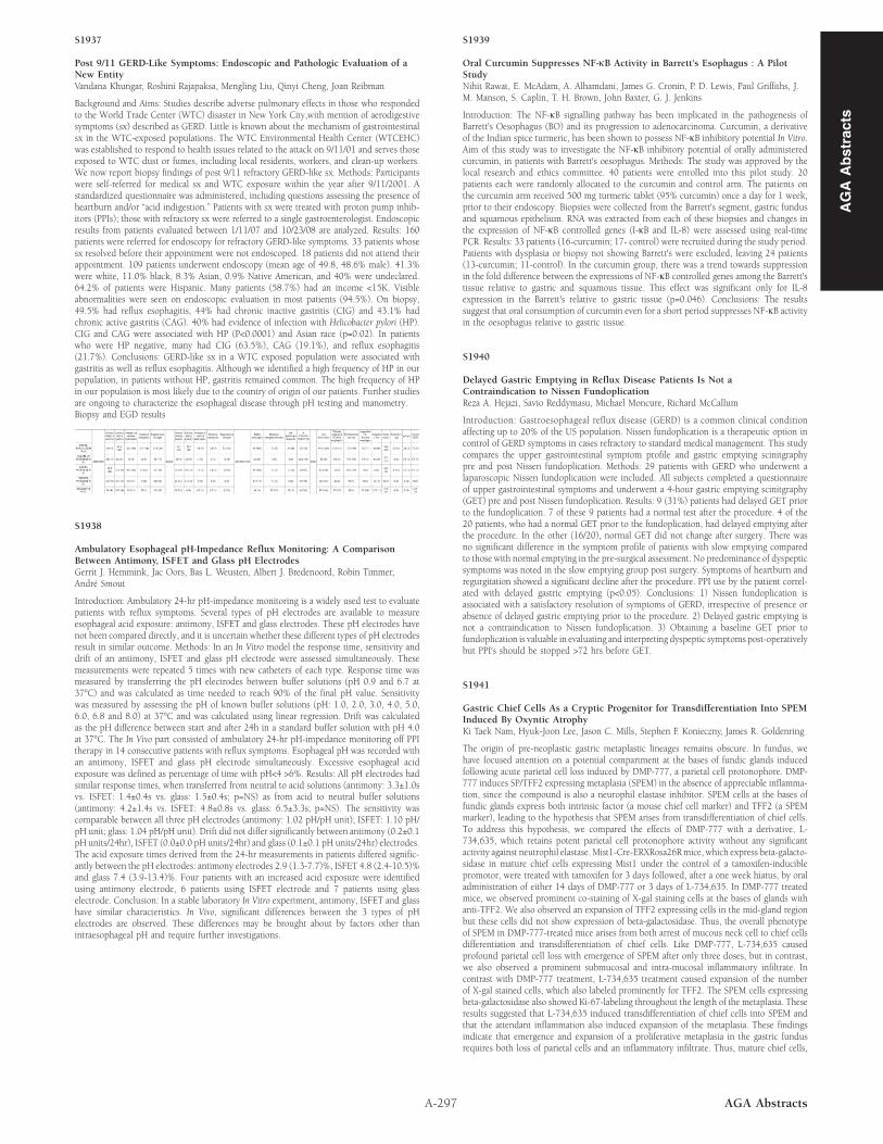
Background and Aims: Studies describe adverse pulmonary effects in those who respondedto the World Trade Center (WTC) disaster in New York City,with mention of aerodigestivesymptoms (sx) described as GERD. Little is known about the mechanism of gastrointestinalsx in the WTC-exposed populations. The WTC Environmental Health Center (WTCEHC)was established to respond to health issues related to the attack on 9/11/01 and serves thoseexposed to WTC dust or fumes, including local residents, workers, and clean-up workers.We now report biopsy findings of post 9/11 refractory GERD-like sx. Methods: Participantswere self-referred for medical sx and WTC exposure within the year after 9/11/2001. Astandardized questionnaire was administered, including questions assessing the presence ofheartburn and/or “acid indigestion.” Patients with sx were treated with proton pump inhib-itors (PPIs); those with refractory sx were referred to a single gastroenterologist. Endoscopicresults from patients evaluated between 1/11/07 and 10/23/08 are analyzed. Results: 160patients were referred for endoscopy for refractory GERD-like symptoms. 33 patients whosesx resolved before their appointment were not endoscoped. 18 patients did not attend theirappointment. 109 patients underwent endoscopy (mean age of 49.8, 48.6% male). 41.3%were white, 11.0% black, 8.3% Asian, 0.9% Native American, and 40% were undeclared.64.2% of patients were Hispanic. Many patients (58.7%) had an income <15K. Visibleabnormalities were seen on endoscopic evaluation in most patients (94.5%). On biopsy,49.5% had reflux esophagitis, 44% had chronic inactive gastritis (CIG) and 43.1% hadchronic active gastritis (CAG). 40% had evidence of infection with Helicobacter pylori (HP).CIG and CAG were associated with HP (P<0.0001) and Asian race (p=0.02). In patientswho were HP negative, many had CIG (63.5%), CAG (19.1%), and reflux esophagitis(21.7%). Conclusions: GERD-like sx in a WTC exposed population were associated withgastritis as well as reflux esophagitis. Although we identified a high frequency of HP in ourpopulation, in patients without HP, gastritis remained common. The high frequency of HPin our population is most likely due to the country of origin of our patients. Further studiesare ongoing to characterize the esophageal disease through pH testing and manometry.Biopsy and EGD results

S1938

Ambulatory Esophageal pH-Impedance Reflux Monitoring: A ComparisonBetween Antimony, ISFET and Glass pH ElectrodesGerrit J. Hemmink, Jac Oors, Bas L. Weusten, Albert J. Bredenoord, Robin Timmer,André Smout

Introduction: Ambulatory 24-hr pH-impedance monitoring is a widely used test to evaluatepatients with reflux symptoms. Several types of pH electrodes are available to measureesophageal acid exposure: antimony, ISFET and glass electrodes. These pH electrodes havenot been compared directly, and it is uncertain whether these different types of pH electrodesresult in similar outcome. Methods: In an In Vitro model the response time, sensitivity anddrift of an antimony, ISFET and glass pH electrode were assessed simultaneously. Thesemeasurements were repeated 5 times with new catheters of each type. Response time wasmeasured by transferring the pH electrodes between buffer solutions (pH 0.9 and 6.7 at37°C) and was calculated as time needed to reach 90% of the final pH value. Sensitivitywas measured by assessing the pH of known buffer solutions (pH: 1.0, 2.0, 3.0, 4.0, 5.0,6.0, 6.8 and 8.0) at 37°C and was calculated using linear regression. Drift was calculatedas the pH difference between start and after 24h in a standard buffer solution with pH 4.0at 37°C. The In Vivo part consisted of ambulatory 24-hr pH-impedance monitoring off PPItherapy in 14 consecutive patients with reflux symptoms. Esophageal pH was recorded withan antimony, ISFET and glass pH electrode simultaneously. Excessive esophageal acidexposure was defined as percentage of time with pH<4 >6%. Results: All pH electrodes hadsimilar response times, when transferred from neutral to acid solutions (antimony: 3.3±1.0svs. ISFET: 1.4±0.4s vs. glass: 1.5±0.4s; p=NS) as from acid to neutral buffer solutions(antimony: 4.2±1.4s vs. ISFET: 4.8±0.8s vs. glass: 6.5±3.3s; p=NS). The sensitivity wascomparable between all three pH electrodes (antimony: 1.02 pH/pH unit); ISFET: 1.10 pH/pH unit; glass: 1.04 pH/pH unit). Drift did not differ significantly between antimony (0.2±0.1pH units/24hr), ISFET (0.0±0.0 pH units/24hr) and glass (0.1±0.1 pH units/24hr) electrodes.The acid exposure times derived from the 24-hr measurements in patients differed signific-antly between the pH electrodes: antimony electrodes 2.9 (1.3-7.7)%, ISFET 4.8 (2.4-10.5)%and glass 7.4 (3.9-13.4)%. Four patients with an increased acid exposure were identifiedusing antimony electrode, 6 patients using ISFET electrode and 7 patients using glasselectrode. Conclusion: In a stable laboratory In Vitro experiment, antimony, ISFET and glasshave similar characteristics. In Vivo, significant differences between the 3 types of pHelectrodes are observed. These differences may be brought about by factors other thanintraesophageal pH and require further investigations.

A-297 AGA Abstracts

S1939

Oral Curcumin Suppresses NF-κB Activity in Barrett's Esophagus : A PilotStudyNihit Rawat, E. McAdam, A. Alhamdani, James G. Cronin, P. D. Lewis, Paul Griffiths, J.M. Manson, S. Caplin, T. H. Brown, John Baxter, G. J. Jenkins

Introduction: The NF-κB signalling pathway has been implicated in the pathogenesis ofBarrett's Oesophagus (BO) and its progression to adenocarcinoma. Curcumin, a derivativeof the Indian spice turmeric, has been shown to possess NF-κB inhibitory potential In Vitro.Aim of this study was to investigate the NF-κB inhibitory potential of orally administeredcurcumin, in patients with Barrett's oesophagus. Methods: The study was approved by thelocal research and ethics committee. 40 patients were enrolled into this pilot study. 20patients each were randomly allocated to the curcumin and control arm. The patients onthe curcumin arm received 500 mg turmeric tablet (95% curcumin) once a day for 1 week,prior to their endoscopy. Biopsies were collected from the Barrett's segment, gastric fundusand squamous epithelium. RNA was extracted from each of these biopsies and changes inthe expression of NF-κB controlled genes (I-κB and IL-8) were assessed using real-timePCR. Results: 33 patients (16-curcumin; 17- control) were recruited during the study period.Patients with dysplasia or biopsy not showing Barrett's were excluded, leaving 24 patients(13-curcumin; 11-control). In the curcumin group, there was a trend towards suppressionin the fold difference between the expressions of NF-κB controlled genes among the Barrett'stissue relative to gastric and squamous tissue. This effect was significant only for IL-8expression in the Barrett's relative to gastric tissue (p=0.046). Conclusions: The resultssuggest that oral consumption of curcumin even for a short period suppresses NF-κB activityin the oesophagus relative to gastric tissue.

S1940

Delayed Gastric Emptying in Reflux Disease Patients Is Not aContraindication to Nissen FundoplicationReza A. Hejazi, Savio Reddymasu, Michael Moncure, Richard McCallum

Introduction: Gastroesophageal reflux disease (GERD) is a common clinical conditionaffecting up to 20% of the US population. Nissen fundoplication is a therapeutic option incontrol of GERD symptoms in cases refractory to standard medical management. This studycompares the upper gastrointestinal symptom profile and gastric emptying scinitgraphypre and post Nissen fundoplication. Methods: 29 patients with GERD who underwent alaparoscopic Nissen fundoplication were included. All subjects completed a questionnaireof upper gastrointestinal symptoms and underwent a 4-hour gastric emptying scinitgraphy(GET) pre and post Nissen fundoplication. Results: 9 (31%) patients had delayed GET priorto the fundoplication. 7 of these 9 patients had a normal test after the procedure. 4 of the20 patients, who had a normal GET prior to the fundoplication, had delayed emptying afterthe procedure. In the other (16/20), normal GET did not change after surgery. There wasno significant difference in the symptom profile of patients with slow emptying comparedto those with normal emptying in the pre-surgical assessment. No predominance of dyspepticsymptoms was noted in the slow emptying group post surgery. Symptoms of heartburn andregurgitation showed a significant decline after the procedure. PPI use by the patient correl-ated with delayed gastric emptying (p<0.05). Conclusions: 1) Nissen fundoplication isassociated with a satisfactory resolution of symptoms of GERD, irrespective of presence orabsence of delayed gastric emptying prior to the procedure. 2) Delayed gastric emptying isnot a contraindication to Nissen fundoplication. 3) Obtaining a baseline GET prior tofundoplication is valuable in evaluating and interpreting dyspeptic symptoms post-operativelybut PPI's should be stopped >72 hrs before GET.

S1941

Gastric Chief Cells As a Cryptic Progenitor for Transdifferentiation Into SPEMInduced By Oxyntic AtrophyKi Taek Nam, Hyuk-Joon Lee, Jason C. Mills, Stephen F. Konieczny, James R. Goldenring

The origin of pre-neoplastic gastric metaplastic lineages remains obscure. In fundus, wehave focused attention on a potential compartment at the bases of fundic glands inducedfollowing acute parietal cell loss induced by DMP-777, a parietal cell protonophore. DMP-777 induces SP/TFF2 expressing metaplasia (SPEM) in the absence of appreciable inflamma-tion, since the compound is also a neurophil elastase inhibitor. SPEM cells at the bases offundic glands express both intrinsic factor (a mouse chief cell marker) and TFF2 (a SPEMmarker), leading to the hypothesis that SPEM arises from transdifferentiation of chief cells.To address this hypothesis, we compared the effects of DMP-777 with a derivative, L-734,635, which retains potent parietal cell protonophore activity without any significantactivity against neutrophil elastase. Mist1-Cre-ERXRosa26Rmice, which express beta-galacto-sidase in mature chief cells expressing Mist1 under the control of a tamoxifen-induciblepromotor, were treated with tamoxifen for 3 days followed, after a one week hiatus, by oraladministration of either 14 days of DMP-777 or 3 days of L-734,635. In DMP-777 treatedmice, we observed prominent co-staining of X-gal staining cells at the bases of glands withanti-TFF2. We also observed an expansion of TFF2 expressing cells in the mid-gland regionbut these cells did not show expression of beta-galactosidase. Thus, the overall phenotypeof SPEM in DMP-777-treated mice arises from both arrest of mucous neck cell to chief cellsdifferentiation and transdifferentiation of chief cells. Like DMP-777, L-734,635 causedprofound parietal cell loss with emergence of SPEM after only three doses, but in contrast,we also observed a prominent submucosal and intra-mucosal inflammatory infiltrate. Incontrast with DMP-777 treatment, L-734,635 treatment caused expansion of the numberof X-gal stained cells, which also labeled prominently for TFF2. The SPEM cells expressingbeta-galactosidase also showed Ki-67-labeling throughout the length of the metaplasia. Theseresults suggested that L-734,635 induced transdifferentiation of chief cells into SPEM andthat the attendant inflammation also induced expansion of the metaplasia. These findingsindicate that emergence and expansion of a proliferative metaplasia in the gastric fundusrequires both loss of parietal cells and an inflammatory infiltrate. Thus, mature chief cells,

AG

AA

bst

ract

s