Embed Size (px)
Citation preview


Sakthikumar Mathivanan
Doctoral Thesis
Director: Prof. Antonio Ferrer Montiel
INSTITUTO DE BIOLOGÍA MOLECULAR Y CELULAR
UNIVERSIDAD MIGUEL HERNANDEZ (ELCHE)
Differential mechanism of TRPV1 sensitization
in peptidergic and nonpeptidergic nociceptors


Dr. Antonio Ferrer Montiel, Catedrático y Director del Instituto de Biología Molecular y
Celular de la Universidad Miguel Hernández de Elche.
DA SU CONFORMIDAD a la lectura de tesis doctoral titulada: “Differential mechanism of
TRPV1 sensitization in peptidergic and nonpeptidergic nociceptors”, presentada por D.
Sakthikumar Mathivanan
Elche, Abril 2016
Fdo: Dr. Antonio Ferrer Montiel
Catedrático Universidad Miguel Hernández


Dr. Antonio Ferrer Montiel, Catedrático de la Universidad Miguel Hernández de Elche,
CERTIFICA que el trabajo de investigación que lleva por título “Differential mechanism of
TRPV1 sensitization in peptidergic and nonpeptidergic nociceptors”, presentado por D.
Sakthikumar Mathivanan para optar al grado de Doctor, ha sido realizado bajo su dirección
en el Instituto de Biología Molecular y Celular de la Universidad Miguel Hernández de
Elche. Considerando que la presente tesis se halla concluida, AUTORIZA su presentación
para que pueda ser juzgada por el tribunal correspondiente.
Y para que así conste a los efectos oportunos, se expide el presente escrito.
Elche, Abril 2016
Fdo: Dr. Antonio Ferrer Montiel
Catedrático Universidad Miguel Hernández


El presente trabajo ha sido realizado en el Instituto de Biología Molecular y Celular (IBMC),
de la Universidad Miguel Hernández de Elche.
Sakthikumar Mathivanan ha sido beneficiario de una beca predoctoral del programa Santiago
Grisolia financiado por la Generalitat Valenciana, GRISOLIA/2010/058
Este trabajo se ha desarrollado gracias a la financiación procedente del Ministerio de
Economía y competitividad y de la Generalitat Valenciana.

INDEX OF CONTENT
INDEX OF ABBREVIATIONS ................................................................................................... i
SUMMARY............................................................................................................................ 1
RESUMEN............................................................................................................................. 2
INTRODUCTION.................................................................................................................... 3
PAIN, AN INTRICATE MECHANISM........................................................................................ 3
Definition................................................................................................................... ................ 3
Classification.......................................................................................................................... .... 3
Pain signalling............................................................................................................. ............... 5
Recognition of nociceptors........................................................................................................ 6
IB4- and IB4+ neurons are electrically distinct............................................................................ 8
Inflammatory sensitization........................................................................................................ 10
Peripheral sensitization................................................................................................ 10
Central sensitization..................................................................................................... 11
Ion channels involved in pain sensation.................................................................................... 13
TRP CHANNELS AS SENSORY RECEPTORS.............................................................................. 15
Differential expression of TRP channels in DRG and TG neurons.............................................. 16
Thermo TRPs.......................................................................................................................... .... 17
TRPA subfamily............................................................................................................. 17
TRPM subfamily............................................................................................................ 18
TRPV subfamily............................................................................................................. 18
TRPV1 AND PAIN....................................................................................................................... 20
TRPV1- A POTENTIAL NOXIOUS SENSOR............................................................................... 21
Structural determinants............................................................................................................. 21
Ankyrin repeat domains................................................................................................ 21
CaM binding domains................................................................................................... 22
PIP2 interaction............................................................................................................ 22

Pore loop and C terminal................................................................................................. 22
Heterotetramers of TRPV1........................................................................................................... 23
MODULATION OF TRPV1............................................................................................................. 23
Agonist induced desensitization................................................................................................... 24
Acute desensitization...................................................................................................... 24
Tachyphylaxis.................................................................................................................. 24
Desensitization through endocytosis.............................................................................. 25
Inflammatory sensitization of TRPV1........................................................................................... 25
TRPV1 phosphorylation................................................................................................... 25
PKC.................................................................................................................... 25
PKA.................................................................................................................... 26
Other kinases..................................................................................................... 26
TRPV1 trafficking and exocytosis.................................................................................... 27
Constitutive TRPV1 expression........................................................................... 28
Regulated exocytosis of TRPV1.......................................................................... 28
OBJECTIVES............................................................................................................................ 32
RESULTS................................................................................................................................. 33
CHARACTERISATION OF RAT NOCICEPTORS........................................................................... 33
ATP INDUCED INFLAMMATORY SENSITIZATION OF TRPV1..................................................... 47
Capsaicin evoked excitability- MEA characterisation..................................................... 47
DD04107- basal TRPV1 activity...................................................................................... 50
ATP induced sensitization of TRPV1.............................................................................. 53
Role of αCGRP and Tac1 on ATP induced inflammatory sensitization of TRPV1........... 61
BK INDUCED INFLAMMATORY SENSITIZATION OF TRPV1....................................................... 70
BK induced sensitization of TRPV1................................................................................. 70
Role of αCGRP and Tac1 on BK induced inflammatory sensitization of TRPV1............. 82
ATP- BK INDUCED INFLAMMATORY SENSITIZATION OF TRPV1............................................... 85

ATP- BK induced sensitization of TRPV1 excitabilty.......................................................... 85
ATP- BK pH 6.2 induced sensitization of TRPV1 excitability.............................................. 88
pH 6.2 potentiates TRPV1 excitability............................................................................... 91
ATP- BK pH 6.2 induced sensitization of TRPV1 excitability
in peptidergic nociceptors................................................................................................. 93
DISCUSSION.............................................................................................................................. 97
CONCLUSIONS.......................................................................................................................... 106
CONCLUSIONES........................................................................................................................ 107
MATERIALS AND METHODS...................................................................................................... 108
Animals...................................................................................................................... ..................... 108
Primary culture of sensory neurons............................................................................................... 108
Patch clamp recordings.................................................................................................................. 109
Electrical properties....................................................................................................................... 109
Micro Electrode Array.................................................................................................................... 110
Immunocytochemistry................................................................................................................... 110
Chemicals.................................................................................................................... ................... 111
Data Analysis..................................................................................................................... ............. 111
REFERENCES............................................................................................................................. 112
ANNEX..................................................................................................................................... 141
ACKNOWLEDGEMENTS............................................................................................................. 142


i
INDEX OF ABBREVIATIONS
2- APB 2-Aminoethoxydiphenyl borate
4- AP 4 amino pyridine
5- HT 5- Hydroxytryptamine
a.a Aminoacids
AHP After hyperpolarization
AITC Allyl isothiocyanate
AKAP A kinase anchoring protein
AMPA α-amino-3-hydroxy-5-methyl-4-isoxazolepropionic acid receptor
Ano 1 Anoctamin 1
AP Action potential
ARD Ankyrin repeat domain
ASIC Acid sensing ion channel
ATP Adenosine triphosphate
BIM Bisindolylmaleimide
BK Bradykinin
BoNT- A Botulinum neurotoxin A
CACC Calcium activated chloride channel
CaCl2 Calcium chloride
CaM Calmodulin
CaMKII Ca2+
/calmodulin-dependent protein kinase II
CCL2 Chemokine ligand
CDK 5 Cyclin dependent kinase 5

ii
CFA Complete freund’s adjuvant
CGRP Calcitonin gene related peptide
Cps Capsaicin
CPZ Capsazepine
CsA- CyP Cyclosporin A- Cyclophilin D
CSP Constitutive secretory pathway
CT Current threshold
DAG Diacylglycerol
DKO Double knockout
DMEM Dulbecco’s modified eagle medium
DMSO Dimethyl sulfoxide
DRG Dorsal root ganglion
EC50 Effective concentration
Erk Extracellular signal-regulated kinase
FBS Fetal bovine serum
GABA Gamma-Aminobutyric acid
GABARAP Gamma-aminobutyric acid type A (GABAA) receptor-associated protein
GDNF Glial cell line derived neurotrophic factor
GPCR G protein coupled receptor
GTP Guanosine-5'-triphosphate
H2O2 Hydrogen peroxide
HBSS Hank’s balanced salt solution
HCN Hyperpolarization-activated cyclic nucleotide-gated (HCN) channel

iii
HEPES 4-(2-hydroxyethyl)-1-piperazineethanesulfonic acid
HIV/ AIDS Human immunodeficiency virus infection and acquired
immune deficiency syndrome
HPETE Hydroperoxyeicosatetraenoic acid
IASP International association for the study of pain
IB4 Isolectin B4
IGF- 1 Insulin growth factor 1
IL- 1β Interleukin- 1β
IP3 Inositol triphosphate
J (pA/pF) Current density
KCl Potassium chloride
KIF13B Kinesin family member 13B
KOH Potassium hydroxide
LDCV Large dense core vesicle
LPA Lysophosphatidic acid
MAPK Mitogen-activated protein kinase
MEA Micro electrode array
MES 2-(N-morpholino) ethanesulfonic acid
MgCl2 Magnesium chloride
mGluR Metabotropic glutamate receptor
ml Milliliter
mM Millimolar
Mrg Mas related g protein coupled receptor

iv
ms Millisecond
mV Millivolt
MYCBP2 Myc-binding protein 2
NaCl Sodium chloride
NADA N-arachidonoyl-dopamine
NaOH Sodium hydroxide
ng Nano gram
NGF Nerve growth factor
NGS Normal goat serum
NK Neurokinin
nM Nano molar
NMDA N-methyl-D-aspartate receptor
OLDA N-oleoyldopamine
P2 Purinergic receptor family
pA picoampere
PAR2 Protease-activated receptor 2
PBS Phosphate buffered saline
pF picoFarad
PGE2 Prostaglandin E2
PGI2 Prostaglandin I2
PI3 kinase Phosphoinositide 3-kinase
PIP2 Phosphatidylinositol 4, 5-bisphosphate
PKA Protein kinase A

v
PKC Protein kinase C
PLC Phospholipase C
PMA Phorbol 12-myristate 13-acetate
RMP Resting membrane potential
RSP Regulated secretory pathway
RT Room temperature
RTX Resiniferatoxin
SAP Saporin
SEM Standard error mean
siRNA small interference ribonucleic acid
SNAP 25 Synaptosomal associated protein - 25
SNARE Soluble N-ethylmaleimide-sensitive factor attachment protein receptor
SP Substance P
Syt IX Synaptotagmin IX
TG Trigeminal ganglion
TGF- β Transforming growth factor β
TM Transmembrane
TNF- α Tumor necrosis factor- α
TP Threshold potential
TrkA Receptor tyrosine kinase A
TRPA Transient receptor potential ankyrin
TRPC Transient receptor potential canonical
TRPM Transient receptor potential melastatin

vi
TRPML Transient receptor potential mucolipin
TRPP Transient receptor potential polycystic
TRPV Transient receptor potential vanilloid
TTX Tetrodotoxin
w/v weight/ volume
µm Micrometer
µM Micromolar
µV Microvolt


SUMMARY


Summary
1
SUMMARY
TRPV1 is a polymodal, non selective cation channel which acts as a major integrator
of painful stimuli in nociceptors. During inflammation, the release of inflammatory mediators
act on TRPV1 leading to enhanced nociceptor excitability and thermal hyperalgesia. Acute
inflammatory sensitization of TRPV1 involves both the modification of channel gating
properties by phosphorylation and recruitment of new channels to the neuronal surface.
Mobilization of TRPV1 channel to the plasma membrane by some pro- inflammatory
mediators occurs through SNARE-dependent exocytosis, but the exact mechanism involved
remains to be elucidated. We hypothesize that the inflammatory recruitment of channels
occurs in the neuronal subpopulation which contains neuropeptides substance P (SP) and
calcitonin gene related peptide (CGRP), also called peptidergic nociceptors.
Therefore, we have investigated the underlying mechanism of pro- inflammatory
mediators Adenosine triphosphate (ATP) and Bradykinin (BK) induced inflammatory
sensitization of TRPV1 in cultured nociceptors containing both peptidergic and
nonpeptidergic subpopulations. We have performed functional analysis using patch clamp
electrophysiology and micro electrode array (MEA) technique. We found that the inhibition
of neuronal exocytosis results in decreased inflammatory sensitization of TRPV1 induced by
both ATP and Bradykinin in peptidergic nociceptors where membrane recruitment of the
channel is essential. In addition, knocking out of αCGRP leads to the reduction of
inflammatory sensitization of TRPV1.
Hence, this study reveals that both ATP and Bradykinin induces regulated exocytosis
of TRPV1 in peptidergic nociceptors where αCGRP plays a significant role. Furthermore, our
result validates the therapeutic potential of DD04107 on lessening inflammatory pain through
modulation of regulated exocytosis of TRPV1.


Resumen
2
RESUMEN
El canal TRPV1 es un receptor polimodal, no selectivo a cationes, el cual actúa como
principal integrador del estímulo doloroso en nociceptores. Durante la inflamación, los
mediadores inflamatorios liberados actúan sobre TRPV1 provocando una excitabilidad
aumentada del nociceptor y una hiperalgesia térmica. La sensibilización inflamatoria aguda
de TRPV1 conlleva tanto la modificación de las propiedades de apertura del canal (“gating”)
por fosforilación como al reclutamiento de nuevos canales a la membrana neuronal. La
movilización del canal TRPV1 a la membrana plasmática provocada por algunos mediadores
pro-inflamatorios tiene lugar a través de exocitosis dependiente de SNARE, aunque el
mecanismo exacto no ha sido todavía esclarecido.
Con el fin de elucidar dicho mecanismo de exocitosis, nos planteamos la hipótesis de
que el reclutamiento inflamatorio del canal TRPV1 ocurría únicamente en la subpoblación
neuronal que contiene los neuropéptidos sustancia P (SP) y el péptido relacionado con el gen
calcitonina (CGRP), también denominada nociceptores peptidérgicos. Así pues, se ha
investigado la sensibilización inflamatoria del canal TRPV1 inducida por los agentes pro-
inflamatorios Trifosfato de adenosina (ATP) y Bradiquinina (BK) en cultivos de nociceptores
tanto peptidérgicos como no peptidérgicos. Para ello, se ha llevado a cabo el análisis
funcional del canal TRPV1 empleando técnicas de electrofisiología patch clamp y MEA
(matriz de microelectrodos). Nuestros resultados muestran que la inhibición de la exocitosis
neuronal por parte del péptido DD04107 provoca una disminución en la sensibilización de
TRPV1 inducida por los agentes pro- inflamatorios ATP y BK únicamente en la
subpoblación peptidérgica. Además, la eliminación de la expresión de αCGRP también
conduce a la reducción de la sensibilización inflamatoria de TRPV1. Así pues, este estudio
revela que tanto ATP como BK inducen la exocitosis regulada de TRPV1 en nociceptores
peptidérgicos donde αCGRP juega un papel significativo. Además, nuestros resultados
validan el potencial terapéutico del péptido DD04107 en la disminución del dolor
inflamatorio a través de la modulación de la exocitosis regulada de TRPV1.


INTRODUCTION


Introduction
3
PAIN, AN INTRICATE MECHANISM
Pain is the major problem of the health community that affects up to 20 % of the
global population. Pain management and treatment are largely unmet which creates enormous
emotional and economical burden to the patients. Extensive studies from peripheral
nociceptors towards their neural circuits in the brain have been carried out to elucidate the
mechanism of pain sensation and its transition from acute to chronic state (1). Perception of
pain is a multidimensional subjective experience which encompasses cognitive and emotional
components (2). Physiologically, pain sensation acts as an alarming system to protect
ourselves from potentially damaging stimuli which consists of adaptive (nociceptive,
inflammatory pain) and maladaptive (neuropathic and functional pain) responses. Multiple
mechanisms induce pain sensation which includes nociception, peripheral sensitization,
phenotypic switches, central sensitization, ectopic excitability, structural reorganization and
decreased inhibition. Studies conducted on human brain imaging technique have shown the
prominent role of brain circuits in pain perception. Besides environmental factor playing a
predominant role in pain sensation, differences in genetic factors with the existence of single
nucleotide polymorphisms also plays a key role in pain perception (3).
Definition
Pain, as a multi component complex syndrome, principally consists of unpleasant
sensorial, physiological, cognitive and behavioural modules, where noxious stimuli are
experienced by the sensory component and transmitted causing acute or potential tissue
damage to the brain (4), (5).
Classification of pain
Pain can be classified depending on its duration or pathophysiology (6). Based on the
duration of pain, it can be either acute or chronic (7). Acute pain is the short lasting pain
sensation due to tissue injury which stimulates nociceptors and usually disappears when the
injury heals (< 6 months). In contrast, chronic pain generally begins as acute pain and
persists for longer periods (> 6 months), persists beyond the expected time of healing.
Introduction
4
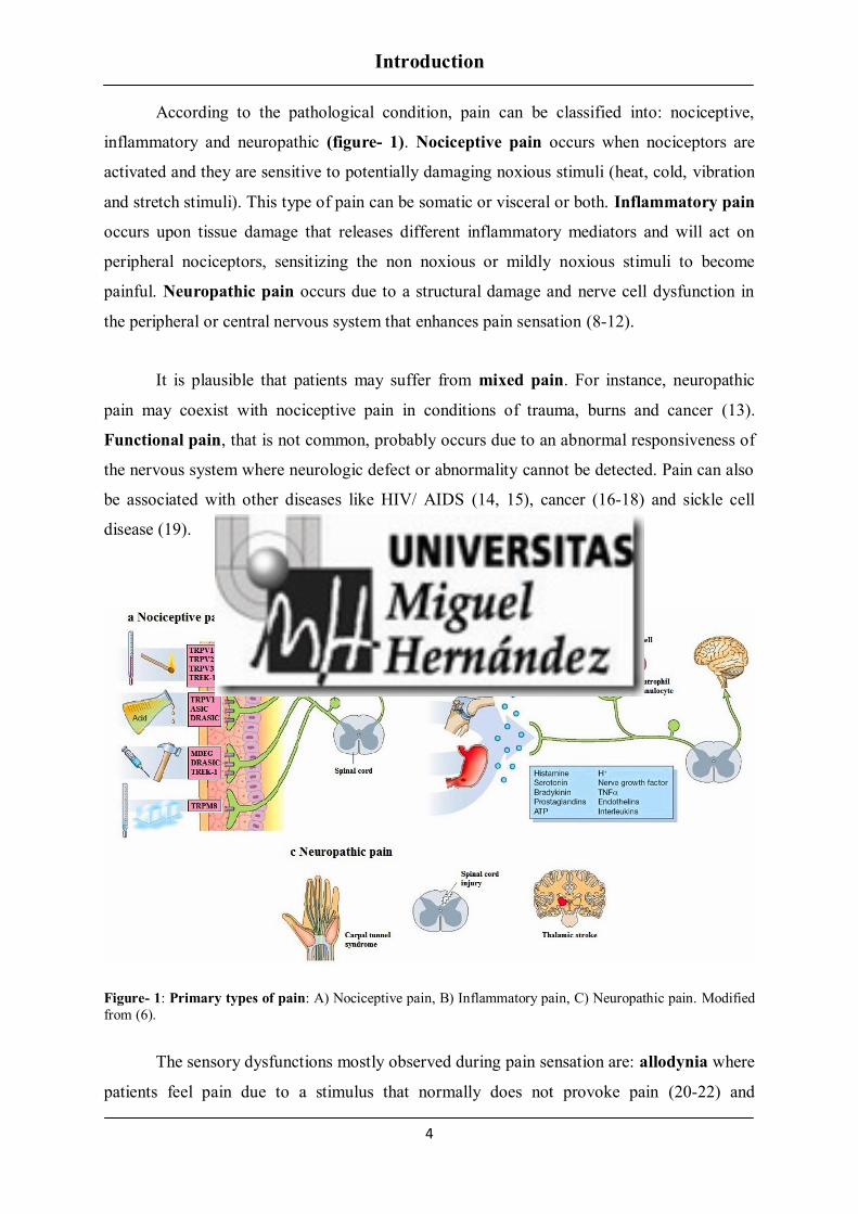
According to the pathological condition, pain can be classified into: nociceptive,
inflammatory and neuropathic (figure- 1). Nociceptive pain occurs when nociceptors are
activated and they are sensitive to potentially damaging noxious stimuli (heat, cold, vibration
and stretch stimuli). This type of pain can be somatic or visceral or both. Inflammatory pain
occurs upon tissue damage that releases different inflammatory mediators and will act on
peripheral nociceptors, sensitizing the non noxious or mildly noxious stimuli to become
painful. Neuropathic pain occurs due to a structural damage and nerve cell dysfunction in
the peripheral or central nervous system that enhances pain sensation (8-12).
It is plausible that patients may suffer from mixed pain. For instance, neuropathic
pain may coexist with nociceptive pain in conditions of trauma, burns and cancer (13).
Functional pain, that is not common, probably occurs due to an abnormal responsiveness of
the nervous system where neurologic defect or abnormality cannot be detected. Pain can also
be associated with other diseases like HIV/ AIDS (14, 15), cancer (16-18) and sickle cell
disease (19).
Figure- 1: Primary types of pain: A) Nociceptive pain, B) Inflammatory pain, C) Neuropathic pain. Modified
from (6).
The sensory dysfunctions mostly observed during pain sensation are: allodynia where
patients feel pain due to a stimulus that normally does not provoke pain (20-22) and

Introduction
5
hyperalgesia where an increased pain response to a normal painful stimulus is observed (23-
25). Frequently, allodynia and hyperalgesia occurs during inflammatory and neuropathic
conditions (26, 27). In addition, paraesthesia and dysesthesia conditions are also observed as
a common symptom of pain where patients feel an abnormal sensation to a stimulus that is
normally not unpleasant like tingling or numbness, burning, skin crawling, or itching. These
symptoms could be spontaneous or evoked (28-31). Normally, paraesthesia occurs during
stroke and transient ischemic attacks (mini-strokes), multiple sclerosis, transverse myelitis
and encephalitis, while dysesthesia occurs mainly due to chronic pain and frightening
diseased conditions.
Pain Signalling
The process of pain perception involves both peripheral and central nervous system.
Peripheral pain machinery comprises pseudounipolar sensory neurons where one end of the
afferent fibers project as free nerve endings to the skin, and the other end projects to different
laminae of the spinal cord. Sensory neurons make connections with dorsal horn neurons of
spinal cord through monosynaptic or multiple interneurons (excitatory or inhibitory) thereby
facilitating pain transmission. Pain perception is processed from dorsal horn neurons to
somatosensory cortex of the brain through the spinothalamic tract (figure- 2).
The processes involved in pain signalling are
Transduction: conversion of noxious (chemical, thermal and mechanical) stimuli
into electrical activity in the peripheral terminals of nociceptive sensory fibers mediated by
specific ion channels expressed in nociceptors.
Conduction: the passage of action potentials from the peripheral terminal along
axons to the central terminal of nociceptor in the central nervous system (afferent) or to the
peripheral terminal (efferent).
Transmission: synaptic transfer and modulation of input from one neuron to another.
Perception: transfer of sensory inflow to specific areas in cortex.

Introduction
6
Figure- 2: Schematic view of main circuits mediating physiological pain. Pain sensing neurons in the
peripheral nervous system have their soma located in the dorsal root ganglia (DRG). These neurons have a
peripheral axon innervating the distal territories (skin, viscera, etc.) where they detect painful stimuli leading to
an action potential that travels along the fibers up to the DRG and then to the first relay in the dorsal spinal cord.
Sensory neurons within the DRGs are diverse. The sensory information is processed locally in neuronal circuitry
within the dorsal horn of the spinal cord before being sent to the thalamus to convey nociceptive information.
Following thalamic filtering, the information is sent to the cortical structures of the pain matrix (32).
Recognition of nociceptors
In 1903, Charles Scott Sherrington, a renowned neurophysiologist, after extensive
research on neurons, stated that skin is provided with a set of free nerve endings and they can
be provoked by noxious stimuli. He coined the term nocicipient (recipient of noxious
stimuli). Later, he phrased nociceptive reaction as these free nerve endings capable of
detecting noxious stimuli (thermal, mechanical and chemical) and tissue damage with the
capability to provoke a reflex action, an autonomic response and pain. The apparatus
responsible to perceive noxious stimulus was identified as nociceptors (33, 34). These
nociceptors are electrically silent, transmit all or none action potentials when stimulated and
belong to subpopulations of primary sensory neurons.
Primary sensory neurons, have their cell bodies located in the trigeminal ganglia
(TG) at the base of the skull whose sensory endings covers head and face, or in the dorsal

Introduction
7
root ganglia (DRGs) outside the spinal cord, with their sensory endings covering the rest of
the body (35, 36). Neurons from DRGs and TGs are classified into three main types (figure-
3):
Large diameter, myelinated, rapidly conducting Aα and Aβ fibers which detect
innocuous stimuli from skin, muscle and joints (37).
Medium diameter, thinly myelinated, more rapidly conducting Aδ fibers.
Small diameter, unmyelinated, slowly conducting C fibers (35, 38).
The medium and small diameter sensory neurons are considered as nociceptors, as
they respond to noxious stimuli or become responsive after inflammatory sensitization (39).
Figure- 3: Primary Sensory Neurons and their synaptic connections in spinal cord. The unmyelinated,
peptidergic C (red) and myelinated Aδ nociceptors (purple) terminate in lamina I and outer lamina II. The
unmyelinated, nonpeptidergic nociceptors (blue) terminate in the inner part of lamina II. Innocuous input carried
by myelinated Aβ fibers (orange) terminates on PKCγ expressing interneurons in the ventral half of the inner
lamina II. Lamina V (purple) receives convergent input from Aδ and Aβ fibers (35).
Aδ fibers carry the first pain response to injury. They consist of type I and type II
fibers, both responding to mechanical stimuli. The difference between these two fibers relies
on their responsiveness to intense heat where type I fiber sense temperature at 53 oC and type
II fiber sense temperature at 43 oC.

Introduction
8
Unmyelinated C fibers are widely considered as nociceptors and they are
heterogeneous. They carry out the second pain responses to injury. Most of them are
polymodal responding to noxious thermal, chemical and mechanical stimuli. Characterisation
of C fiber nociceptors based on neuroanatomical, histochemical and molecular analysis
further classifies them into two subsets based on their dependency on growth factor and the
presence of neuropeptides. These are peptidergic and nonpeptidergic subpopulations (40).
Genetic transneuronal tracing studies proved that peptidergic and nonpeptidergic nociceptors
process nociceptive information through parallel, perhaps independent pathways (41). These
unmyelinated neurons, project their axons distinctly on both peripheral and central targets.
The first subset is a peptidergic population of C fibers which contains the
neuropeptides SP and CGRP, expresses TrkA neurotropin receptor and responds to nerve
growth factor (NGF) (42, 43). Almost all small C fiber nociceptors require NGF for their
survival during prenatal and postnatal life (44, 45). The peripheral axons of peptidergic
nociceptors terminates at the stratum spinosum of glabrous skin, and its central axons
terminates at lamina I and outerpart of lamina II of the dorsal horn of the spinal cord. The
second subset is a nonpeptidergic population of C fibers which expresses c-Ret neurotropin
receptor, is supported by glial derived neurotropic factor (GDNF), contains G protein-
coupled receptors of the Mas related genes (Mrg) family (46) and can be selectively labelled
with the α- D- galactosyl- binding lectin IB4 (IB4+). They also express purinergic receptor
subtype P2X3, a specific subtype of ATP gated ion channel. During early postnatal life a
subpopulation of C fiber nociceptors undergoes a shift in growth factor dependence, where
neurons which does not express the TrkA receptor and bind to IB4 becomes GDNF-
dependent for their survival (47). Nonpeptidergic peripheral axons terminate in superficial
stratum granulosum of the epidermis and its central axons terminate in the inner lamina II of
the dorsal horn of the spinal cord (48).
IB4- (peptidergic)
and IB4
+ (nonpeptidergic) neurons are electrically distinct
In primary sensory neurons (nociceptors) different ion channels are expressed and
transmit pain signals to both peripheral and central targets. Hence, they are considered as a
potential target for pain pharmacotherapy. Ion channels including classical voltage gated Na+,
K+, Ca
2+ channels, transient receptor potential (TRP), purinergic (P2), acid- gated ion channel
(ASIC) at the periphery and excitatory (Glutamergic), inhibitory (GABAergic)

Introduction
9
neurotransmitter channels at the central connections and are primarily involved in pain
signalling. Experimental pain studies have shown that few ion channels and their isoforms are
involved in stimulus detection, action potential initiation, propagation in nociceptors and
synaptic transmission in the dorsal horn neurons which are being marked as a therapeutic
target to impede pain signalling mechanisms.
Ion channels contributing to maintain the resting membrane potential of nociceptors
are 4- amino pyridine (4-AP) sensitive Voltage gated Kv channels, Kv7 (KCNQ), 2-pore leak
K+ channels (K2P), Na
+- activated K
+ channels, hyperpolarization activated cyclic nucleotide
gated (HCN) channels, low voltage- activated T-type Ca2+
channels and Voltage- gated Na+
channels (49). In general, changes in the membrane potential result from resting membrane
potential (Erest) which is due to the steady state interaction of a number of membrane
conductances, mostly represented by ion channels with a small contribution by electrogenic
pumps (50, 51). During tissue injury or inflammation the cell bodies of injured C fibers and
surrounding non injured C fibers can become hyper excitable and generate spontaneous
action potentials (AP) thereby transmitting pain signals from the periphery to the central
targets (52, 53). An increase in excitability is mainly initiated through activation of Nav
channels expressed in primary sensory neurons. Molecular study confirms that the DRG
neurons express multiple sodium channel subtypes. Furthermore, electrophysiological studies
have proved that one or more tetrodotoxin resistant (TTX-R) sodium channels can support
action potential conduction in the unmyelinated C fibers. Two prominent TTX- R sodium
channel subtypes Nav 1.8 and 1.9 are found to be expressed in C fibers, where Nav 1.8 is
responsible for producing the majority of the current underlying the depolarization phase of
action potential and is critically important for the production of multiple APs (54, 55).
Functional characterisation of IB4 binding neurons revealed that IB4- and IB4
+
neurons exhibit different electrical properties. In vitro studies found that IB4+ neurons had
longer duration action potentials and larger TTX resistant currents compared to IB4- neurons
(56). Consistently, in vivo studies from rat DRGs- C fiber nociceptors revealed that IB4+
compared to IB4- neurons had longer somatic action potential durations, slower conduction
velocities, and more negative membrane potentials (57). Generation of multiple APs differs
from IB4- to IB4
+ due to the slower inactivation of Nav 1.8 channel in IB4
- compared to IB4
+
neurons (58). Negative membrane potential of IB4+ neurons are primarily due to the selective
expression of TREK2 channel. Knockdown experiments using siRNA for TREK2

Introduction
10
depolarized the resting membrane potential by approximately 10 mV, suggesting that TREK2
is responsible for maintaining hyperpolarizing membrane potential in IB4+ neurons (59).
Inflammatory Sensitization
Although nociceptors functions as a protective system from potentially damaging
noxious stimuli, during inflammatory conditions they exhibit enhanced responses and
become more sensitive to normal sensory input. Tissue injury or inflammation results in
changes in the chemical environment of the nociceptors. Cells surrounding the damaged
tissues mediate the release of different inflammatory mediators that act on the peripheral
terminals and sensitize nociceptors, a process termed as peripheral sensitization (60). Like
peripheral neurons, dorsal horn neurons in the spinal cord can also be sensitized upon
inflammation, a process termed as central sensitization in which synaptic transfer from the
nociceptor central terminal to the dorsal horn neurons is amplified and facilitated by an
increase in membrane excitability, synaptic efficacy, or a reduced inhibition (61).
Peripheral Sensitization
Tissue injury triggers the release of inflammatory mediators from activated
nociceptors and surrounding non neuronal cells like keratinocytes, fibroblasts, mast cells,
monocytes, macrophages, neutrophils, lymphocytes, basophils, platelets, endothelial cells and
satellite glial cells (62-75).
The cocktail of inflammatory soup produced during tissue injury consists of ATP,
protons (75), BK (76), Histamine (77, 78), Serotonin (79), Prostaglandin E2 (PGE2) (80),
glutamate, NGF (81), SP, CGRP and pro inflammatory cytokines like tumor necrosis factor-
α (TNF-α) and interleukins (82). These inflammatory mediators can act directly on ion
channels/ receptors (activators) or modulate the function of ion channels (sensitizers) by
either reducing the activation threshold or increasing the responsiveness through
phosphorylation mechanism (figure- 4).

Introduction
11
Figure- 4: Tissue injury and infection cause inflammation via plasma extravasation and infiltration of
immune cells into the damaged tissue. The infiltrated immune cells and resident cells, including mast cells,
macrophages and keratinocytes, release several inflammatory mediators, such as bradykinin, prostaglandins, H+,
ATP, NGF, pro-inflammatory cytokines (TNFα), interleukin-1β (IL-1β) and IL-6) and pro-inflammatory
chemokines (CC-chemokine ligand 2 (CCL2), CXC-chemokine ligand 1 (CXCL1) and CXCL5). These released
inflammatory mediators either directly activates or modulates different ion channels expressed in nociceptors,
leading to enhanced neuronal excitability (peripheral sensitization). Activation of nociceptors also releases SP
and CGRP, which are involved in the generation of neurogenic inflammation (60).
Central Sensitization
Sensitization of central neurons also involves phosphorylation of ion channels/
receptors through activation of intracellular kinases similar to the peripheral sensitization
process (61). There are both excitatory (glutamatergic) and inhibitory (GABAergic)
interneurons within the spinal cord, which influence the pain transmission (figure- 5).

Introduction
12
Glutamate is an excitatory amino acid neurotransmitter which is released from
sensory afferents along with CGRP and SP in response to acute or persistent noxious stimuli.
Glutamate exerts an excitatory effect on post synaptic spinal neurons acting through α-amino-
3-hydroxy-5-methyl-4-isoxazolepropionic acid (AMPA) receptor, N-methyl-D-aspartate
(NMDA) receptor and metabotropic glutamate receptor (mGluR) and numerous studies have
been reported the role of glutamate in regulating spinal nociception process (83, 84).
ϒ- aminobutyric acid (GABA) is an inhibitory neurotransmitter released by
interneurons within the spinal cord, where it controls and limits the post synaptic
transmission of sensory input. GABA acts through GABAA and GABAB receptors in the
spinal dorsal horn. Inhibition of GABA increases pain hypersensitivity. Furthermore,
pathologic loss of inhibition (disinhibition) can also lead to increased excitability and pain
(85). Studies have reported the role of GABAA and GABAB receptors on mediating inhibitory
effect of GABA on different pain conditions (86-88).
Figure- 5: Spinal cord (central) sensitization, steps involved in inflammation induced central sensitization is
as follows, 1. Glutamate/NMDA receptor-mediated sensitization, where normally silent NMDA glutamate
receptors will be activated leading to signalling of cascade of events that will increase the excitability of the output neuron and facilitate the transmission of pain messages to the brain. 2. Disinhibition, inflammation
induces disinhibition of GABA, resulting in hyperalgesia. 3. Microglial activation. Peripheral nerve injury
promotes release of ATP and the chemokine fractalkine that will stimulate microglial cells. Hence activation of
microglial cells releases numerous soluble factors which promote increased excitability and enhanced pain in
response to both noxious and innocuous stimulation (that is, hyperalgesia and allodynia) (35).

Introduction
13
Ion channels involved in pain sensation
Primary peripheral ion channels expressed in DRG neurons and involved in
inflammatory pain signalling are voltage gated Na+ channels, which are responsible for the
AP depolarization. Of nine different Nav, four isoforms have been extremely involved in pain
sensation. They are NaV 1.3 (89, 90), Nav 1.7 (91), NaV 1.8 (55, 92, 93) and NaV 1.9 (92, 94).
Potassium channels are important regulators of resting membrane potential and action
potential repolarization in DRG neurons. DRG neurons expresses multiple types of KV
channels (KV 1, 2, 3, 4, 7, 9) (95) that are involved in different pain models (96, 97). Kv
channels are the key determinants of neuronal firing frequency and spike duration. Defect in
the activity of Kv channels leads to hyperexcitability of the neurons. Another subfamily of
potassium channels are two pore potassium channels (K2P), which contributes to maintain
the hyperpolarized resting membrane potential. DRG neurons express several K2P channel
subtypes (TRESK, TRAAK, TASK, TREK, and THIK) (98-100). Ca2+
activated K+ (KCa)
channels and Na+ - activated K
+ (KNa) channels were found as important determinants of after
hyperpolarization following an action potential. KCa channels include large (BK),
intermediate (IK) and small (SK) conductance channels (101, 102). The role of these
channels in afferent pain signalling is yet to be explored, though few studies shows that they
interact with other channels in regulating the excitability pattern (103, 104).
Na+ and K
+ ions plays a prominent role in pain signalling mechanism; similarly Ca
2+
ions also plays an important role in pain signalling through the activation of Cav channels.
Primary afferent neurons express multiple types of Cav channels like L, T, N, R, P/ Q- types
with specific sub cellular expression pattern and functions (105-107). This suggests a
significant role of Cav channels in pain processing. Likewise, Ca2+
activated chloride
channels expression in DRGs and their conductance contributes to after depolarization
suggests its possible role in regulating the excitability of the afferent pain pathway (108,
109). For instance, expression of Ca2+
activated chloride current is altered in response to
nerve injury, due to increased expression in large and medium sized DRGs. Another example
is the Ca2+
activated Cl-
channel Ano1 (TMEM16A) known to be a key player in heat
sensitivity (110).

Introduction
14
Hyperpolarization- activated cyclic nucleotide- gated (HCN) channels are cation
channels that open at negative membrane potential, contributing to neuronal excitability. All
the four subtypes (HCN1, HCN2, HCN3 and HCN4) (111) are expressed in DRG neurons.
The role of HCN channels in pain signalling have been explored in inflammatory and
neuropathic pain models (112, 113).
Purinergic (P2X) receptor also contributes to pain signalling. P2X is a ligand gated
ion channel family (P2X1-7), activated by extracellular ATP. Tissue damage induces the
release of ATP from neuronal and surrounding non neuronal cell, acts on P2X receptor
specifically P2X3, and evokes pain sensation (75). P2X3 is expressed in DRG neurons (114).
Numerous studies have reported the pathologic role of P2X3 on pain sensation (115-118).
Tissue acidosis is associated with inflammation leading to drop in extracellular pH.
Proton plays a significant role in pain sensation where decrease in pH (<6) activates
nociceptors gated through acid sensing ion channel (ASIC). ASIC are Na+ channels
belonging to degenerin/ ENaC superfamily. Different subtype expression of ASIC has been
found on DRG cell bodies and sensory terminals, playing a vital role in nociception and
mechanosensation (119-121) of which ASIC 3 is widely expressed in nociceptors (122, 123).
In recent years, immense studies on subfamilies of Transient Receptor Potential
(TRP) channels have explored their prominent role in inflammatory pain signalling. Different
inflammatory mediators sensitize or modulate the function of TRPV1 and TRPA1 in primary
sensory neurons leading to inflammatory hyperalgesia. Both TRPV1 and TRPA1 are
polymodal ion channels that are expressed in medium and small diameter sensory neurons.
TRPV1 and TRPA1 exhibit higher selectivity for Ca2+
rather than Na+
ions. TRPV1 plays a
significant role in inflammatory sensitization where some inflammatory mediators directly
activate TRPV1 and some inflammatory mediators modulate the gating of TRPV1 through
phosphorylation mechanism or increase the number of TRPV1 channels available to be
activated on the nociceptor surface membrane (124-126). Similarly, TRPA1 can also be
sensitized during inflammatory conditions. Different inflammatory mediators like PAR2,
TNF-α, bacterial endotoxins like lipopolysaccharide activate and sensitize TRPA1 mediated
inflammatory signalling (127-130). Furthermore, inflammation is also initiated by activation
of sensory neurons and the subsequent release of proinflammatory neuropeptides like SP and
CGRP, defined as neurogenic inflammation.

Introduction
15
TRP CHANNELS AS SENSORY RECEPTORS
Transient Receptor Potential (TRP) channels were first identified in Drosophila
photoreceptor cells by Cosens & Manning (131, 132). TRP channels are a superfamily of ion
channels with 28 mammalian members classified into six related subfamilies namely, TRPC
(Canonical), TRPV (Vanilloid), TRPM (Melastatin), TRPA (Ankyrin), TRPP (Polycystin)
and TRPML (Mucolipin) (figure- 6) (133-137).
Figure- 6: Phylogenetic tree of the mammalian transient receptor potential (TRP) channel superfamily.
TRPC (canonical), TRPM (melastatin), TRPV (vanilloid), TRPA (ankyrin), TRPP (polycystin), and TRPML
(mucolipin) are the only identified sub families in mammals (138).
The membrane topology of TRP channels resembles to the superfamily of voltage
gated channels. Mammalian TRP channels exist as homo or heterotetramers of four
individual subunits containing six transmembrane segments (TMs) with the putative pore
loop between TM5- TM6 for ion permeation (139-143). The amino and carboxy terminal
regions are located intracellularly, containing recognised domains and motifs that are
involved in differential functions of the channel. TRP channels present limited selectivity to
cations, hence termed as ligand gated cationic channels (144). Most TRP channels are highly
selective for Ca2+
and Mg2+
ions (145).

Introduction
16
Ten TRP subfamilies have been reported to be expressed in primary sensory neurons.
They function as noxious stimuli sensors and have significant role in pain transmission. They
are TRPV1, TRPV2, TRPV3, TRPV4, TRPA1, TRPM2, TRPM3, TRPM8, TRPC1, and
TRPC6 (133, 146, 147).
Differential expression of TRP channels in DRG and TG neurons
Of these TRP channels expressed in primary sensory neurons, TRPV1, TRPV4,
TRPA1 and TRPM8 are expressed predominantly in C-fiber small diameter neurons (figure-
7), which express the nociceptive cell markers CGRP or Isolectin B4 (148-153), whereas
TRPV2 and TRPC1 were found to be expressed in Aβ and Aδ fibers (154, 155). Furthermore,
expression of TRPM3 in TRPV1+ neurons suggests that TRPM3 might also express in CGRP
and IB4 binding neurons (156). Besides the varied expression of individual TRP channels,
extensive co-expression of TRP channels is observed in primary sensory neurons. TRPV1 has
been found to be co expressed more with TRPA1 (157), also with TRPV4 (152) and TRPM3
(156). Co expression of TRPV2 with TRPV1 is limited (158) and very little co-expression of
TRPM8 with TRPV1 has been observed (159).
Figure- 7: Noxious and thermal stimuli act directly on the peripheral terminals of nociceptors to activate
sensory fibers. Many of the transduction channels that convert thermal, mechanical or chemical stimuli into
electrical activity are transient receptor potential (TRP) channels. They are TRPV1, TRPA1, TRPM8, TRPV3, TRPV4. Some TRP channels are expressed on keratinocytes and these cells may respond to noxious thermal
stimuli by releasing ATP that then acts on the nociceptor (160).

Introduction
17
Thermo TRPs
Thermo sensory channels also termed as thermoTRPs, define a subfamily of TRP
channels that are activated by changes in the environmental temperature, from noxious cold
(<15 oC) to heat (>52
oC) (figure- 8). They are polymodal and act as transducers of noxious
temperatures like heat and cold, mechanical and chemical stimuli. Numerous studies have
reported the potential role of thermoTRP channels in nociceptive and neuropathic pain and
their functional modulation by proinflammatory mediators leading to enhanced pain sensation
(105, 161, 162).
Figure- 8: Classification of thermosensing nociceptive TRP channels in mammalian sensory neurons. The
upper row of individual boxes denotes chemical/ mechanical activators of marked TRP channels. The lower
panel depicts the magnitude of channel activity upon activation by temperature of the independent TRP channels
shown (163).
TRPA subfamily
Transient Receptor Potential Ankyrin 1 (TRPA1) is the only identified member of
TRPA subfamily in mammals. TRPA1 (1119 aa), is a non selective cation permeable ion
channel (Ca2+
, Na+, K
+) activated by temperature less than 17
oC (164). TRPA1 can be
directly activated by mustard oil (allyl isothiocynate- AITC), cinnamaldehyde, acrolein,
allicin, endogenous ligand like hydrogen peroxide (H2O2) and indirectly by bradykinin

Introduction
18
through phospholipase C (PLC) - Ca2+
pathway (157, 165-171). TRPA1 is expressed in both
medium sized Aδ fibers and small sized C fibers (172). Many studies have reported the role
of TRPA1 in inflammatory pain and mechanical hyperalgesia (173-177).
TRPM subfamily
Melastatin- related transient receptor potential (TRPM) cation channel family consists
of eight mammalian members (TRPM1- 8) with different physiological functions (178, 179).
Of these both TRPM8 and TRPM3 have a significant role in nociception.
TRPM8- Transient Receptor Potential Melastatin 8 (1104 aa) is a cold sensitive,
non selective cation permeable ion channel (Ca2+
, Ba2+
, Na+, K
+), activated by temperature
range between 17 oC- 25
oC (180, 181). TRPM8 can be activated by cooling compounds like
menthol, eucalyptol, illicin, camphor, WS-12 (182-186). TRPM8 is expressed in both
medium sized Aδ and small sized C fibers of DRG and TG sensory neurons. TRPM8 does
not coexpress with TRPV1 and TRPA1 (172, 187). TRPM8 mediates analgesia in
inflammatory and neuropathic pain models (188, 189). Recently TRPM8 was discovered as
neuronal osmosensor for regulating normal eye blinking in mice (190).
TRPM3- Transient Receptor Potential Melastatin 3 (1732 aa) is a recently
identified heat sensitive, non selective cation permeable ion channel (Ca2+
, Na+, K
+) activated
by temperature >30 oC (191). TRPM3 can be activated by nifedipine, neurosteroid
pregnenolone sulphate (PS), membrane depolarization, clotrimazole, CIM0216 (156, 191,
192). TRPM3 is expressed in DRG and TG sensory neurons (156). TRPM3 frequently
coexpress with TRPV1 and TRPA1. TRPM3 activation leads to neuropeptide release from
peptidergic subpopulations similar to TRPV1 and TRPA1 activation (192).
TRPV subfamily
Based on the structure and function, mammalian TRPV channels are subdivided into
six types, TRPV1, TRPV2, TRPV3, TRPV4, TRPV5 and TRPV6 (193). TRPV1, 2, 3, and 4
are non selective cation channels activated by diverse stimuli, whereas TRPV5 and TRPV6
are selective for Ca2+
ions and are strictly regulated by intracellular Ca2+
concentration. All

Introduction
19
TRPV channels are blocked by ruthenium red. Of these six subtypes of TRPV family
TRPV1, TRPV2, TRPV3 and TRPV4 belong to a subset of thermo sensory channels or
thermo- TRPs.
Structurally all TRPV channels are similar to most of the TRP channel family
members, with six transmembrane segments (TM1- TM6) forming a pore loop between TM5
and TM6 region (139). The N and C termini are located in the cytoplasmic region and the
channel exists as homo or heterotetramer.
TRPV1- Transient Receptor Potential Vanilloid 1 (838 aa) is a non selective cation
channel activated by temperature above 43 oC (194). TRPV1 is also activated by capsaicin,
protons (194) and other endovanilloids like anandamide (195), N-Arachidonoyl dopamine
(NADA) (196), N- Oleoyldopamine (OLDA) (197). TRPV1 is expressed in both medium
sized Aδ fibers and small sized C fibers including peptidergic and nonpeptidergic
subpopulations in rats (198) whereas in adult mice TRPV1 is primarily restricted to
peptidergic sub populations (199). Numerous studies have reported the role of TRPV1 in
inflammatory pain (124, 200-202). TRPV1 knockout animals exhibited impaired
inflammatory thermal hyperalgesia (124).
TRPV2- Transient Receptor Potential Vanilloid 2 (764 aa) is a cation channel
activated by temperature above 52 oC (148) and also by mechanical stretch (203).
Chemically, murine and rat but not human TRPV2 is activated by 2- amino ethoxydiphenyl
borate (2-APB) (204). TRPV2 expression is concentrated in a subset of medium to large
sized DRG neurons and is independent of TRPV1 expression (155, 205, 206). An up
regulation of TRPV2 channel is also observed after inflammation (206, 207).
TRPV3- Transient Receptor Potential Vanilloid 3 (790 aa) is a cation channel
activated by temperature >39 oC (208). TRPV3 is chemically activated by 2- amino
ethoxydiphenyl borate (2- APB) (209), Camphor (210) and Drofenine (211). TRPV3 is found
to be strongly immunoreactive in large diameter DRG neurons (208). An upregulation of
TRPV3 channel is observed after traumatic and diabetic neuropathy (212).
TRPV4- Transient Receptor Potential Vanilloid 4 (871 aa) is a cation channel
activated by temperature with moderate heat of around 24 oC, cell swelling (213-215) and

Introduction
20
mechanical stimuli (216). Chemically TRPV4 can be activated by 4α-phorbol 12, 13-
didecanoate (4α- PDD) (217, 218), anandamide and arachadonic acid (217). TRPV4 channel
is expressed in large diameter sensory neurons (149). Numerous studies have found that the
expression of TRPV4 is upregulated after inflammation or nerve injury (147, 152, 219-222).
TRPV1 AND PAIN
The pungent compound capsaicin provokes intense burning sensation and pain when
applied on the skin. Furthermore, studies on primary sensory neurons found that exposure to
capsaicin induced an inward current leading to depolarization of the nociceptor membrane
suggesting its possible role in pain (223-225). Later, capsaicin has been recognized as a sole
activator of TRPV1, a primary nocisensor in nociceptors. Since then many studies were
performed to understand the role of TRPV1 and its molecular mechanism in pain sensation.
During inflammatory conditions, the released inflammatory mediators at the injured site
sensitize TRPV1 through a reduction in the temperature and protons activation thresholds,
leading to thermal hyperalgesia. Experimental evidences showed that the mice lacking
TRPV1 (TRPV1-/-
) exhibited impaired thermal nociception and inflammatory hyperalgesia
(124, 126, 226, 227). Furthermore, transgenic mice studies showed that knockdown of
TRPV1 using short hairpin RNA displayed lack of capsaicin induced nocifensive behaviour
and reduced sensitivity to noxious heat similar to TRPV1 knockout mice (228).
In order to treat TRPV1 mediated pain, in earlier day’s capsaicin was used due to its
ability to desensitize TRPV1. The mechanism of desensitization involves both channel
desensitization and cellular toxicity due to prolonged calcium influx. Capsaicin was also
proved to be effective for treating pain caused by osteoarthritis, rheumatoid arthritis and
peripheral neuropathy such as diabetic neuropathy. But the wide spread of this treatment has
been reduced due to its higher burning sensation and erythema. This led to the identification
of small molecules either agonists or antagonists of TRPV1 to treat different
pathophysiological pain conditions like inflammatory pain, migraine, osteoarthritis pain,
dental pain, HIV neuropathy associated pain, cluster headache, neuropathic pain and bone
cancer pain (229-235). Many TRPV1 target drugs are currently in clinical phase.

Introduction
21
TRPV1 – A POTENTIAL NOXIOUS SENSOR
Transient Receptor Potential Vanilloid type 1, the founding member of TRP
channels, is a polymodal thermoTRP channel, non selective for cations (Ca2+
, Na+, Mg
2+)
(194, 236). TRPV1 is considered as a major integrator of nociceptive signals in primary
sensory neurons. The channel was first identified as a capsaicin and heat sensitive ion
channel in nociceptive sensory neurons (76, 237) and later confirmed by using expression
cloning strategy (194). Noxious heat directly gates TRPV1 channel at ~43 oC (238).
Capsaicin, a pungent chemical found in chilli peppers solely activates TRPV1 of TRP family
through an intramembrane binding site (239, 240). Other exogenous stimuli include protons
(241-243), resiniferotoxin (244), retinoids (245), lysophosphatidic acid (LPA) (246) and also
endogenous lipophilic vanilloid ligands such as Anandamide (195), 12-
hydroperoxyeicosatetraenoic acid (12-HPETE) (247), 15- HPETE, N- arachidonoyldopamine
(NADA), N-oleoyldopamine (OLDA), Diacylglycerol (248), Allicin (168, 249) can activate
TRPV1. Furthermore, TRPV1 can also be activated by strong depolarization, where
application of depolarizing voltages at room temperature produces an outward current.
TRPV1 voltage sensitivity can be functionally modulated by increasing the temperature or
the concentration of vanilloid which produce leftward shift of voltage dependence (250).
Structural determinants of TRPV1
TRPV1 is a membrane protein of 838 aminoacids. TRPV1 was believed to be
structurally similar to voltage gated K+ channels, since it displays six transmembrane
domains, with a pore loop between TM5- TM6 and intracellular N and C termini. Later,
structure of TRPV1 ion channel was studied by using electron cryo- microscopy. This
substantiated that TRPV1 contains repeated ankyrin domains (ARD) at the cytoplasmic N
terminal followed by six transmembrane segments with a pore loop between TM5 and TM6
and a TRP domain (23- 25 amino acid) which is found in many TRP family members
followed by a cytoplasmic C terminal domain (251) (figure- 9).
Ankyrin repeat domains (ARD). The N terminus contains six repeated ankyrin
domains (TRPV1- ARD) which are responsible for many protein- protein interactions and
also participate in the formation of tetrameric channel. The N terminal domain is also an
important region which determines the desensitization of the channel, containing competitive

Introduction
22
binding sites for ATP (which prevents desensitization) and Calmodulin (CaM) (induces
desensitization) (252). Basal TRPV1 ARD is ATP bound, upon activation of TRPV1, Ca2+
and Mg2+
enters the cell, where Mg2+
chelates ATP, prompting ARD accessible for Ca2+
-
CaM.
CaM binding sites. Calmodulin (CaM) directly binds to TRPV1 upon activation by
Ca2+
(253). CaM interacts with both N- (122- 189 aa) and C- termini (767- 801 aa) of
TRPV1. Primary function of Ca2+
/ CaM on TRPV1 activity is to decrease the open
probability, thus operating as a TRPV1 channel inhibitor (253). The interaction of CaM at N
terminus is Ca2+
dependent whereas at C terminus is Ca2+
independent. In addition,
experiments using CaM antibody, abolished CaM mediated TRPV1 inhibition, which
confirms its vital role in desensitization (252, 254).
PIP2 interaction. Phosphotidylinositol 4, 5- bisphosphate (PIP2) binds directly on C-
terminus of TRPV1 and competes with CaM for binding. TRPV1 activation leads to increase
in Ca2+
ions induces PLC activation which depletes PIP2 in the membrane making the site
accessible for CaM, thereby lessening TRPV1 channel activity. Intracellular application of
ATP generates depleted PIP2 and increases the maximal conductance of TRPV1 (252, 255).
Pore loop & C terminal. TRPV1 pore region is formed by the transmembrane
domains TM5 and TM6 and the connecting loop. Mutations in the pore region affects both
ion permeation (256) and direct channel gating (257). Activation of TRPV1 involves
plasticity at the upper and lower gate of the pore. This implicates that dual gating mechanism
of TRPV1 occurs upon binding of different agonists (258). The C terminus was found to be
essential for TRPV1 functioning and plays a prominent role in tetramerization (259, 260),
channel gating (261-264) and for the interaction with other proteins like tubulin (265). A
complete mutational mapping of TRPV1 reveals the function of mutation at distinct sites
affecting channel activity from activation to functional modulation of the channel (137).

Introduction
23
Figure- 9: Transient receptor potential vanilloid type 1 (TRPV1) channel topology highlighting key
residues and aminoacids involved in gating function evoked by different stimuli are indicated. The TRP
domain conserved in TRP channels is required for PIP2 activation; Black circles, phosphorylation sites involved
in sensitizing actions of protein kinase C (PKC) and protein kinase A (PKA) (266).
Heterotetramers of TRPV1
Although TRPV1 is predominantly expressed as homotetramer, TRPV1 can also be
expressed as heterotetramers with TRPV2 (267, 268) and TRPV3 (208). Moreover, recent
studies discovered the existence of a functional interaction between TRPV1 and TRPA1
heteromers (269).
MODULATION OF TRPV1 FUNCTION
TRPV1 channels suffer two major modulatory actions:
1) Agonist induced desensitization
2) Inflammatory sensitization

Introduction
24
Agonist induced desensitization
Desensitization is a common process found in all cell surface proteins including ion
channels after prolonged stimuli activation. This mechanism acts as a protective system that
prevents potential excitotoxicity of the cell due to continuous activation and inhibits
increased entry of Ca2+
ions through negative feedback mechanism.
TRPV1 response to agonists and the presence of external Ca2+
displays two distinct
mechanism of desensitization (270, 271).
1. Acute desensitization: inactivation of the current during a prolonged application
of capsaicin.
As mentioned above, acute desensitization is critically dependent on external Ca2+
and
abolished by removal of Ca2+
. Ca2+
induces activation of Ca2+
/calmodulin- dependent serine/
threonine phosphatase 2B (Calcineurin), which dephosphorylates the channel and induces
strong desensitization (271, 272). Conversely, Ca2+
dependent desensitization of TRPV1
channel can be reversed by activating PKA (272) and PKC (273). S116 is a potent
phosphorylation site for the reversal of desensitization by PKA and this phosphorylation site
is ligand independent. A similar mechanism is also observed when TRPV1 is activated by
protons (pH 5) (274).
2. Tachyphylaxis: diminution of the maximal current amplitude during successive
deliveries of the same agonist concentration.
Similar to acute desensitization, tachyphylaxis of TRPV1 current is also dependent on
the presence of external Ca2+
(275). Tachyplylaxis is abolished when cells are exposed to
ATP/ GTP containing internal solution (270). Capsaicin induced TRPV1 tachyphylaxis is
also mediated by calcium activated calcineurin and it is sensitive to CsA- CyP (Cyclosporin
A- Cyclophilin A), an inhibitor of calcineurin (protein phosphatase B). Moreover,
pretreatment of cells with forskolin, an activator of adenylyl cyclase, reduces tachyphylaxis
of TRPV1. Similar mechanism is also observed in proton induced tachyphylaxis of TRPV1
(272).
Introduction
25
3. Desensitization through endocytosis:
Prolonged activation of TRPV1 by its agonists capsaicin and resiniferotoxin (RTX)
induces desensitization through internalization of the receptor which is due to the activation
of endocytosis and lysosomal degradation pathway. This process requires TRPV1 activation
and calcium influx through the receptor and it is strongly modulated by PKA dependent
phosphorylation, the same manner as in acute desensitization and tachyphylaxis process
(276).
Inflammatory sensitization of TRPV1
Inflammatory pain is initiated by tissue damage/ inflammation. It is characterized by
pain hypersensitivity at the site of damage and also at adjacent tissue. This is mainly
accompanied by allodynia (stimuli that would not produce pain normally) and hyperalgesia
(enhanced pain sensation to a normal painful stimulus). Sensitization of TRPV1 is one among
the prominent mechanisms involved during inflammatory conditions. Most of the
inflammatory mediators like ATP, BK, PG, serotonin, adenosine, acidic pH are released in
vivo during tissue inflammation and can sensitize TRPV1 channel activity through
phosphorylation mechanism and increased expression of new TRPV1 channels in the
membrane through exocytosis.
1. TRPV1 phosphorylation mechanisms
TRPV1 can be phosphorylated by several kinases including PKC, PKA, Ca2+
/CaM-
dependent kinase II (CaM kinase II), PI3 kinase and Src kinase (figure- 10).
Protein kinase C (PKC). Phosphorylation of TRPV1 by PKC is triggered by the
activation of metabotropic G protein coupled receptors (GPCRs) upon binding of the ligand
(inflammatory mediators). This induces downstream intracellular signalling cascade by
stimulating PLC which in turn converts PIP2 to IP3 (acting on IP3R on ER, releasing calcium
from internal stores) and DAG (which activates PKC). Henceforth, putative phosphorylation
sites of desensitized TRPV1 become phosphorylated and exhibit potentiated TRPV1 currents.
Sensitization of TRPV1 by PKC also reduces the temperature threshold of TRPV1 activation.

Introduction
26
Abundant experimental studies unravelled the mechanism of PKC induced
potentiation of TRPV1. PKC directly modulates desensitized TRPV1 channel, by
phosphorylating the putative sites S502 and S800 (273). Different inflammatory mediators
induce phosphorylation of TRPV1 channel through PKC signalling like ATP (277), BK (76,
278, 279), PGI2 (80), PGE2 through EP1 receptor (80), Histamine (280), 5-
Hydroxytryptamine (5 HT) through 5- HT2A receptor (79), Endothelin (281), LPA
(Lysophosphatidic Acid) (282), Activin (283), Macrophage inflammatory protein 1α (CCL-3)
(284), Prokinectin (285) and SP (286, 287).
Protein kinase A (PKA). Phosphorylation of TRPV1 by PKA is triggered by the
activation of specific GPCRs upon binding of the ligand (inflammatory mediators) which
induces downstream intracellular signalling cascade by stimulating Gs adenylyl cyclase –
PKA pathway.
PGE2 through EP4 receptor (80) and 5- Hydroxytryptamine (5 HT) through 5- HT7
receptor (79) induce PKA mediated sensitization of TRPV1. PKA dependent phosphorylation
potentially regulates the interaction of TRPV1 with PKA anchoring protein – AKAP 150
(rodent ortholog) and AKAP79 (human ortholog). Hence, PKA mediated thermal
hypersensitivity is dependent on AKAP 79/ 150 (288, 289). Furthermore, PKA mediated
phosphorylation plays a significant role in TRPV1 desensitization, where S116 putative site
for desensitization becomes phosphorylated.
Sensitization by other kinases. Together with PKC and PKA, other phosphorylation
mechanisms also potentiates TRPV1 currents. NGF induced activation of PI3 kinase,
CAMKII, ERK and Src kinase pathways can phosphorylate TRPV1 (290-295). Chemokine
ligand (CCL2) - also potentiates TRPV1 currents through PI3 kinase pathway (296).

Introduction
27
Figure- 10: Regulation of TRPV1 function and expression by proinflammatory mediators. Acute post-
translation modification of transient receptor potential vanilloid 1 function. Activation of PLC C/PKC, PKA,
CAMK, and other intracellular signaling cascades increase TRPV1 activity and cytosolic Ca2+ levels (297).
2. TRPV1 trafficking and exocytosis
Membrane proteins expression, including ion channel, is controlled by altering the
number of proteins expressed at the cell surface to maintain the homeostasis. This process is
mediated either by the release of newly synthesized proteins or by the activation of their
degradation pathway. TRPV1 expression, similar to other proteins, is regulated by differential
factors. Experimental evidences show that expression level of TRPV1 plays a vital role in
pain hypersensitivity under chronic conditions (298). Increased TRPV1 levels have been
observed in neuropathic pain conditions. Akin to other proteins, TRPV1 expression can be
regulated by both constitutive and regulated secretory pathway. In constitutive secretory
pathway (CSP), TRPV1 containing secretory vesicles are constantly transported and fused
with the membrane without forming storage pool. Constitutive vesicles release is independent
of Ca2+
and external signal. Nevertheless, constitutive vesicles are regulated by cascade of
protein- protein interactions. In regulated secretory pathway (RSP), secretory materials are

Introduction
28
accumulated in secretory vesicles as storage sites. These vesicles are arrested later and
proceeds only when appropriate stimulus is applied mostly Ca2+
(299).
Constitutive TRPV1 expression. Structurally, TRPV1 interacts with numerous
protein partners which enhances the surface expression and stability of the receptor. GABAA
receptor associated protein (GABARAP) interacts with TRPV1, forming a signal complex
which enhances channel trafficking and membrane expression. Furthermore, the presence of
GABARAP increases the interaction of tubulin with the C terminus of TRPV1 (300). Cyclin
dependent kinase 5 (CDK5), a main promoter of motor cargo association, positively regulates
TRPV1 surface localization. CDK5 phosphorylates KIF13B (kinesin 3 family member 13B),
a major protein involved in the intracellular transport of various cargos. KIF13B strongly
interacts with TRPV1 carrying vesicles and promotes the transportation of TRPV1 to the
membrane (301). In addition, Transforming growth factor- β (TGF-β) an inflammatory
cytokine potentiates Cdk5 activity which in turn enhances TRPV1 activity. This corroborates
the role of Cdk5 in nociceptive pain transduction (302). Studies also reported that functional
interaction with TRPV1 also induces trafficking and increased membrane expression. One
such example is P85β subunit of PI3 kinase which strongly interacts with TRPV1 and
enhances NGF induced membrane insertion of TRPV1 channel (293-295).
As stated above, in addition to constitutive secretion, degradation pathways also
regulate membrane expression of TRPV1. E3 ubiquitin ligase MYCBP2 (Myc- binding
protein 2), known to be involved in receptor and ion channel internalization, specifically
regulates thermal hyperalgesia through internalization of TRPV1 in primary sensory neurons.
Lack of MYCBP2 activates p38 MAPK that leads to prolonged thermal hyperalgesia.
Furthermore, loss of MYCBP2 also prevents capsaicin induced TRPV1 desensitization and
its internalization (303). Recent studies on HeLa cells have been found that degradation of
TRPV1 is also mediated by autophagy (298).
Regulated exocytosis of TRPV1. In addition to the constitutive secretion of TRPV1
protein, increased TRPV1 expression is also observed in the plasma membrane through
vesicular exocytosis upon stimulation (regulated). This pathway supports rapid modulation of
TRPV1 protein and its role in enhanced nociception (figure- 11).

Introduction
29
Studies of TRPV1 interaction with vesicular proteins using yeast two hybrid
screening model revealed that the N- terminus of TRPV1 strongly interacts with two
vesicular proteins namely Snapin and Synaptotagmin IX (Syt IX). These proteins associate
with SNARE complex, a major regulator of neuronal exocytosis. Moreover, coexpression of
TRPV1 with Syt IX in primary DRG cultures validates the stronger interaction of TRPV1
with vesicular proteins (304).
As mentioned before, PKC a major kinase, activated during inflammatory conditions
potentiates TRPV1 currents. Application of BoNT A, a blocker of regulated exocytosis,
potently abolishes the sensitization of TRPV1 current by PKC. This suggests an increased
release of new TRPV1 channels from the vesicles to the membrane through SNARE
mediated exocytosis. In trigeminal ganglion neurons, application of BoNT A abolishes the
induced release of CGRP by both inflammatory mediators and depolarization (305).
Numerous studies have reported the anti nociceptive action of BoNT A to treat different pain
conditions (306-309). Despite, its high potential therapeutic usage, BoNT A produces higher
neurotoxicity. Studies have been performed to identify and synthesize peptides to inhibit
SNARE complex formation thereby controlling Ca2+
induced exocytotic release. Extensive
studies on SNAP 25, a major protein involved in SNARE complex formation, revealed that a
synthetic peptide patterned after the N terminus of SNAP 25 protein is a potent inhibitor of
SNARE complex formation. This peptide inhibits Ca2+
induced neuronal exocytosis by
disrupting the binary complex formed by SNAP 25 and syntaxin (310, 311).

Introduction
30
Figure- 11: Regulation of TRPV1 function and expression by proinflammatory mediators. 1) Rapid
receptor translocation to the cell surface from the vesicular reservoir (left side). 2) Long-term upregulation of
protein levels by transcription/translation process (right side) (297).
Furthermore, in vitro experiments revealed that inflammatory mediators can sensitize
TRPV1 through exocytosis and phosphorylation mechanism. Inflammatory mediators like
ATP, NGF and Insulin growth factor-1 (IGF-1) induce TRPV1 sensitization through the
release of new channels from the vesicles to the plasma membrane. Sensitization of TRPV1
through exocytosis is inhibited by using the peptide DD04107, an inhibitor of neuronal
exocytosis (312). This suggests a strong anti nociceptive potential of the peptide upon
inflammatory sensitization. TRPV1 sensitization by BK, Interleukin- 1β (IL- 1β) and artemin
is insensitive to the peptide DD04107. This suggests a possible modulatory role on TRPV1
channel gating through phosphorylation mechanism. In addition, in vivo studies confirm that
the peptide DD04107 exhibits prolonged antinociceptive activity on different pain models
(CFA, osteosarcoma, chemotherapy and diabetic neuropathy) (313).
Aforesaid studies provide an insight that inflammatory potentiation of TRPV1 is
mediated by two distinct mechanisms; channel gating regulation (phosphorylation) and

Introduction
31
release of new TRPV1 channels from the vesicles to the plasma membrane. It is still unclear
if inflammatory sensitization of TRPV1 in nociceptor subpopulations follows a general
mechanism or a distinct one exists.


OBJECTIVES


Objectives
32
GENERAL OBJECTIVES
We hypothesize that the inflammatory recruitment of TRPV1 channels can occur in
peptidergic nociceptors. An earlier study revealed that inflammatory sensitization of TRPV1
by ATP is exocytosis dependent whereas BK induced sensitization of TRPV1 is exocytosis
independent (312). ATP and BK signal by activating Gαq/ 11 pathways through GPCR and
stimulating the β isoforms of PLC (PLC β), which in turn catalyzes the hydrolysis of PIP2,
resulting in the generation of IP3 and DAG. IP3 regulates intracellular Ca2+
concentration
through Ca2+
release from internal stores and DAG regulates PKC. Ca2+
ions and PKC
participate in diverse signalling mechanism.
We wanted to elucidate the mechanism behind inflammatory sensitization of TRPV1
in nociceptors upon exposure to ATP and BK. Furthermore, we have also explored the
possible involvement of two neuropeptides αCGRP and SP on sensitizing TRPV1 activity, by
using a double knockout mice model of αCGRP-/-
xTac1-/-
together with patch clamp and
micro electrode array techniques.
SPECIFIC OBJECTIVES
To characterise the excitable properties of nociceptive neurons and its modulation by
ATP and BK.
To evaluate ATP mediated inflammatory potentiation of TRPV1.
To examine BK mediated inflammatory potentiation of TRPV1.
To assess the extent of inflammatory soup (ATP- BK pH 6.2) induced potentiation of
TRPV1 evoked excitability.


RESULTS


Results
33
CHARACTERISATION OF RAT NOCICEPTORS
For the characterisation of intrinsic membrane properties of nociceptors, DRG
neurons from neonatal rats were used. Classification of subpopulation of nociceptors was
done using IB4- Alexa 568 that binds to alive neurons and discriminates between peptidergic
(does not bind to the plant lectin B4) and nonpeptidergic (binds to the plant lectin B4)
subpopulations as shown in figure- 12. From our DRG cultures, 60 % of the neurons were
positive for both TRPV1 and IB4. Experiments were made using patch clamp technique in
whole cell configuration mode. Nociceptors (IB4- and IB4
+) were held at -60 mV.
Figure- 12: Representative images of neonatal rat DRG neurons labelled with IB4-alexa 568 (red) and
TRPV1 (green). A) Immunocytochemical images of IB4- (TRPV1+) peptidergic and IB4+ (TRPV1+) nonpeptidergic neurons. B) Enlarged view of co staining of IB4 alexa 568 (red) with TRPV1 (green). Scale bar-
20μm.
Mean cell capacitance (pF) of IB4- and IB4
+ neurons was not significantly different.
Figure- 13 shows representative current traces from neurons (IB4-, IB4
+) voltage clamped
at -60 mV and stimulated with depolarizing voltage steps from -70 mV to +60 mV to
evaluate voltage dependent ion channels, mainly Nav and Kv. As seen, both nociceptor types
evoked Nav and Kv type currents. To ensure patched cells were neurons we used this
protocol before and after experiments. Note also that IB4 binding does not affect functional
properties of DRG neurons as previously reported (56).
A) B)

Results
34
IB4- IB4+
2nA
0.1s
-60 mV
-70 mV
+50 mV
Figure- 13: Representative traces of ionic currents evoked in IB4- (black) and IB4
+ (red) neurons. Cells
from IB4- and IB4+ sub populations were injected with depolarizing voltage steps from -70 mV to +60 mV,
which evoked typical neuronal Na+- K+ currents.
Differential changes in resting membrane potential and evoked discharges in IB4- and
IB4+ neurons
Electrical properties of peptidergic and nonpeptidergic neurons were analyzed for the
following parameters: resting membrane potential, threshold potential, current threshold,
amplitude of action potential, overshoot of action potential and duration of action potential as
shown in figure- 14. To characterise the excitability of primary sensory neurons, we have
examined primarily the spontaneous activities (within 300 ms) and evoked discharges
(activated by 1 s, 300 pA depolarizing current pulse) in nociceptors having both peptidergic
(IB4-) and nonpeptidergic (IB4
+) subpopulations.

Results
35
RMP
0mV
TP
A
B
C
RMP- Resting Membrane Potential (mV)
TP- Threshold Potential
CT- Current Threshold (pA)
A- Amplitude of action potential
B- Overshoot of action potential
C- Duration of action potential (ms)
Figure- 14: Measured values for an action potential recorded from small-sized DRG neurons (IB4-, IB4
+).
A: amplitude of action potential, B: overshoot of action potential, C: duration of action potential, RMP: Resting
membrane potential, TP: Threshold potential, CT: Current Threshold.
We have not observed spontaneous discharges in either subpopulation. Interestingly,
IB4- neurons displayed more depolarized resting membrane potentials than IB4
+ neurons as
shown in figure- 15 (A). The hyperpolarized resting potential of IB4+ could be due to the
selective expression of TREK2, a leak K+ channel that augments the K
+ permeability in these
nociceptors. Evoked action potential upon current injection was readily observed in IB4-
nociceptors as shown in figure- 15 (B). As seen, 77% (33/43) of IB4- nociceptors exhibited
evoked action potentials, compared with 21% (10/48) of IB4+, consistent with the enhanced
excitability of peptidergic sensory neurons.

Results
36
0.0 0.1 0.2 0.3-70
-60
-50
-40
-30IB4-
IB4+
Time[s]
mV
0.0 0.5 1.0
-50
0
50IB4-
IB4+
Time[s]
mV
Figure- 15: Membrane resting potential and evoked discharges in small-sized DRG neurons from
neonatal rats. (A) Representative resting membrane potential of IB4- (red) and IB4+ (blue) neurons. Note that
peptidergic neurons in neonatal rats had more depolarized resting membrane potential and nonpeptidergic
neurons had more hyperpolarized. (B) Representative traces of evoked discharges. Injection of 300 pA current
in IB4- peptidergic neurons (red) evoked trains of action potential, whereas in IB4+ nonpeptidergic neurons
(blue) very few or no action potential trains were evoked.
IB4- neurons require lower current threshold to evoke an action potential than IB4
+
neurons
To examine eventual changes in the amount of currents required (CT- current
threshold) to evoke an action potential, in both IB4- and IB4
+ sub populations, we injected a
100 ms step of depolarizing current pulse from 0 pA to 200 pA. We observed that all IB4-
neurons (43/43, 100%) required low current threshold (<50 pA) (figure- 16 A), whereas IB4+
neurons required distinct threshold currents to evoke action potential ranging from low
current threshold (7/48, 14%) (<50 pA) to medium current threshold (2/48, 4%) (50-100 pA),
and high current threshold (30/48, 62%) (>100 pA) (figure- 16 B). Statistical analysis
confirmed that IB4- neurons had significantly lower current threshold (21±2 pA) to evoke
action potential compared to IB4+ neurons (142±21 pA) (***- p<0.001) (figure- 16 C). This
result validates that IB4- neurons display enhanced excitability properties.
A) B)

Results
37
IB4-IB4+
20pA 200pA50mV
100ms0pA 0pA
IB4
-IB4
+0
50
100
150
***
20
20
cu
rren
t (p
A)
Figure- 16: Changes in current threshold (CT) in nociceptors from neonatal rat DRGs. A) 20 pA current
injection was sufficient to evoke an action potential in IB4- neurons, B) 200 pA current injection evoked action
potential in IB4+ neurons. C) Mean threshold currents for IB4
- and IB4
+ neurons. Number above the bars
represent the number of neurons recorded, Number of cultures = 3. Statistical analysis was performed by
unpaired Student’s t- test (***- p<0.001).
Distinct electrogenic properties of IB4- peptidergic and IB4
+ nonpeptidergic rat DRG
neurons
As shown in figure- 17 IB4- neurons had a significantly depolarized resting
membrane potential (-45±1 mV) compared to IB4+ neurons (-54±2 mV) (***- p<0.001).
Notably, a higher number of action potentials were evoked in IB4- neurons (16±2) than in
IB4+ neurons (6±1) (***- p<0.001). Similarly, IB4
- neurons had lower amplitude of evoked
action potential (92±4 mV) compared to IB4+
neurons (106±4 mV) (*- p<0.05). In addition,
the duration of action potential (ms) was slightly shorter (20±0.1 ms) in IB4- compared to
A) B)
C)

Results
38
IB4+
neurons (21±0.2 ms). Other parameters of action potentials like the threshold potential
for IB4- neurons (-26±1 mV) compared to IB4
+ neurons (-26±1 mV) and the overshoot for
IB4- neurons (49±2 mV) compared to IB4
+ neurons (52±2 mV) were not changed.
IB4-
IB4+
-60
-40
-20
0
***
20
20
Resti
ng
Mem
bra
ne P
ote
nti
al (m
V)
IB4- IB4+0
5
10
15
20
***
20
20
Acti
on
po
ten
tial n
um
bers
(sp
ikes/s
)
IB4-
IB4+
0
50
100
150*
2020
Am
plitu
de o
f acti
on
po
ten
tial (m
V)
IB4- IB4+0
5
10
15
20
25
20 20
Du
rati
on
of
acti
on
po
ten
tial (m
s)
IB4-
IB4+
-30
-20
-10
0
20 20
Th
resh
old
of
acti
on
po
ten
tial (m
V)
IB4-
IB4+
0
20
40
6020
20
Overs
ho
ot
of
acti
on
po
ten
tial (m
V)
Figure- 17: Intrinsic electrogenic properties of IB4- (peptidergic) and IB4
+ (nonpeptidergic) rat DRG
neurons. Summary of A- resting membrane potential, B- number of spikes, C- amplitude of action potential, D-
duration of action potential, E- action potential threshold, F- overshoot of action potential evoked in IB4- and
IB4+ neurons. Numbers above the bars represent the number of neurons tested, Number of cultures = 3. Statistical analysis was performed by unpaired Student’s t- test (*- p <0.05, ***- p<0.001).
A) B)
C) D)
E) F)

Results
39
NGF does not alter basal electrogenic properties of peptidergic and nonpeptidergic
nociceptors
The differences we have observed in electrical properties of peptidergic and
nonpeptidergic subpopulations reflect the possible involvement of differential ion channels.
Nonetheless, they might be the result of different neurotrophic factors acting on both
subpopulations and modulation of their electrical properties. In this regard IB4- neurons are
NGF dependent while IB4+ neurons shift towards GDNF dependency postnatally (between
P4 to P10) (47).
To investigate the possibility that NGF alters the electrogenic properties of
nociceptive neurons, subsequently we have measured evoked action potentials on peptidergic
and nonpeptidergic neurons, which were cultured in the absence of NGF. There was no
statistical difference observed between peptidergic and nonpeptidergic neurons with respect
to cell capacitance (pF) in the absence of NGF. As shown in figure- 18 peptidergic and
nonpeptidergic subpopulations exhibited similar differences in the electrical properties as
recorded in the presence of the neurotrophin. These properties includes the resting membrane
potential, current threshold, threshold potential, action potential (amplitude, overshoot, and
duration), and number of spikes akin to the cells exposed to NGF as shown in table- 1.
IB4- IB4+
100pA 100pA
0pA 0pA
50mV
100ms
Figure- 18: Representative traces of evoked action potentials in IB4- (red) and IB4
+ (blue) neurons
cultured in the absence of NGF. Nociceptors were injected with 100 pA current for 100 ms to evoke a train of
action potentials.

Results
40
Electrical parameters
(+) NGF (-) NGF
IB4- IB4+ IB4- IB4+
RMP (mV) -45 ± 1 (20) -54 ± 2 (20)*** -46 ± 2 (13) -59 ± 2 (8) ***
CT (pA) 21 ± 2 (20) 142 ± 20(20)*** 30 ± 6 (13) 215 ± 33 (8) ***
TP (mV) -26 ± 1 (20) -26 ± 1 (20) -21 ± 2 (13) -24 ± 3 (8)
AP amplitude 92 ± 4 (20) 106 ± 4 (20)* 78 ± 4 (13) 106 ± 4 (8) ***
AP overshoot 49 ± 2 (20) 52 ± 2 (20) 44 ± 4 (13) 48 ± 4 (8)
AP duration (ms) 20 ± 0.1(20) 21 ± 0.2 (20) 20 ± 0.5 (13) 20 ± 0.2 (8)
No of spikes 16 ± 2 (20) 6 ±1.3(20)*** 9 ± 3(13) 3 ± 2(8)
Table- 1: Electrical properties of IB4- and IB4
+ neurons cultured in the presence and absence of NGF.
RMP- resting membrane potential, CT- current threshold, TP- threshold potential, AP- action potential. Data are
expressed as mean ± SEM (n). Statistical analysis was performed by unpaired Student’s t- test (*- p <0.05, ***-
p<0.001).
Effect of ATP and BK on resting membrane potential and spontaneous activity in IB4-
and IB4+ neurons
Next we have characterised the functional changes induced by ATP and BK on
excitability of nociceptors from both peptidergic and nonpeptidergic subpopulations. Before
exposing the neurons (IB4- and IB4
+) to inflammatory mediators, their basal electrical
properties were measured.
Later, spontaneous activity and changes in the membrane resting potential induced by
both ATP and BK were individually assessed. Neurons were perfused with inflammatory
mediators 10 µM ATP or 1 µM BK, applied continuously for 4 minutes (from 1st minute until
5th minute). DRG neurons in general are silent and exposure of neurons to ATP or BK leads
to activation of purinergic and BK receptors, which induces a change in the membrane ionic
currents leading to cell depolarization.
ATP induced modulation of nociceptor excitability
For ATP, 18 neurons in IB4- and 14 neurons in IB4
+ were tested. The response
typically began within few seconds of ATP application. Mostly, depolarization of the
membrane returned to basal level in seconds in both IB4- and IB4
+. Application of ATP
induced robust depolarization of the membrane resting potential in peptidergic subpopulation

Results
41
(-37±4 mV) compared to control (-42±2 mV). No such changes were observed in
nonpeptidergic subpopulation where both control (-54±2 mV) and ATP (-57±3 mV)
exhibited similar resting membrane potential. The magnitude of depolarization induced by
ATP was significantly higher in IB4- neurons (20±5 mV) compared to IB4
+ neurons (11±1
mV) (*- p<0.05) as shown in figure- 19. Application of ATP activated most of the neurons
tested (ATP responsive neurons: IB4-- 13/18, IB4
+- 14/14).
IB4-
0 60 120 180 240 300-60
-50
-40
-30
ATP
Time [s]
mV
IB4+
0 60 120 180 240 300
-60
-50
-40
ATP
Time[s]
mV
IB4- IB4+
-60
-40
-20
0
Control
ATP
*
18
14
Re
sti
ng
Me
mb
ran
e P
ote
nti
al (m
V)
IB4-
IB4+
0
10
20
30 *13/ 18
14/14
Me
an
de
po
lari
za
tio
n (
mV
)
Figure- 19: ATP modulates excitability of IB4- and IB4
+ nociceptors from neonatal rat DRGs. A)
Representative trace from peptidergic neuron exhibiting spontaneous activity with 10 μM ATP, B)
representative trace from nonpeptidergic neuron exhibiting spontaneous activity with 10 μM ATP. ATP was
applied from 60 seconds until 300 seconds, C) Resting membrane potential before and after 10 μM ATP D)
Mean depolarization rate of peptidergic and nonpeptidergic neurons induced by 10 μM ATP. Numbers above
the bars represent number of neurons tested, Number of cultures= 3. Statistical analysis was performed by
paired and unpaired Student’s t- test (*- p <0.05).
BK induced modulation of nociceptor excitability
For BK, 18 neurons in IB4-
and 14 neurons in IB4+ were tested. The response
typically began within few seconds to one minute of BK application. Application of BK in
IB4- neurons shown enhanced action potential discharge until washout, whereas IB4
+ neurons
A) B)
C) D)

Results
42
exhibited little depolarization of the membrane. BK induced robust depolarization of the
membrane resting potential in peptidergic subpopulation (-36±4 mV) compared to control
(-42±2 mV). No such changes were observed in nonpeptidergic subpopulation where both
control (-54±2 mV) and BK (-59±2 mV) exhibited similar resting membrane potential. The
magnitude of depolarization was significantly higher in IB4- neurons (29±7 mV) exposed to
BK compared to IB4+ neurons (8±2 mV) (**- p<0.01) as shown in figure- 20. Application of
BK activated most of the neurons tested (BK responsive neurons: IB4-- 13/18, IB4
+- 11/14).
IB4-
0 60 120 180 240 300-50
0
50
BK
Time[s]
mV
IB4+
0 60 120 180 240 300-65
-60
-55
-50
-45
-40BK
Time[s]
mV
IB4-
IB4+
-60
-40
-20
0Control
BK
*
18
14
Re
sti
ng
Me
mb
ran
e P
ote
nti
al (m
V)
IB4-
IB4+
0
10
20
30
40**
13/ 18
11/ 14
Me
an
de
po
lari
za
tio
n (
mV
)
Figure- 20: BK modulates excitability of IB4- and IB4
+ nociceptors from neonatal rat DRGs. A)
Representative trace from peptidergic neuron exhibiting spontaneous activity with 1 μM BK, B) representative
trace from nonpeptidergic neuron exhibiting spontaneous activity with 1 μM BK. BK was applied from 60
seconds until 300 seconds. C) Resting membrane potential before and after 1 μM BK , D) Mean depolarization
rate of peptidergic and nonpeptidergic neurons induced by 1 μM BK. Numbers above the bars represent the
number of neurons tested, Number of cultures = 3. Statistical analysis was performed by paired and unpaired
Student’s t- test (*- p <0.05, **- p <0.01).
A) B)
C) D)

Results
43
Inflammatory mediators induced changes in evoked excitability of rat nociceptors
Furthermore, we wanted to examine the modulatory role of inflammatory mediators
(ATP and BK) on evoked excitability of nociceptors. We injected a depolarizing current of
100 pA for 100 ms. Neurons (IB4- and IB4
+) were initially examined for evoked excitability
in basal conditions (vehicle). Inflammatory mediators (ATP and BK) were then applied
individually for 5 minutes and again tested for changes in evoked excitability. Increase in
excitability or sensitization by inflammatory mediators is associated with lower current
threshold requirement to evoke an action potential and more evoked action potentials in
response to previously induced stimuli. From our experiments, we did not observe any huge
changes in the excitability of IB4- neurons after the exposure to inflammatory mediators: only
a little number of neurons with an increase in evoked spikes was observed for both ATP (1/18
neurons) and BK (2/18 neurons). The rest of the IB4- neurons had the same number of
evoked spikes. Most of the neurons in IB4+
subpopulation exhibited either one or two evoked
action potentials under basal conditions (vehicle). After exposure to inflammatory mediators,
an increase in the number of neurons with evoked spikes was observed for both ATP (6/14
neurons) and BK (4/14 neurons) as shown in figure- 21 and figure- 22.

Results
44
-40 mV-34 mV
-49 mV -38 mV
vehicle after 10M ATP
IB4-
IB4+
100pA 100pA
100pA 100pA
50mV
100ms
0pA 0pA
0pA 0pA
Figure- 21: ATP modulates evoked excitability of IB4- and IB4
+ somata to depolarizing current.
Representative traces of 100 ms depolarizing current injection at a strength of 100 pA delivered to the soma
through the recording electrode before and after application of 10 μM ATP (applied for 4 minutes) exhibited
depolarization of membrane resting potential and increase in the number of spikes evoked, Top: IB4- Bottom:
IB4+.
-30 mV -17 mV
-60 mV -41 mV
100pA 100pA
100pA100pA
vehicle after 1M BK
IB4-
IB4+
50mV
100ms
0pA
0pA
0pA
0pA
Figure- 22: BK modulates evoked responses of IB4- and IB4
+ somata to depolarizing current.
Representative traces of 100 ms depolarizing current injection at a strength of 100 pA delivered to the soma
through the recording electrode before and after application of 1 μM BK (applied for 4 minutes) exhibited
depolarization of membrane resting potential and increase in the number of spikes evoked, Top: IB4- Bottom:
IB4+.

Results
45
Facilitatory effect of combined application of ATP and BK on nociceptive neuron
excitability
Preceding experiments revealed that individual applications of ATP and BK induced
changes in the membrane resting potential of peptidergic subpopulations. No such effect was
observed in nonpeptidergic subpopulations after ATP and BK exposure, although the number
of evoked action potentials was increased. Furthermore, both ATP and BK exhibited robust
spontaneous activity on peptidergic neurons and significantly increased their mean
depolarization as well. Notwithstanding, nonpeptidergic neurons also exhibited spontaneous
activity after ATP and BK exposure, although it was lesser compared to peptidergic neurons.
Under inflammatory conditions, release of different inflammatory mediators markedly
increases the excitability of nociceptors. As mentioned before, both ATP and BK strongly
depolarized the membrane resting potential in peptidergic subpopulations but had no effect
on nonpeptidergic neurons. Since in all our experiments we applied inflammatory mediators
separately and they exert their effect on excitability through discrete receptor signalling
mechanisms. We questioned whether co application of both inflammatory mediators ATP and
BK may facilitate enhanced spontaneous action potential firing and depolarize the resting
membrane potential in both peptidergic and nonpeptidergic subpopulations. Akin to earlier
experiments, we used the same protocol and measured the electrical properties of nociceptive
neurons before and after application of 10 µM ATP- 1 µM BK. As shown in figure- 23,
exposure of neurons to ATP- BK exhibited potent spontaneous activity in peptidergic
subpopulations. In addition, nonpeptidergic neurons also exhibited higher depolarization rate
though no spontaneous activity was observed (IB4-: 41±7 vs IB4
+: 14±3 mV). Noticeably,
ATP- BK induced strong depolarization of membrane resting potential in peptidergic
(Control: -53±2 vs ATP- BK: -44±8 mV) and in nonpeptidergic neurons (Control: -60±3 vs
ATP- BK: -55±3 mV) as well. In resume, combined application of ATP and BK enhanced the
excitability of both peptidergic and nonpeptidergic subpopulations. Both inflammatory
mediators induce depolarization of the nociceptor through inhibition of Kv7 channels and
increasing the activity of TRPV1 and CACC, consistent with the effects observed (314, 315).
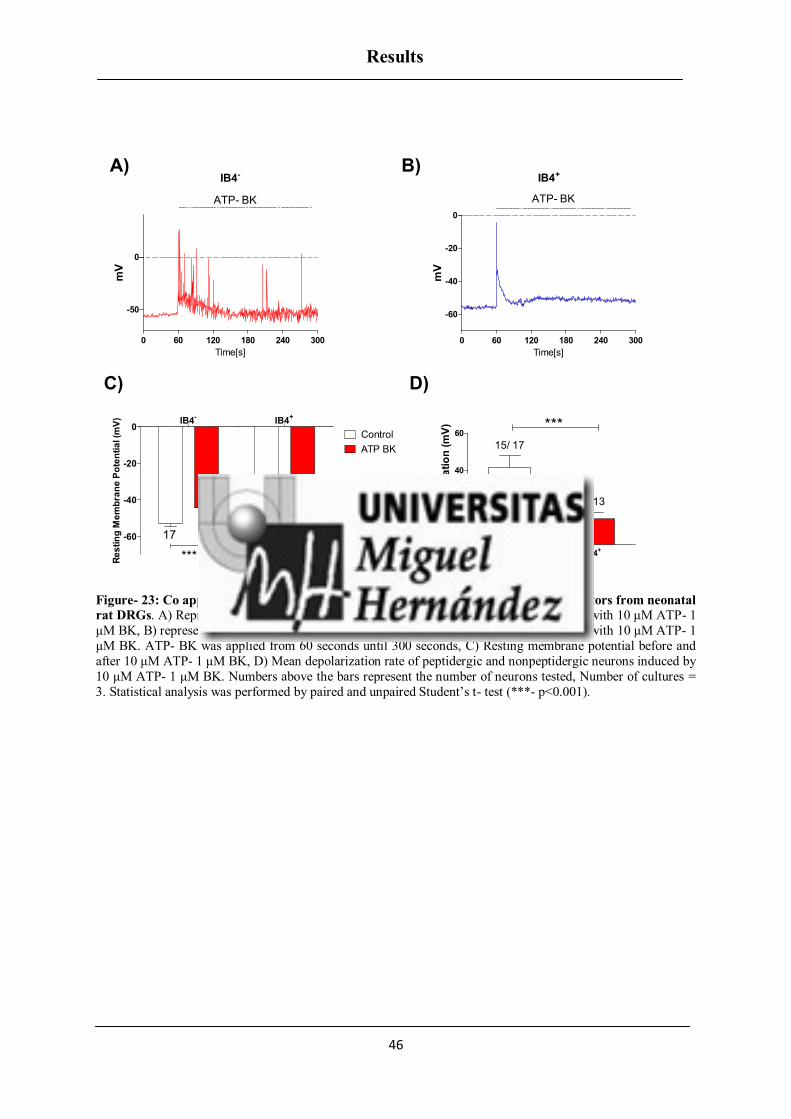
Results
46
IB4-
0 60 120 180 240 300
-50
0
ATP- BK
Time[s]
mV
IB4+
0 60 120 180 240 300
-60
-40
-20
0
ATP- BK
Time[s]
mV
IB4-
IB4+
-60
-40
-20
0Control
ATP BK
******
1713
Re
sti
ng
Me
mb
ran
e P
ote
nti
al (m
V)
IB4- IB4+0
20
40
60
15/ 17
13/ 13
***M
ea
n d
ep
ola
riza
tio
n (
mV
)
Figure- 23: Co application of ATP- BK modulates excitability of IB4- and IB4
+ nociceptors from neonatal
rat DRGs. A) Representative trace from peptidergic neuron exhibiting spontaneous activity with 10 μM ATP- 1
μM BK, B) representative trace from nonpeptidergic neuron exhibiting spontaneous activity with 10 μM ATP- 1
μM BK. ATP- BK was applied from 60 seconds until 300 seconds, C) Resting membrane potential before and
after 10 μM ATP- 1 μM BK, D) Mean depolarization rate of peptidergic and nonpeptidergic neurons induced by
10 μM ATP- 1 μM BK. Numbers above the bars represent the number of neurons tested, Number of cultures =
3. Statistical analysis was performed by paired and unpaired Student’s t- test (***- p<0.001).
A) B)
C) D)

Results
47
ATP INDUCED INFLAMMATORY SENSITIZATION OF TRPV1
Capsaicin evoked neuronal spikes on whole neuronal population using micro electrode
array
Prior experiments on peptidergic and nonpeptidergic nociceptors from neonatal rat
DRGs corroborated that enhanced excitability occurs primarily in peptidergic subpopulations,
while nonpeptidergic subpopulations have a milder increase in excitability upon exposure to
the inflammatory mediators ATP/BK. Furthermore, we characterised the excitability of
neurons using Micro Electrode Array (MEA) technique where DRG neurons formed a cluster
of cultured network resembling in vivo pattern. This technique does not allow differentiating
subpopulations. MEA technique has been used in our experiments to study inflammation
induced sensitization of TRPV1 evoked excitability. All our experiments evaluated capsaicin
induced activation of TRPV1 and its sensitization by inflammatory mediators like ATP, BK
and co- application of ATP- BK. We also evaluated tissue acidosis condition that occurs
during inflammatory microenvironment, by applying ATP- BK at pH 6.2. Prior to our
sensitization experiments, we characterised capsaicin induced TRPV1 spikes, followed by
desensitization of TRPV1 evoked spikes induced by repetitive capsaicin applications on DRG
neurons.
Capsaicin evoked neuronal firing in silent nociceptors
The spike signal obtained in MEA was either from single neuron or a group of
neurons surrounding the electrode. Figure- 24 shows planar microelectrode array (A and B),
where neurons are in contact with the electrodes (C and D) and a capsaicin evoked spike (E).
In MEA chips, smaller the distance between the cell and the electrode, higher the
extracellular signals.

Results
48
Figure- 24: Capsaicin evoked neuronal spikes measured using microelectrode array. (A, B) Planar micro
electrode array, (C) cultured neonatal rat DRG neurons over a substrate array of planar microelectrodes
(Multichannel Systems, Reutlingen, Germany) 2 days after seeding. The electrodes are 30 μm wide; electrode spacing is 200 μm. (D) Cluster of cells coupled to a microelectrode of the array and (E) a trace of 500 nM
Capsaicin evoked spike.
Capsaicin evoked neuronal spikes were measured from neonatal rat DRG neurons
which are silent without any spontaneous activity. As shown in figure- 25 before capsaicin
(Cps) application no spikes were evoked. During capsaicin application downward neuronal
spikes were evoked through activation of TRPV1, which were completely removed after
washing out the vanilloid.
Figure- 25: Representative traces of 500 nM Capsaicin evoked neuronal firing. Exposure to 500 nM
Capsaicin on silent neonatal rat DRG neurons evoked electrical activity mediated through the opening of
TRPV1 channels. Capsaicin was applied for 15 seconds. Neurons did not evoke any electrical activity after washing capsaicin with standard external solution.
A) B) C)
D) E)

Results
49
Capsaicin induced tachyphylaxis of TRPV1 evoked neuronal spikes
It is reported that the repeated activation of TRPV1, leads to desensitization of the
channel activity, a phenomenon known as tachyphylaxis (270). We wanted to study the
process of TRPV1 mediated tachyphylaxis of neuronal spikes in our MEA system. Three
repetitive applications of 500 nM Cps for 15 seconds resulted in desensitization of TRPV1
mediated neuronal spikes as shown in figure- 26. A continuous protocol was used for 16
minutes where three repetitive pulses of 500 nM capsaicin was applied (P1 –1st pulse at 1
st
minute, P2- 2nd
pulse at 5th minute, P3- 3
rd pulse at 14:30 minutes). Upon consecutive
application of capsaicin, TRPV1 evoked neuronal spikes exhibited heterogeneous responses
with either a partial or complete desensitization of neuronal spikes. This protocol was used
for all the MEA experiments. Later, data was processed to extract and compare Mean Spike
Frequency for all the three pulses (P1, P2, and P3) using offline MC RACK program.
Figure- 26: Capsaicin induced tachyphylaxis of TRPV1 evoked neuronal spikes. A) An example of tachyphylaxis to three consecutive applications of 500 nM capsaicin (horizontal bars) given at 5 min intervals.
B, C) represents enlarged view of capsaicin evoked neuronal firing from first pulse. TRPV1 mediated neuronal
firing activity was determined by comparison of all the three pulses (P1, P2, P3) in each group and represented
as Mean spike frequency (Hz).
The brief application of capsaicin pulses for 15 seconds was insufficient to extract the
mean spike frequency. There was a delay in some electrodes between the initial exposure to
capsaicin and the activation of neuronal spikes, which ranges from 4 to 5 seconds. This could
be due to the time required for capsaicin to reach all the electrodes. Furthermore, the
A)
B)
C)

Results
50
heterogeneous response to capsaicin suggests a possible involvement of other membrane
associated ion channels. For this reason, we have applied a time frame during analysis of
MEA data to analyse extra 30 seconds after capsaicin application for all the three pulses (P1,
P2, P3).
Peptide DD04107 does not interfere with basal TRPV1 activity
To study the effect of inhibiting neuronal exocytosis on TRPV1 sensitization, we used
a botulinomimetic peptide DD04107 (EEMQRR), which disrupts the SNARE complex
formation. First we wanted to characterise the peptide under control conditions to know if it
interferes with the basal TRPV1 activity, by using both MEA and patch clamp techniques.
For MEA experiments DRG neurons were preincubated with 20 µM palmitoylated DD04107
for one hour. Application of 500 nM capsaicin on DRG neurons seeded in MEA evoked
neuronal spikes mediated through TRPV1 (P1). Re-application of capsaicin after 5 minutes
interval twice (P2, P3) resulted either in a response of greatly reduced neuronal spikes or in
no response, which indicated that desensitization of TRPV1 had occurred. The mean spike
frequency of a second pulse of capsaicin never exceeded the frequency of neuronal spikes of
the first pulse and it was further reduced with the third pulse (P3). Thus the desensitization of
neuronal spikes evoked by TRPV1 was not readily reversible. Preincubation of neurons with
DD04107 did not affect capsaicin evoked neuronal spikes and its desensitization as shown in
figure- 27.

Results
51
Figure- 27: DD04107 does not affect TRPV1 evoked neuronal spikes and its tachyphylaxis. Application of
capsaicin (500 nM, 15 s) on neonatal rat DRG neurons induced TRPV1 evoked neuronal excitability in both
vehicle and DD04107 treated groups (1st pulse- P1). Repeated exposure of DRG neurons to capsaicin (2nd pulse (P2) and 3rd pulse (P3)) evoked fewer action potentials compared to the 1st pulse due to the desensitization of
TRPV1 channel activity.
We also tested the effect of nonpalmitoylated DD04107 on basal TRPV1 activity in
IB4- and IB4
+ nociceptors using patch clamp technique under whole cell configuration mode.
In both subpopulations 100 μM nonpalmitoylated DD04107 (represented as Peptide) was
applied through patch pipette, and the recordings were started 10 minutes after forming the
seal. Application of 1 μM capsaicin induced TRPV1 currents on both subpopulations, which
also exhibited desensitization upon repetitive capsaicin application. Neurons treated with
nonpalmitoylated DD04107 also exhibited activation of TRPV1 currents and its
desensitization in both subpopulations as shown in figure- 28.

Results
52
Cps Cps
Cps Cps
Vehicle
Peptide
50pA 10pA
100sec 100sec
IB4- IB4+
Figure- 28: Representative voltage clamp recordings of currents elicited by capsaicin (1 μM, 10 s) in IB4-
and IB4+ nociceptors treated with nonpalmitoylated DD04107 (A, B) 100 µM nonpalmitoylated DD04107
represented as Peptide was given through the patch pipette and incubated for 10 minutes after forming the seal.
Three repetitive pulses of capsaicin at 1 µM concentration were given for the continuous protocol.
In addition, we wanted to check if nonpalmitoylated DD04107 (Peptide) interferes
with the basal TRPV1 current density in nociceptors. We calculated the ratio of initial
TRPV1 current responses (pA) to their corresponding cell capacitance (pF) and compared
between Vehicle and Peptide treated groups in both subpopulations (IB4-, IB4
+). As shown in
figure- 29, DD04107 did not affect basal TRPV1 current density in both IB4- and IB4
+
nociceptors.
A) B)

Results
53
IB4-
IB4+
0
2
4
6
VehiclePeptide
34 26
28 24C
urr
en
t d
en
sit
y J
(p
A/p
F)
Figure- 29: Nonpalmitoylated DD04107 (Peptide) does not affect basal TRPV1 currents in IB4- and IB4
+
neurons. Current density was calculated by taking the ratio of initial TRPV1 current responses (pA) to their
corresponding cell capacitance (pF). Data are expressed as mean ± SEM. The numbers above the bars represent
the total number of neurons registered. Number of cultures = 3. Statistical analysis was performed by two- way
ANOVA with Bonferroni’s post hoc test.
Pharmacological blockage of neuronal exocytosis reduces ATP induced sensitization of
TRPV1 excitability
ATP induced sensitization of TRPV1 excitability in rat nociceptors was evaluated
using MEA technique. As seen before, consecutive application of capsaicin led to
desensitization in the number of spikes evoked and desensitized responses could be
significantly recovered with ATP sensitization (10 μM ATP was applied between P2 and P3)
as shown in figure- 30 (Vehicle). ATP induced sensitization of TRPV1 excitability was
blocked by 20 μM DD04107, an inhibitor of neuronal exocytosis, as shown in figure- 30
(DD04107). This indicates that ATP induced TRPV1 sensitization was predominantly due to
the release of new TRPV1 channels from the vesicles and their insertion into the plasma
membrane of DRG neurons. Although we observed a less significant potentiation of TRPV1
activity by ATP in DD04107 treated group (*-p<0.05) compared to Vehicle group (***-
p<0.001), we still have potentiation of TRPV1 evoked excitability (figure- 31). This
confirms that there is a subpopulation of nociceptors involved in channel recruitment.
Therefore, it was noteworthy to evaluate the fold increase in the potentiation of TRPV1
evoked spikes induced by ATP between Vehicle and DD04107 treated groups.

Results
54
Figure- 30: DD04107 abrogates, ATP sensitized TRPV1- mediated nociceptor excitability. Representative
traces of potentiation of capsaicin (500 nM, 15 s) responses elicited by applying 10 μM of ATP between the second (P2) and third (P3) vanilloid pulse (top), in neonatal rat DRG neurons. Preincubation of the neurons with
DD04107 (bottom) also induced potentiation of TRPV1 but it was less significant compared to Vehicle (top).
Vehicle DD04107 Vehicle DD041070
1
2
3P1
P2
P3
*
***
Control ATP
40
17
3541
Mean
Sp
ike F
req
uen
cy (
Hz)
Figure- 31: DD04107 abrogates ATP induced inflammatory sensitization of TRPV1 mediated neuronal
firing in rat DRG neurons. Effect of 20 µM DD04107 on 10 μM ATP mediated potentiation of TRPV1
evoked neuronal firing activity was determined by comparison of all the three pulses (P1, P2, P3) in each groups
and represented as Mean spike frequency (Hz). Data are expressed as mean ± SEM. The numbers above the bars
represent the total number of electrodes that responded. Number of cultures = 3. Statistical analysis was
performed by one way ANOVA repeated measures with Bonferroni’s post hoc test (***- p<0.001, *- p<0.05).
We have calculated the ratio of third pulse to second pulse (P3/P2) in both Vehicle
and DD04107 treated groups exposed to ATP. Then, the impact of DD04107 on blocking the

Results
55
potentiation of TRPV1 channel induced by ATP treatment was compared between Vehicle
and DD04107 groups as shown in figure- 32. A significant blockage of TRPV1 potentiation
on ATP treated neurons pre incubated with DD04107 (2.6±0.5) compared to the Vehicle
(5.5±1.5) (**- p<0.01) was observed. Although ATP induced TRPV1 exocytosis is mainly
due to the release of intracellular Ca2+
, no such potentiation was observed under control
conditions, which confirms that external Ca2+
ions entering through TRPV1 were not
involved.
Control ATP
0
2
4
6
8Vehicle
DD04107
*** **
1919
15
29
Fo
ld P
ote
nti
ati
on
Figure- 32: ATP mediated TRPV1 sensitization requires channel recruitment in a subset of nociceptors.
Effect of 20 µM DD04107 on 10 μM ATP mediated fold potentiation of TRPV1 activity was determined by
comparison of capsaicin-mediated neuronal spikes before and after ATP incubation (ratio P3/P2) of the groups
(vehicle, DD04107). Data are expressed as mean ± SEM. The numbers above the bars represent the total
number of electrodes that responded. Number of cultures = 3. Statistical analysis was performed by two- way
ANOVA with Bonferroni’s post hoc test (**- p<0.01, ***- p<0.001).
Neuronal exocytosis is responsible for ATP induced TRPV1 sensitization in IB4-
neurons but not for IB4+
neurons
Using MEA technique, we found that ATP induced sensitization of TRPV1
excitability was due to the release of new TRPV1 channels and their insertion into the
membrane in a subset of nociceptors. Next we wanted to know which subtype of nociceptor
was involved in mobilization of TRPV1 channels upon ATP exposure. For that purpose ATP
induced TRPV1 sensitization was analysed in both peptidergic (IB4-) and nonpeptidergic
(IB4+) subpopulations. Capsaicin induced desensitization of TRPV1 currents in IB4
-
nociceptors was potentiated by 10 μM ATP as shown in figure- 33 A (Vehicle). Notably,

Results
56
ATP induced TRPV1 sensitization in IB4- nociceptors was blocked by 100 μM
nonpalmitoylated DD04107 (represented as Peptide) delivered to the neuronal cytosol
through the pipette as shown in figure- 33 A (Peptide). Similarly in IB4+ neurons, ATP
induced TRPV1 sensitization was observed, but it was insensitive to the blocker of neuronal
exocytosis peptide as shown in figure- 33 (B).
Cps Cps
Cps Cps
ATP ATP
ATP ATP
Vehicle
Peptide
IB4- IB4+
50pA 10pA
100sec 100sec
Figure- 33: Nonpalmitoylated DD04107 (Peptide) mediated abrogation of ATP induced TRPV1
sensitization occurs primarily in peptidergic nociceptors. Representative voltage clamp recordings of ATP
induced sensitization of TRPV1 currents elicited by capsaicin (1 μM, 10 s) and the abrogating effect of the
peptide in IB4- and IB4+ nociceptors (A, B). Cells were held at -60 mV. 100 µM nonpalmitoylated DD04107
was given through the patch pipette and incubated for 10 minutes after forming the seal. Three repetitive pulses of capsaicin at 1 µM concentration were given for the continuous protocol. 10 μM ATP was applied between
second and third pulse of capsaicin.
Fold potentiation of TRPV1 currents was calculated by taking the ratio of P3 to P2.
As shown in figure- 34 both IB4- and IB4
+ subpopulations had no significant changes in fold
potentiation between Vehicle and nonpalmitoylated DD04107 groups under control
conditions. In IB4-
nociceptors, fold increase in current intensity was significantly higher
after ATP incubation compared to control Vehicle group (*- p<0.05). ATP induced increase
in TRPV1 potentiation was significantly blocked by 100 μM nonpalmitoylated DD04107
(**- p<0.01). Similarly, in IB4+ nociceptors fold increase in current intensity was
significantly higher after ATP incubation compared to control Vehicle group (**- p<0.01),
A) B)

Results
57
whereas ATP induced increase in TRPV1 potentiation was not blocked by 100 μM
nonpalmitoylated DD04107 although a tendency was observed. This could be due to the
possible involvement of differential TRPV1 sensitization mechanism involved in IB4-
(exocytosis) and IB4
+ (phosphorylation) nociceptors.
Control ATP Control ATP0.0
0.5
1.0
1.5
2.0Vehicle
Peptide
**
* **
IB4-
IB4+
27 15
19
2010
7
15
16
Fo
ld P
ote
nti
ati
on
Figure- 34: Nonpalmitoylated DD04107 lessens ATP induced inflammatory sensitization of TRPV1
channel in peptidergic but not in nonpeptidergic nociceptors. Effect of 100 µM nonpalmitoylated DD04107
on 10 μM ATP mediated fold potentiation of TRPV1 activity was determined by comparison of capsaicin-mediated current intensities before and after incubation of ATP (ratio P3/P2) of the groups (Control Vehicle,
Control Peptide, ATP Vehicle, ATP Peptide) for both IB4- and IB4+ subpopulations. Data are expressed as mean
± SEM. The numbers above the bars represent the total number of neurons registered. Number of cultures = 4.
Statistical analysis was performed by two-way ANOVA with Bonferroni’s post hoc test (*- p<0.05, **- p<0.01).
Further we focussed on the fold potentiation (ratio P3/P2) to check the percentage of
neurons that exhibited sensitization of TRPV1 induced by ATP in IB4- and IB4
+
subpopulations, as shown in figure- 35. There was a significant difference in the percentage
of neurons that exhibited potentiated response to capsaicin after incubation with ATP,
compared between Vehicle and nonpalmitoylated DD04107 treated groups. In fact in IB4-
group, exposure to ATP induced 30 % of neurons in the Vehicle group with potentiated
response, whereas in DD04107 treated group we observed a significant blockage of ATP
induced TRPV1 potentiation with only 5 % of neurons being potentiated. In IB4+ group, a
higher percentage of neurons exhibited potentiation of TRPV1 currents induced by ATP in
both Vehicle (60 % of neurons) and DD04107 treated groups (40 % of neurons). This
substantiates that there is a possible involvement of two distinct potentiation mechanisms in
IB4- (through exocytosis) and IB4
+ (through channel gating mechanism) subpopulations.

Results
58
Vehicle Peptide Vehicle Peptide0
20
40
60
80
100desensitized
same response
sensitized
ATP- IB4+ATP- IB4
-
Perc
en
tag
e o
f N
eu
ron
s
Figure- 35: Percentage of Neurons with ATP induced potentiation of TRPV1 activity in peptidergic and
nonpeptidergic nociceptors treated with Vehicle and nonpalmitoylated DD04107 (Peptide). Percentage of
neurons with potentiated TRPV1 activity when treated with 10 µM ATP were determined in both peptidergic
and nonpeptidergic rat DRG neurons. The values of the ratio P3/P2 in both vehicle and peptide treated groups
were calculated. The criteria is when the ratio is lesser than 0.9 the neurons are desensitized; ratio of 0.9-1.1 there is no significant difference between the second and third pulses; ratio greater than 1.1 the channel is
getting sensitized with ATP.
ATP mediated TRPV1 potentiation in IB4+ but not in IB4
- subpopulation is PKC
dependent
Since we found that the potentiation of TRPV1 by ATP was due to the differential
mechanism involved in IB4- (primarily due to exocytosis) and IB4
+ (primarily due to channel
gating) neurons, we could not exclude the possibility of phosphorylation mechanism involved
in both subpopulations. To confirm that we used a general PKC inhibitor and examined
whether PKC is involved in ATP induced TRPV1 potentiation in both IB4- and IB4
+ neurons.
Bisindolylmaleimide (BIM) was used at 1 μM concentration and given through the patch
pipette (10 minutes incubation after forming the seal). Application of three repetitive pulses
of 1 μM capsaicin induced desensitization of TRPV1 channel that was significantly recovered
by the application of 10 µM ATP between the second and third pulses. As shown in figure-
36 ATP induced sensitization of TRPV1 currents was not affected in IB4- neurons treated
with BIM, whereas in IB4+ neurons, potentiation of TRPV1 currents by ATP was strongly
inhibited by BIM.

Results
59
ATP ATP
ATP ATP
Cps Cps
Cps Cps
50pA
100sec
50pA
100sec
Vehicle
BIM
IB4- IB4+
Figure- 36: PKC inhibitor (BIM) prevents the potentiation of TRPV1 induced by ATP in IB4+ but not in
IB4- neurons. Representative voltage clamp recordings of currents elicited by capsaicin (1 μM, 10 s) in IB4-
and IB4+ nociceptors (A, B). Cells were held at -60 mV. 1 μM BIM was given through the patch pipette and
incubated for 10 minutes after forming the seal. Three repetitive pulses of capsaicin at 1 µM concentration were
given for the continuous protocol. 10 μM ATP was applied between second and third pulse of capsaicin.
Fold potentiation of TRPV1 currents was calculated by taking the ratio of P3 to P2.
Figure- 37 shows that in IB4- and IB4
+ subpopulations under control conditions no
significant changes in fold potentiation were observed in both Vehicle and BIM treated
groups. In IB4- nociceptors, fold increase in current intensity was significantly higher after
ATP incubation (*- p<0.05) compared to control Vehicle group. ATP induced TRPV1
potentiation was not affected by 1 μM BIM. Similarly, in IB4+ nociceptors fold increase in
current intensity was significantly higher after ATP incubation (**- p<0.01) compared to
control Vehicle group. ATP induced TRPV1 potentiation was significantly blocked by 1 μM
BIM (*- p<0.05).
A) B)

Results
60
Control ATP Control ATP0.0
0.5
1.0
1.5Vehicle
BIM*
** *
IB4-
IB4+
3312
2323
119
24
23F
old
Po
ten
tiati
on
Figure- 37: BIM (PKC inhibitor) abolishes ATP induced inflammatory sensitization of TRPV1 channel in
nonpeptidergic nociceptors. Effect of 1 µM BIM on 10 μM ATP mediated fold potentiation of TRPV1 activity
was determined by comparison of capsaicin-mediated current intensities before and after incubation of ATP (ratio P3/P2) of the groups (Control Vehicle, Control BIM, ATP Vehicle, ATP BIM) for both IB4- and IB4+
subpopulations. Data are expressed as mean ± SEM. The numbers above the bars represent the total number of
neurons registered. Number of cultures = 4. Statistical analysis was performed by two-way ANOVA with
Bonferroni’s post hoc test (*- p<0.05, **- p<0.01).
In addition, we focused on the fold potentiation (ratio P3/P2) to check the percentage
of IB4- and IB4
+ neurons that exhibited potentiation of TRPV1 induced by ATP as shown in
figure- 38. There was a significant difference in the percentage of neurons that exhibited
potentiated response to capsaicin after incubation with ATP, compared with control groups.
In fact, in IB4- group exposure to ATP induced 18 % of neurons with a potentiated response
in the Vehicle group. We have not observed blockage of ATP induced TRPV1 potentiation in
BIM treated neurons which showed 13 % of neurons with a potentiated response, although a
tendency was observed. Similarly, in IB4+
group exposure to ATP induced a higher
percentage of neurons with potentiated responses in Vehicle group (16 % of neurons),
whereas neurons treated with BIM exhibited a complete blockage of TRPV1 potentiation
induced by ATP. Hence, it confirms that ATP induced sensitization of TRPV1 in IB4+
nonpeptidergic neurons is PKC dependent, but not in IB4- peptidergic neurons where TRPV1
exocytosis is primarily involved, though a possible role of PKC cannot be excluded.

Results
61
Vehicle BIM Vehicle BIM0
20
40
60
80
100desensitized
same response
sensitized
ATP- IB4-
ATP- IB4+
Perc
en
tag
e o
f N
eu
ron
s
Figure- 38: Percentage of Neurons with ATP induced potentiation of TRPV1 activity in peptidergic and
nonpeptidergic nociceptors treated with Vehicle and BIM. Percentage of neurons with potentiated TRPV1 activity when treated with 10 µM ATP was determined in both peptidergic and nonpeptidergic rat DRG
neurons. The value of the ratio P3/P2 in both Vehicle and BIM treated groups was calculated. The criteria is
when the ratio is lesser than 0.9 the neurons are desensitized; ratio of 0.9-1.1 there is no significant difference
between the second and third pulses; ratio greater than 1.1 the channel is getting sensitized with ATP.
Role of αCGRP and Tac1 on ATP induced inflammatory sensitization of TRPV1
Experiments from rat DRG neurons have confirmed that peptidergic nociceptors are
primarily involved in recruiting new TRPV1 channels from the vesicles to the plasma
membrane upon ATP exposure. We further extended our studies to know the functional
mechanism behind exocytotic release of TRPV1 during inflammation. We focused on the
role of two primary neuropeptides, αCGRP and Tac1 (encoding SP) in sensitizing TRPV1.
We hypothesize that there is a possible involvement of these two neuropeptides in sorting
TRPV1 channels in large dense core vesicles (LDCVs) and driving their regulated exocytosis
upon inflammatory (ATP) insult. We used αCGRP-/-
and Tac1-/-
gene knockout mice and
generated a double knockout mice model of αCGRP-/-
xTac1-/-
(referred to as DKO). We
studied ATP induced potentiation of TRPV1 in αCGRP-/-
xTac1-/-
compared with wild type
mice.
Recently, it has been reported that TRPV1 is primarily restricted to peptidergic
subpopulation during development in mice and it does not co localise with IB4 (a marker for
nonpeptidergic nociceptors) (199). All our experiments were done in adult mice for both wild

Results
62
type and double knockout models with 12- 15 weeks of age to be sure that the observed
TRPV1 responses we studied were from peptidergic nociceptors. We studied ATP induced
potentiation of TRPV1 evoked neuronal spikes in wild type and DKO mice models using
MEA technique.
Removal of neuropeptides did not affect basal TRPV1 responses and the intrinsic
membrane properties of nociceptors
Before performing sensitization experiments we wanted to know if the removal of
neuropeptides potentially affects the basal TRPV1 responses and the electrical properties of
nociceptors. Capsaicin responses and intrinsic electrogenic properties in both wild type and
DKO mice were studied using patch clamp technique.
A capsaicin dose response curve on TRPV1+
DRG neurons from wild type and DKO
mice exhibited no significant changes with no alteration in the EC 50 (0.7±0.1 µM for WT vs
0.5±0.1 µM for DKO) as shown in figure- 39.
-7 -6 -50.0
0.5
1.0
CGRP-/-
xTac1-/-
WT
Capsaicin (log M)
No
rmalise
d c
urr
en
t d
en
sit
y
J (
pA
/pF
)
Figure- 39: Capsaicin dose-response curves from wild type and αCGRP-/-
xTac1-/-
DRG at -60 mV. Ionic
currents evoked by different concentrations of capsaicin were measured in wild type and αCGRP-/-xTac1-/-
nociceptors, by patch clamp in whole-cell configuration and standard external solution was used.
Also we studied electrogenic properties of the nociceptors from wild type and DKO
mice positive for TRPV1. As shown in table- 2 we did not observe any significant changes in

Results
63
the electrical properties measured for resting membrane potential, current threshold, threshold
potential, action potential amplitude, action potential overshoot, action potential duration and
after hyperpolarization amplitude in both wild type and DKO mice.
WT αCGRP-/-
xTac1-/-
RMP (mV) -45.6 ± 2.3 (13) -44.9 ± 2.2 (21)
CT (pA) 12.5 ± 2.5 (13) 14.7 ± 3.0 (21)
TP (mV) -32.8 ± 1.6 (13) -33.5 ± 1.8 (21)
AP amplitude (mV) 67.0 ± 4.1 (13) 68.6 ± 4.3 (21)
AP overshoot (mV) 23.0 ± 2.6 (13) 19.5 ± 2.5 (21)
AP duration (ms) 21.5 ± 0.4 (13) 21.2 ± 0.5 (21)
AHP amplitude (mV) 9.2 ± 1.3 (13) 8.9 ± 1.6 (21)
Table- 2: Intrinsic electrogenic properties of TRPV1 sensitive DRG neurons in wild type and αCGRP-/-
xTac1-/-
mice. RMP: resting membrane potential; CT: the depolarized current threshold for evoking the 1st action potential; TP: threshold potential; AHP: afterhyperpolarization; AP: action potential. No changes were
observed in αCGRP-/- x Tac1-/- compared to wild type DRG neurons as assessed by the intrinsic electrogenic
parameters. Data are expressed as mean ± SEM (n). Statistical analysis was performed by Unpaired Student’s t-
test.
ATP induced sensitization of neuronal firing mediated through TRPV1 in wild type and
αCGRP-/-
xTac1-/-
mice nociceptors
ATP induced sensitization of TRPV1 excitability in nociceptors from wild type and
DKO mice DRG neurons was evaluated using MEA technique. Application of capsaicin led
to activation of TRPV1 channel in DRG neurons, inducing changes in ionic flow, which was
detected as a train of evoked neuronal spikes, as shown in Vehicle for both wild type and
DKO mice neurons (figure- 40 A and B) and represented as Mean Spike Frequency (Hz). In
wild type nociceptors, repetitive capsaicin pulses induced desensitization of the vanilloid-
evoked electrical responses. Desensitized capsaicin responses could be recovered with ATP
sensitization and blocked by DD04107 (figure- 40 A and C). In DKO nociceptors, capsaicin
also induced neuronal activity. Vanilloid-induced TRPV1 tachyphylaxia was partially
overturned by ATP (figure- 40 B) and virtually insensitive to DD04107 (figure- 40 D).

Results
64
Figure- 40: ATP induced sensitization of neuronal firing mediated through TRPV1 in wild type and
αCGRP-/-
xTac1-/-
mice nociceptors. Repetitive application of capsaicin 500 nM induced tachyphylaxis of
TRPV1 mediated neuronal firing and it was reversed by sensitizing the neurons with 10 µM ATP in wild type
and αCGRP-/-xTac1-/- mice nociceptors.
DD04107 abrogates ATP induced sensitization of TRPV1 evoked neuronal spikes in
wild type nociceptors
Consecutive application of capsaicin led to desensitization in the number of spikes
evoked and desensitized responses could be significantly recovered (***- p<0.001) with ATP
sensitization (10 μM ATP was applied between P2 and P3) in wild type nociceptors. Some
neurons exposed to ATP evoked fast neuronal spikes, due to the activation of ionotropic
P2X3 receptor, which is widely expressed in nociceptors. ATP induced sensitization of
TRPV1 evoked excitability from wild type DRG neurons was blocked by DD04107,
indicating that ATP induced TRPV1 sensitization was mainly due to the insertion of new
TRPV1 channels into the plasma membrane, as shown in figure- 41.
A) C)
B) D)

Results
65
Vehicle DD04107 Vehicle DD041070
1
2
3P1
P2
P3
Control ATP
5020
67
31
***M
ean
Sp
ike F
req
uen
cy (
Hz)
Figure- 41: DD04107 abolishes ATP induced inflammatory sensitization of TRPV1 mediated neuronal
firing in wild type mice DRG neurons. Effect of 20 µM DD04107 on 10 μM ATP mediated potentiation of
TRPV1 evoked neuronal firing activity was determined by comparison of all the three pulses (P1, P2, P3) in
each group and represented as Mean spike frequency (Hz). Data are expressed as mean ± SEM. The numbers
above the bars represent the total number of electrodes that responded. Number of cultures = 4. Statistical
analysis was performed by one way ANOVA repeated measures with Bonferroni’s post hoc test (***- p<0.001).
DD04107 did not affect ATP induced sensitization of TRPV1 evoked neurons spikes in
αCGRP-/-
xTac1-/-
nociceptors
In DKO mice neurons also repetitive application of capsaicin induced desensitization
in the number of spikes evoked and it was significantly recovered (*- p<0.05) with ATP
sensitization, though it was less significant compared to wild type DRG neurons. However
DRG neurons from DKO mice model did not show any blockage of TRPV1 potentiation after
treatment with DD04107 as we see a significant potentiation of TRPV1 activity (*- p<0.05)
by ATP in DD04107 treated group as shown in figure- 42. Hence these results show the
significant role of neuropeptides on promoting TRPV1 exocytosis. The remnant potentiation
we observed in DD04107 treated group could be due to the modification of channel gating
through phosphorylation process and not through TRPV1 exocytosis.
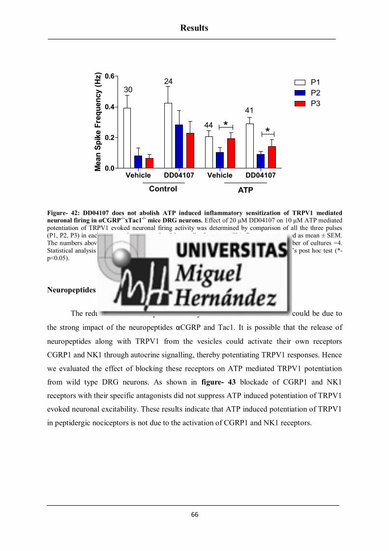
Results
66
Vehicle DD04107 Vehicle DD041070.0
0.2
0.4
0.6P1
P2
P3
Control ATP
3024
44
41
**
Mean
Sp
ike F
req
uen
cy (
Hz)
Figure- 42: DD04107 does not abolish ATP induced inflammatory sensitization of TRPV1 mediated
neuronal firing in αCGRP-/-
xTac1-/-
mice DRG neurons. Effect of 20 µM DD04107 on 10 μM ATP mediated
potentiation of TRPV1 evoked neuronal firing activity was determined by comparison of all the three pulses
(P1, P2, P3) in each group and represented as Mean spike frequency (Hz). Data are expressed as mean ± SEM.
The numbers above the bars represent the total number of electrodes that responded. Number of cultures =4.
Statistical analysis was performed by one way ANOVA repeated measures with Bonferroni’s post hoc test (*-
p<0.05).
Neuropeptides did not sensitize TRPV1 through autocrine signalling
The reduction of TRPV1 potentiation by ATP in DKO mice models could be due to
the strong impact of the neuropeptides αCGRP and Tac1. It is possible that the release of
neuropeptides along with TRPV1 from the vesicles could activate their own receptors
CGRP1 and NK1 through autocrine signalling, thereby potentiating TRPV1 responses. Hence
we evaluated the effect of blocking these receptors on ATP mediated TRPV1 potentiation
from wild type DRG neurons. As shown in figure- 43 blockade of CGRP1 and NK1
receptors with their specific antagonists did not suppress ATP induced potentiation of TRPV1
evoked neuronal excitability. These results indicate that ATP induced potentiation of TRPV1
in peptidergic nociceptors is not due to the activation of CGRP1 and NK1 receptors.

Results
67
ATP CGRP8-37 CP96345
0.0
0.5
1.0
1.5
P1
P2
P353
28
26
***
*M
ean
Sp
ike F
req
uen
cy (
Hz)
Figure- 43: Neuropeptides CGRP and SP does not sensitize TRPV1 through autocrine signalling in wild
type nociceptors. Effect of αCGRP (250 nM CGRP 8-37 ) and Tac1 (10 μM CP96345) receptor antagonists was
studied by co application with 10 μM ATP on potentiation of TRPV1 evoked neuronal firing activity,
determined by comparison of all the three pulses (P1, P2, P3) in each group and represented as Mean spike
frequency (Hz). Data are expressed as mean ± SEM. The numbers above the bars represent the total number of
electrodes responded. Number of cultures = 3. Statistical analysis was performed by one way ANOVA repeated
measures with Bonferroni’s post hoc test (*- p<0.05, **- p<0.01).
αCGRP is essential for ATP induced sensitization of TRPV1
In order to know which neuropeptide was involved in regulating TRPV1 exocytosis,
we next studied the role of each neuropeptide in the nociceptors from αCGRP-/-
and Tac1-/-
single knockout mice. We evaluated ATP induced potentiation of TRPV1 evoked spikes in
single knockouts compared with wild type mice. Our result shows that capsaicin induced
desensitization of TRPV1 evoked neuronal spikes was completely reversed by ATP in
nociceptors from Tac1-/-
mice. Nevertheless, abrogation of αCGRP expression eliminated
ATP induced increase in mean spike frequency of TRPV1 evoked neuronal firing as shown in
figure- 44. Together, these results indicate that αCGRP expression is necessary for ATP
induced TRPV1 potentiation in peptidergic nociceptors, whereas SP plays a marginal role.

Results
68
Control ATP Control ATP Control ATP
0.0
0.5
1.0
1.5
2.0
P1
P2
P3
Wild type -CGRP-/-
Tac1-/-
*
*58 66
3428
19 46
Mean
Sp
ike F
req
uen
cy (
Hz)
Figure- 44: αCGRP but not Tac1 is involved in ATP induced TRPV1 sensitization. Effect of 10 μM ATP
mediated potentiation of TRPV1 evoked neuronal firing activity was determined by comparison of all the three
pulses (P1, P2, P3) in each group (wild type, αCGRP-/- and Tac1-/-) and represented as Mean spike frequency
(Hz). Data are expressed as mean ± SEM. The numbers above the bars represent the total number of electrodes
that responded. Number of cultures = 4. Statistical analysis was performed by one way ANOVA repeated
measures with Bonferroni’s post hoc test (*- p<0.05).
Experimental depolarization does not sensitize TRPV1
We found that ATP induced TRPV1 exocytosis is mainly due to the elevation of
intracellular Ca2+
ions, which activates SNARE complex machinery. Many studies on DRG
neurons revealed that experimental depolarization stimulated by higher KCl concentration
could induce exocytosis by increasing intracellular Ca2+
concentration. Such depolarizing
stimuli could induce the release of neuropeptides CGRP and SP. We found that the removal
of these two neuropeptides significantly affects ATP induced TRPV1 potentiation. Thus, we
questioned whether electrical stimulation of the neurons through depolarization could induce
exocytosis of TRPV1 carrying vesicles and potentiate TRPV1 activity similar to ATP. Hence
we performed experiments on nociceptors from wild type mice and compared depolarization
induced TRPV1 potentiation with ATP treated groups. As shown in figure- 45 depolarizing
the neurons with 40 mM KCl for 8 minutes did not potentiate TRPV1 evoked neuronal firing
compared to ATP treated neurons which showed a significant potentiation (***- p<0.001) of
TRPV1 spikes. It evidently demonstrates that the inflammatory sensitization of TRPV1 due

Results
69
to the recruitment of new channels from LDCVs to the neuronal surface is specific for
inflammatory mediator ATP, but not for depolarizing pulses.
Control KCl ATP
0.0
0.2
0.4
0.6
0.8
1.0P1
P2
P3
6778
72
***
Mean
Sp
ike F
req
uen
cy (
Hz)
Figure- 45: Depolarization of nociceptors does not sensitize TRPV1 in wild type mice. Effect of 10 μM
ATP and 40 mM KCl mediated potentiation of TRPV1 evoked neuronal firing activity was determined by
comparison of all the three pulses (P1, P2, P3) in each group and represented as Mean spike frequency (Hz).
Data are expressed as mean ± SEM. The numbers above the bars represent the total number of electrodes that
responded. Number of cultures = 3. Statistical analysis was performed by one way ANOVA repeated measures
with Bonferroni’s post hoc test (***- p<0.001).

Results
70
BK INDUCED INFLAMMATORY SENSITIZATION OF TRPV1
Blockage of neuronal exocytosis also lessens BK induced TRPV1 excitability
Earlier experiments with ATP revealed that DD04107 significantly reduced the
sensitization of TRPV1, which primarily occurs in peptidergic nociceptors. Furthermore,
knocking down of the genes αCGRP and Tac1 corresponding to the neuropeptides αCGRP
and SP had a high impact on exocytotic inflammatory sensitization of TRPV1, with αCGRP
playing a major role in TRPV1 vesicular release. As we cited earlier bradykinin (BK), a
potent inflammatory mediator, is also released during tissue injury. BK is reportedly involved
in increasing the membrane expression and trafficking of opioid receptors in sensory neurons
(316). Previous studies from our group reported that DD04107 abolished only ATP but not
BK mediated TRPV1 potentiation suggesting that not all pro inflammatory mediators
promote the exocytotic release of TRPV1 (312).
In addition, recent studies revealed that BK induced CGRP release is dose dependent:
acute incubation (10 minutes) of DRG neurons with 1 μM BK enhanced the release of CGRP
(317). Akin to ATP, BK also exerts its action through Gαq/11 signalling pathway activated by
BK2 receptor. Henceforth, we proposed that BK could also induce sensitization of TRPV1
through exocytotic vesicular release of new TRPV1 channels.
In order to unravel the mechanism behind BK induced TRPV1 sensitization in rat
DRG neurons, we performed both MEA and patch clamp technique on IB4- and IB4
+
nociceptors, similar to ATP experiments. In MEA experiments consecutive application of
capsaicin led to desensitization in the number of spikes evoked and desensitized responses
could be significantly recovered after BK sensitization (1 μM BK was applied between P2
and P3) (figure- 46 Vehicle). BK induced sensitization of TRPV1 evoked excitability was
abrogated by DD04107 (figure- 46 DD04107). This indicates that BK induced TRPV1
sensitization was mainly due to the release of new TRPV1 channels from the vesicles and
their insertion into the plasma membrane in a subset of nociceptors.

Results
71
Figure- 46: DD04107 abrogates, BK sensitized TRPV1 evoked nociceptor excitability. Representative traces
of potentiation of capsaicin (500 nM, 15 s) responses elicited by applying 1 μM of BK between the second (P2)
and third (P3) vanilloid pulse (top), in neonatal rat DRG neurons. Preincubation of the neurons with 20 μM
DD04107 (bottom) also potentiated TRPV1 but it was less significant compared to Vehicle (top).
Though we saw a less significant potentiation of TRPV1 activity by BK in DD04107
treated group (*- p<0.05) compared to Vehicle group (***- p<0.001), we still observed
potentiation of TRPV1 evoked excitability as shown in figure- 47. Hence it was important to
compare the fold increase in BK induced potentiation of TRPV1 between Vehicle and
DD04107 treated groups.

Results
72
Vehicle DD04107 Vehicle DD041070.0
0.2
0.4
0.6
0.8P1
P2
P3***
*
Control BK
40
24
40
65
Mean
Sp
ike F
req
uen
cy (
Hz)
Figure- 47: DD04107 lessens BK induced inflammatory sensitization of TRPV1 in neonatal Rat DRG
neurons. Effect of 20 µM DD04107 on 1 μM BK mediated potentiation of TRPV1 evoked neuronal firing
activity was determined by comparison of all the three pulses (P1, P2, P3) in each group and represented as
Mean spike frequency (Hz). Data are expressed as mean ± SEM. The numbers above the bars represent the total
number of electrodes that responded. Number of cultures = 3. Statistical analysis was performed by one way
ANOVA repeated measures with Bonferroni’s post hoc test (*- p<0.05, ***- p<0.001).
We analysed the ratio of third pulse to second pulse (P3/P2) in both vehicle and
DD04107 treated groups exposed to BK. Then, the impact of DD04107 on blocking the
potentiation of TRPV1 channel induced by BK treatment was compared between Vehicle and
DD04107 groups. We observed a significant blockage of BK induced TRPV1 potentiation on
neurons pre incubated with DD04107 (3±0.4) compared to the Vehicle (7±2.5) (*- p<0.05) as
shown in figure- 48. BK induced TRPV1 exocytosis was mainly due to the release of
intracellular Ca2+
. No such potentiation was observed under control conditions, which
confirms that external Ca2+
ions entered through TRPV1 were not involved.

Results
73
Control BK
0
2
4
6
8
10Vehicle
DD04107
** *
14 19
18
24
Fo
ld P
ote
nti
ati
on
Figure- 48: BK mediated TRPV1 sensitization requires channel recruitment in a subset of nociceptors.
Effect of 20 µM DD04107 on 1 μM BK mediated fold potentiation of TRPV1 activity was determined by
comparison of capsaicin-mediated neuronal spikes before and after incubation of BK (ratio P3/P2) of the groups
(Vehicle, DD04107). Data are expressed as mean ± SEM. The numbers above the bars represent the total
number of electrodes that responded. Number of cultures = 3. Statistical analysis was performed by two- way
ANOVA with Bonferroni’s post hoc test (*- p<0.05, **- p<0.01).
Neuronal exocytosis is responsible for BK induced TRPV1 sensitization in IB4- neurons
but not in IB4+ neurons
Using MEA technique, we found that BK induced sensitization of TRPV1 excitability
was due to the release of new TRPV1 channels and their insertion into the membrane in a
subset of nociceptors. We wanted to identify which subtype of nociceptors (peptidergic or
nonpeptidergic) was involved in TRPV1 mobilization upon BK exposure using patch clamp
technique. Capsaicin induced desensitized ionic currents in IB4- nociceptors were potentiated
by 1 μM BK (BK was applied between P2 and P3) as shown in figure- 49 A Vehicle.
Notably, BK induced TRPV1 sensitization in IB4- sensory neurons was blocked by 100 μM
nonpalmitoylated DD04107 (represented as Peptide) delivered to the neuronal cytosol
through the pipette as shown in figure- 49 A Peptide. In contrast to IB4- neurons, BK
induced TRPV1 sensitization in IB4+ sensory neurons showed differential responses, since in
IB4+
neurons we did not observe significant TRPV1 potentiation as shown in figure- 49 B.

Results
74
Cps BK Cps BK
Cps BK Cps BK
Vehicle
Peptide
IB4-IB4+
50pA
100sec
50pA
100sec
Figure- 49: Non-palmitoylated DD04107 (Peptide) mediated abrogation of BK induced TRPV1
sensitization occurs primarily in peptidergic nociceptors. Representative voltage clamp recordings of
currents elicited by capsaicin (1 μM, 10 s) in IB4- and IB4+ nociceptors (A, B). Cells were held at -60 mV. 100
µM non-palmitoylated DD04107 was given through the patch pipette and incubated for 10 minutes after forming the seal. Three repetitive pulses of capsaicin at 1 µM concentration were given for the continuous
protocol. 1 μM BK was applied between second and third pulse of capsaicin.
Fold potentiation of TRPV1 currents was calculated by taking the ratio of P3 to P2
(P3/P2- since BK was applied between P2 and P3). Figure- 50 shows that in IB4- and IB4
+
subpopulations no significant changes in fold potentiation were observed under control
conditions in both Vehicle and non-palmitoylated DD04107 groups. In IB4- nociceptors, fold
increase in TRPV1 current intensity was significantly higher after BK incubation compared
to control Vehicle group (*- p<0.05). BK induced increase in TRPV1 potentiation was
significantly blocked by 100 μM nonpalmitoylated DD04107 (**- p<0.01). In IB4+
nociceptors no significant potentiation of BK induced fold increase in current intensity was
observed.
A) B)

Results
75
Control BK Control BK0.0
0.5
1.0
Vehicle
Peptide
2715
23
2014
913
13
* **
IB4- IB4
+
Fo
ld P
ote
nti
ati
on
Figure- 50: Nonpalmitoylated DD04107 (Peptide) lessens BK induced inflammatory sensitization of
TRPV1 channel in IB4-. Effect of 100 µM non-palmitoylated DD04107 on 1 μM BK mediated fold
potentiation of TRPV1 activity was determined by comparison of capsaicin-mediated current intensities before
and after incubation of BK (ratio P3/P2) of the groups (Control Vehicle, Control Peptide, BK Vehicle, BK
Peptide) for both IB4- and IB4+ subpopulations. Data are expressed as mean ± SEM. The numbers above the
bars represent total neurons analyzed. Number of cultures = 4. Statistical analysis was performed by two-way
ANOVA with Bonferroni’s post test (*- p<0.05, **- p<0.01).
In addition, we focused on the fold potentiation (ratio P3/P2) to check the percentage
of IB4- and IB4
+ neurons that exhibited potentiation of TRPV1 induced by BK as shown in
figure- 51. There was a significant difference in the percentage of neurons that showed
potentiation of capsaicin induced currents after BK incubation compared with control Vehicle
group. In fact, in IB4- group 44 % of neurons showed a potentiated response after the third
pulse of capsaicin, whereas we observed a higher blockage of capsaicin induced currents in
nonpalmitoylated DD04107 treated group exposed to BK, with only 15 % of neurons
exhibiting potentiation. In IB4+ group, we did not observe any neurons potentiated by BK in
Vehicle group, whereas in nonpalmitoylated DD04107 group 23 % of neurons were
potentiated by BK. The differential potentiated responses that we observed in IB4+ neurons
could be due to the lesser expression of BK1 receptor, or co expression of B2/B1 receptor in
a subpopulation of IB4+ neurons. Hence, we found that the mechanism behind BK induced
TRPV1 potentiation in IB4- neurons is primarily due to exocytosis; although PKC mediated
phosphorylation could play a vital role in sensitization process.

Results
76
Vehicle Peptide Vehicle Peptide0
20
40
60
80
100desensitized
same response
sensitized
BK- IB4-
BK- IB4+
Perc
en
tag
e o
f N
eu
ron
s
Figure- 51: Percentage of Neurons with BK induced potentiation of TRPV1 activity in peptidergic and
nonpeptidergic nociceptors treated with vehicle and non-palmitoylated DD04107 (Peptide). Percentage of
neurons with potentiated TRPV1 activity when treated with 1 µM BK was determined in both the sub
populations of peptidergic and nonpeptidergic rat DRG neurons. The values of the ratio P3/P2 in both vehicle and peptide treated groups was calculated. The criteria is when the ratio is lesser than 0.9 the neurons are
desensitized; ratio of 0.9-1.1 there is no significant difference between the second and third pulses; ratio greater
than 1.1 the channel is getting sensitized with BK.
BK induced TRPV1 potentiation in IB4- subpopulation is PKC dependent
Since we found that BK significantly potentiates TRPV1 currents through exocytosis
in IB4- subpopulations, we wanted to further elucidate if exocytosis is the prime mechanism
involved in this sensitization process. Earlier studies proved that extracellular application of
BK activates bradykinin receptor which is involved in the intracellular modification of
TRPV1 sensitization. Numerous studies reported that BK induced sensitization of TRPV1
current is mainly due to the direct phosphorylation of TRPV1 channel at S800 by PKCε (318,
319). Thus similar to ATP experiments, we wanted to examine the impact of inhibiting PKC
in BK induced TRPV1 potentiation.
We used Bisindolylmaleimide (BIM), a general PKC inhibitor, as a blocker in
peptidergic nociceptors alone, since in nonpeptidergic neurons we did not observe any
significant TRPV1 potentiation by BK. BIM was used at 1 μM concentration and given
through patch pipette (10 minutes incubation after forming the seal). Then we applied three
repetitive pulses of capsaicin which induced desensitization of TRPV1 channel that was
significantly recovered by the application of BK between the second and third pulses. As

Results
77
shown by the traces of IB4- neurons registered in figure- 52, application of BK induced
sensitization of capsaicin activated currents and it was strongly inhibited in the neurons
treated with BIM.
Cps
Cps
Vehicle
BIM
BK
BK
50pA
100sec
IB4-
Figure- 52: BIM (PKC inhibitor) abolishes BK induced inflammatory sensitization of TRPV1 channel in
rat peptidergic DRG neurons. Representative voltage clamp recordings of currents elicited by capsaicin (1
μM, 10 s) in IB4- nociceptors. Cells were held at -60 mV. 1 µM BIM was given through the patch pipette and
incubated for 10 minutes after forming the seal. Three repetitive pulses of capsaicin at 1 µM concentration were
given for the continuous protocol. 1 μM BK was applied between second and third pulse of capsaicin.
Fold potentiation of TRPV1 currents was calculated by taking the ratio of P3 to P2
(P3/P2- since BK was applied between P2 and P3). Figure- 53 shows no significant changes
in fold potentiation were observed under control conditions in both Vehicle and BIM treated
groups. In IB4- nociceptors, fold increase in current intensity was significantly higher after
BK incubation compared to control Vehicle group (**- p<0.01). BK induced increase in
TRPV1 potentiation was significantly blocked by 1 μM BIM (*- p<0.05).

Results
78
Control BK0.0
0.5
1.0
Vehicle
BIM
1010
15
19
** *
IB4-
Fo
ld P
ote
nti
ati
on
Figure- 53: BIM (PKC inhibitor) abolishes BK induced inflammatory sensitization of TRPV1 channel on
rat peptidergic DRG neurons. Effect of 1 µM BIM on 1 μM BK mediated fold potentiation of TRPV1 activity
was determined by comparison of capsaicin-mediated current intensities before and after incubation of BK (ratio
P3/P2) of the groups (Control Vehicle, Control BIM, BK Vehicle, and BK BIM) for IB4- subpopulations. Data
are expressed as mean ± SEM. The numbers above the bars represent the total number of neurons registered.
Number of cultures = 4. Statistical analysis was performed by two-way ANOVA with Bonferroni’s post hoc test
(*- p<0.05, **- p<0.01).
Further we focused on the fold potentiation (ratio P3/P2) to check the percentage of
IB4- neurons that exhibited BK induced potentiation of TRPV1 as shown in figure- 54. There
was a significant difference in the percentage of neurons that exhibited potentiated response
to capsaicin after BK incubation compared with control Vehicle group. In fact, in IB4- group
exposure to BK showed 25 % of neurons with a potentiated response after the third pulse of
capsaicin (ratio P3/P2) in the Vehicle group. We observed a complete blockage of BK
induced TRPV1 potentiation by BIM. Hence two different mechanisms of potentiation of
TRPV1 by BK in IB4- nociceptors exist, where BK can induce both exocytosis and
phosphorylation of TRPV1 channel.

Results
79
Vehicle BIM0
20
40
60
80
100desensitized
same response
sensitized
BK- IB4-
Perc
en
tag
e o
f N
eu
ron
s
Figure- 54: Percentage of Neurons with BK induced potentiation of TRPV1 activity in peptidergic
nociceptors treated with Vehicle and BIM. Percentage of neurons with potentiated TRPV1 activity when
treated with 1 µM BK was determined in peptidergic sub population of rat DRG neurons. The values of the ratio
P3/P2 in both vehicle and BIM treated groups was calculated by taking the ratio of P3 (third pulse of capsaicin)
to P2 (second pulse of Capsaicin). The criteria is when the ratio is lesser than 0.9 the neurons are desensitized;
ratio of 0.9-1.1 there is no significant difference between the second and third pulses; ratio greater than 1.1 the
channel is getting sensitized with BK.
BK2 receptor contributes largely for BK mediated TRPV1 sensitization
Prior experiments with BK revealed that peptide DD04107 abrogated sensitization of
TRPV1 mediated neuronal firing in nociceptors. Furthermore, patch clamp experiments in
IB4- and IB4
+ neurons, showed that BK potently sensitized TRPV1 currents in peptidergic
subpopulation which was sensitive to both inhibition of neuronal exocytosis and PKC. In
contrast, no traceable potentiation was observed in IB4+ subpopulations; consistent with the
literature that BK induced potentiation is mainly mediated through BK2 receptors. BK2R is
expressed constitutively in peptidergic nociceptors, whereas BK1 receptor expression is
inducible during inflammation in nonpeptidergic nociceptors, thus contributing for chronic
pain conditions. Nevertheless, many studies reported a basal constitutive expression of BK1
receptor, although it was less compared to BK2 receptor expression (320, 321).
In order to unravel which BK receptor subtype is involved in sensitization of TRPV1
in nociceptors we performed MEA experiments. A continuous protocol was used for 16
minutes where three repetitive pulses of 500 nM capsaicin were applied. Consecutive

Results
80
application of capsaicin led to desensitization in the number of spikes evoked and
desensitized responses could be significantly recovered with BK sensitization (1 μM BK was
applied between P2 and P3). DRG neurons exposed to BK evoked slow and consistent
neuronal spikes until washout, through the activation of metabotropic B2 or B1 receptors
(figure- 55). Akin to BK, BK2 receptor agonist ([Phe8ψ(CH-NH)-Arg
9]-Bradykinin) was
applied between P2 and P3 and significantly potentiated TRPV1 mediated neuronal spikes.
Furthermore, to explore the role of basal BK1 receptor on BK induced TRPV1 sensitization,
BK1 receptor agonist (Sar-[D- Phe8]-des-Arg
9-Bradykinin) was applied between P2 and P3
which significantly potentiated TRPV1 mediated neuronal spikes. Application of both the
agonists (BK1 and BK2) evoked neuronal spikes through activation of their corresponding
receptors, although activation of BK1 receptor evoked less neuronal spikes compared to BK2
receptor.
Figure- 55: BK1 and BK2 receptor agonists on sensitization of TRPV1 evoked neuronal spikes in
neonatal rat nociceptors. Representative traces of potentiation of capsaicin (500 nM, 15 s) responses elicited
by applying 1 μM of BK and its agonists 1 μM BK1 (Sar-[D- Phe8]-des-Arg9-Bradykinin) and 1 μM BK2
([Phe8ψ(CH-NH)-Arg9]-Bradykinin) between the second (P2) and third (P3) vanilloid pulse in neonatal rat DRG
neurons.

Results
81
There was a significant potentiation of TRPV1 evoked neuronal spikes observed after
BK application (***- p<0.001) (figure- 56). Specific activation of BK receptors- BK1 (*-
p<0.05) and BK2 (***- p<0.001) also exhibited significant potentiation of TRPV1 evoked
neuronal spikes.
Control BK 1M BKR1- 1M BKR2- 1M
0.0
0.5
1.0
1.5P1
P2
P3***
*
***
4760
92
46
Mean
Sp
ike F
req
uen
cy (
Hz)
Figure- 56: BK1 and BK2 receptor agonists on sensitization of TRPV1 evoked neuronal spikes in
neonatal rat nociceptors. Effect of 1 μM BK1 (Sar-[D- Phe8]-des-Arg9-Bradykinin) and 1 μM BK2
([Phe8ψ(CH-NH)-Arg9]-Bradykinin) receptor agonists was determined by comparison of all the three pulses (P1,
P2, P3) in each group and represented as Mean spike frequency (Hz). Data are expressed as mean ± SEM. The
numbers above the bars represent the total number of electrodes that responded. Number of cultures = 3. Statistical analysis was performed by one way ANOVA repeated measures with Bonferroni’s post hoc test (***-
p<0.001, *-p<0.05).
In order to know which receptor induced response is similar to that evoked by BK we
analysed the ratio of third pulse to second pulse (P3/P2) (since BK and its agonists was
applied between P2 and P3). We observed a significant increase in fold potentiation of
TRPV1 evoked neuronal spikes in BK treated groups 2.6±0.4 compared to control 1±0.2
(**p<0.01). Similar increase in fold potentiation of TRPV1 evoked neuronal spikes was
observed upon activation of BK2 receptor 3±0.3 compared to control (***-p<0.001). No
significant increase in fold potentiation of TRPV1 evoked neuronal spikes was observed in
neurons treated with BK1 receptor agonist (figure- 57).
Though the degree of sensitization was less compared to both BK and BK2 receptor
agonist, a possible involvement of BK1 receptor on TRPV1 sensitization cannot be excluded.
This could be due to the experimental conditions, where in vitro application of BK
specifically activates BK2 receptor, but not BK1 receptor.

Results
82
Control BK BKR1 BKR20
1
2
3
4
30
28
53
30*****
Fo
ld P
ote
nti
ati
on
Figure- 57: BK2 receptor is mainly involved in BK induced sensitization of TRPV1 evoked neuronal
spikes in neonatal rat nociceptors. Fold potentiation of BK and its agonists induced potentiation of TRPV1
mediated neuronal firing activity was calculated by taking the ratio of P3 to P2 (ratio P3/P2). Data are expressed
as mean ± SEM. The numbers above the bars represent the total number of electrodes that responded. Number
of cultures = 3. Statistical analysis was performed by one- way ANOVA followed by Bonferroni’s post hoc test
(**- p<0.01, ***- p<0.001).
Role of αCGRP and Tac1 on BK induced sensitization of TRPV1 evoked neuronal
spikes
In addition, we wanted to examine the modulatory role of αCGRP on BK induced
sensitization of TRPV1, since from our earlier experiments with ATP we validated that
αCGRP plays a major role on TRPV1 sensitization through exocytosis. Also experiments
from neonatal rat DRG neurons confirmed that BK induced TRPV1 potentiation involves
exocytotic release of new TRPV1 channel carrying vesicles. We proposed that akin to ATP,
BK could also require αCGRP for TRPV1 vesicular release.
BK induced sensitization of TRPV1 excitability in nociceptors from wild type, Tac1-/-
and αCGRP-/-
knockout mice were evaluated using MEA technique.
DD04107 abrogates BK induced sensitization of TRPV1 evoked neuronal spikes
In wild type nociceptors, repetitive capsaicin pulses induced desensitization of the
vanilloid-evoked electrical responses. Consecutive application of capsaicin led to

Results
83
desensitization in the number of spikes evoked and desensitized responses were significantly
recovered (*- p<0.05) with BK sensitization (1 μM BK was applied between P2 and P3) in
wild type. Some neurons exposed to BK evoked fast neuronal spikes, due to the activation of
metabotropic BK2 receptor, which is widely expressed in nociceptors. BK induced TRPV1
sensitization of excitability was blocked by DD04107, indicating that BK induced TRPV1
sensitization was mainly due to the insertion of new TRPV1 channels into the plasma
membrane of nociceptors as shown in figure- 58.
Vehicle DD04107 Vehicle DD041070.0
0.1
0.2
0.3
0.4
0.5P1
P2
P343 73
78
109
*
Control BK
Mean
Sp
ike F
req
uen
cy (
Hz)
Figure- 58: DD04107 abrogates BK induced inflammatory sensitization of TRPV1 mediated neuronal
firing in wild type mice nociceptors. Effect of 20 µM DD04107 on 1 μM BK mediated potentiation of TRPV1
evoked neuronal firing activity was determined by comparison of all the three pulses (P1, P2, P3) in each group
and represented as Mean spike frequency (Hz). Data are expressed as mean ± SEM. The numbers above the bars
represent the total number of electrodes that responded. Number of cultures = 4. Statistical analysis was
performed by one way ANOVA repeated measures with Bonferroni’s post hoc test (*- p<0.05).
αCGRP is essential for BK induced TRPV1 sensitization
In order to know which neuropeptide is involved in regulating TRPV1 exocytosis, we
next studied the role of each neuropeptide in the nociceptors from αCGRP-/-
and Tac1-/-
single
knockout mice. We evaluated BK induced potentiation of TRPV1 evoked spikes in single
knockout mice. Our result shows that capsaicin induced desensitization of TRPV1 evoked
neuronal spikes was completely reversed by BK in nociceptors from Tac1-/-
mice (***-
p<0.001) as shown in figure- 59.

Results
84
Control BK
0.0
0.2
0.4
0.6
0.8P1
P2
P3
172 192
***
Mean
Sp
ike F
req
uen
cy (
Hz)
Figure- 59: Silencing of Tac1 in nociceptors does not affect BK induced inflammatory sensitization of
TRPV1 mediated neuronal firing. 1 μM BK mediated potentiation of TRPV1 evoked neuronal firing activity
was determined by comparison of all the three pulses (P1, P2, P3) in control and BK groups and represented as
Mean spike frequency (Hz). Data are expressed as mean ± SEM. The numbers above the bars represent the total number of electrodes that responded. Number of cultures = 3. Statistical analysis was performed by one way
ANOVA repeated measures with Bonferroni’s post hoc test (***- p< 0.001).
Nevertheless, abrogation of αCGRP expression eliminated BK induced increase in
mean spike frequency of TRPV1 evoked neuronal firing as shown in figure- 60. Together,
these results indicate that αCGRP expression is necessary for BK induced TRPV1
potentiation in peptidergic nociceptors.
Control BK0.0
0.2
0.4
0.6P1
P2
P3
83
140
Mean
Sp
ike F
req
uen
cy (
Hz)
Figure- 60: Silencing of αCGRP-/-
abrogates BK induced inflammatory sensitization of TRPV1 mediated
neuronal firing. 1 μM BK mediated potentiation of TRPV1 evoked neuronal firing activity was determined by
comparison of all the three pulses (P1, P2, P3) in control and BK groups and represented as Mean spike
frequency (Hz). Data are expressed as mean ± SEM. The numbers above the bars represent the total number of
electrodes that responded. Number of cultures = 4. Statistical analysis was performed by one way ANOVA
repeated measures with Bonferroni’s post hoc test.

Results
85
FACILITATORY EFFECTS OF ATP AND BK CO- APPLICATION ON
INFLAMMATORY SENSITIZATION OF TRPV1
Blockage of neuronal exocytosis reduces ATP- BK induced TRPV1 excitability
Until now we have evaluated the effect of pro- inflammatory mediators ATP and BK
individually on potentiation of TRPV1. We found that the mechanism of TRPV1 potentiation
by ATP and BK is different in both peptidergic and nonpeptidergic nociceptors. DD04107
significantly abrogated TRPV1 potentiation induced by ATP and BK in peptidergic
subpopulations. The ultimate aim of our experiments was to study the impact of blocking
neuronal exocytosis in a complicated environment like tissue injury, where the immune cells
adjacent to the primary sensory neurons discharge a mixture of inflammatory compounds
with a drop in pH leading to tissue acidosis. Hence we wanted to test the effect of these two
inflammatory mediators ATP and BK, applied concurrently in the form of a simple cocktail,
on potentiation of TRPV1 evoked spikes and its abrogation by DD04107.
ATP- BK induced sensitization of TRPV1 excitability in rat nociceptors was
evaluated using MEA technique. Consecutive application of capsaicin led to desensitization
in the number of spikes evoked and desensitized responses could be significantly recovered
with ATP- BK sensitization (10 μM ATP- 1 μM BK was applied between P2 and P3) as
shown in figure- 61 (Vehicle). ATP-BK induced sensitization of TRPV1 evoked spikes was
blocked by DD04107 (ATP- BK Peptide). This corroborated that ATP- BK induced TRPV1
sensitization is mainly due to the insertion of new TRPV1 channels into the plasma
membrane in a subset of nociceptors as shown in figure- 61 (DD04107).

Results
86
Figure- 61: DD04107 abrogates ATP-BK sensitized TRPV1- mediated nociceptor excitability.
Representative traces of potentiation of capsaicin (500 nM, 15 s) responses elicited by applying the cocktail of
10 μM ATP- 1 μM BK between the second (P2) and third (P3) vanilloid pulse, in neonatal rat DRG neurons
(top). Preincubation of the neurons with DD04107 (bottom) also potentiated TRPV1 but it was less significant compared to Vehicle (top).
Though we saw a less significant potentiation of TRPV1 activity by ATP- BK in
DD04107 (*- p<0.05) compared to Vehicle group (***- p<0.001), we still observed
potentiation of TRPV1 evoked spikes as shown in figure- 62. Hence it was important to
compare ATP- BK induced fold increase in the potentiation of TRPV1 between Vehicle and
DD04107 treated groups.

Results
87
Vehicle DD04107 Vehicle DD041070.0
0.2
0.4
0.6
0.8
1.0P1
P2
P3
23
1356
59
***
*
Control ATP- BK
Mean
Sp
ike F
req
uen
cy (
Hz)
Figure- 62: DD04107 abrogates ATP- BK induced inflammatory sensitization of TRPV1 mediated
neuronal firing in rat DRG neurons. Effect of 20 µM DD04107 on 10 μM ATP, 1 μM BK mediated
potentiation of TRPV1 evoked neuronal firing activity was determined by comparison of all the three pulses
(P1, P2, P3) in each group and represented as Mean spike frequency (Hz). Data are expressed as mean ± SEM.
The numbers above the bars represent the total number of electrodes that responded. Number of cultures = 3.
Statistical analysis was performed by one way ANOVA repeated measures with Bonferroni’s post hoc test (*-
p<0.05, ***- p<0.001).
To quantify the extent of TRPV1 potentiation, we calculated the ratio of third pulse to
second pulse (P3/P2) in both Vehicle and DD04107 treated groups exposed to ATP- BK.
Then, the impact of DD04107 on blocking the potentiation of TRPV1 channel induced by
ATP- BK treatment was compared between Vehicle and DD04107 groups as shown in
figure- 63. We observed a significant blockage of TRPV1 potentiation on ATP- BK treated
neurons pre incubated with DD04107 (5±0.6) compared to Vehicle (12±3) (*- p<0.05).

Results
88
Vehicle DD041070
5
10
15 43
32
*
ATP- BK
Fo
ld P
ote
nti
ati
on
Figure- 63: ATP- BK mediated TRPV1 sensitization requires channel recruitment in a subset of
nociceptors. Effect of 20 µM DD04107 on 10 μM ATP - 1 μM BK mediated potentiation of TRPV1 activity was determined by comparison of capsaicin mediated neuronal spikes before and after incubation of ATP- BK
(ratio P3/P2) of the groups Vehicle and DD04107. Data are expressed as mean ± SEM. The numbers above the
bars represent the total number of electrodes that responded. Number of cultures = 3. Statistical analysis was
performed by Unpaired Student’s t- test (*- p<0.05).
DD04107 does not abrogate ATP- BK induced TRPV1 sensitization in rat nociceptors
under acidic conditions
Combined application of ATP- BK significantly potentiated TRPV1 evoked neuronal
excitability which was abolished by blocking exocytotic release of TRPV1 channels with
DD04107. In addition, we wanted to apply our inflammatory cocktail in conditions similar to
tissue acidosis microenvironment. It has been reported that pH 6.1 was sufficient to evoke
nociceptive pain and to facilitate the action of inflammatory mediators (322). Nevertheless,
experimenting at low pH is a complex mechanism, since DRG neurons express proton
sensitive ASIC channels that can be activated at pH 6.4. Furthermore, TRPV1 itself can be
activated by protons at pH 5.6. Therefore we didn’t neglect that the resultant inflammatory
cocktail actions could also be due to the involvement of activation of ASIC channels, but we
directed our study towards the functional modulation of TRPV1.

Results
89
Hence we prepared our cocktail ATP- BK at pH 6.2 and studied their effect on
potentiation of TRPV1 evoked neuronal excitability and its abrogation by DD04107.
Application of ATP- BK pH 6.2 undergoes a complex mechanism where purinergic,
bradykinin and proton receptors are activated and exert their effect in both depolarization of
the membrane potential and potentiation of TRPV1.
ATP- BK pH 6.2 induced sensitization of TRPV1 excitability in rat nociceptors was
evaluated using MEA technique. Consecutive application of capsaicin led to desensitization
in the number of spikes evoked and desensitized responses could be significantly recovered
with ATP- BK pH 6.2 treatment (10 μM ATP- 1 μM BK- pH 6.2 was applied between P2 and
P3) as shown in figure- 64 (Vehicle). ATP-BK pH 6.2 induced TRPV1 sensitization of
excitability was not blocked by DD04107 as shown in figure- 64 (DD04107).
Figure- 64: DD04107 does not affect ATP-BK sensitized TRPV1- mediated nociceptor excitability at pH
6.2. Representative traces of potentiation of capsaicin (500 nM, 15 s) responses elicited by applying the cocktail
of 10 μM ATP- 1 μM BK at pH 6.2 between the second (P2) and third (P3) vanilloid pulse (top), in neonatal rat
DRG neurons. Pre incubation of the neurons with the peptide DD04107 (bottom) had significantly higher potentiation of TRPV1 compared to Vehicle (top).
We observed a significantly higher potentiation of TRPV1 evoked neuronal spikes in
DD04107 compared to the Vehicle groups as shown in figure- 65. To compare ATP- BK pH
6.2 induced fold increase in the potentiation of TRPV1 between Vehicle and DD04107

Results
90
treated groups, we calculated the ratio of third pulse to second pulse (P3/P2) in both Vehicle
and DD04107 treated groups exposed to ATP- BK pH 6.2.
Vehicle DD04107 Vehicle DD041070.0
0.5
1.0
P1
P2
P3
2848
219
208***
***
Control ATP- BK pH 6.2
Mean
Sp
ike F
req
uen
cy (
Hz)
Figure- 65: DD04107 peptide does not affect ATP, BK –pH 6.2 induced inflammatory sensitization of
TRPV1 mediated neuronal firing in rat DRG neurons. Effect of 20 µM DD04107 on 10 μM ATP, 1 μM BK
at pH 6.2 mediated potentiation of TRPV1 evoked neuronal firing activity was determined by comparison of all
the three pulses (P1, P2, P3) in each group and represented as Mean spike frequency (Hz). Data are expressed as
mean ± SEM. The numbers above the bars represent the total number of electrodes that responded. Number of
cultures = 3. Statistical analysis was performed by one way ANOVA repeated measures with Bonferroni’s post hoc test (***- p<0.001).
Then, we compared the ratio P3/P2 of Vehicle and DD04107 treated groups exposed
to ATP- BK pH 6.2. We observed a significantly higher TRPV1 potentiation on ATP- BK pH
6.2 treated neurons pre incubated with DD04107 (8.5±1.3) compared to the Vehicle (5.6±0.6)
(*- p<0.05) as shown in figure- 66.
This could be due to the presence of distinct subpopulations of nociceptors in the
culture, where pH 6.2 could function as a modulator of TRPV1. Hence the blockage effect of
DD04107 that we observed earlier at physiological pH 7.4 could be suppressed by the
sensitization of TRPV1 induced by pH 6.2 in both nonpeptidergic C-fibers and medium sized
Aδ fibers.

Results
91
Vehicle DD041070
2
4
6
8
10
ATP- BK pH 6.2
199
208
*
Fo
ld P
ote
nti
ati
on
Figure- 66: Inhibition of neuronal exocytosis does not affect ATP- BK –pH 6.2 induced inflammatory
sensitization of TRPV1 mediated neuronal firing in rat DRG neurons. Effect of 20 µM DD04107 on 10 μM
ATP– 1 μM BK with pH 6.2 mediated potentiation of TRPV1 activity was determined by comparison of
capsaicin-mediated neuronal spikes before and after incubation of ATP- BK pH 6.2 (ratio P3/P2) of the groups
Vehicle and DD04107. Data are expressed as mean ± SEM. The numbers above the bars represent the total
number of electrodes that responded. Number of cultures = 3. Statistical analysis was performed by Unpaired,
Students t- test (*- p<0.05).
pH 6.2 potentiates TRPV1 excitability
Since we did not see an impact of DD04107 on ATP- BK pH 6.2 cocktail, we focused
our experiments on pH 6.2 alone, to know if a reduction in pH could modulate TRPV1
activity. It has been reported that pH 6.2 enhances TRPV1 activity by decreasing the EC50 of
the agonist from μM to nM (323). Hence we used pH 6.2 as a sensitizing stimulus and
studied its effect on potentiation of TRPV1 evoked neuronal excitability.
As we mentioned before, we did not exclude the possibility that the application of pH
6.2 could also activate proton receptors, but our experiments are directed towards the
functional modulation of TRPV1.
pH 6.2 induced sensitization of TRPV1 excitability in rat nociceptors was
evaluated using MEA technique. A continuous protocol was used where three repetitive
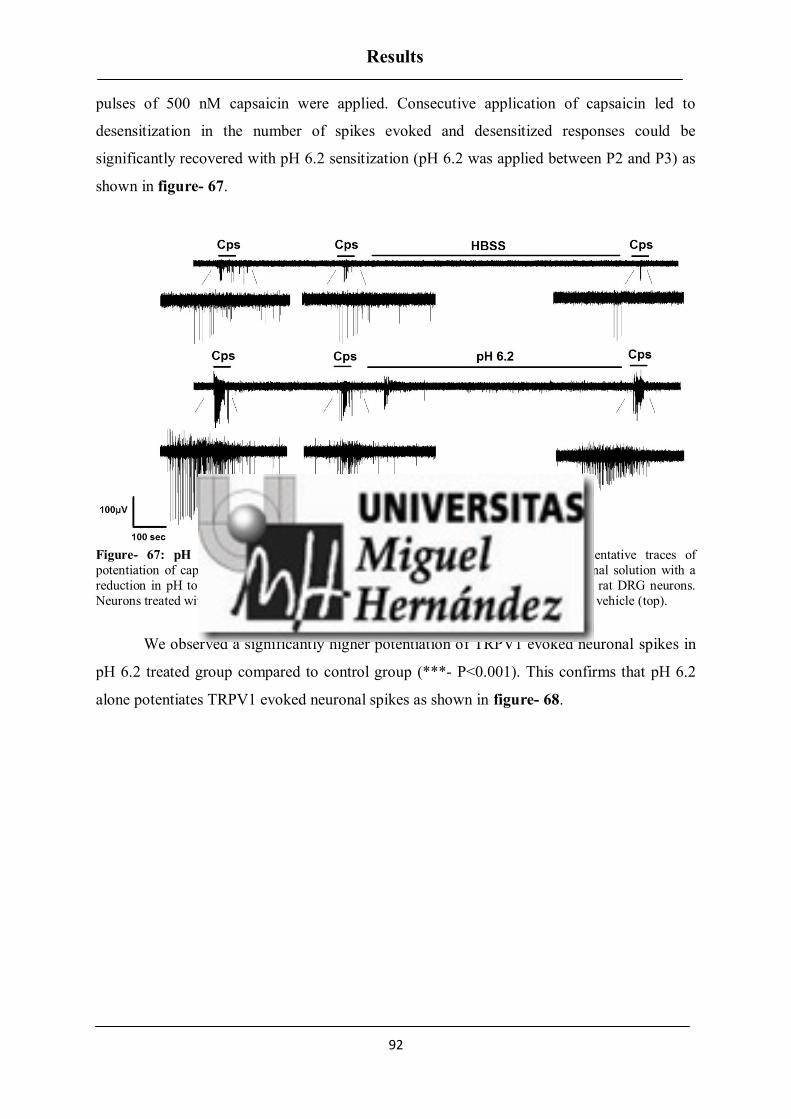
Results
92
pulses of 500 nM capsaicin were applied. Consecutive application of capsaicin led to
desensitization in the number of spikes evoked and desensitized responses could be
significantly recovered with pH 6.2 sensitization (pH 6.2 was applied between P2 and P3) as
shown in figure- 67.
Figure- 67: pH 6.2 potentiates TRPV1- mediated nociceptor excitability. Representative traces of
potentiation of capsaicin (500 nM, 15 s) responses elicited by applying the standard external solution with a
reduction in pH to 6.2 between the second (P2) and third (P3) vanilloid pulse, in neonatal rat DRG neurons.
Neurons treated with the pH 6.2 (bottom) had significant potentiation of TRPV1 compared to vehicle (top).
We observed a significantly higher potentiation of TRPV1 evoked neuronal spikes in
pH 6.2 treated group compared to control group (***- P<0.001). This confirms that pH 6.2
alone potentiates TRPV1 evoked neuronal spikes as shown in figure- 68.

Results
93
Control pH 6.2
0.0
0.2
0.4
0.6
0.8P1
P2
P3***51
196
Mean
Sp
ike F
req
uen
cy (
Hz)
Figure- 68: pH 6.2 induces inflammatory sensitization on TRPV1 mediated neuronal firing in rat DRG
neurons. Effect of pH 6.2 on TRPV1 evoked neuronal firing activity was determined by comparison of all the
three pulses (P1, P2, and P3) in each group and represented as Mean spike frequency (Hz). Data are expressed
as mean ± SEM. The numbers above the bars represent the total number of electrodes that responded. Number
of cultures = 3. Statistical analysis was performed by one way ANOVA repeated measures with Bonferroni’s
post hoc test (***- p<0.001).
Blockage of neuronal exocytosis reduces ATP- BK pH 6.2 induced TRPV1 excitability
in rat peptidergic nocieptors
From our earlier experiment, we did not see a blockage effect of DD04107 on ATP-
BK pH 6.2 mediated TRPV1 potentiation. This could be due to its activity on differential
subpopulation of DRG neurons. We wanted to know if this mechanism of TRPV1
potentiation by ATP- BK pH 6.2 is common for all subpopulations of DRG neurons. From
our previous experiments with ATP and BK, we found that potentiation of TRPV1 in
peptidergic neurons is primarily due to the recruitment of new functional TRPV1 channels
from the vesicles to the membrane and it is abrogated by DD04107. This corroborates that the
distinct signalling pathways are involved in sensitization of TRPV1 in both peptidergic and
nonpeptidergic nociceptors. Hence we wanted to further elucidate whether ablation of
nonpeptidergic neurons from the DRG neuronal culture could enhance the potential blockade
effect of DD04107 on ATP- BK pH 6.2 induced potentiation of TRPV1 in peptidergic
nociceptors. For that we used IB4 SAP toxin which removes completely all IB4+ neurons,
leaving only IB4- neurons. The experiments were done 48 hours after toxin treatment.

Results
94
ATP- BK pH 6.2 induced sensitization of TRPV1 excitability in peptidergic
nociceptors was evaluated using MEA technique. A continuous protocol was used where
three repetitive pulses of 500 nM capsaicin was applied. Consecutive application of capsaicin
led to desensitization in the number of spikes evoked as shown in figure- 69 (Vehicle &
DD04107) under control conditions and desensitized responses could be significantly
recovered with ATP- BK pH 6.2 sensitization (10 μM ATP- 1 μM BK- pH 6.2 was applied
between P2 and P3) as shown in figure- 70 (Vehicle). ATP-BK pH 6.2 induced TRPV1
sensitization of excitability was blocked by DD04107, this sensitization was mainly due to
the insertion of new TRPV1 channels into the plasma membrane of peptidergic nociceptors
as shown in figure- 70 (DD04107).
Figure- 69: Representative MEA recordings of capsaicin induced action potentials and desensitization in
IB4 saporin treated nociceptors. Application of capsaicin (500 nM, 15 s) on neonatal rat DRG neurons
induced TRPV1 evoked neuronal excitability in both vehicle and DD04107 treated groups (1st pulse- P1) in IB4 Saporin treated nociceptors. Repeated exposure of DRG neurons to capsaicin (2nd pulse (P2) and 3rd pulse (P3))
evoked fewer action potentials compared to the 1st pulse due to the desensitization of TRPV1 channel activity.

Results
95
Figure- 70: DD04107 abrogates, ATP-BK pH 6.2 sensitized TRPV1- mediated nociceptor excitability in
peptidergic rat DRG neurons. Representative traces of potentiation of capsaicin (500 nM, 15 s) responses elicited by applying the cocktail of 10 μM ATP- 1 μM BK with less pH 6.2 between the second (P2) and third
(P3) vanilloid pulse (top), in peptidergic neonatal rat DRG neurons. Preincubation of the neurons with DD04107
(bottom) also had potentiation of TRPV1 but it was less significant compared to Vehicle (top).
Though we saw a less significant potentiation of ATP- BK pH 6.2 induced TRPV1
activity in DD04107 treated group (*- p<0.05) compared to Vehicle group (***- p<0.001),
we still observed potentiation of TRPV1 evoked excitability as shown in figure- 71. Hence it
was important to compare ATP- BK pH 6.2 induced fold increase in the potentiation of
TRPV1 between Vehicle and DD04107 treated groups.

Results
96
Vehicle DD04107 Vehicle DD041070.0
0.5
1.0
1.5P1
P2
P3
***
*29
2152 68
Control ATP- BK pH6.2
Mean
Sp
ike F
req
uen
cy (
Hz)
Figure- 71: DD04107 abolishes ATP, BK –pH 6.2 induced inflammatory sensitization of TRPV1 mediated
neuronal firing in IB4 saporin treated rat DRG neurons. Effect of 20 µM DD04107 on 10 μM ATP, 1 μM BK pH 6.2 mediated potentiation of TRPV1 evoked neuronal firing activity was determined by comparison of
all the three pulses (P1, P2, P3) in each group and represented as Mean spike frequency (Hz). Data are
expressed as mean ± SEM. The numbers above the bars represent the total number of electrodes that responded.
Number of cultures = 3. Statistical analysis was performed by one way ANOVA repeated measures with
Bonferroni’s post hoc test (*- p<0.05, ***- p<0.001).
We calculated the ratio of third pulse to second pulse (P3/P2) in both Vehicle and
DD04107 treated groups exposed to ATP- BK pH 6.2. Then, the impact of DD04107 on
blocking the potentiation of TRPV1 channel by ATP- BK pH 6.2 treated neurons was
compared between Vehicle and DD04107 groups as shown in figure- 72 (A). We observed a
significant blockage of ATP- BK pH 6.2 induced TRPV1 potentiation on peptidergic DRG
neurons pre incubated with DD04107 (2±0.3) compared to the Vehicle (2.8±0.3) (*- p<0.05).
Experiments from neonatal rat DRG neurons exposed to Saporin alone (which does not
eliminate nonpeptidergic neurons and served as a control) did not show any blockage effect
of DD04107, in addition, neurons treated with DD04107 (6.2±1.9) showed significantly
higher potentiation of TRPV1 evoked neuronal activity compared to the Vehicle (2±0.8) (*-
p<0.05) as shown in figure- 72 (B).

Results
97
Vehicle DD041070
2
4
6
8
10 *
14
12
ATP- BK pH6.2 (Saporin)
Fo
ld P
ote
nti
ati
on
Vehicle DD041070
1
2
3
ATP- BK pH 6.2 (IB4 SAP)
49
46
*
Fo
ld P
ote
nti
ati
on
Figure- 72: ATP- BK at pH 6.2 mediated TRPV1 sensitization requires channel recruitment in a subset of
nociceptors in IB4 Saporin treated (A) but not in Saporin treated (B) rat nociceptors. Effect of 20 µM
DD04107 on 10 μM ATP – 1 μM BK with pH 6.2 mediated potentiation of TRPV1 activity was determined by
comparison of capsaicin-mediated neuronal spikes before and after incubation of ATP- BK pH 6.2 (ratio P3/P2)
of the groups Vehicle and DD04107 in both IB4 Saporin and Saporin treated neonatal rat DRG neurons . Data
are expressed as mean ± SEM. The numbers above the bars represent the total number of electrodes that responded. Number of cultures = 3. Statistical analysis was performed by Unpaired Student’s t- test (*-
p<0.05).
A) B)


DISCUSSION


Discussion
97
DISCUSSION
Ion channels play an important role in controlling physiological activity. Activation of
ion channels lead to alteration in the membrane potential. During pathophysiological
conditions an up regulation or down regulation of the ion channel activity is detected and
represented as an increase or decrease in the number of action potentials. Similarly, a
considerable increase in the excitability of neurons represents the underlying mechanism in
sensitization of nociceptors (324). During tissue injury, a plethora of algogen factors are
released from damaged nociceptors and surrounding immune cells. These inflammatory
mediators will act on their receptors on sensory neurons, thus leading to modulation of ion
channel gating, thereby provoking an enhanced pain sensation (325). Inflammation can
sensitize nociceptors through short term modifications induced by either a decrease in
excitability threshold of the ion channels or activation of phosphorylation mechanisms (326).
One such primary ion channel involved in sensitization of nociceptor is TRPV1, a
thermoTRP channel which is primarily involved in heat sensation (194). Many studies on
inflammatory sensitization of TRPV1 suggest its significant role in nociceptive pain
signalling pathway (124, 202).
Earlier studies have reported that inflammatory sensitization of TRPV1 could involve
differential mechanism from the recruitment of new TRPV1 channels to the plasma
membrane through regulated exocytosis (304) to the modulation of channel gating through
phosphorylation (327). Experiments using blockers of regulated exocytosis BoNTA and
DD04107 revealed that these blockers potently reduce increased surface expression of
TRPV1 induced by algogens. Hence the nociceptors exhibit reduced TRPV1 activity
confirming that exocytotic mobilization of TRPV1 is a key mechanism in sensitizing a subset
of nociceptors upon an inflammatory insult. A previous study from our group reported that
some inflammatory mediators like ATP, NGF and IGF-1 can sensitize TRPV1 through
exocytosis of new channels, whereas BK, Artemin and IL-1β sensitize TRPV1 through
phosphorylation mechanism (312). Due to their higher heterogeneity, which subpopulation of
nociceptor is involved in TRPV1 exocytosis and phosphorylation mechanisms remains
unclear.

Discussion
98
Therefore, the principal objective of this work is to unravel the underlying mechanism
behind inflammatory sensitization of TRPV1 in nociceptors. For this purpose, we have
studied the mechanism of two inflammatory mediators ATP and BK on TRPV1 sensitization
in peptidergic and nonpeptidergic nociceptor subpopulations.
We have found that:
1. Both ATP and BK sensitize TRPV1 through exocytosis in peptidergic nociceptors.
2. αCGRP is essential for both ATP and BK induced exocytotic mobilization of TRPV1
in peptidergic nociceptors.
Regulated exocytosis of TRPV1 in nociceptors was examined by using a hexamer
lipopeptide DD04107 (EEMQRR), an inhibitor of neuronal exocytosis. This peptide is
patterned after the N-terminus of SNAP 25 protein which inhibits SNARE complex
formation thereby specifically blocking regulated exocytosis (328). In addition, peptide
DD04107 was found to exhibit long lasting anti nociceptive potential in distinct animal pain
models in vivo (313). From experimental conditions (in vitro), we have confirmed that the
peptide does not interfere neither with the basal electrical properties of the nociceptors nor
the initial TRPV1 current densities in both peptidergic and nonpeptidergic subpopulations.
TRPV1 MODULATION BY ATP
Initially, we explored the mechanism of ATP mediated TRPV1 potentiation. ATP, the
prominent cytosolic content of keratinocytes and sensory neurons, is released upon tissue
damage and excites nearby nociceptors (75). ATP evoked excitatory action potentials in
nociceptors are due to the activation of purinergic P2X and P2Y receptors (315). In
nociceptors, the purinergic receptor families P2X3, P2Y1 and P2Y2 receptors are widely
expressed. Although most of the TRPV1+ sensory neurons express P2X3 receptor, they do
not contribute for TRPV1 sensitization (329). P2Y1 was found to be strongly colocalized
with IB4, whereas it was weakly colocalized with TRPV1. Initial studies on ATP induced
TRPV1 sensitization revealed that P2Y1 receptor was involved in sensitizing TRPV1
currents through PLC- DAG/ IP3 pathway induced by Gαq/11 signalling protein (329). To
further validate the role of P2Y1 receptors on TRPV1 sensitization, experiments were done in
P2Y1 knockout mice (P2Y1-/-
). ATP induced TRPV1 sensitization was preserved in P2Y1-/-

Discussion
99
mice, suggesting a lack of P2Y1 receptor involvement on TRPV1 potentiation. In sensory
neurons, P2Y2 receptor was strongly expressed with TRPV1 suggesting that P2Y2 can
functionally interact with TRPV1 (172) thus contributing for TRPV1 evoked thermal
hyperalgesia. Furthermore, pharmacological activation of P2Y2 receptors in sensory neurons
exhibited robust TRPV1 sensitization (277). Other studies on P2Y2 receptors also
corroborated their potential role in TRPV1 sensitization and nociception. During CFA
induced inflammation, P2Y2 was the only receptor found to be elevated, thus contributing to
significant ATP responses in sensory neurons leading to enhanced thermal hyperalgesia
(330). Another study on kidney projecting sensory neurons validated the significant role of
P2Y2 receptors on ATP induced TRPV1 sensitization (331). Thus ATP induced TRPV1
potentiation seems to be chiefly mediated by P2Y2, although we cannot exclude the possible
involvement of P2Y1 receptors on sensitizing TRPV1 channel activity. Moreover, activation
of P2Y2 receptor stimulates pertusis toxin insensitive Gαq/11 protein which associates with
PLC- β3 catalysing PIP2 and activating IP3 and DAG- PKC pathways (332). Gαq/11 induced
PLC-β3 activation is crucial for ATP mediated pain signalling since PLC-β3 is primarily
expressed in DRG neurons among other isoforms of PLC β (333).
From our experiments on neonatal rat DRG neurons, we observed a high impact of
inhibiting neuronal exocytosis on lessening ATP induced sensitization of TRPV1 evoked
neuronal spikes. This suggests a major role of exocytotic mobilization of new TRPV1
channels in a subset of the whole neuronal population. Furthermore, targeted differentiation
of peptidergic and nonpeptidergic neurons using IB4 marker revealed that two distinct
mechanism of ATP induced TRPV1 sensitization is involved. Inhibitor of neuronal
exocytosis – DD04107 completely abolished TRPV1 sensitization in peptidergic neurons but
had no effect on nonpeptidergic neurons. Additionally, TRPV1 sensitization in peptidergic
neurons is PKC independent, whereas in nonpeptidergic neurons ATP induced TRPV1
sensitization is PKC dependent. This confirms the exocytotic release of new vesicles as a
prime mechanism in peptidergic and phosphorylation mechanism in nonpeptidergic
subpopulations upon ATP induced TRPV1 sensitization.
Since we found that in peptidergic nociceptors TRPV1 was sensitized mainly through
exocytosis, we hypothesized that the inflammatory recruitment of TRPV1 channel could
occur through release of TRPV1 channel from the vesicles containing neuropeptides CGRP

Discussion
100
and SP. Many studies suggested the potential role of these neuropeptides in sorting receptors/
ion channels into large dense core vesicles (LDCVs) (334-336). The presence of TRPV1
along with CGRP and SP suggests their possible involvement in sorting TRPV1 into the
LDCVs and driving its regulated exocytosis. We performed experiments on wild type and
αCGRP-/-
xTac1-/-
(DKO) mice in order to know the potential role of neuropeptides on ATP
induced TRPV1 sensitization. In adult wild type mice, TRPV1 expression is primarily
restricted to peptidergic nociceptors (199). Notably, experiments from adult wild type mice
exhibited a strong sensitization of TRPV1 evoked spikes induced by ATP. Such sensitization
was completely abolished by inhibiting neuronal exocytosis thus corroborating that ATP
induced TRPV1 exocytosis is specific for peptidergic nociceptors. Nociceptors from DKO
mice also exhibited ATP induced sensitization of TRPV1 evoked neuronal spikes which was
insensitive to the neuronal exocytosis inhibitor, suggesting a significant role of neuropeptides
on TRPV1 release through exocytosis. Furthermore, experiments from single knockout
animal (Tac1 and αCGRP) revealed that αCGRP but not SP is essential for ATP induced
regulated exocytosis of TRPV1. Since the released neuropeptides can also sensitize TRPV1
in primary sensory neurons, we did not exclude the possibility that the released neuropeptides
could act through autocrine signalling mechanism. Indeed, earlier studies revealed that SP
released upon TRPV1 activation sensitizes the receptor through both autocrine and paracrine
mechanism. SP binds to NK1 receptor and activates PKCε which induces phosphorylation of
TRPV1. NK1 receptor strongly colocalises with TRPV1 in unmyelinated small diameter
neurons (337). SP induced TRPV1 sensitization further enhances capsaicin evoked SP release
from cultured rat DRG neurons (286). In addition, activation of NK2 receptor in afferent
neurons also leads to PKC induced phosphorylation of TRPV1 leading to sensitization of
capsaicin activated currents (287). Though, no direct evidence of CGRP involvement on
sensitizing TRPV1 channel was reported, a vital role of CGRP on sensitizing nociceptors has
been extensively described (305). From our experiments we pharmacologically blocked both
CGRP and NK1 receptors in vitro using specific antagonists and found that no autocrine
signalling mechanism of neuropeptides was involved in sensitization of TRPV1. This
validates the prime role of TRPV1 release in ATP induced sensitization process.
It has been reported that, akin to the activation of TRPV1, a strong depolarization
stimuli can also induce CGRP release. All these stimuli require external Ca2+
ions to release
CGRP. Studies on trigeminal sensory neurons showed that an elevated K+ concentration can

Discussion
101
induce increase in CGRP release which is Ca2+
dependent. We tested if depolarization of
neurons could mimic a similar mechanism of ATP induced TRPV1 exocytosis, since
depolarization is also an important underlying signal for dense core vesicle exocytosis (338).
Experiments on wild type mice showed that depolarization of the neurons with elevated
external K+ concentration [40 mM] did not induce sensitization of TRPV1 evoked spikes.
Hence, we confirmed that TRPV1 exocytosis is a specific mechanism for ATP induced
inflammatory signalling.
TRPV1 MODULATION BY BK
Bradykinin is another endogenous pain causing potent algogenic nonapeptide released
during tissue damage (318). BK belongs to kinin family whose production is enhanced in
tissues during pathophysiological conditions leading to activation of kallikrein (339). BK is
released from mast cells and basophils (340). Bradykinin exerts its action through B1 and B2
receptors. Both B1 and B2 are metabotropic receptors which trigger second messenger
signalling (341).
B2 receptor expression is ubiquitous and constitutive, whereas B1 receptor is
expressed only in inflammatory conditions. Although in DRG and TG neurons constitutive
B1 receptor expression was found to be inconsistent, few studies have identified the
constitutive expression of B1 receptor. B1 receptor was found to be expressed preferentially
in IB4+, but not in CGRP containing C fibers of dorsal root ganglion (321). Recent studies
from TG neurons disclosed that B1 and B2 receptors were functionally expressed, although
immunoreactivity of B1 was weak when compared to B2 (342).
Prior to the identification and cloning of TRPV1 channel, studies on DRG neurons
reported that a novel heat activated current was observed. Sensitization of this heat activated
current was observed upon exposure to bradykinin, which was mediated by PKC (76).
Furthermore, bradykinin increased the percentage of neurons responding to capsaicin (343).
After the identification of TRPV1 in sensory neurons, studies revealed that BK exposure led
to lowering of temperature threshold to activate TRPV1 channel. The reduction in
temperature was up to 24 oC which is under the normal physiological temperature thus
leading to activation of the channel at room temperature. All these mechanisms were PKC

Discussion
102
dependent, specifically through PKCε (278, 344). In addition, interaction of TRPV1 and
PKCε with AKAP 79/150 protein was found to be important for bradykinin induced
sensitization of TRPV1 (319).
The amino acid positions S502 and S800 of TRPV1 are the putative phosphorylation
sites responsible for PKC mediated sensitization of capsaicin responses in sensory neurons.
Of these phosphorylation sites, S800 was identified as a major contributor for bradykinin
induced hypersensitivity of TRPV1 in sensory neurons (345).
Earlier studies from our group also reported that BK can sensitize TRPV1 through
phosphorylation mechanism and not through exocytosis (312). Numerous studies on
nociceptors have reported the possible involvement of BK in trafficking and exocytosis.
Likewise BK induced trafficking of opioid receptors in nociceptors was extensively studied
(316, 346). BK has also been involved in exocytotic release of CGRP in primary sensory
neurons (305). Another study on DRG neurons showed that acute incubation (10 minutes) of
BK (1 μM) markedly increased the release of CGRP in a concentration dependent manner
(317). In our experiments we tested whether Bradykinin (1 μM), which is coupled to Gαq/11
protein signalling, can induce exocytosis of TRPV1 similar to ATP in peptidergic
subpopulations.
Experiments on BK mediated TRPV1 potentiation from neonatal rat DRG neurons
revealed that BK can induce TRPV1 exocytosis similar to ATP. A high impact of the peptide
in blocking TRPV1 sensitization from whole neuronal population was observed, suggesting a
major role of exocytotic mobilization of new TRPV1 channels in a subset of nociceptors. In
addition, targeted differentiation of peptidergic and nonpeptidergic neurons using IB4 marker
revealed that a different mechanism is involved in BK induced TRPV1 sensitization.
Inhibitor of neuronal exocytosis (DD04107) completely abolished TRPV1 sensitization
through regulated exocytosis in peptidergic neurons but had no effect on nonpeptidergic
neurons. Additionally, no significant TRPV1 sensitization was observed in nonpeptidergic
neurons by BK. This could be due to the differential expression of BK receptors in nociceptor
subpopulations, where B2 is specifically expressed in peptidergic and B1 is specifically
expressed in nonpeptidergic. Furthermore, a small population of IB4+ neurons showed
positive immunoreactivity for B2 receptors (342), which suggests that the observed TRPV1

Discussion
103
sensitization by BK in nonpeptidergic nociceptors could be due to the expression of B2
receptor. Since it is known that BK induces phosphorylation of TRPV1, we studied if
exocytosis of TRPV1 is the prime mechanism involved in peptidergic nociceptors similar to
ATP. We observed that in peptidergic neurons BK induced TRPV1 sensitization is also PKC
dependent, suggesting that a dual mechanism of TRPV1 sensitization (exocytotic release of
new vesicles and phosphorylation) is involved in peptidergic subpopulation. Also PKC was
reported as one of the major activator for regulated exocytosis of TRPV1 (304).
Though BK induced TRPV1 sensitization is primarily mediated by B2 receptor,
contribution of B1 receptor on TRPV1 sensitization cannot be ruled out. By
pharmacologically activating BK receptors we found that B2 receptor is predominantly
involved in BK induced TRPV1 sensitization with a less contribution from B1 receptor.
Specific agonist for B2 receptor mediated TRPV1 sensitization was similar to BK activation.
Since we found that BK induced TRPV1 sensitization in peptidergic nociceptors is
primarily mediated through regulated exocytosis, we extended our studies to identify the
possible involvement of αCGRP on BK induced TRPV1 sensitization.
Notably, experiments from adult wildtype mice exhibited strong sensitization of
TRPV1 induced by BK. Such sensitization was completely abolished by inhibiting neuronal
exocytosis corroborating that BK induced TRPV1 exocytosis is specific for peptidergic
nociceptors. Conversely, studies on nociceptors from single knockout mice revealed that
αCGRP
but not SP is essential for BK induced TRPV1 sensitization. These results
corroborate with our ATP experiments, suggesting that both ATP and BK activate Gαq/11
signalling protein and induce TRPV1 exocytosis in peptidergic nociceptors. Recently in vivo
studies to understand the role of Gαq/11 signalling protein on nociceptor sensitization induced
by algogens were performed using Gαq-/-
, Gα11-/-
and Gαq/11-/-
knockout mice models. The
results showed that both ATP and BK induced thermal hyperalgesia was preserved in Gα11-/-
deficient mice, whereas it was completely abolished in Gαq-/-
and Gαq/11-/-
deficient mice. This
confirms that the major common signalling mechanism involved in ATP and BK induced
nociceptor sensitization is mediated through Gαq protein and provokes thermal hyperalgesia,
where TRPV1 could play a significant role (347). This study supports our hypothesis that
both ATP and BK induce their effect on nociceptors through Gαq/11 protein with similar
mechanism.

Discussion
104
FACILITATORY EFFECT OF ATP- BK ON TRPV1 MODULATION
Furthermore, we studied the combinatorial effect of ATP and BK on facilitating
TRPV1 sensitization under acidic pH conditions. This is to mimic the tissue acidosis
microenvironment during inflammatory conditions. Algogens are known to interact with
protons and facilitate their effects in different animal models. Bradykinin functions as a co-
operative algogen with other inflammatory mediators to evoke pain sensation (348). In
addition, combinatorial applications of BK with other inflammatory mediators were found to
significantly increase the release of CGRP in DRG neurons. This release of CGRP could not
be enhanced further by acidification. Exposure of DRG neurons to acidic pH also induced
CGRP release which was lower compared to inflammatory mediators. Both, low pH and
inflammatory mediators induced CGRP release, are dependent of external Ca2+
. Also the
inflammatory mediators were proposed to act as endogenous activators for TRPV1 since
TRPV1 antagonist Capsazepine (CPZ) inhibited inflammatory mediators induced release of
CGRP in DRG neurons (349).
From our experiments on neonatal rat DRG neurons, combined application of both
ATP and BK significantly increased TRPV1 evoked neuronal spikes which were abrogated
by blocking neuronal exocytosis. This validates the major role of TRPV1 exocytosis similar
to what we have observed earlier in our individual experiments with ATP and BK. In
addition, we increased the strength of inflammatory mediators by decreasing the extracellular
pH to 6.2, to mimic the acidic microenvironment conditions observed during tissue injury.
We found that combined application of ATP and BK at pH 6.2 significantly increased
TRPV1 evoked neuronal spikes which were insensitive to neuronal exocytosis inhibitor. This
could be due to the decrease in extracellular pH, in fact numerous studies have reported that
acidic pH 6.1 can potentiate TRPV1 currents by strongly reducing the EC50 of capsaicin from
μM to nM concentrations (323). We proposed that the reduced pH alone could sensitize
TRPV1 evoked spikes and we confirmed from our experiments that exposure of TRPV1
desensitized neurons to pH 6.2, led to significant sensitization of TRPV1. From our results
we confirmed that DD04107 significantly abrogates TRPV1 sensitization in peptidergic
subpopulations and the reduction of pH could enhance its sensitizing effect of TRPV1
channel in other subpopulations of primary sensory neurons. In rat DRG neurons TRPV1 is
expressed in both C fibers (peptidergic and nonpeptidergic) and Aδ fibers. We proposed that

Discussion
105
the pH 6.2 induced TRPV1 modulation is mainly occurring in nonpeptidergic and Aδ fibers.
Then DD04107 could inhibit ATP and BK at pH 6.2 induced TRPV1 sensitization in
peptidergic subpopulations. This was proved by using IB4 saporin toxin which selectively
eliminates IB4+
neurons (350). Studies reported that injection of IB4 saporin toxin eliminated
acute and chronic pain conditions in vivo (351, 352). We found that ATP, BK and pH 6.2
induced TRPV1 sensitization on remaining IB4-
neurons was significantly inhibited by
blocking neuronal exocytosis. As a control, in neurons treated with saporin toxin alone we
did not observe any significant blockage of TRPV1 sensitization by DD04107. This result
corroborates the significant role of DD04107 on alleviating inflammatory sensitization of
TRPV1 in peptidergic nociceptors.
Hence in conclusion, this study reveals the involvement of distinct signalling
mechanism in TRPV1 sensitization induced by algogens in peptidergic and nonpeptidergic
nociceptors. Furthermore, it substantiates the potential therapeutic role of DD04107 to
alleviate inflammatory pain induced by sensitization of TRPV1.


CONCLUSIONS


Conclusions
106
Inflammatory mediators (ATP/ BK) induce enhanced excitability in peptidergic
nociceptors, while in nonpeptidergic nociceptors induce a moderate effect.
TRPV1 sensitization induced by inflammatory mediators (ATP/ BK) occurs primarily
through exocytotic recruitment of channels from large dense core vesicles (LDCVs)
to the plasma membrane in peptidergic nociceptors however, in nonpeptidergic
nociceptors TRPV1 is sensitized through differential mechanism which also includes
phosphorylation of the channel.
Deletion of αCGRP but not Tac1 expression impairs inflammatory TRPV1
recruitment to the surface of peptidergic nociceptors, leading to abrogation of TRPV1
sensitization. This corroborates the potential role of αCGRP in promoting regulated
exocytosis of TRPV1.
TRPV1 is co- trafficked with αCGRP in LDCVs in peptidergic nociceptors.
Inhibition of LDCV exocytosis is a valuable therapeutic strategy to treat pain, as it
concurrently reduces the release of pro- inflammatory peptides and the membrane
recruitment of TRPV1 channels.
DD04107 abolishes TRPV1 sensitization induced by an inflammatory
microenvironment (in vitro), thus substantiating the therapeutic potential of DD04107
to alleviate inflammatory pain induced by sensitization of TRPV1.


Conclusiones
107
Los mediadores inflamatorios (ATP/ BK) provocan una excitabilidad mayor en
neuronas peptidérgicas mientras que en neuronas no peptidérgicas producen un
efecto moderado.
La sensibilización de TRPV1 inducida por los mediadores inflamatorios (ATP/ BK)
en neuronas peptidérgicas se produce principalmente a través de exocitosis regulada
del canal desde vesículas de centro denso (LDCVs) a la membrana plasmática. En el
caso de nociceptores no peptidérgicos la sensibilización del TRPV1 es causada
principalmente por fosforilación del canal.
El silenciamiento del gen αCGRP pero no de Tac1, provoca una disminución de
TRPV1 en la membrana neuronal suprimiendo completamente la sensibilización
inflamatoria del canal, lo que indica el papel potencial de este neuropéptido en
promover la exocitosis de TRPV1.
TRPV1 es transportado a la membrana neuronal en LDCVs junto con el neuropéptido
αCGRP en nociceptores peptidérgicos.
La inhibición de la exocitosis de LDCVs puede ser una estrategia terapéutica muy
eficaz en el tratamiento del dolor ya que reduce la liberación de péptidos pro-
inflamatorios y la presencia de TRPV1 en la membrana neuronal.
DD04107 suprime la sensibilización de TRPV1 inducida por el microambiente
inflamatorio (in vitro), demostrando el potencial papel terapéutico del péptido
DD04107 en la disminución del dolor inflamatorio inducido por la sensibilización de
TRPV1.


MATERIALS AND METHODS


Materials and Methods
108
All procedures were approved by the Institutional Animal and Ethical Committee of
the University Miguel Hernández de Elche, in accordance with the guidelines of the
Economic European Community, the National Institutes of Health, and the Committee for
Research and Ethical Issues of the International Association for the Study of Pain.
Animals. Animals were kept in a controlled environment (21-23 ºC, 12h light/dark cycle),
and had food and water available ad libitum. Neonatal Wistar rats and wild type C57BL/6J
mice were purchased from in house bred stock (originally from Harlan Laboratories).
αCGRP-deficient mice (B6;129-Calcatm
) were kindly provided by Dr. J.P. Changeaux. Tac1-
deficient mice (B6.Cg-Tac1tm1Bbm
/J) were purchased from The Jackson Laboratory. Double
knockout mice αCGRP and Tac1 genes were in house generated by crossing homozygous
αCGRP and Tac1 null mice to obtain first heterozygous and later homozygous deficient mice
(B6; B6; 129-Calcatm
Tac1tm1Bbm
/J).
Primary culture of sensory neurons. DRG from neonatal Wistar rats (3-5 days old) or adult
male 12-15 week-aged mice (strains: C57BL/6J, αCGRP-/-
, Tac1-/-
, and αCGRP-/-
xTac1-/-
mice), cultured following previously described protocols with some modification (290, 353).
Neonatal rat ganglia were digested with 0.25% (w/v) collagenase (type IA) in DMEM-
glutamax (Invitrogen) with 1% penicillin-streptomycin (P/S) (5000 U/mL, Invitrogen) for 1 h
(37 ºC, 5% CO2). Isolated mouse DRG were incubated with 0.67% (w/v) collagenase type XI
and 3% (w/v) dispase (Gibco) in INC mix medium (in mM): 155 NaCl, 1.5 K2HPO4, 5.6
HEPES, 4.8 NaHEPES, 5 glucose) for 1 h (37 ºC, 5% CO2). After digestion, rat and mouse
DRG were mechanically dissociated using a glass Pasteur pipette. Single cell suspension was
passed through a 100 µM cell strainer, and washed with DMEM glutamax plus 10% fetal
bovine serum (FBS) (Invitrogen) and 1% P/S. Cells were seeded at the required density for
each experiment on 12 mm cover-glass slides, or microelectrode array chambers previously
coated with poly-L-lysine (8.33 µg/ml) and laminin (5 µg/ml). After 2 h, medium was
replaced with DMEM glutamax, 10% FBS and 1% P/S, supplemented with mouse 2.5s NGF
50 ng/mL (Promega), and 1.25 µg/mL cytosine arabinoside when required (37 ºC, 5% CO2).
If it is not specified, all experiments were made 48 h after cell seeding. To deplete Isolectin
IB4+ neurons, rat DRG were cultured in the presence of 10 nM IB4-saporin or saporin as
control (Advanced Targeting Systems) for 48 h.

Materials and Methods
109
Patch clamp recordings. Whole-cell voltage clamp was made in neurons seeded on
coverslips, placed in RC-25 chamber and connected to a external perfusion system at RT.
HBSS external solution (in mM): 140 NaCl, 4 KCl, 2 CaCl2, 2 MgCl2, 10 HEPES, 5 D-
glucose, 20 mannitol, pH 7.4 (adjusted with NaOH). Internal pipette solution (in mM): 144
KCl, 2 MgCl2, 10 HEPES, 5 EGTA, pH 7.2 (adjusted with KOH). For the experiments with
less pH (pH 6.2) HEPES was replaced with MES. Membrane currents were acquired by using
EPC10 HEKA Patch amplifier (HEKA Electronics). Cells were held at a resting membrane
potential of -60 mV, and at a sampling rate of 2.5 Hz. Patch glass pipettes with O.D 1.5 mm
x I.D. 1.17 mm (Harvard Instruments) made with Sutter Pippete puller Equipment (Sutter
Instruments) with 3-6 MΩ resistance. Data acquisition and offline analysis with PatchMaster
software (HEKA Electronics). Rat DRG neurons labelled with IB4-alexa 568 (10 μg/ml, 10
min, RT) in external solution (Molecular Probes, Invitrogen), followed by two 5 min-washes,
and visually identified through x20 air objective (Aixiovert 200 inverted microscope, Carl
Zeiss), with an excitation filter ET545 and an emision filter ET605 (CHR-49004, Laser 2000
SAD). Neuronal viabiliy determined through typical neuronal Na+-K
+ currents. TRPV1
desensitization was induced by three repetitive 10s-pulse of 1 µM capsaicin using perfusion
system (2 mL/min flux). 10 µM ATP or 1 μM BK was applied for 8 min between P2 and P3
pulse. Potentiation of TRPV1-mediated currents was calculated as ratio P3/P2 current peak.
Capsaicin dose-response was determined in wild type and αCGRP-/-
xTac1-/-
neurons with
resting membrane potential below -40 mV.
Electrical properties. Electrical properties of the neuron were determined 2 minutes after
establishing whole cell access using current clamp mode. For current-clamp recording, cells
were held at 0 pA. Cells were assessed for the presence of spontaneous activity for 1 min and
processed to measure electrogenic properties. Both IB4- and IB4
+ nociceptors were processed
for electrogenic properties before ATP and BK application. Firing threshold was measured
first by injecting a series of 100 ms depolarizing current in 10 pA steps from 0 pA to elicit the
first action potential. To further examine the neurons firing properties, a depolarizing current
injection in 100 ms, 40 pA (IB4-) and 100 pA (IB4
+) was delivered to elicit an action
potential which was analysed for the following intrinsic membrane properties: threshold
potential (mV), amplitude of action potential (mV), duration of action potential (ms),
overshoot of action potential (mV). 10 μM ATP and 1 μM BK was applied to IB4- and IB4
+
nociceptors for 4 minutes to observe both ATP and BK induced spontaneous neuronal

Materials and Methods
110
excitabilities. RMP was checked before and after ATP, BK application. Mean depolarization
was calculated by selecting the maximum depolarized voltage observed upon ATP and BK
application in IB4- and IB4
+ nociceptors. ATP and BK induced changes in electrically evoked
action potentials were measured before (Vehicle) and during ATP and BK application.
Nociceptors (IB4- and IB4
+) were injected with 100 pA for 100 ms, 300 pA for 1 sec to
measure electrically evoked action potentials.
Microelectrode Array (MEA). Extracellular recordings were made using multiple electrode
planar arrays of 60-electrode thin MEA chips, with 30 μm diameter electrodes and, 200 μm
inter-electrode spacing with an integrated reference electrode (Multichannel Systems GmbH).
The electrical activity of primary sensory neuron was recorded by the MEA1060 System
(Multi Channel Systems GmbH, http://www.multichannelsystems.com), and MC_Rack
software version 4.3.0 at a sampling rate of 25 kHz. TRPV1-mediated neuronal firing activity
was evoked by three repetitive 15s-applications of 500 nM capsaicin, using continuous
perfusion system (2 mL/min flux). 10 µM ATP or 1 μM BK in external solution was perfused
between P2 and P3 for 8 min. For the experiments on combined application of ATP- BK on
TRPV1 sensitization, 10 μM ATP- 1 μM BK was perfused between P2 and P3 for 8 min. For
the experiments on bradykinin receptor agonists, selective bradykinin receptor agonists for
BK1 and BK2 was applied between P2 and P3 for 8 min. Data were analyzed using
MC_RACK spike sorter and Neuroexplorer Software (Nex Technologies). An evoked spike
was defined when the amplitude of the neuronal electrical activity overcame a threshold set at
-20 μV. The recorded signals were then processed to extract mean spike frequency.
Immunocytochemistry. For immunocytochemistry, cultured neonatal DRG seeded on
coverslips washed with PBS, were fixed with 4% w/v paraformaldehyde for 20 min, washed,
permeabilized with 0.3% Triton X-100 for 5 min at RT, and then blocked with 5% NGS for 1
h at RT. Primary antibody was incubated in 5% NGS in PBS overnight at 4 ºC. Once washed
with PBS-Tween 0.05%, samples were incubated with secondary antibodies in blocking
solution (1 h, RT). Samples were mounted with Mowiol® (Calbiochem). Primary antibody:
rabbit anti-TRPV1 (Alomone, 1.6 µg/mL). Secondary antibody: Isolectin B4-Alexa 568
(Molecular Probes, 10 µg/mL). Samples visualized with an inverted confocal microscope
(Zeiss LSM 5 Pascal, Carl Zeiss). Number of positive stained neurons determined by

Materials and Methods
111
capturing at least 5 images per sample, and then analysed with Zen lite 2012 software (Zeiss)
or Image J software (NIH), under blinded conditions.
Chemicals. DD04107 (BCN Peptides) freshly prepared at 20 mM in H2O. For microelectrode
array, cells were incubated with compound 20 µM DD04107 in HBSS for 1 h (37 ºC, 5%
CO2). For electrophysiological recordings, non-palmitoylated DD04107 (BCN Peptides) at
100 µM concentration was prepared in standard internal solution from 10 mM stock solution
in H2O. Peptide or vehicle were applied through the patch pipette 10 min before recording.
ATP was dissolved in H2O at 10 mM and further diluted in HBSS at 10 μM. The CGRP
receptor 1 antagonist CGRP8-37 (Tocris Bioscience, R&D Systems) dissolved in 30%
acetonitrile (v/v) at 100 µM and further diluted in HBSS at 250 nM. The neurokinin receptor
1 antagonist CP96345 (Tocris Bioscience, R&D Systems) dissolved in DMSO at 20 mM and
further diluted in HBSS at 10 µM. Both antagonists applied with ATP. BIM was dissolved in
DMSO at 1mM and further diluted in HBSS at 1 μM. BK was dissolved in H2O at 1 mM and
further diluted in HBSS at 1 μM. The BK1 receptor agonist- Sar-[D- Phe8]-des-Arg
9-
Bradykinin, BK2 receptor agonist- [Phe8ψ(CH-NH)-Arg
9]-Bradykinin (Tocris Bioscience,
R&D Systems) was dissolved in H2O at 1 mM and further diluted in HBSS at 1 μM. When
not specified, all chemicals were obtained from Sigma-Aldrich.
Data analysis. All data are expressed as mean ± SEM, with n as number of registered cells
and N as the number of experiments. The percentage of sensitized neurons was calculated
considering those cells that exhibited a fold potentiation above 1.1. Statistical analysis was
made using 1-way ANOVA or 2-ways ANOVA as required followed by Bonferroni’s post-
hoc test using Graph Pad Prism 5.0 (Graph-Pad). For the experiments on characterization of
nociceptors using patch clamp, paired and unpaired student’s t-test was used. MEA data were
analysed by Bonferroni’s post-hoc test as paired values through comparison of the responses
of each electrode along time. Fold potentiation (ratio P3/P2) was analyzed by unpaired
student’s t- test *p<0.05,
**p<0.01,
***p<0.001.


REFERENCES


References
112
1. Tracey I & Mantyh PW (2007) The cerebral signature for pain perception and its
modulation. Neuron 55(3):377-391.
2. Baliki MN & Apkarian AV (2015) Nociception, Pain, Negative Moods, and Behavior
Selection. Neuron 87(3):474-491.
3. Motavaf M, Safari S, & Alavian SM (2013) Understanding of molecular pain
medicine: genetic basis of variation in pain sensation and analgesia response.
Anesthesiology and pain medicine 2(3):104-106.
4. Woolf CJ (2010) What is this thing called pain? The Journal of clinical investigation
120(11):3742-3744.
5. Raouf R, Quick K, & Wood JN (2010) Pain as a channelopathy. The Journal of
clinical investigation 120(11):3745-3752.
6. Scholz J & Woolf CJ (2002) Can we conquer pain? Nature neuroscience 5
Suppl:1062-1067.
7. Grichnik KP & Ferrante FM (1991) The difference between acute and chronic pain.
The Mount Sinai journal of medicine, New York 58(3):217-220.
8. Treede RD, et al. (2008) Neuropathic pain: redefinition and a grading system for
clinical and research purposes. Neurology 70(18):1630-1635.
9. Khan N & Smith MT (2015) Neurotrophins and Neuropathic Pain: Role in
Pathobiology. Molecules (Basel, Switzerland) 20(6):10657-10688.
10. Dykes RW (1975) Nociception. Brain research 99(2):229-245.
11. Ferreira SH (1981) Local control of inflammatory pain. Agents and actions 11(6-
7):636-638.
12. Woolf CJ (1993) The pathophysiology of peripheral neuropathic pain--abnormal
peripheral input and abnormal central processing. Acta neurochirurgica.
Supplementum 58:125-130.
13. Marchettini P, Lacerenza M, Mauri E, & Marangoni C (2006) Painful peripheral
neuropathies. Current neuropharmacology 4(3):175-181.
14. Marcus KS, Kerns RD, Rosenfeld B, & Breitbart W (2000) HIV/AIDS-related pain as
a chronic pain condition: implications of a biopsychosocial model for comprehensive
assessment and effective management. Pain medicine (Malden, Mass.) 1(3):260-273.
15. Parker R, Stein DJ, & Jelsma J (2014) Pain in people living with HIV/AIDS: a
systematic review. Journal of the International AIDS Society 17:18719.

References
113
16. Lozano-Ondoua AN, Symons-Liguori AM, & Vanderah TW (2013) Cancer-induced
bone pain: Mechanisms and models. Neuroscience letters 557 Pt A:52-59.
17. Falk S & Dickenson AH (2014) Pain and nociception: mechanisms of cancer-induced
bone pain. Journal of clinical oncology : official journal of the American Society of
Clinical Oncology 32(16):1647-1654.
18. Selvaraj D, et al. (2015) A Functional Role for VEGFR1 Expressed in Peripheral
Sensory Neurons in Cancer Pain. Cancer cell 27(6):780-796.
19. Wang ZJ, Wilkie DJ, & Molokie R (2010) Neurobiological mechanisms of pain in
sickle cell disease. Hematology / the Education Program of the American Society of
Hematology. American Society of Hematology. Education Program 2010:403-408.
20. Jensen TS & Finnerup NB (2014) Allodynia and hyperalgesia in neuropathic pain:
clinical manifestations and mechanisms. The Lancet. Neurology 13(9):924-935.
21. Janig W (2011) Mechanical allodynia generated by stimulation of unmyelinated
afferent nerve fibres. The Journal of physiology 589(Pt 18):4407-4408.
22. Chaplan SR, Bach FW, Pogrel JW, Chung JM, & Yaksh TL (1994) Quantitative
assessment of tactile allodynia in the rat paw. Journal of neuroscience methods
53(1):55-63.
23. Sandkuhler J (2009) Models and mechanisms of hyperalgesia and allodynia.
Physiological reviews 89(2):707-758.
24. Pedersen JL & Kehlet H (1998) Hyperalgesia in a human model of acute
inflammatory pain: a methodological study. Pain 74(2-3):139-151.
25. Hargreaves K, Dubner R, Brown F, Flores C, & Joris J (1988) A new and sensitive
method for measuring thermal nociception in cutaneous hyperalgesia. Pain 32(1):77-
88.
26. Bridges D, Thompson SW, & Rice AS (2001) Mechanisms of neuropathic pain.
British journal of anaesthesia 87(1):12-26.
27. Ren K & Dubner R (1999) Inflammatory Models of Pain and Hyperalgesia. ILAR
journal / National Research Council, Institute of Laboratory Animal Resources
40(3):111-118.
28. De Iuliis F, Taglieri L, Salerno G, Lanza R, & Scarpa S (2015) Taxane induced
neuropathy in patients affected by breast cancer: Literature review. Critical reviews in
oncology/hematology 96(1):34-45.
29. Ochoa JL & Torebjork HE (1980) Paraesthesiae from ectopic impulse generation in
human sensory nerves. Brain : a journal of neurology 103(4):835-853.

References
114
30. Levitt M & Levitt JH (1981) The deafferentation syndrome in monkeys: dysesthesias
of spinal origin. Pain 10(2):129-147.
31. Brull SJ, Atanassoff PG, Silverman DG, Zhang J, & Lamotte RH (1999) Attenuation
of experimental pruritus and mechanically evoked dysesthesiae in an area of
cutaneous allodynia. Somatosensory & motor research 16(4):299-303.
32. Kuner R (2010) Central mechanisms of pathological pain. Nature medicine
16(11):1258-1266.
33. Sherrington CS & Laslett EE (1903) Observations on some spinal reflexes and the
interconnection of spinal segments. The Journal of physiology 29(1):58-96.
34. Trafton JA, Abbadie C, Marek K, & Basbaum AI (2000) Postsynaptic signaling via
the [mu]-opioid receptor: responses of dorsal horn neurons to exogenous opioids and
noxious stimulation. The Journal of neuroscience : the official journal of the Society
for Neuroscience 20(23):8578-8584.
35. Basbaum AI, Bautista DM, Scherrer G, & Julius D (2009) Cellular and molecular
mechanisms of pain. Cell 139(2):267-284.
36. Le Pichon CE & Chesler AT (2014) The functional and anatomical dissection of
somatosensory subpopulations using mouse genetics. Frontiers in neuroanatomy
8:21.
37. Djouhri L, Bleazard L, & Lawson SN (1998) Association of somatic action potential
shape with sensory receptive properties in guinea-pig dorsal root ganglion neurones.
The Journal of physiology 513 ( Pt 3):857-872.
38. Julius D & Basbaum AI (2001) Molecular mechanisms of nociception. Nature
413(6852):203-210.
39. Schmidt R, et al. (1995) Novel classes of responsive and unresponsive C nociceptors
in human skin. The Journal of neuroscience : the official journal of the Society for
Neuroscience 15(1 Pt 1):333-341.
40. Nagy JI & van der Kooy D (1983) Effects of neonatal capsaicin treatment on
nociceptive thresholds in the rat. The Journal of neuroscience : the official journal of
the Society for Neuroscience 3(6):1145-1150.
41. Braz JM, Nassar MA, Wood JN, & Basbaum AI (2005) Parallel "pain" pathways arise
from subpopulations of primary afferent nociceptor. Neuron 47(6):787-793.
42. Snider WD & McMahon SB (1998) Tackling pain at the source: new ideas about
nociceptors. Neuron 20(4):629-632.
References
115
43. Averill S, McMahon SB, Clary DO, Reichardt LF, & Priestley JV (1995)
Immunocytochemical localization of trkA receptors in chemically identified
subgroups of adult rat sensory neurons. The European journal of neuroscience
7(7):1484-1494.
44. Ruit KG, Elliott JL, Osborne PA, Yan Q, & Snider WD (1992) Selective dependence
of mammalian dorsal root ganglion neurons on nerve growth factor during embryonic
development. Neuron 8(3):573-587.
45. Smeyne RJ, et al. (1994) Severe sensory and sympathetic neuropathies in mice
carrying a disrupted Trk/NGF receptor gene. Nature 368(6468):246-249.
46. Dong X, Han S, Zylka MJ, Simon MI, & Anderson DJ (2001) A diverse family of
GPCRs expressed in specific subsets of nociceptive sensory neurons. Cell 106(5):619-
632.
47. Molliver DC, et al. (1997) IB4-binding DRG neurons switch from NGF to GDNF
dependence in early postnatal life. Neuron 19(4):849-861.
48. Luo W, et al. (2007) A hierarchical NGF signaling cascade controls Ret-dependent
and Ret-independent events during development of nonpeptidergic DRG neurons.
Neuron 54(5):739-754.
49. Du X, et al. (2014) Control of somatic membrane potential in nociceptive neurons and
its implications for peripheral nociceptive transmission. Pain 155(11):2306-2322.
50. Koike H, Eisenstadt M, & Schwartz JH (1972) Axonal transport of newly synthesized
acetylcholine in an identified neuron of Aplysia. Brain research 37(1):152-159.
51. Lamas JA, Reboreda A, & Codesido V (2002) Ionic basis of the resting membrane
potential in cultured rat sympathetic neurons. Neuroreport 13(5):585-591.
52. Zhang JM, Song XJ, & LaMotte RH (1999) Enhanced excitability of sensory neurons
in rats with cutaneous hyperalgesia produced by chronic compression of the dorsal
root ganglion. Journal of neurophysiology 82(6):3359-3366.
53. Song XJ, Zhang JM, Hu SJ, & LaMotte RH (2003) Somata of nerve-injured sensory
neurons exhibit enhanced responses to inflammatory mediators. Pain 104(3):701-709.
54. Blair NT & Bean BP (2002) Roles of tetrodotoxin (TTX)-sensitive Na+ current, TTX-
resistant Na+ current, and Ca2+ current in the action potentials of nociceptive sensory
neurons. The Journal of neuroscience : the official journal of the Society for
Neuroscience 22(23):10277-10290.

References
116
55. Renganathan M, Cummins TR, & Waxman SG (2001) Contribution of Na(v)1.8
sodium channels to action potential electrogenesis in DRG neurons. Journal of
neurophysiology 86(2):629-640.
56. Stucky CL & Lewin GR (1999) Isolectin B(4)-positive and -negative nociceptors are
functionally distinct. The Journal of neuroscience : the official journal of the Society
for Neuroscience 19(15):6497-6505.
57. Fang X, et al. (2006) Intense isolectin-B4 binding in rat dorsal root ganglion neurons
distinguishes C-fiber nociceptors with broad action potentials and high Nav1.9
expression. The Journal of neuroscience : the official journal of the Society for
Neuroscience 26(27):7281-7292.
58. Choi JS, Dib-Hajj SD, & Waxman SG (2007) Differential slow inactivation and use-
dependent inhibition of Nav1.8 channels contribute to distinct firing properties in
IB4+ and IB4- DRG neurons. Journal of neurophysiology 97(2):1258-1265.
59. Acosta C, et al. (2014) TREK2 expressed selectively in IB4-binding C-fiber
nociceptors hyperpolarizes their membrane potentials and limits spontaneous pain.
The Journal of neuroscience : the official journal of the Society for Neuroscience
34(4):1494-1509.
60. Ji RR, Xu ZZ, & Gao YJ (2014) Emerging targets in neuroinflammation-driven
chronic pain. Nature reviews. Drug discovery 13(7):533-548.
61. Latremoliere A & Woolf CJ (2009) Central sensitization: a generator of pain
hypersensitivity by central neural plasticity. The journal of pain : official journal of
the American Pain Society 10(9):895-926.
62. Levy D, Burstein R, Kainz V, Jakubowski M, & Strassman AM (2007) Mast cell
degranulation activates a pain pathway underlying migraine headache. Pain 130(1-
2):166-176.
63. Folgueras AR, et al. (2009) Metalloproteinase MT5-MMP is an essential modulator
of neuro-immune interactions in thermal pain stimulation. Proceedings of the
National Academy of Sciences of the United States of America 106(38):16451-16456.
64. Suzuki A, Suzuki R, Furuno T, Teshima R, & Nakanishi M (2004) N-cadherin plays a
role in the synapse-like structures between mast cells and neurites. Biological &
pharmaceutical bulletin 27(12):1891-1894.
65. Ottosson A & Edvinsson L (1997) Release of histamine from dural mast cells by
substance P and calcitonin gene-related peptide. Cephalalgia : an international
journal of headache 17(3):166-174.

References
117
66. Cui JG, Holmin S, Mathiesen T, Meyerson BA, & Linderoth B (2000) Possible role
of inflammatory mediators in tactile hypersensitivity in rat models of
mononeuropathy. Pain 88(3):239-248.
67. Sorkin LS, Xiao WH, Wagner R, & Myers RR (1997) Tumour necrosis factor-alpha
induces ectopic activity in nociceptive primary afferent fibres. Neuroscience
81(1):255-262.
68. Shubayev VI, et al. (2006) TNFalpha-induced MMP-9 promotes macrophage
recruitment into injured peripheral nerve. Molecular and cellular neurosciences
31(3):407-415.
69. Shubayev VI & Myers RR (2000) Upregulation and interaction of TNFalpha and
gelatinases A and B in painful peripheral nerve injury. Brain research 855(1):83-89.
70. Guerrero AT, et al. (2008) Involvement of LTB4 in zymosan-induced joint
nociception in mice: participation of neutrophils and PGE2. Journal of leukocyte
biology 83(1):122-130.
71. Perretti M, Ahluwalia A, Flower RJ, & Manzini S (1993) Endogenous tachykinins
play a role in IL-1-induced neutrophil accumulation: involvement of NK-1 receptors.
Immunology 80(1):73-77.
72. Costigan M, et al. (2009) T-cell infiltration and signaling in the adult dorsal spinal
cord is a major contributor to neuropathic pain-like hypersensitivity. The Journal of
neuroscience : the official journal of the Society for Neuroscience 29(46):14415-
14422.
73. Jang JH, et al. (2010) Nociceptive sensitization by complement C5a and C3a in
mouse. Pain 148(2):343-352.
74. Ringkamp M, et al. (1994) Activated human platelets in plasma excite nociceptors in
rat skin, in vitro. Neuroscience letters 170(1):103-106.
75. Cook SP & McCleskey EW (2002) Cell damage excites nociceptors through release
of cytosolic ATP. Pain 95(1-2):41-47.
76. Cesare P & McNaughton P (1996) A novel heat-activated current in nociceptive
neurons and its sensitization by bradykinin. Proceedings of the National Academy of
Sciences of the United States of America 93(26):15435-15439.
77. Rudick CN, Bryce PJ, Guichelaar LA, Berry RE, & Klumpp DJ (2008) Mast cell-
derived histamine mediates cystitis pain. PloS one 3(5):e2096.
78. Barbara G, et al. (2007) Mast cell-dependent excitation of visceral-nociceptive
sensory neurons in irritable bowel syndrome. Gastroenterology 132(1):26-37.

References
118
79. Ohta T, et al. (2006) Potentiation of transient receptor potential V1 functions by the
activation of metabotropic 5-HT receptors in rat primary sensory neurons. The
Journal of physiology 576(Pt 3):809-822.
80. Moriyama T, et al. (2005) Sensitization of TRPV1 by EP1 and IP reveals peripheral
nociceptive mechanism of prostaglandins. Molecular pain 1:3.
81. Caroleo MC, Costa N, Bracci-Laudiero L, & Aloe L (2001) Human
monocyte/macrophages activate by exposure to LPS overexpress NGF and NGF
receptors. Journal of neuroimmunology 113(2):193-201.
82. Uceyler N, Rogausch JP, Toyka KV, & Sommer C (2007) Differential expression of
cytokines in painful and painless neuropathies. Neurology 69(1):42-49.
83. Osgood DB, Harrington WF, Kenney EV, & Harrington JF (2013) The utility of
ionotropic glutamate receptor antagonists in the treatment of nociception induced by
epidural glutamate infusion in rats. Surgical neurology international 4:106.
84. Wozniak KM, Rojas C, Wu Y, & Slusher BS (2012) The role of glutamate signaling
in pain processes and its regulation by GCP II inhibition. Current medicinal chemistry
19(9):1323-1334.
85. Woolf CJ (2004) Pain: moving from symptom control toward mechanism-specific
pharmacologic management. Annals of internal medicine 140(6):441-451.
86. Knabl J, et al. (2008) Reversal of pathological pain through specific spinal GABAA
receptor subtypes. Nature 451(7176):330-334.
87. Gangadharan V, et al. (2009) Conditional gene deletion reveals functional redundancy
of GABAB receptors in peripheral nociceptors in vivo. Molecular pain 5:68.
88. Gwak YS & Hulsebosch CE (2011) GABA and central neuropathic pain following
spinal cord injury. Neuropharmacology 60(5):799-808.
89. Black JA, et al. (1999) Upregulation of a silent sodium channel after peripheral, but
not central, nerve injury in DRG neurons. Journal of neurophysiology 82(5):2776-
2785.
90. Waxman SG, Kocsis JD, & Black JA (1994) Type III sodium channel mRNA is
expressed in embryonic but not adult spinal sensory neurons, and is reexpressed
following axotomy. Journal of neurophysiology 72(1):466-470.
91. Dib-Hajj SD, Yang Y, Black JA, & Waxman SG (2013) The Na(V)1.7 sodium
channel: from molecule to man. Nature reviews. Neuroscience 14(1):49-62.

References
119
92. Binshtok AM, et al. (2008) Nociceptors are interleukin-1beta sensors. The Journal of
neuroscience : the official journal of the Society for Neuroscience 28(52):14062-
14073.
93. Hudmon A, et al. (2008) Phosphorylation of sodium channel Na(v)1.8 by p38
mitogen-activated protein kinase increases current density in dorsal root ganglion
neurons. The Journal of neuroscience : the official journal of the Society for
Neuroscience 28(12):3190-3201.
94. Dib-Hajj S, Black JA, Cummins TR, & Waxman SG (2002) NaN/Nav1.9: a sodium
channel with unique properties. Trends in neurosciences 25(5):253-259.
95. Rasband MN, et al. (2001) Distinct potassium channels on pain-sensing neurons.
Proceedings of the National Academy of Sciences of the United States of America
98(23):13373-13378.
96. Zhao X, et al. (2013) A long noncoding RNA contributes to neuropathic pain by
silencing Kcna2 in primary afferent neurons. Nature neuroscience 16(8):1024-1031.
97. Zheng Q, et al. (2013) Suppression of KCNQ/M (Kv7) potassium channels in dorsal
root ganglion neurons contributes to the development of bone cancer pain in a rat
model. Pain 154(3):434-448.
98. Marsh B, Acosta C, Djouhri L, & Lawson SN (2012) Leak K(+) channel mRNAs in
dorsal root ganglia: relation to inflammation and spontaneous pain behaviour.
Molecular and cellular neurosciences 49(3):375-386.
99. Kang G, et al. (2006) cAMP sensor Epac as a determinant of ATP-sensitive
potassium channel activity in human pancreatic beta cells and rat INS-1 cells. The
Journal of physiology 573(Pt 3):595-609.
100. Tulleuda A, et al. (2011) TRESK channel contribution to nociceptive sensory neurons
excitability: modulation by nerve injury. Molecular pain 7:30.
101. Mongan LC, et al. (2005) The distribution of small and intermediate conductance
calcium-activated potassium channels in the rat sensory nervous system.
Neuroscience 131(1):161-175.
102. Bahia PK, et al. (2005) A functional role for small-conductance calcium-activated
potassium channels in sensory pathways including nociceptive processes. The Journal
of neuroscience : the official journal of the Society for Neuroscience 25(14):3489-
3498.

References
120
103. Gao SB, et al. (2008) Slack and Slick KNa channels are required for the depolarizing
afterpotential of acutely isolated, medium diameter rat dorsal root ganglion neurons.
Acta pharmacologica Sinica 29(8):899-905.
104. Huang F, et al. (2013) TMEM16C facilitates Na(+)-activated K+ currents in rat
sensory neurons and regulates pain processing. Nature neuroscience 16(9):1284-1290.
105. Bourinet E, et al. (2014) Calcium-permeable ion channels in pain signaling.
Physiological reviews 94(1):81-140.
106. Beedle AM, et al. (2004) Agonist-independent modulation of N-type calcium
channels by ORL1 receptors. Nature neuroscience 7(2):118-125.
107. Yusaf SP, Goodman J, Pinnock RD, Dixon AK, & Lee K (2001) Expression of
voltage-gated calcium channel subunits in rat dorsal root ganglion neurons.
Neuroscience letters 311(2):137-141.
108. Kenyon JL (2000) The reversal potential of Ca(2+)-activated Cl(-) currents indicates
that chick sensory neurons accumulate intracellular Cl(-). Neuroscience letters
296(1):9-12.
109. Andre S, et al. (2003) Axotomy-induced expression of calcium-activated chloride
current in subpopulations of mouse dorsal root ganglion neurons. Journal of
neurophysiology 90(6):3764-3773.
110. Cho H, et al. (2012) The calcium-activated chloride channel anoctamin 1 acts as a
heat sensor in nociceptive neurons. Nature neuroscience 15(7):1015-1021.
111. Matsuyoshi H, et al. (2006) Expression of hyperpolarization-activated cyclic
nucleotide-gated cation channels in rat dorsal root ganglion neurons innervating
urinary bladder. Brain research 1119(1):115-123.
112. Weng X, Smith T, Sathish J, & Djouhri L (2012) Chronic inflammatory pain is
associated with increased excitability and hyperpolarization-activated current (Ih) in
C- but not Adelta-nociceptors. Pain 153(4):900-914.
113. Emery EC, Young GT, Berrocoso EM, Chen L, & McNaughton PA (2011) HCN2 ion
channels play a central role in inflammatory and neuropathic pain. Science (New York,
N.Y.) 333(6048):1462-1466.
114. Kobayashi K, Yamanaka H, & Noguchi K (2013) Expression of ATP receptors in the
rat dorsal root ganglion and spinal cord. Anatomical science international 88(1):10-
16.
115. Burnstock G (2013) Purinergic mechanisms and pain--an update. European journal of
pharmacology 716(1-3):24-40.

References
121
116. McGaraughty S & Jarvis MF (2005) Antinociceptive properties of a non-nucleotide
P2X3/P2X2/3 receptor antagonist. Drug news & perspectives 18(8):501-507.
117. Wang S, et al. (2015) Adrenergic signaling mediates mechanical hyperalgesia through
activation of P2X3 receptors in primary sensory neurons of rats with chronic
pancreatitis. American journal of physiology. Gastrointestinal and liver physiology
308(8):G710-719.
118. Krimon S, et al. (2013) P2X3 receptors induced inflammatory nociception modulated
by TRPA1, 5-HT3 and 5-HT1A receptors. Pharmacology, biochemistry, and behavior
112:49-55.
119. Price MP, et al. (2000) The mammalian sodium channel BNC1 is required for normal
touch sensation. Nature 407(6807):1007-1011.
120. Price MP, et al. (2001) The DRASIC cation channel contributes to the detection of
cutaneous touch and acid stimuli in mice. Neuron 32(6):1071-1083.
121. Poirot O, Berta T, Decosterd I, & Kellenberger S (2006) Distinct ASIC currents are
expressed in rat putative nociceptors and are modulated by nerve injury. The Journal
of physiology 576(Pt 1):215-234.
122. Deval E & Lingueglia E (2015) Acid-Sensing Ion Channels and nociception in the
peripheral and central nervous systems. Neuropharmacology 94:49-57.
123. Martinez-Rojas VA, Barragan-Iglesias P, Rocha-Gonzalez HI, Murbartian J, &
Granados-Soto V (2014) Role of TRPV1 and ASIC3 in formalin-induced secondary
allodynia and hyperalgesia. Pharmacological reports : PR 66(6):964-971.
124. Huang J, Zhang X, & McNaughton PA (2006) Inflammatory pain: the cellular basis
of heat hyperalgesia. Current neuropharmacology 4(3):197-206.
125. Roberts LA & Connor M (2006) TRPV1 antagonists as a potential treatment for
hyperalgesia. Recent patents on CNS drug discovery 1(1):65-76.
126. Watanabe M, Ueda T, Shibata Y, Kumamoto N, & Ugawa S (2015) The role of
TRPV1 channels in carrageenan-induced mechanical hyperalgesia in mice.
Neuroreport 26(3):173-178.
127. Bautista DM, Pellegrino M, & Tsunozaki M (2013) TRPA1: A gatekeeper for
inflammation. Annual review of physiology 75:181-200.
128. Dai Y, et al. (2007) Sensitization of TRPA1 by PAR2 contributes to the sensation of
inflammatory pain. The Journal of clinical investigation 117(7):1979-1987.
129. Brain SD (2011) TRPV1 and TRPA1 channels in inflammatory pain: elucidating
mechanisms. Annals of the New York Academy of Sciences 1245:36-37.

References
122
130. Meseguer V, et al. (2014) TRPA1 channels mediate acute neurogenic inflammation
and pain produced by bacterial endotoxins. Nature communications 5:3125.
131. Cosens DJ & Manning A (1969) Abnormal electroretinogram from a Drosophila
mutant. Nature 224(5216):285-287.
132. Minke B (2010) The history of the Drosophila TRP channel: the birth of a new
channel superfamily. Journal of neurogenetics 24(4):216-233.
133. Sousa-Valente J, Andreou AP, Urban L, & Nagy I (2014) Transient receptor potential
ion channels in primary sensory neurons as targets for novel analgesics. British
journal of pharmacology 171(10):2508-2527.
134. Levine JD & Alessandri-Haber N (2007) TRP channels: targets for the relief of pain.
Biochimica et biophysica acta 1772(8):989-1003.
135. Stucky CL, et al. (2009) Roles of transient receptor potential channels in pain. Brain
research reviews 60(1):2-23.
136. Moran MM, McAlexander MA, Biro T, & Szallasi A (2011) Transient receptor
potential channels as therapeutic targets. Nature reviews. Drug discovery 10(8):601-
620.
137. Winter Z, et al. (2013) Functionally important amino acid residues in the transient
receptor potential vanilloid 1 (TRPV1) ion channel--an overview of the current
mutational data. Molecular pain 9:30.
138. Nilius B, Owsianik G, Voets T, & Peters JA (2007) Transient receptor potential cation
channels in disease. Physiological reviews 87(1):165-217.
139. Owsianik G, Talavera K, Voets T, & Nilius B (2006) Permeation and selectivity of
TRP channels. Annual review of physiology 68:685-717.
140. Vriens J, Owsianik G, Voets T, Droogmans G, & Nilius B (2004) Invertebrate TRP
proteins as functional models for mammalian channels. Pflugers Archiv : European
journal of physiology 449(3):213-226.
141. Clapham DE (2003) TRP channels as cellular sensors. Nature 426(6966):517-524.
142. Schaefer M (2005) Homo- and heteromeric assembly of TRP channel subunits.
Pflugers Archiv : European journal of physiology 451(1):35-42.
143. Hellwig N, Albrecht N, Harteneck C, Schultz G, & Schaefer M (2005) Homo- and
heteromeric assembly of TRPV channel subunits. Journal of cell science 118(Pt
5):917-928.
144. Nilius B & Owsianik G (2011) The transient receptor potential family of ion channels.
Genome biology 12(3):218.

References
123
145. Venkatachalam K & Montell C (2007) TRP channels. Annual review of biochemistry
76:387-417.
146. Vandewauw I, Owsianik G, & Voets T (2013) Systematic and quantitative mRNA
expression analysis of TRP channel genes at the single trigeminal and dorsal root
ganglion level in mouse. BMC neuroscience 14:21.
147. Alessandri-Haber N, Dina OA, Chen X, & Levine JD (2009) TRPC1 and TRPC6
channels cooperate with TRPV4 to mediate mechanical hyperalgesia and nociceptor
sensitization. The Journal of neuroscience : the official journal of the Society for
Neuroscience 29(19):6217-6228.
148. Caterina MJ, Rosen TA, Tominaga M, Brake AJ, & Julius D (1999) A capsaicin-
receptor homologue with a high threshold for noxious heat. Nature 398(6726):436-
441.
149. Alessandri-Haber N, et al. (2003) Hypotonicity induces TRPV4-mediated nociception
in rat. Neuron 39(3):497-511.
150. Patapoutian A, Peier AM, Story GM, & Viswanath V (2003) ThermoTRP channels
and beyond: mechanisms of temperature sensation. Nature reviews. Neuroscience
4(7):529-539.
151. Jordt SE, et al. (2004) Mustard oils and cannabinoids excite sensory nerve fibres
through the TRP channel ANKTM1. Nature 427(6971):260-265.
152. Cao DS, Yu SQ, & Premkumar LS (2009) Modulation of transient receptor potential
Vanilloid 4-mediated membrane currents and synaptic transmission by protein kinase
C. Molecular pain 5:5.
153. Chen Y, et al. (2013) Temporomandibular joint pain: a critical role for Trpv4 in the
trigeminal ganglion. Pain 154(8):1295-1304.
154. Elg S, Marmigere F, Mattsson JP, & Ernfors P (2007) Cellular subtype distribution
and developmental regulation of TRPC channel members in the mouse dorsal root
ganglion. The Journal of comparative neurology 503(1):35-46.
155. Lewinter RD, Skinner K, Julius D, & Basbaum AI (2004) Immunoreactive TRPV-2
(VRL-1), a capsaicin receptor homolog, in the spinal cord of the rat. The Journal of
comparative neurology 470(4):400-408.
156. Vriens J, et al. (2011) TRPM3 is a nociceptor channel involved in the detection of
noxious heat. Neuron 70(3):482-494.

References
124
157. Bautista DM, et al. (2005) Pungent products from garlic activate the sensory ion
channel TRPA1. Proceedings of the National Academy of Sciences of the United
States of America 102(34):12248-12252.
158. Gibbs JL, Melnyk JL, & Basbaum AI (2011) Differential TRPV1 and TRPV2 channel
expression in dental pulp. Journal of dental research 90(6):765-770.
159. Abe J, et al. (2005) TRPM8 protein localization in trigeminal ganglion and taste
papillae. Brain research. Molecular brain research 136(1-2):91-98.
160. Patapoutian A, Tate S, & Woolf CJ (2009) Transient receptor potential channels:
targeting pain at the source. Nature reviews. Drug discovery 8(1):55-68.
161. Salat K, Moniczewski A, & Librowski T (2013) Transient receptor potential channels
- emerging novel drug targets for the treatment of pain. Current medicinal chemistry
20(11):1409-1436.
162. Vay L, Gu C, & McNaughton PA (2012) The thermo-TRP ion channel family:
properties and therapeutic implications. British journal of pharmacology 165(4):787-
801.
163. Mickle AD, Shepherd AJ, & Mohapatra DP (2015) Sensory TRP channels: the key
transducers of nociception and pain. Progress in molecular biology and translational
science 131:73-118.
164. Story GM, et al. (2003) ANKTM1, a TRP-like channel expressed in nociceptive
neurons, is activated by cold temperatures. Cell 112(6):819-829.
165. Merrill AW, Cuellar JM, Judd JH, Carstens MI, & Carstens E (2008) Effects of
TRPA1 agonists mustard oil and cinnamaldehyde on lumbar spinal wide-dynamic
range neuronal responses to innocuous and noxious cutaneous stimuli in rats. Journal
of neurophysiology 99(2):415-425.
166. Bautista DM, et al. (2006) TRPA1 mediates the inflammatory actions of
environmental irritants and proalgesic agents. Cell 124(6):1269-1282.
167. Cheah EY, Burcham PC, Mann TS, & Henry PJ (2014) Acrolein relaxes mouse
isolated tracheal smooth muscle via a TRPA1-dependent mechanism. Biochemical
pharmacology 89(1):148-156.
168. Macpherson LJ, et al. (2005) The pungency of garlic: activation of TRPA1 and
TRPV1 in response to allicin. Current biology : CB 15(10):929-934.
169. Trevisan G, et al. (2014) TRPA1 receptor stimulation by hydrogen peroxide is critical
to trigger hyperalgesia and inflammation in a model of acute gout. Free radical
biology & medicine 72:200-209.

References
125
170. Sawada Y, Hosokawa H, Matsumura K, & Kobayashi S (2008) Activation of transient
receptor potential ankyrin 1 by hydrogen peroxide. The European journal of
neuroscience 27(5):1131-1142.
171. Bandell M, et al. (2004) Noxious cold ion channel TRPA1 is activated by pungent
compounds and bradykinin. Neuron 41(6):849-857.
172. Kobayashi K, et al. (2005) Distinct expression of TRPM8, TRPA1, and TRPV1
mRNAs in rat primary afferent neurons with adelta/c-fibers and colocalization with
trk receptors. The Journal of comparative neurology 493(4):596-606.
173. Wang YY, Chang RB, Waters HN, McKemy DD, & Liman ER (2008) The
nociceptor ion channel TRPA1 is potentiated and inactivated by permeating calcium
ions. The Journal of biological chemistry 283(47):32691-32703.
174. Yu S, Gao G, Peterson BZ, & Ouyang A (2009) TRPA1 in mast cell activation-
induced long-lasting mechanical hypersensitivity of vagal afferent C-fibers in guinea
pig esophagus. American journal of physiology. Gastrointestinal and liver physiology
297(1):G34-42.
175. Petrus M, et al. (2007) A role of TRPA1 in mechanical hyperalgesia is revealed by
pharmacological inhibition. Molecular pain 3:40.
176. Gregus AM, et al. (2012) Spinal 12-lipoxygenase-derived hepoxilin A3 contributes to
inflammatory hyperalgesia via activation of TRPV1 and TRPA1 receptors.
Proceedings of the National Academy of Sciences of the United States of America
109(17):6721-6726.
177. Lennertz RC, Kossyreva EA, Smith AK, & Stucky CL (2012) TRPA1 mediates
mechanical sensitization in nociceptors during inflammation. PloS one 7(8):e43597.
178. Fleig A & Penner R (2004) The TRPM ion channel subfamily: molecular, biophysical
and functional features. Trends in pharmacological sciences 25(12):633-639.
179. Zholos A (2010) Pharmacology of transient receptor potential melastatin channels in
the vasculature. British journal of pharmacology 159(8):1559-1571.
180. McKemy DD, Neuhausser WM, & Julius D (2002) Identification of a cold receptor
reveals a general role for TRP channels in thermosensation. Nature 416(6876):52-58.
181. Bautista DM, et al. (2007) The menthol receptor TRPM8 is the principal detector of
environmental cold. Nature 448(7150):204-208.
182. Tsavaler L, Shapero MH, Morkowski S, & Laus R (2001) Trp-p8, a novel prostate-
specific gene, is up-regulated in prostate cancer and other malignancies and shares

References
126
high homology with transient receptor potential calcium channel proteins. Cancer
research 61(9):3760-3769.
183. Mahieu F, et al. (2007) TRPM8-independent menthol-induced Ca2+ release from
endoplasmic reticulum and Golgi. The Journal of biological chemistry 282(5):3325-
3336.
184. Bandell M, et al. (2006) High-throughput random mutagenesis screen reveals TRPM8
residues specifically required for activation by menthol. Nature neuroscience
9(4):493-500.
185. Ma S, G G, Ak VE, Jf D, & H H (2008) Menthol derivative WS-12 selectively
activates transient receptor potential melastatin-8 (TRPM8) ion channels. Pakistan
journal of pharmaceutical sciences 21(4):370-378.
186. Selescu T, Ciobanu AC, Dobre C, Reid G, & Babes A (2013) Camphor activates and
sensitizes transient receptor potential melastatin 8 (TRPM8) to cooling and icilin.
Chemical senses 38(7):563-575.
187. Dhaka A, Earley TJ, Watson J, & Patapoutian A (2008) Visualizing cold spots:
TRPM8-expressing sensory neurons and their projections. The Journal of
neuroscience : the official journal of the Society for Neuroscience 28(3):566-575.
188. Proudfoot CJ, et al. (2006) Analgesia mediated by the TRPM8 cold receptor in
chronic neuropathic pain. Current biology : CB 16(16):1591-1605.
189. Liu B, et al. (2013) TRPM8 is the principal mediator of menthol-induced analgesia of
acute and inflammatory pain. Pain 154(10):2169-2177.
190. Quallo T, et al. (2015) TRPM8 is a neuronal osmosensor that regulates eye blinking
in mice. 6:7150.
191. Vriens J, et al. (2014) Opening of an alternative ion permeation pathway in a
nociceptor TRP channel. Nature chemical biology 10(3):188-195.
192. Held K, et al. (2015) Activation of TRPM3 by a potent synthetic ligand reveals a role
in peptide release. Proceedings of the National Academy of Sciences of the United
States of America 112(11):E1363-1372.
193. Vennekens R, Owsianik G, & Nilius B (2008) Vanilloid transient receptor potential
cation channels: an overview. Current pharmaceutical design 14(1):18-31.
194. Caterina MJ, et al. (1997) The capsaicin receptor: a heat-activated ion channel in the
pain pathway. in Nature, pp 816-824.
195. Zygmunt PM, et al. (1999) Vanilloid receptors on sensory nerves mediate the
vasodilator action of anandamide. Nature 400(6743):452-457.

References
127
196. Huang SM, et al. (2002) An endogenous capsaicin-like substance with high potency
at recombinant and native vanilloid VR1 receptors. Proceedings of the National
Academy of Sciences of the United States of America 99(12):8400-8405.
197. Chu CJ, et al. (2003) N-oleoyldopamine, a novel endogenous capsaicin-like lipid that
produces hyperalgesia. The Journal of biological chemistry 278(16):13633-13639.
198. Mitchell K, et al. (2010) Ablation of rat TRPV1-expressing Adelta/C-fibers with
resiniferatoxin: analysis of withdrawal behaviors, recovery of function and molecular
correlates. Molecular pain 6:94.
199. Cavanaugh DJ, et al. (2011) Restriction of transient receptor potential vanilloid-1 to
the peptidergic subset of primary afferent neurons follows its developmental
downregulation in nonpeptidergic neurons. The Journal of neuroscience : the official
journal of the Society for Neuroscience 31(28):10119-10127.
200. Planells-Cases R, Garcia-Sanz N, Morenilla-Palao C, & Ferrer-Montiel A (2005)
Functional aspects and mechanisms of TRPV1 involvement in neurogenic
inflammation that leads to thermal hyperalgesia. Pflugers Archiv : European journal
of physiology 451(1):151-159.
201. Siemens J, et al. (2006) Spider toxins activate the capsaicin receptor to produce
inflammatory pain. Nature 444(7116):208-212.
202. Yu L, et al. (2008) The role of TRPV1 in different subtypes of dorsal root ganglion
neurons in rat chronic inflammatory nociception induced by complete Freund's
adjuvant. Molecular pain 4:61.
203. Muraki K, et al. (2003) TRPV2 is a component of osmotically sensitive cation
channels in murine aortic myocytes. Circulation research 93(9):829-838.
204. Neeper MP, et al. (2007) Activation properties of heterologously expressed
mammalian TRPV2: evidence for species dependence. The Journal of biological
chemistry 282(21):15894-15902.
205. Leffler A, Linte RM, Nau C, Reeh P, & Babes A (2007) A high-threshold heat-
activated channel in cultured rat dorsal root ganglion neurons resembles TRPV2 and
is blocked by gadolinium. The European journal of neuroscience 26(1):12-22.
206. Shimosato G, et al. (2005) Peripheral inflammation induces up-regulation of TRPV2
expression in rat DRG. Pain 119(1-3):225-232.
207. Frederick J, Buck ME, Matson DJ, & Cortright DN (2007) Increased TRPA1,
TRPM8, and TRPV2 expression in dorsal root ganglia by nerve injury. Biochemical
and biophysical research communications 358(4):1058-1064.

References
128
208. Smith GD, et al. (2002) TRPV3 is a temperature-sensitive vanilloid receptor-like
protein. Nature 418(6894):186-190.
209. Chung MK, Lee H, Mizuno A, Suzuki M, & Caterina MJ (2004) 2-
aminoethoxydiphenyl borate activates and sensitizes the heat-gated ion channel
TRPV3. The Journal of neuroscience : the official journal of the Society for
Neuroscience 24(22):5177-5182.
210. Sherkheli MA, Vogt-Eisele AK, Weber K, & Hatt H (2013) Camphor modulates
TRPV3 cation channels activity by interacting with critical pore-region cysteine
residues. Pakistan journal of pharmaceutical sciences 26(3):431-438.
211. Deering-Rice CE, et al. (2014) Drofenine: A 2-APB Analogue with Greater
Selectivity for Human TRPV3. Pharmacology research & perspectives 2(5):e00062.
212. Facer P, et al. (2007) Differential expression of the capsaicin receptor TRPV1 and
related novel receptors TRPV3, TRPV4 and TRPM8 in normal human tissues and
changes in traumatic and diabetic neuropathy. BMC neurology 7:11.
213. Arniges M, Vazquez E, Fernandez-Fernandez JM, & Valverde MA (2004) Swelling-
activated Ca2+ entry via TRPV4 channel is defective in cystic fibrosis airway
epithelia. The Journal of biological chemistry 279(52):54062-54068.
214. Vriens J, et al. (2004) Cell swelling, heat, and chemical agonists use distinct pathways
for the activation of the cation channel TRPV4. Proceedings of the National Academy
of Sciences of the United States of America 101(1):396-401.
215. Becker D, Blase C, Bereiter-Hahn J, & Jendrach M (2005) TRPV4 exhibits a
functional role in cell-volume regulation. Journal of cell science 118(Pt 11):2435-
2440.
216. Liedtke W & Friedman JM (2003) Abnormal osmotic regulation in trpv4-/- mice.
Proceedings of the National Academy of Sciences of the United States of America
100(23):13698-13703.
217. Watanabe H, et al. (2002) Activation of TRPV4 channels (hVRL-2/mTRP12) by
phorbol derivatives. The Journal of biological chemistry 277(16):13569-13577.
218. Gao X, Wu L, & O'Neil RG (2003) Temperature-modulated diversity of TRPV4
channel gating: activation by physical stresses and phorbol ester derivatives through
protein kinase C-dependent and -independent pathways. The Journal of biological
chemistry 278(29):27129-27137.

References
129
219. Segond von Banchet G, et al. (2013) Neuronal IL-17 receptor upregulates TRPV4 but
not TRPV1 receptors in DRG neurons and mediates mechanical but not thermal
hyperalgesia. Molecular and cellular neurosciences 52:152-160.
220. Wang C, et al. (2011) Nuclear factor-kappa B mediates TRPV4-NO pathway
involved in thermal hyperalgesia following chronic compression of the dorsal root
ganglion in rats. Behavioural brain research 221(1):19-24.
221. Liu TT, Bi HS, Lv SY, Wang XR, & Yue SW (2010) Inhibition of the expression and
function of TRPV4 by RNA interference in dorsal root ganglion. Neurological
research 32(5):466-471.
222. Zhang Y, et al. (2008) A transient receptor potential vanilloid 4 contributes to
mechanical allodynia following chronic compression of dorsal root ganglion in rats.
Neuroscience letters 432(3):222-227.
223. Dray A (1992) Neuropharmacological mechanisms of capsaicin and related
substances. Biochemical pharmacology 44(4):611-615.
224. Bevan S & Szolcsanyi J (1990) Sensory neuron-specific actions of capsaicin:
mechanisms and applications. Trends in pharmacological sciences 11(8):330-333.
225. Nagy JI, Iversen LL, Goedert M, Chapman D, & Hunt SP (1983) Dose-dependent
effects of capsaicin on primary sensory neurons in the neonatal rat. The Journal of
neuroscience : the official journal of the Society for Neuroscience 3(2):399-406.
226. Li L, Hasan R, & Zhang X (2014) The basal thermal sensitivity of the TRPV1 ion
channel is determined by PKCbetaII. The Journal of neuroscience : the official
journal of the Society for Neuroscience 34(24):8246-8258.
227. Mitchell K, et al. (2014) Nociception and inflammatory hyperalgesia evaluated in
rodents using infrared laser stimulation after Trpv1 gene knockout or resiniferatoxin
lesion. Pain 155(4):733-745.
228. Christoph T, et al. (2008) Investigation of TRPV1 loss-of-function phenotypes in
transgenic shRNA expressing and knockout mice. Molecular and cellular
neurosciences 37(3):579-589.
229. Wei NN, et al. (2016) Selective Activation of Nociceptor TRPV1 Channel and
Reversal of Inflammatory Pain in Mice by a Novel Coumarin Derivative Muralatin L
from Murraya alata. The Journal of biological chemistry 291(2):640-651.
230. Ghilardi JR, et al. (2005) Selective blockade of the capsaicin receptor TRPV1
attenuates bone cancer pain. The Journal of neuroscience : the official journal of the
Society for Neuroscience 25(12):3126-3131.

References
130
231. Szallasi A, Cortright DN, Blum CA, & Eid SR (2007) The vanilloid receptor TRPV1:
10 years from channel cloning to antagonist proof-of-concept. Nature reviews. Drug
discovery 6(5):357-372.
232. Brito R, Sheth S, Mukherjea D, Rybak LP, & Ramkumar V (2014) TRPV1: A
Potential Drug Target for Treating Various Diseases. Cells 3(2):517-545.
233. Jara-Oseguera A, Simon SA, & Rosenbaum T (2008) TRPV1: on the road to pain
relief. Current molecular pharmacology 1(3):255-269.
234. White JP, Urban L, & Nagy I (2011) TRPV1 function in health and disease. Current
pharmaceutical biotechnology 12(1):130-144.
235. Brandt MR, Beyer CE, & Stahl SM (2012) TRPV1 Antagonists and Chronic Pain:
Beyond Thermal Perception. Pharmaceuticals (Basel, Switzerland) 5(2):114-132.
236. Chung MK, Guler AD, & Caterina MJ (2008) TRPV1 shows dynamic ionic
selectivity during agonist stimulation. Nature neuroscience 11(5):555-564.
237. Reichling DB & Levine JD (1997) Heat transduction in rat sensory neurons by
calcium-dependent activation of a cation channel. Proceedings of the National
Academy of Sciences of the United States of America 94(13):7006-7011.
238. Caterina MJ & Julius D (2001) The vanilloid receptor: a molecular gateway to the
pain pathway. Annual review of neuroscience 24:487-517.
239. Jordt SE & Julius D (2002) Molecular basis for species-specific sensitivity to "hot"
chili peppers. Cell 108(3):421-430.
240. Jung J, et al. (1999) Capsaicin binds to the intracellular domain of the capsaicin-
activated ion channel. The Journal of neuroscience : the official journal of the Society
for Neuroscience 19(2):529-538.
241. Tominaga M, et al. (1998) The cloned capsaicin receptor integrates multiple pain-
producing stimuli. Neuron 21(3):531-543.
242. Ryu S, Liu B, Yao J, Fu Q, & Qin F (2007) Uncoupling proton activation of vanilloid
receptor TRPV1. The Journal of neuroscience : the official journal of the Society for
Neuroscience 27(47):12797-12807.
243. Aneiros E, et al. (2011) The biophysical and molecular basis of TRPV1 proton gating.
The EMBO journal 30(6):994-1002.
244. Raisinghani M, Pabbidi RM, & Premkumar LS (2005) Activation of transient receptor
potential vanilloid 1 (TRPV1) by resiniferatoxin. The Journal of physiology 567(Pt
3):771-786.

References
131
245. Yin S, et al. (2013) Retinoids activate the irritant receptor TRPV1 and produce
sensory hypersensitivity. The Journal of clinical investigation 123(9):3941-3951.
246. Nieto-Posadas A, et al. (2012) Lysophosphatidic acid directly activates TRPV1
through a C-terminal binding site. Nature chemical biology 8(1):78-85.
247. Hwang SW, et al. (2000) Direct activation of capsaicin receptors by products of
lipoxygenases: endogenous capsaicin-like substances. Proceedings of the National
Academy of Sciences of the United States of America 97(11):6155-6160.
248. Woo DH, et al. (2008) Direct activation of transient receptor potential vanilloid
1(TRPV1) by diacylglycerol (DAG). Molecular pain 4:42.
249. Salazar H, et al. (2008) A single N-terminal cysteine in TRPV1 determines activation
by pungent compounds from onion and garlic. Nature neuroscience 11(3):255-261.
250. Voets T, et al. (2004) The principle of temperature-dependent gating in cold- and
heat-sensitive TRP channels. Nature 430(7001):748-754.
251. Liao M, Cao E, Julius D, & Cheng Y (2013) Structure of the TRPV1 ion channel
determined by electron cryo-microscopy. Nature 504(7478):107-112.
252. Lishko PV, Procko E, Jin X, Phelps CB, & Gaudet R (2007) The ankyrin repeats of
TRPV1 bind multiple ligands and modulate channel sensitivity. Neuron 54(6):905-
918.
253. Rosenbaum T, Gordon-Shaag A, Munari M, & Gordon SE (2004) Ca2+/calmodulin
modulates TRPV1 activation by capsaicin. The Journal of general physiology
123(1):53-62.
254. Lau SY, Procko E, & Gaudet R (2012) Distinct properties of Ca2+-calmodulin
binding to N- and C-terminal regulatory regions of the TRPV1 channel. The Journal
of general physiology 140(5):541-555.
255. Grycova L, et al. (2012) Integrative binding sites within intracellular termini of
TRPV1 receptor. PloS one 7(10):e48437.
256. Garcia-Martinez C, Morenilla-Palao C, Planells-Cases R, Merino JM, & Ferrer-
Montiel A (2000) Identification of an aspartic residue in the P-loop of the vanilloid
receptor that modulates pore properties. The Journal of biological chemistry
275(42):32552-32558.
257. Bohlen CJ, et al. (2010) A bivalent tarantula toxin activates the capsaicin receptor,
TRPV1, by targeting the outer pore domain. Cell 141(5):834-845.

References
132
258. Cao E, Cordero-Morales JF, Liu B, Qin F, & Julius D (2013) TRPV1 channels are
intrinsically heat sensitive and negatively regulated by phosphoinositide lipids.
Neuron 77(4):667-679.
259. Garcia-Sanz N, et al. (2004) Identification of a tetramerization domain in the C
terminus of the vanilloid receptor. The Journal of neuroscience : the official journal
of the Society for Neuroscience 24(23):5307-5314.
260. Zhang XF, et al. (2011) Coexpression and activation of TRPV1 suppress the activity
of the KCNQ2/3 channel. The Journal of general physiology 138(3):341-352.
261. Garcia-Sanz N, et al. (2007) A role of the transient receptor potential domain of
vanilloid receptor I in channel gating. The Journal of neuroscience : the official
journal of the Society for Neuroscience 27(43):11641-11650.
262. Valente P, et al. (2008) Identification of molecular determinants of channel gating in
the transient receptor potential box of vanilloid receptor I. FASEB journal : official
publication of the Federation of American Societies for Experimental Biology
22(9):3298-3309.
263. Gregorio-Teruel L, Valente P, Gonzalez-Ros JM, Fernandez-Ballester G, & Ferrer-
Montiel A (2014) Mutation of I696 and W697 in the TRP box of vanilloid receptor
subtype I modulates allosteric channel activation. The Journal of general physiology
143(3):361-375.
264. Gregorio-Teruel L, et al. (2015) The Integrity of the TRP Domain Is Pivotal for
Correct TRPV1 Channel Gating. Biophysical journal 109(3):529-541.
265. Goswami C & Hucho T (2007) TRPV1 expression-dependent initiation and regulation
of filopodia. Journal of neurochemistry 103(4):1319-1333.
266. Szolcsanyi J & Sandor Z (2012) Multisteric TRPV1 nocisensor: a target for
analgesics. Trends in pharmacological sciences 33(12):646-655.
267. Liapi A & Wood JN (2005) Extensive co-localization and heteromultimer formation
of the vanilloid receptor-like protein TRPV2 and the capsaicin receptor TRPV1 in the
adult rat cerebral cortex. The European journal of neuroscience 22(4):825-834.
268. Rutter AR, Ma QP, Leveridge M, & Bonnert TP (2005) Heteromerization and
colocalization of TrpV1 and TrpV2 in mammalian cell lines and rat dorsal root
ganglia. Neuroreport 16(16):1735-1739.
269. Fischer MJ, et al. (2014) Direct evidence for functional TRPV1/TRPA1 heteromers.
Pflugers Archiv : European journal of physiology 466(12):2229-2241.

References
133
270. Koplas PA, Rosenberg RL, & Oxford GS (1997) The role of calcium in the
desensitization of capsaicin responses in rat dorsal root ganglion neurons. The Journal
of neuroscience : the official journal of the Society for Neuroscience 17(10):3525-
3537.
271. Docherty RJ, Yeats JC, Bevan S, & Boddeke HW (1996) Inhibition of calcineurin
inhibits the desensitization of capsaicin-evoked currents in cultured dorsal root
ganglion neurones from adult rats. Pflugers Archiv : European journal of physiology
431(6):828-837.
272. Mohapatra DP & Nau C (2005) Regulation of Ca2+-dependent desensitization in the
vanilloid receptor TRPV1 by calcineurin and cAMP-dependent protein kinase. The
Journal of biological chemistry 280(14):13424-13432.
273. Mandadi S, et al. (2004) Activation of protein kinase C reverses capsaicin-induced
calcium-dependent desensitization of TRPV1 ion channels. Cell calcium 35(5):471-
478.
274. Bhave G, et al. (2002) cAMP-dependent protein kinase regulates desensitization of
the capsaicin receptor (VR1) by direct phosphorylation. Neuron 35(4):721-731.
275. Vyklicky L, et al. (2008) Calcium-dependent desensitization of vanilloid receptor
TRPV1: a mechanism possibly involved in analgesia induced by topical application of
capsaicin. Physiological research / Academia Scientiarum Bohemoslovaca 57 Suppl
3:S59-68.
276. Sanz-Salvador L, Andres-Borderia A, Ferrer-Montiel A, & Planells-Cases R (2012)
Agonist- and Ca2+-dependent desensitization of TRPV1 channel targets the receptor
to lysosomes for degradation. The Journal of biological chemistry 287(23):19462-
19471.
277. Moriyama T, et al. (2003) Possible involvement of P2Y2 metabotropic receptors in
ATP-induced transient receptor potential vanilloid receptor 1-mediated thermal
hypersensitivity. The Journal of neuroscience : the official journal of the Society for
Neuroscience 23(14):6058-6062.
278. Sugiura T, Tominaga M, Katsuya H, & Mizumura K (2002) Bradykinin lowers the
threshold temperature for heat activation of vanilloid receptor 1. Journal of
neurophysiology 88(1):544-548.
279. Sikand P & Premkumar LS (2007) Potentiation of glutamatergic synaptic
transmission by protein kinase C-mediated sensitization of TRPV1 at the first sensory
synapse. The Journal of physiology 581(Pt 2):631-647.

References
134
280. Kajihara Y, et al. (2010) Histamine potentiates acid-induced responses mediating
transient receptor potential V1 in mouse primary sensory neurons. Neuroscience
166(1):292-304.
281. Plant TD, et al. (2007) Endothelin potentiates TRPV1 via ETA receptor-mediated
activation of protein kinase C. Molecular pain 3:35.
282. Pan HL, Zhang YQ, & Zhao ZQ (2010) Involvement of lysophosphatidic acid in bone
cancer pain by potentiation of TRPV1 via PKCepsilon pathway in dorsal root
ganglion neurons. Molecular pain 6:85.
283. Zhu W, Xu P, Cuascut FX, Hall AK, & Oxford GS (2007) Activin acutely sensitizes
dorsal root ganglion neurons and induces hyperalgesia via PKC-mediated potentiation
of transient receptor potential vanilloid I. The Journal of neuroscience : the official
journal of the Society for Neuroscience 27(50):13770-13780.
284. Zhang N, et al. (2005) A proinflammatory chemokine, CCL3, sensitizes the heat- and
capsaicin-gated ion channel TRPV1. Proceedings of the National Academy of
Sciences of the United States of America 102(12):4536-4541.
285. Vellani V, et al. (2006) Sensitization of transient receptor potential vanilloid 1 by the
prokineticin receptor agonist Bv8. The Journal of neuroscience : the official journal
of the Society for Neuroscience 26(19):5109-5116.
286. Tang HB, Li YS, Miyano K, & Nakata Y (2008) Phosphorylation of TRPV1 by
neurokinin-1 receptor agonist exaggerates the capsaicin-mediated substance P release
from cultured rat dorsal root ganglion neurons. Neuropharmacology 55(8):1405-1411.
287. Sculptoreanu A, Aura Kullmann F, & de Groat WC (2008) Neurokinin 2 receptor-
mediated activation of protein kinase C modulates capsaicin responses in DRG
neurons from adult rats. The European journal of neuroscience 27(12):3171-3181.
288. Schnizler K, et al. (2008) Protein kinase A anchoring via AKAP150 is essential for
TRPV1 modulation by forskolin and prostaglandin E2 in mouse sensory neurons. The
Journal of neuroscience : the official journal of the Society for Neuroscience
28(19):4904-4917.
289. Efendiev R, Bavencoffe A, Hu H, Zhu MX, & Dessauer CW (2013) Scaffolding by
A-kinase anchoring protein enhances functional coupling between adenylyl cyclase
and TRPV1 channel. The Journal of biological chemistry 288(6):3929-3937.
290. Bonnington JK & McNaughton PA (2003) Signalling pathways involved in the
sensitisation of mouse nociceptive neurones by nerve growth factor. The Journal of
physiology 551(Pt 2):433-446.

References
135
291. Zhuang ZY, Xu H, Clapham DE, & Ji RR (2004) Phosphatidylinositol 3-kinase
activates ERK in primary sensory neurons and mediates inflammatory heat
hyperalgesia through TRPV1 sensitization. The Journal of neuroscience : the official
journal of the Society for Neuroscience 24(38):8300-8309.
292. Wang ZY, et al. (2014) Activation of CB1 inhibits NGF-induced sensitization of
TRPV1 in adult mouse afferent neurons. Neuroscience 277:679-689.
293. Zhang X, Huang J, & McNaughton PA (2005) NGF rapidly increases membrane
expression of TRPV1 heat-gated ion channels. The EMBO journal 24(24):4211-4223.
294. Zhu W & Oxford GS (2007) Phosphoinositide-3-kinase and mitogen activated protein
kinase signaling pathways mediate acute NGF sensitization of TRPV1. Molecular and
cellular neurosciences 34(4):689-700.
295. Stein AT, Ufret-Vincenty CA, Hua L, Santana LF, & Gordon SE (2006)
Phosphoinositide 3-kinase binds to TRPV1 and mediates NGF-stimulated TRPV1
trafficking to the plasma membrane. The Journal of general physiology 128(5):509-
522.
296. Kao DJ, et al. (2012) CC chemokine ligand 2 upregulates the current density and
expression of TRPV1 channels and Nav1.8 sodium channels in dorsal root ganglion
neurons. Journal of neuroinflammation 9:189.
297. Devesa I, et al. (2011) Role of the transient receptor potential vanilloid 1 in
inflammation and sepsis. Journal of inflammation research 4:67-81.
298. Ahn S, Park J, An I, Jung SJ, & Hwang J (2014) Transient receptor potential cation
channel V1 (TRPV1) is degraded by starvation- and glucocorticoid-mediated
autophagy. Molecules and cells 37(3):257-263.
299. Gerber SH & Sudhof TC (2002) Molecular determinants of regulated exocytosis.
Diabetes 51 Suppl 1:S3-11.
300. Lainez S, et al. (2010) GABAA receptor associated protein (GABARAP) modulates
TRPV1 expression and channel function and desensitization. FASEB journal : official
publication of the Federation of American Societies for Experimental Biology
24(6):1958-1970.
301. Xing BM, et al. (2012) Cyclin-dependent kinase 5 controls TRPV1 membrane
trafficking and the heat sensitivity of nociceptors through KIF13B. The Journal of
neuroscience : the official journal of the Society for Neuroscience 32(42):14709-
14721.
References
136
302. Utreras E, et al. (2013) TGF-beta1 sensitizes TRPV1 through Cdk5 signaling in
odontoblast-like cells. Molecular pain 9:24.
303. Holland S, et al. (2011) The ubiquitin ligase MYCBP2 regulates transient receptor
potential vanilloid receptor 1 (TRPV1) internalization through inhibition of p38
MAPK signaling. The Journal of biological chemistry 286(5):3671-3680.
304. Morenilla-Palao C, Planells-Cases R, Garcia-Sanz N, & Ferrer-Montiel A (2004)
Regulated exocytosis contributes to protein kinase C potentiation of vanilloid receptor
activity. The Journal of biological chemistry 279(24):25665-25672.
305. Meng J, Wang J, Lawrence G, & Dolly JO (2007) Synaptobrevin I mediates
exocytosis of CGRP from sensory neurons and inhibition by botulinum toxins reflects
their anti-nociceptive potential. Journal of cell science 120(Pt 16):2864-2874.
306. Cui M, Khanijou S, Rubino J, & Aoki KR (2004) Subcutaneous administration of
botulinum toxin A reduces formalin-induced pain. Pain 107(1-2):125-133.
307. Sidaway P (2015) Pain: BoNT-A reduces pain in patients with treatment refractory
IC/BPS. Nature reviews. Urology 12(6):300.
308. Kim DW, Lee SK, & Ahnn J (2015) Botulinum Toxin as a Pain Killer: Players and
Actions in Antinociception. Toxins 7(7):2435-2453.
309. Shimizu T, et al. (2012) Reduction of TRPV1 expression in the trigeminal system by
botulinum neurotoxin type-A. Neurobiology of disease 48(3):367-378.
310. Blanes-Mira C, et al. (2003) Identification of SNARE complex modulators that
inhibit exocytosis from an alpha-helix-constrained combinatorial library. The
Biochemical journal 375(Pt 1):159-166.
311. Blanes-Mira C, et al. (2002) A synthetic hexapeptide (Argireline) with antiwrinkle
activity. International journal of cosmetic science 24(5):303-310.
312. Camprubi-Robles M, Planells-Cases R, & Ferrer-Montiel A (2009) Differential
contribution of SNARE-dependent exocytosis to inflammatory potentiation of TRPV1
in nociceptors. FASEB journal : official publication of the Federation of American
Societies for Experimental Biology 23(11):3722-3733.
313. Ponsati B, et al. (2012) An inhibitor of neuronal exocytosis (DD04107) displays long-
lasting in vivo activity against chronic inflammatory and neuropathic pain. The
Journal of pharmacology and experimental therapeutics 341(3):634-645.
314. Liu B, et al. (2010) The acute nociceptive signals induced by bradykinin in rat
sensory neurons are mediated by inhibition of M-type K+ channels and activation of
Ca2+-activated Cl- channels. The Journal of clinical investigation 120(4):1240-1252.

References
137
315. Yousuf A, Klinger F, Schicker K, & Boehm S (2011) Nucleotides control the
excitability of sensory neurons via two P2Y receptors and a bifurcated signaling
cascade. Pain 152(8):1899-1908.
316. Pettinger L, Gigout S, Linley JE, & Gamper N (2013) Bradykinin controls pool size
of sensory neurons expressing functional delta-opioid receptors. The Journal of
neuroscience : the official journal of the Society for Neuroscience 33(26):10762-
10771.
317. Supowit SC, Zhao H, Katki KA, Gupta P, & Dipette DJ (2011) Bradykinin and
prostaglandin E(1) regulate calcitonin gene-related peptide expression in cultured rat
sensory neurons. Regulatory peptides 167(1):105-111.
318. Cesare P, Dekker LV, Sardini A, Parker PJ, & McNaughton PA (1999) Specific
involvement of PKC-epsilon in sensitization of the neuronal response to painful heat.
Neuron 23(3):617-624.
319. Zhang X, Li L, & McNaughton PA (2008) Proinflammatory mediators modulate the
heat-activated ion channel TRPV1 via the scaffolding protein AKAP79/150. Neuron
59(3):450-461.
320. Ma QP (2001) The expression of bradykinin B(1) receptors on primary sensory
neurones that give rise to small caliber sciatic nerve fibres in rats. Neuroscience
107(4):665-673.
321. Wotherspoon G & Winter J (2000) Bradykinin B1 receptor is constitutively expressed
in the rat sensory nervous system. Neuroscience letters 294(3):175-178.
322. Steen KH, Issberner U, & Reeh PW (1995) Pain due to experimental acidosis in
human skin: evidence for non-adapting nociceptor excitation. Neuroscience letters
199(1):29-32.
323. Jordt SE, Tominaga M, & Julius D (2000) Acid potentiation of the capsaicin receptor
determined by a key extracellular site. Proceedings of the National Academy of
Sciences of the United States of America 97(14):8134-8139.
324. Gold MS & Traub RJ (2004) Cutaneous and colonic rat DRG neurons differ with
respect to both baseline and PGE2-induced changes in passive and active
electrophysiological properties. Journal of neurophysiology 91(6):2524-2531.
325. Mamet J, Baron A, Lazdunski M, & Voilley N (2002) Proinflammatory mediators,
stimulators of sensory neuron excitability via the expression of acid-sensing ion
channels. The Journal of neuroscience : the official journal of the Society for
Neuroscience 22(24):10662-10670.

References
138
326. Kidd BL & Urban LA (2001) Mechanisms of inflammatory pain. British journal of
anaesthesia 87(1):3-11.
327. Srinivasan R, et al. (2008) Protein kinase C epsilon contributes to basal and
sensitizing responses of TRPV1 to capsaicin in rat dorsal root ganglion neurons. The
European journal of neuroscience 28(7):1241-1254.
328. Blanes-Mira C, et al. (2004) Small peptides patterned after the N-terminus domain of
SNAP25 inhibit SNARE complex assembly and regulated exocytosis. Journal of
neurochemistry 88(1):124-135.
329. Tominaga M, Wada M, & Masu M (2001) Potentiation of capsaicin receptor activity
by metabotropic ATP receptors as a possible mechanism for ATP-evoked pain and
hyperalgesia. Proceedings of the National Academy of Sciences of the United States of
America 98(12):6951-6956.
330. Malin SA, et al. (2008) Thermal nociception and TRPV1 function are attenuated in
mice lacking the nucleotide receptor P2Y2. Pain 138(3):484-496.
331. Wang H, Wang DH, & Galligan JJ (2010) P2Y2 receptors mediate ATP-induced
resensitization of TRPV1 expressed by kidney projecting sensory neurons. American
journal of physiology. Regulatory, integrative and comparative physiology
298(6):R1634-1641.
332. Strassheim D & Williams CL (2000) P2Y2 purinergic and M3 muscarinic
acetylcholine receptors activate different phospholipase C-beta isoforms that are
uniquely susceptible to protein kinase C-dependent phosphorylation and inactivation.
The Journal of biological chemistry 275(50):39767-39772.
333. Han SK, Mancino V, & Simon MI (2006) Phospholipase Cbeta 3 mediates the
scratching response activated by the histamine H1 receptor on C-fiber nociceptive
neurons. Neuron 52(4):691-703.
334. Guan JS, et al. (2005) Interaction with vesicle luminal protachykinin regulates surface
expression of delta-opioid receptors and opioid analgesia. Cell 122(4):619-631.
335. Zhang X, Bao L, & Ma GQ (2010) Sorting of neuropeptides and neuropeptide
receptors into secretory pathways. Progress in neurobiology 90(2):276-283.
336. Zhao B, et al. (2011) Transport of receptors, receptor signaling complexes and ion
channels via neuropeptide-secretory vesicles. Cell research 21(5):741-753.
337. Zhang H, et al. (2007) Neurokinin-1 receptor enhances TRPV1 activity in primary
sensory neurons via PKCepsilon: a novel pathway for heat hyperalgesia. The Journal

References
139
of neuroscience : the official journal of the Society for Neuroscience 27(44):12067-
12077.
338. Xia X, Lessmann V, & Martin TF (2009) Imaging of evoked dense-core-vesicle
exocytosis in hippocampal neurons reveals long latencies and kiss-and-run fusion
events. Journal of cell science 122(Pt 1):75-82.
339. Dray A & Perkins M (1993) Bradykinin and inflammatory pain. Trends in
neurosciences 16(3):99-104.
340. Raidoo DM & Bhoola KD (1998) Pathophysiology of the kallikrein-kinin system in
mammalian nervous tissue. Pharmacology & therapeutics 79(2):105-127.
341. Mizumura K, Sugiura T, Katanosaka K, Banik RK, & Kozaki Y (2009) Excitation
and sensitization of nociceptors by bradykinin: what do we know? Experimental brain
research 196(1):53-65.
342. Kawaguchi A, et al. (2015) Functional expression of bradykinin B1 and B2 receptors
in neonatal rat trigeminal ganglion neurons. Frontiers in cellular neuroscience 9:229.
343. Stucky CL, Abrahams LG, & Seybold VS (1998) Bradykinin increases the proportion
of neonatal rat dorsal root ganglion neurons that respond to capsaicin and protons.
Neuroscience 84(4):1257-1265.
344. Premkumar LS & Ahern GP (2000) Induction of vanilloid receptor channel activity
by protein kinase C. Nature 408(6815):985-990.
345. Wang S, Joseph J, Ro JY, & Chung MK (2015) Modality-specific mechanisms of
protein kinase C-induced hypersensitivity of TRPV1: S800 is a polymodal
sensitization site. Pain 156(5):931-941.
346. Patwardhan AM, et al. (2005) Bradykinin-induced functional competence and
trafficking of the delta-opioid receptor in trigeminal nociceptors. The Journal of
neuroscience : the official journal of the Society for Neuroscience 25(39):8825-8832.
347. Wirotanseng LN, Kuner R, & Tappe-Theodor A (2013) Gq rather than G11
preferentially mediates nociceptor sensitization. Molecular pain 9:54.
348. Vyklicky L, et al. (1998) Inflammatory mediators at acidic pH activate capsaicin
receptors in cultured sensory neurons from newborn rats. Journal of neurophysiology
79(2):670-676.
349. Averbeck B, Izydorczyk I, & Kress M (2000) Inflammatory mediators release
calcitonin gene-related peptide from dorsal root ganglion neurons of the rat.
Neuroscience 98(1):135-140.

References
140
350. Vulchanova L, et al. (2001) Cytotoxic targeting of isolectin IB4-binding sensory
neurons. Neuroscience 108(1):143-155.
351. Alvarez P, Gear RW, Green PG, & Levine JD (2012) IB4-saporin attenuates acute
and eliminates chronic muscle pain in the rat. Experimental neurology 233(2):859-
865.
352. Taylor AM, Osikowicz M, & Ribeiro-da-Silva A (2012) Consequences of the ablation
of nonpeptidergic afferents in an animal model of trigeminal neuropathic pain. Pain
153(6):1311-1319.
353. Baker MD & Bostock H (1997) Low-threshold, persistent sodium current in rat large
dorsal root ganglion neurons in culture. Journal of neurophysiology 77(3):1503-1513.


ANNEX


Annex
141
1. Devesa I, Ferrándiz-Huertas C, Mathivanan S, Wolf C, Luján R, Changeux JP,
Ferrer-Montiel A. (2014) αCGRP is essential for algesic exocytotic mobilization of
TRPV1 channels in peptidergic nociceptors. PNAS 111(51):18345-50
2. Mathivanan S, Devesa I, Changeux JP and Ferrer- Montiel A. Bradykinin induces
TRPV1 exocytotic recruitment in peptidergic nociceptors. (Manuscript submitted).
3. Ferrandiz-Huertas C, Mathivanan S, Wolf CJ, Devesa I, Ferrer-Montiel A.(2014)
Trafficking of ThermoTRP Channels. Membranes (Basel) 4(3):525-64.


ACKNOWLEDGEMENTS


Acknowledgements
142
First and foremost, I would like to thank my supervisor Prof. Antonio Ferrer Montiel
for giving me an opportunity to do my doctoral thesis in his laboratory, for his constant
support and encouragement, for introducing me to the field of pain research and
electrophysiology. Also for permitting me to collaborate with different projects of the lab
apart from my thesis work.
I am extremely thankful to Dr. Isabel Devesa for her continuous support and being a
backbone throughout my thesis with her valuable suggestions.
Thanks to the PhD program committee of IBMC- UMH for helping me to organise
my work every six months.
Thanks to the people from Secretaria of IBMC (May, Raquel, Javier and Carmen) for
assisting me with the paper works without any hesitation whenever I was in need.
I never thought of leaving my country for my career and to be honest I had no idea
about Spain and the people. But here I am completing my six years of stay in Spain. I am just
thinking to rewind my clock to 6 years back in my life to start my experience again in Spain.
I can’t even imagine how I ended up in Spain without having any idea about the people and
their customs especially the language. Amidst my nervousness of stepping into a new country
I should say the warm reception I have had here I am sure I will not see in any other country.
If your work environment is good you can really achieve what you have aimed for. I
feel I am gifted to work with these lovely people of my lab for supporting me all these years,
for tolerating and taking care of me. Just words cannot describe my experience with these
lovely colleagues/ friends. I am thankful to all our present and past group members Luci,
Roberto S/P, Aaron, Roberto torre, Maria grazia, Isa, Cloti, Gema, Maite, Paco, Vero, Ainara,
Christoph, Nuria, Pier, Jan, Antonio zafra, Monica, Gregorio, Asia, Carlo, Carmen, Efren,
Laura, Magdalena, Jose, Eva. In other words I can say thanks to Sensory Neurobiology
Group just to see the reaction of Prof. De la Torre . Also thanks to Angeles, Lourdes and
Estefania from IBMC for the wonderful time. Although I felt missing my home during initial
days later all these people made me to feel happy. Now Spain has become my second home

Acknowledgements
143
(It was so crazier for me to get angry when Spain lost in the league matches of FIFA 2014
and the people around me were looking like why this Indian is so angry than us).
I am thankful to Luci, Roberto S/P and Aaron for helping me to settle down in Elche.
I must say they were the first people who made me to feel comfortable and helping with all
the paper works and house hunting (well I was lucky to find a house in just 4 days of my
arrival and staying there for all these years).
I would like to remember the wonderful moments I have had in Spain. They are
always the sweet memories. Right from Ola que ace? Just potato, Brave potatoes, Ey man,
My Mobile in La boda de Gema (the best wedding party I have enjoyed in my lifetime), El
Karma, El Chiringuito, La morcilla (my morcilla rankings in Elche), jamon Serrano, How to
make Mojitho class from prof. De la torre, our Wednesday movies and dinner, after work
drinks on Fridays, Sangria, chori- chori, my swimming classes, those chungo moments,
evening meeting with my friend George in the parque, more fiestas and more Resaca !!! It is
really hard to leave my second home but I have to move on with these memories. There is
always space to meet again, the world is so small and there are more flights. Who knows
Ryanair will start a flight service from India to Europe in future.
How can I miss my wonderful Spanish Sobrinos and Sobrinas? A special thanks to
them for their love and for considering me as an Indian Tio. For sure I am going to miss them
toooooooo...
My heartfelt thanks to my friends Henrique, Cheo, Silvia, Laura, Alicia, Pablo,
Estefania, Rayco, Ana, Espartaco, Silvia in Elche for having wonderful t ime.
A Special thanks to Maria grazia, Cloti, Maite and to my sister Ammu for helping me
in thesis correction.
Thanks to the amable people of Spain for treating me well and making me to feel
good at all times.
A special thanks to Dr. Rema, Dr. Soumya, Dr.Sayali from National Brain Research
Centre, India for their support and helping me to get an initial research experience.

Acknowledgements
144
Thanks to all my seniors for their guidance in my research career.
Thanks to Dr.Sadiq, Dr. Gowthaman, Mr. Arulkumaran, and Dr. Preetham for
introducing me into life sciences.
Thanks to my friends Balaji, Sivaraj, Ethiraj, Thamizh and Narendar for their
continuous support and for being with me always.
And last but not least thanks to Appa, Amma, Ammu, Chinna, Chitti, Chittappa, Vivi,
Somu and to my grandma for their unconditional love and endless support.