Embed Size (px)
Citation preview
Screening for triploidy by fetal nuchal translucency andmaternal serum free b-hCG and PAPP-A at 10±14 weeks ofgestation
Kevin Spencer1*, Adolfo W. J. Liao2, Hara Skentou2, Simona Cicero2 and Kypros H. Nicolaides2
1Endocrine Unit, Clinical Biochemistry Department, Harold Wood Hospital, Essex, UK2Harris Birthright Research Centre for Fetal Medicine, King's College Hospital, London, UK
In 25 cases of triploidy at 10±14 weeks of gestation, compared with 947 controls, the median multiple of themedian (MoM) fetal nuchal translucency (NT) thickness was signi®cantly increased (1.89 MoM), andmaternal serum total and free b-human chorionic gonadotrophin (hCG) were increased (3.13 MoM and4.59 MoM respectively), alpha fetoprotein (AFP) was increased (2.14 MoM), and pregnancy associatedplasma protein A (PAPP-A) was decreased (0.12 MoM). There are two types of triploidy. In type I, wherethe additional chromosome set is of paternal origin, the placenta is partially molar and the fetus is relativelywell-grown. Type II, where the extra chromosome set is of maternal origin, is characterized by a smallnormal looking placenta and severe asymmetrical fetal growth restriction. In type I triploidy there wasincreased fetal NT (2.76 MoM), maternal serum total hCG (4.91 MoM), free b-hCG (8.04 MoM), and AFP(3.22 MoM), and mildly decreased PAPP-A (0.75 MoM). In type II triploidy fetal NT was not increased(0.88 MoM), and there was a decrease in maternal serum total hCG (0.16 MoM), free b-hCG (0.18 MoM),PAPP-A (0.06 MoM) and AFP (0.77 MoM). We conclude that a large proportion of triploidy cases of bothphenotypes could be identi®ed in the ®rst trimester using NT, maternal serum free b-hCG and PAPP-A witha combination of trisomy 21 risk and an atypicality approach. Copyright # 2000 John Wiley & Sons, Ltd.
KEY WORDS: triploidy; biochemical screening; ultrasound screening; prenatal screening; nuchal translucency;free b-hCG; PAPP-A; ®rst trimester
INTRODUCTION
Trisomy 21 pregnancies are associated with increasedmaternal age, increased fetal nuchal translucency (NT)thickness, increased maternal serum free b-humanchorionic gonadotrophin (b-hCG) and decreasedmaternal serum pregnancy associated plasma protein-A (PAPP-A). Screening for trisomy 21 at 10±14 weeksof gestation by a combination of maternal age, fetalNT and maternal serum b-hCG and PAPP-A,identi®es about 90% of affected pregnancies for ascreen positive rate of 5% (Spencer et al., 1999).
Trisomy 18 is characterized by increased fetal NTand decreased maternal serum free b-hCG and PAPP-A (Sherod et al., 1997, Spencer et al., 1992; 1993; 1994;Brizot et al., 1994; 1995; Biagiotti et al., 1998).Furthermore screening by a combination of all threemarkers can identify 86±89% of affected pregnanciesfor a 0.5±1.0% false positive rate (Tul et al., 1999).Trisomy 13 is associated with increased NT andreduced levels of maternal serum free b-hCG andPAPP-A; screening by a combination of all threemarkers can identify 84±90% of affected pregnanciesfor a 0.1±0.5% false positive rate (Spencer et al.,2000a). In cases of sex chromosomal anomalies,particularly Turner's syndrome, the pattern ofincreased NT, normal maternal serum free b-hCG
and low PAPP-A can also be utilized to identify over90% of cases (Spencer et al., 2000b).
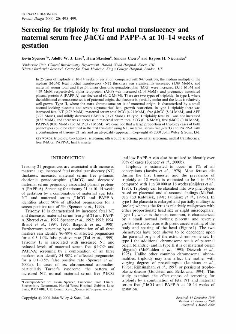
Triploidy is estimated to occur in 1% of allconceptions (Jacobs et al., 1978). Most fetuses dieduring the ®rst trimester and the prevalence oftriploidy at 12 weeks is estimated to be 1 in 3500compared with 1 in 30 000 at 16 weeks (Snijders et al.,1995). Triploidy can be classi®ed into two phenotypesbased on placental and ultrasound ®ndings (McFad-den and Kalousek, 1991; Jauniaux et al., 1996a). Intype I the placenta is enlarged and partially multicystic(molar) whereas the fetus is relatively well-grown witheither proportionate head size or slight microcephaly.Type II, which is the most common, is characterizedby a small normal looking placenta and severelygrowth restricted fetus with pronounced wasting of thebody and sparing of the head (Figure 1). The twophenotypes have been shown to be dependent uponthe parental origin of the extra chromosome set. Intype I the additional chromosome set is of paternalorigin (diandric) and in type II it is of maternal origin(digynic) (McFadden et al., 1993; Dietzsch et al.,1995). Unlike other common chromosomal abnor-malities, triploidy may also affect the mother withvarying degrees of pre-eclampsia (Jauniaux et al.,1996a; Rijhsinghani et al., 1997) or persistent tropho-blastic disease (Goldstein and Berkowitz, 1994). Thisstudy examines the effectiveness of screening fortriploidy by a combination of fetal NT and maternalserum free b-hCG and PAPP-A at 10±14 weeks ofgestation.
*Correspondence to: Kevin Spencer, Endocrine Unit, ClinicalBiochemistry Department, Harold Wood Hospital, Gubbins Lane,Essex, RM3 0BE, UK. E-mail: [email protected]
PRENATAL DIAGNOSIS
Prenat Diagn 2000; 20: 495±499.
Copyright # 2000 John Wiley & Sons, Ltd. Received: 14 December 1999Revised: 17 February 2000
Accepted: 4 March 2000

METHODS
Since 1994 maternal serum samples were collected atthe Harris Birthright Centre from women prior tochorionic villus sampling (CVS) because of advancedmaternal age or increased risk for chromosomalabnormality after NT measurement at 10±14 weeks.Serum was stored at x20uC. At the time of ultrasoundexamination crown±rump length (CRL) and NT weremeasured as previously described (Snijders et al.,1998). Maternal serum samples were available from22 cases of triploidy. In addition, three cases oftriploidy were identi®ed as part of prospective ®rsttrimester screening in the OSCAR clinic at HaroldWood Hospital (Spencer, 1999). Maternal age, weight,duration of the pregnancy based on last menstrualperiod and all ultrasound ®ndings were collected in adatabase. Outcome of pregnancy and fetal karyotypeswere added as soon as available.
During the same period serum from women attend-ing the Harris Birthright Centre for the assessment ofrisk for chromosomal abnormality or for CVS werealso taken and the same data were entered in thedatabase. From the stored sera 947 controls wereselected with matching for maternal and gestationalage. The inclusion criteria were normal karyotype atCVS or birth of a baby without abnormalities. Thesecontrols have been part of previous studies (Spenceret al., 1999; 2000a; 2000b; Tul et al., 1999).
Maternal serum free b-hCG, PAPP-A, AFP andtotal hCG were measured using the Kryptor analyser
Ð a rapid random access immunoassay analyser usingtime resolved ampli®ed cryptate emmission (TRACE)technology and the CIS automated immuno¯uorescentassays (CIS UK Ltd., High Wycombe, Bucks, UK).The stored samples were measured over a period of®ve days. The between day precision of these assayshas been previously reported (Spencer et al., 1999;2000c). The three samples collected during prospectivescreening were analysed within 20 min of bloodcollection.
Statistical analysis
Regression analysis was carried out to derive therelationship between marker levels and gestationalage. All marker measurements were converted toMoMs using the derived medians from normalpregnancies at the same gestation as assessed byCRL. Correction of each MoM for maternal weightwas also performed using the reciprocal-linear regres-sion weight correction procedure of Neveux et al.(1996). Statistical analysis of data was performed usingMicrosoft Excel 97 and Analyse-It (Smart Software,Leeds, U.K.), a statistical software add-in.
RESULTS
There were no signi®cant differences between thetriploid and control pregnancies in maternal age,gestational age based on fetal CRL, and sample
Figure 1ÐSevere asymmetrical growth restriction in a 13 week fetus with triploidy. The placenta looks normal
K. SPENCER ET AL.496
Copyright # 2000 John Wiley & Sons, Ltd. Prenat Diagn 2000; 20: 495±499.

storage time (Table 1). In the triploidy pregnancies,compared to the controls, the median fetal NT (1.89MoM), and maternal serum free b-hCG (4.59 MoM),total hCG (3.13 MoM) and AFP (2.14 MoM) weresigni®cantly increased ( p<0.0001) and PAPP-A (0.12MoM) was decreased ( p<0.0001). In 15 cases theplacentas were enlarged with molar changes and in 7they appeared normal on ultrasound (in 3 cases noplacental morphology was recorded). In the patientswith regular menstrual dates and certain dates of theirlast period, the difference in gestation estimated bydates and from fetal crown-rump length was calcu-lated as an index of fetal growth restriction. In thegroup with a molar placenta the de®cit in fetal growth(median=8, range 4±12 days) was smaller than inthose with a normal looking placenta (median=19,range 12±25). The individual marker values in the 25cases of triploidy are shown in Table 2 along with themethod by which each case was originally identi®edand the two phenotypes are compared in Table 3.
All 15 cases of type I triploidy would have beenidenti®ed using an algorithm for trisomy 21 (Spenceret al., 1999) and a risk cut off of 1 in 300. In the 10cases of type II triploidy seven of the cases would havebeen identi®ed using an algorithm for trisomy 21(Spencer et al., 1999), primarily as a result of the lowPAPP-A and the advanced maternal age (median age=36 years). If a non age related procedure, such as themethod of atypicality (Wright et al., 1993), had beenused and the atypicality threshold set to identify 1% ofunaffected cases (mahalanobis distance of 11.3 for athree marker system), all 10 of our cases would havebeen identi®ed.
DISCUSSION
The ®ndings of this study indicate that the twophenotypic types of triploidy demonstrate strikingdifferences both in fetal NT and maternal serum total
Table 1ÐMean (range) maternal age, gestational age, crown±rump lenth and length of storage of the samples from thetriploidy (n=25) and normal (n=947) pregnancies
Triploidy Controls
Maternal age (years) 33.4 (23±41) 35.1 (15±47)Gestational age (days) 86.2 (73±97) 85.1 (72±99)Crown±rump length (mm) 49.4 (39±73.8) 60.4 (38±85)Median length of storage (days) 557 (0±305) 546 (102±1811)
Table 2ÐIndividual maker levels (as MoM) in 25 cases of triploidy classi®ed by phenotype and the method of originalidenti®cation
Case NT MoM Free b-hCG MoM PAPP-A MoM AFP MoM Total hCG MoM Phenotype Identi®cation
1 1.89 3.68 0.01 3.19 6.09 1 US & placenta2 5.73 4.13 1.58 1.41 3.13 1 US & placenta3 1.61 5.35 1.44 3.06 4.12 1 US & placenta4 2.83 5.69 0.38 4.26 4.15 1 US & biochem5 2.05 6.54 1.04 16.18 2.47 1 US & placenta6 2.22 8.04 0.12 1.89 4.91 1 US & placenta7 3.27 9.48 0.93 2.14 5.11 1 US & placenta8 5.05 10.12 0.83 4.61 7.58 1 US & placenta9 1.45 14.41 0.31 17.82 5.37 1 US & placenta
10 3.93 16.11 0.36 2.72 5.65 1 US & placenta11 1.90 20.07 0.01 6.73 12.66 1 US & placenta12 2.56 12.60 0.75 3.56 10.27 1 US & biochem13 3.85 8.25 1.03 3.22 4.59 1 US14 2.91 4.59 0.50 4.26 2.56 1 US15 2.76 6.64 0.95 2.44 4.31 1 US16 0.93 0.04 0.08 0.78 0.05 2 US & IUGR17 0.87 0.06 0.05 0.51 0.15 2 T18 risk biochem18 0.50 0.07 0.01 0.13 0.07 2 US & IUGR19 0.70 0.10 0.01 1.89 0.09 2 US & IUGR20 1.13 0.36 0.06 0.57 0.21 2 US & IUGR21 1.15 0.47 0.02 0.58 0.19 2 US & IUGR22 0.61 0.48 0.09 0.87 0.16 2 T18 risk biochem23 0.79 0.25 0.11 0.97 0.23 2 ?24 0.99 0.11 0.06 0.75 0.25 2 ?25 0.89 0.31 0.12 1.06 0.15 2 ?
US, ultrasound; IUGR, intrauterine growth restriction.
FIRST TRIMESTER SCREENING FOR TRIPLOIDY 497
Copyright # 2000 John Wiley & Sons, Ltd. Prenat Diagn 2000; 20: 495±499.

and free b-hCG, PAPP-A and AFP at 10±14 weeks ofgestation. In type I, where the placenta is partiallymolar and the fetus is relatively well-grown, there isincreased fetal NT and maternal serum total hCG, freeb-hCG and AFP with mildly decreased PAPP-A. TypeII, characterized by a small normal looking placentaand severe asymmetrical fetal growth restriction, isassociated with normal fetal NT and markedlydecreased maternal serum total hCG, free b-hCGand PAPP-A with mildly decreased AFP.
Previous biochemical studies in triploid pregnanciesduring the second trimester have also shown thatmaternal serum hCG can either be low or high. Thus,®ve studies on a total of 10 triploid pregnanciesreported very low levels of total hCG and unconju-gated oestriol (Bogart et al., 1989; Kohn et al., 1991;Mason et al., 1992; Fejgin et al., 1992; 1993). Twostudies on a total of seven cases of triploidy reportedthat in some pregnancies total hCG can be very lowand in others it can be very high (Oyer and Canick,1992; Muller et al., 1993). Jacobs et al. (1982) reportedthat in triploidy with partial molar placentas andelevated maternal serum hCG the extra haploid set ofchromosomes is paternally derived; and Schmidt et al.(1994) reported that in triploidy with low maternalserum total hCG and oestriol the extra haploid set ofchromosomes is maternally derived. These ®ndingsrepresent an example of genomic imprinting inhumans with the extra paternal set of chromosomesover expressing hCG (McFadden and Kalousek, 1991;McFadden et al., 1993; 1998; Dietzsch et al., 1995;Goshen, 1994).
In the second trimester ultrasound examinationrevealing partial molar changes in the placenta, orsevere asymmetrical fetal growth restriction in thepresence of an apparently normal placenta, has beensuggested as an indicator of triploidy (Jaunieux et al.,1996a). However, molar changes in the placenta areless easily detectable in the ®rst than in the second orthird trimesters (Jaunieux et al., 1996b). As for fetalNT, in a series of 18 fetuses with triploidy at 10±14weeks the NT was above the 95th centile in 67% ofcases; although molar changes in the placenta werenoted in only 33% of cases, 85% had elevated maternalserum total hCG (Jaunieux et al., 1997). In anextension of this series, fetal NT was above the 95thcentile in 59% of 32 cases of triploidy (Snijders et al.,1998).
In ®rst trimester screening for chromosomal defectsby a combination of fetal NT and maternal serum free
b-hCG and PAPP-A, type I triploidy will be identi®edsince the marker patterns are very similar to those seenin trisomy 21 (Spencer et al., 1999). However, in typeII, despite the biochemical pattern being very similarto that in trisomy 18 and trisomy 13 (Tul et al., 1999;Spencer et al., 2000a), the near normal NT wouldnegate their detection. However, this problem could beovercome by paying attention to fetal symmetryduring the ®rst trimester scan, especially in cases ofvery low free b-hCG and PAPP-A, since type II fetusesdemonstrate severe asymmetrical growth restriction(Figure 1). Another possible way to increase thechances of detection of type II triploidy cases wouldbe to use the concept of atypicality, which has beenproposed for use in second trimester screening (Wrightet al., 1993). This concept assesses the probability of apattern of results having arisen from the trisomy 21population or the normal population. If the prob-ability of arising from either group is low the patternof results are considered atypical (i.e. they are neithernormal or trisomy 21 like). The function de®ningatypicality (the Mahalanobis distance) is alreadycalculated in conventional trisomy 21 risk algorithms.Setting the atypicality threshold to identify 1% ofunaffected cases (Mahalanobis distance of 11.3 for athree marker system) in our series of type II triploidyall 10 cases would have been identi®ed. Anotheralternative approach would be to devise speci®calgorithms for type II triploidy; however this approachwould require considerably more data on affectedcases.
Screening for trisomy 21 at 10±14 weeks of gestationby a combination of maternal age, fetal NT andmaternal serum free b-hCG and PAPP-A, identi®esabout 90% of pregnancies with trisomy 21, trisomy 18,trisomy 13 and sex chromosome abnormalities for ascreen positive rate of about 6% (Spencer et al., 1999;2000a; 2000b; Tul et al., 1999). The ®ndings of thisstudy indicate that the same method of screening canidentify more than 90% of fetuses with triploidy ofboth phenotypes.
ACKNOWLEDGMENTS
We acknowledge the support of CIS in providing theinstrument and reagents for this study. The work atthe Harris Birthright Research Centre is supported bya grant from the Fetal Medicine Foundation (Charityno. 1037116).
Table 3ÐMedian (95% con®dence interval) fetal nuchal translucency thickness and maternal serum markers in the twophenotypes of triploidy at 10±14 weeks of gestation
Parameter Type I Type II Comparison
NT MoM 2.76 (1.90 to 3.85) 0.88 (0.61 to 1.13) p<0.0001Free b-hCG MoM 8.04 (4.59 to 12.60) 0.18 (0.06 to 0.47) P<0.0001Total hCG MoM 4.91 (4.12 to 6.09) 0.16 (0.08 to 0.23) P<0.0001PAPP-A MoM 0.75 (0.31 to 1.03) 0.06 (0.01 to 0.11) p<0.0001AFP MoM 3.22 (2.44 to 4.60) 0.77 (0.51 to 1.06) p<0.0001
K. SPENCER ET AL.498
Copyright # 2000 John Wiley & Sons, Ltd. Prenat Diagn 2000; 20: 495±499.

REFERENCES
Biagiotti R, Cariati E, Brizzi L, Capelli G, D'Agata A. 1998.Maternal serum screening for trisomy 18 in the ®rst trimester ofpregnancy. Prenat Diagn 18: 907±913.
Bogart MH, Golbus MS, Sorg ND, Jones OW. 1989. Humanchorionic gonadotropin levels in pregnancies with aneuploidfetuses. Prenat Diagn 9: 379±384.
Brizot ML, Snijders RJM, Bersinger NA, Kuhn P, Nicolaides KH.1994. Maternal serum pregnancy-associated plasma protein A andfetal nuchal translucency thickness for the predicition of fetaltrisomies in early pregnancies. Obstet Gynecol 84: 918±922.
Brizot ML, Snijders RJM, Butler J, Bersinger NA, Nicolaides KH.1995. Maternal serum hCG and nuchal translucency thickness forthe prediction of fetal trisomies in the ®rst trimester of pregnancy.Br J Obstet Gynaecol 102: 127±132.
Dietzsch E, Ramsay M, Christianson AL, Henderson BD, de RavelTJL. 1995. Maternal origin of extra haploid set of chromosomesin third trimester triploid fetuses. Am J Med Genet 58: 360±364.
Fejgin MD, Amiel A, Golberger S, Barnes I, Zer T, Kohn G. 1992.Placental insuf®ciency as a possible cause of low maternal serumhuman chorionic gonadotropin and low maternal serum uncon-jugated estriol levels in triploidy. Am J Obstet Gynecol 167:766±767.
Fejgin MD, Amiel A, Kohn G. 1993. More about low maternalserum human chorionic gonadotropin and unconjugated estriolvalues in triploidy. Am J Obstet Gynecol 168: 1641.
Goldstein DP, Berkowitz RS. 1994. Current management ofcomplete and partial molar pregnancy. J Reprod Med 39:139±146.
Goshen R. 1994. The genomic basis of the b subunit of humanchorionic gonadotropin diversity in triploidy. Am J ObstetGynecol 170: 700±701.
Jacobs PA, Angell RR, Buchanan IM, Hassold TJ, Matsuyama JS,Manuel B. 1978. The origin of human triploids. Ann Hum Genet42: 49±57.
Jacobs PA, Szulman AE, Funkhouser J, Matsuura JS, Wilson CC.1982. Human triploidy: relationship between parental origin ofthe additional haploid complement and development of partialhydatidiform mole. Ann Hum Genet 46: 223±231.
Jauniaux E, Brown R, Rodeck C, Nicolaides KH. 1996a. Prenataldiagnosis of triploidy during the second trimester of pregnancy.Obstet Gynecol 83: 983±989.
Jauniaux E, Kadri R, Hustin J. 1996b. Partial mole and triploidy:screening patients with ®rst trimester spontaneous abortions.Obstet Gynecol 88: 616±619.
Jauniaux E, Brown R, Snijders RJM, Noble P, Nicolaides KH. 1997.Early prenatal diagnosis of triploidy. Am J Obstet Gynecol 176:550±554.
Kohn G, Zamir R, Zer T, Amiel A, Fejgin M. 1991. Signi®cance ofvery low maternal serum human chorionic gonadotropin inprenatal diagnosis of triploidy. Prenat Diagn 11: 277.
Mason G, Linton C, Cuckle H, Holding S. 1992. Low maternalserum human chorionic gonadotrophin and unconjugated oestriolin a triploidy pregnancy. Prenat Diagn 12: 545±546.
McFadden DE, Kalousek DK. 1991. Two different phenotypes offetuses with chromosomal triploidy: correlation with parentalorigin of the extra haploid set. Am J Med Genet 38: 535±538.
McFadden DE, Kwong LC, Yam IYL, Langlois S. 1993. Parentalorigin of triploidy in human fetuses: evidence for genomicimprinting. Hum Genet 92: 465±469.
McFadden DE, Lockitch G, Langlois S. 1998. Triple screen intriploidy. Am J Hum Genet 63 (Suppl.): A168.
Muller F, Aegerter P, Boue A. 1993. Prospective maternal serumhuman chorionic gonadotropin screening for the risk of fetalchromosome anomalies and of subsequent fetal and neonataldeaths. Prenat Diagn 13: 29±43.
Neveux LM, Palomaki GE, Larivee DA, Knight GJ, Haddow JE.1996. Re®nements in managing maternal weight adjustment forprenatal screening results. Prenat Diagn 16: 1115±1119.
Oyer CE, Canick JA. 1992. Maternal serum hCG levels in triploidy:variability and need to consider molar tissue. Prenat Diagn 12:627±629.
Rijhsinghani A, Yankowitz J, Strauss RA, Kuller JA, Patel S,Williamson RA. 1997. Risk of preeclampsia in second trimestertriploid pregnancies. Obstet Gynecol 90: 884±888.
Schmidt D, Shaffer LG, McCaskill C, Rose E, Greenberg F. 1994.Very low maternal serum chorionic gonadotropin levels inassociation with fetal triploidy. Am J Obstet Gynecol 170: 77±80.
Sherod C, Sebire NJ, Soares W, Snijders RJM, Nicolaides KH. 1997.Prenatal diagnosis of trisomy 18 at the 10±14 week ultrasoundscan. Ultrasound Obstet Gynecol 10: 387±390.
Snijders RJM, Sebire NJ, Nicolaides KH. 1995. Maternal age andgestational age-speci®c risk for chromosomal defects. Fetal DiagnTher 10: 356±367.
Snijders RJM, Noble P, Sebire N, Souka A, Nicolaides KH, for theFetal Medicine Foundation First Trimester Screening Group.1998. UK multicentre project on assessment of risk of trisomy 21by maternal age and fetal nuchal-translucency thickness at 10±14weeks of gestation. Lancet 351: 343±346.
Spencer K. 1999. OSCAR Ð One Stop Clinic for Assessment ofRisk for fetal abnormalities. Down's Screening News 6: 10.
Spencer K, Macri JN, Aitken DA, Connor JM. 1992. Free b-hCG as®rst trimester marker for fetal trisomy. Lancet 339: 1480.
Spencer K, Mallard AS, Coombes EJ, Macri JN. 1993. Prenatalscreening for trisomy 18 with free beta human chorionicgonadotrophin as a marker. BMJ 307: 1455±1458.
Spencer K, Aitken DA, Croslley JA, McCaw G, Berry E, AndersonR, Connor JM, Macri JN. 1994. First trimester biochemicalscreening for trisomy 21: the role of free beta hCG, alphafetoprotein and pregnancy associated plasma protein A. Ann ClinBiochem 31: 447±454.
Spencer K, Souter V, Tul N, Snijders R, Nicolaides KH. 1999. Arapid screening program for trisomy 21 at 10±14 weeks using fetalnuchal translucency, maternal serum free b-hCG and PAPP-A.Ultrasound Obstet Gynecol 13: 231±237.
Spencer K, Ong C, Skentou H, Liao AW, Nicolaides KH. 2000a.Screening for trisomy 13 by fetal nuchal translucency andmaternal serum free b-hCG and PAPP-A at 10±14 weeks ofgestation. Prenat Diagn (in press).
Spencer K, Tul N, Nicolaides KH. 2000b. Maternal serum free betahCG and PAPP-A in fetal sex chromsome defects in the ®rsttrimester. Prenat Diagn (in press).
Spencer K, Berry E, Crossley JA, Aitken DA, Nicolaides KH. 2000c.Is maternal serum total hCG a marker of trisomy 21 in the ®rsttrimester of pregnancy. Prenat Diagn (in press).
Tul N, Spencer K, Noble P, Chan C, Nicolaides KH. 1999c.Screening for trisomy 18 by fetal nuchal translucency andmaternal serum free b-hCG and PAPP-A at 10±14 weeks ofgestation. Prenat Diagn 19: 1035±1042.
Wright DE, Reynolds TM, Donovan CM. 1993. Assessment ofatypicality: an adjunct to prenatal screening for Down's syndromethat facilitates detection of other abnormalities. Ann Clin Biochem30: 578±583.
FIRST TRIMESTER SCREENING FOR TRIPLOIDY 499
Copyright # 2000 John Wiley & Sons, Ltd. Prenat Diagn 2000; 20: 495±499.