Embed Size (px)
Citation preview

Self-Assembly of Bovine �-Casein below theIsoelectric pH
IRINA PORTNAYA,§,‡ EINAV BEN-SHOSHAN,§,‡ URI COGAN,‡ RAFAIL KHALFIN,|
DEBORAH FASS,⊥ ORY RAMON,‡ AND DGANIT DANINO*,‡
Department of Biotechnology and Food Engineering and Department of Chemical Engineering,Technion - Israel Institute of Technology, 32000, Israel; and Department of Structural Biology,
Weizmann Institute of Science, Rehovot 76100, Israel
�-Casein is an intrinsically unstructured amphiphilic protein that self-assembles into micelles at neutralpH. This paper reports that �-casein self-organizes into micelles also under acidic conditions. Theprotein association behavior and micelle characteristics at pH 2.6, well below the pI, are presented.The pH was found to strongly affect the micelle shape and dimensions. Cryogenic transmissionelectron microscopy (cryo-TEM) experiments revealed disk-like micelles of 20–25 nm in length and∼3.5 nm in height in acidic conditions. An aggregation number of 6 was determined by sedimentationequilibrium under these conditions. Isothermal titration calorimetry experiments verified the associationbelow the pI and allowed determination of the micellization enthalpy, the critical micellar concentration,and the micellization relative cooperativity (MR). Small-angle X-ray scattering results at concentrationsbelow the critical micellization concentration (CMC) suggest that the monomeric protein is likely in apremolten globule state at low pH. Calculations of the protein charge at acidic and neutral pH reveala similar high net charge but considerable differences in the charge distribution along the proteinbackbone. Overall the results show that �-casein is amphiphilic at low pH, but the distribution ofcharge along the protein chain creates packing constraints that affect the micelle organization, leadingat concentrations above the CMC to the formation of disk micelles.
KEYWORDS: �-casein; micellization; self-assembly; acidic pH; cryo-TEM; ITC
INTRODUCTION
�-Casein is a 24 kDa calcium-sensitive phosphoproteindisplaying self-assembly behavior. Recently, it has been clas-sified as an “intrinsically unstructured/disordered” protein (IUP)(1). Members of this family are characterized by a distinct aminoacid composition. High charge and low hydrophobicity lead toan open conformation and lack of tertiary structure. High prolinecontent further promotes extended conformations by beingincompatible with protein secondary structures and decreasingthe degrees of freedom of the polypeptide backbone. Disulfidebonds, which cross-link and constrain the polypeptide, aregenerally absent (2).
Whereas most IUPs lack hydrophobic regions, above theisoelectric pH (pI) �-casein contains a large, hydrophobicC-terminal domain. Because it also has a polar, negativelycharged N-terminal domain, �-casein is amphiphilic (3). There-
fore, in contrast to typical IUPs that remain in a monomericstate in solution, �-casein self-assembles at physiologicalconditions into micelles.
Studies related to caseins have generally been performedunder physiological conditions, where stable milk casein mi-celles exert biological functions (1, 4–11). Similarly, the self-assembly of �-casein into micelles was mostly investigatedabove the pI, in the pH range of 6.5–7.0. The associationmechanism and its mode of cooperativity, as well as the structureof the protein monomers and the morphology of the micelles,were studied by numerous spectroscopic, scattering, and mi-croscopy techniques (4, 5, 11–14) and found to be stronglyaffected by temperature and, to a lesser degree, by pH,concentration, ionic strength, and solvent composition (6, 9, 11).
This paper examines, for the first time, the physicochemicalcharacteristics of �-casein at low pH and its association behaviorinto stable micelles in acidic environment. The study aims toachieve more insight into the distinct properties of �-casein asa self-assembling unstructured protein.
Our study shows that �-casein associates into stable micellesat low pH. The self-assembly process and structural character-istics of the monomers and the micelles in acidic conditions,below the protein pI, were studied by several techniques. Theshape and dimensions of the micelles were characterized by
* Corresponding author [telephone (972 4) 829 2143; fax (972 4)829 3399; e-mail [email protected]].
§ Equal contribution.‡ Department of Biotechnology, Technion - Israel Institute of
Technology.| Department of Chemical Engineering, Technion - Israel Institute
of Technology.⊥ Weizmann Institute of Science.
2192 J. Agric. Food Chem. 2008, 56, 2192–2198
10.1021/jf072630r CCC: $40.75 2008 American Chemical SocietyPublished on Web 02/28/2008

cryogenic transmission electron microscopy (cryo-TEM). Thecritical micellar concentration (CMC), the relative cooperativityof the association process, and the enthalpy of micellization weredetermined by isothermal titration calorimetery (ITC). Themicellar aggregation number (Nagg) was calculated from sedi-mentation equilibrium experiments, and the monomer confor-mation was determined from small-angle X-ray scattering(SAXS) measurements. The experiments were performed at 24°C in dilute lactic acid solution or aqueous HCl solution at pH2.6 and a low ionic strength (IS) of 0.002. Our findings at lowpH were compared with the characteristic behavior of �-caseinunder physiological conditions (neutral pH). The effect of ionicstrength on the protein assembly process at neutral pH is alsostudied.
EXPERIMENTAL PROCEDURES
Materials. Bovine �-casein (>99%; Sigma-Aldrich) was dissolvedin diluted lactic acid solution and the pH was adjusted to 2.6. Additionalsolutions were prepared in pH 7.0 phosphate buffer containing 5.65mM Na2HPO4 and 3.05 mM NaH2PO4 (buffer A) and in buffer Acontaining 0.08 M NaCl (buffer B). All compounds were from Merck.The lactic acid solution and buffer A were characterized by low ionicstrengths of 0.002 and 0.02, respectively. The ionic strength of bufferB was 0.1 (Table 1). Each protein solution was filtered through a porousmembrane of 0.45 µm to avoid large protein aggregates. Proteinsolutions were prepared at concentrations ranging from 0.1 to 20 mg/mL (0.0041-0.83 mM). The protein concentration was determined fromabsorbance at 280 nm using an Ultrospec 2000 UV-visible spectro-photometer (Pharmacia Biotech), using an extinction coefficient of4.6(1%) (9).
Methods. Cryo-TEM. Specimens were prepared in the controlledenvironment vitrification system (CEVS) (15) at 24 °C and 100%relative humidity to avoid loss of volatiles. First, the solutions wereincubated in the CEVS at the desired temperature for 1 h. Then, a 7µL drop of each solution was placed on a TEM copper grid coveredwith a perforated carbon film (Pelco International) and blotted withfilter paper to form a thin liquid film of the sample (100–200 nm thick).The thinned sample was plunged into liquid ethane at its freezingtemperature (-183 °C) to form a vitrified specimen and then transferredto liquid nitrogen (-196 °C) for storage. The vitrified specimens wereexamined in a Philips CM120 transmission electron microscopeoperating at an accelerating voltage of 120 kV. We used an OxfordCT3500 (Oxford Instruments) cryoholder that maintained the specimensbelow -175 °C during sample transfer and observation. Images wererecorded digitally on a cooled Gatan MultiScan 791 CCD camera usingDigitalMicrograph 3.1 software (Gatan) in the low-dose imaging modeto minimize beam exposure and electron-beam radiation damage (16).
ITC. ITC measurements were performed with a VP-ITC calorimeter(MicroCal) at a temperature of 24 °C. The reaction cell (V ) 1.43mL) was filled with degassed solvent (lactic acid at pH 2.6, or phosphatebuffer at pH 7.0). The injector-stirrer syringe (289 µL) was loaded witha �-casein micellar solution (20 mg/mL). The micellar solution wasinjected into the reaction cell in 28 steps of 10 µL aliquots each, andthe heat flow was measured. During the titration, the stirring speedwas 310 rpm. The duration of each injection was 20 s, and theequilibration time between consecutive injections was 3 min. Such aninterval was sufficient to equilibrate the reaction cell after every
injection. Each experiment was performed at least three times.Calorimetric data analysis was carried out using Origin 5.0 software(MicroCal).
Analytical Ultracentrifugation. Sedimentation equilibrium experi-ments were performed at 24 °C using a Beckman Optima XL-A (PaloAlto, CA) analytical centrifuge at 6000, 10000, and 12000 rpm for thelow-pH solutions and at 4000, 6000, and 8000 rpm for the pH 7.0solutions. Data were collected at 280 nm. The �-casein solutions werestudied at concentrations ranging from 0.2 to 10.0 mg/mL at pH 2.6and from 0.2 to 2 mg/mL at pH 7.0 and an ionic strength of 0.1. Paststudies showed that the protein self-assembly is not affected by pressureand, therefore, it is not speed-dependent (7, 17).
The average apparent molecular weight of the micelles Mj w,app at thevarious protein concentrations was calculated from the expression
Mw,app )d ln(c)
dr2
2RT
ω2(1- νF)(1)
where c is the concentration (assumed to be proportional to absorbance)at radius r, ω the angular velocity (radians), T the temperature in K,and R the gas constant (g cm2/gmol min2 K). The partial specific volumeνj of the solute was taken to be 0.742 cm3/g3, and a solution density Fof 1.0044 g/cm3 was measured. At �-casein concentrations of 2 mg/mL and above under low pH conditions, the plot of the natural logarithmof the measured absorbance versus the square of the radius from theaxis of rotation was not linear. To estimate Nagg, the limiting slopetoward the outer edge of the sample cell was used to provide d ln(c)/dr2. The molecular weight calculated using this slope was divided bythe monomer molecular weight calculated from the �-casein amino acidsequence (24000).
SAXS. SAXS data were obtained using a slit collimated Kratkycamera (A. Paar, Graz, Austria) with a one-dimensional sensitivedetector (Ni-filtered, Cu KR radiation, operating at 40 kV and 25 mA).The wave vector h is defined as
h) 4π sinθλ
(2)
where λ is the wavelength (λ ) 0.154 nm) and 2θ the scattering angle.Samples were placed in the camera within a ∼2 mm glass capillary.Experiments were performed in a vacuum of 5 × 10⁻² Torr at 24 (0.1 °C.
The scattering intensity I(h) was normalized with regard to time,solid angle, first beam intensity, capillary thickness, transmission, andThompson factor, and the scattering from the solvent and emptycapillary were subtracted. Long exposures of 44 h were needed to obtainaccurate data and good statistics.
Statistical Analysis. For each of the methods applied here, a statisticalanalysis of the data was performed, based on at least three separatereplicate experiments. The standard error of the ITC data was found tobe no more than 5% for the CMC and MR values and no more than3% for ∆Hdemic. The standard error of the analytical ultracentrifugationdata is 5%, and that of the Rg is 4%. The analysis supports the statisticalsignificance and validity of the results.
RESULTS AND DISCUSSION
Cryo-TEM. Cryo-TEM is becoming a central technique inthe study of micellar assemblies and nanoparticles in solutionbecause it provides the morphology and dimensions of theparticles, directly, at high resolution, and in their native(hydrated) state. In the present study, cryo-TEM provided twomajor findings. First, it revealed that �-casein self-assemblesinto micelles below the pI. Second, it showed that these micelleshave a disk-like shape, a morphology that is rather unique amongself-assembling amphiphiles, whether surfactants, block-copolymers, or proteins. As an example, Figure 1 shows themicelles in diluted lactic acid solution at pH 2.6. These micelleshave a round cross-section 20–25 nm in diameter and a widthof 3–4 nm. The vitrified ice thickness (150–200 nm) in the fieldof view is an order of magnitude larger than the micelle size;
Table 1. Parameters of �-Casein Micellization as a Function of pH andIonic Strength at 24 °C
system characteristics CMC
pH ionic strength ∆Hdemic, kJ/mol mM mg/mL MR, mM Nagg
2.6 0.002 -17.9 0.079 1.89 0.14 ∼67.0 0.02 -18.58 0.094 2.26 0.11 NDa
7.0 0.1 -40.53 0.039 0.98 0.06 ∼20
a Not determined.
�-Casein Micellization J. Agric. Food Chem., Vol. 56, No. 6, 2008 2193

hence, the micelles are distributed in the film randomly, withouta preferred orientation. From the many projections seen in theimage, it is clear that the micelles are uniform. We further foundthat their shape and dimensions, at least within the range ofconcentration studied (10-40 mg/mL), are independent ofconcentration. Flat disk assemblies of similar dimensions alsoform by �-casein in HCl solution at the same temperature andpH in the absence of salt (low IS, not shown), suggesting thatthe anion type and its interaction with the protein do notsignificantly affect the morphology of the micelles or theirdimensions.
As mentioned above, the self-organization of �-casein intomicelles under physiological conditions has been widely studied.Indeed, scattering techniques (13, 14) and cryo-TEM (18)showed that at room temperature in the pH range of 6.5–7.0, inphosphate buffer as well as in water, �-casein self-assemblesinto oblate micelles with a diameter of ∼13 nm. We furthershowed by cryo-TEM that under those conditions the micelleshave a nonuniform packing (18), in contrast with the uniformappearance of the micelles in the acidic environment (Figure1). To better understand the organization of �-casein at low pHinto disk-like assemblies, we calculated the net charge and itsdistribution along the protein backbone at pH 2.6. We alsostudied the monomer conformation and the self-organizationcharacteristics (CMC, mode of self-assembly, cooperativity) of�-casein by ITC, analytical ultracentrifugation, and SAXS, asdescribed below.
ITC. ITC is a sensitive method, directly providing both theheat of demicellization (∆Hmic ) -∆Hdemic) and the CMC in asingle experiment (18, 19). We previously used this techniqueto characterize the ∆Hmic and the CMC of �-casein in phosphatebuffer (pH 7.0) and ionic strength of 0.1. Data analysis alsoprovided the relative cooperativity, MR, which defines theprotein concentration range over which the micellization processtakes place (18). MR depends on temperature, pH, and ionicstrength. Its increase and decrease are indicative of decreaseand increase of the cooperativity, respectively.
In a typical demicellization experiment, a micellar solutionis titrated into buffer placed in the ITC cell, and the heat flowis measured as a function of time. Such an experiment,performed on �-casein at pH 2.6, is presented in Figure 2A.
Three factors contribute to the exothermic enthalpy changesobserved at the initial injections: micelle dilution, demicelliza-tion, and dilution of individual �-casein molecules. The enthalpychanges decrease in magnitude as more protein is added andthe concentration in the ITC cell increases. Eventually (finalinjections), the concentration in the cell exceeds the CMC andonly micelle dilution contributes to the heat flow (18, 19).
In Figure 2B the heat of the reaction, obtained by integratingthe peaks of the individual injections given in Figure 2A, isplotted against the �-casein concentration in the cell. A slowincrease in the reaction enthalpy is observed, resulting in MRof 0.14 mM (Figure 2 and Table 1), which is more than twicethan the value found at pH 7.0 and IS of 0.1 (Table 1) (18).Figure 2B also presents the heat of demicellization, ∆Hdemic,which equals the enthalpy difference between the two asymp-totes (19) of the sigmoid fit of the experimental data (obtainedby using the Origin software). It is shown that at 24 °C ∆Hdemic
is ∼-17.9 kJ/mol (Table 1), relatively small compared withthe -40.53 kJ/mol found at pH 7.0 and IS of 0.1 (18).
In Figure 2C we present the first derivative of the reactionenthalpy versus the �-casein concentration in the cell. The CMC,obtained from the concentration at which the first derivative ofthe reaction heat displays a maximum (19–22), was determinedto be 1.89 mg/mL (Figure 2C and Table 1) at pH 2.6. Thisvalue is approximately twice the CMC found at pH 7 and ionicstrength 0.1 (Table 1) (18).
The ITC measurements presented in Figure 2 support thecryo-TEM findings; that is, they confirm the self-assembly of�-casein into micelles at acidic pH (2.6) and low ionic strength(0.002). However, the small ∆Hdemic, the high CMC, and thelarge MR indicate that the driving forces for micellization under
Figure 1. Cryo-TEM image of 20 mg/mL �-casein micelles at 24 °C, pH2.6, and low ionic strength in lactic acid solution. Bar ) 50 nm.
Figure 2. Titration of micellar (20 mg/mL) �-casein solution in dilutedlactic acid (pH 2.6) and very low ionic strength (0.002) into lactic acidsolution, having the same pH and ionic strength, at 24 °C: (A) calorimetrictraces; (B) reaction enthalpy versus �-casein concentration in the cell;(C) first derivative of curve B calculated from the interpolated values.
2194 J. Agric. Food Chem., Vol. 56, No. 6, 2008 Portnaya et al.

acidic conditions are reduced compared with those at physi-ological pH and high IS (18).
To separate the effect of pH from that of IS, we also studied�-casein solutions at pH 7.0 and low ionic strength (0.02). InFigure 3 we plot the first derivatives of the reaction enthalpiesversus the protein concentration for the three compositionsstudied (see also Table 1). From these graphs the CMC valuesare evaluated. The positions and magnitudes of the peaks ofthe two low ionic strength solutions (pH 2.6 and 7.0) are almostidentical and wider than the peak of the high IS solution.Additionally, their ∆Hdemic are of similar magnitudes and arelower relative to the sample with high ionic strength. Likewise,the CMC values of the two low ionic strength samples aresimilar and about twice as high as the CMC of the sample withIS 0.1.
The differences in the micellization parameters of �-caseinsolutions between low (0.02) and high (0.1) ionic strength (at24 °C and 20 mg/mL) can be explained by the screening ofelectrostatic repulsion forces at high IS, which lowers therepulsions between head-groups, thereby enabling micellizationat a lower concentration. Therefore, at low IS, ∆Hdemic has alower magnitude than at high IS, whereas the CMC and MRare significantly larger. Thus, the larger value of MR at low ISsignifies decreased micellization cooperativity.
Sedimentation Equilibrium. To determine the aggregationnumber of the micelles at pH 2.6, analytical ultracentrifugationexperiments were conducted at various protein concentrations(see Table 2) and at pH 7.0 and IS of 0.1. Sample data areplotted in Figure 4. At concentrations lower than the CMC,determined by ITC to be 1.89 mg/mL (0.079 mM, Table 1), astraight line was obtained. The aggregation numbers calculatedfrom the slope of this line and eq 1 confirmed that the protein
is monomeric at these concentrations (see Table 2). Atconcentrations higher than the CMC, two regions could bedefined, indicating the presence of two protein populations:monomers at relatively short radii (i.e., in Figure 4 at r < 6.5or r2 < 43) and assemblies at large radii. The two limiting slopestoward the inner and outer edges of the sample cell were usedto calculate the minimal and maximal apparent molecularweights, respectively. The micelles at pH 2.6 are characterizedby a small aggregation number of 3 around the CMC and 6 athigher concentrations (Table 2). In contrast, using the sametechnique we measured Nagg of 20 at pH 7.0 and ionic strength0.1, and values of 17–23 were reported in the literature by Evanset al. (12) and Mikheeva et al. (9) at comparable temperature,pH, and ionic strength. Thus, compared with assembly at neutralpH, assembly at low pH is characterized by two special features:the micelles are flat and disk-like in shape, and they have alow molecular weight. To understand the origin of theseproperties, we calculated the charge and charge distribution atpH 2.6 and 7.0.
Evaluation of the Net Charge and Charge Distributionof �-Casein at pH 2.6. As mentioned, �-casein has beencharacterized as an intrinsically unstructured protein. IUPs canexist in a random coil conformation, resembling denaturedglobular proteins, or in a premolten globule state exhibiting somesecondary structure (2, 23). Indeed, �-casein is thought to havean open rheomorphic structure at low concentrations. However,in contrast to other IUPs, �-casein is amphiphilic and henceself-assembles into micelles. Micellization processes are drivenprimarily by hydrophobic interactions, whereas electrostatic andsteric repulsive forces oppose the association process andstabilize the formed structures (9, 11). In �-casein, close toneutral pH, a high negative charge of -14.8 is concentratedmainly in the first 50 amino acids of the N-terminal domain(Figure 5A) (7, 24, 25), whereas the C terminus has only afew charged groups and a small net charge and is rich inhydrophobic groups (7, 24, 25).
To estimate the net charge and the charge distribution of�-casein at pH 2.6, we used a procedure similar to that reportedby Ribadeau et al. (25). The degree of protonation, θi,A of theindividual titratable sites was calculated from the Henderson–Hasselbach equation (26). This equation is not valid when strongelectrostatic interactions exist between titratable sites but canbe used when the protein is in an unfolded state (27). Because�-casein is intrinsically unstructured, the equation is appropriatefor calculating its net charge at low pH.
Figure 3. CMC of �-casein at different pH values and ionic strengths: 1,pH 2.6, ionic strength 0.002; 2, pH 7, ionic strength 0.1; 3, pH 7, ionicstrength 0.02.
Table 2. Analytical Ultracentrifugation Results of �-Casein Solutions at pH2.6 and Ionic Strength 0.002a
population a population b
concn, mg/mL M̄a Nagg M̄b Nagg
0.2 16800 ∼1 16800 ∼10.7 16200 ∼1 16200 ∼11.0 14200 ∼1 26400 ∼12.0 25900 ∼1 76700 ∼35.0 24000 ∼1 142600 ∼610.0 26600 ∼1 135100 ∼6
a The micelle aggregation numbers were obtained by dividing the measuredaggregate molecular weight by the monomer molecular weight (24000) calculatedfrom the �-casein amino acid sequence.
Figure 4. Determination of �-casein aggregation number from theultracentrifugation data, following eq 1: pH 2.6, protein concentration 5mg/mL, 0.002 IS.
�-Casein Micellization J. Agric. Food Chem., Vol. 56, No. 6, 2008 2195
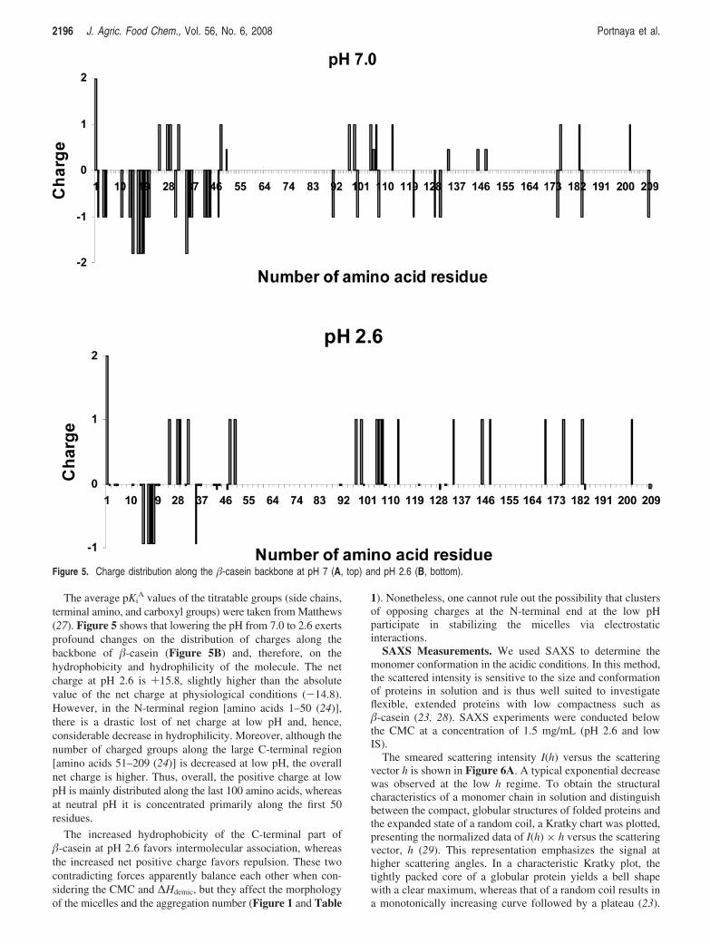
The average pKiA values of the titratable groups (side chains,
terminal amino, and carboxyl groups) were taken from Matthews(27). Figure 5 shows that lowering the pH from 7.0 to 2.6 exertsprofound changes on the distribution of charges along thebackbone of �-casein (Figure 5B) and, therefore, on thehydrophobicity and hydrophilicity of the molecule. The netcharge at pH 2.6 is +15.8, slightly higher than the absolutevalue of the net charge at physiological conditions (-14.8).However, in the N-terminal region [amino acids 1–50 (24)],there is a drastic lost of net charge at low pH and, hence,considerable decrease in hydrophilicity. Moreover, although thenumber of charged groups along the large C-terminal region[amino acids 51–209 (24)] is decreased at low pH, the overallnet charge is higher. Thus, overall, the positive charge at lowpH is mainly distributed along the last 100 amino acids, whereasat neutral pH it is concentrated primarily along the first 50residues.
The increased hydrophobicity of the C-terminal part of�-casein at pH 2.6 favors intermolecular association, whereasthe increased net positive charge favors repulsion. These twocontradicting forces apparently balance each other when con-sidering the CMC and ∆Hdemic, but they affect the morphologyof the micelles and the aggregation number (Figure 1 and Table
1). Nonetheless, one cannot rule out the possibility that clustersof opposing charges at the N-terminal end at the low pHparticipate in stabilizing the micelles via electrostaticinteractions.
SAXS Measurements. We used SAXS to determine themonomer conformation in the acidic conditions. In this method,the scattered intensity is sensitive to the size and conformationof proteins in solution and is thus well suited to investigateflexible, extended proteins with low compactness such as�-casein (23, 28). SAXS experiments were conducted belowthe CMC at a concentration of 1.5 mg/mL (pH 2.6 and lowIS).
The smeared scattering intensity I(h) versus the scatteringvector h is shown in Figure 6A. A typical exponential decreasewas observed at the low h regime. To obtain the structuralcharacteristics of a monomer chain in solution and distinguishbetween the compact, globular structures of folded proteins andthe expanded state of a random coil, a Kratky chart was plotted,presenting the normalized data of I(h) × h versus the scatteringvector, h (29). This representation emphasizes the signal athigher scattering angles. In a characteristic Kratky plot, thetightly packed core of a globular protein yields a bell shapewith a clear maximum, whereas that of a random coil results ina monotonically increasing curve followed by a plateau (23).
Figure 5. Charge distribution along the �-casein backbone at pH 7 (A, top) and pH 2.6 (B, bottom).
2196 J. Agric. Food Chem., Vol. 56, No. 6, 2008 Portnaya et al.

A monotonic increase in I(h) × h versus h followed by apeak and a mild decrease for the 1.5 mg/mL solution (pH 2.6)are shown in Figure 6B. According to Longhi and co-workers(28), who studied the C-terminal domain of the measles virusnucleoprotein, this intermediate shape between the two char-acteristic plots of globular and random coil states suggests thepresence of an intermediate premolten globule conformationwith some residual structure. This interpretation is also in linewith the studies of Farrell and co-workers (30, 31), whichsuggested the existence of limited but defined secondarystructures in �-casein. Thus, our SAXS findings suggest thatunder acidic conditions �-casein exists in a premolten globulestate.
SAXS measurements can also be used to evaluate the radiusof gyration, Rg, estimated from the Guinier approximation (32):
I(h)) I(0) eh2Rg2 ⁄3 (3)
To increase the accuracy, we evaluated the radius of gyrationfrom the smeared SAXS data using the ITR program packagedeveloped by Glatter (33).
Rg of �-casein monomers (1.5 mg/mL, pH 2.6, IS 0.002) wasfound to be 5.50 ( 0.5 nm. This value is nearly identical to thevalue 5.40 ( 0.3 nm reported at pH 7.0 and IS of 0.1 by Evanset al. (12) and only slightly smaller than the value of 5.87 (0.2 nm reported in the presence of 4 M guanidyl chloride (atthe same pH and IS), which represents the denaturated state of�-casein (24). Thus, we find that �-casein at low pH, as inneutral pH, is in a premolten globule conformation and has anRg close in value to that of the fully denatured state of theprotein.
Why Do Disk-Shaped Micelles with Lower AggregationNumber Form in Acidic pH? Berry and Creamer (34)confirmed that the main driving force for the endothermic self-assembly of �-casein at pH ∼7 is the hydrophobic C-terminalregion. They showed that removal of the last 20 amino acidsfrom the C terminus (�-casein 1–189) destroyed the ability ofthe protein to associate into micelles at physiological conditions,whereas deletion of segments from the N terminus (�-casein29–209) did not decrease the ability of the modified protein toself-assemble.
We show that below the pI, at pH 2.6, the charge of theN-terminal domain decreases and that of the C-terminal regionincreases, but at the same time the overall number of chargedgroups in the C-terminal region decreases (see Figure 5B).These contradicting contributions balance each other, leadingto association at concentration and enthalpy of similar magnitudeas in neutral pH at low IS (Table 1). However, the high chargedensity along the C-terminal portion and the electrostaticrepulsion between the monomers (and likely increase in protein
backbone rigidity) do not enable changes in the size or shapeof the micelles or in the monomer conformation during the earlystages of the assembly process as the change proposed by Farrelland co-workers (7) at neutral pH.
Conclusions. �-Casein is an intrinsically unstructured proteinthat, in contrast to other IUPs, self-assembles into detergent-like micelles at physiological conditions. We show here thatthe ability of �-casein to self-organize into micelles is preservedeven at low pH. The charge and its distribution along the proteinbackbone vary significantly with the pH and strongly affect theprotein amphiphilicity and as a result the micelle morphology,dimensions, and aggregation number. Cryo-TEM shows that flat,disk-like micelles form at pH 2.6, compared with the spheroidalmicelles that exist at physiological pH. Analytical ultracentrifu-gation experiments indicate on only 6 monomers per micelle atthe low pH, whereas about 20 monomers constitute a micelleat neutral pH. The formation of disk-like micelles can beexplained by packing constraints that result from strong intra-and intermolecular repulsion forces between the unscreenedcharges along the �-casein backbone.
The decrease in the protein amphiphilicity and the spread ofcharges along the protein backbone lead us to question thevalidity of applying the block copolymer micellization model[the dual theory by Horne (8)] to describe the aggregationprocess at low pH. Also interesting is the effect of ionic strengthon the assembly process and the protein characteristics. Wefound that the CMC and heat of demicellization were hardlyaffected by the pH when the IS was low, but they weresignificantly altered when the IS was increased under pH ∼7.Future studies will test the effect of IS on the assembly.
ACKNOWLEDGMENT
The cryo-TEM work was performed at the “Cryo-TEMHannah and George Krumholz Laboratory for AdvancedMicroscopy” at the Technion. We thank Dr. Yoav D. Livneyfor contributing discussions.
LITERATURE CITED
(1) Tompa, P. Trends Biochem. Sci. 2002, 27 (10), 527–533.(2) Uversky, V. N. Eur. J. Biochem. 2002, 269 (1), 2–12.(3) Swaisgood, H. E. In AdVanced Dairy Chemistry; Fox, P.,
McSweeney, P. L. H., Eds.; Kluwer Academic/Plenum: NewYork, 2003; pp 139–201.
(4) Arima, S.; Niki, R.; Takase, K. J. Dairy Res. 1979, 46 (2), 281–282.
(5) Buchheim, W.; Schmidt, D. G. J. Dairy Res. 1979, 46 (2), 277–280.
(6) de Kruif, C. G.; Grinberg, V. Y. Colloid Surf. A: Physicochem.Eng. Asp. 2002, 210 (2–3), 183–190.
Figure 6. Normalized SAXS I(h) (right) and the corresponding Kratky plot, I(h) × h (left) versus the wave vector h: protein concentration, 1.5 mg/mL;pH 2.6; and low ionic strength.
�-Casein Micellization J. Agric. Food Chem., Vol. 56, No. 6, 2008 2197

(7) Farrell, H. M.; Wickham, E. D.; Unruh, J. J.; Qi, P. X.; Hoagland,P. D. Food Hydrocolloids 2001, 15 (4–6), 341–354.
(8) Horne, D. S. Curr. Opin. Colloid Interface Sci. 2002, 7 (5), 456–461.
(9) Mikheeva, L. M.; Grinberg, N. V.; Grinberg, V. Y.; Khokhlov,A. R.; de Kruif, C. G. Langmuir 2003, 19 (7), 2913–2921.
(10) Niki, V. R.; Takase, K.; Arima, S. Milchwiss.-Milk Sci. Int. 1977,32, 577–582.
(11) O’Connell, J. E.; Grinberg, V. Y.; de Kruif, C. G. J. ColloidInterface Sci. 2003, 258 (1), 33–39.
(12) Evans, M. T. A.; Phillips, M. C.; Jones, M. N. Biopolymers 1979,18 (5), 1123–1140.
(13) Kajiwara, K.; Niki, R.; Urakawa, H.; Hiragi, Y.; Donkai, N.;Nagura, M. BBA-Proteins Struct. Mol. Enzymol. 1988, 955 (2),128–134.
(14) Thurn, A.; Burchard, W.; Niki, R. Colloid Polym. Sci. 1987, 265(8), 653–666.
(15) Bellare, J. R.; Davis, H. T.; Scriven, L. E.; Talmon, Y. J. ElectronMicrosc. Technique 1988, 10 (1), 87–111.
(16) Danino, D.; Bernheim-Groswasser, A.; Talmon, Y. Colloid Surf.A-Physicochem. Eng. Asp. 2001, 183, 113–122.
(17) Payens, T. A. J.; Brinkhui, J. A.; Vanmarkw, B. W. BBA-ProteinStruct. 1969, 175 (2), 434–437.
(18) Portnaya, I.; Cogan, U.; Livney, Y. D.; Ramon, O.; Shimoni, K.;Rosenberg, M.; Danino, D. J. Agric. Food Chem. 2006, 54, 5555–5561.
(19) Paula, S.; Sus, W.; Tuchtenhagen, J.; Blume, A. J. Phys. Chem.1995, 99, 11742–11751.
(20) Dai, S.; Tam, K. C. Langmuir 2004, 20, 2177–2183.(21) Moroi, Y. Micelles: Theoretical and Applied Aspects; Moroi, Y.,
Ed.; Plenum: New York, 1992; pp 47–50.(22) Tanford, C. The Hydrophobic Effect: Formation of Micelles and
Biological Membranes, 2nd ed.; Wiley: New York, 1980.
(23) Receveur-Brechot, V.; Bourhis, J. M.; Uversky, V. N.; Canard,B.; Longhi, S. Proteins 2006, 62 (1), 24–45.
(24) Aschi, A.; Gharbi, A.; Daoud, M.; Douillard, R.; Calmettes, P.Polym. Int. 2007, 56 (5), 606–612.
(25) Ribadeau Dumas, B.; Brignon, G.; Grosclaude, F.; Mercier, J. C.Eur. J. Biochem. 1972, 25 (3), 505–514.
(26) Kundrotas, P. J.; Karshikoff, A. Protein Sci. 2002, 11 (7), 1681–1686.
(27) Matthews, H. R. In Biochemistry: A Short Course; Miesfeld, R. L.,Freedland, R. A., Eds.; Wiley: 1997; p 505.
(28) Longhi, S.; Receveur-Brechot, V.; Karlin, D.; Johansson, K.;Darbon, H.; Bhella, D.; Yeo, R.; Finet, S.; Canard, B. J. Biol.Chem. 2003, 278 (20), 18638–18648.
(29) Glatter, O.; Kratky, O. In Small Angle X-ray Scattering; Glatter,O., Ed.; Academic Press: London, U.K., 1982; p 515.
(30) Qi, P. X.; Wickham, E. D.; Farrell, H. M. Protein J. 2004, 23(6), 389–402.
(31) Qi, P. X.; Wickham, E. D.; Piotrowski, E. G.; Fagerquist, C. K.;Farrell, H. M. Protein J. 2005, 24 (7–8), 431–444.
(32) Guinier, A., Fournet, G., Eds. Small Angle Scattering of X-rays;Wiley: New York, 1955.
(33) Glatter, O.; Gruber, K. J. Appl. Crystallogr. 1993, 26, 512–518.(34) Berry, G. P.; Creamer, L. K. Biochemistry 1975, 14, 3542–3545.
Received for review September 5, 2007. Revised manuscript receivedDecember 16, 2007. Accepted December 24, 2007. This work wassupported in part by the Israel Science Foundation of the IsraelAcademy of Sciences and Humanities and the RBNI at the Technion.I.P. and R.K. acknowledge the support of a joint grant from the Centerfor Absorption in Science of the Ministry of Immigrant Absorptionand the Committee for Planning and Budgeting of the Council forHigher Education under the framework of the KAMEA program.
JF072630R
2198 J. Agric. Food Chem., Vol. 56, No. 6, 2008 Portnaya et al.