Embed Size (px)
Citation preview
Akademie věd České republiky
Teze disertace k získání vědeckého titulu "doktor věd"
ve skupině chemických věd
Singlet oxygen producing sensitizers: from molecular photophysics
to photofunctional materials
Komise pro obhajoby doktorských disertací v oboru Anorganická chemie
Kamil Lang
Ústav anorganické chemie AVČR, v.v.i.
Řež, 2014
Contents
Résumé ..................................................................................................................................2
Abbreviations .........................................................................................................................3
1. Foreword ............................................................................................................................4
2. Photosensitized reactions of molecular oxygen ...................................................................5
3. Porphyrinic sensitizers ........................................................................................................6
3.1. Noncovalently bound porphyrin sensitizers in solutions ...............................................8
3.1.1. Calixarenes ...........................................................................................................8
3.1.2. Cyclodextrins ...................................................................................................... 10
4. Novel sensitizers ............................................................................................................... 12
4.1. Hexanuclear molybdenum cluster complexes ............................................................. 12
4.2. Boron hydride clusters ............................................................................................... 14
5. Photofunctional materials ................................................................................................. 17
5.1. Layered hydroxides .................................................................................................... 18
5.1.1. Layered double hydroxides ................................................................................. 18
5.1.2. Layered zinc hydroxide salts ............................................................................... 28
5.1.3. Layered rare-earth hydroxides ............................................................................. 30
5.2. Polymers .................................................................................................................... 31
6. Conclusions ...................................................................................................................... 34
7. Acknowledgments ............................................................................................................ 36
8. Publications that form the basis of the thesis ..................................................................... 36
9. References ........................................................................................................................ 39

2
Résumé
This thesis reviews my research on the photophysical and photochemical properties of
porphyrin sensitizers confined in water-soluble host-guest complexes or in host matrices
including layered hydroxides and polymers. The properties of photoactive porphyrins
constrained within hosts are altered and cannot be directly extrapolated from the known
behavior of their individual components. The sensitizers studied were mostly of the porphyrin
type; however, two other groups of novel sensitizers, hexanuclear molybdenum clusters and
boron clusters, were also investigated. Particular attention was paid to the sensitizers
producing singlet oxygen by energy transfer from their excited triplet states to molecular
oxygen. Because the kinetic parameters of the triplet states predetermine the formation of
singlet oxygen, the excited-state dynamics of the sensitizers were characterized by steady-
state and time-resolved spectroscopy techniques.
The development of a new class of photofunctional hybrid materials was based on
porphyrin sensitizers intercalated in layered metal hydroxides. The metal hydroxide layers of
layered hydroxides can be viewed as spacers separating compartments containing the
porphyrin molecules, whose arrangement imposes specific structural, thermal, chemical, and
photochemical properties on the materials. Investigation of the embedded porphyrin
sensitizers in polymeric nanofiber materials and layered hydroxide/polymer composites, in
which layered hydroxides serve as nanofillers and nanocontainers of photoactive porphyrins,
allowed for the fabrication of eco-friendly and gas permeable polymers with bactericidal and
virucidal surfaces that are activated by visible light. We also prepared and characterized the
first metal-organic frameworks with singlet oxygen-sensitizing ability. Our research on
layered metal hydroxides led to the development of new methods of hydroxide delamination
and to the use of hydroxide nanosheets for the preparation of functional films (for
luminescent, photocatalytic, and electrochemical activity) with an adjustable thickness.
The unifying theme of the research was to utilize the photophysical and photochemical
properties of the embedded sensitizers for the development of materials whose function is
triggered by visible light, i.e., materials with bactericidal surfaces or materials acting as solid
sensitizers or oxygen sensors.
3
Abbreviations
CD Cyclodextrin
clxm p-Sulfonatocalix[m]arene (m = 4,6,8)
DS Dodecyl sulfate
LDHs Layered double hydroxides
LZHs Layered zinc hydroxide salts
MOF Metal-organic framework
MgRAl-LDH-PdTPPC Layered double hydroxide with a Mg/Al molar ratio R and
intercalated with PdTPPC (and analogously for other LDHs)
LZH-TPPS Layered zinc hydroxide intercalated with TPPS
LREHs Layered rare-earth hydroxides
LEuH-PdTPPS Layered europium hydroxide intercalated with PdTPPS
MTPPC, MTPPS, MTPP Metal complexes of TPPC, TPPS, or TPP
PDT Photodynamic therapy
PU Polyurethane
SBU Secondary building unit
SODF Singlet oxygen-sensitized delayed fluorescence
TPP 5,10,15,20-Tetraphenylporphyrin
TPPS 5,10,15,20-Tetrakis(4-sulfonatophenyl)porphyrin
TPPC 5,10,15,20-Tetrakis(4-phosphonatophenyl)porphyrin
TMPyPn 5,10,15,20-Tetrakis(N-methylpyridinium-n-yl)porphyrin
XRD X-ray diffraction
ZnPc Zinc phthalocyanine
Symbols
f , Φp, T Quantum yields of fluorescence, phosphorescence, or triplet states
Quantum yield of singlet oxygen formation
4
1. Foreword
Photosensitized production of singlet oxygen from molecular oxygen has found far-
reaching applications in chemistry and biology. The reaction is based on the excitation of a
sensitizer molecule by light, followed by energy transfer to the ground triplet state oxygen
which, in turn, is excited to its singlet state. Singlet oxygen, a highly reactive species, is
involved in numerous oxidative processes and can cause serious damage to biological
materials. The oxidative potential of singlet oxygen can also be exploited for use in chemical
syntheses, for the photodynamic treatment of cancer, and for antimicrobial photodynamic
therapy. Singlet oxygen reactions have become an interdisciplinary topic that spans
photophysics, quantum chemistry, photochemistry, photobiology, and photomedicine.1,2,3
The observed poor correlation between photophysical parameters of sensitizers and their
photosensitizing efficacy in a complex environment turned my attention to the interactions of
sensitizers with surrounding entities. Understanding these interactions could help explain the
unexpected differences in sensitizer photophysical and photochemical properties. In chemical
terms, the noncovalent interactions of sensitizers with the microenvironment may play a key
role in controlling the sensitizer properties. The research progressed along two parallel
courses:
1. Noncovalent interactions of sensitizers and their photophysical and photochemical
properties. Noncovalent binding of porphyrin sensitizers to biopolymers (proteins,
nucleic acids) and host molecules, such as cyclodextrins and calixarenes, does not
restrict the formation of excited states, but it influences their yields and the kinetics of
their reactions.4 Evidently, careful assessment of the binding effects on the molecular
level is a rational way to understand photosensitized processes in complex
environments. We also investigated nanostructured and polymeric host matrices with
embedded porphyrins to reduce their undesired aggregation and to eliminate a
“hostile” environment that can affect the porphyrin sensitizing activity.
2. Photofunctional materials. We investigated hybrid materials based on porphyrins
intercalated in layered metal hydroxides [A1]. Layered hydroxides were found to act
as hosts that keep the photoactive molecules organized in a two-dimensional
expandable interlayer space and separated from the surroundings. Such hybrids are not
only easy to use as transport materials; they also turn out to be suitable for the
fabrication of light-triggered materials with bactericidal functionality. Polymeric
nanofiber materials doped with porphyrins have bactericidal and virucidal surfaces
5
upon visible light irradiation. Molybdenum cluster complexes were found to be
promising molecules for the preparation of luminescent materials and oxygen sensors.
2. Photosensitized reactions of molecular oxygen
Oxygen molecules, having absorption in the vacuum UV region, can be hardly excited by
direct exposure to common sources of UV/Vis radiation. The indirect path of oxygen
excitation, the photosensitized reaction, is therefore the main pathway involving excited forms
of oxygen. Photosensitized oxygen reactions are classified as Type I and Type II according to
the nature of the quencher.5 Quenching of the excited sensitizer by molecular oxygen
proceeds as energy transfer, yielding singlet oxygen, or as electron transfer, yielding the
superoxide radical anion O2- (Type II reactions).
Sensitization. A photosensitized reaction involves the excitation of a sensitizer into higher
electronic states by light. Processes occurring after excitation can be summarized as follows:
excited singlet states, 1P*, decay to ground states,
1P, by fluorescence (Eq. 1) and vibrational
relaxation. Intersystem crossing leads to triplet states, 3P, (Eq. 2) that are deactivated
spontaneously via vibrational relaxation, phosphorescence emission (Eq. 3), or by interaction
with oxygen to form O2(1g) and O2(
1Σg
+) (Eq. 4). The higher excited state, O2(
1Σg
+), has a
very short lifetime; hence, the relatively long-lived O2(1g), referred to as “singlet oxygen”, is
generally accepted as a decisive species involved in photosensitized reactions (Figure 1). The
excited singlet states of most sensitizers are too short-lived to be effectively quenched by
oxygen to form singlet oxygen. Detailed discussion of all of these processes can be found
elsewhere.3,4
1P*
1P + hf (1)
1P*
3P (2)
3P
1P + hp (3)
3P + O2(
g
-)
1P + O2(
1g) (4)
O2(1g) O2(
g
-) + hp (5)
Sensitizers. Numerous dyes and complexes produce singlet oxygen and are considered
suitable for photodynamic therapy (PDT); nevertheless, the majority of the sensitizers
investigated in this context have a porphyrinic structure. The reasons for this preference are
the extensive knowledge of porphyrin chemistry and their inherent similarity to natural
porphyrins in living matter. For photodynamic applications, the sensitizers should possess
6
certain characteristics, such as absorption and emission in the red spectral region to minimize
interference with the living tissue, high quantum yield of singlet oxygen formation (ΦΔ), low
toxicity, and good chemical and photochemical stability.
My research is focused on porphyrins and related macrocycles in solutions4 and solid
materials [A1] (Sections 3 and 5), hexanuclear molybdenum cluster complexes as relevant
alternatives to the sensitizers so far used (Section 4.1), and boron clusters (Section 4.2.).
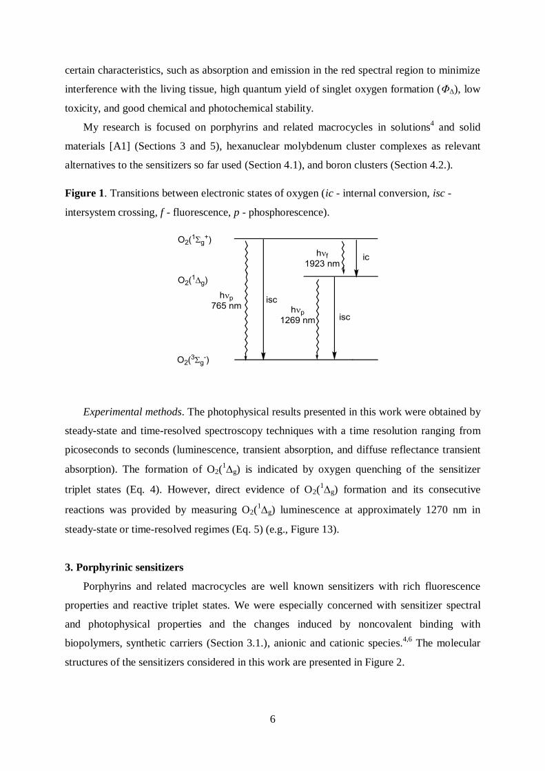
Figure 1. Transitions between electronic states of oxygen (ic - internal conversion, isc -
intersystem crossing, f - fluorescence, p - phosphorescence).
Experimental methods. The photophysical results presented in this work were obtained by
steady-state and time-resolved spectroscopy techniques with a time resolution ranging from
picoseconds to seconds (luminescence, transient absorption, and diffuse reflectance transient
absorption). The formation of O2(1g) is indicated by oxygen quenching of the sensitizer
triplet states (Eq. 4). However, direct evidence of O2(1g) formation and its consecutive
reactions was provided by measuring O2(1g) luminescence at approximately 1270 nm in
steady-state or time-resolved regimes (Eq. 5) (e.g., Figure 13).
3. Porphyrinic sensitizers
Porphyrins and related macrocycles are well known sensitizers with rich fluorescence
properties and reactive triplet states. We were especially concerned with sensitizer spectral
and photophysical properties and the changes induced by noncovalent binding with
biopolymers, synthetic carriers (Section 3.1.), anionic and cationic species.4,6
The molecular
structures of the sensitizers considered in this work are presented in Figure 2.
7
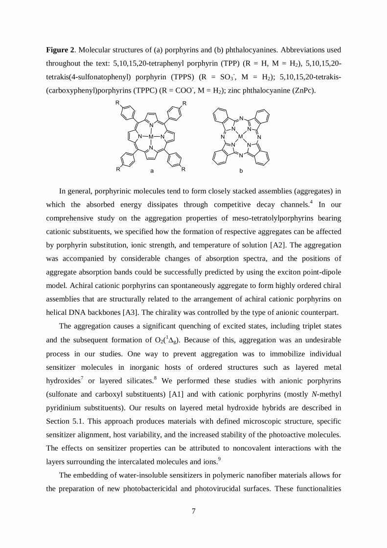
Figure 2. Molecular structures of (a) porphyrins and (b) phthalocyanines. Abbreviations used
throughout the text: 5,10,15,20-tetraphenyl porphyrin (TPP) (R = H, M = H2), 5,10,15,20-
tetrakis(4-sulfonatophenyl) porphyrin (TPPS) (R = SO3-, M = H2); 5,10,15,20-tetrakis-
(carboxyphenyl)porphyrins (TPPC) (R = COO-, M = H2); zinc phthalocyanine (ZnPc).
In general, porphyrinic molecules tend to form closely stacked assemblies (aggregates) in
which the absorbed energy dissipates through competitive decay channels.4 In our
comprehensive study on the aggregation properties of meso-tetratolylporphyrins bearing
cationic substituents, we specified how the formation of respective aggregates can be affected
by porphyrin substitution, ionic strength, and temperature of solution [A2]. The aggregation
was accompanied by considerable changes of absorption spectra, and the positions of
aggregate absorption bands could be successfully predicted by using the exciton point-dipole
model. Achiral cationic porphyrins can spontaneously aggregate to form highly ordered chiral
assemblies that are structurally related to the arrangement of achiral cationic porphyrins on
helical DNA backbones [A3]. The chirality was controlled by the type of anionic counterpart.
The aggregation causes a significant quenching of excited states, including triplet states
and the subsequent formation of O2(1g). Because of this, aggregation was an undesirable
process in our studies. One way to prevent aggregation was to immobilize individual
sensitizer molecules in inorganic hosts of ordered structures such as layered metal
hydroxides7 or layered silicates.
8 We performed these studies with anionic porphyrins
(sulfonate and carboxyl substituents) [A1] and with cationic porphyrins (mostly N-methyl
pyridinium substituents). Our results on layered metal hydroxide hybrids are described in
Section 5.1. This approach produces materials with defined microscopic structure, specific
sensitizer alignment, host variability, and the increased stability of the photoactive molecules.
The effects on sensitizer properties can be attributed to noncovalent interactions with the
layers surrounding the intercalated molecules and ions.9
The embedding of water-insoluble sensitizers in polymeric nanofiber materials allows for
the preparation of new photobactericidal and photovirucidal surfaces. These functionalities
8
are based on the combination of the photosensitizing activity of porphyrins with the high
surface area, flexibility, lightness, and good light and oxygen penetrability of nanofibers
(Section 5.2.).
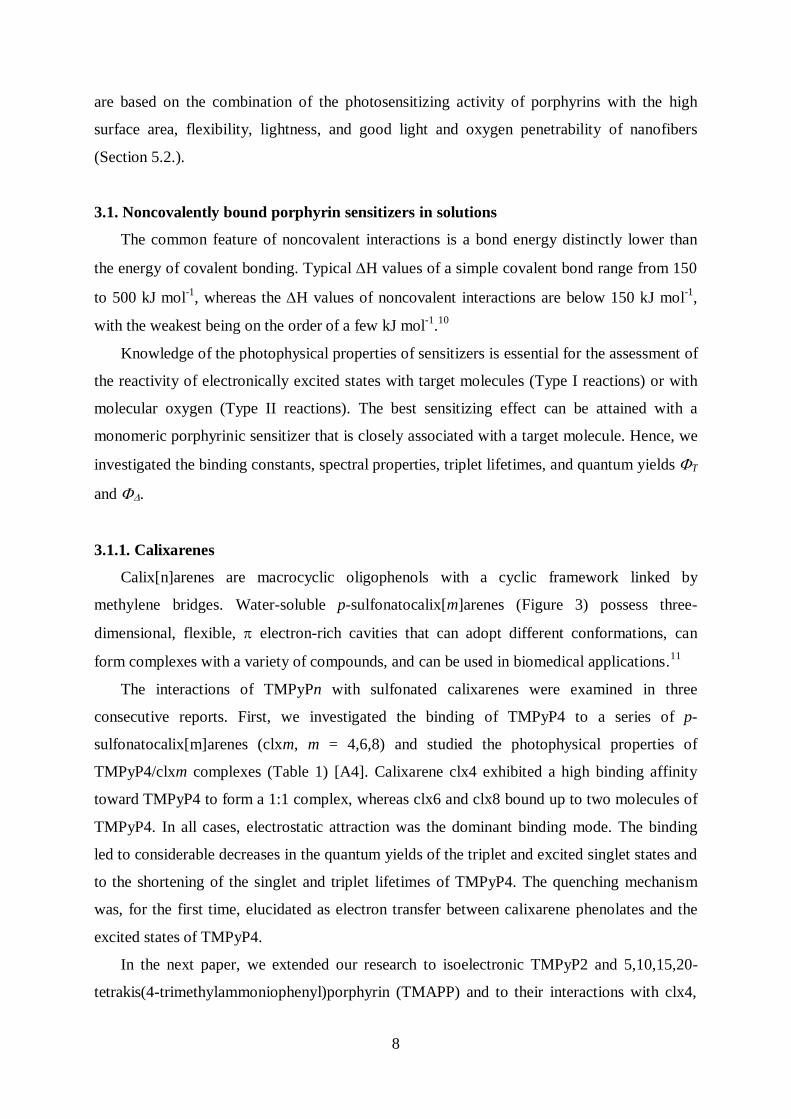
3.1. Noncovalently bound porphyrin sensitizers in solutions
The common feature of noncovalent interactions is a bond energy distinctly lower than
the energy of covalent bonding. Typical H values of a simple covalent bond range from 150
to 500 kJ mol-1
, whereas the H values of noncovalent interactions are below 150 kJ mol-1
,
with the weakest being on the order of a few kJ mol-1
.10
Knowledge of the photophysical properties of sensitizers is essential for the assessment of
the reactivity of electronically excited states with target molecules (Type I reactions) or with
molecular oxygen (Type II reactions). The best sensitizing effect can be attained with a
monomeric porphyrinic sensitizer that is closely associated with a target molecule. Hence, we
investigated the binding constants, spectral properties, triplet lifetimes, and quantum yields T
and .
3.1.1. Calixarenes
Calix[n]arenes are macrocyclic oligophenols with a cyclic framework linked by
methylene bridges. Water-soluble p-sulfonatocalix[m]arenes (Figure 3) possess three-
dimensional, flexible, electron-rich cavities that can adopt different conformations, can
form complexes with a variety of compounds, and can be used in biomedical applications.11
The interactions of TMPyPn with sulfonated calixarenes were examined in three
consecutive reports. First, we investigated the binding of TMPyP4 to a series of p-
sulfonatocalix[m]arenes (clxm, m = 4,6,8) and studied the photophysical properties of
TMPyP4/clxm complexes (Table 1) [A4]. Calixarene clx4 exhibited a high binding affinity
toward TMPyP4 to form a 1:1 complex, whereas clx6 and clx8 bound up to two molecules of
TMPyP4. In all cases, electrostatic attraction was the dominant binding mode. The binding
led to considerable decreases in the quantum yields of the triplet and excited singlet states and
to the shortening of the singlet and triplet lifetimes of TMPyP4. The quenching mechanism
was, for the first time, elucidated as electron transfer between calixarene phenolates and the
excited states of TMPyP4.
In the next paper, we extended our research to isoelectronic TMPyP2 and 5,10,15,20-
tetrakis(4-trimethylammoniophenyl)porphyrin (TMAPP) and to their interactions with clx4,
9
thiacalix[4]arene-p-tetrasulfonate, and sulfonylcalix[4]arene-p-tetrasulfonate [A5]. We
observed the quenching of the excited states of TMPyP2. In the case of TMAPP, the binding
led to extended porphyrin aggregation.
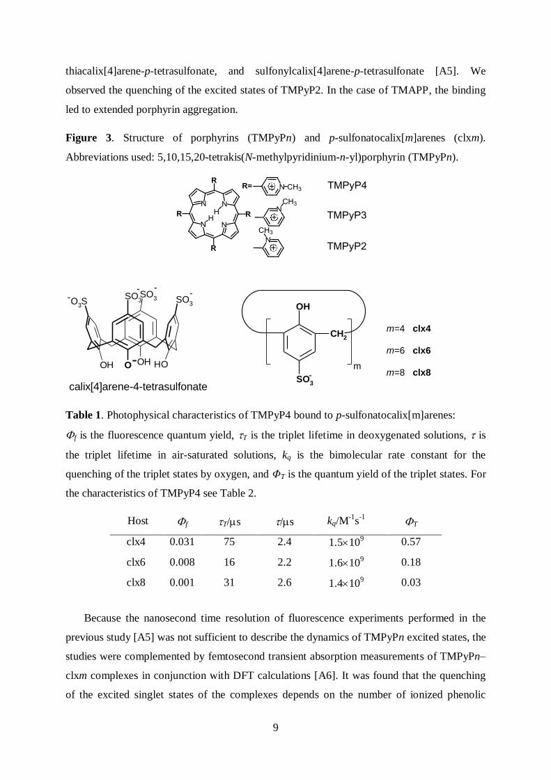
Figure 3. Structure of porphyrins (TMPyPn) and p-sulfonatocalix[m]arenes (clxm).
Abbreviations used: 5,10,15,20-tetrakis(N-methylpyridinium-n-yl)porphyrin (TMPyPn).
OHOHOOH
SO3 SO
3O3S
SO3- -- -
OH
SO3
CH2
calix[4]arene-4-tetrasulfonate
m=4 clx4
m=6 clx6
m=8 clx8-m
Table 1. Photophysical characteristics of TMPyP4 bound to p-sulfonatocalix[m]arenes:
f is the fluorescence quantum yield, T is the triplet lifetime in deoxygenated solutions, is
the triplet lifetime in air-saturated solutions, kq is the bimolecular rate constant for the
quenching of the triplet states by oxygen, and ΦT is the quantum yield of the triplet states. For
the characteristics of TMPyP4 see Table 2.
Host f T/s /s kq/M-1
s-1
T
clx4 0.031 75 2.4 1.5109 0.57
clx6 0.008 16 2.2 1.6109 0.18
clx8 0.001 31 2.6 1.4109 0.03
Because the nanosecond time resolution of fluorescence experiments performed in the
previous study [A5] was not sufficient to describe the dynamics of TMPyPn excited states, the
studies were complemented by femtosecond transient absorption measurements of TMPyPn–
clxm complexes in conjunction with DFT calculations [A6]. It was found that the quenching
of the excited singlet states of the complexes depends on the number of ionized phenolic
10
groups of clxm and can be correlated with the partial electron density transfer from O- to
peripheral methylpyridinium substituents rather than to the porphyrin ring.
The key conclusions are as follows:
- The ultrafast excited-state dynamics of TMPyPn are related to the redistribution of
electron density within the complexes. The electronic density redistribution decreases in
the order TMPyP4 > TMPyP3 > TMPyP2, and the same order is observed for the increase
in the fluorescence lifetimes.
- In the case of TMPyPn–clxm complexes, partial intramolecular charge separation
proceeds predominantly from the ionized O- groups of clxm to the methylpyridinium
peripheral substituents of TMPyPn. The electron density redistribution within the
TMPyP4–clxm (m = 4, 6) complexes in the ground and excited S1 states increases with
the number of O- groups in clxm and can be linked to the increase of quenching efficiency
of the S1 states. The quenching of the S1 states of TMPyPn–clxm (n = 2 and 3) can be
described similarly as in the TMPyP4 complexes, i.e., by the electron density transfer to
methylpyridinium substituents.
- Differences in the excited-state dynamics of the complexes of TMPyPn positional isomers
with clxm are related to the steric and electron-accepting effects of N-methylpyridinium
substituents. The most effective quenching in the series of clx8 complexes is observed for
TMPyP2.
- Clx4 is a potential carrier for cationic porphyrins because a moderate influence on the
excited states is compensated by the high solubility and stability of the complexes.
3.1.2. Cyclodextrins
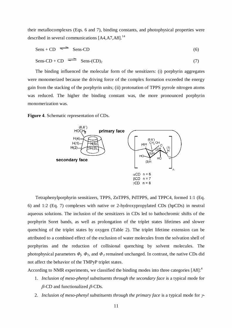
The most common cyclodextrins (CDs) consist of 6, 7, or 8 -1,4-D-glucopyranose units
arranged in a truncated cone and denoted as -CD, -CD or -CD, respectively (Figure 4).12
The interior, lined with C–H groups and glycosidic oxygen bridges, is hydrophobic in
comparison with the exterior, which is hydrophilic due to the presence of hydroxyl groups.
Consequently, CDs are water-soluble and form inclusion host–guest complexes with a variety
of molecules. Cyclodextrins are frequently used to improve the resistance of pharmaceuticals
to thermal and oxidative degradation, to limit side effects, to increase solubility, and to
prevent aggregation.13
We were concerned with the carrier function of CDs for porphyrin
sensitizers.
The binding stoichiometries of water-soluble TPPS, TPPC, and TMPyP4, and some of
11
their metallocomplexes (Eqs. 6 and 7), binding constants, and photophysical properties were
described in several communications [A4,A7,A8].14
Sens + CD Sens-CD (6)
Sens-CD + CD Sens-(CD)2 (7)
The binding influenced the molecular form of the sensitizers: (i) porphyrin aggregates
were monomerized because the driving force of the complex formation exceeded the energy
gain from the stacking of the porphyrin units; (ii) protonation of TPPS pyrrole nitrogen atoms
was reduced. The higher the binding constant was, the more pronounced porphyrin
monomerization was.
Figure 4. Schematic representation of CDs.
Tetraphenylporphyrin sensitizers, TPPS, ZnTPPS, PdTPPS, and TPPC4, formed 1:1 (Eq.
6) and 1:2 (Eq. 7) complexes with native or 2-hydroxypropylated CDs (hpCDs) in neutral
aqueous solutions. The inclusion of the sensitizers in CDs led to bathochromic shifts of the
porphyrin Soret bands, as well as prolongation of the triplet states lifetimes and slower
quenching of the triplet states by oxygen (Table 2). The triplet lifetime extension can be
attributed to a combined effect of the exclusion of water molecules from the solvation shell of
porphyrins and the reduction of collisional quenching by solvent molecules. The
photophysical parameters Φf, ΦT, and ΦΔ remained unchanged. In contrast, the native CDs did
not affect the behavior of the TMPyP triplet states.
According to NMR experiments, we classified the binding modes into three categories [A8]:4
1. Inclusion of meso-phenyl substituents through the secondary face is a typical mode for
-CD and functionalized -CDs.
2. Inclusion of meso-phenyl substituents through the primary face is a typical mode for -
12
CDs. The size of the secondary face is larger than that of -CDs and, therefore, the
inclusion of the meso-phenyl substituent from the primary face is more favorable.
3. Non-specific external binding was, for the first time, described for cationic TMPyP4
with native, per-methylated, sulfonated, and dimethyl-sulfonated CDs [A8]. The high
affinity of TMPyP4 toward sulfonated CDs was due to ion pairing between the N-
methylpyridinium-4-yl peripheral substituents of TMPyP and the sulfonate groups.
We found that anionic tetraphenylporphyrins bound with CDs remain good sensitizers
and are protected against the influence of the environment. Thus, CDs are suitable carriers of
the porphyrin sensitizers.
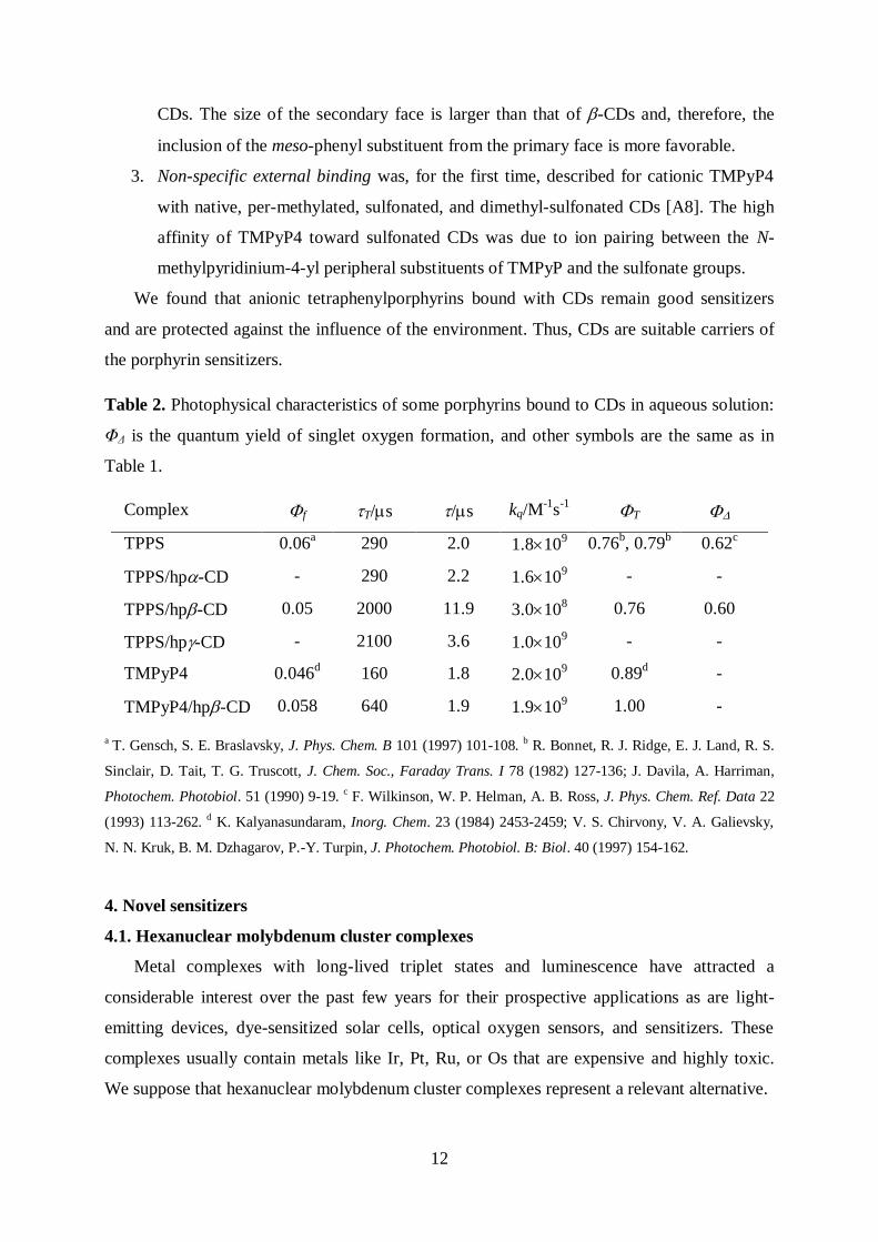
Table 2. Photophysical characteristics of some porphyrins bound to CDs in aqueous solution:
ΦΔ is the quantum yield of singlet oxygen formation, and other symbols are the same as in
Table 1.
Complex f T/s /s kq/M-1
s-1
T
TPPS 0.06a
290 2.0 1.8109 0.76
b, 0.79
b 0.62
c
TPPS/hp-CD - 290 2.2 1.610
9 - -
TPPS/hp-CD 0.05 2000 11.9 3.010
8 0.76 0.60
TPPS/hp-CD - 2100 3.6 1.0109 - -
TMPyP4 0.046d 160 1.8 2.010
9 0.89
d -
TMPyP4/hp-CD 0.058 640 1.9 1.9109 1.00 -
a T. Gensch, S. E. Braslavsky, J. Phys. Chem. B 101 (1997) 101-108. b R. Bonnet, R. J. Ridge, E. J. Land, R. S.
Sinclair, D. Tait, T. G. Truscott, J. Chem. Soc., Faraday Trans. I 78 (1982) 127-136; J. Davila, A. Harriman,
Photochem. Photobiol. 51 (1990) 9-19. c F. Wilkinson, W. P. Helman, A. B. Ross, J. Phys. Chem. Ref. Data 22
(1993) 113-262. d K. Kalyanasundaram, Inorg. Chem. 23 (1984) 2453-2459; V. S. Chirvony, V. A. Galievsky,
N. N. Kruk, B. M. Dzhagarov, P.-Y. Turpin, J. Photochem. Photobiol. B: Biol. 40 (1997) 154-162.
4. Novel sensitizers
4.1. Hexanuclear molybdenum cluster complexes
Metal complexes with long-lived triplet states and luminescence have attracted a
considerable interest over the past few years for their prospective applications as are light-
emitting devices, dye-sensitized solar cells, optical oxygen sensors, and sensitizers. These
complexes usually contain metals like Ir, Pt, Ru, or Os that are expensive and highly toxic.
We suppose that hexanuclear molybdenum cluster complexes represent a relevant alternative.
13
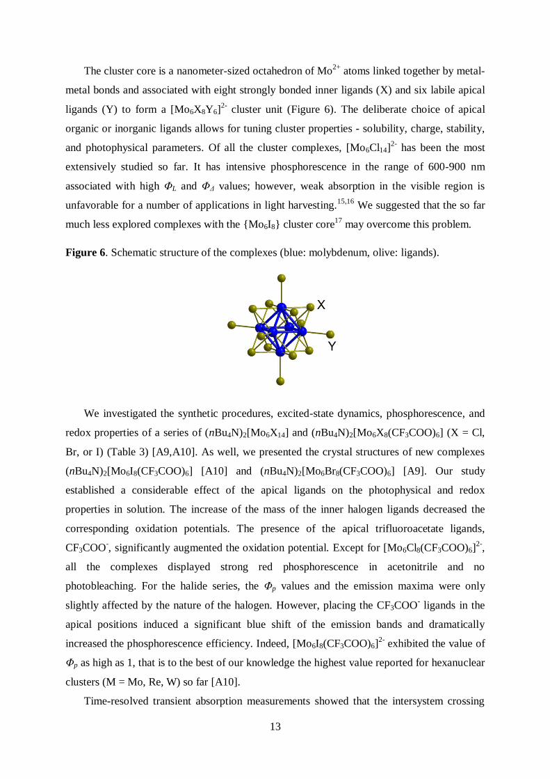
The cluster core is a nanometer-sized octahedron of Mo2+
atoms linked together by metal-
metal bonds and associated with eight strongly bonded inner ligands (X) and six labile apical
ligands (Y) to form a [Mo6X8Y6]2-
cluster unit (Figure 6). The deliberate choice of apical
organic or inorganic ligands allows for tuning cluster properties - solubility, charge, stability,
and photophysical parameters. Of all the cluster complexes, [Mo6Cl14]2-
has been the most
extensively studied so far. It has intensive phosphorescence in the range of 600-900 nm
associated with high ΦL and ΦΔ values; however, weak absorption in the visible region is
unfavorable for a number of applications in light harvesting.15,16
We suggested that the so far
much less explored complexes with the {Mo6I8} cluster core17
may overcome this problem.
Figure 6. Schematic structure of the complexes (blue: molybdenum, olive: ligands).
We investigated the synthetic procedures, excited-state dynamics, phosphorescence, and
redox properties of a series of (nBu4N)2[Mo6X14] and (nBu4N)2[Mo6X8(CF3COO)6] (X = Cl,
Br, or I) (Table 3) [A9,A10]. As well, we presented the crystal structures of new complexes
(nBu4N)2[Mo6I8(CF3COO)6] [A10] and (nBu4N)2[Mo6Br8(CF3COO)6] [A9]. Our study
established a considerable effect of the apical ligands on the photophysical and redox
properties in solution. The increase of the mass of the inner halogen ligands decreased the
corresponding oxidation potentials. The presence of the apical trifluoroacetate ligands,
CF3COO-, significantly augmented the oxidation potential. Except for [Mo6Cl8(CF3COO)6]
2-,
all the complexes displayed strong red phosphorescence in acetonitrile and no
photobleaching. For the halide series, the Φp values and the emission maxima were only
slightly affected by the nature of the halogen. However, placing the CF3COO- ligands in the
apical positions induced a significant blue shift of the emission bands and dramatically
increased the phosphorescence efficiency. Indeed, [Mo6I8(CF3COO)6]2-
exhibited the value of
Φp as high as 1, that is to the best of our knowledge the highest value reported for hexanuclear
clusters (M = Mo, Re, W) so far [A10].
Time-resolved transient absorption measurements showed that the intersystem crossing
14
from the excited singlet states was ultrafast with time constants between < 120 fs and 1.68 ps
and led to hot triplet states. The cooling of the hot triplet states, not observed only for
[Mo6I8(CF3COO)6]2-
, occurred at a ps time scale and was attributed to electronic redistribution
over the triplet state sublevels. The formation of O2(1g), suggested earlier indirectly on the
basis of photooxidation experiments for some complexes,16
was revised by direct
measurements of O2(1Δg). The high ΦΔ values reflect the efficiency of the intersystem
crossing.
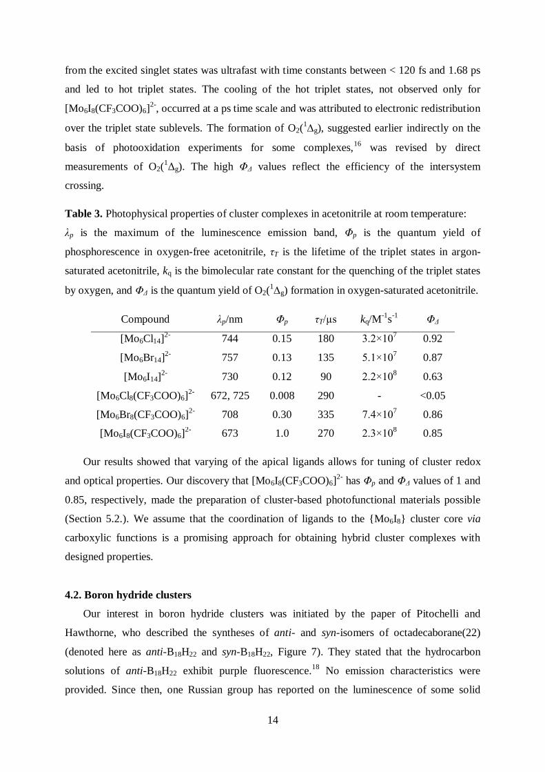
Table 3. Photophysical properties of cluster complexes in acetonitrile at room temperature:
λp is the maximum of the luminescence emission band, Φp is the quantum yield of
phosphorescence in oxygen-free acetonitrile, τT is the lifetime of the triplet states in argon-
saturated acetonitrile, kq is the bimolecular rate constant for the quenching of the triplet states
by oxygen, and ΦΔ is the quantum yield of O2(1g) formation in oxygen-saturated acetonitrile.
Compound λp/nm Φp τT/µs kq/M-1
s-1
ΦΔ
[Mo6Cl14]2-
744 0.15 180 3.2×10⁷ 0.92
[Mo6Br14]2-
757 0.13 135 5.1×10⁷ 0.87
[Mo6I14]2-
730 0.12 90 2.2×10⁸ 0.63
[Mo6Cl8(CF3COO)6]2-
672, 725 0.008 290 - <0.05
[Mo6Br8(CF3COO)6]2-
708 0.30 335 7.4×10⁷ 0.86
[Mo6I8(CF3COO)6]2-
673 1.0 270 2.3×10⁸ 0.85
Our results showed that varying of the apical ligands allows for tuning of cluster redox
and optical properties. Our discovery that [Mo6I8(CF3COO)6]2-
has Φp and ΦΔ values of 1 and
0.85, respectively, made the preparation of cluster-based photofunctional materials possible
(Section 5.2.). We assume that the coordination of ligands to the {Mo6I8} cluster core via
carboxylic functions is a promising approach for obtaining hybrid cluster complexes with
designed properties.
4.2. Boron hydride clusters
Our interest in boron hydride clusters was initiated by the paper of Pitochelli and
Hawthorne, who described the syntheses of anti- and syn-isomers of octadecaborane(22)
(denoted here as anti-B18H22 and syn-B18H22, Figure 7). They stated that the hydrocarbon
solutions of anti-B18H22 exhibit purple fluorescence.18
No emission characteristics were
provided. Since then, one Russian group has reported on the luminescence of some solid
15
borane and carborane clusters.19
In fact, the photophysics of boron hydride clusters is
practically unknown. In light of the growing interest in the applications of boron clusters in
biology and medicine, we pursued the detailed structural, spectral, and photophysical
characterization of B18H22 skeletons [A11,A12].
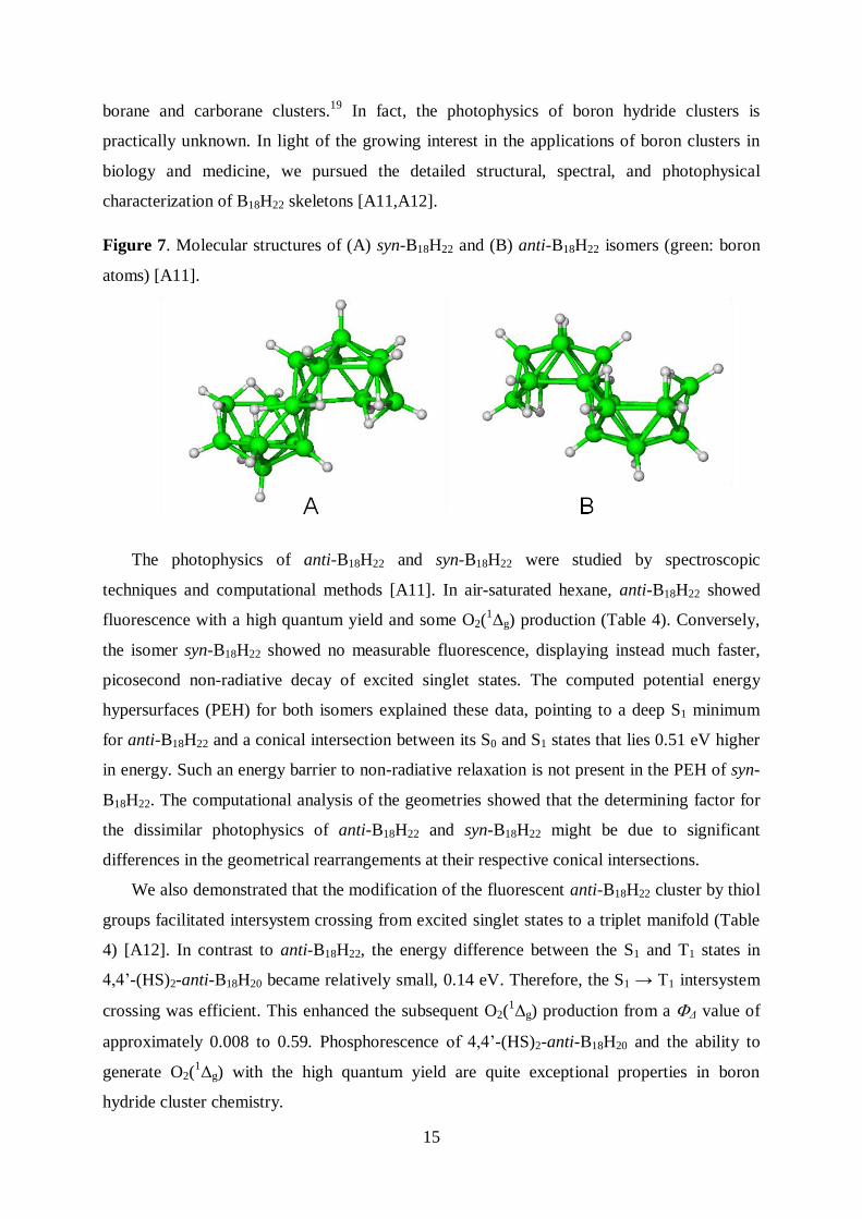
Figure 7. Molecular structures of (A) syn-B18H22 and (B) anti-B18H22 isomers (green: boron
atoms) [A11].
The photophysics of anti-B18H22 and syn-B18H22 were studied by spectroscopic
techniques and computational methods [A11]. In air-saturated hexane, anti-B18H22 showed
fluorescence with a high quantum yield and some O2(1Δg) production (Table 4). Conversely,
the isomer syn-B18H22 showed no measurable fluorescence, displaying instead much faster,
picosecond non-radiative decay of excited singlet states. The computed potential energy
hypersurfaces (PEH) for both isomers explained these data, pointing to a deep S1 minimum
for anti-B18H22 and a conical intersection between its S0 and S1 states that lies 0.51 eV higher
in energy. Such an energy barrier to non-radiative relaxation is not present in the PEH of syn-
B18H22. The computational analysis of the geometries showed that the determining factor for
the dissimilar photophysics of anti-B18H22 and syn-B18H22 might be due to significant
differences in the geometrical rearrangements at their respective conical intersections.
We also demonstrated that the modification of the fluorescent anti-B18H22 cluster by thiol
groups facilitated intersystem crossing from excited singlet states to a triplet manifold (Table
4) [A12]. In contrast to anti-B18H22, the energy difference between the S1 and T1 states in
4,4’-(HS)2-anti-B18H20 became relatively small, 0.14 eV. Therefore, the S1 → T1 intersystem
crossing was efficient. This enhanced the subsequent O2(1Δg) production from a Δ value of
approximately 0.008 to 0.59. Phosphorescence of 4,4’-(HS)2-anti-B18H20 and the ability to
generate O2(1Δg) with the high quantum yield are quite exceptional properties in boron
hydride cluster chemistry.
16
Table 4. Photophysical properties of syn-B18H22 (syn) and anti-B18H22 (anti) in air-saturated
hexane and 4,4’-(HS)2-anti-B18H20 (antiSH) in air-saturated cyclohexane: λmax is the
absorption band maximum, λf is the fluorescence band maximum, f is the fluorescence
quantum yield, τf is the lifetime of the excited singlet states, λp is the phosphorescence band
maximum, T is the quantum yield of the triplet states, τT is the lifetime of the triplet states in
oxygen-free solution, Δ is the quantum yield of O2(1Δg) formation.
Abs. spectra
λmax/nm
Excited singlet states Triplet states O2(1Δg)
λf/nm f f/ns p/nm T τT/µs Δ
syn 229, 308 - ~ 0 0.075 - - < 2 ~0.008
anti 215, 272, 329 407 0.97 11.2 - <0.03 6±2 ~0.008
antiSH 316, 381, 536 -a
0.94 596 -a
14 0.59
a Not measured.
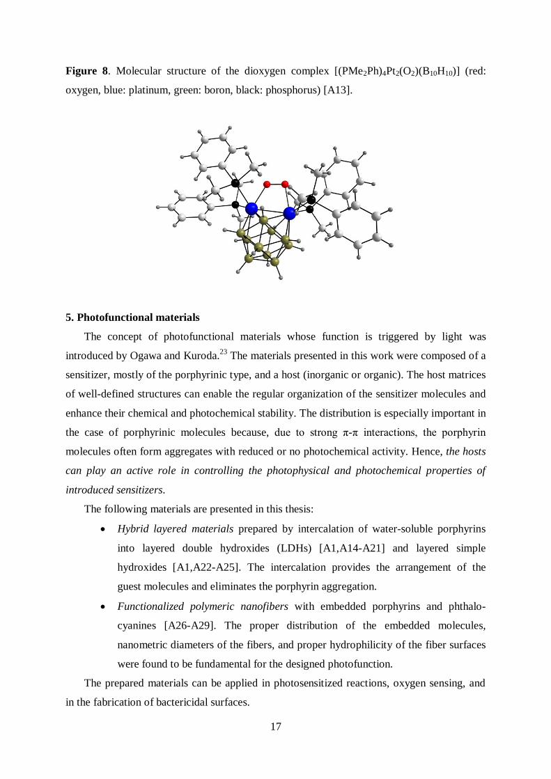
Of the two known dioxygen complexes with a unique peroxo-trapezoid structure, only in
the [(PMe2Ph)4Pt2(O2)(B10H10)] is dioxygen bound reversibly (Figure 8) [A13].20,21
Transient
absorption spectroscopy revealed that dioxygen is released after irradiation on a picosecond
timescale with a quantum yield as high as 0.58. The binding/release steps can be repeated
many times; dioxygen is released under continuous irradiation, and the photoproduced
bimetallaborane cluster [(PMe2Ph)4Pt2(B10H10)] rebinds dioxygen in a dark period to again
form the original [(PMe2Ph)4Pt2(O2)(B10H10)]. Unexpectedly, we observed the formation of
O2(1Δg) with a low quantum yield ΦΔ < 0.01. In this case, the photosensitized formation of
O2(1Δg) can be excluded because the complex itself has no photosensitizing properties. A
plausible explanation was earlier suggested by Seip and Brauer for oxodiperoxo Mo
complexes.22
Singlet oxygen is generated by an allowed transition from an upper singlet state
of a peroxo complex, whereas the competitive intersystem crossing to a photochemically
active triplet state yields dioxygen in the ground triplet state. The very low value of ΦΔ
indicates that the O2(1Δg) channel is of minor importance. To the best of our knowledge,
[(PMe2Ph)4Pt2(O2)(B10H10)] is the only example of a complex with the metal-metal bond that
binds dioxygen and releases it upon irradiation. The compound has potential to serve as a
light-triggered local and instantaneous source of dioxygen.
17
Figure 8. Molecular structure of the dioxygen complex [(PMe2Ph)4Pt2(O2)(B10H10)] (red:
oxygen, blue: platinum, green: boron, black: phosphorus) [A13].
5. Photofunctional materials
The concept of photofunctional materials whose function is triggered by light was
introduced by Ogawa and Kuroda.23
The materials presented in this work were composed of a
sensitizer, mostly of the porphyrinic type, and a host (inorganic or organic). The host matrices
of well-defined structures can enable the regular organization of the sensitizer molecules and
enhance their chemical and photochemical stability. The distribution is especially important in
the case of porphyrinic molecules because, due to strong π-π interactions, the porphyrin
molecules often form aggregates with reduced or no photochemical activity. Hence, the hosts
can play an active role in controlling the photophysical and photochemical properties of
introduced sensitizers.
The following materials are presented in this thesis:
Hybrid layered materials prepared by intercalation of water-soluble porphyrins
into layered double hydroxides (LDHs) [A1,A14-A21] and layered simple
hydroxides [A1,A22-A25]. The intercalation provides the arrangement of the
guest molecules and eliminates the porphyrin aggregation.
Functionalized polymeric nanofibers with embedded porphyrins and phthalo-
cyanines [A26-A29]. The proper distribution of the embedded molecules,
nanometric diameters of the fibers, and proper hydrophilicity of the fiber surfaces
were found to be fundamental for the designed photofunction.
The prepared materials can be applied in photosensitized reactions, oxygen sensing, and
in the fabrication of bactericidal surfaces.
18
5.1. Layered hydroxides
The hybrid materials, prepared from layered metal hydroxides, exhibit a compositional
variability that makes them promising for a wide range of applications, including catalysis,
photovoltaics, polymer nanocomposites, sensing, luminescence, magnetism, and
photooxidation. The materials are supposed to be biocompatible carriers that can be used in
drug stabilization and delivery systems.
In 1989, Park et al. and Dutta et al. first described the synthesis and certain properties of
the LDH-porphyrin and LDH-phthalocyanine intercalation compounds, respectively.24,25
Since then, other authors have studied intercalated anionic porphyrins and phthalocyanines,
particularly those intercalated into LDHs, and they have contributed to a better understanding
of the synthetic methods, structure, and properties imposed by intercalated molecules.7,26,27
The photophysical and photochemical properties imposed by intercalated porphyrinic
compounds have only been studied to a limited extent [A1]. This work summarizes our
achievements concerning layered hydroxide hybrids and their synthesis, flexibility, and
photofunctionality.
We studied LDHs (Section 5.1.1.), layered zinc hydroxide salts (LZHs) (Section 5.1.2.),
and layered rare-earth hydroxides (Section 5.1.3.). Because this work describes many layered
hydroxide materials, several notations are used to characterize the composition of the
hydroxide layers and the type of intercalated porphyrins. For instance, MgRAl-LDH-PdTPPC
represents layered double hydroxide with a Mg/Al molar ratio R and intercalated with
PdTPPC. Analogously, LZH-TPPS is layered zinc hydroxide intercalated with TPPS and
LEuH-PdTPPS is layered europium hydroxide intercalated with PdTPPS.
Investigated items:
- Syntheses of layered hydroxide-porphyrin hybrids.
- Structure of layered hydroxide-porphyrin hybrids.
- Absorption and luminescence properties of layered hydroxide-porphyrin hybrids.
- Reactivity of the triplet states of intercalated porphyrins and the formation of O2(1Δg).
- Synthesis and properties of composites based on layered hydroxide-porphyrin
hybrids.
5.1.1. Layered double hydroxides
The crystal structure of LDHs consists of positively charged metal hydroxide layers and
19
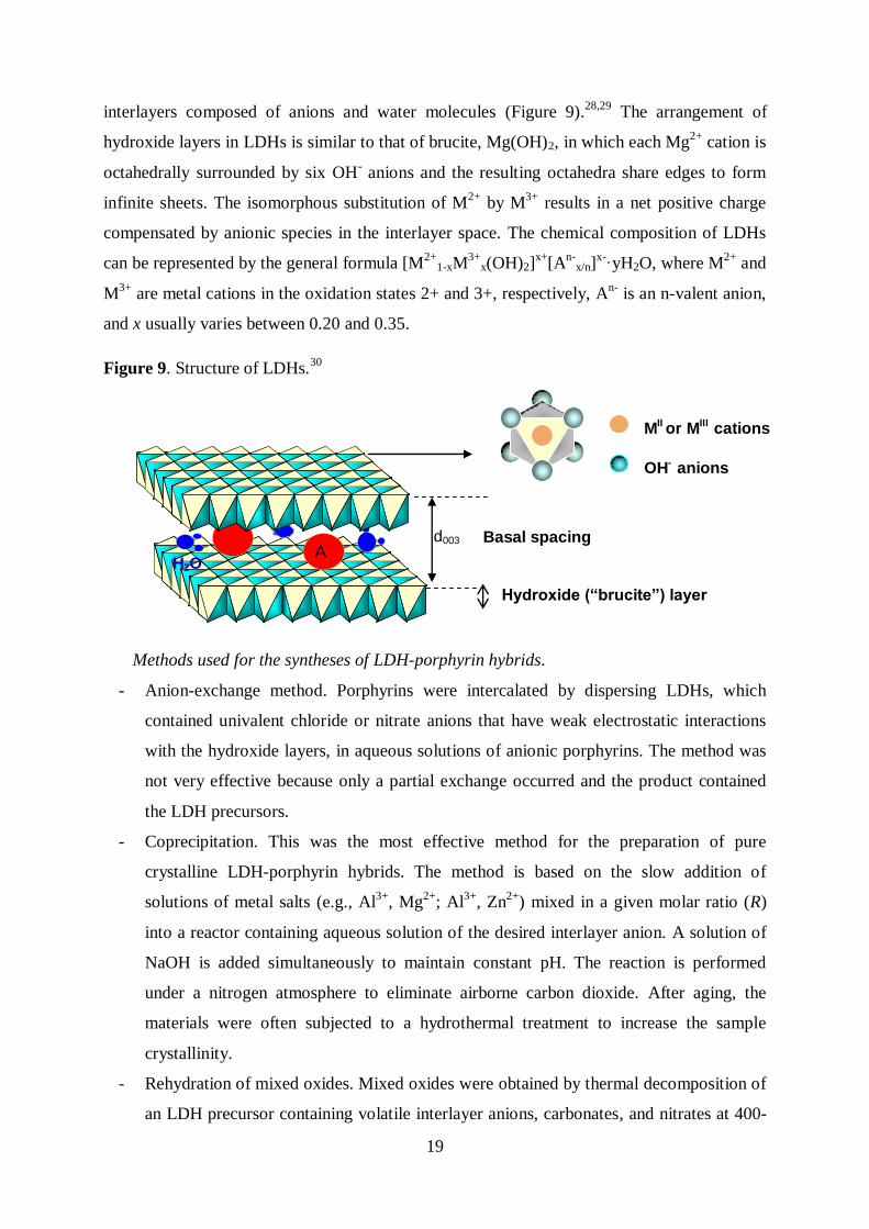
interlayers composed of anions and water molecules (Figure 9).28,29
The arrangement of
hydroxide layers in LDHs is similar to that of brucite, Mg(OH)2, in which each Mg2+
cation is
octahedrally surrounded by six OH- anions and the resulting octahedra share edges to form
infinite sheets. The isomorphous substitution of M2+
by M3+
results in a net positive charge
compensated by anionic species in the interlayer space. The chemical composition of LDHs
can be represented by the general formula [M2+
1-xM3+
x(OH)2]x+
[An-
x/n]x-·yH2O, where M
2+ and
M3+
are metal cations in the oxidation states 2+ and 3+, respectively, An-
is an n-valent anion,
and x usually varies between 0.20 and 0.35.
Figure 9. Structure of LDHs.30
Methods used for the syntheses of LDH-porphyrin hybrids.
- Anion-exchange method. Porphyrins were intercalated by dispersing LDHs, which
contained univalent chloride or nitrate anions that have weak electrostatic interactions
with the hydroxide layers, in aqueous solutions of anionic porphyrins. The method was
not very effective because only a partial exchange occurred and the product contained
the LDH precursors.
- Coprecipitation. This was the most effective method for the preparation of pure
crystalline LDH-porphyrin hybrids. The method is based on the slow addition of
solutions of metal salts (e.g., Al3+
, Mg2+
; Al3+
, Zn2+
) mixed in a given molar ratio (R)
into a reactor containing aqueous solution of the desired interlayer anion. A solution of
NaOH is added simultaneously to maintain constant pH. The reaction is performed
under a nitrogen atmosphere to eliminate airborne carbon dioxide. After aging, the
materials were often subjected to a hydrothermal treatment to increase the sample
crystallinity.
- Rehydration of mixed oxides. Mixed oxides were obtained by thermal decomposition of
an LDH precursor containing volatile interlayer anions, carbonates, and nitrates at 400-
OH- anions
MII or MIII cations
d003
Hydroxide (“brucite”) layer
Basal spacing A-
H2O
20
600 °C. Thereafter, mixed oxides were rehydrated in aqueous porphyrin solution.
However, the products were not fully intercalated with porphyrins, and partial
metalation of the porphyrin core also occurred.
Arrangement of porphyrins in the LDH interlayer space [A14,A15]. The alignment of
intercalated molecules in the galleries of LDHs can be deduced from the analysis of powder
XRD patterns. In the case of intercalated porphyrins, the basal spacings d003 (Figure 9) varied
between 21 and 23 Å, indicating that the size of the interlayer space is comparable to that of
the intercalated macrocycle units.
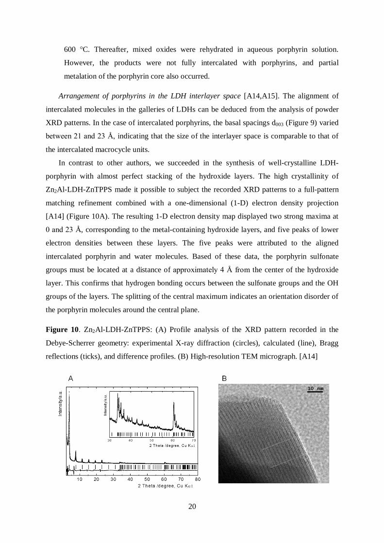
In contrast to other authors, we succeeded in the synthesis of well-crystalline LDH-
porphyrin with almost perfect stacking of the hydroxide layers. The high crystallinity of
Zn2Al-LDH-ZnTPPS made it possible to subject the recorded XRD patterns to a full-pattern
matching refinement combined with a one-dimensional (1-D) electron density projection
[A14] (Figure 10A). The resulting 1-D electron density map displayed two strong maxima at
0 and 23 Å, corresponding to the metal-containing hydroxide layers, and five peaks of lower
electron densities between these layers. The five peaks were attributed to the aligned
intercalated porphyrin and water molecules. Based of these data, the porphyrin sulfonate
groups must be located at a distance of approximately 4 Å from the center of the hydroxide
layer. This confirms that hydrogen bonding occurs between the sulfonate groups and the OH
groups of the layers. The splitting of the central maximum indicates an orientation disorder of
the porphyrin molecules around the central plane.
Figure 10. Zn2Al-LDH-ZnTPPS: (A) Profile analysis of the XRD pattern recorded in the
Debye-Scherrer geometry: experimental X-ray diffraction (circles), calculated (line), Bragg
reflections (ticks), and difference profiles. (B) High-resolution TEM micrograph. [A14]
21
The arrangement of ZnTPPS in Zn2Al-LDH-ZnTPPS was also studied by high-resolution
transmission electron microscopy (HRTEM). The distance between the observed parallel
fringes was in good agreement with the d003 value of 23.03 Å determined by powder XRD
(Figure 10B). The HRTEM micrographs revealed additional lattice fringes with a fringe
separation of approximately 8 Å. This separation indicates an increase of electron density in
the middle of the interlayer space and confirms the alignment of Zn ions of the ZnTPPS units
in the central plane.
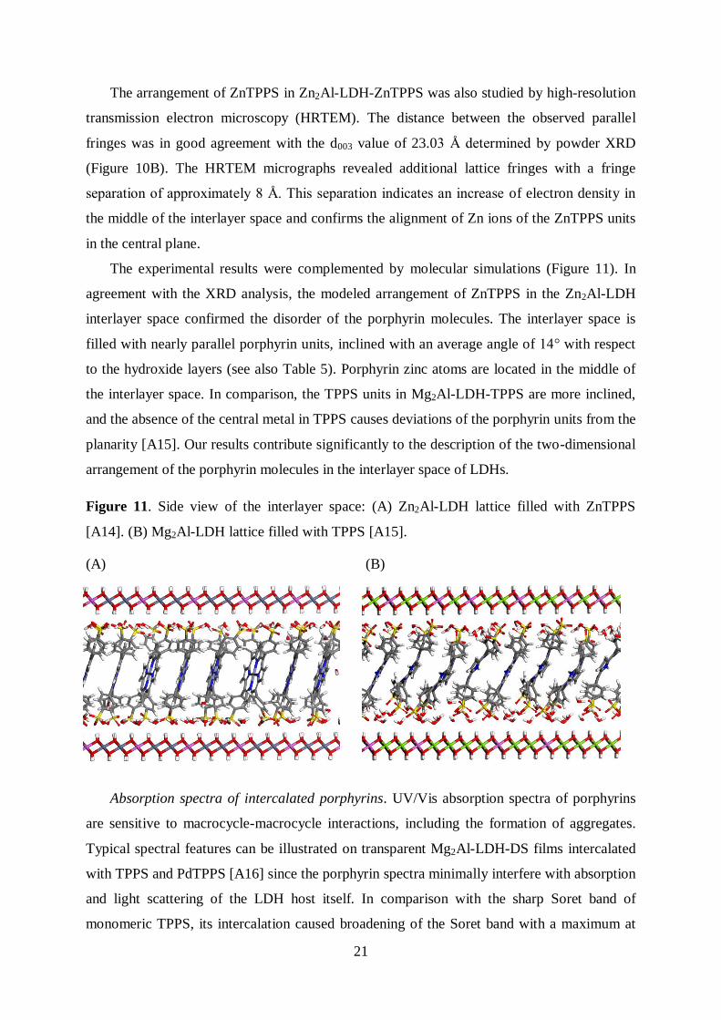
The experimental results were complemented by molecular simulations (Figure 11). In
agreement with the XRD analysis, the modeled arrangement of ZnTPPS in the Zn2Al-LDH
interlayer space confirmed the disorder of the porphyrin molecules. The interlayer space is
filled with nearly parallel porphyrin units, inclined with an average angle of 14° with respect
to the hydroxide layers (see also Table 5). Porphyrin zinc atoms are located in the middle of
the interlayer space. In comparison, the TPPS units in Mg2Al-LDH-TPPS are more inclined,
and the absence of the central metal in TPPS causes deviations of the porphyrin units from the
planarity [A15]. Our results contribute significantly to the description of the two-dimensional
arrangement of the porphyrin molecules in the interlayer space of LDHs.
Figure 11. Side view of the interlayer space: (A) Zn2Al-LDH lattice filled with ZnTPPS
[A14]. (B) Mg2Al-LDH lattice filled with TPPS [A15].
(A) (B)
Absorption spectra of intercalated porphyrins. UV/Vis absorption spectra of porphyrins
are sensitive to macrocycle-macrocycle interactions, including the formation of aggregates.
Typical spectral features can be illustrated on transparent Mg2Al-LDH-DS films intercalated
with TPPS and PdTPPS [A16] since the porphyrin spectra minimally interfere with absorption
and light scattering of the LDH host itself. In comparison with the sharp Soret band of
monomeric TPPS, its intercalation caused broadening of the Soret band with a maximum at
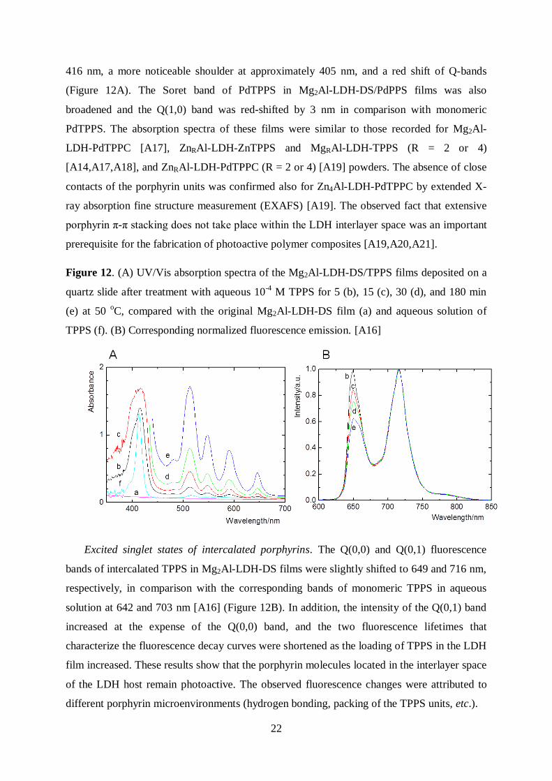
22
416 nm, a more noticeable shoulder at approximately 405 nm, and a red shift of Q-bands
(Figure 12A). The Soret band of PdTPPS in Mg2Al-LDH-DS/PdPPS films was also
broadened and the Q(1,0) band was red-shifted by 3 nm in comparison with monomeric
PdTPPS. The absorption spectra of these films were similar to those recorded for Mg2Al-
LDH-PdTPPC [A17], ZnRAl-LDH-ZnTPPS and MgRAl-LDH-TPPS (R = 2 or 4)
[A14,A17,A18], and ZnRAl-LDH-PdTPPC (R = 2 or 4) [A19] powders. The absence of close
contacts of the porphyrin units was confirmed also for Zn4Al-LDH-PdTPPC by extended X-
ray absorption fine structure measurement (EXAFS) [A19]. The observed fact that extensive
porphyrin π-π stacking does not take place within the LDH interlayer space was an important
prerequisite for the fabrication of photoactive polymer composites [A19,A20,A21].
Figure 12. (A) UV/Vis absorption spectra of the Mg2Al-LDH-DS/TPPS films deposited on a
quartz slide after treatment with aqueous 10-4
M TPPS for 5 (b), 15 (c), 30 (d), and 180 min
(e) at 50 oC, compared with the original Mg2Al-LDH-DS film (a) and aqueous solution of
TPPS (f). (B) Corresponding normalized fluorescence emission. [A16]
Excited singlet states of intercalated porphyrins. The Q(0,0) and Q(0,1) fluorescence
bands of intercalated TPPS in Mg2Al-LDH-DS films were slightly shifted to 649 and 716 nm,
respectively, in comparison with the corresponding bands of monomeric TPPS in aqueous
solution at 642 and 703 nm [A16] (Figure 12B). In addition, the intensity of the Q(0,1) band
increased at the expense of the Q(0,0) band, and the two fluorescence lifetimes that
characterize the fluorescence decay curves were shortened as the loading of TPPS in the LDH
film increased. These results show that the porphyrin molecules located in the interlayer space
of the LDH host remain photoactive. The observed fluorescence changes were attributed to
different porphyrin microenvironments (hydrogen bonding, packing of the TPPS units, etc.).
23
We showed that TPPS intercalated in Mg2Al-LDH-DS films emits singlet oxygen-
sensitized delayed fluorescence (SODF) [A16], similar to TPP embedded in polymeric
nanofibers [A26] (Section 5.2.). The repopulation of excited singlet states, 1P*, proceeds via
the reaction of triplet states, 3P, with O2(
1g) (Eq. 8) in solids with high concentration of the
immobilized 3P, high local concentration of generated O2(
1g), and adequate oxygen mobility.
The analysis of SODF decay curves allowed for estimation of the O2(1g) lifetime in the LDH
interlayer space (see below).
3P + O2(
1g) →
1P* + O2(
3g
-) (8)
Triplet states of intercalated porphyrins. The effects of the hydroxide layers on the triplet
state dynamics predetermine the efficacy of the sensitized O2(1g) generation (Eq. 4). The
triplet states of intercalated TPPS in Mg2Al-LDH-DS films decayed with a lifetime of
approximately 500 μs in the absence of oxygen, which is comparable to the lifetime of TPPS
in solution. The result indicates that the hydroxide layers do not quench the triplet states in a
measurable way [A16,A18]. The decay was accelerated by oxygen but to a lesser extent than
in a solvent, due to slower oxygen diffusion in the solid LDH matrix. Because PdTPPS is
phosphorescent, the triplet states of intercalated PdTPPS in Mg2Al-LDH-DS films were
probed by luminescence spectroscopy [A16]. These measurements showed that the triplet
state quenching by oxygen did not follow first-order kinetics. This observation was
interpreted by a broad distribution of the triplet lifetimes. The kinetic heterogeneity of the
PdTPPS triplet states was much larger than that of intercalated TPPS.
The porphyrin triplet states in Mg2Al-LDH-PdTPPC and Mg2Al-LDH-TPPS powders
were probed by transient diffuse reflectance spectroscopy [A17]. Difference absorption
spectra showed typical features of the porphyrin triplet states, i.e., a broad positive absorption
band in the ~400-550 nm region arising from the triplet-triplet absorption, which is partially
depleted by the ground state absorption associated with the Soret band (~400-420 nm) and Q-
bands (~500-520 nm). In the absence of oxygen, the observed decay curves were fitted by
biexponential functions. It can indicate different locations of intercalated porphyrins and/or
conformational deformations of the porphyrin ring yielding distinctly different triplet states.
Comparison of the triplet state lifetimes of powder samples (7.1 and 68 s for Mg2Al-LDH-
TPPS, 3.1 and 50 s for Mg2Al-LDH-PdTPPC) with those of free porphyrins in solution
indicate deactivation of the triplet states by the LDH host. This is a different behavior than
that described above for the LDH films. Probably, remaining intercalated dodecyl sulfate
anions used for the pre-expansion of the LDH interlayer space minimizes quenching effects of
24
the hydroxide layers. The triplet state decay was accelerated by oxygen also in the powder
samples, confirming that the sensitizer molecules were accessible to oxygen. The fact that the
decay curves in air were fairly fitted with a biexponential function corroborated that the
pertinent porphyrin triplet states have different oxygen accessibility.
Time-resolved spectroscopic techniques were also applied to assess the triplet state
behavior of the porphyrin sensitizers in LDH/polymer composites [A19]. For example, the
triplet state lifetimes of TPPS in polyurethane composites decreased from 1.4–3.0 ms in
vacuum to 60–100 μs in oxygen atmosphere. These relatively long lifetimes confirm the
limited diffusion of oxygen in the composites.
Singlet oxygen produced by intercalated porphyrins. Typical results confirming the
production of O2(1g) by intercalated TPPS in Mg2Al-LDH-DS films are presented in Figure
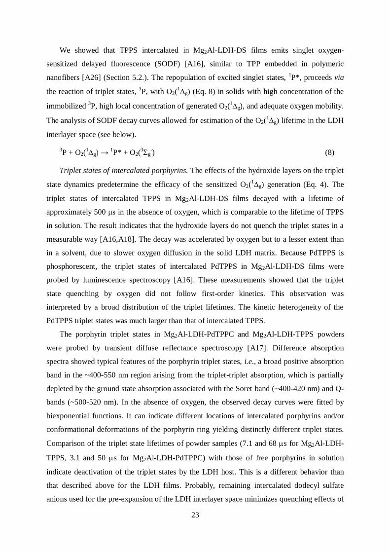
13A [A16]. The luminescence curves recorded at 1270 nm decreased monoexponentially,
giving apparent O2(1g) lifetimes of 7 and approximately 20 µs in oxygen and in air,
respectively. The corresponding luminescence band of O2(1g) is shown in the inset. The fact
that the lifetimes were affected by oxygen pressure and that the triplet lifetimes of TPPS
obtained by transient absorption spectroscopy had comparable values implies that the rate-
determining step of O2(1g) formation is the decay of the parental TPPS triplet states (see Eq.
4) [A16, A18]. The intrinsic lifetime of O2(1g) was estimated by kinetic analysis of the
SODF curves - the lifetime was approximately 0.3 µs, which is much shorter than the lifetime
of O2(1g) in water (Δ ~ 3.5 μs). This result points to the vibrational quenching of O2(
1g) by
the OH groups coordinated to the metal centers of the hydroxide layers and by water
molecules confined in the interlayer space. The short lifetime of O2(1g) accounts for the low
probability of O2(1g) reactions with molecules surrounding the LDH host.
LDH powders intercalated with TPPS, PdTPPS, or PdTPPC sensitizers also produced
O2(1g) [A17]. This ability was not affected by sample crystallinity. Mg2Al-LDH-PdTPPC
powders in particular were found to be good producers of O2(1g) [A17]. The O2(
1g)
lifetimes recovered from the fitting of the time-resolved luminescence were 10-50 μs longer
than those of their precursor triplet states. This indicates that the O2(1g) molecules generated
in the interior of the LDHs have enough time to diffuse out of the LDH host. It was proven in
the suspensions of Mg2Al-LDH-PdTPPC, in which the lifetimes of produced O2(1g) were
governed by the solvent itself.
Intercalated ZnTPPS and ZnTPPC behaved differently [A14, see also for LZH-ZnTPPS
in A22]. Although zinc porphyrins are effective sensitizers in solution, when intercalated into
25
LDHs, no detectable luminescence of O2(1g) was observed. We infer that O2(
1g), if
produced, disappears very quickly, within several microseconds after its formation.
Consequently, ZnTPPS and ZnTPPC are not suitable sensitizers for the preparation of
photofunctional layered hydroxides.
Figure 13. (A) Luminescence signals of O2(1Δg) in air (black line) and in an oxygen
atmosphere (blue line) after the excitation of TPPS in the Mg2Al-LDH-DS/TPPS film. The
red lines are least squares monoexponential fits. Inset: Emission band of O2(1Δg) in air. [A16]
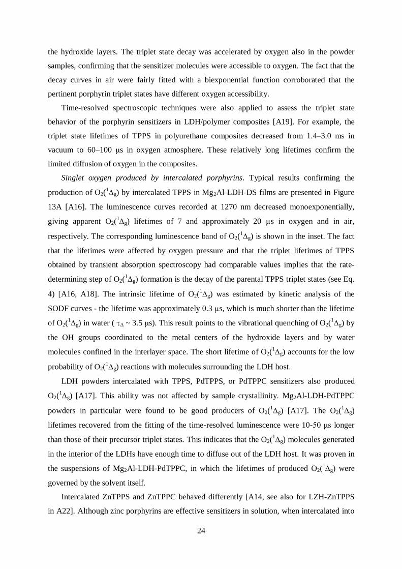
(B) Time dependence of the 1O2 luminescence signal at 1270 nm of the Mg2Al-LDH-PdTPPC
sample in O2 when fresh (a), dehydrated (b), rehydrated by exposure to ambient air (c, red
line), and dehydrated again (d, red line) (exc= 425 nm, ~1 mJ/pulse). [A17]
As has been mentioned, the potential application of sensitizer-doped solids as O2(1g)
sources depends not only on the O2(1g) yield but also on its lifetime. As shown above, the
LDH host may influence the lifetime of the photoproduced O2(1g). Consequently, structural
and composition changes of the host may significantly influence the oxidative efficacy of the
material. To pursue this issue, we investigated in detail the formation of O2(1g) sensitized by
PdTPPC in dried Mg2Al-LDH-PdTPPC (Figure 13B) [A17]. Because water is an efficient
quencher of O2(1g), longer O2(
1g) lifetimes were expected for the dehydrated samples.
Surprisingly, we observed the opposite behavior; the removal of the interlayer water inhibited
the production of O2(1g). Because the population of the porphyrin triplet states and their
relaxation kinetics were not significantly affected by dehydration, the only explanation was
that the removal of interlayer water dramatically increases the vibrational quenching of
O2(1g) by the hydroxyl LDH groups. The capability of the dehydrated LDHs to
26
photogenerate long-lived O2(1g) was recovered after the exposure of the dried samples to
atmospheric humidity. This finding presents Mg2Al-LDH-PdTPPC as a source of O2(1g)
whose oxidative activity can be modulated by the dehydration/rehydration cycles.
Composite materials. The ability of intercalated porphyrins to produce O2(1g) upon
irradiation with visible light and the known cytotoxicity of O2(1g) led to the idea to utilize
the nanocontainer and nanofiller aspects of LDHs for the preparation of LDH-
porphyrin/polymer composites for bactericidal coatings [A19,A20,A21]. We investigated
ZnRAl-LDH and MgRAl-LDH functions in polyurethane (PU), poly(butylene succinate), or
poly(butyl methacrylate) composites.
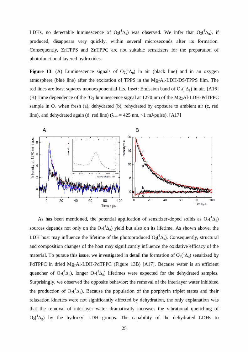
PdTPPC and TPPS were intercalated into the LDH hosts by the coprecipitation procedure
and then used as fillers in two eco-friendly polymers, namely PU and poly(butylene
succinate) [A19]. LDH-porphyrin/polymer composites were prepared either by a solvent
cast/cross-linking technique or by melt-compounding with different porphyrin-LDH filler
loadings (up to 1.3 wt.%). Both X-ray diffraction and transmission electron microscopy
measurements indicated that the porphyrin-LDH fillers are well dispersed in the polymer
matrices and that the porphyrin molecules remain intercalated within the LDH hydroxide
layers (Figure 14A).
Figure 14. (A) TEM/bright field micrograph of the Zn3Al-LDH-PdTPPC/PU composite.
(B) Decay of the PdTPPC triplet states in the Zn2Al-LDH-PdTPPC/PU composite film in
vacuum (red) and in an oxygen atmosphere (black) recorded at 480 nm. [A19]
LDHs intercalated with anions containing reactive vinyl groups, namely acrylate,
methacrylate, 2-acrylamido-2-methyl-1-propanesulfonate, and 4-vinylbenzoate,
27
polymerization initiator - 4,4’-azobis(4-cyanopentanoate), and hydrophobic DS, were
prepared by anion exchange reactions, in which Mg2Al-LDH-NO3 and Zn2Al-LDH-NO3 were
used as precursors [A20]. These hybrid LDHs were used as co-monomers or initiators for the
preparation of LDH/poly(butyl methacrylate) nanocomposites; the preparations were
performed by in situ emulsion polymerization and solution polymerization in 1-methyl-2-
pyrolidone or in a mixture of dimethylformamide and formamide. We obtained
nanostructured hybrid materials containing a low amount of the inorganic filler (1 – 5 wt.%);
the best dispersion of LDH nanoparticles was obtained in the products prepared by solution
polymerization in a mixture of dimethylformamide and formamide (50/50 v/v). Furthermore,
LDHs co-intercalated with monomers (methacrylate or 2-acrylamido-2-methyl-1-
propanesulfonate) and hydrophobic dodecyl sulfate anions were prepared; at least a low
amount of monomer anions had to be intercalated to achieve LDH delamination in the
composites. The addition of LDHs decreased the gas permeability of the nanocomposite
membranes, but the mechanical properties were not affected. The LDH/poly(butyl
methacrylate) nanocomposites producing O2(1g) were obtained by the partial intercalation of
TPPS in LDHs.
Bactericidal properties. The composites were suggested as platforms for the fabrication
of bactericidal surfaces activated by visible light. Because the polymer matrix restricts the
diffusion of oxygen and partially quenches O2(1g), the cytotoxic effect on the composite
surface is due to O2(1g) produced in a narrow surface layer. It is promising that the surface
concentration of O2(1g) can be tuned by the amount of intercalated sensitizers and/or by the
amount of fillers.
The photostability and photobactericidal properties of Zn2Al-LDH-PdTPPC/PU
composite films were studied to establish their applicability as new photodynamic surfaces
[A21]. These films comprised PdTPPC intercalated in Zn2Al-LDH and dispersed into the PU
matrix (1 wt.%). We showed that the Zn2Al-LDH host enhanced the chemical stability of
PdTPPC by minimizing photobleaching and aggregation effects. Investigation of the kinetics
of the quenching of the triplet states by oxygen indicated slow diffusion of oxygen within the
composite (Figure 14B). The effects of O2(1g) on the stability of the PU matrix were
examined by measuring oxygen consumption and carbon dioxide production under
accelerated aging conditions. Oxygen uptake experiments coupled with attenuated total
reflectance-FTIR spectroscopy indicated the probable formation of hydroxylated
photoproducts, but there were no detrimental effects on the microstructure and viscoelastic
28
properties of the PU composite.
In vitro antimicrobial tests showed that Staphylococcus aureus growth on the composite
surfaces is inhibited by white light irradiation. We also observed total inhibition of
Pseudomonas aeruginosa growth, which indicates the efficacy of the surface against biofilm
formation.
To summarize, the activity of the surfaces can be tuned by adjusting the amount of filler.
The potential applications of such photodynamic surfaces, which are sterile under white light,
are of great interest in all cases in which it is desirable to maintain microbial populations at a
low level.
5.1.2. Layered zinc hydroxide salts
In contrast to LDHs, layered simple hydroxides consist of hydroxide layers containing a
single type of metal cation, with a positive charge created by hydroxyl vacancies. The
representatives of this group are M2(OH)3(An–
)1/n (M = Co, Cu, Ni) and layered zinc
hydroxide salts Zn5(OH)8(An-
)2/n (LZH).31,32
In the structure of LZHs, a quarter of the
octahedrally coordinated zinc ions is replaced by tetrahedrally coordinated zinc ions below
and above the plane, thus forming a triple-deck architecture. The attractive aspects of LZHs
are their simple synthesis, a high anion-exchange capacity (3.2 meq g-1
for LZH-NO3) that is
comparable to LDHs, the possibility of mixing Zn ions with other metal ions (Ni2+
, Co2+
), and
simple transformation to a wide band gap semiconductor, ZnO.
Synthesis of LZH-porphyrin hybrids. LZH-porphyrin hybrids were prepared by
precipitation because ion exchange of nitrate anions in [Zn5(OH)8(NO3)2]·2H2O (abbreviated
hereafter as LZH-NO3) for porphyrins led to the formation of ZnO [A22].
During the investigation of intercalation reactions, we discovered that LZHs can
delaminate into individual hydroxide layers [A23]. This process provided positively charged
hydroxide nanosheets that restack to form hydroxide films of the original LZH structure. The
most promising results were obtained with LZHs intercalated with dodecyl sulfate anions
(LZH-DS) and LZH-NO3 delaminated in butanol at 60 °C and delaminated in formamide at
room temperature, respectively. The former method produced hydroxide nanosheets with a
lateral size of approximately 20 nm. The latter method yielded nanosheets with a size
decreasing from approximately 70 nm to 10 nm after two weeks of aging; the thickness of
these nanosheets remained nearly constant (3-4 nm). The solvothermal transformation of the
hydroxide nanosheets produced nanosheets of ZnO with a thickness of 0.6 - 0.7 nm,
corresponding to two or three stacked ZnO tetrahedra layers [A24]. The lateral size varied
29
between 15 and 25 nm. The ZnO nanosheets were arranged into transparent films with a large
{001} surface area by dip-coating and inkjet printing.33
Such orientation of the high-energy
facets {001} allowed for the preparation of transparent, ultrathin ZnO films with superior
photocatalytic degradation activity toward 4-chlorophenol.
Arrangement of porphyrins in the LZH interlayer space [A22]. The properties and the
arrangement of the porphyrin molecules in the interlayer space were explored by a
combination of experimental techniques and molecular simulations. The XRD patterns of
LZH-ZnTPPS confirmed the identity of the hydroxide layers and revealed the layered
structure with an interlayer space comparable in size to that of ZnTPPS. Molecular
simulations of LZH-ZnTPPS showed that the interlayer space is filled with the porphyrin
guests: (i) porphyrin planes are inclined with respect to the hydroxide layer normal by
approximately 22°; (ii) the angle between the porphyrin planes varies between 0 (parallel
arrangement) and 20°; (iii) porphyrin units are tightly arranged; (iv) hydrogen bond
interactions between the porphyrin sulfonate groups and the layer OH groups are similar to
those observed in LDHs (Section 5.1.1.). The arrangement of ZnTPPS in LZH and Zn2Al
LDH hosts is summarized in Table 5.
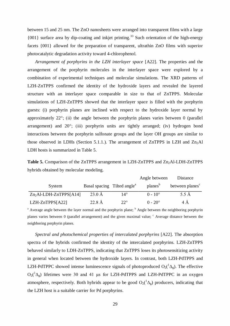
Table 5. Comparison of the ZnTPPS arrangement in LZH-ZnTPPS and Zn2Al-LDH-ZnTPPS
hybrids obtained by molecular modeling.
System Basal spacing Tilted anglea
Angle between
planesb
Distance
between planesc
Zn2Al-LDH-ZnTPPS[A14] 23.0 Å 14° 0 - 10° 5.5 Å
LZH-ZnTPPS[A22] 22.8 Å 22° 0 - 20° 4 Å
a Average angle between the layer normal and the porphyrin plane; b Angle between the neighboring porphyrin
planes varies between 0 (parallel arrangement) and the given maximal value; c Average distance between the
neighboring porphyrin planes.
Spectral and photochemical properties of intercalated porphyrins [A22]. The absorption
spectra of the hybrids confirmed the identity of the intercalated porphyrins. LZH-ZnTPPS
behaved similarly to LDH-ZnTPPS, indicating that ZnTPPS loses its photosensitizing activity
in general when located between the hydroxide layers. In contrast, both LZH-PdTPPS and
LZH-PdTPPC showed intense luminescence signals of photoproduced O2(1Δg). The effective
O2(1Δg) lifetimes were 30 and 41 μs for LZH-PdTPPS and LZH-PdTPPC in an oxygen
atmosphere, respectively. Both hybrids appear to be good O2(1Δg) producers, indicating that
the LZH host is a suitable carrier for Pd porphyrins.
30
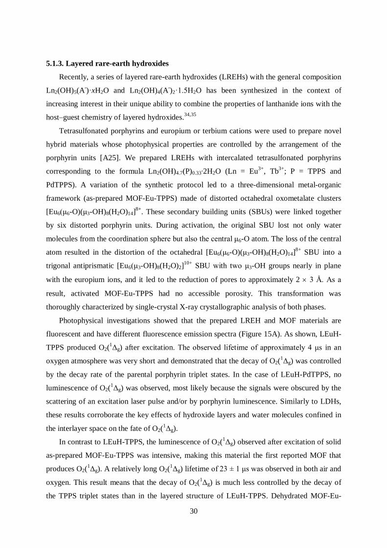
5.1.3. Layered rare-earth hydroxides
Recently, a series of layered rare-earth hydroxides (LREHs) with the general composition
Ln2(OH)5(A-)·xH2O and Ln2(OH)4(A
-)2·1.5H2O has been synthesized in the context of
increasing interest in their unique ability to combine the properties of lanthanide ions with the
host–guest chemistry of layered hydroxides.34,35
Tetrasulfonated porphyrins and europium or terbium cations were used to prepare novel
hybrid materials whose photophysical properties are controlled by the arrangement of the
porphyrin units [A25]. We prepared LREHs with intercalated tetrasulfonated porphyrins
corresponding to the formula Ln2(OH)4.7(P)0.33∙2H2O (Ln = Eu3+
, Tb3+
; P = TPPS and
PdTPPS). A variation of the synthetic protocol led to a three-dimensional metal-organic
framework (as-prepared MOF-Eu-TPPS) made of distorted octahedral oxometalate clusters
[Eu6(μ6-O)(μ3-OH)8(H2O)14]8+
. These secondary building units (SBUs) were linked together
by six distorted porphyrin units. During activation, the original SBU lost not only water
molecules from the coordination sphere but also the central μ6-O atom. The loss of the central
atom resulted in the distortion of the octahedral [Eu6(μ6-O)(μ3-OH)8(H2O)14]8+
SBU into a
trigonal antiprismatic [Eu6(μ3-OH)8(H2O)2]10+
SBU with two μ3-OH groups nearly in plane
with the europium ions, and it led to the reduction of pores to approximately 2 3 Å. As a
result, activated MOF-Eu-TPPS had no accessible porosity. This transformation was
thoroughly characterized by single-crystal X-ray crystallographic analysis of both phases.
Photophysical investigations showed that the prepared LREH and MOF materials are
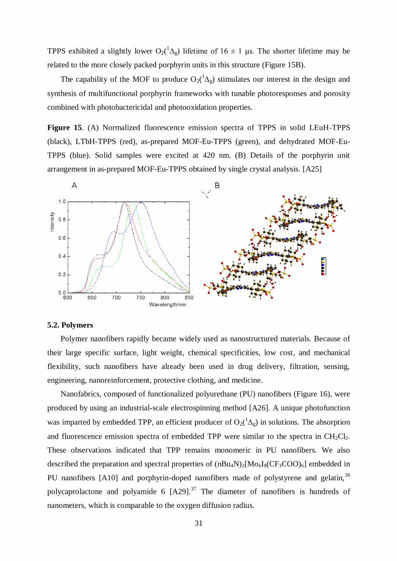
fluorescent and have different fluorescence emission spectra (Figure 15A). As shown, LEuH-
TPPS produced O2(1Δg) after excitation. The observed lifetime of approximately 4 μs in an
oxygen atmosphere was very short and demonstrated that the decay of O2(1Δg) was controlled
by the decay rate of the parental porphyrin triplet states. In the case of LEuH-PdTPPS, no
luminescence of O2(1Δg) was observed, most likely because the signals were obscured by the
scattering of an excitation laser pulse and/or by porphyrin luminescence. Similarly to LDHs,
these results corroborate the key effects of hydroxide layers and water molecules confined in
the interlayer space on the fate of O2(1Δg).
In contrast to LEuH-TPPS, the luminescence of O2(1Δg) observed after excitation of solid
as-prepared MOF-Eu-TPPS was intensive, making this material the first reported MOF that
produces O2(1Δg). A relatively long O2(
1Δg) lifetime of 23 ± 1 μs was observed in both air and
oxygen. This result means that the decay of O2(1Δg) is much less controlled by the decay of
the TPPS triplet states than in the layered structure of LEuH-TPPS. Dehydrated MOF-Eu-
31
TPPS exhibited a slightly lower O2(1Δg) lifetime of 16 ± 1 μs. The shorter lifetime may be
related to the more closely packed porphyrin units in this structure (Figure 15B).
The capability of the MOF to produce O2(1g) stimulates our interest in the design and
synthesis of multifunctional porphyrin frameworks with tunable photoresponses and porosity
combined with photobactericidal and photooxidation properties.
Figure 15. (A) Normalized fluorescence emission spectra of TPPS in solid LEuH-TPPS
(black), LTbH-TPPS (red), as-prepared MOF-Eu-TPPS (green), and dehydrated MOF-Eu-
TPPS (blue). Solid samples were excited at 420 nm. (B) Details of the porphyrin unit
arrangement in as-prepared MOF-Eu-TPPS obtained by single crystal analysis. [A25]
5.2. Polymers
Polymer nanofibers rapidly became widely used as nanostructured materials. Because of
their large specific surface, light weight, chemical specificities, low cost, and mechanical
flexibility, such nanofibers have already been used in drug delivery, filtration, sensing,
engineering, nanoreinforcement, protective clothing, and medicine.
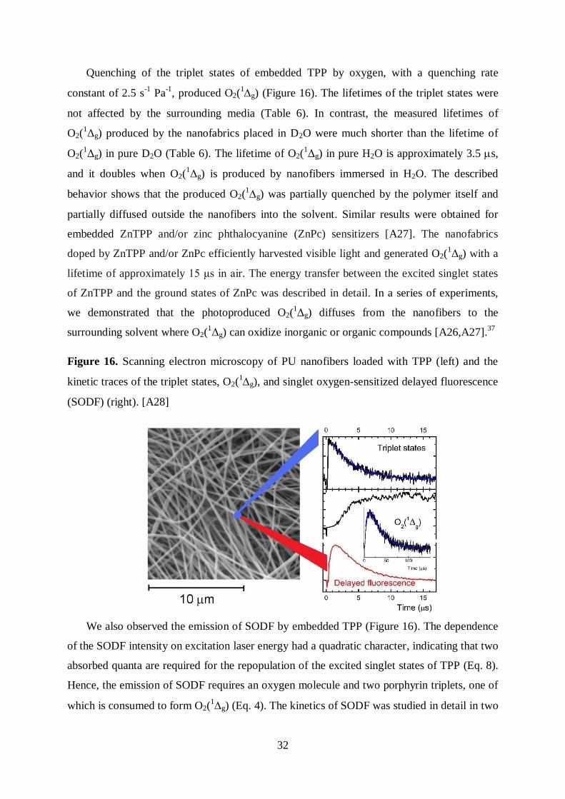
Nanofabrics, composed of functionalized polyurethane (PU) nanofibers (Figure 16), were
produced by using an industrial-scale electrospinning method [A26]. A unique photofunction
was imparted by embedded TPP, an efficient producer of O2(1g) in solutions. The absorption
and fluorescence emission spectra of embedded TPP were similar to the spectra in CH2Cl2.
These observations indicated that TPP remains monomeric in PU nanofibers. We also
described the preparation and spectral properties of (nBu4N)2[Mo6I8(CF3COO)6] embedded in
PU nanofibers [A10] and porphyrin-doped nanofibers made of polystyrene and gelatin,36
polycaprolactone and polyamide 6 [A29].37
The diameter of nanofibers is hundreds of
nanometers, which is comparable to the oxygen diffusion radius.
32
Quenching of the triplet states of embedded TPP by oxygen, with a quenching rate
constant of 2.5 s-1
Pa-1
, produced O2(1g) (Figure 16). The lifetimes of the triplet states were
not affected by the surrounding media (Table 6). In contrast, the measured lifetimes of
O2(1Δg) produced by the nanofabrics placed in D2O were much shorter than the lifetime of
O2(1Δg) in pure D2O (Table 6). The lifetime of O2(
1Δg) in pure H2O is approximately 3.5 s,
and it doubles when O2(1Δg) is produced by nanofibers immersed in H2O. The described
behavior shows that the produced O2(1g) was partially quenched by the polymer itself and
partially diffused outside the nanofibers into the solvent. Similar results were obtained for
embedded ZnTPP and/or zinc phthalocyanine (ZnPc) sensitizers [A27]. The nanofabrics
doped by ZnTPP and/or ZnPc efficiently harvested visible light and generated O2(1Δg) with a
lifetime of approximately 15 μs in air. The energy transfer between the excited singlet states
of ZnTPP and the ground states of ZnPc was described in detail. In a series of experiments,
we demonstrated that the photoproduced O2(1Δg) diffuses from the nanofibers to the
surrounding solvent where O2(1Δg) can oxidize inorganic or organic compounds [A26,A27].
37
Figure 16. Scanning electron microscopy of PU nanofibers loaded with TPP (left) and the
kinetic traces of the triplet states, O2(1g), and singlet oxygen-sensitized delayed fluorescence
(SODF) (right). [A28]
We also observed the emission of SODF by embedded TPP (Figure 16). The dependence
of the SODF intensity on excitation laser energy had a quadratic character, indicating that two
absorbed quanta are required for the repopulation of the excited singlet states of TPP (Eq. 8).
Hence, the emission of SODF requires an oxygen molecule and two porphyrin triplets, one of
which is consumed to form O2(1g) (Eq. 4). The kinetics of SODF was studied in detail in two
33
consecutive communications [A28].36
Because the intensity and kinetics of SODF are
strongly affected by oxygen concentration in the microenvironment, SODF is a sensitive
indicator of the oxygen content. We proposed that SODF spectroscopy could be used for
spatially resolved imaging of O2(1g) inside polymeric nanofibers and other materials.
Table 6. Lifetimes of the porphyrin triplet states () and corresponding lifetimes of O2(1Δg)
() produced by TPP embedded in PU nanofibers in air or immersed in air-saturated
solutions [A26].
Surroundings /s /s
Measured Literaturea
Air 18.1 21.2 -b
D2O 18.3 18.6 68
H2O 18.0 6.3 3.5
H2O,0.01M NaN3 17.9, 2.0 c 4.7 -
a Lifetime of O2(1Δg) in the given environment. b Radiative lifetime of unperturbed O2(
1Δg) is 72 min.
c Biexponential decay.
Photoproduced O2(1g) imparted bactericidal [A26,A27]
37,38 and virucidal [A29]
properties to porphyrin-doped nanofabrics. The bactericidal properties were demonstrated by
testing with the DH5 Escherichia coli strain. In control experiments that used doped
nanofabrics in the dark or nanofabrics without the incorporated sensitizer exposed to light or
kept in the dark, the growth of the bacteria was not affected. The strong photovirucidal effects
toward non-enveloped polyomaviruses and enveloped baculoviruses were observed on the
surfaces of hydrophilic PU and polycaprolactone nanofibers with embedded TPP. In all cases,
we observed that hydrophilicity of the nanofiber surfaces was a prerequisite of nanofiber
activity. Surprisingly, the nanofibers exhibited post-irradiation bactericidal properties after
long periods of irradiation due to the minor generation of H2O2.37
The nanofibers are excellent hosts of photoactive compounds. Considering the industrial
scale of nanofabric production and the fact that bacteria cannot pass through a nanostructured
material because they are retained at the surface, the sensitizer-doped nanofabrics are
potentially useful as sterile materials.
34
6. Conclusions
The presented research concentrated on porphyrinic sensitizers whose spectral and
photophysical properties are affected by noncovalent interactions within either molecular
assemblies or matrices, which can be inorganic or organic. Many of our results concerning the
structure and photophysical properties of free and embedded sensitizers, their intercalation,
the delamination of layered hydroxides, and the fabrication of polymeric composites and
oriented layers were new and were published for the first time by our team. The investigated
topics were:
A) Synthesis, characterization, and structure of the molecular assemblies and hybrid
materials. The sensitizers were studied in solutions, dispersions, and embedded in the
solid materials (powders, films, nanofibers).
B) Properties of the sensitizer molecules. Elementary photoprocesses occurring after
excitation of the sensitizers (free or bound) were investigated and correlated with the
nature of the sensitizer environment. The structural aspects, absorption spectra,
luminescence, and sensitizing properties of the molecular assemblies and
photofunctional materials were summarized in our review papers [A1].4,20
The
organization of the sensitizers in the materials is an important parameter because these
molecules tend to form closely stacked aggregates with reduced photochemical
activity. Some of the studied sensitizers have excellent brightness (i.e., product of
molar absorption coefficient and luminescence quantum yield), high yields of O2(1g),
and emission in the red region, and they can serve as a platform for the development
of novel oxygen sensors, singlet oxygen sensitizers, and UV/Red light convertors.
C) Functions of the materials. Some of the investigated materials are good solid-state
sensitizers that produce O2(1g), and they have bactericidal, virucidal, and
photocatalytic activities. They also exhibit interesting electrochemical properties.
Hence, our research may motivate applications of these materials in the area of
photoactive surfaces, catalysts, sensors, molecular magnets, and even batteries.
The main results:
- Effect of container molecules. The host molecules influence the spectral and photophysical
properties of the sensitizers, especially the lifetimes and quenching rate constants of the
sensitizer excited states. It is difficult, however, to form a general model of all the effects
because numerous, sometimes opposite factors are combined together: e.g., aggregation,
restriction of internal movement, protonation, electron transfer, polarity, and preorganization
35
of a binding site. These studies also stimulated the development of new cluster-type
sensitizers in our laboratory.
- Concept of photofunctional layered metal hydroxides. The observation that porphyrin π-π
stacking does not occur within the interlayer space of layered hydroxides is essential for the
concept of photoactive materials. Further positive effects of the hydroxide matrix on
intercalated porphyrins are enhanced thermal and chemical stability and reduction of
photobleaching. Intercalation affects photophysical and photochemical properties; however,
such important features as luminescence and the quenching of the porphyrin triplet states by
oxygen remain preserved. The quenching confirms that the porphyrin molecules are
accessible to oxygen and can generate O2(1g). In many cases, singlet oxygen lifetimes were
quite short, even shorter than the lifetime of O2(1g) in water (Δ ~ 3.5 μs). This points to the
quenching of O2(1g) by the OH groups, which are coordinated to the metal centers of the
hydroxide layers, and the water molecules confined in the interlayer space.
- Description of the two-dimensional arrangement of the porphyrin molecules in the
interlayer space of layered double hydroxides and layered zinc hydroxide salts by
experimental and molecular simulation methods.
- Nanocontainer and nanofiller aspects of LDHs. The function of the new LDH-
porphyrin/polymer composites is based on the ability of intercalated porphyrins to produce
O2(1g) upon irradiation with visible light. The amount of O2(
1g) at the composite surface
can be tuned by varying the filler loading and the amount of intercalated porphyrin in the
filler. Since O2(1g) is cytotoxic, these composites provide a platform for the fabrication of
bactericidal surfaces functioning under daylight.
- Bactericidal and virucidal nanofibers. Considering the industrial scale of the nanofabric
production and the fact that bacteria cannot pass through a nanostructured material and are
detained on its surface, the porphyrin-doped nanofabrics are prospective materials that would
remain sterile while irradiated by visible light.
- Transformation of layered rare-earth hydroxide to three-dimensional MOFs. The MOF has
framework-dependent fluorescent properties. The ability of the MOF to produce long-lived
O2(1g) motivates the design of novel MOFs with tuned photoresponses and porosity.
- Delamination of layered metal hydroxides to hydroxide nanosheets and their use for the
preparation of functional films with adjustable thicknesses. The functions of the
nanostructured films prepared from the hydroxide/oxide nanosheets are wide-ranging: (i)
luminescent ultrathin films, (ii) transparent films constructed from ZnO nanosheets with
36
enhanced photocatalytic activity due to the nanosheet orientations,33
and (iii)
electrochemically active ultrathin films with reproducible and stable charge-discharge
properties in an alkaline electrolyte.39
7. Acknowledgments
K. Lang thanks many colleagues who participated in various parts of the presented research
and whose names appear in the cited communications.
8. Publications that form the basis of the thesis
[A1] J. Demel, K. Lang: Layered hydroxide-porphyrin hybrid materials: synthesis, structure,
and properties. Eur. J. Inorg. Chem. (2012) 5154-5164.
[A2] P. Kubát, K. Lang, K. Procházková, P. Anzenbacher, Jr.: Self-aggregates of cationic
meso-tetratolylporphyrins in aqueous solutions. Langmuir 19 (2003) 422-428.
[A3] V. Král, F. P. Schmidtchen, K. Lang, M. Berger: Anion-controlled assembly of
porphyrin-bicyclic guanidine conjugates. Org. Lett. 4 (2002) 51-54.
[A4] K. Lang, P. Kubát, P. Lhoták, J. Mosinger, D. M. Wagnerová: Photophysical properties
and photoinduced electron transfer within host-guest complexes of 5, 10, 15, 20-
tetrakis(4-N-methylpyridyl)porphyrin with water-soluble calixarenes and cyclodextrins.
Photochem. Photobiol. 74 (2001) 558-565.
[A5] P. Kubát, K. Lang, P. Lhoták, P. Janda, J. Sýkora, P. Matějíček, M. Hof, K. Procházka,
Z. Zelinger: Porphyrin/calixarene self-assemblies in aqueous solution. J. Photochem.
Photobiol. A: Chem. 198 (2008) 18-25.
[A6] P. Kubát, J. Šebera, S. Záliš, J. Langmaier, M. Fuciman, T. Polívka, K. Lang: Charge
transfer in porphyrin-calixarene complexes: ultrafast kinetics, cyclic voltammetry, and
DFT calculations. Phys. Chem. Chem. Phys. 13 (2011) 6916-6923.
[A7] J. Mosinger, M. Deumié, K. Lang, P. Kubát, D. M. Wagnerová: Supramolecular
sensitizer: complexation of meso-tetrakis(4-sulfonatophenyl)porphyrin with 2-
hydroxypropyl-cyclodextrins. J. Photochem. Photobiol. A: Chem. 130 (2000) 13-20.
[A8] J. Mosinger, L. Slavětínská, K. Lang, P. Coufal, P. Kubát: Cyclodextrin carriers of
positively charged porphyrin sensitizers. Org. Biomol. Chem. 7 (2009) 3797-3804.
[A9] K. Kirakci, P. Kubát, J. Langmaier, T. Polívka, M. Fuciman, K. Fejfarová, K. Lang: A
comparative study of the redox and excited state properties of (nBu4N)2[Mo6X14] and
(nBu4N)2[Mo6X8(CF3COO)6] (X = Cl, Br, or I). Dalton Trans. 42 (2013) 7224-7232.
37
[A10] K. Kirakci, P. Kubát, M. Dušek, K. Fejfarová, V. Šícha, J. Mosinger, K. Lang: Highly
luminescent hexanuclear molybdenum cluster: a promising candidate toward photoactive
materials. Eur. J. Inorg. Chem. (2012) 3107-3111.
[A11] M. . S. Londesborough, D. Hnyk, J. Bould, L. Serrano-Andrés, V. Sauri, J. M. liva,
P. Kubát, T. Polívka, K. Lang: Distinct photophysics of the isomers of B18H22 explained.
Inorg. Chem. 51 (2012) 1471−1479.
[A12] V. Saurí, J. M. liva, D. Hnyk, J. Bould, J. Braborec, M. Merchán, P. Kubát, I.
Císařová, K. Lang, M. . S. Londesborough: Tuning the photophysical properties of
anti-B18H22: efficient intersystem crossing between excited singlet and triplet states in
new 4,4’-(HS)2-anti-B18H20. Inorg. Chem. 52 (2013) 9266-9274.
[A13] J. Bould, T. Baše, M. . S. Londesborough, L. A. ro, R. Macías, J. D. Kennedy, P.
Kubát, M. Fuciman, T. Polívka, K. Lang: Reversible capture of small molecules on
bimetallaborane clusters: synthesis, structural characterization, and photophysical
aspects. Inorg. Chem. 50 (2011) 7511-7523.
[A14] E. Káfuňková, C. Taviot- uého, P. Bezdička, M. Klementová, P. Kovář, P. Kubát, J.
Mosinger, M. Pospíšil, K. Lang: Porphyrins intercalated in Zn/Al and Mg/Al layered
double hydroxides: properties and structural arrangement. Chem. Mater. 22 (2010)
2481-2490.
[A15] P. Kovář, M. Pospíšil, E. Káfuňková, K. Lang, F. Kovanda: Mg-Al layered double
hydroxide intercalated with porphyrin anions: molecular simulations and experiments. J.
Mol. Model. 16 (2010) 223-233.
[A16] M. Jiřičková, J. Demel, P. Kubát, J. Hostomský, F. Kovanda, K. Lang: Photoactive
self-standing films made of layered double hydroxides with arranged porphyrin
molecules. J. Phys. Chem. C 115(44) (2011) 21700–21706.
[A17] K. Lang, P. Bezdička, J. L. Bourdelande, J. Hernando, I. Jirka, E. Káfuňková, F.
Kovanda, P. Kubát, J. Mosinger, D. M. Wagnerová: Layered double hydroxides with
intercalated porphyrins as photofunctional materials: subtle structural changes modify
singlet oxygen production. Chem. Mater. 19 (2007) 3822-3829.
[A18] K. Lang, P. Kubát, J. Mosinger, J. Bujdák, M. Hof, P. Janda, J. Sýkora, N. Iyi:
Photoactive Oriented Films of Layered Double Hydroxides. Phys. Chem. Chem. Phys.
10 (2008) 4429-4434.
[A19] E. Káfuňková, K. Lang, P. Kubát, M. Klementová, J. Mosinger, M. Šlouf, A.-L.
Troutier-Thuilliez, F. Leroux, V. Verney, C. Taviot- uého: Porphyrin-layered double
38
hydroxide/polymer composites as novel ecological photoactive surfaces. J. Mater.
Chem. 20 (2010) 9423-9432.
[A20] F. Kovanda, E. Jindová, K. Lang, P. Kubát, Z. Sedláková: Preparation of layered
double hydroxides intercalated with organic anions and their application in
LDH/poly(butyl methacrylate) nanocomposites. Appl. Clay Sci. 48 (2010) 260-270.
[A21] M. Merchán, T. Sothea uk, P. Kubát, K. Lang, C. Coelho, V. Verney, S. Commereuc,
F. Leroux, V. Sol, C. Taviot- uého: Photostability and photobactericidal properties of
porphyrin-layered double hydroxide-polyurethane composite films. J. Mater. Chem. B 1
(2013) 2139-2146.
[A22] J. Demel, P. Kubát, I. Jirka, P. Kovář, M. Pospíšil, K. Lang: Inorganic-organic hybrid
materials: layered zinc hydroxide salts with intercalated porphyrin sensitizers. J. Phys.
Chem. C 114 (2010) 16321-16328.
[A23] J. Demel, J. Pleštil, P. Bezdička, P. Janda, M. Klementová, K. Lang: Layered zinc
hydroxide salts: Delamination, preferred orientation of hydroxide lamellae, and
formation of ZnO nanodiscs. J. Colloid Inter. Sci. 360 (2011) 532-539.
[A24] J. Demel, J. Pleštil, P. Bezdička, P. Janda, M. Klementová, K. Lang: Few-layer ZnO
nanosheets: preparation, properties, and films with exposed {001} facets. J. Phys. Chem.
C 115 (2011) 24702-24706.
[A25] J. Demel, P. Kubát, F. Millange, J. Marrot, I. Císařová, K. Lang: Lanthanide-porphyrin
hybrids: from layered structure to metal-organic frameworks with photophysical
properties. Inorg. Chem. 52 (2013) 2779-2786.
[A26] J. Mosinger, . Jirsák, P. Kubát, K. Lang, B. Mosinger: Bactericidal nanofabrics based
on photoproduction of singlet oxygen. J. Mater. Chem. 17(2) (2007) 164 – 166.
[A27] J. Mosinger, K. Lang, P. Kubát, J. Sýkora, M. Hof, L. Plíštil, B. Mosinger Jr.:
Photofunctional polyurethane nanofabrics doped by zinc tetraphenylporphyrin and zinc
phthalocyanine photosensitizers. J. Fluoresc. 19 (2009) 705–713.
[A28] J. Mosinger, K. Lang, L. Plíštil, S. Jesenská, J. Hostomský, Z. Zelinger, P. Kubát:
Fluorescent polyurethane nanofabrics: a source of singlet oxygen and oxygen sensing.
Langmuir 26 (2010) 10050-10056.
[A29] Y. Lhotáková, L. Plíštil, A. Morávková, P. Kubát, K. Lang, J. Forstová, J. Mosinger:
Virucidal nanofiber textiles based on photosensitized production of singlet oxygen.
PLoS ONE 7(11) (2012) e49226.
39
9. References
1. J. Zhao, W. Wu, J. Sun, S. Guo, Chem. Soc. Rev. 42 (2013) 5323-5351.
2. P. R. Ogilby, Chem. Soc. Rev. 39 (2010) 3181-3209.
3. R. Schmidt, Photochem. Photobiol. 82 (2006) 1161-1177.
4. K. Lang, J. Mosinger, D. M. Wagnerová, Coord. Chem. Rev. 248 (2004) 321-350.
5. C. S. Foote, Photochem. Photobiol. 54 (1991) 659.
6. See e.g., a) P. Kubát, K. Lang, P. Anzenbacher Jr., K. Jursíková, V. Král, B. Ehrenberg, J.
Chem. Soc., Perkin Trans. 1 (2000) 933-941; b) M. Dudič, P. Lhoták, I. Stibor, K. Lang, P.
Prošková, Org. Lett. 5 (2003) 149-152; c) P. Kubát, K. Lang, P. Janda, P. Anzenbacher,
Langmuir 21 (2005) 9714-9720; d) V. Novakova, P. Zimcik, M. Miletin, L. Vachova, K.
Kopecky, K. Lang, P. Chábera, T. Polívka, Phys. Chem. Chem. Phys. 12 (2010) 2555-2563;
e) V. Novakova, L. Lochman, I. Zajícová, K. Kopecky, M. Miletin, K. Lang, K. Kirakci, P.
Zimcik, Chem. Eur. J. 19 (2013) 5025-5028.
7. S. Bonnet, C. Forano, A. de Roy, J. P. Besse, P. Maillard, M. Momenteau, Chem. Mater. 8
(1996) 1962-1968.
8. a) A. Čeklovský, A. Czímerová, K. Lang, J. Bujdák, J. Luminescence 129 (2009) 912-918;
b) J. Bujdák, J. Jurečeková, H. Bujdáková, K. Lang, F. Šeršen, Environ. Sci. Technol. 43
(2009) 6202-6207; c) A. Čeklovský, A. Czímerová, K. Lang, J. Bujdák, Pure Appl. Chem. 81
(2009) 1385-1396; d) J. Bujdák, M. Danko, D. Chorvát Jr., A. Czímerová, J. Sýkora, K. Lang,
Applied Clay Sci. 65–66 (2012) 152–157.
9. E. A. Lissi, M. V. Encinas, E. Lemp, M. A. Rubio, Chem. Rev. 93 (1993) 699-723.
10. M. Nishio, M. Hirota, Y. Umezawa, The CH/ Interaction; Evidence, Nature, and
Consequences. Wiley-VCH Publishers, New York 1998.
11. F. Perret, A. W. Coleman, Chem. Commun. 47 (2011) 7303–7319.
12. J. Szejtli, Chem. Rev. 98 (1998) 1743-1753.
13. K. A. Connors, Chem. Rev. 97 (1997) 1325-1357.
14. J. Mosinger, V. Kliment, Jr., J. Sejbal, P. Kubát, K. Lang, J. Porphyrins Phthalocyanines
6 (2002) 514-526.
15. A. W. Maverick, J. S. Najdzionek, D. Mackenzie, D. G. Nocera, H. B. Gray, J. Am. Chem.
Soc. 105 (1983) 1878-1882.
16. J. A. Jackson, C. Turro, M. D. Newsham, D. G. Nocera, J. Phys. Chem. 94 (1990) 4500-
4507.
40
17. a) L. M. Robinson, D. F. Shriver, J. Coord. Chem. 37 (1996) 119-129; b) O. A.
Adamenko, G. V. Loukova, V. A. Smirnov, Russ. Chem. Bull. 51 (2002) 994-997; c) N.
Prokopuk, C. S. Weinert, D. P. Siska, C. L. Stern, D. F. Shriver, Angew. Chem., Int. Ed. 39
(2000) 3312-3315, d) L. F. Szczepura, K. A. Ketcham, B. A. Ooro, J. A. Edwards, J. N.
Templeton, D. L. Cedeno, A. J. Jircitano, Inorg. Chem. 47 (2008) 7271-7278.
18. A. R. Pitochelli, M. F. Hawthorne, J. Am. Chem. Soc. 84 (1962) 3218.
19. V. V. Volkov, E. A. Il'inchik, O. V. Volkov, O. O. Yuryeva, Chem. Sustainable Dev. 8
(2000) 185-191.
20. D. M. Wagnerová, K. Lang, Coord. Chem. Rev. 255 (2011) 2904-2911.
21. a) J. Bould, Y. M. McInnes, M. J. Carr, J. D. Kennedy, Chem. Commun. (2004) 2380-
2381; b) J. Bould, C. A. Kilner, J. D. Kennedy, Dalton Trans. (2005) 1574-1582.
22. M. Seip, H.-D. Brauer, J. Photochem. Photobiol. A: Chem. 79 (1994) 19-24.
23. M. Ogawa, K. Kuroda, Chem. Rev. 95 (1995) 399-438.
24. I. Y. Park, K. Kuroda, C. Kato, Chem. Lett. (1989) 2057-2058.
25. P. K. Dutta, M. Puri, J. Phys. Chem. 93 (1989) 376-381.
26. L. Ukrainczyk, M. Chibwe, T. J. Pinnavaia, S. A. Boyd, J. Phys. Chem. 98 (1994) 2668–
2676.
27. D. Yan, S. Qin, L. Chen, J. Lu, J. Ma, M. Wei, D. G. Evans, X. Duan, Chem. Commun. 46
(2010) 8654–8656.
28. F. Leroux, C. Taviot- uého, J. Mater. Chem. 15 (2005) 3628-3642.
29. V. Rives, (Ed.), Layered Double Hydroxides: Present and Future, Nova Science
Publishers, New York 2001, pp. 251-434.
30. E. Káfuňková, Porphyrin-layered double hydroxide hybrids as novel photofunctional
materials, PhD Thesis, Charles University in Prague, Praha, Czech Republic, 2010.
31. G. G. C. Arizaga, K. G. Satyanarayana, F. Wypych, Solid State Ionics 178 (2007) 1143-
1162.
32. G. Rogez, C. Massobrio, P. Rabu, M. Drillon, Chem. Soc. Rev. 40 (2011) 1031-1058.
33. J. Hynek, V. Kalousek, R. Žouželka, P. Bezdička, P. Dzik, J. Rathouský, J. Demel, K.
Lang, Langmuir 30 (2014) 380-386.
34. F. Geng, R. Ma, T. Sasaki, Acc. Chem. Res. 43 (2010) 1177-1185.
35. L. J. McIntyre, T. J. Prior, A. M. Fogg, Chem. Mater. 22 (2010) 2635-2645.
36. J. Mosinger, K. Lang, J. Hostomský, J. Franc, J. Sýkora, M. Hof, P. Kubát, J. Phys. Chem.
B 114 (2010) 15773–15779.

41
37. S. Jesenská, L. Plíštil, P. Kubát, K. Lang, L. Brožová, Š. Popelka, L. Szatmáry, J.
Mosinger, J. Biomed. Mater. Res. A 99A (2011) 676-683.
38. P. Henke, K. Lang, P. Kubát, J. Sýkora, M. Šlouf, J. Mosinger, ACS Appl. Mater.
Interfaces 5 (2013) 3776-3783.
39. a) B. Schneiderová, J. Demel, J. Pleštil, P. Janda, J. Bohuslav, D. Ihiawakrim, . Ersen,
G. Rogez, K. Lang, J. Mater. Chem. A 1 (2013) 11429-11437; b) B. Schneiderová, J. Demel,
J. Pleštil, H. Tarábková, J. Bohuslav, K. Lang, Dalton Trans. (2014), in press.


![singlet-triplet anticrossings 3He : precise determination ...He by means of Electric Field Induced Singlet-Triplet Anticrossings [5]. The coupling between 1 D and 3D states occurs](https://img.pdfslide.tips/doc/110x75/5e8835bb70def00de7745e00/singlet-triplet-anticrossings-3he-precise-determination-he-by-means-of-electric.jpg)