Embed Size (px)
Citation preview
Small and Large Intestinal pathology,
part 3Manar Hajeer, MD, FRCPath
University of Jordan, School of medicine
Diseases of the intestines
Intestinal obstruction
Vascular disorders
Malabsorptive diseases and infections
Inflammatory bowel disease.
Polyps and neoplastic diseases
COLONIC POLYPS AND
NEOPLASTIC DISEASE
Colon is most common site for polyps
Sessile polyp: no stalk
Pedunculated polyp: stalk.
Neoplastic polyps: adenoma.
Non neoplastic polyps: inflammatory, hamartomatous, or hyperplastic
Inflammatory Polyps
Solitary rectal ulcer syndrome.
Recurrent abrasion and ulceration of the overlying rectal mucosa.
Chronic cycles of injury and healing give a polypoid mass of inflamed and
reactive mucosal tissue.
Hamartomatous Polyps
Sporadic or syndromatic.
Disorganized, tumor-like growth composed of mature cell types normally
present at that site.
Juvenile Polyps
Peutz-Jeghers Syndrome
Juvenile Polyps
Most common hamartomatous polyp
Sporadic are solitary.
Children younger than 5 years of age
Rectum.
Syndromic are multiple.
3 to as many as 100. Mean age 5 years
Autosomal dominant syndrome of juvenile polyposis
Transforming growth factor-β (TGF-β) mutation.
Increased risk for colonic adenocarcinoma.
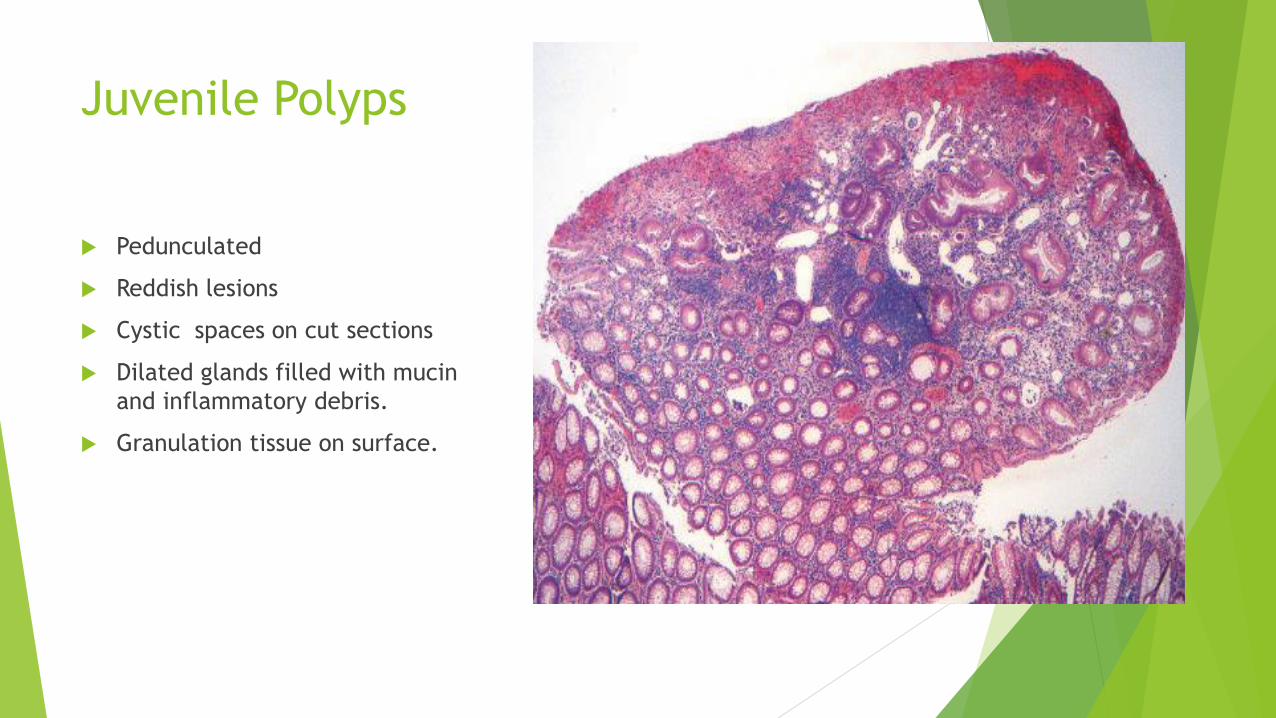
Juvenile Polyps
Pedunculated
Reddish lesions
Cystic spaces on cut sections
Dilated glands filled with mucin
and inflammatory debris.
Granulation tissue on surface.
Peutz-Jeghers Syndrome
Autosomal dominant, rare
Mean age: 10-15 years.
Multiple gastrointestinal hamartomatous polyps
Most common in the small intestine.
Mucocutaneous hyperpigmentation
Increased risk for several malignancies: colon, pancreas, breast, lung,
ovaries, uterus, and testes,
LKB1/STK11 gene mutation.
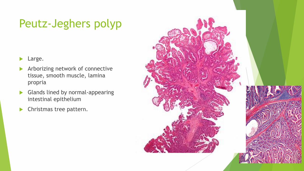
Peutz-Jeghers polyp
Large.
Arborizing network of connective
tissue, smooth muscle, lamina
propria
Glands lined by normal-appearing
intestinal epithelium
Christmas tree pattern.
Hyperplastic Polyps
Common
5th-6th decade.
Decreased epithelial turnover and delayed shedding of surface epithelium >>>
pileup of goblet cells & epithelial overcrowding
No malignant potential
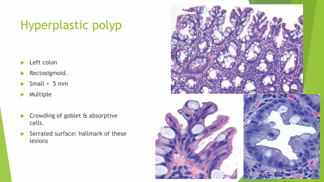
Hyperplastic polyp
Left colon
Rectosigmoid.
Small < 5 mm
Multiple
Crowding of goblet & absorptive
cells.
Serrated surface: hallmark of these
lesions
Adenomas
Most common and clinically important
Increase with age.
Definition: presence of epithelial dysplasia (low or high).
Precursor for majority of colorectal adenocarcinomas
Most adenomas DO NOT progress to carcinoma.
USA: screening colonoscopy starts at 50 yrs.
Earlier screening with family history.
Western diets and lifestyles increase risk.
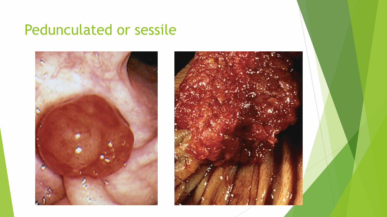
Pedunculated or sessile
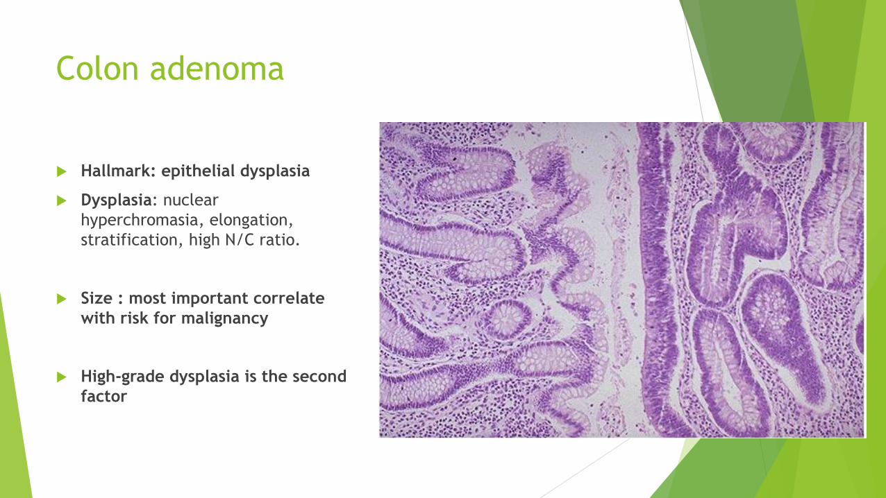
Colon adenoma
Hallmark: epithelial dysplasia
Dysplasia: nuclear
hyperchromasia, elongation,
stratification, high N/C ratio.
Size : most important correlate
with risk for malignancy
High-grade dysplasia is the second
factor
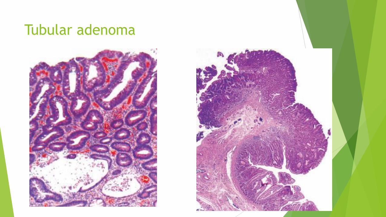
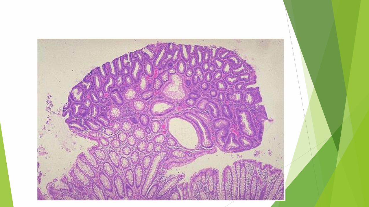
Tubular adenoma
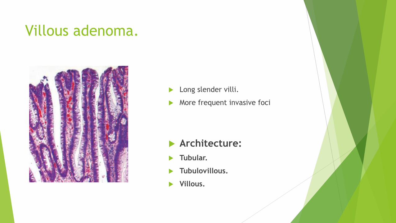
Villous adenoma.
Long slender villi.
More frequent invasive foci
Architecture:
Tubular.
Tubulovillous.
Villous.
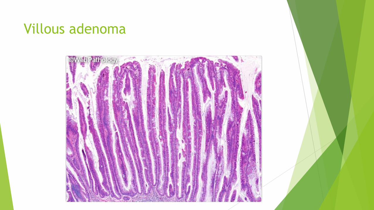
Villous adenoma
Familial Syndromes
Syndromes associated with colonic polyps and increased rates of colon cancer
Genetic basis.
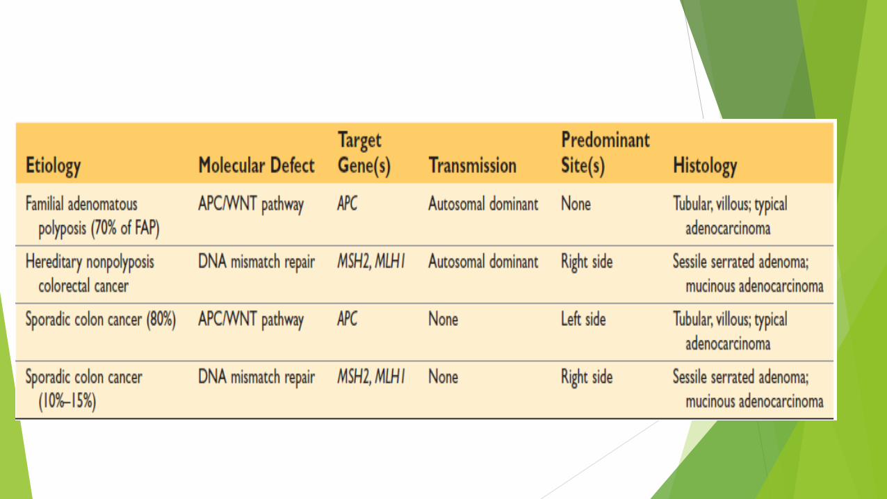
Familial Adenomatous Polyps (FAP)
Hereditary Nonpolyposis Colorectal Cancer (HNPCC)
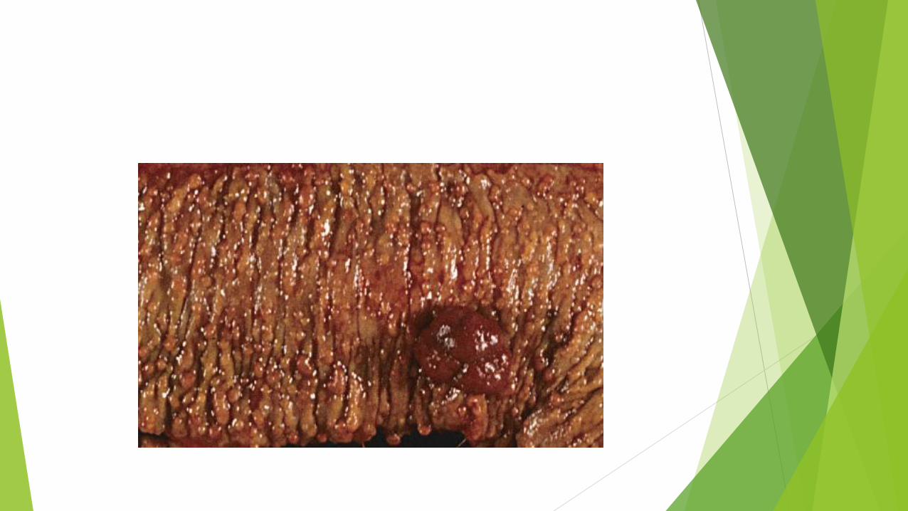
Familial adenomatous polyposis FAP
Autosomal dominant.
Numerous colorectal adenomas: teenage years.
Mutation in APC gene.
At least 100 polyps are necessary for a diagnosis of classic FAP.
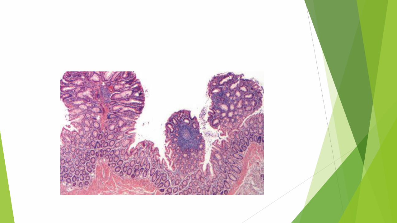
Morphologically similar to sporadic adenomas
100% of patients develop colorectal carcinoma, IF UNTREATED, often before
age of 30.
Standard therapy: prophylactic colectomy before 20 Year of age.
Risk for extraintestinal manifestations,
Variants of FAP: Gardner syndrome and Turcot syndrome.
Gardner syndrome: intestinal polyps + osteomas (mandible, skull, and long
bones); epidermal cysts; desmoid and thyroid tumors; and dental
abnormalities.
Turcot syndrome: intestinal adenomas and CNS tumors (medulloblastomas
>> glioblastomas )
Hereditary Nonpolyposis Colorectal
Cancer: HNPCC, Lynch syndrome
Clustering of tumors: Colorectum, endometrium, stomach, ovary, ureters, brain, small bowel, hepatobiliary tract, and skin
Colon cancer at younger age than sporadic cancers
Right colon with excessive mucin production .
Adenomas are present, BUT POLYPOSIS IS NOT.
Inherited germ line mutations in DNA mismatch repair genes.
Accumulation of mutations in microsatellite DNA (short repeating sequences)
Resulting in microsatellite instability
Majority of cases involve either MSH2 or MLH1.
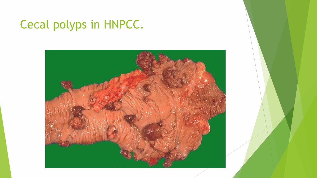
Cecal polyps in HNPCC.
Colonic Adenocarcinoma
Most common malignancy of the gastrointestinal tract
Small intestine is uncommonly involved by neoplasia.
Peak: 60 to 70 years
20% under 50 years.
Developed countries lifestyles and diet.
Low intake of vegetable fiber and high intake of carbohydrates and
fat.
Aspirin or other NSAIDs have a protective effect.
Cyclooxygenase-2 (COX-2) promotes epithelial proliferation.
Pathogenesis
Heterogeneous molecular events.
Sporadic >>>> familial.
Two pathways:
APC/β-catenin pathway >> increased WNT signaling
Microsatellite instability pathway >> defects in DNA mismatch repair
Stepwise accumulation of multiple mutations
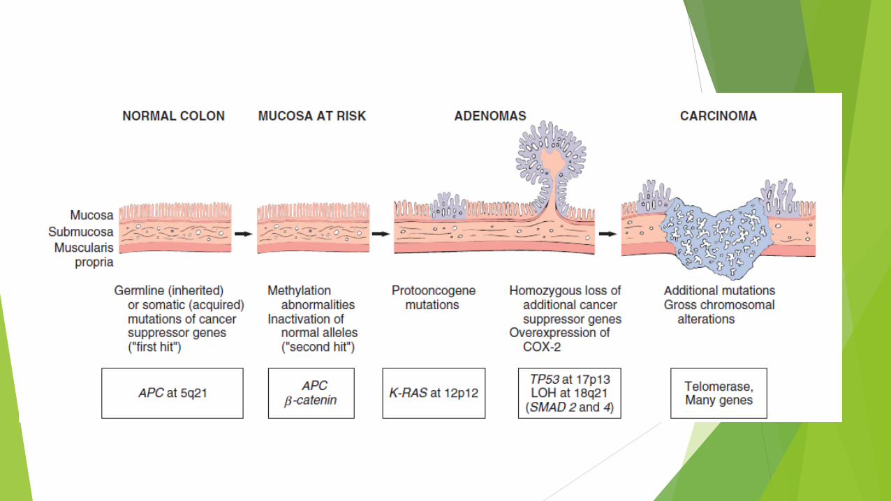
The APC/β-catenin pathway:
chromosomal instability
Classic adenoma carcinoma sequence.
80% of sporadic colon tumors
Mutation of the APC tumor suppressor gene: EARLY EVENT
APC is a key negative regulator of β-catenin, a component of the WNT
signaling pathway.
Both copies of APC should be inactivated for adenoma to develop (1st and 2nd
hits).
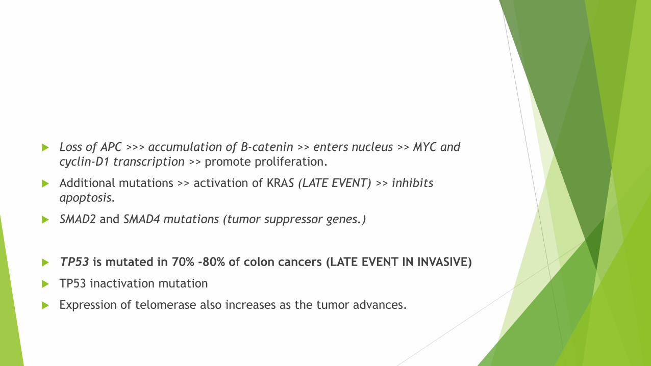
Loss of APC >>> accumulation of B-catenin >> enters nucleus >> MYC and
cyclin-D1 transcription >> promote proliferation.
Additional mutations >> activation of KRAS (LATE EVENT) >> inhibits
apoptosis.
SMAD2 and SMAD4 mutations (tumor suppressor genes.)
TP53 is mutated in 70% -80% of colon cancers (LATE EVENT IN INVASIVE)
TP53 inactivation mutation
Expression of telomerase also increases as the tumor advances.
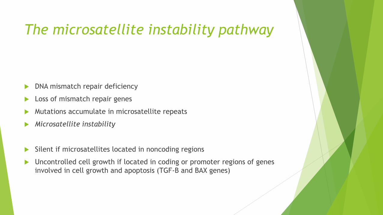
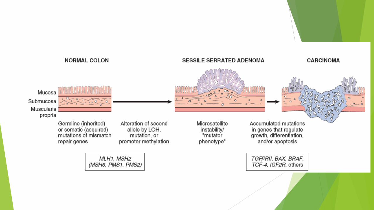
The microsatellite instability pathway
DNA mismatch repair deficiency
Loss of mismatch repair genes
Mutations accumulate in microsatellite repeats
Microsatellite instability
Silent if microsatellites located in noncoding regions
Uncontrolled cell growth if located in coding or promoter regions of genes
involved in cell growth and apoptosis (TGF-B and BAX genes)
MORPHOLOGY
Proximal colon tumors: polypoid, exophytic masses
Proximal colon: rarely cause obstruction.
Distal colon: annular lesions “napkin ring” constrictions & narrowing
Tall columnar cells of dysplastic epithelium forming GLANDS with strong
desmoplastic response.
Necrotic debris are typical.
Some tumors give abundant mucin.
Some form signet ring cells.
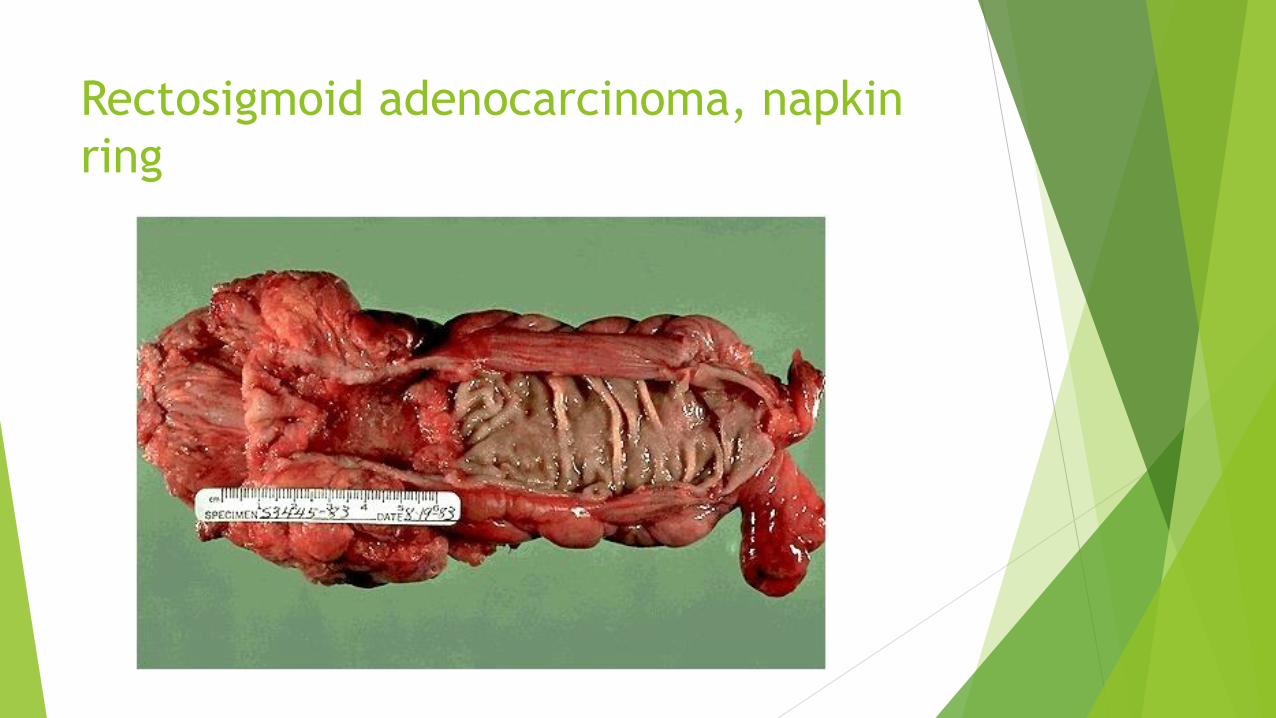
Rectosigmoid adenocarcinoma, napkin
ring
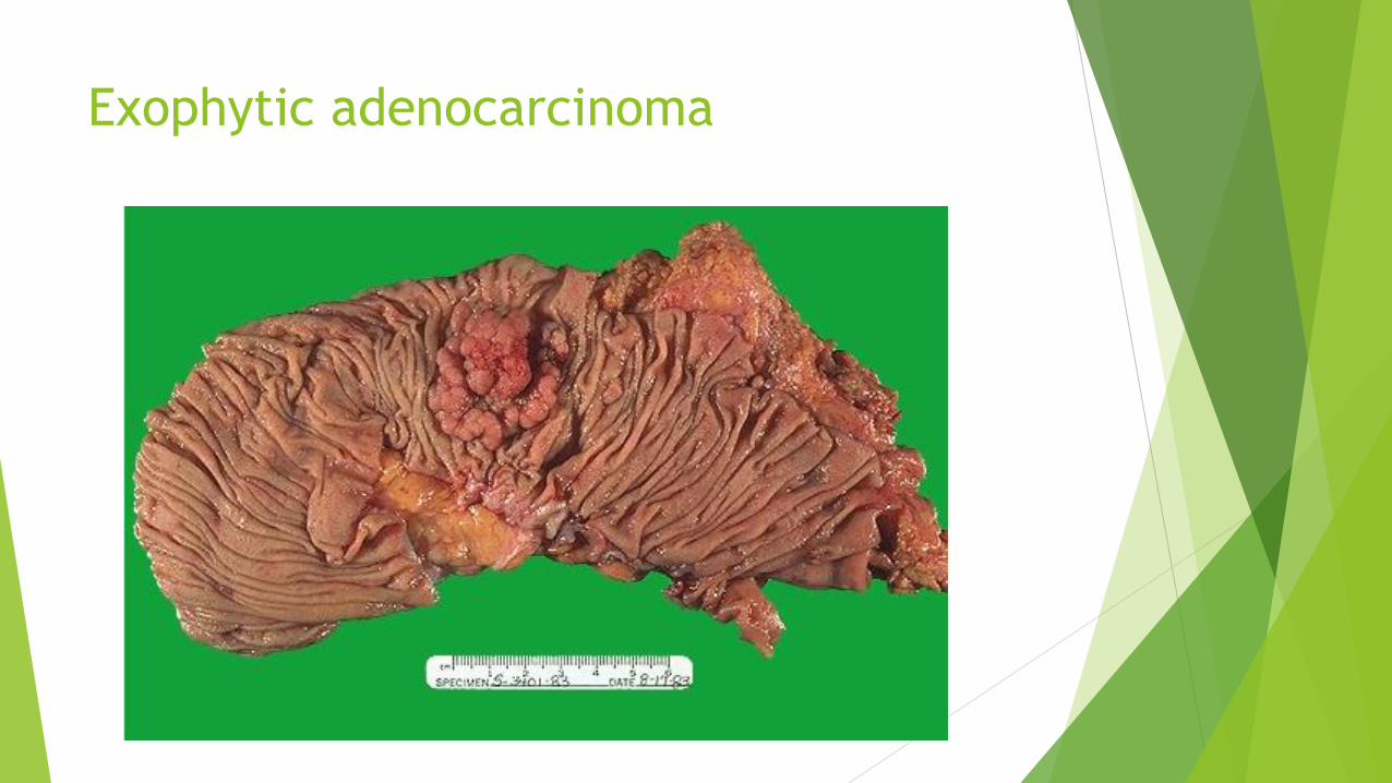
Exophytic adenocarcinoma
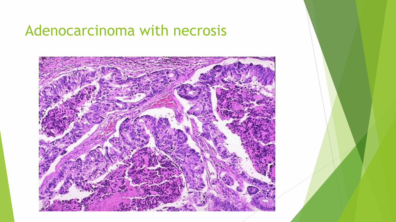
Adenocarcinoma with necrosis
Clinical Features
Endoscopic screening >> cancer prevention
Early cancer is asymptomatic !!!!!!!!
Cecal and right side cancers: Fatigue and weakness (iron deficiency anemia)
Iron-deficiency anemia in an older male or postmenopausal female is
gastrointestinal cancer until proven otherwise.
Left sided carcinomas: occult bleeding, changes in bowel habits, cramping
left lower-quadrant discomfort.
Poor differentiation and mucinous histology >> poor prognosis
Most important two prognostic factors are
Depth of invasion
Lymph node metastasis.
Distant metastases (lung and liver) can be resected.
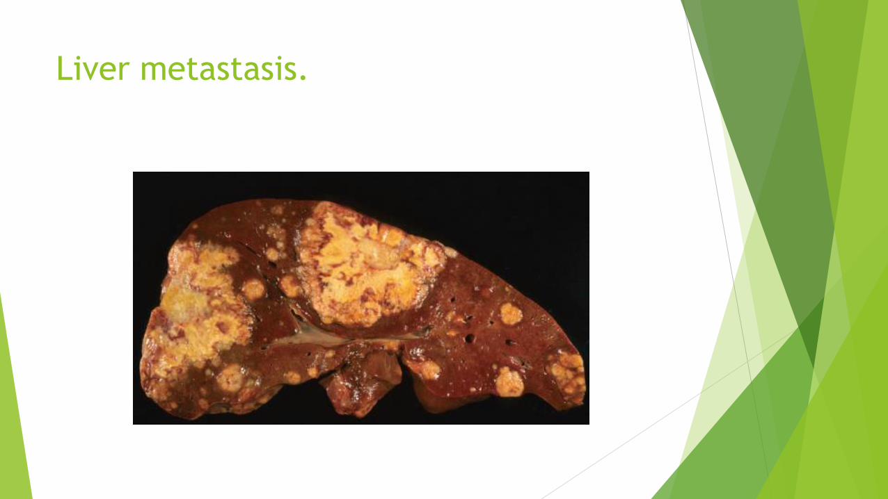
Liver metastasis.
Appendix
Normal true diverticulum of the cecum
ACUTE APPENDICITIS
TUMORS OF THE APPENDIX
ACUTE APPENDICITIS
Most common in adolescents and young adults.
May occur in any age.
Difficult to confirm preoperatively
DDx:
Mesenteric lymphadenitis,
Acute salpingitis,
Ectopic pregnancy,
Mittelschmerz (pain associated with ovulation),
Meckel diverticulitis.
Luminal obstruction in 50-80% of cases >> increased luminal pressure >>
impaired venous drainage >> ischemic injury & stasis associated bacterial
proliferation >>> inflammatory response rich in neutrophils & edema.
Obstruction by fecalith, less commonly : gallstone, tumor, worms….
Diagnosis requires neutrophilic infiltration of the muscularis propria
Acute suppurative appendicitis >> more severe >> focal abscess
formation.
Acute gangrenous appendicitis >> necrosis and ulceration.
Clinical Features
Early acute appendicitis: periumbilical pain
Later: pain localizes to the right lower quadrant,
Nausea, vomiting, low-grade fever, mildly leukocytosis.
A classic physical finding is McBurney’s sign (McBurney’s point).
Signs and symptoms are often absent, creating difficulty in clinical
diagnosis.
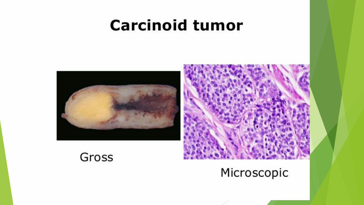
TUMORS OF THE APPENDIX
The most common tumor: carcinoid (neuroendocrine tumor)
Incidentally found during surgery or on examination of a resected appendix
Distal tip of the appendix
Nodal metastases & distant spread are rare.