Embed Size (px)
Citation preview
CLARISSA HARUMI OMORI
Efeitos de um programa de exercícios físicos em crianças e
adolescentes com dermatomiosite juvenil
Tese apresentada à Faculdade de Medicina da
Universidade de São Paulo para obtenção do título
de Doutora em Ciências
Programa: Pediatria
Orientador: Prof. Dr. Clovis Artur Almeida da Silva
Coorientador: Prof. Dr. Bruno Gualano
São Paulo
2014
Dados Internacionais de Catalogação na Publicação (CIP)
Preparada pela Biblioteca da Faculdade de Medicina da Universidade de São Paulo
reprodução autorizada pelo autor
Omori, Clarissa Harumi Efeitos de um programa de exercícios físicos em crianças e adolescentes com dermatomiosite juvenil / Clarissa Harumi Omori. -- São Paulo, 2014.
Tese(doutorado)--Faculdade de Medicina da Universidade de São Paulo.
Programa de Pediatria.
Orientador: Clovis Artur Almeida da Silva. Coorientador: Bruno Gualano. Descritores: 1.Dermatomiosite 2.Exercício 3.Criança 4.Adolescente
5.Estudos prospectivos 6.Estudos longitudinais 7.Qualidade de vida 8.Treinamento de resistência
USP/FM/DBD-138/14
“O que fizemos apenas por nós mesmos morre conosco; o que
fizemos pelos outros e pelo mundo permanece e é imortal.”
Albert Pike

Dedicatória
Aos meus pais, Rosa e Nelson, pelos valiosos princípios a mim
passados e pela educação por meio do amor e da abnegação;
Ao meu irmão, Ivam, pela amizade e cumplicidade;
Ao meu amado, Decio, por cada gesto de carinho e
compreensão;
Às minhas filhas, Carolina e Fernanda, por serem a luz da minha
vida.

Agradecimentos
A todos os pacientes que possibilitaram a realização deste estudo;
A Clovis Artur Almeida da Silva, querido professor e orientador, por
ser o meu maior exemplo de competência e dedicação;
A Bruno Gualano, pela coorientação e por sua generosidade;
Às admiráveis Adriana Maluf Elias Sallum, Lúcia Maria de Arruda
Campos, Nádia Emi Aikawa, Adriana Almeida de Jesus e Kátia Tomie Kozu,
por todos os ensinamentos sobre a reumatologia pediátrica e sobre a vida;
Aos amigos e melhores companheiros de complementação: Fernanda
Jusan Fiorot, João Carlos Diniz, Roberta Tavares Almeida, Vanessa Ramos
Guissa e Cíntia Maria Michelin Dinato;
Aos pós-graduandos, residentes e ex-residentes da unidade de
Reumatologia Pediátrica do Instituto da Criança, por toda a alegria e pelo
companheirismo;
À Ana Lucia de Sá Pinto, à Maria Beatriz de Perondi, ao Hamilton
Roschel e ao Danilo Marcelo Leite do Prado, pela assistência neste projeto e
por todos os ensinamentos sobre o exercício físico;
À Rosa Maria Rodrigues Pereira, por todos os ensinamentos sobre o
metabolismo ósseo;
Ao Luiz Perandini, à Thalita Dassouki, à Cleonice Santos Nolasco de
Castro e a toda a equipe de educadores físicos do LACRE, pela colaboração
e pelo apoio dedicados a mim e aos pacientes;
À Valéria de Falco Caparbo e à Lilian Takayama, pela realização das
densitometrias ósseas;
À Mariza Yoshikawa, pela ajuda nas pesquisas bibliográficas;
Ao Nivaldo Rocha, por todas as fotocópias realizadas desde a época
do internato;
A todos os meus amigos e parentes, que completam a minha vida.
Esta tese está de acordo com as seguintes normas, em vigor no momento
desta publicação:
Referências: adaptado de International Committee of Medical Journals
Editors (Vancouver)
Universidade de São Paulo. Faculdade de Medicina. Divisão de Biblioteca e
Documentação. Guia de apresentação de dissertações, teses e monografias.
Elaborado por Annelise Carneiro da Cunha, Maria Julia de A.L. Freddi,
Maria F. Crestana, Marinalva de Souza Aragão, Suely Campos Cardoso,
Valéria Vilhena. 3ª ed. São Paulo. Divisão de Biblioteca e Documentação:
2011.
Abreviaturas dos títulos dos periódicos de acordo com List of Journals
Indexed in Index Medicus.
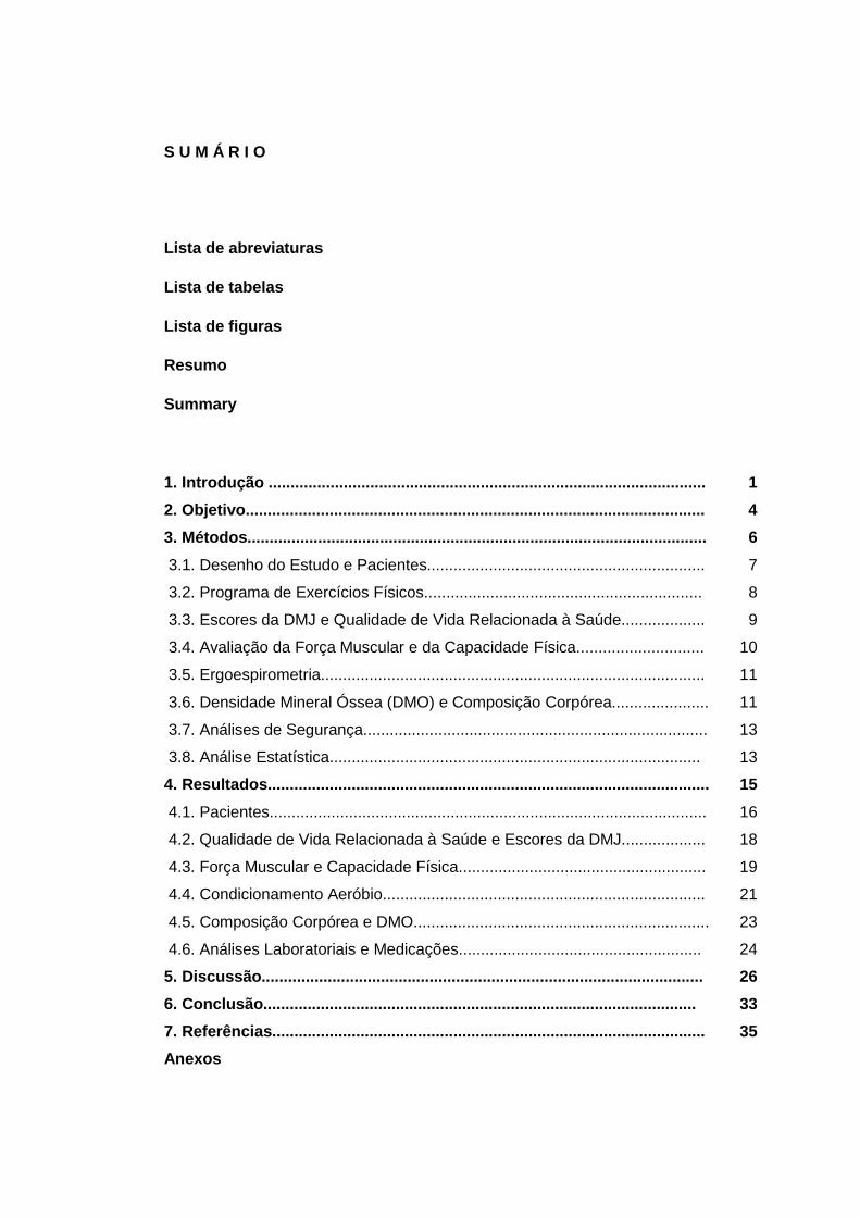
S U M Á R I O
Lista de abreviaturas
Lista de tabelas
Lista de figuras
Resumo
Summary
1. Introdução ................................................................................................... 1
2. Objetivo........................................................................................................ 4
3. Métodos........................................................................................................ 6
3.1. Desenho do Estudo e Pacientes............................................................... 7
3.2. Programa de Exercícios Físicos............................................................... 8
3.3. Escores da DMJ e Qualidade de Vida Relacionada à Saúde................... 9
3.4. Avaliação da Força Muscular e da Capacidade Física............................. 10
3.5. Ergoespirometria....................................................................................... 11
3.6. Densidade Mineral Óssea (DMO) e Composição Corpórea...................... 11
3.7. Análises de Segurança.............................................................................. 13
3.8. Análise Estatística.................................................................................... 13
4. Resultados.................................................................................................... 15
4.1. Pacientes................................................................................................... 16
4.2. Qualidade de Vida Relacionada à Saúde e Escores da DMJ................... 18
4.3. Força Muscular e Capacidade Física........................................................ 19
4.4. Condicionamento Aeróbio......................................................................... 21
4.5. Composição Corpórea e DMO................................................................... 23
4.6. Análises Laboratoriais e Medicações....................................................... 24
5. Discussão.................................................................................................... 26
6. Conclusão.................................................................................................. 33
7. Referências.................................................................................................. 35
Anexos
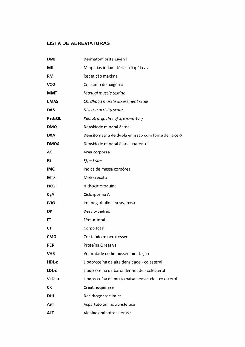
LISTA DE ABREVIATURAS
DMJ Dermatomiosite juvenil
MII Miopatias inflamatórias idiopáticas
RM Repetição máxima
VO2 Consumo de oxigênio
MMT Manual muscle testing
CMAS Childhood muscle assessment scale
DAS Disease activity score
PedsQL Pediatric quality of life inventory
DMO Densidade mineral óssea
DXA Densitometria de dupla emissão com fonte de raios-X
DMOA Densidade mineral óssea aparente
AC Área corpórea
ES Effect size
IMC Índice de massa corpórea
MTX Metotrexato
HCQ Hidroxicloroquina
CyA Ciclosporina A
IVIG Imunoglobulina intravenosa
DP Desvio-padrão
FT Fêmur total
CT Corpo total
CMO Conteúdo mineral ósseo
PCR Proteína C reativa
VHS Velocidade de hemossedimentação
HDL-c Lipoproteína de alta densidade - colesterol
LDL-c Lipoproteína de baixa densidade - colesterol
VLDL-c Lipoproteína de muito baixa densidade - colesterol
CK Creatinoquinase
DHL Desidrogenase lática
AST Aspartato aminotransferase
ALT Alanina aminotransferase
LISTA DE TABELAS
Tabela 1 - Características demográficas e medicações.................... 17
Tabela 2 - Efeitos de um programa de exercícios físicos
supervisionados nos parâmetros de saúde e qualidade
de vida relacionada à saúde em pacientes com
dermatomiosite juvenil......................................................
19
Tabela 3 - Efeitos de um programa de exercícios físicos
supervisionados na DMOA, CMO, índice de massa
magra, e índice de massa gorda em pacientes com
dermatomiosite juvenil......................................................
24
Tabela 4 - Efeitos de um programa de exercícios físicos
supervisionados nos parâmetros laboratoriais em
pacientes com dermatomiosite juvenil..............................
25
LISTA DE FIGURAS
Figura 1 - Força muscular e capacidade física nos momentos inicial
(PRE intervenção) e após 12 semanas de treinos (POS
intervenção). * indica p< 0,05 comparado com a
avaliação inicial...................................................................
20
Figura 2 - Parâmetros de condicionamento aeróbio nos momentos
inicial (PRE intervenção) e após 12 semanas de treinos
(POS intervenção). * indica p< 0,05 comparado com a
avaliação inicial...................................................................
22
R E S U M O
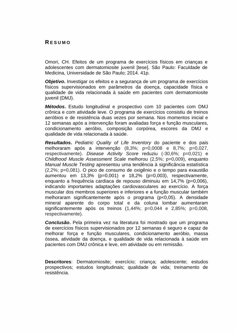
Omori, CH. Efeitos de um programa de exercícios físicos em crianças e adolescentes com dermatomiosite juvenil [tese]. São Paulo: Faculdade de Medicina, Universidade de São Paulo; 2014. 41p.
Objetivo. Investigar os efeitos e a segurança de um programa de exercícios físicos supervisionados em parâmetros da doença, capacidade física e qualidade de vida relacionada à saúde em pacientes com dermatomiosite juvenil (DMJ).
Métodos. Estudo longitudinal e prospectivo com 10 pacientes com DMJ crônica e com atividade leve. O programa de exercícios consistiu de treinos aeróbios e de resistência duas vezes por semana. Nos momentos inicial e 12 semanas após a intervenção foram avaliadas força e função musculares, condicionamento aeróbio, composição corpórea, escores da DMJ e qualidade de vida relacionada à saúde.
Resultados. Pediatric Quality of Life Inventory do paciente e dos pais melhoraram após a intervenção (8,3%; p=0,0008 e 8,7%; p=0,027, respectivamente). Disease Activity Score reduziu (-30,6%; p=0,021) e Childhood Muscle Assessment Scale melhorou (2,5%; p=0,009), enquanto Manual Muscle Testing apresentou uma tendência à significância estatística (2,2%; p=0,081). O pico de consumo de oxigênio e o tempo para exaustão aumentou em 13,3% (p=0,001) e 18,2% (p=0,003), respectivamente, enquanto a frequência cardíaca de repouso diminuiu em 14,7% (p=0,006), indicando importantes adaptações cardiovasculares ao exercício. A força muscular dos membros superiores e inferiores e a função muscular também melhoraram significantemente após o programa (p<0,05). A densidade mineral aparente do corpo total e da coluna lombar aumentaram significantemente após os treinos (1,44%; p=0,044 e 2,85%; p=0,008, respectivamente).
Conclusão. Pela primeira vez na literatura foi mostrado que um programa de exercícios físicos supervisionados por 12 semanas é seguro e capaz de melhorar força e função musculares, condicionamento aeróbio, massa óssea, atividade da doença, e qualidade de vida relacionada à saúde em pacientes com DMJ crônica e leve, em atividade ou em remissão.
Descritores: Dermatomiosite; exercício; criança; adolescente; estudos prospectivos; estudos longitudinais; qualidade de vida; treinamento de resistência.
S U M M A R Y
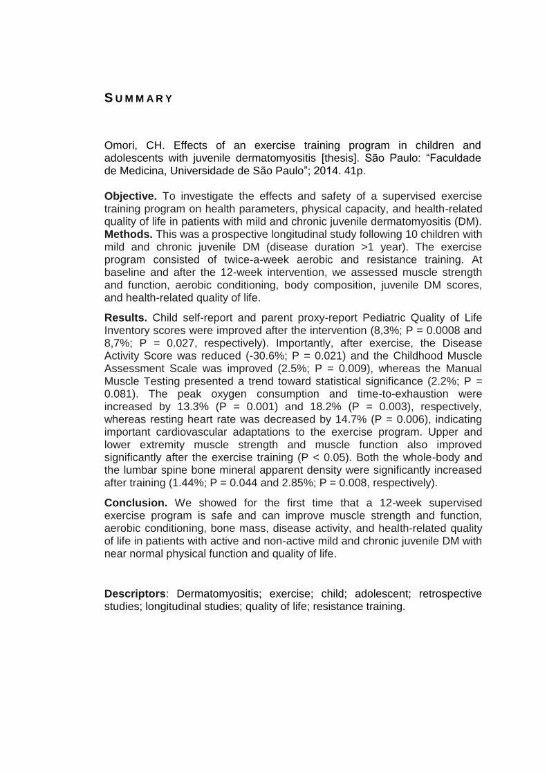
Omori, CH. Effects of an exercise training program in children and adolescents with juvenile dermatomyositis [thesis]. São Paulo: “Faculdade de Medicina, Universidade de São Paulo”; 2014. 41p. Objective. To investigate the effects and safety of a supervised exercise training program on health parameters, physical capacity, and health-related quality of life in patients with mild and chronic juvenile dermatomyositis (DM). Methods. This was a prospective longitudinal study following 10 children with mild and chronic juvenile DM (disease duration >1 year). The exercise program consisted of twice-a-week aerobic and resistance training. At baseline and after the 12-week intervention, we assessed muscle strength and function, aerobic conditioning, body composition, juvenile DM scores, and health-related quality of life.
Results. Child self-report and parent proxy-report Pediatric Quality of Life Inventory scores were improved after the intervention (8,3%; P = 0.0008 and 8,7%; P = 0.027, respectively). Importantly, after exercise, the Disease Activity Score was reduced (-30.6%; P = 0.021) and the Childhood Muscle Assessment Scale was improved (2.5%; P = 0.009), whereas the Manual Muscle Testing presented a trend toward statistical significance (2.2%; P = 0.081). The peak oxygen consumption and time-to-exhaustion were increased by 13.3% (P = 0.001) and 18.2% (P = 0.003), respectively, whereas resting heart rate was decreased by 14.7% (P = 0.006), indicating important cardiovascular adaptations to the exercise program. Upper and lower extremity muscle strength and muscle function also improved significantly after the exercise training (P < 0.05). Both the whole-body and the lumbar spine bone mineral apparent density were significantly increased after training (1.44%; P = 0.044 and 2.85%; P = 0.008, respectively).
Conclusion. We showed for the first time that a 12-week supervised exercise program is safe and can improve muscle strength and function, aerobic conditioning, bone mass, disease activity, and health-related quality of life in patients with active and non-active mild and chronic juvenile DM with near normal physical function and quality of life.
Descriptors: Dermatomyositis; exercise; child; adolescent; retrospective studies; longitudinal studies; quality of life; resistance training.

1. INTRODUÇÃO
Introdução
2
Dermatomiosite Juvenil (DMJ) é uma doença rara e pertence a um
grupo heterogêneo de doenças conhecidas como Miopatias Inflamatórias
Idiopáticas (MII), que compartilham sintomas comuns, como fraqueza
muscular proximal e inflamação não supurativa da musculatura esquelética
(1,2). O prognóstico da DMJ em termos de função física é geralmente
adequado. No entanto, muitos pacientes continuam a apresentar doença
crônica e evoluem com sequelas da doença (3). Neste sentido, tem sido
especulado que o treinamento físico possa promover benefícios funcionais
para esses pacientes, levando a melhora na sua qualidade de vida (3-5).
Anteriormente, o exercício físico não era permitido para pacientes
com MII pelo receio de que essa atividade pudesse elevar a inflamação
muscular ou piorar a atividade da doença. Atualmente, entretanto, estudos
mostraram que o treinamento físico pode melhorar ou pelo menos estabilizar
a força e função musculares sem alterar marcadores inflamatórios ou
atividade da doença (6-10). Portanto, o exercício físico tem sido reconhecido
como uma estratégia terapêutica promissora em pacientes com MII.
Entretanto, o papel do exercício na DMJ tem sido pouco explorado.
Além disto, foi mostrado que pacientes com DMJ apresentam baixas
massas magra e óssea, fraqueza importante, condicionamento aeróbio
deficiente, e importante intolerância ao exercício (11-13). Uma variedade de
mecanismos fisiopatológicos explica a capacidade física diminuída nos
pacientes com DMJ, como a concentração intramuscular elevada de
Introdução
3
citocinas pró-inflamatórias, processo inflamatório sistêmico, inflamação de
capilares musculares, hipoatividade, e efeito deletério do tratamento com
glicocorticóides na remodelação das massas muscular e óssea (14).
O exercício físico regular pode beneficiar pacientes com DMJ
reduzindo inflamação sistêmica e reatividade vascular, aumentando massas
muscular e óssea, e melhorando condicionamento aeróbio, função
neuromuscular e força muscular. É possível especular que essas
adaptações possam levar a uma melhora na qualidade de vida desses
pacientes.
Até o momento, apenas um estudo de caso avaliou os efeitos do
treinamento físico em uma criança com DMJ, sem atividade da doença,
acompanhada na Unidade de Reumatologia Pediátrica do Instituto da
Criança do Hospital das Clínicas da Faculdade de Medicina da Universidade
de São Paulo (ICr-HC-FMUSP). Como resultado de um programa
supervisionado com exercícios aeróbios e de resistência, a criança
apresentou melhora da força, da função muscular, e do condicionamento
aeróbio sem exacerbação da doença, comprovando o potencial do exercício
como um tratamento da DMJ (15). No entanto, nenhum estudo longitudinal
foi realizado com uma coorte de pacientes.
2. OBJETIVO
Objetivo
5
1. Avaliar longitudinalmente a segurança e os efeitos de um programa de
exercícios físicos supervisionados nos parâmetros da doença,
capacidade física, massa óssea e qualidade de vida relacionada à saúde
em pacientes com DMJ.

3. MÉTODOS
Métodos
7
3.1. Desenho do Estudo e Pacientes
Esse foi um estudo prospectivo, longitudinal, realizado entre janeiro
de 2009 e julho de 2011 em pacientes acompanhados na Unidade de
Reumatologia Pediátrica do ICr-HC-FMUSP. Todos os 30 pacientes com
DMJ (idade superior a sete anos e inferior a 18 anos), residentes na cidade
de São Paulo, e seguidos no presente serviço foram convidados a participar
do estudo. Os critérios de exclusão foram: envolvimento cardíaco (arritmia,
hipertensão arterial, insuficiência cardíaca, distúrbios de condução,
miocardite ou pericardite), desnutrição, doença renal crônica, doença
pulmonar crônica, e realização de atividade física regular.
Todos os pacientes tinham o diagnóstico definitivo de DMJ de acordo
com os critérios de Bohan e Peter (16). A amostra consistiu de crianças e
adolescentes de 7 a 18 anos incompletos, com DMJ ativa ou inativa, leve
(força muscular e qualidade de vida próximas ao normal) e crônica (duração
da doença maior de um ano) (17).
Os pacientes foram submetidos a um programa de exercícios físicos
supervisionados por 12 semanas, duas vezes por semana. Nos momentos
inicial e após 12 semanas de intervenção foram avaliados os escores da
DMJ, a qualidade de vida relacionada à saúde, função e força musculares,
condicionamento aeróbio, composição corpórea, e parâmetros clínicos e
Métodos
8
laboratoriais. O estudo teve aprovação do Comitê de Ética do HC-FMUSP
(aprovação número 0730/08) e todos os pais e pacientes assinaram o termo
de consentimento livre e esclarecido.
3.2. Programa de Exercícios Físicos
O programa consistiu de exercícios aeróbios e de resistência
supervisionados com duração de 12 semanas. As sessões de treinos eram
compostas de um aquecimento de 5 minutos, seguido por treinos de
resistência com duração de 20 minutos, treino aeróbio em esteira por 30
minutos e exercícios de alongamento por 5 minutos. As sessões eram
realizadas duas vezes por semana e foram monitoradas por um professor de
educação física e por um reumatologista pediátrico. O programa foi realizado
no Laboratório de Avaliação e Condicionamento em Reumatologia (LACRE)
da Disciplina de Reumatologia do HC-FMUSP. Os treinos de resistência
eram compostos por exercícios de supino, leg press, puxada costas, cadeira
extensora e remada sentada. Os pacientes eram incentivados a realizar três
sessões de 8-12 repetições máximas (RM) de cada exercício, exceto
durante a primeira semana quando eles realizaram duas sessões de 15-20
RM. A partir da segunda semana uma progressão na carga absoluta do
exercício era implementada quando o paciente era capaz de realizar mais de
12 repetições. A intensidade do treino aeróbio era determinada pela
Métodos
9
frequência cardíaca correspondente a 70% do consumo de oxigênio máximo
(VO2 pico). A adesão ao programa foi monitorada em cada sessão por um
membro do estudo.
3.3. Escores da DMJ e Qualidade de Vida Relacionada à
Saúde
Os escores da DMJ e os parâmetros de qualidade de vida
relacionados à saúde incluíram: Manual Muscle Testing (MMT) (18),
Childhood Muscle Assessment Scale (CMAS) (19), Disease Activity Score
(DAS) (20) e Pediatric Quality of Life Inventory (PedsQL) versão 4.0 (21). O
PedsQL apresenta três diferentes instrumentos de apresentação de acordo
com a faixa etária e foi aplicado conforme a idade de cada paciente. Todos
os instrumentos foram aplicados por um único reumatologista pediátrico, que
desconhecia as outras avaliações da pesquisa no momento das aplicações.
Métodos
10
3.4. Avaliação da Força Muscular e da Capacidade Física
Os pacientes realizaram três sessões de familiarização para testes de
força com intervalo de pelo menos 72 horas. Previamente ao teste de 1-RM,
foram realizadas duas sessões leves de aquecimento com intervalo de 2
minutos. Os pacientes tiveram até cinco tentativas para determinação da
carga de 1-RM, com intervalo de 3 minutos entre elas. Os testes de 1-RM
foram determinados para os exercícios de leg press e supino. A força
isométrica foi avaliada pelo dinamômetro de mão (Sanny).
A avaliação da capacidade física foi realizada através dos testes de
sentar-e-levantar, assim como levantar-e-caminhar. O teste de sentar-e-
levantar avalia o número máximo que o indivíduo consegue levantar-se de
uma cadeira sem apoio com altura padrão (45 cm), em 30 segundos. O teste
de levantar-e-caminhar avalia o tempo que o indivíduo necessita para
levantar-se de uma cadeira padrão sem apoio, andar sobre uma linha no
chão por três metros, virar-se, voltar para a cadeira e sentar-se novamente.
Métodos
11
3.5. Ergoespirometria
Todos os pacientes realizaram testes cardiopulmonares de
incremento em esteira antes e após o programa de exercícios. O pico de
consumo de oxigênio (VO2) obtido foi aceito quando dois de três critérios
foram atingidos: presença de platô de VO2, razão de troca respiratória de
1:1, e/ou exaustão referida pelo paciente. Frequência cardíaca de repouso e
tempo para exaustão também foram avaliados como medidas de adaptação
cardiovascular ao treino e tolerância ao exercício, respectivamente.
3.6. Densidade Mineral Óssea (DMO) e Composição
Corpórea
DMO (g/cm2) e escores Z foram determinados por densitometria de
dupla emissão com fonte de raios-X (DXA) através de um modelo de
densitômetro Hologic QDR 4500 Discovery. Foi calculada a superfície óssea
(cm2) através do software fornecido com o densitômetro. A massa óssea foi
analisada na coluna lombar (L1-L4), no fêmur total e no corpo total. A análise
do corpo total foi realizada sem considerar a cabeça, pois este é um fator
que leva a aumento da variabilidade em crianças (22). A fim de minimizar o
fator de confusão do tamanho do esqueleto nas medidas da DXA, foi
Métodos
12
calculado o volume tridimensional aproximado da densidade óssea, a
densidade mineral óssea aparente (DMOA; g/cm3). A DMOA foi obtida
dividindo-se a DMO de uma determinada região (coluna, fêmur total, ou
corpo total) pela raiz quadrada da área corpórea correspondente (AC;
DMOA=DMO/AC) (23,24). Todas as medidas foram realizadas pelo mesmo
técnico que desconhecia as outras etapas da pesquisa. Os erros de precisão
para as medidas da DMO foram determinados de acordo com os protocolos
padronizados pela Sociedade Internacional para Densitometria Clínica (25).
Alterações menos significativas com 95% de confiança foram de 0,033g/cm2
na coluna lombar, 0,039g/cm2 no fêmur total e 0,020g/cm2 no corpo total.
A composição corpórea [massa magra, massa gorda, e percentual
(%) de gordura] foi avaliada pelo mesmo densitômetro com a utilização de
um programa de software pediátrico. Os índices de massa magra e massa
gorda foram calculados dividindo-se a massa magra (Kg) ou a massa gorda
pela altura, em metros quadrados (m2) (26).
Métodos
13
3.7. Análises de Segurança
As análises de segurança incluíram a avaliação de parâmetros
laboratoriais de inflamação, funções renal e hepática, hemograma, enzimas
musculares, perfil lipídico e perfil glicêmico nos momentos inicial e 48 horas
após o último treino. Os possíveis episódios de reativação da doença foram
monitorados durante o estudo de acordo com os critérios já descritos
(16,17). Os pacientes foram examinados semanalmente por um
reumatologista pediátrico a fim de avaliar sintomas como exaustão
excessiva, dor, lesão osteoarticular, dor muscular, ou algum outro evento
adverso. Os pacientes foram orientados a reportar qualquer tipo de evento
adverso.
3.8. Análise Estatística
O teste t pareado foi utilizado para avaliar possíveis diferenças entre
os momentos pré e pós-intervenção. Effect size (ES) foi estimado para todas
as variáveis dependentes para determinação da significância prática dos
achados. O ES foi obtido através da divisão dos dados pós-intervenção
menos os dados pré-intervenção pelo desvio-padrão médio, de acordo com
descrição prévia (27). O nível de significância foi previamente estipulado em
Métodos
14
valores de P menores ou iguais a 0,05. Os dados foram expressos em média
DP.
4. RESULTADOS
Resultados
16
4.1. Pacientes
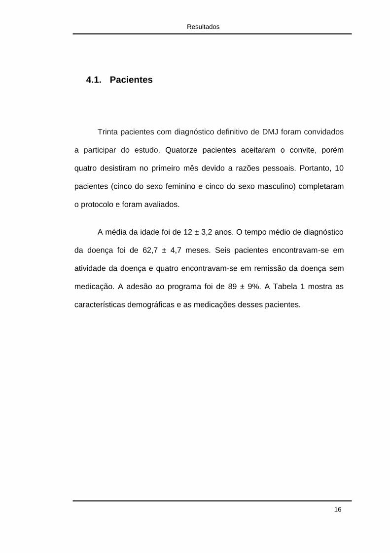
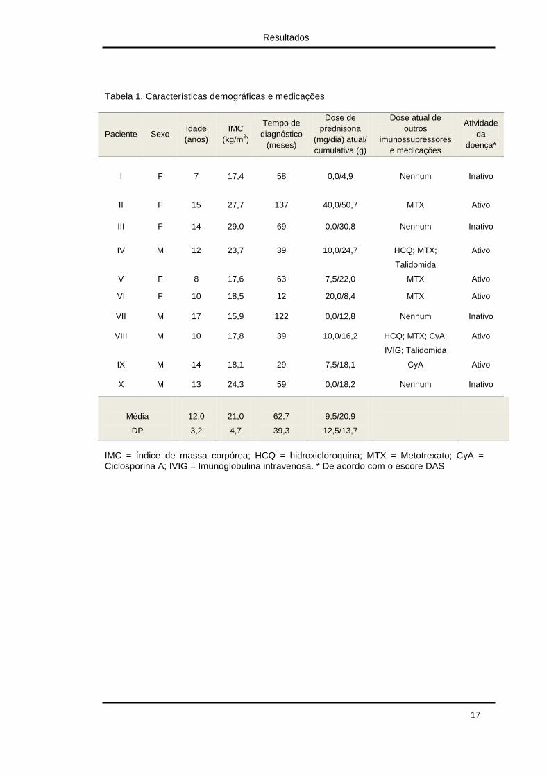
Trinta pacientes com diagnóstico definitivo de DMJ foram convidados
a participar do estudo. Quatorze pacientes aceitaram o convite, porém
quatro desistiram no primeiro mês devido a razões pessoais. Portanto, 10
pacientes (cinco do sexo feminino e cinco do sexo masculino) completaram
o protocolo e foram avaliados.
A média da idade foi de 12 ± 3,2 anos. O tempo médio de diagnóstico
da doença foi de 62,7 ± 4,7 meses. Seis pacientes encontravam-se em
atividade da doença e quatro encontravam-se em remissão da doença sem
medicação. A adesão ao programa foi de 89 ± 9%. A Tabela 1 mostra as
características demográficas e as medicações desses pacientes.
Resultados
17
Tabela 1. Características demográficas e medicações
Paciente Sexo Idade
(anos)
IMC
(kg/m2)
Tempo de
diagnóstico
(meses)
Dose de
prednisona
(mg/dia) atual/
cumulativa (g)
Dose atual de
outros
imunossupressores
e medicações
Atividade
da
doença*
I
F
7
17,4
58
0,0/4,9
Nenhum
Inativo
II F 15 27,7 137 40,0/50,7 MTX Ativo
III F 14 29,0 69 0,0/30,8 Nenhum Inativo
IV M 12 23,7 39 10,0/24,7 HCQ; MTX;
Talidomida
Ativo
V F 8 17,6 63 7,5/22,0 MTX Ativo
VI F 10 18,5 12 20,0/8,4 MTX Ativo
VII M 17 15,9 122 0,0/12,8 Nenhum Inativo
VIII M 10 17,8 39 10,0/16,2 HCQ; MTX; CyA;
IVIG; Talidomida
Ativo
IX M 14 18,1 29 7,5/18,1 CyA Ativo
X M 13 24,3 59 0,0/18,2 Nenhum Inativo
Média
DP
12,0
3,2
21,0
4,7
62,7
39,3
9,5/20,9
12,5/13,7
IMC = índice de massa corpórea; HCQ = hidroxicloroquina; MTX = Metotrexato; CyA = Ciclosporina A; IVIG = Imunoglobulina intravenosa. * De acordo com o escore DAS
Resultados
18
4.2. Qualidade de Vida Relacionada à Saúde e Escores da
DMJ
PedsQL da criança e dos pais melhoraram significantemente após a
intervenção (8,3%; ES 0,67, p=0,0008 e 8,7%; ES 0,68, p=0,027,
respectivamente). DAS reduziu significantemente após a intervenção (-
31,6%; ES -0,29, p=0,021). CMAS apresentou aumento discreto, mas
estatisticamente significante após o programa (2,5%; ES 0,28, p=0,009),
enquanto o MMT apresentou uma tendência à significância estatística (2,2%;
ES 0,42, p=0,080) (Tabela 2).
Resultados
19
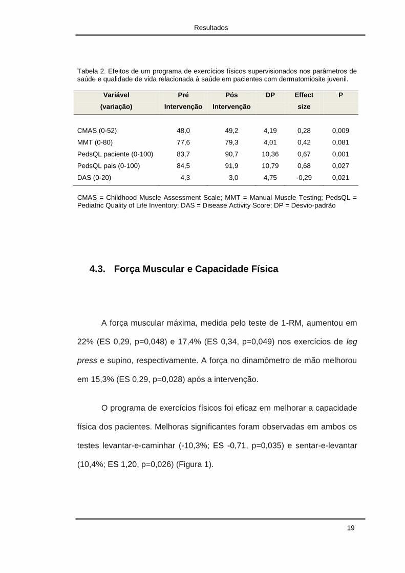
Tabela 2. Efeitos de um programa de exercícios físicos supervisionados nos parâmetros de saúde e qualidade de vida relacionada à saúde em pacientes com dermatomiosite juvenil.
Variável
(variação)
Pré
Intervenção
Pós
Intervenção
DP Effect
size
P
CMAS (0-52)
48,0
49,2
4,19
0,28
0,009
MMT (0-80) 77,6 79,3 4,01 0,42 0,081
PedsQL paciente (0-100) 83,7 90,7 10,36 0,67 0,001
PedsQL pais (0-100) 84,5 91,9 10,79 0,68 0,027
DAS (0-20) 4,3 3,0 4,75 -0,29 0,021
CMAS = Childhood Muscle Assessment Scale; MMT = Manual Muscle Testing; PedsQL = Pediatric Quality of Life Inventory; DAS = Disease Activity Score; DP = Desvio-padrão
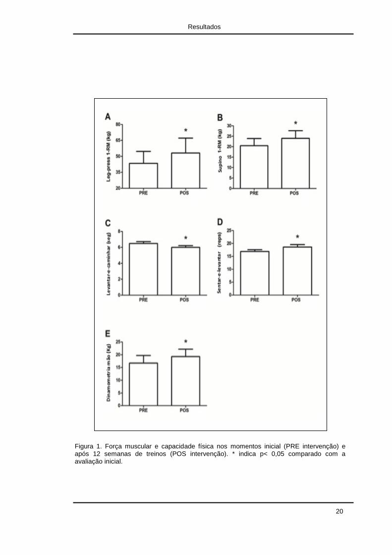
4.3. Força Muscular e Capacidade Física
A força muscular máxima, medida pelo teste de 1-RM, aumentou em
22% (ES 0,29, p=0,048) e 17,4% (ES 0,34, p=0,049) nos exercícios de leg
press e supino, respectivamente. A força no dinamômetro de mão melhorou
em 15,3% (ES 0,29, p=0,028) após a intervenção.
O programa de exercícios físicos foi eficaz em melhorar a capacidade
física dos pacientes. Melhoras significantes foram observadas em ambos os
testes levantar-e-caminhar (-10,3%; ES -0,71, p=0,035) e sentar-e-levantar
(10,4%; ES 1,20, p=0,026) (Figura 1).
Resultados
20
Figura 1. Força muscular e capacidade física nos momentos inicial (PRE intervenção) e após 12 semanas de treinos (POS intervenção). * indica p< 0,05 comparado com a avaliação inicial.
Resultados
21
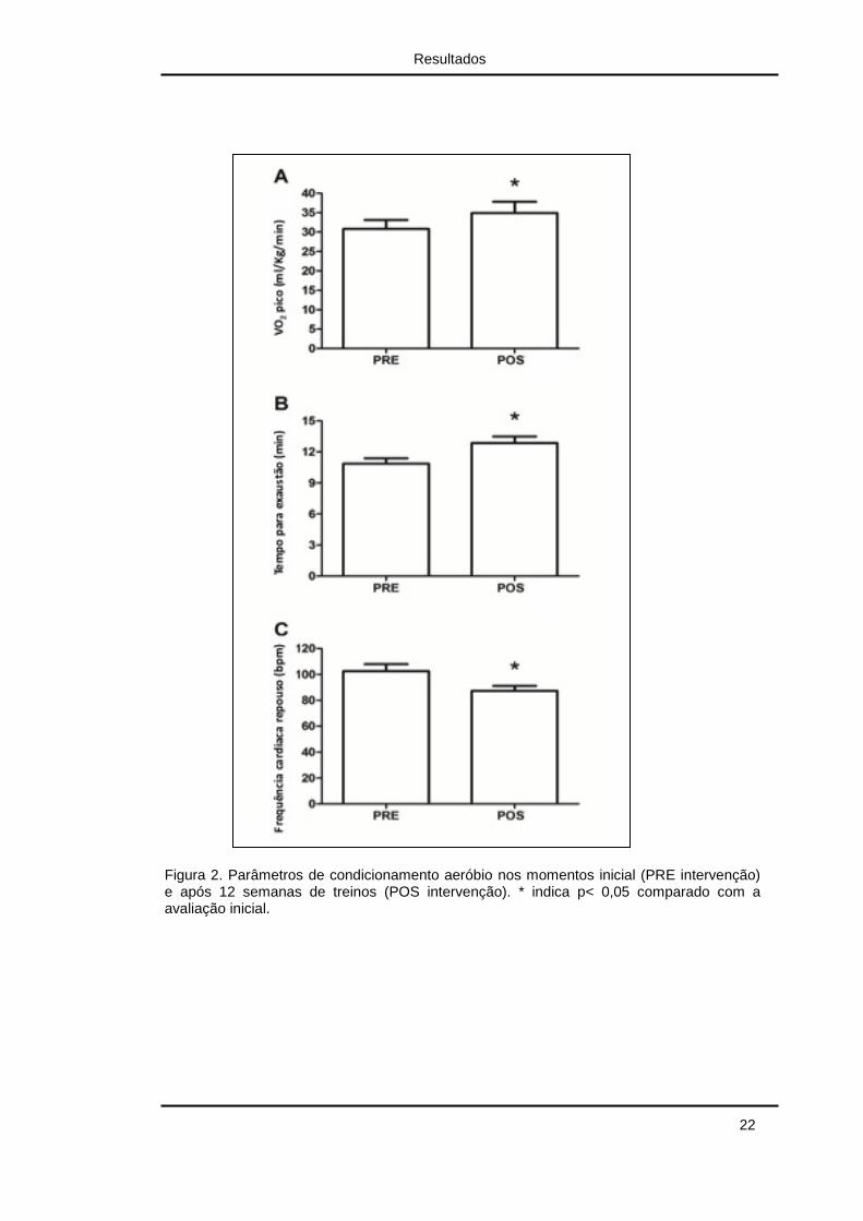
4.4. Condicionamento Aeróbio
O condicionamento aeróbio, avaliado pela ergoespirometria, melhorou
significantemente após o programa. O pico de VO2 e o tempo para exaustão
aumentaram em 13,3% (ES 0,49, p=0,001) e 18,2% (ES 0,94, p=0,003),
respectivamente. A frequência cardíaca de repouso diminuiu em 14,7% (ES -
0,91, p=0,008), indicando adaptações cardiovasculares importantes ao
exercício (Figura 2).
Resultados
22
Figura 2. Parâmetros de condicionamento aeróbio nos momentos inicial (PRE intervenção) e após 12 semanas de treinos (POS intervenção). * indica p< 0,05 comparado com a avaliação inicial.
Resultados
23
4.5. Composição Corpórea e DMO
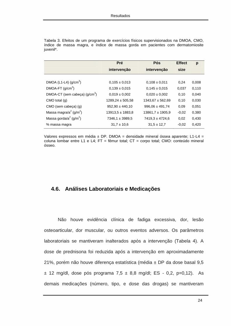
DMOA na coluna lombar e corpo total melhoraram significantemente
após o programa (2,85%; ES 0,24, p=0,008 e 1,44%; ES 0,11, p=0,044,
respectivamente). O conteúdo mineral ósseo total melhorou
significantemente quando a cabeça foi considerada na análise (4,2%; ES
0,10, p=0,03), enquanto uma tendência foi observada quando a cabeça foi
retirada da analise (4,5%; ES 0,09, p=0,051) (Tabela 3). Não foram
observadas diferenças significativas na DMOA do fêmur total (4,1%; ES
0,37, p=0,115), no índice de massa muscular esquelética relativa (-0,37%;
ES -0,02, p=0,39), no índice de massa gorda relativa (1,0%; ES 0,01,
p=0,43), na porcentagem de massa gorda (-0,69%; ES -0,02, p=0,42), e nos
escores Z da DMO na coluna lombar (-3,3%; ES 0,02, p=0,30), fêmur total
(2,9%; ES 0,01, p=0,44) e corpo total (11%; ES 0,17, p=0,09).
Resultados
24
Tabela 3. Efeitos de um programa de exercícios físicos supervisionados na DMOA, CMO, índice de massa magra, e índice de massa gorda em pacientes com dermatomiosite juvenil*.
Pré
intervenção
Pós
intervenção
Effect
size
p
DMOA (L1-L4) (g/cm3)
0,105 ± 0,013
0,108 ± 0,011
0,24
0,008
DMOA-FT (g/cm3) 0,139 ± 0,015 0,145 ± 0,015 0,037 0,110
DMOA-CT (sem cabeça) (g/cm3) 0,019 ± 0,002 0,020 ± 0,002 0,10 0,040
CMO total (g) 1289,24 ± 505,58 1343,67 ± 562,69 0,10 0,030
CMO (sem cabeça) (g) 952,90 ± 440,10 996,06 ± 491,74 0,09 0,051
Massa magra/a2 (g/m
2) 13913,5 ± 1883,8 13861,7 ± 1905,9 -0,02 0,380
Massa gorda/a2 (g/m
2) 7346,1 ± 3989,5 7419,3 ± 4724,6 0,02 0,430
% massa magra 31,7 ± 10,6 31,5 ± 12,7 -0,02 0,420
Valores expressos em média ± DP. DMOA = densidade mineral óssea aparente; L1-L4 = coluna lombar entre L1 e L4; FT = fêmur total; CT = corpo total; CMO: conteúdo mineral ósseo.
4.6. Análises Laboratoriais e Medicações
Não houve evidência clínica de fadiga excessiva, dor, lesão
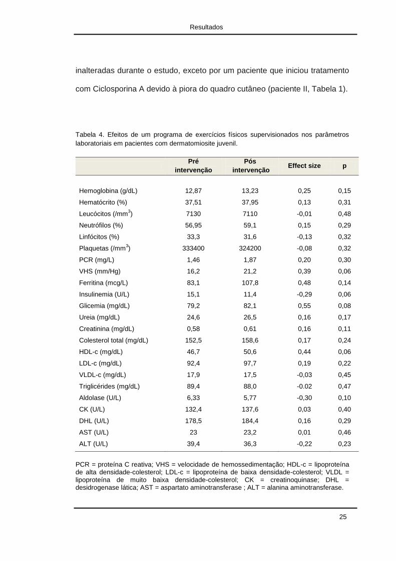
osteoarticular, dor muscular, ou outros eventos adversos. Os parâmetros
laboratoriais se mantiveram inalterados após a intervenção (Tabela 4). A
dose de prednisona foi reduzida após a intervenção em aproximadamente
21%, porém não houve diferença estatística (média ± DP da dose basal 9,5
± 12 mg/dl, dose pós programa 7,5 ± 8,8 mg/dl; ES - 0,2, p=0,12). As
demais medicações (número, tipo, e dose das drogas) se mantiveram
Resultados
25
inalteradas durante o estudo, exceto por um paciente que iniciou tratamento
com Ciclosporina A devido à piora do quadro cutâneo (paciente II, Tabela 1).
Tabela 4. Efeitos de um programa de exercícios físicos supervisionados nos parâmetros
laboratoriais em pacientes com dermatomiosite juvenil.
Pré
intervenção
Pós
intervenção Effect size p
Hemoglobina (g/dL)
12,87
13,23
0,25
0,15
Hematócrito (%) 37,51 37,95 0,13 0,31
Leucócitos (/mm3) 7130 7110 -0,01 0,48
Neutrófilos (%) 56,95 59,1 0,15 0,29
Linfócitos (%) 33,3 31,6 -0,13 0,32
Plaquetas (/mm3) 333400 324200 -0,08 0,32
PCR (mg/L) 1,46 1,87 0,20 0,30
VHS (mm/Hg) 16,2 21,2 0,39 0,06
Ferritina (mcg/L) 83,1 107,8 0,48 0,14
Insulinemia (U/L) 15,1 11,4 -0,29 0,06
Glicemia (mg/dL) 79,2 82,1 0,55 0,08
Ureia (mg/dL) 24,6 26,5 0,16 0,17
Creatinina (mg/dL) 0,58 0,61 0,16 0,11
Colesterol total (mg/dL) 152,5 158,6 0,17 0,24
HDL-c (mg/dL) 46,7 50,6 0,44 0,06
LDL-c (mg/dL) 92,4 97,7 0,19 0,22
VLDL-c (mg/dL) 17,9 17,5 -0,03 0,45
Triglicérides (mg/dL) 89,4 88,0 -0.02 0,47
Aldolase (U/L) 6,33 5,77 -0,30 0,10
CK (U/L) 132,4 137,6 0,03 0,40
DHL (U/L) 178,5 184,4 0,16 0,29
AST (U/L) 23 23,2 0,01 0,46
ALT (U/L) 39,4 36,3 -0,22 0,23
PCR = proteína C reativa; VHS = velocidade de hemossedimentação; HDL-c = lipoproteína de alta densidade-colesterol; LDL-c = lipoproteína de baixa densidade-colesterol; VLDL = lipoproteína de muito baixa densidade-colesterol; CK = creatinoquinase; DHL = desidrogenase lática; AST = aspartato aminotransferase ; ALT = alanina aminotransferase.

5. DISCUSSÃO
Discussão
27
Este foi o primeiro estudo que avaliou os efeitos de um programa de
exercícios físicos em uma coorte de pacientes com DMJ. Foi demonstrado
que o treinamento físico melhorou vários sintomas da DMJ, como fraqueza
muscular, falta de condicionamento aeróbio, atrofia muscular e perda óssea.
Uma melhora significante na qualidade de vida relacionada à saúde também
foi observada sem exacerbação ou reativação da doença. Esses dados
inéditos fornecem evidências de que o exercício pode ser seguro e eficaz em
pacientes com DMJ leve e crônica.
O exercício físico é classicamente recomendado como tratamento de
várias doenças, como o diabetes mellitus tipo 2 e a hipertensão arterial (28).
Mais recentemente, houve um aumento no reconhecimento da proposta
terapêutica do exercício nas doenças reumatológicas, principalmente em
adultos. A atividade física tem sido prescrita como tratamento da
osteoporose (29), osteoartrite (30), esclerodermia (31), fibromialgia (32), e
artrite reumatóide (33). Além disso, o exercício físico foi estudado como
sendo promissor no tratamento das MIIs (34). No entanto, evidências para a
recomendação de atividade física para o tratamento da DMJ são limitadas a
poucos estudos transversais, mostrando uma capacidade física prejudicada
nesta doença (11-13). De fato, estudos longitudinais são necessários para
confirmar a relevância do exercício nos pacientes com DMJ.
Neste sentido, foi demonstrado que pacientes com DMJ apresentam
menor tolerância ao exercício e menor VO2 pico quando comparados a
Discussão
28
indivíduos saudáveis (11-13). Takken e colaboradores (12) demonstraram
que o VO2 pico (dados expressos em valores absolutos e relativos ao peso
corpóreo) foi o parâmetro mais prejudicado na DMJ quando a doença está
ativa. Foi observado em nosso estudo que houve uma melhora importante
no tempo para exaustão na ergoespirometria, que é um parâmetro
relacionado à tolerância a fadiga e capacidade física. Além disso, nós
também observamos um incremento no VO2 pico e uma diminuição na
frequência cardíaca de repouso, ambos os parâmetros clássicos de
adaptação cardiovascular ao treinamento aeróbio. Esses achados sugerem
um importante efeito terapêutico da atividade física, atenuando a falta de
condicionamento aeróbio observado em pacientes com DMJ.
O programa de exercícios melhorou significantemente a força
muscular máxima dos membros superiores e inferiores (1-RM e
dinamometria de mão) e a função muscular (testes levantar-e-caminhar,
assim como sentar-e-levantar). A melhora nas medidas objetivas de força e
função musculares foram paralelas as dos escores da DMJ. Em crianças
pré-púberes saudáveis, os exercícios de resistência agem principalmente
melhorando os parâmetros neurais (coordenação intra e intermuscular) em
comparação com os morfológicos (área transversal do músculo e tipos das
fibras) (35). Certamente os efeitos positivos da atividade física no sistema
neuromuscular devem ter relevância clinica no tratamento da DMJ, uma vez
que a atrofia e a fraqueza musculares são as principais preocupações
nesses pacientes.
Discussão
29
Outro achado interessante neste estudo foi a melhora na DMOA após
o programa. A DMOA é uma medida de densidade volumétrica, com o
objetivo de minimizar o efeito do crescimento na análise longitudinal em
crianças (23-24). Está bem estabelecido que crianças apresentam uma alta
taxa de remodelação óssea e que a atividade física regular leva a
modificações mais importantes na massa óssea em estágios pré-púberes
comparado a idades mais avançadas, o que parcialmente explica a elevada
resposta da massa óssea após o programa neste estudo (36,37). Apesar de
esse achado ter sido significante estatisticamente, em números absolutos o
ganho de massa óssea foi pequeno. Isso é corroborado pelo resultado do
ES, que mostrou um tamanho do efeito pequeno, revelando uma baixa
significância prática. Porém, sabe-se que em indivíduos saudáveis há
evidências de que o exercício físico é eficaz em diminuir progressivamente a
perda de massa óssea, sendo este outro motivo para que a atividade física
possa ser prescrita para pacientes com DMJ.
Há uma preocupação empírica de que o exercício poderia causar
reativação da doença em pacientes com MII. No entanto, um número
crescente de estudos refutou essa possibilidade em adultos com
dermatomiosite, polimiosite, e miosite por corpúsculos de inclusão. Em
crianças com DMJ ativa ou em remissão, Maillard e colaboradores (38)
demonstraram que uma única sessão de exercícios não alterava o grau de
inflamação muscular, fornecendo evidências preliminares de que um
programa de exercícios poderia ser seguro para pacientes com DMJ.
Discussão
30
Recentemente, nosso grupo relatou uma ausência de eventos adversos em
uma criança com DMJ em remissão que realizou o programa de atividades
físicas por 16 semanas. O presente estudo reafirma que o exercício é seguro
na DMJ, pois não houve eventos adversos após o programa de atividades
nos pacientes com DMJ crônica ativa ou em remissão. Além disso, é
importante observar que o instrumento de atividade da doença (DAS)
reduziu depois da intervenção, sugerindo que um programa de exercícios
regulares supervisionados pode de fato diminuir ao invés de aumentar a
atividade da doença.
O prognóstico dos pacientes com DMJ tem substancialmente
melhorado nas ultimas décadas. Como consequência, o foco clínico e
científico tem mudado de mortalidade para morbidade e qualidade de vida.
Neste contexto, os efeitos do treinamento físico em melhorar a qualidade de
vida relacionada à saúde neste estudo é de grande relevância. A
restauração da capacidade física do paciente pode ser o primeiro passo para
um estilo de vida mais ativo.
Obviamente, pais, reumatologistas pediátricos e outros profissionais
da saúde devem continuar a incentivar a inserção progressiva da atividade
física na vida dos pacientes. Consequentemente, poderia se esperar
sintomas menos graves, melhor qualidade de vida e inclusive uma melhor
capacidade física (14). Essa recomendação tem sido há bastante tempo
almejada em uma grande variedade de doenças, onde o exercício tem sido
promovido como um “medicamento” (39).
Discussão
31
O presente estudo estende essa recomendação para a DMJ, para a
qual o exercício pode ser prescrito como uma terapia adjuvante e lúdica,
com a necessidade de uma padronização dos exercícios para todos os
pacientes, levando-se em conta a sua individualidade e habilidades.
É importante enfatizar que esse estudo possui limitações. Primeiro,
houve uma ausência de grupo controle, sendo que influências da maturação
e do curso natural da doença nos resultados não puderam ser avaliados. No
entanto, como se tratou de um estudo de curta duração, alguns desses
aspectos podem ser minimizados. Além disso, a DMJ é uma doença
heterogênea que pode se beneficiar de comparações intra e não entre
pacientes. Isso pode ser observado levando-se em conta as amostras
pequenas em estudos com DMJ devido à baixa prevalência dessa doença.
Nossa amostra foi composta de pacientes com doença em atividade e em
remissão com o objetivo de maximizar a validade externa deste estudo. No
entanto, o pequeno número de pacientes nestes subgrupos (ativo e inativo)
nos impediu comparar as respostas ao exercício em função da atividade da
doença.
Entretanto, como este estudo envolveu apenas pacientes com DMJ
crônica e leve, esses achados não podem ser extrapolados aos pacientes
com fraqueza muscular importante e para aqueles com diagnóstico recente.
Estudos controlados, randomizados e com amostras maiores e mais
diversificadas (ativas, em remissão, refratárias) devem ser priorizados para
identificar os efeitos promissores do exercício na DMJ.
Discussão
32
Além disto, o objetivo deste estudo não foi avaliar os mecanismos
intrínsecos pelos quais o exercício possa melhorar a capacidade física na
DMJ. Supostamente, o exercício poderia atenuar uma variedade de
sintomas da DMJ reduzindo a inflamação local e sistêmica, melhorando a
capacidade oxidativa muscular, reduzindo a reabsorção óssea e/ou
aumentando a formação óssea, e promovendo o anabolismo protéico. Esses
mecanismos devem ser testados em estudos futuros para melhorar a
prescrição do exercício para pacientes com DMJ. Assim sendo, os
resultados do presente estudo ampliam o conhecimento na literatura do
papel terapêutico do exercício nas MII para a DMJ.

6. CONCLUSÃO
Conclusão
34
1. Um programa de exercícios físicos supervisionados de 12 semanas foi
seguro e melhorou força e função musculares, atividade da doença,
massa muscular, condicionamento aeróbio, massa óssea e qualidade de
vida relacionada à saúde em pacientes com DMJ crônica e de leve
intensidade.
7. REFERÊNCIAS
Referências
36
1. Pignone A, Fiori G, Del Rosso A, Generini S, Matucci-Cerinic M. The
pathogenesis of inflammatory muscle diseases: on the cutting edge among
the environment, the genetic background, the immune response and the
dysregulation of apoptosis. Autoimmun Rev 2002;1:226 –32.
2. Rider LG. The heterogeneity of juvenile myositis. Autoimmun Rev
2007;6:241–7.
3. Feldman BM, Rider LG, Reed AM, Pachman LM. Juvenile
dermatomyositis and other idiopathic inflammatory myopa- thies of childhood.
Lancet 2008;371:2201–12.
4. De Salles Painelli V, Gualano B, Artioli GG, de Sa Pinto AL, Bonfa E,
Lancha AH, et al. The possible role of physical exercise on the treatment of
idiopathic inflammatory myopathies. Autoimmun Rev 2009;8:355–9.
5. Harris-Love MO. Physical activity and disablement in the idiopathic
inflammatory myopathies. Curr Opin Rheumatol 2003;15:679 –90.
6. Alexanderson H, Dastmalchi M, Esbjornsson-Liljedahl M, Opava CH,
Lundberg IE. Benefits of intensive resistance training in patients with chronic
polymyositis or dermatomyositis. Arthritis Rheum 2007;57:768–77.
7. Arnardottir S, Alexanderson H, Lundberg IE, Borg K. Sporadic inclusion
body myositis: pilot study on the effects of a home exercise program on
muscle function, histopathology and inflammatory reaction. J Rehabil Med
Referências
37
2003;35:31–5.
8. Gualano B, Neves M Jr, Lima FR, de Sa Pinto AL, Laurentino G, Borges
C, et al. Resistance training with vascular occlusion in inclusion body
myositis: a case study. Med Sci Sports Exerc 2010;42:250–4.
9. Nader GA, Dastmalchi M, Alexanderson H, Grundtman C, Gernapudi R,
Esbjornsson M, et al. A longitudinal, integrated, clinical, histological and
mRNA profiling study of resistance exercise in myositis. Mol Med
2010;16:455–64.
10. Spector SA, Lemmer JT, Koffman BM, Fleisher TA, Feuerstein IM, Hurley
BF, et al. Safety and efficacy of strength training in patients with sporadic
inclusion body myositis. Muscle Nerve 1997;20:1242– 8.
11. Takken T, Spermon N, Helders PJ, Prakken AB, van der Net J. Aerobic
exercise capacity in patients with juvenile dermatomyositis. J Rheumatol
2003;30:1075–80.
12. Takken T, van der Net J, Engelbert RH, Pater S, Helders PJ.
Responsiveness of exercise parameters in children with inflammatory
myositis. Arthritis Rheum 2008;59:59–64.
13. Takken T, van der Net J, Helders PJ. Anaerobic exercise capacity in
patients with juvenile-onset idiopathic inflammatory myopathies. Arthritis
Rheum 2005;53:173–7.
Referências
38
14. Gualano B, Sa Pinto AL, Perondi B, Leite Prado DM, Omori C, Almeida
RT, et al. Evidence for prescribing exercise as treatment in pediatric
rheumatic diseases. Autoimmun Rev 2010; 9:569 –73.
15. Omori CH, Prado DML, Gualano B, Sallum AE, Pinto ALS, Roschel H, et
al. Responsiveness to exercise training in juvenile dermatomyositis: a twin-
case study. BMC Musculoeskelet Disord 2010;25(1):270.
16. Bohan A, Peter JB. Polymyositis and dermatomyositis (second of two
parts). N Engl J Med 1975;292:403–7.
17. Ruperto N, Pistorio A, Ravelli A, Rider LG, Pilkington C, Oliveira S, et al,
for the Paediatric Rheumatology International Trials Organisation (PRINTO)
and the Pediatric Rheu- matology Collaborative Study Group (PRCSG). The
Paediatric Rheumatology International Trials Organisation provisional criteria
for the evaluation of response to therapy in juvenile dermatomyositis. Arthritis
Care Res (Hoboken) 2010;62:1533– 41.
18. Kendall F, McCreary EK, Provance PG. Muscles: testing and function.
Baltimore: Williams & Willkins; 1993.
19. Lovell DJ, Lindsley CB, Rennebohm RM, Ballinger SH, Bow- yer SL,
Giannini EH, et al, and the Juvenile Dermatomyositis Disease Activity
Collaborative Study Group. Development of validated disease activity and
damage indices for the juvenile idiopathic inflammatory myopathies. II. The
Childhood Myositis Assessment Scale (CMAS): a quantitative tool for the
Referências
39
evaluation of muscle function. Arthritis Rheum 1999:42: 2213–9.
20. Bode RK, Klein-Gitelman MS, Miller ML, Lechman TS, Pachman LM.
Disease activity score for children with juvenile dermatomyositis: reliability
and validity evidence. Arthritis Rheum 2003, 49(1):7-15.
21. Klatchoian DA, Len CA, Terreri MT, Silva M, Itamoto C, Ciconelli RM, et
al. Quality of life of children and adolescents from Sao Paulo: reliability and
validity of the Brazilian ver- sion of the Pediatric Quality of Life Inventory
version 4.0 Generic Core Scales. J Pediatr (Rio J) 2008;84:308–15.
22. Taylor A, Konrad PT, Norman ME, Harcke HT. Total body bone mineral
density in young children: influence of head bone mineral density. J Bone
Miner Res 1997;12:652–5.
23. Carter DR, Bouxsein ML, Marcus R. New approaches for interpreting
projected bone densitometry data. J Bone Miner Res 1992;7:137– 45.
24. Crabtee N, Leonard M, Zemel B. Dual-energy x-ray absorp- tiometry. In:
Sawyer A, Bachrach L, Fung E, editors. Bone densitometry in growing
patients: guidelines for clinical practice. Totowa (NJ): Humana Press; 2007.
p. 41–57.
25. Baim S, Wilson CR, Lewiecki EM, Luckey MM, Downs RW Jr, Lentle BC.
Precision assessment and radiation safety for dual-energy x-ray
absorptiometry: position paper of the Inter- national Society for Clinical
Densitometry. J Clin Densitom 2005;8:371– 8.
Referências
40
26. Goulding A, Taylor RW, Grant AM, Jones S, Taylor BJ, Williams SM.
Relationships of appendicular LMI and total body LMI to bone mass and
physical activity levels in a birth cohort of New Zealand five-year olds. Bone
2009;45:455–9.
27. Cohen J. Statistical power analysis for the behavioral sciences. 2nd ed.
Hillsdale (CA): Lawrence Erlbaum Associates; 1988.
28. Pedersen BK, Saltin B. Evidence for prescribing exercise as therapy in
chronic disease. Scand J Med Sci Sports 2006; Suppl 1:3–63.
29. Babatunde OO, Forsyth JJ, Gidlow CJ. A meta-analysis of brief high-
impact exercises for enhancing bone health in premenopausal women.
Osteoporos Int 2012;23:109–19.
30. Altman RD. Early management of osteoarthritis. Am J Manag Care
2010;16 Suppl:S41–7.
31. Pinto AL, Oliveira NC, Gualano B, Christmann RB, PainelliVS, Artioli GG,
et al. Efficacy and safety of concurrent training in systemic sclerosis. J
Strength Cond Res 2011;25:1423–8.
32. Sanudo B, Galiano D, Carrasco L, de Hoyo M, McVeigh JG. Effects of a
prolonged exercise program on key health outcomes in women with
fibromyalgia: a randomized controlled trial. J Rehabil Med 2011;43:521–6.
Referências
41
33. Kelley GA, Kelley KS, Hootman JM, Jones DL. Effects of community-
deliverable exercise on pain and physical function in adults with arthritis and
other rheumatic diseases: a meta-analysis. Arthritis Care Res (Hoboken)
2011;63:79–93.
34. Alexanderson H, Lundberg IE. The role of exercise in the rehabilitation of
idiopathic inflammatory myopathies. Curr Opin Rheumatol 2005;17:164–71.
35. Faigenbaum AD, Kraemer WJ, Blimkie CJ, Jeffreys I, Micheli LJ, Nitka M,
et al. Youth resistance training: updated positionstatement paper from the
national strength and conditioning association. J Strength Cond Res 2009;
23:S60–79.
36. Guadalupe-Grau A, Fuentes T, Guerra B, Calbet JA. Exercise and bone
mass in adults. Sports Med 2009;39:439–68.
37. Bailey DA, McKay HA, Mirwald RL, Crocker PR, Faulkner RA. A six-year
longitudinal study of the relationship of physical activity to bone mineral
accrual in growing children: the University of Saskatchewan bone mineral
accrual study. J Bone Miner Res 1999;14:1672–9.
38. Maillard SM, Jones R, Owens CM, Pilkington C, Woo PM, Wedderburn
LR, et al. Quantitative assessments of the effects of a single exercise session
on muscles in juvenile dermatomyositis. Arthritis Rheum 2005;53:558–64.
39. Booth FW, Zwetsloot KA. Basic concepts about genes, inactivity and
aging. Scand J Med Sci Sports 2011;20:1–4.
ANEXOS
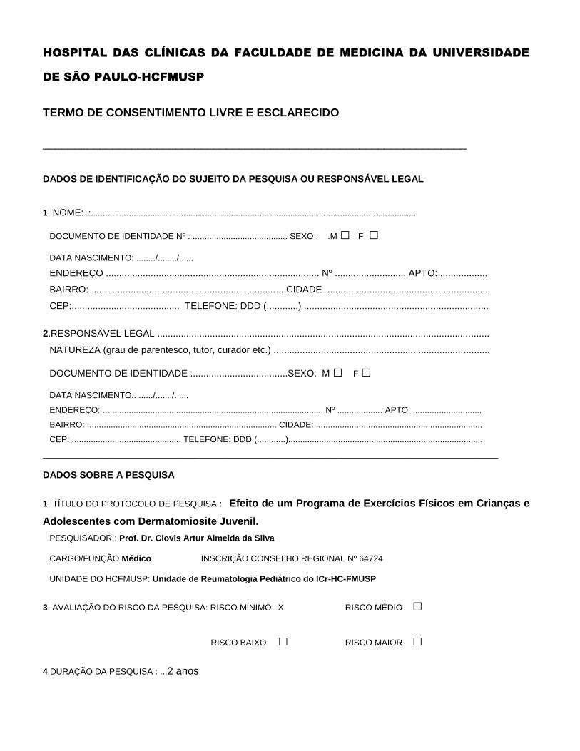
HOSPITAL DAS CLÍNICAS DA FACULDADE DE MEDICINA DA UNIVERSIDADE
DE SÃO PAULO-HCFMUSP
TERMO DE CONSENTIMENTO LIVRE E ESCLARECIDO
___________________________________________________________________
DADOS DE IDENTIFICAÇÃO DO SUJEITO DA PESQUISA OU RESPONSÁVEL LEGAL
1. NOME: .:............................................................................. ...........................................................
DOCUMENTO DE IDENTIDADE Nº : ........................................ SEXO : .M □ F □
DATA NASCIMENTO: ......../......../......
ENDEREÇO ................................................................................. Nº ........................... APTO: ..................
BAIRRO: ........................................................................ CIDADE .............................................................
CEP:......................................... TELEFONE: DDD (............) ......................................................................
2.RESPONSÁVEL LEGAL ..............................................................................................................................
NATUREZA (grau de parentesco, tutor, curador etc.) ..................................................................................
DOCUMENTO DE IDENTIDADE :....................................SEXO: M □ F □
DATA NASCIMENTO.: ....../......./......
ENDEREÇO: ............................................................................................. Nº ................... APTO: .............................
BAIRRO: ................................................................................ CIDADE: ......................................................................
CEP: .............................................. TELEFONE: DDD (............)..................................................................................
________________________________________________________________________________________________
DADOS SOBRE A PESQUISA
1. TÍTULO DO PROTOCOLO DE PESQUISA : Efeito de um Programa de Exercícios Físicos em Crianças e
Adolescentes com Dermatomiosite Juvenil.
PESQUISADOR : Prof. Dr. Clovis Artur Almeida da Silva
CARGO/FUNÇÃO Médico INSCRIÇÃO CONSELHO REGIONAL Nº 64724
UNIDADE DO HCFMUSP: Unidade de Reumatologia Pediátrico do ICr-HC-FMUSP
3. AVALIAÇÃO DO RISCO DA PESQUISA: RISCO MÍNIMO X RISCO MÉDIO □
RISCO BAIXO □ RISCO MAIOR □
4.DURAÇÃO DA PESQUISA : ...2 anos
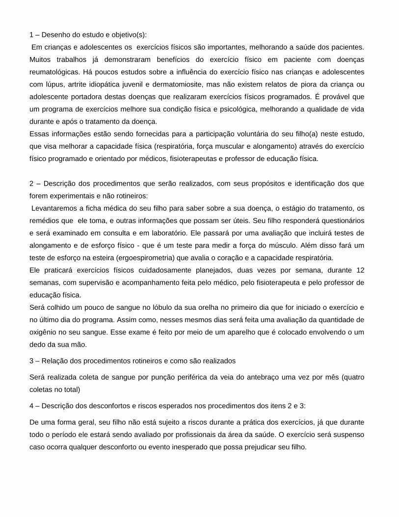
1 – Desenho do estudo e objetivo(s):
Em crianças e adolescentes os exercícios físicos são importantes, melhorando a saúde dos pacientes.
Muitos trabalhos já demonstraram benefícios do exercício físico em paciente com doenças
reumatológicas. Há poucos estudos sobre a influência do exercício físico nas crianças e adolescentes
com lúpus, artrite idiopática juvenil e dermatomiosite, mas não existem relatos de piora da criança ou
adolescente portadora destas doenças que realizaram exercícios físicos programados. É provável que
um programa de exercícios melhore sua condição física e psicológica, melhorando a qualidade de vida
durante e após o tratamento da doença.
Essas informações estão sendo fornecidas para a participação voluntária do seu filho(a) neste estudo,
que visa melhorar a capacidade física (respiratória, força muscular e alongamento) através do exercício
físico programado e orientado por médicos, fisioterapeutas e professor de educação física.
2 – Descrição dos procedimentos que serão realizados, com seus propósitos e identificação dos que
forem experimentais e não rotineiros:
Levantaremos a ficha médica do seu filho para saber sobre a sua doença, o estágio do tratamento, os
remédios que ele toma, e outras informações que possam ser úteis. Seu filho responderá questionários
e será examinado em consulta e em laboratório. Ele passará por uma avaliação que incluirá testes de
alongamento e de esforço físico - que é um teste para medir a força do músculo. Além disso fará um
teste de esforço na esteira (ergoespirometria) que avalia o coração e a capacidade respiratória.
Ele praticará exercícios físicos cuidadosamente planejados, duas vezes por semana, durante 12
semanas, com supervisão e acompanhamento feita pelo médico, pelo fisioterapeuta e pelo professor de
educação física.
Será colhido um pouco de sangue no lóbulo da sua orelha no primeiro dia que for iniciado o exercício e
no último dia do programa. Assim como, nesses mesmos dias será feita uma avaliação da quantidade de
oxigênio no seu sangue. Esse exame é feito por meio de um aparelho que é colocado envolvendo o um
dedo da sua mão.
3 – Relação dos procedimentos rotineiros e como são realizados
Será realizada coleta de sangue por punção periférica da veia do antebraço uma vez por mês (quatro
coletas no total)
4 – Descrição dos desconfortos e riscos esperados nos procedimentos dos itens 2 e 3:
De uma forma geral, seu filho não está sujeito a riscos durante a prática dos exercícios, já que durante
todo o período ele estará sendo avaliado por profissionais da área da saúde. O exercício será suspenso
caso ocorra qualquer desconforto ou evento inesperado que possa prejudicar seu filho.
Avaliação da força muscular e respiratória - O exame para avaliar a força dos músculos e o exame
para avaliar a capacidade respiratória podem ocasionar algum cansaço na hora do exame. Seu filho(a)
também poderá sentir um pouco de dor nos músculos até 2 dias depois do teste.
Programa de Exercícios - Durante o programa de exercícios ele poderá sentir cansaço na primeira
semana, o que provavelmente não acontecerá nas outras semanas de exercício.
Exame de sangue – a coleta de sangue poderá causar um leve desconforto no local da picada, que
deve desaparecer em um dia.
5 – Benefícios para o participante
Seu filho será avaliado e iremos desenvolver um programa de exercícios adequado à sua condição
clínica e física. Participando dele com regularidade ele poderá melhorar sua condição física, o que irá
permitir que possa fazer suas atividades diárias com mais disposição. Adquirindo mais força muscular e
capacidade respiratória.
6 – Relação de procedimentos alternativos que possam ser vantajosos, pelos quais o paciente pode
optar;
Seu filho passará por um programa de exercícios cuidadosamente desenvolvido para a sua condição
física e clínica que poderá levar a melhora da sua capacidade de executar as tarefas do dia-a-dia e
diminuir a sensação de cansaço. Será acompanhado por médicos, fisioterapeutas e professores de
educação física durante todo o período. Qualquer sintoma ou mudança na sua condição física, poderá
ser avaliado e tratado de forma adequada.
7 – Garantia de acesso:
Em qualquer etapa do estudo, você terá acesso aos profissionais responsáveis pela pesquisa para
esclarecimento de eventuais dúvidas. O principal investigador é o Dr Clovis Artur Almeida da Silva.
que pode ser encontrado no endereço Instituto da Criança do Hospital das Clínicas da Faculdade de
Medicina da Universidade de São Paulo na Rua Dr Eneas Carvalho de Aguiar, 647 Telefone(s)
3069-8675. Se você tiver alguma consideração ou dúvida sobre a ética da pesquisa, entre em contato
com o Comitê de Ética em Pesquisa (CEP) – Rua Ovídio Pires de Campos, 225 – 5º andar – tel: 3069-
6442 ramais 16, 17, 18 ou 20, FAX: 3069-6442 ramal 26 – E-mail: [email protected]
8 – É garantida a liberdade da retirada de consentimento a qualquer momento e deixar de participar do
estudo, sem qualquer prejuízo à continuidade de seu tratamento na Instituição;
09 – Direito de confidencialidade – As informações obtidas serão analisadas em conjunto com outros
pacientes, não sendo divulgado a identificação de nenhum paciente;
10 – Direito de ser mantido atualizado sobre os resultados parciais das pesquisas, quando em estudos
abertos, ou de resultados que sejam do conhecimento dos pesquisadores;
11 – Despesas e compensações: não há despesas pessoais para o participante em qualquer fase do
estudo, incluindo exames e consultas. Também não há compensação financeira relacionada à sua
participação. Se existir qualquer despesa adicional, ela será absorvida pelo orçamento da pesquisa.
12 - Compromisso do pesquisador de utilizar os dados e o material coletado somente para esta
pesquisa.
Acredito ter sido suficientemente informado a respeito das informações que li ou que foram lidas para
mim, descrevendo o estudo “ Efeito de um Programa de Exercícios Físicos em Crianças e Adolescentes
com Dermatomiosite Juvenil”.
Eu discuti com o Dr Clovis Artur Almeida da Silva sobre a minha decisão em participar nesse estudo.
Ficaram claros para mim quais são os propósitos do estudo, os procedimentos a serem realizados, seus
desconfortos e riscos, as garantias de confidencialidade e de esclarecimentos permanentes. Ficou claro
também que minha participação é isenta de despesas e que tenho garantia do acesso a tratamento
hospitalar quando necessário. Concordo voluntariamente em participar deste estudo e poderei retirar o
meu consentimento a qualquer momento, antes ou durante o mesmo, sem penalidades ou prejuízo ou
perda de qualquer benefício que eu possa ter adquirido, ou no meu atendimento neste Serviço.
-------------------------------------------------
Assinatura do paciente/representante legal Data / /
-------------------------------------------------------------------------
Assinatura da testemunha Data / /
para casos de pacientes menores de 18 anos, analfabetos, semi-analfabetos ou portadores de
deficiência auditiva ou visual.
(Somente para o responsável do projeto)
Declaro que obtive de forma apropriada e voluntária o Consentimento Livre e Esclarecido deste paciente
ou representante legal para a participação neste estudo.
-------------------------------------------------------------------------
Assinatura do responsável pelo estudo Data / /
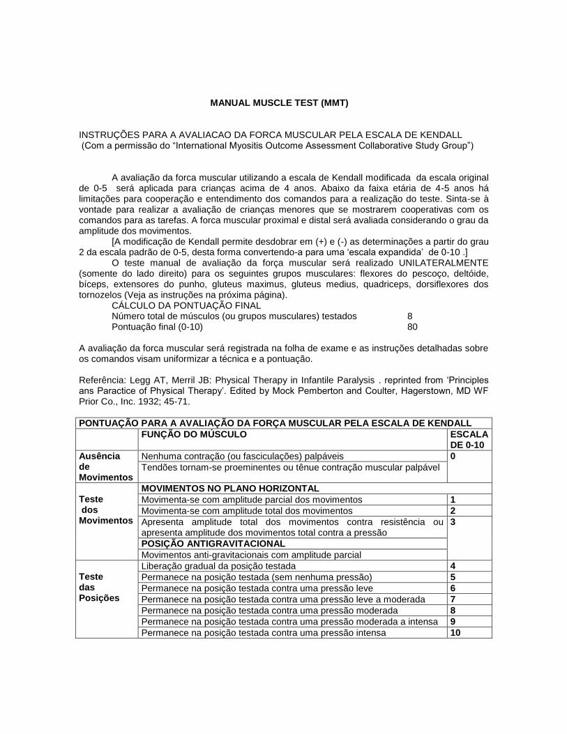
MANUAL MUSCLE TEST (MMT) INSTRUÇÕES PARA A AVALIACAO DA FORCA MUSCULAR PELA ESCALA DE KENDALL (Com a permissão do “International Myositis Outcome Assessment Collaborative Study Group”)
A avaliação da forca muscular utilizando a escala de Kendall modificada da escala original de 0-5 será aplicada para crianças acima de 4 anos. Abaixo da faixa etária de 4-5 anos há limitações para cooperação e entendimento dos comandos para a realização do teste. Sinta-se à vontade para realizar a avaliação de crianças menores que se mostrarem cooperativas com os comandos para as tarefas. A forca muscular proximal e distal será avaliada considerando o grau da amplitude dos movimentos. [A modificação de Kendall permite desdobrar em (+) e (-) as determinações a partir do grau 2 da escala padrão de 0-5, desta forma convertendo-a para uma ‘escala expandida’ de 0-10 .] O teste manual de avaliação da força muscular será realizado UNILATERALMENTE (somente do lado direito) para os seguintes grupos musculares: flexores do pescoço, deltóide, bíceps, extensores do punho, gluteus maximus, gluteus medius, quadriceps, dorsiflexores dos tornozelos (Veja as instruções na próxima página). CÁLCULO DA PONTUAÇÃO FINAL Número total de músculos (ou grupos musculares) testados 8 Pontuação final (0-10) 80 A avaliação da forca muscular será registrada na folha de exame e as instruções detalhadas sobre os comandos visam uniformizar a técnica e a pontuação. Referência: Legg AT, Merril JB: Physical Therapy in Infantile Paralysis . reprinted from ‘Principles ans Paractice of Physical Therapy’. Edited by Mock Pemberton and Coulter, Hagerstown, MD WF Prior Co., Inc. 1932; 45-71.
PONTUAÇÃO PARA A AVALIAÇÃO DA FORÇA MUSCULAR PELA ESCALA DE KENDALL
FUNÇÃO DO MÚSCULO ESCALA DE 0-10
Ausência de Movimentos
Nenhuma contração (ou fasciculações) palpáveis 0
Tendões tornam-se proeminentes ou tênue contração muscular palpável
Teste dos Movimentos
MOVIMENTOS NO PLANO HORIZONTAL
Movimenta-se com amplitude parcial dos movimentos 1
Movimenta-se com amplitude total dos movimentos 2
Apresenta amplitude total dos movimentos contra resistência ou apresenta amplitude dos movimentos total contra a pressão
3
POSIÇÃO ANTIGRAVITACIONAL
Movimentos anti-gravitacionais com amplitude parcial
Teste das Posições
Liberação gradual da posição testada 4
Permanece na posição testada (sem nenhuma pressão) 5
Permanece na posição testada contra uma pressão leve 6
Permanece na posição testada contra uma pressão leve a moderada 7
Permanece na posição testada contra uma pressão moderada 8
Permanece na posição testada contra uma pressão moderada a intensa 9
Permanece na posição testada contra uma pressão intensa 10
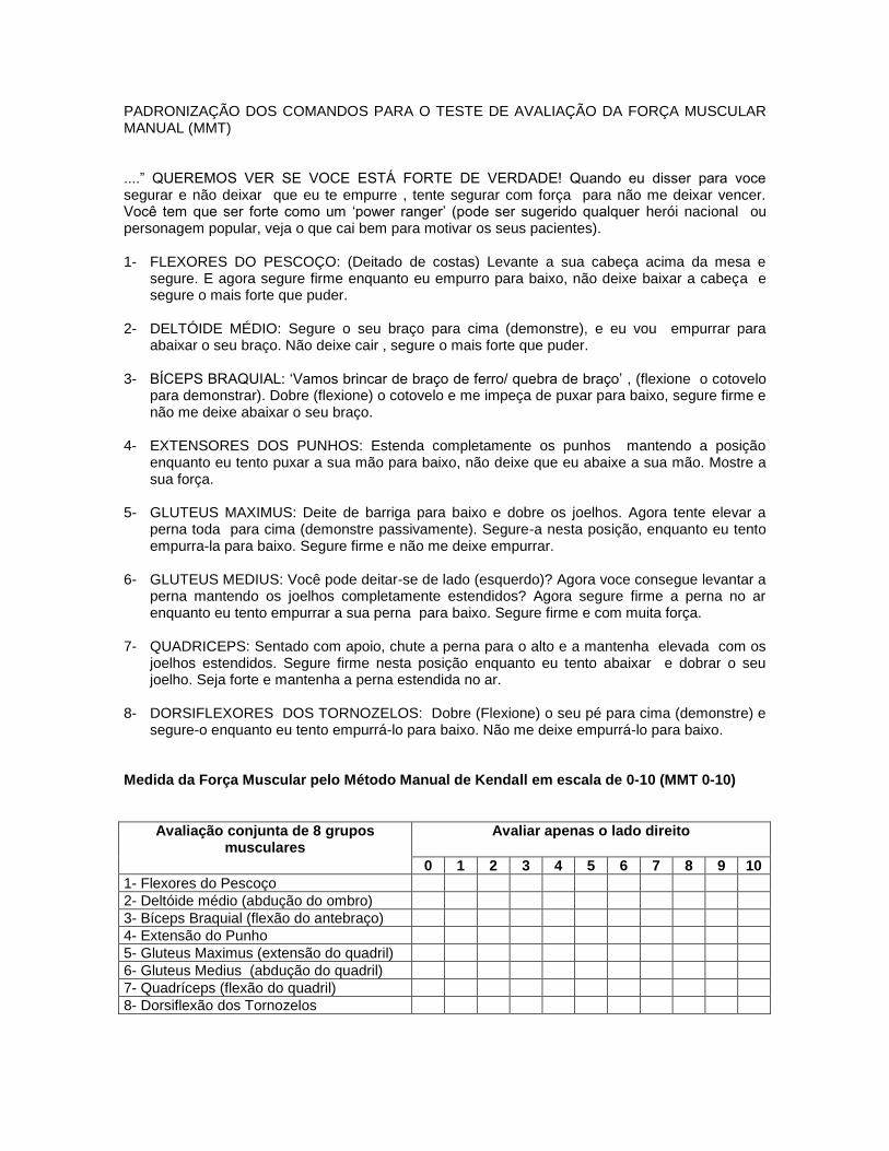
PADRONIZAÇÃO DOS COMANDOS PARA O TESTE DE AVALIAÇÃO DA FORÇA MUSCULAR MANUAL (MMT) ....” QUEREMOS VER SE VOCE ESTÁ FORTE DE VERDADE! Quando eu disser para voce segurar e não deixar que eu te empurre , tente segurar com força para não me deixar vencer. Você tem que ser forte como um ‘power ranger’ (pode ser sugerido qualquer herói nacional ou personagem popular, veja o que cai bem para motivar os seus pacientes). 1- FLEXORES DO PESCOÇO: (Deitado de costas) Levante a sua cabeça acima da mesa e
segure. E agora segure firme enquanto eu empurro para baixo, não deixe baixar a cabeça e segure o mais forte que puder.
2- DELTÓIDE MÉDIO: Segure o seu braço para cima (demonstre), e eu vou empurrar para
abaixar o seu braço. Não deixe cair , segure o mais forte que puder. 3- BÍCEPS BRAQUIAL: ‘Vamos brincar de braço de ferro/ quebra de braço’ , (flexione o cotovelo
para demonstrar). Dobre (flexione) o cotovelo e me impeça de puxar para baixo, segure firme e não me deixe abaixar o seu braço.
4- EXTENSORES DOS PUNHOS: Estenda completamente os punhos mantendo a posição
enquanto eu tento puxar a sua mão para baixo, não deixe que eu abaixe a sua mão. Mostre a sua força.
5- GLUTEUS MAXIMUS: Deite de barriga para baixo e dobre os joelhos. Agora tente elevar a
perna toda para cima (demonstre passivamente). Segure-a nesta posição, enquanto eu tento empurra-la para baixo. Segure firme e não me deixe empurrar.
6- GLUTEUS MEDIUS: Você pode deitar-se de lado (esquerdo)? Agora voce consegue levantar a
perna mantendo os joelhos completamente estendidos? Agora segure firme a perna no ar enquanto eu tento empurrar a sua perna para baixo. Segure firme e com muita força.
7- QUADRICEPS: Sentado com apoio, chute a perna para o alto e a mantenha elevada com os
joelhos estendidos. Segure firme nesta posição enquanto eu tento abaixar e dobrar o seu joelho. Seja forte e mantenha a perna estendida no ar.
8- DORSIFLEXORES DOS TORNOZELOS: Dobre (Flexione) o seu pé para cima (demonstre) e
segure-o enquanto eu tento empurrá-lo para baixo. Não me deixe empurrá-lo para baixo. Medida da Força Muscular pelo Método Manual de Kendall em escala de 0-10 (MMT 0-10)
Avaliação conjunta de 8 grupos musculares
Avaliar apenas o lado direito
0 1 2 3 4 5 6 7 8 9 10
1- Flexores do Pescoço
2- Deltóide médio (abdução do ombro)
3- Bíceps Braquial (flexão do antebraço)
4- Extensão do Punho
5- Gluteus Maximus (extensão do quadril)
6- Gluteus Medius (abdução do quadril)
7- Quadríceps (flexão do quadril)
8- Dorsiflexão dos Tornozelos
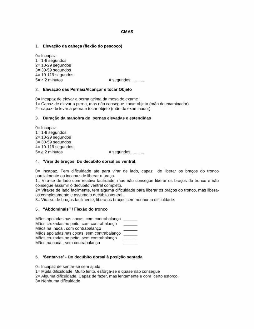
CMAS
1. Elevação da cabeça (flexão do pescoço) 0= Incapaz 1= 1-9 segundos 2= 10-29 segundos 3= 30-59 segundos 4= 10-119 segundos
5= 2 minutos # segundos ............ 2. Elevação das Pernas/Alcançar e tocar Objeto 0= Incapaz de elevar a perna acima da mesa de exame 1= Capaz de elevar a perna, mas não consegue tocar objeto (mão do examinador) 2= capaz de levar a perna e tocar objeto (mão do examinador) 3. Duração da manobra de pernas elevadas e estendidas 0= Incapaz 1= 1-9 segundos 2= 10-29 segundos 3= 30-59 segundos 4= 10-119 segundos
5= 2 minutos # segundos ............ 4. ‘Virar de bruços’ Do decúbito dorsal ao ventral. 0= Incapaz. Tem dificuldade ate para virar de lado, capaz de liberar os braços do tronco parcialmente ou incapaz de liberar o braço. 1= Vira-se de lado com relativa facilidade, mas não consegue liberar os braços do tronco e não consegue assumir o decúbito ventral completo. 2= Vira-se de lado facilmente, tem alguma dificuldade para liberar os braços do tronco, mas libera-os completamente e assume o decúbito ventral. 3= Vira-se de bruços facilmente, libera os braços sem nenhuma dificuldade. 5. “Abdominais” / Flexão do tronco Mãos apoiadas nas coxas, com contrabalanço ______ Mãos cruzadas no peito, com contrabalanço ______ Mãos na nuca , com contrabalanço ______ Mãos apoiadas nas coxas, sem contrabalanço ______ Mãos cruzadas no peito, sem contrabalanço ______ Mãos na nuca , sem contrabalanço ______ 6. ‘Sentar-se’ - Do decúbito dorsal à posição sentada 0= Incapaz de sentar-se sem ajuda 1= Muita dificuldade. Muito lento, esforça-se e quase não consegue 2= Alguma dificuldade. Capaz de fazer, mas lentamente e com certo esforço. 3= Nenhuma dificuldade
2
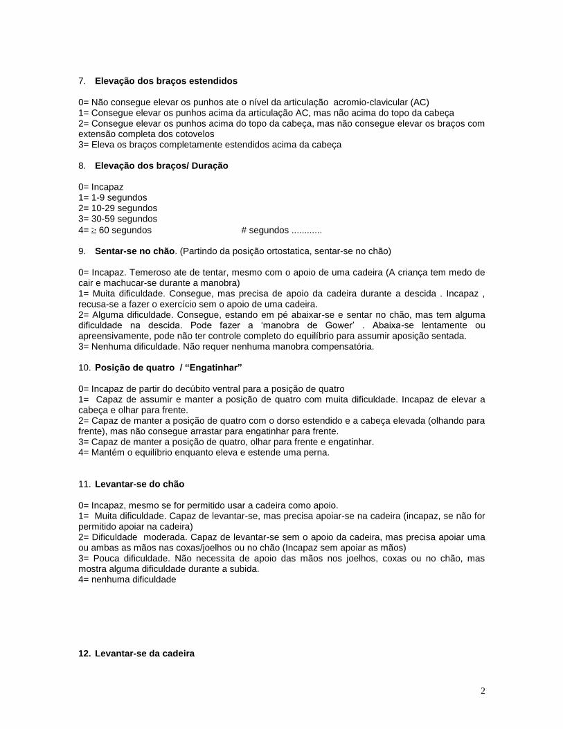
7. Elevação dos braços estendidos 0= Não consegue elevar os punhos ate o nível da articulação acromio-clavicular (AC) 1= Consegue elevar os punhos acima da articulação AC, mas não acima do topo da cabeça 2= Consegue elevar os punhos acima do topo da cabeça, mas não consegue elevar os braços com extensão completa dos cotovelos 3= Eleva os braços completamente estendidos acima da cabeça 8. Elevação dos braços/ Duração 0= Incapaz 1= 1-9 segundos 2= 10-29 segundos 3= 30-59 segundos
4= 60 segundos # segundos ............ 9. Sentar-se no chão. (Partindo da posição ortostatica, sentar-se no chão) 0= Incapaz. Temeroso ate de tentar, mesmo com o apoio de uma cadeira (A criança tem medo de cair e machucar-se durante a manobra) 1= Muita dificuldade. Consegue, mas precisa de apoio da cadeira durante a descida . Incapaz , recusa-se a fazer o exercício sem o apoio de uma cadeira. 2= Alguma dificuldade. Consegue, estando em pé abaixar-se e sentar no chão, mas tem alguma dificuldade na descida. Pode fazer a ‘manobra de Gower’ . Abaixa-se lentamente ou apreensivamente, pode não ter controle completo do equilíbrio para assumir aposição sentada. 3= Nenhuma dificuldade. Não requer nenhuma manobra compensatória. 10. Posição de quatro / “Engatinhar” 0= Incapaz de partir do decúbito ventral para a posição de quatro 1= Capaz de assumir e manter a posição de quatro com muita dificuldade. Incapaz de elevar a cabeça e olhar para frente. 2= Capaz de manter a posição de quatro com o dorso estendido e a cabeça elevada (olhando para frente), mas não consegue arrastar para engatinhar para frente. 3= Capaz de manter a posição de quatro, olhar para frente e engatinhar. 4= Mantém o equilíbrio enquanto eleva e estende uma perna. 11. Levantar-se do chão 0= Incapaz, mesmo se for permitido usar a cadeira como apoio. 1= Muita dificuldade. Capaz de levantar-se, mas precisa apoiar-se na cadeira (incapaz, se não for permitido apoiar na cadeira) 2= Dificuldade moderada. Capaz de levantar-se sem o apoio da cadeira, mas precisa apoiar uma ou ambas as mãos nas coxas/joelhos ou no chão (Incapaz sem apoiar as mãos) 3= Pouca dificuldade. Não necessita de apoio das mãos nos joelhos, coxas ou no chão, mas mostra alguma dificuldade durante a subida. 4= nenhuma dificuldade 12. Levantar-se da cadeira
3
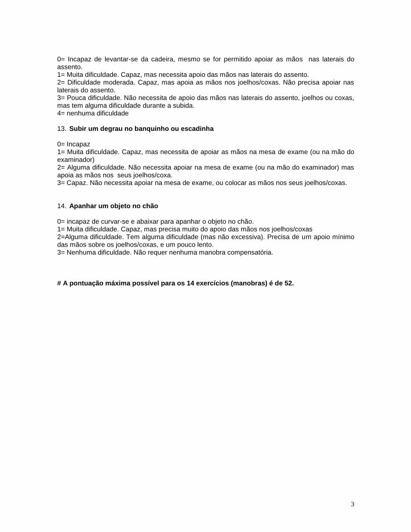
0= Incapaz de levantar-se da cadeira, mesmo se for permitido apoiar as mãos nas laterais do assento. 1= Muita dificuldade. Capaz, mas necessita apoio das mãos nas laterais do assento. 2= Dificuldade moderada. Capaz, mas apoia as mãos nos joelhos/coxas. Não precisa apoiar nas laterais do assento. 3= Pouca dificuldade. Não necessita de apoio das mãos nas laterais do assento, joelhos ou coxas, mas tem alguma dificuldade durante a subida. 4= nenhuma dificuldade 13. Subir um degrau no banquinho ou escadinha 0= Incapaz 1= Muita dificuldade. Capaz, mas necessita de apoiar as mãos na mesa de exame (ou na mão do examinador) 2= Alguma dificuldade. Não necessita apoiar na mesa de exame (ou na mão do examinador) mas apoia as mãos nos seus joelhos/coxa. 3= Capaz. Não necessita apoiar na mesa de exame, ou colocar as mãos nos seus joelhos/coxas. 14. Apanhar um objeto no chão 0= incapaz de curvar-se e abaixar para apanhar o objeto no chão. 1= Muita dificuldade. Capaz, mas precisa muito do apoio das mãos nos joelhos/coxas 2=Alguma dificuldade. Tem alguma dificuldade (mas não excessiva). Precisa de um apoio mínimo das mãos sobre os joelhos/coxas, e um pouco lento. 3= Nenhuma dificuldade. Não requer nenhuma manobra compensatória. # A pontuação máxima possível para os 14 exercícios (manobras) é de 52.
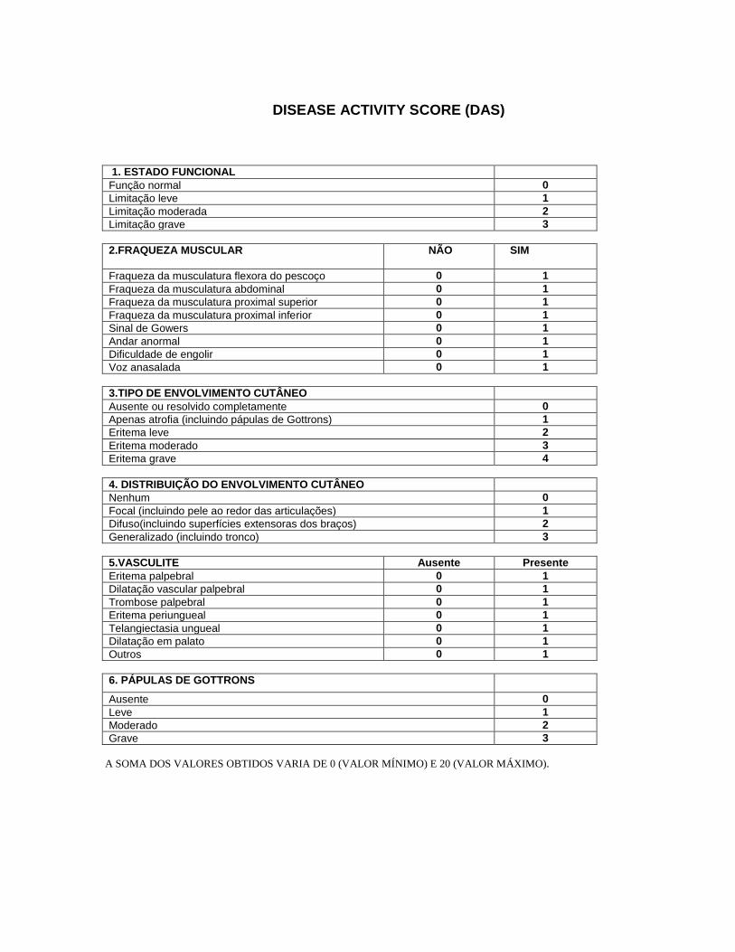
DISEASE ACTIVITY SCORE (DAS)
1. ESTADO FUNCIONAL
Função normal 0
Limitação leve 1
Limitação moderada 2
Limitação grave 3
2.FRAQUEZA MUSCULAR NÃO SIM
Fraqueza da musculatura flexora do pescoço 0 1
Fraqueza da musculatura abdominal 0 1
Fraqueza da musculatura proximal superior 0 1
Fraqueza da musculatura proximal inferior 0 1
Sinal de Gowers 0 1
Andar anormal 0 1
Dificuldade de engolir 0 1
Voz anasalada 0 1
3.TIPO DE ENVOLVIMENTO CUTÂNEO
Ausente ou resolvido completamente 0
Apenas atrofia (incluindo pápulas de Gottrons) 1
Eritema leve 2
Eritema moderado 3
Eritema grave 4
4. DISTRIBUIÇÃO DO ENVOLVIMENTO CUTÂNEO
Nenhum 0
Focal (incluindo pele ao redor das articulações) 1
Difuso(incluindo superfícies extensoras dos braços) 2
Generalizado (incluindo tronco) 3
5.VASCULITE Ausente Presente
Eritema palpebral 0 1
Dilatação vascular palpebral 0 1
Trombose palpebral 0 1
Eritema periungueal 0 1
Telangiectasia ungueal 0 1
Dilatação em palato 0 1
Outros 0 1
6. PÁPULAS DE GOTTRONS
Ausente 0
Leve 1
Moderado 2
Grave 3
A SOMA DOS VALORES OBTIDOS VARIA DE 0 (VALOR MÍNIMO) E 20 (VALOR MÁXIMO).
PPeeddssQQLL TTMM
QQuueessttiioonnáárriioo PPeeddiiááttrriiccoo
ddee QQuuaalliiddaaddee ddee VViiddaa
VVeerrssããoo 44..00
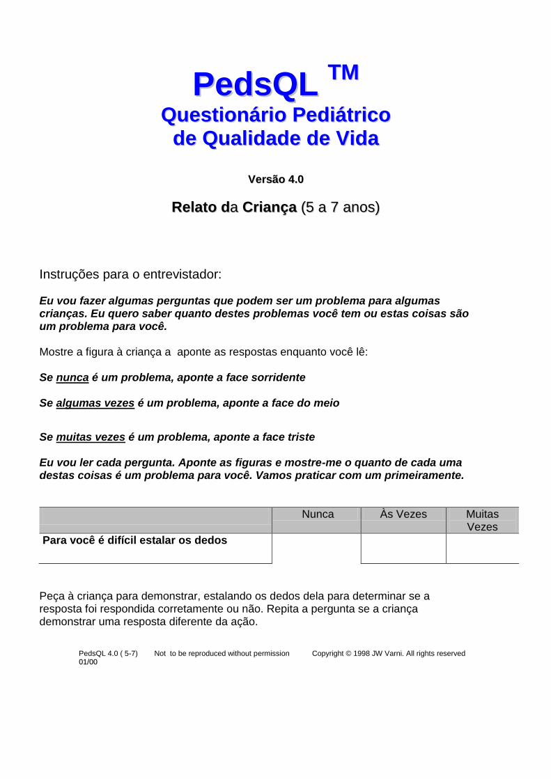
RReellaattoo ddaa CCrriiaannççaa ((55 aa 77 aannooss))
Instruções para o entrevistador:
Eu vou fazer algumas perguntas que podem ser um problema para algumas crianças. Eu quero saber quanto destes problemas você tem ou estas coisas são um problema para você. Mostre a figura à criança a aponte as respostas enquanto você lê: Se nunca é um problema, aponte a face sorridente Se algumas vezes é um problema, aponte a face do meio
Se muitas vezes é um problema, aponte a face triste Eu vou ler cada pergunta. Aponte as figuras e mostre-me o quanto de cada uma destas coisas é um problema para você. Vamos praticar com um primeiramente.
Nunca Às Vezes Muitas Vezes
Para você é difícil estalar os dedos
Peça à criança para demonstrar, estalando os dedos dela para determinar se a resposta foi respondida corretamente ou não. Repita a pergunta se a criança demonstrar uma resposta diferente da ação.
PedsQL 4.0 ( 5-7) Not to be reproduced without permission Copyright © 1998 JW Varni. All rights reserved 0011//0000

Pense em como você tem se sentido nas últimas semanas. Por favor ouça cada frase com atenção e conte-me quanto de cada um destes problemas você tem tido. Depois de ler o item, faça um gesto em direção às figuras.Se a criança hesitar ou parecer não saber como responder,leia a resposta enquanto aponta para as caras.
Capacidade Física (problemas com...) Nunca Às Vezes Muitas
Vezes
1.Para você é difícil andar 0 2 4
2.Para você é difícil correr 0 2 4
3.Para você é difícil praticar esportes ou exercícios 0 2 4
4.Para você é difícil levantar coisas grandes 0 2 4
5.Para você é difícil tomar banho de banheira ou chuveiro sozinho
0 2 4
6.Fazer as tarefas do dia-a-dia da casa (como pegar seus brinquedos)
0 2 4
7.Você tem machucado ou dor (Onde ? ________) 0 2 4
8. Você alguma vez já se sentiu cansado demais para brincar
0 2 4
Aspecto Emocional (problemas com...) Nunca Às Vezes Muitas
Vezes
1.Você se sente assustado 0 2 4
2.Você se sente triste 0 2 4
3.Você se sente nervoso 0 2 4
4. Você tem dificuldade para dormir 0 2 4
5.Você se preocupa com o que vai acontecer com você
0 2 4
Aspecto Social (problemas com...) Nunca Às Vezes Muitas
Vezes
1. É difícil para você conviver com outras crianças 0 2 4
2. Outras crianças dizem que não querem brincar com você
0 2 4
3. Outras crianças te provocam 0 2 4
4. Outras crianças fazem coisas que você não consegue fazer
0 2 4
5. É difícil para você acompanhar a brincadeira com outras crianças
0 2 4
Atividade Escolar (problemas com...) Nunca Às Vezes Muitas
vezes
1.É difícil para você prestar atenção na aula 0 2 4
2. Você esquece as coisas 0 2 4
3.É difícil para você fazer os trabalhos da classe 0 2 4
4. Você falta na escola por não se sentir bem 0 2 4
5. Você falta na escola por ter que ir ao médico ou hospital
0 2 4
PedsQL 4.0 ( 5-7) Not to be reproduced without permission Copyright © 1998 JW Varni. All rights reserved 01/00
Quanto cada uma destas coisas é problema para você?
Nunca As vezes Muitas vezes
PedsQL 4.0 ( 5-7) Not to be reproduced without permission Copyright © 1998 JW Varni. All rights reserved 01/00
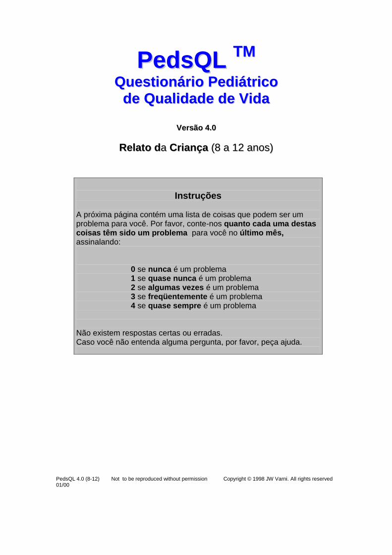
PPeeddssQQLL TTMM
QQuueessttiioonnáárriioo PPeeddiiááttrriiccoo
ddee QQuuaalliiddaaddee ddee VViiddaa
VVeerrssããoo 44..00
RReellaattoo ddaa CCrriiaannççaa ((88 aa 1122 aannooss))
Instruções
A próxima página contém uma lista de coisas que podem ser um problema para você. Por favor, conte-nos quanto cada uma destas coisas têm sido um problema para você no último mês, assinalando:
0 se nunca é um problema 1 se quase nunca é um problema 2 se algumas vezes é um problema 3 se freqüentemente é um problema 4 se quase sempre é um problema
Não existem respostas certas ou erradas. Caso você não entenda alguma pergunta, por favor, peça ajuda.
PedsQL 4.0 (8-12) Not to be reproduced without permission Copyright © 1998 JW Varni. All rights reserved 01/00
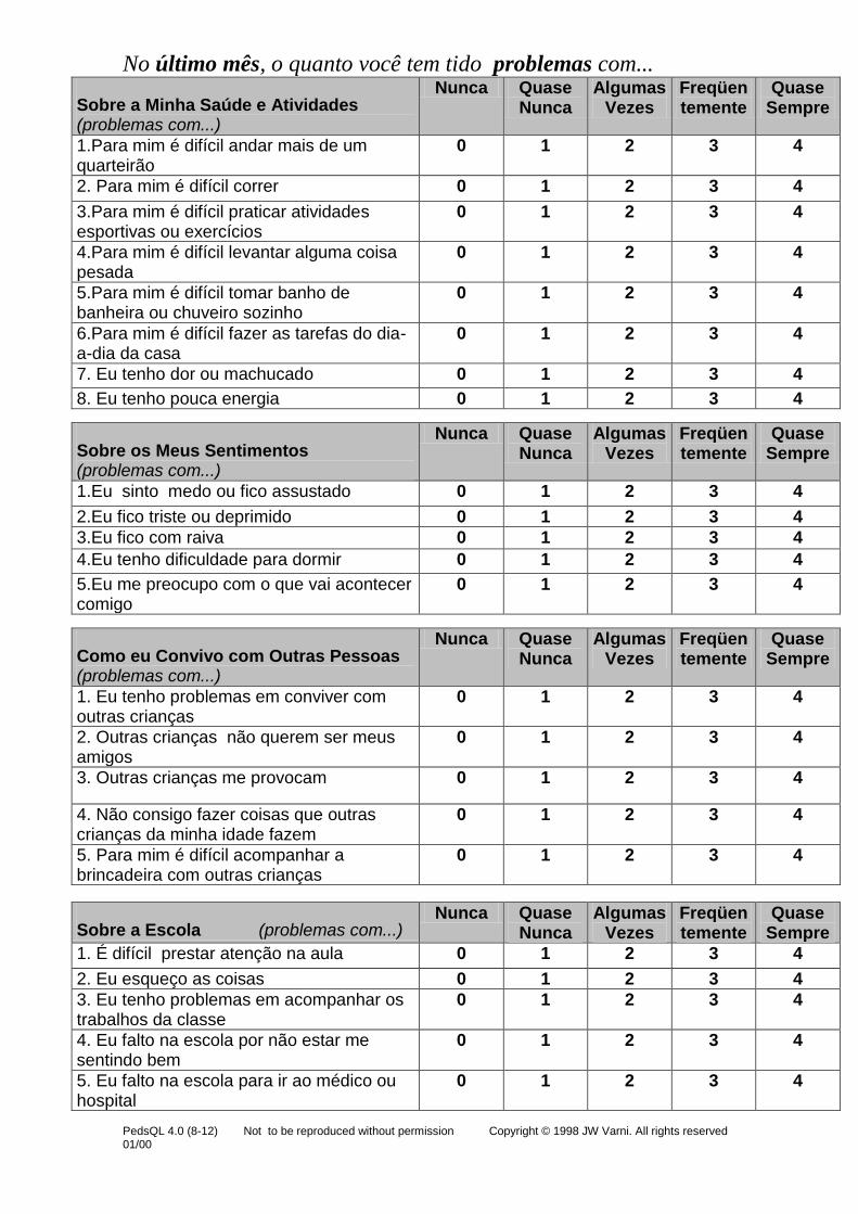
No último mês, o quanto você tem tido problemas com...
Sobre a Minha Saúde e Atividades (problemas com...)
Nunca Quase Nunca
Algumas Vezes
Freqüentemente
Quase Sempre
1.Para mim é difícil andar mais de um quarteirão
0 1 2 3 4
2. Para mim é difícil correr 0 1 2 3 4
3.Para mim é difícil praticar atividades esportivas ou exercícios
0 1 2 3 4
4.Para mim é difícil levantar alguma coisa pesada
0 1 2 3 4
5.Para mim é difícil tomar banho de banheira ou chuveiro sozinho
0 1 2 3 4
6.Para mim é difícil fazer as tarefas do dia-a-dia da casa
0 1 2 3 4
7. Eu tenho dor ou machucado 0 1 2 3 4
8. Eu tenho pouca energia 0 1 2 3 4
Sobre os Meus Sentimentos (problemas com...)
Nunca Quase Nunca
Algumas Vezes
Freqüentemente
Quase Sempre
1.Eu sinto medo ou fico assustado 0 1 2 3 4
2.Eu fico triste ou deprimido 0 1 2 3 4
3.Eu fico com raiva 0 1 2 3 4
4.Eu tenho dificuldade para dormir 0 1 2 3 4
5.Eu me preocupo com o que vai acontecer comigo
0 1 2 3 4
Como eu Convivo com Outras Pessoas (problemas com...)
Nunca Quase Nunca
Algumas Vezes
Freqüentemente
Quase Sempre
1. Eu tenho problemas em conviver com outras crianças
0 1 2 3 4
2. Outras crianças não querem ser meus amigos
0 1 2 3 4
3. Outras crianças me provocam 0 1 2 3 4
4. Não consigo fazer coisas que outras crianças da minha idade fazem
0 1 2 3 4
5. Para mim é difícil acompanhar a brincadeira com outras crianças
0 1 2 3 4
Sobre a Escola (problemas com...) Nunca Quase
Nunca Algumas
Vezes Freqüentemente
Quase Sempre
1. É difícil prestar atenção na aula 0 1 2 3 4
2. Eu esqueço as coisas 0 1 2 3 4
3. Eu tenho problemas em acompanhar os trabalhos da classe
0 1 2 3 4
4. Eu falto na escola por não estar me sentindo bem
0 1 2 3 4
5. Eu falto na escola para ir ao médico ou hospital
0 1 2 3 4
PedsQL 4.0 (8-12) Not to be reproduced without permission Copyright © 1998 JW Varni. All rights reserved 01/00
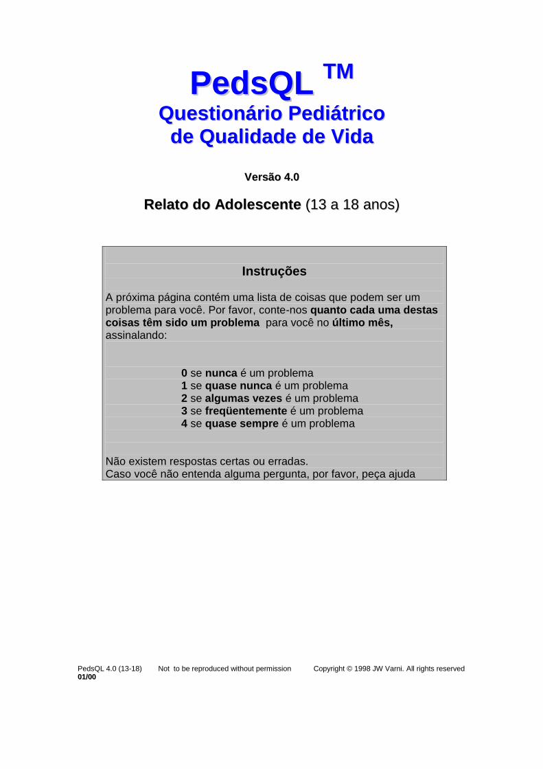
PPeeddssQQLL TTMM
QQuueessttiioonnáárriioo PPeeddiiááttrriiccoo
ddee QQuuaalliiddaaddee ddee VViiddaa
VVeerrssããoo 44..00
RReellaattoo ddoo AAddoolleesscceennttee ((1133 aa 1188 aannooss))
Instruções
A próxima página contém uma lista de coisas que podem ser um problema para você. Por favor, conte-nos quanto cada uma destas coisas têm sido um problema para você no último mês, assinalando:
0 se nunca é um problema 1 se quase nunca é um problema 2 se algumas vezes é um problema 3 se freqüentemente é um problema 4 se quase sempre é um problema
Não existem respostas certas ou erradas. Caso você não entenda alguma pergunta, por favor, peça ajuda
PedsQL 4.0 (13-18) Not to be reproduced without permission Copyright © 1998 JW Varni. All rights reserved 0011//0000
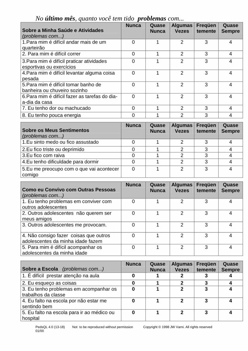
No último mês, quanto você tem tido problemas com...
Sobre a Minha Saúde e Atividades (problemas com...)
Nunca Quase Nunca
Algumas Vezes
Freqüentemente
Quase Sempre
1.Para mim é difícil andar mais de um quarteirão
0 1 2 3 4
2. Para mim é difícil correr 0 1 2 3 4
3.Para mim é difícil praticar atividades esportivas ou exercícios
0 1 2 3 4
4.Para mim é difícil levantar alguma coisa pesada
0 1 2 3 4
5.Para mim é difícil tomar banho de banheira ou chuveiro sozinho
0 1 2 3 4
6.Para mim é difícil fazer as tarefas do dia-a-dia da casa
0 1 2 3 4
7. Eu tenho dor ou machucado 0 1 2 3 4
8. Eu tenho pouca energia 0 1 2 3 4
Sobre os Meus Sentimentos (problemas com...)
Nunca Quase Nunca
Algumas Vezes
Freqüentemente
Quase Sempre
1.Eu sinto medo ou fico assustado 0 1 2 3 4
2.Eu fico triste ou deprimido 0 1 2 3 4
3.Eu fico com raiva 0 1 2 3 4
4.Eu tenho dificuldade para dormir 0 1 2 3 4
5.Eu me preocupo com o que vai acontecer comigo
0 1 2 3 4
Como eu Convivo com Outras Pessoas (problemas com...)
Nunca Quase Nunca
Algumas Vezes
Freqüentemente
Quase Sempre
1. Eu tenho problemas em conviver com outros adolescentes
0 1 2 3 4
2. Outros adolescentes não querem ser meus amigos
0 1 2 3 4
3. Outros adolescentes me provocam. 0 1 2 3 4
4. Não consigo fazer coisas que outros adolescentes da minha idade fazem
0 1 2 3 4
5. Para mim é difícil acompanhar os adolescentes da minha idade
0 1 2 3 4
Sobre a Escola (problemas com...) Nunca Quase
Nunca Algumas
Vezes Freqüentemente
Quase Sempre
1. É difícil prestar atenção na aula 0 1 2 3 4
2. Eu esqueço as coisas 0 1 2 3 4
3. Eu tenho problemas em acompanhar os trabalhos da classe
0 1 2 3 4
4. Eu falto na escola por não estar me sentindo bem
0 1 2 3 4
5. Eu falto na escola para ir ao médico ou hospital
0 1 2 3 4
PedsQL 4.0 (13-18) Not to be reproduced without permission Copyright © 1998 JW Varni. All rights reserved 01/00
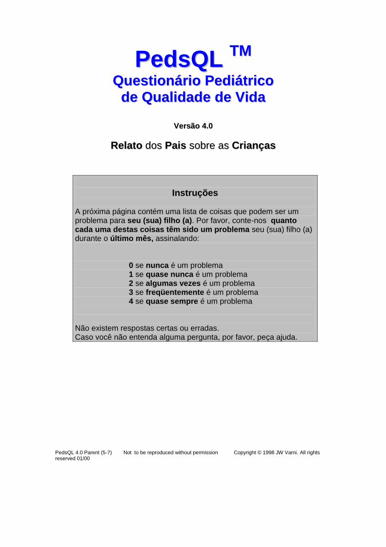
PPeeddssQQLL TTMM
QQuueessttiioonnáárriioo PPeeddiiááttrriiccoo
ddee QQuuaalliiddaaddee ddee VViiddaa
VVeerrssããoo 44..00
RReellaattoo ddooss PPaaiiss ssoobbrree aass CCrriiaannççaass
Instruções
A próxima página contém uma lista de coisas que podem ser um problema para seu (sua) filho (a). Por favor, conte-nos quanto cada uma destas coisas têm sido um problema seu (sua) filho (a) durante o último mês, assinalando:
0 se nunca é um problema 1 se quase nunca é um problema 2 se algumas vezes é um problema 3 se freqüentemente é um problema 4 se quase sempre é um problema
Não existem respostas certas ou erradas. Caso você não entenda alguma pergunta, por favor, peça ajuda.
PedsQL 4.0 Parent (5-7) Not to be reproduced without permission Copyright © 1998 JW Varni. All rights reserved 01/00
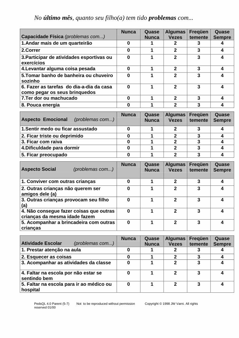
No último mês, quanto seu filho(a) tem tido problemas com...
Capacidade Física (problemas com...) Nunca Quase
Nunca Algumas
Vezes Freqüentemente
Quase Sempre
1.Andar mais de um quarteirão 0 1 2 3 4
2.Correr 0 1 2 3 4
3.Participar de atividades esportivas ou exercícios
0 1 2 3 4
4.Levantar alguma coisa pesada 0 1 2 3 4
5.Tomar banho de banheira ou chuveiro sozinho
0 1 2 3 4
6. Fazer as tarefas do dia-a-dia da casa como pegar os seus brinquedos
0 1 2 3 4
7.Ter dor ou machucado 0 1 2 3 4
8. Pouca energia 0 1 2 3 4
Aspecto Emocional (problemas com...) Nunca Quase
Nunca Algumas
Vezes Freqüentemente
Quase Sempre
1.Sentir medo ou ficar assustado 0 1 2 3 4
2. Ficar triste ou deprimido 0 1 2 3 4
3. Ficar com raiva 0 1 2 3 4
4.Dificuldade para dormir 0 1 2 3 4
5. Ficar preocupado 0 1 2 3 4
Aspecto Social (problemas com...) Nunca Quase
Nunca Algumas
Vezes Freqüentemente
Quase Sempre
1. Conviver com outras crianças 0 1 2 3 4
2. Outras crianças não querem ser amigos dele (a)
0 1 2 3 4
3. Outras crianças provocam seu filho (a)
0 1 2 3 4
4. Não consegue fazer coisas que outras crianças da mesma idade fazem
0 1 2 3 4
5. Acompanhar a brincadeira com outras crianças
0 1 2 3 4
Atividade Escolar (problemas com...) Nunca Quase
Nunca Algumas
Vezes Freqüentemente
Quase Sempre
1. Prestar atenção na aula 0 1 2 3 4
2. Esquecer as coisas 0 1 2 3 4
3. Acompanhar as atividades da classe 0 1 2 3 4
4. Faltar na escola por não estar se sentindo bem
0 1 2 3 4
5. Faltar na escola para ir ao médico ou hospital
0 1 2 3 4
PedsQL 4.0 Parent (5-7) Not to be reproduced without permission Copyright © 1998 JW Varni. All rights reserved 01/00
Exercise Training in Juvenile DermatomyositisCLARISSA HARUMI OMORI, CLOVIS ARTUR ALMEIDA SILVA, ADRIANA MALUF ELIAS SALLUM,ROSA MARIA RODRIGUES PEREIRA, ANA LUCIA DE SA PINTO, HAMILTON ROSCHEL, AND
BRUNO GUALANO
Objective. To investigate the effects of a supervised exercise training program on health parameters, physical capacity,and health-related quality of life in patients with mild and chronic juvenile dermatomyositis (DM).Methods. This was a prospective longitudinal study following 10 children with mild and chronic juvenile DM (diseaseduration >1 year). The exercise program consisted of twice-a-week aerobic and resistance training. At baseline and afterthe 12-week intervention, we assessed muscle strength and function, aerobic conditioning, body composition, juvenile DMscores, and health-related quality of life.Results. Child self-report and parent proxy-report Pediatric Quality of Life Inventory scores were improved after theintervention (!40.3%; P " 0.001 and !48.2%; P " 0.049, respectively). Importantly, after exercise, the Disease ActivityScore was reduced (!26.9%; P " 0.026) and the Childhood Muscle Assessment Scale was improved (#2.5%; P " 0.009),whereas the Manual Muscle Test presented a trend toward statistical significance (#2.2%; P " 0.081). The peak oxygenconsumption and time-to-exhaustion were increased by 13.3% (P " 0.001) and 18.2% (P " 0.003), respectively, whereasresting heart rate was decreased by 14.7% (P " 0.006), indicating important cardiovascular adaptations to the exerciseprogram. Upper and lower extremity muscle strength and muscle function were also significantly improved after theexercise training (P < 0.05). Both the whole-body and the lumbar spine bone mineral apparent density were significantlyincreased after training (1.44%; P " 0.044 and 2.85%; P " 0.008, respectively).Conclusion. We showed for the first time that a 12-week supervised exercise program is safe and can improve musclestrength and function, aerobic conditioning, bone mass, disease activity, and health-related quality of life in patients withactive and nonactive mild and chronic juvenile DM with near normal physical function and quality of life.
INTRODUCTION
Juvenile dermatomyositis (DM) belongs to a heterogeneousgroup of diseases known as idiopathic inflammatory my-opathies (IIM) that share common symptoms, such as aproximal muscle weakness and a nonsuppurative inflam-mation of the skeletal muscle (1,2). The prognosis of juve-nile DM in terms of physical function is often positive.However, many patients continue to present chronic ill-
ness and suffer sequelae of the disease (3). In this regard, ithas been speculated that exercise training could promotefunctional benefits for these patients, ultimately leading toimprovements in their quality of life (3–5).
In the past, exercise training was not allowed for pa-tients with IIM because of the fear that the physical activ-ity could either increase the muscle inflammation or en-hance the disease activity. Currently, however, a growingbody of studies has indicated that the exercise training canimprove or at least stabilize the muscle strength and func-tion without affecting markers of inflammation or diseaseactivity (6–10). Therefore, exercise training has been rec-ognized as a promising therapeutic strategy in the treat-ment of IIM patients. Nonetheless, the role of exercise injuvenile DM patients has been clearly underexplored.
It has been shown that juvenile DM patients often expe-rience low lean and bone mass, marked weakness, pooraerobic conditioning, and strong exercise intolerance(11–13). A variety of physiopathologic mechanisms mayexplain the lower exercise capacity in juvenile DM, in-cluding the excessive concentration of intramuscular pro-inflammatory cytokines, the systemic inflammation pro-cess, the inflammation of the capillaries in the muscle, thehypoactivity, and the deleterious effect of glucocorticoid
Dr. Silva’s work was supported by the Fundacao deAmparo a Pesquisa do Estado de Sao Paulo (08/58238-4), theConselho Nacional de Desenvolvimento Cientıfico e Tec-nologico (302724/2011-7), and the Federico Foundation. Dr.Pereira’s work was supported by the Conselho Nacional deDesenvolvimento Cientıfico e Tecnologico (300559/2009-7)and the Federico Foundation.
Clarissa Harumi Omori, MD, Clovis Artur Almeida Silva,MD, PhD, Adriana Maluf Elias Sallum, MD, Rosa MariaRodrigues Pereira, MD, PhD, Ana Lucia de Sa Pinto, MD,PhD, Hamilton Roschel, PhD, Bruno Gualano, PhD: Univer-sity of Sao Paulo, Sao Paulo, Brazil.
Address correspondence to Bruno Gualano, PhD, AvenueMello de Moraes, 65 Butanta, Sao Paulo, SP Brazil, CEP:05508-030. E-mail: [email protected].
Submitted for publication October 26, 2011; accepted inrevised form March 20, 2012.
Arthritis Care & ResearchVol. 64, No. 8, August 2012, pp 1186–1194DOI 10.1002/acr.21684© 2012, American College of Rheumatology
ORIGINAL ARTICLE
1186
treatment on the bone and muscle mass turnover (14).Theoretically, the regular exercise training could benefitjuvenile DM patients by reducing systemic inflammationand vascular reactivity, increasing bone and muscle mass,and improving aerobic conditioning, neuromuscular func-tion, and muscle strength. It is also reasonable to speculatethat these adaptations altogether could lead to improve-ments in health-related quality of life.
To our knowledge, only a single case study has evalu-ated the effects of exercise training in a child with nonac-tive juvenile DM. As a result of a supervised programcomprising both aerobic and strengthening exercises, thechild exhibited greater strength, muscle function, and aer-obic conditioning without experiencing exacerbation ofthe disease, thereby confirming the potential of exercise asa treatment in juvenile DM (15). However, no longitudinalstudy has been performed with a cohort of patients.
Thus, the aim of this study was to prospectively inves-tigate for the first time the efficacy and safety of a super-vised exercise training program in mild and chronic juve-nile DM patients.
PATIENTS AND METHODS
Study design and patients. This was a prospective, lon-gitudinal, quasiexperimental study conducted betweenJanuary 2009 and July 2011 in Sao Paulo, Brazil (ClinicalHospital, School of Medicine, University of Sao Paulo). Allof the 30 juvenile DM patients (age !7 years) followed atour clinic were invited to participate in this trial. Fourteenpatients with juvenile DM accepted the invitation, but 4withdrew within the first month of training due to per-sonal reasons. Therefore, 10 completed the protocol andwere evaluated. The sample consisted of children ages7–17 years with active or nonactive mild (near normalphysical function and quality of life) and chronic (diseaseduration !1 year) juvenile DM according to previouslydescribed criteria (16,17). All of the patients were diag-nosed with definite juvenile DM according to the Bohanand Peter criteria (15), i.e., symmetric weakness of theproximal musculature; characteristic cutaneous changesconsisting of heliotrope and/or Gottron’s papules; eleva-tion of the serum level of !1 of the following skeletalmuscle enzymes: creatine kinase, aspartate aminotransfer-ase, lactate dehydrogenase, and aldolase; muscle biopsydocumenting histologic evidence of necrosis; fiber size
variation, particularly perifascicular atrophy, degenera-tion and regeneration, and a mononuclear inflammatoryinfiltrate; and typical electromyography abnormalities.None of the patients presented 1) cardiovascular involve-ment (e.g., arrhythmias, arterial hypertension, heart fail-ure, conduction disturbances, myocarditis, or pericardi-tis), 2) undernourishment, or 3) kidney or pulmonarychronic diseases. The patients were not engaged in anyform of exercise for at least 6 months prior to and duringthe study. Table 1 shows the patients’ demographic char-acteristics and their drug regimen.
The patients underwent a twice-a-week, 12-week super-vised exercise training program. At baseline and afterthe intervention we assessed juvenile DM scores, health-related quality of life, muscle strength and function, aero-bic conditioning, body composition, and laboratory andclinical parameters. The study was approved by the localethics committee and all of the patients’ parents signed thewritten informed consent.
Exercise training program. The exercise program con-sisted of 12 weeks of supervised aerobic and resistanceexercises. Training sessions were composed of a 5-minutetreadmill warmup followed by 20 minutes of resistancetraining, 30 minutes of treadmill aerobic training, and 5minutes of stretching exercises. Exercise sessions occurred2 times a week and were monitored by a fitness profes-sional (HR) and a pediatric rheumatologist (CHO). Theexercise program was performed in an intrahospital gym-nasium (Laboratory of Assessment and Conditioning inRheumatology, Clinical Hospital, School of Medicine,University of Sao Paulo, Brazil). The resistance trainingprogram was comprised of bench press, leg press, latissi-mus dorsi pulldown, leg extension, and seated row exer-cises. Patients were required to perform 3 sets of 8–12repetition maximum (RM), except during the first weekwhen a reduced volume of 2 sets of 15–20 RM for eachexercise was performed. From the second week on, pro-gression in the absolute exercise load was implementedwhen the patient was able to perform more than 12 repe-titions. Aerobic training intensity was set at the corre-sponding heart rate of 70% of the peak oxygen consump-tion (VO2peak). The adherence to the exercise programwas monitored on a session basis by a member of theresearch staff. The mean " SD percent adherence rate was89% " 9%.
Juvenile DM scores and health-related quality of life.Juvenile DM scores and health-related quality of life pa-rameters included the Manual Muscle Test (MMT) (18),Childhood Muscle Assessment Scale (CMAS) (19), DiseaseActivity Score (DAS) (20), and Pediatric Quality of LifeInventory (PedsQL) version 4.0 Generic Core Scales (21).
Muscle strength and physical function assessments.The patients underwent 3 familiarization sessions forstrength tests separated by at least 72 hours. Prior to the1-RM test, 2 light warmup sets interspaced for 2 minuteswere performed. Then the patients had up to 5 attempts toachieve the 1-RM load, with a 3-minute interval between
Significance & Innovations! This is the first study to demonstrate that exercise
training is a safe and effective strategy to improveoverall health and quality of life in chronically andmildly affected juvenile dermatomyositis (DM) pa-tients.
! Exercise could be suggested as a nonpharmaco-logic adjuvant treatment in juvenile DM symp-toms.
Effects of Exercise in Juvenile DM 1187
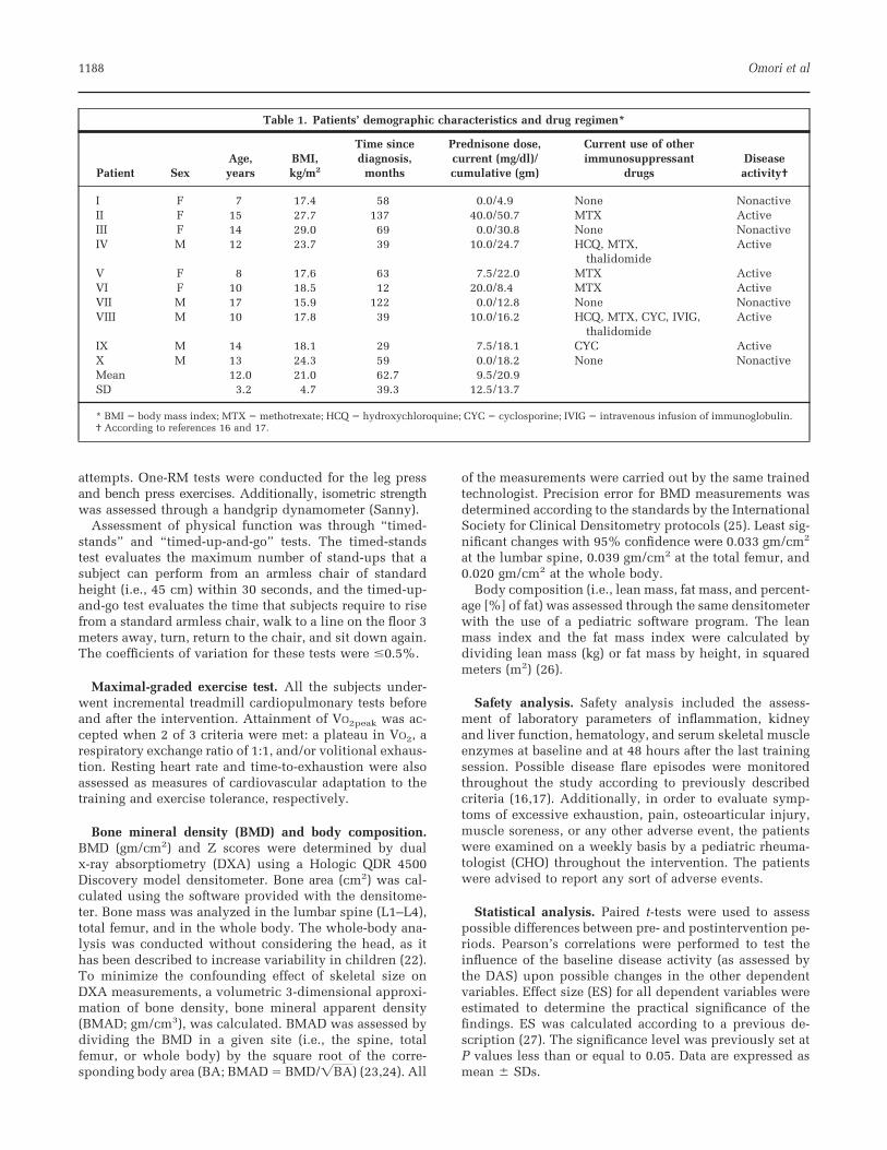
attempts. One-RM tests were conducted for the leg pressand bench press exercises. Additionally, isometric strengthwas assessed through a handgrip dynamometer (Sanny).
Assessment of physical function was through “timed-stands” and “timed-up-and-go” tests. The timed-standstest evaluates the maximum number of stand-ups that asubject can perform from an armless chair of standardheight (i.e., 45 cm) within 30 seconds, and the timed-up-and-go test evaluates the time that subjects require to risefrom a standard armless chair, walk to a line on the floor 3meters away, turn, return to the chair, and sit down again.The coefficients of variation for these tests were "0.5%.
Maximal-graded exercise test. All the subjects under-went incremental treadmill cardiopulmonary tests beforeand after the intervention. Attainment of VO2peak was ac-cepted when 2 of 3 criteria were met: a plateau in VO2, arespiratory exchange ratio of 1:1, and/or volitional exhaus-tion. Resting heart rate and time-to-exhaustion were alsoassessed as measures of cardiovascular adaptation to thetraining and exercise tolerance, respectively.
Bone mineral density (BMD) and body composition.BMD (gm/cm2) and Z scores were determined by dualx-ray absorptiometry (DXA) using a Hologic QDR 4500Discovery model densitometer. Bone area (cm2) was cal-culated using the software provided with the densitome-ter. Bone mass was analyzed in the lumbar spine (L1–L4),total femur, and in the whole body. The whole-body ana-lysis was conducted without considering the head, as ithas been described to increase variability in children (22).To minimize the confounding effect of skeletal size onDXA measurements, a volumetric 3-dimensional approxi-mation of bone density, bone mineral apparent density(BMAD; gm/cm3), was calculated. BMAD was assessed bydividing the BMD in a given site (i.e., the spine, totalfemur, or whole body) by the square root of the corre-sponding body area (BA; BMAD # BMD/$BA) (23,24). All
of the measurements were carried out by the same trainedtechnologist. Precision error for BMD measurements wasdetermined according to the standards by the InternationalSociety for Clinical Densitometry protocols (25). Least sig-nificant changes with 95% confidence were 0.033 gm/cm2
at the lumbar spine, 0.039 gm/cm2 at the total femur, and0.020 gm/cm2 at the whole body.
Body composition (i.e., lean mass, fat mass, and percent-age [%] of fat) was assessed through the same densitometerwith the use of a pediatric software program. The leanmass index and the fat mass index were calculated bydividing lean mass (kg) or fat mass by height, in squaredmeters (m2) (26).
Safety analysis. Safety analysis included the assess-ment of laboratory parameters of inflammation, kidneyand liver function, hematology, and serum skeletal muscleenzymes at baseline and at 48 hours after the last trainingsession. Possible disease flare episodes were monitoredthroughout the study according to previously describedcriteria (16,17). Additionally, in order to evaluate symp-toms of excessive exhaustion, pain, osteoarticular injury,muscle soreness, or any other adverse event, the patientswere examined on a weekly basis by a pediatric rheuma-tologist (CHO) throughout the intervention. The patientswere advised to report any sort of adverse events.
Statistical analysis. Paired t-tests were used to assesspossible differences between pre- and postintervention pe-riods. Pearson’s correlations were performed to test theinfluence of the baseline disease activity (as assessed bythe DAS) upon possible changes in the other dependentvariables. Effect size (ES) for all dependent variables wereestimated to determine the practical significance of thefindings. ES was calculated according to a previous de-scription (27). The significance level was previously set atP values less than or equal to 0.05. Data are expressed asmean " SDs.
Table 1. Patients’ demographic characteristics and drug regimen*
Patient SexAge,years
BMI,kg/m2
Time sincediagnosis,
months
Prednisone dose,current (mg/dl)/cumulative (gm)
Current use of otherimmunosuppressant
drugsDisease
activity†
I F 7 17.4 58 0.0/4.9 None NonactiveII F 15 27.7 137 40.0/50.7 MTX ActiveIII F 14 29.0 69 0.0/30.8 None NonactiveIV M 12 23.7 39 10.0/24.7 HCQ, MTX,
thalidomideActive
V F 8 17.6 63 7.5/22.0 MTX ActiveVI F 10 18.5 12 20.0/8.4 MTX ActiveVII M 17 15.9 122 0.0/12.8 None NonactiveVIII M 10 17.8 39 10.0/16.2 HCQ, MTX, CYC, IVIG,
thalidomideActive
IX M 14 18.1 29 7.5/18.1 CYC ActiveX M 13 24.3 59 0.0/18.2 None NonactiveMean 12.0 21.0 62.7 9.5/20.9SD 3.2 4.7 39.3 12.5/13.7
* BMI # body mass index; MTX # methotrexate; HCQ # hydroxychloroquine; CYC # cyclosporine; IVIG # intravenous infusion of immunoglobulin.† According to references 16 and 17.
1188 Omori et al
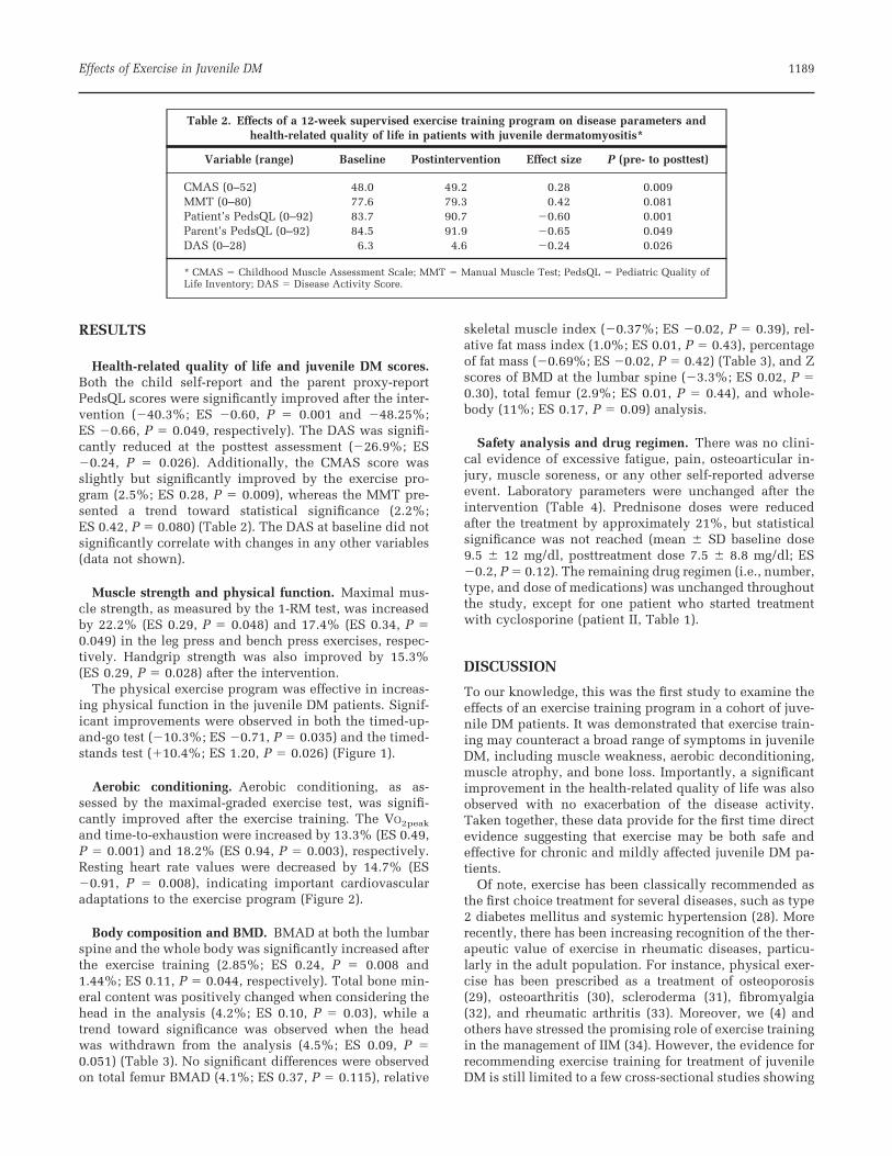
RESULTS
Health-related quality of life and juvenile DM scores.Both the child self-report and the parent proxy-reportPedsQL scores were significantly improved after the inter-vention (%40.3%; ES %0.60, P # 0.001 and %48.25%;ES %0.66, P # 0.049, respectively). The DAS was signifi-cantly reduced at the posttest assessment (%26.9%; ES%0.24, P # 0.026). Additionally, the CMAS score wasslightly but significantly improved by the exercise pro-gram (2.5%; ES 0.28, P # 0.009), whereas the MMT pre-sented a trend toward statistical significance (2.2%;ES 0.42, P # 0.080) (Table 2). The DAS at baseline did notsignificantly correlate with changes in any other variables(data not shown).
Muscle strength and physical function. Maximal mus-cle strength, as measured by the 1-RM test, was increasedby 22.2% (ES 0.29, P # 0.048) and 17.4% (ES 0.34, P #0.049) in the leg press and bench press exercises, respec-tively. Handgrip strength was also improved by 15.3%(ES 0.29, P # 0.028) after the intervention.
The physical exercise program was effective in increas-ing physical function in the juvenile DM patients. Signif-icant improvements were observed in both the timed-up-and-go test (%10.3%; ES %0.71, P # 0.035) and the timed-stands test (&10.4%; ES 1.20, P # 0.026) (Figure 1).
Aerobic conditioning. Aerobic conditioning, as as-sessed by the maximal-graded exercise test, was signifi-cantly improved after the exercise training. The VO2peak
and time-to-exhaustion were increased by 13.3% (ES 0.49,P # 0.001) and 18.2% (ES 0.94, P # 0.003), respectively.Resting heart rate values were decreased by 14.7% (ES%0.91, P # 0.008), indicating important cardiovascularadaptations to the exercise program (Figure 2).
Body composition and BMD. BMAD at both the lumbarspine and the whole body was significantly increased afterthe exercise training (2.85%; ES 0.24, P # 0.008 and1.44%; ES 0.11, P # 0.044, respectively). Total bone min-eral content was positively changed when considering thehead in the analysis (4.2%; ES 0.10, P # 0.03), while atrend toward significance was observed when the headwas withdrawn from the analysis (4.5%; ES 0.09, P #0.051) (Table 3). No significant differences were observedon total femur BMAD (4.1%; ES 0.37, P # 0.115), relative
skeletal muscle index (%0.37%; ES %0.02, P # 0.39), rel-ative fat mass index (1.0%; ES 0.01, P # 0.43), percentageof fat mass (%0.69%; ES %0.02, P # 0.42) (Table 3), and Zscores of BMD at the lumbar spine (%3.3%; ES 0.02, P #0.30), total femur (2.9%; ES 0.01, P # 0.44), and whole-body (11%; ES 0.17, P # 0.09) analysis.
Safety analysis and drug regimen. There was no clini-cal evidence of excessive fatigue, pain, osteoarticular in-jury, muscle soreness, or any other self-reported adverseevent. Laboratory parameters were unchanged after theintervention (Table 4). Prednisone doses were reducedafter the treatment by approximately 21%, but statisticalsignificance was not reached (mean " SD baseline dose9.5 " 12 mg/dl, posttreatment dose 7.5 " 8.8 mg/dl; ES%0.2, P # 0.12). The remaining drug regimen (i.e., number,type, and dose of medications) was unchanged throughoutthe study, except for one patient who started treatmentwith cyclosporine (patient II, Table 1).
DISCUSSION
To our knowledge, this was the first study to examine theeffects of an exercise training program in a cohort of juve-nile DM patients. It was demonstrated that exercise train-ing may counteract a broad range of symptoms in juvenileDM, including muscle weakness, aerobic deconditioning,muscle atrophy, and bone loss. Importantly, a significantimprovement in the health-related quality of life was alsoobserved with no exacerbation of the disease activity.Taken together, these data provide for the first time directevidence suggesting that exercise may be both safe andeffective for chronic and mildly affected juvenile DM pa-tients.
Of note, exercise has been classically recommended asthe first choice treatment for several diseases, such as type2 diabetes mellitus and systemic hypertension (28). Morerecently, there has been increasing recognition of the ther-apeutic value of exercise in rheumatic diseases, particu-larly in the adult population. For instance, physical exer-cise has been prescribed as a treatment of osteoporosis(29), osteoarthritis (30), scleroderma (31), fibromyalgia(32), and rheumatic arthritis (33). Moreover, we (4) andothers have stressed the promising role of exercise trainingin the management of IIM (34). However, the evidence forrecommending exercise training for treatment of juvenileDM is still limited to a few cross-sectional studies showing
Table 2. Effects of a 12-week supervised exercise training program on disease parameters andhealth-related quality of life in patients with juvenile dermatomyositis*
Variable (range) Baseline Postintervention Effect size P (pre- to posttest)
CMAS (0–52) 48.0 49.2 0.28 0.009MMT (0–80) 77.6 79.3 0.42 0.081Patient’s PedsQL (0–92) 83.7 90.7 %0.60 0.001Parent’s PedsQL (0–92) 84.5 91.9 %0.65 0.049DAS (0–28) 6.3 4.6 %0.24 0.026
* CMAS # Childhood Muscle Assessment Scale; MMT # Manual Muscle Test; PedsQL # Pediatric Quality ofLife Inventory; DAS # Disease Activity Score.
Effects of Exercise in Juvenile DM 1189
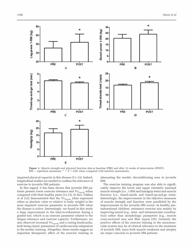
impaired physical capacity in this disease (11–13). Indeed,longitudinal studies are needed to confirm the relevance ofexercise to juvenile DM patients.
In this regard, it has been shown that juvenile DM pa-tients present lower exercise tolerance and VO2peak whencompared with their healthy peers (11,13). In fact, Takkenet al (12) demonstrated that the VO2peak (data expressedeither as absolute value or relative to body weight) is themost impaired exercise parameter in juvenile DM whenthe disease is active. Interestingly, we found in this studya large improvement in the time-to-exhaustion during agraded test, which is an exercise parameter related to thefatigue tolerance and exercise capacity. Furthermore, wealso observed increased VO2peak and a resting bradicardia,both being classic parameters of cardiovascular adaptationto the aerobic training. Altogether, these results suggest animportant therapeutic effect of the exercise training in
attenuating the aerobic deconditioning seen in juvenileDM.
The exercise training program was also able to signifi-cantly improve the lower and upper extremity maximalmuscle strength (i.e., 1-RM and handgrip tests) and musclefunction (i.e., timed-stands and timed-up-and-go tests).Interestingly, the improvements in the objective measuresof muscle strength and function were paralleled by theimprovements in the juvenile DM scores. In healthy pre-maturational children, resistance exercise acts mainly byimproving neural (e.g., inter- and intramuscular coordina-tion) rather than morphologic parameters (e.g., musclecross-sectional area and fiber types) (35). Certainly thepositive effects of the exercise training in the neuromus-cular system may be of clinical relevance in the treatmentof juvenile DM, since both muscle weakness and atrophyare major concerns to juvenile DM patients.
Figure 1. Muscle strength and physical function data at baseline (PRE) and after 12 weeks of intervention (POST).RM # repetition maximum; * # P ' 0.05 when compared with baseline assessments.
1190 Omori et al
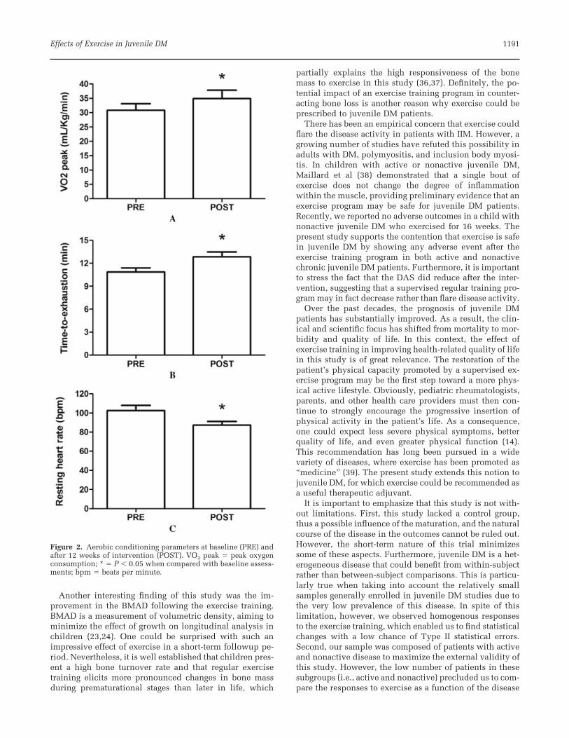
Another interesting finding of this study was the im-provement in the BMAD following the exercise training.BMAD is a measurement of volumetric density, aiming tominimize the effect of growth on longitudinal analysis inchildren (23,24). One could be surprised with such animpressive effect of exercise in a short-term followup pe-riod. Nevertheless, it is well established that children pres-ent a high bone turnover rate and that regular exercisetraining elicits more pronounced changes in bone massduring prematurational stages than later in life, which
partially explains the high responsiveness of the bonemass to exercise in this study (36,37). Definitely, the po-tential impact of an exercise training program in counter-acting bone loss is another reason why exercise could beprescribed to juvenile DM patients.
There has been an empirical concern that exercise couldflare the disease activity in patients with IIM. However, agrowing number of studies have refuted this possibility inadults with DM, polymyositis, and inclusion body myosi-tis. In children with active or nonactive juvenile DM,Maillard et al (38) demonstrated that a single bout ofexercise does not change the degree of inflammationwithin the muscle, providing preliminary evidence that anexercise program may be safe for juvenile DM patients.Recently, we reported no adverse outcomes in a child withnonactive juvenile DM who exercised for 16 weeks. Thepresent study supports the contention that exercise is safein juvenile DM by showing any adverse event after theexercise training program in both active and nonactivechronic juvenile DM patients. Furthermore, it is importantto stress the fact that the DAS did reduce after the inter-vention, suggesting that a supervised regular training pro-gram may in fact decrease rather than flare disease activity.
Over the past decades, the prognosis of juvenile DMpatients has substantially improved. As a result, the clin-ical and scientific focus has shifted from mortality to mor-bidity and quality of life. In this context, the effect ofexercise training in improving health-related quality of lifein this study is of great relevance. The restoration of thepatient’s physical capacity promoted by a supervised ex-ercise program may be the first step toward a more phys-ical active lifestyle. Obviously, pediatric rheumatologists,parents, and other health care providers must then con-tinue to strongly encourage the progressive insertion ofphysical activity in the patient’s life. As a consequence,one could expect less severe physical symptoms, betterquality of life, and even greater physical function (14).This recommendation has long been pursued in a widevariety of diseases, where exercise has been promoted as“medicine” (39). The present study extends this notion tojuvenile DM, for which exercise could be recommended asa useful therapeutic adjuvant.
It is important to emphasize that this study is not with-out limitations. First, this study lacked a control group,thus a possible influence of the maturation, and the naturalcourse of the disease in the outcomes cannot be ruled out.However, the short-term nature of this trial minimizessome of these aspects. Furthermore, juvenile DM is a het-erogeneous disease that could benefit from within-subjectrather than between-subject comparisons. This is particu-larly true when taking into account the relatively smallsamples generally enrolled in juvenile DM studies due tothe very low prevalence of this disease. In spite of thislimitation, however, we observed homogenous responsesto the exercise training, which enabled us to find statisticalchanges with a low chance of Type II statistical errors.Second, our sample was composed of patients with activeand nonactive disease to maximize the external validity ofthis study. However, the low number of patients in thesesubgroups (i.e., active and nonactive) precluded us to com-pare the responses to exercise as a function of the disease
Figure 2. Aerobic conditioning parameters at baseline (PRE) andafter 12 weeks of intervention (POST). VO2 peak # peak oxygenconsumption; * # P ' 0.05 when compared with baseline assess-ments; bpm # beats per minute.
Effects of Exercise in Juvenile DM 1191
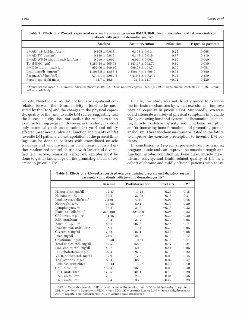
activity. Nonetheless, we did not find any significant cor-relation between the disease activity at baseline (as mea-sured by the DAS) and the changes in the physical capac-ity, quality of life, and juvenile DM scores, suggesting thatthe disease activity does not predict the responses to anexercise training program. However, as this study involvedonly chronically (disease duration !1 year) and mildlyaffected (near normal physical function and quality of life)juvenile DM patients, no extrapolation of the present find-ings can be done to patients with exacerbated muscleweakness and who are early in their disease course. Fur-ther randomized controlled trials with larger and diversi-fied (e.g., active, remissive, refractory) samples must bedone to gather knowledge on the promising effects of ex-ercise in juvenile DM.
Finally, this study was not directly aimed to examinethe intrinsic mechanisms by which exercise can improvephysical capacity in juvenile DM. Supposedly, exercisecould attenuate a variety of physical symptoms in juvenileDM by reducing local and systemic inflammation, enhanc-ing muscle oxidative capacity, reducing bone resorptionand/or increasing bone formation, and promoting proteinanabolism. These mechanisms must be tested in the futureto improve the exercise prescription to juvenile DM pa-tients.
In conclusion, a 12-week supervised exercise trainingprogram is safe and can improve the muscle strength andfunction, aerobic conditioning, bone mass, muscle mass,disease activity, and health-related quality of life in acohort of chronic and mildly affected patients with active
Table 3. Effects of a 12-week supervised exercise training program on BMAD, BMC, lean mass index, and fat mass index inpatients with juvenile dermatomyositis*
Baseline Postintervention Effect size P (pre- to posttest)
BMAD (L1–L4) (gm/cm3) 0.105 " 0.013 0.108 " 0.011 0.24 0.008BMAD-TF (gm/cm3) 0.139 " 0.015 0.145 " 0.015 0.37 0.110BMAD-WB (without head) (gm/cm3) 0.019 " 0.002 0.020 " 0.002 0.10 0.040Total BMC (gm) 1,289.24 " 505.58 1,343.67 " 562.79 0.10 0.030BMC (without head) (gm) 952.90 " 440.10 996.06 " 491.74 0.09 0.051Lean mass/h2 (gm/m2) 1,3913.5 " 1,883.8 1,3861.7 " 1,905.9 %0.02 0.380Fat mass/h2 (gm/m2) 7,346.1 " 3,989.5 7,419.3 " 4,724.6 0.02 0.430Percentage of fat mass 31.7 " 10.6 31.5 " 12.7 %0.02 0.420
* Values are the mean " SD unless indicated otherwise. BMAD # bone mineral apparent density; BMC # bone mineral content; TF # total femur;WB # whole body.
Table 4. Effects of a 12-week supervised exercise training program on laboratory serumparameters in patients with juvenile dermatomyositis*
Baseline Postintervention Effect size P
Hemoglobin, gm/dl 12.87 13.23 0.25 0.15Hematocrit, % 37.51 37.95 0.13 0.31Leukocytes, cells/mm3 7,130 7,110 %0.01 0.48Neutrophils, % 56.95 59.1 0.15 0.29Lymphocytes, % 33.3 31.6 %0.13 0.32Platelets, cells/mm3 333,400 324,200 %0.08 0.32CRP level, mg/liter 1.46 1.87 0.20 0.30ESR, mm/hour 16.2 21.2 0.39 0.06Ferritin, #g/liter 83.1 107.8 0.48 0.14Insulinemia, units/liter 15.1 11.4 %0.29 0.06Glycemia, mg/dl 79.2 82.1 0.55 0.08Urea, mg/dl 24.6 26.5 0.16 0.17Creatinine, mg/dl 0.58 0.61 0.16 0.11Total cholesterol, mg/dl 152.5 158.6 0.17 0.24HDL cholesterol, mg/dl 46.7 50.6 0.44 0.06LDL cholesterol, mg/dl 92.4 97.7 0.19 0.22VLDL cholesterol, mg/dl 17.9 17.5 %0.03 0.45Triglycerides, mg/dl 89.4 88.0 %0.02 0.47Aldolase, units/liter 6.33 5.77 %0.30 0.10CK, units/liter 132.4 137.6 0.03 0.40LDH, units/liter 178.5 184.4 0.16 0.29AST, units/liter 23 23.2 0.01 0.46ALT, units/liter 39.4 36.3 %0.22 0.23
* CRP # C-reactive protein; ESR # erythrocyte sedimentation rate; HDL # high-density lipoprotein;LDL # low-density lipoprotein; VLDL # very LDL; CK # creatine kinase; LDH # lactate dehydrogenase;AST # aspartate aminotransferase; ALT # alanine aminotransferase.
1192 Omori et al
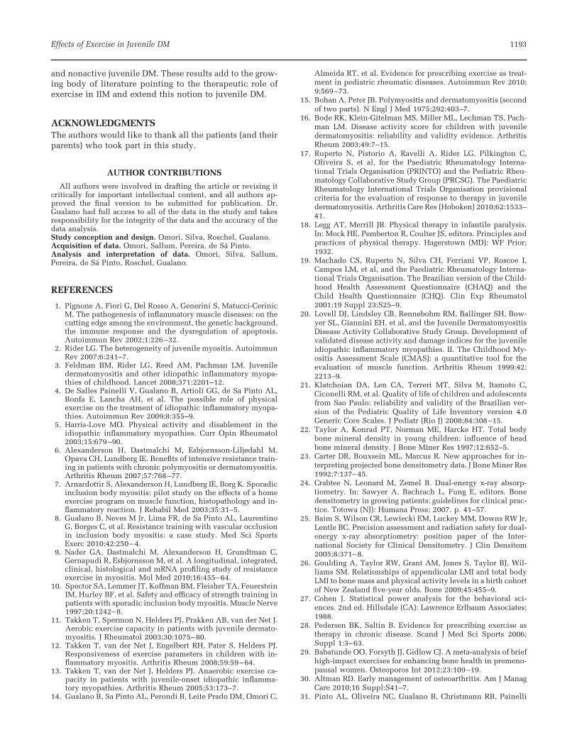
and nonactive juvenile DM. These results add to the grow-ing body of literature pointing to the therapeutic role ofexercise in IIM and extend this notion to juvenile DM.
ACKNOWLEDGMENTSThe authors would like to thank all the patients (and theirparents) who took part in this study.
AUTHOR CONTRIBUTIONSAll authors were involved in drafting the article or revising it
critically for important intellectual content, and all authors ap-proved the final version to be submitted for publication. Dr.Gualano had full access to all of the data in the study and takesresponsibility for the integrity of the data and the accuracy of thedata analysis.Study conception and design. Omori, Silva, Roschel, Gualano.Acquisition of data. Omori, Sallum, Pereira, de Sa Pinto.Analysis and interpretation of data. Omori, Silva, Sallum,Pereira, de Sa Pinto, Roschel, Gualano.
REFERENCES
1. Pignone A, Fiori G, Del Rosso A, Generini S, Matucci-CerinicM. The pathogenesis of inflammatory muscle diseases: on thecutting edge among the environment, the genetic background,the immune response and the dysregulation of apoptosis.Autoimmun Rev 2002;1:226–32.
2. Rider LG. The heterogeneity of juvenile myositis. AutoimmunRev 2007;6:241–7.
3. Feldman BM, Rider LG, Reed AM, Pachman LM. Juveniledermatomyositis and other idiopathic inflammatory myopa-thies of childhood. Lancet 2008;371:2201–12.
4. De Salles Painelli V, Gualano B, Artioli GG, de Sa Pinto AL,Bonfa E, Lancha AH, et al. The possible role of physicalexercise on the treatment of idiopathic inflammatory myopa-thies. Autoimmun Rev 2009;8:355–9.
5. Harris-Love MO. Physical activity and disablement in theidiopathic inflammatory myopathies. Curr Opin Rheumatol2003;15:679–90.
6. Alexanderson H, Dastmalchi M, Esbjornsson-Liljedahl M,Opava CH, Lundberg IE. Benefits of intensive resistance train-ing in patients with chronic polymyositis or dermatomyositis.Arthritis Rheum 2007;57:768–77.
7. Arnardottir S, Alexanderson H, Lundberg IE, Borg K. Sporadicinclusion body myositis: pilot study on the effects of a homeexercise program on muscle function, histopathology and in-flammatory reaction. J Rehabil Med 2003;35:31–5.
8. Gualano B, Neves M Jr, Lima FR, de Sa Pinto AL, LaurentinoG, Borges C, et al. Resistance training with vascular occlusionin inclusion body myositis: a case study. Med Sci SportsExerc 2010;42:250–4.
9. Nader GA, Dastmalchi M, Alexanderson H, Grundtman C,Gernapudi R, Esbjornsson M, et al. A longitudinal, integrated,clinical, histological and mRNA profiling study of resistanceexercise in myositis. Mol Med 2010;16:455–64.
10. Spector SA, Lemmer JT, Koffman BM, Fleisher TA, FeuersteinIM, Hurley BF, et al. Safety and efficacy of strength training inpatients with sporadic inclusion body myositis. Muscle Nerve1997;20:1242–8.
11. Takken T, Spermon N, Helders PJ, Prakken AB, van der Net J.Aerobic exercise capacity in patients with juvenile dermato-myositis. J Rheumatol 2003;30:1075–80.
12. Takken T, van der Net J, Engelbert RH, Pater S, Helders PJ.Responsiveness of exercise parameters in children with in-flammatory myositis. Arthritis Rheum 2008;59:59–64.
13. Takken T, van der Net J, Helders PJ. Anaerobic exercise ca-pacity in patients with juvenile-onset idiopathic inflamma-tory myopathies. Arthritis Rheum 2005;53:173–7.
14. Gualano B, Sa Pinto AL, Perondi B, Leite Prado DM, Omori C,
Almeida RT, et al. Evidence for prescribing exercise as treat-ment in pediatric rheumatic diseases. Autoimmun Rev 2010;9:569–73.
15. Bohan A, Peter JB. Polymyositis and dermatomyositis (secondof two parts). N Engl J Med 1975;292:403–7.
16. Bode RK, Klein-Gitelman MS, Miller ML, Lechman TS, Pach-man LM. Disease activity score for children with juveniledermatomyositis: reliability and validity evidence. ArthritisRheum 2003;49:7–15.
17. Ruperto N, Pistorio A, Ravelli A, Rider LG, Pilkington C,Oliveira S, et al, for the Paediatric Rheumatology Interna-tional Trials Organisation (PRINTO) and the Pediatric Rheu-matology Collaborative Study Group (PRCSG). The PaediatricRheumatology International Trials Organisation provisionalcriteria for the evaluation of response to therapy in juveniledermatomyositis. Arthritis Care Res (Hoboken) 2010;62:1533–41.
18. Legg AT, Merrill JB. Physical therapy in infantile paralysis.In: Mock HE, Pemberton R, Coulter JS, editors. Principles andpractices of physical therapy. Hagerstown (MD): WF Prior;1932.
19. Machado CS, Ruperto N, Silva CH, Ferriani VP, Roscoe I,Campos LM, et al, and the Paediatric Rheumatology Interna-tional Trials Organisation. The Brazilian version of the Child-hood Health Assessment Questionnaire (CHAQ) and theChild Health Questionnaire (CHQ). Clin Exp Rheumatol2001;19 Suppl 23:S25–9.
20. Lovell DJ, Lindsley CB, Rennebohm RM, Ballinger SH, Bow-yer SL, Giannini EH, et al, and the Juvenile DermatomyositisDisease Activity Collaborative Study Group. Development ofvalidated disease activity and damage indices for the juvenileidiopathic inflammatory myopathies. II. The Childhood My-ositis Assessment Scale (CMAS): a quantitative tool for theevaluation of muscle function. Arthritis Rheum 1999:42:2213–9.
21. Klatchoian DA, Len CA, Terreri MT, Silva M, Itamoto C,Ciconelli RM, et al. Quality of life of children and adolescentsfrom Sao Paulo: reliability and validity of the Brazilian ver-sion of the Pediatric Quality of Life Inventory version 4.0Generic Core Scales. J Pediatr (Rio J) 2008;84:308–15.
22. Taylor A, Konrad PT, Norman ME, Harcke HT. Total bodybone mineral density in young children: influence of headbone mineral density. J Bone Miner Res 1997;12:652–5.
23. Carter DR, Bouxsein ML, Marcus R. New approaches for in-terpreting projected bone densitometry data. J Bone Miner Res1992;7:137–45.
24. Crabtee N, Leonard M, Zemel B. Dual-energy x-ray absorp-tiometry. In: Sawyer A, Bachrach L, Fung E, editors. Bonedensitometry in growing patients: guidelines for clinical prac-tice. Totowa (NJ): Humana Press; 2007. p. 41–57.
25. Baim S, Wilson CR, Lewiecki EM, Luckey MM, Downs RW Jr,Lentle BC. Precision assessment and radiation safety for dual-energy x-ray absorptiometry: position paper of the Inter-national Society for Clinical Densitometry. J Clin Densitom2005;8:371–8.
26. Goulding A, Taylor RW, Grant AM, Jones S, Taylor BJ, Wil-liams SM. Relationships of appendicular LMI and total bodyLMI to bone mass and physical activity levels in a birth cohortof New Zealand five-year olds. Bone 2009;45:455–9.
27. Cohen J. Statistical power analysis for the behavioral sci-ences. 2nd ed. Hillsdale (CA): Lawrence Erlbaum Associates;1988.
28. Pedersen BK, Saltin B. Evidence for prescribing exercise astherapy in chronic disease. Scand J Med Sci Sports 2006;Suppl 1:3–63.
29. Babatunde OO, Forsyth JJ, Gidlow CJ. A meta-analysis of briefhigh-impact exercises for enhancing bone health in premeno-pausal women. Osteoporos Int 2012;23:109–19.
30. Altman RD. Early management of osteoarthritis. Am J ManagCare 2010;16 Suppl:S41–7.
31. Pinto AL, Oliveira NC, Gualano B, Christmann RB, Painelli
Effects of Exercise in Juvenile DM 1193
VS, Artioli GG, et al. Efficacy and safety of concurrent trainingin systemic sclerosis. J Strength Cond Res 2011;25:1423–8.
32. Sanudo B, Galiano D, Carrasco L, de Hoyo M, McVeigh JG.Effects of a prolonged exercise program on key health out-comes in women with fibromyalgia: a randomized controlledtrial. J Rehabil Med 2011;43:521–6.
33. Kelley GA, Kelley KS, Hootman JM, Jones DL. Effects ofcommunity-deliverable exercise on pain and physical func-tion in adults with arthritis and other rheumatic diseases: ameta-analysis. Arthritis Care Res (Hoboken) 2011;63:79–93.
34. Alexanderson H, Lundberg IE. The role of exercise in therehabilitation of idiopathic inflammatory myopathies. CurrOpin Rheumatol 2005;17:164–71.
35. Faigenbaum AD, Kraemer WJ, Blimkie CJ, Jeffreys I, MicheliLJ, Nitka M, et al. Youth resistance training: updated position
statement paper from the national strength and conditioningassociation. J Strength Cond Res 2009;23:S60–79.
36. Guadalupe-Grau A, Fuentes T, Guerra B, Calbet JA. Exerciseand bone mass in adults. Sports Med 2009;39:439–68.
37. Bailey DA, McKay HA, Mirwald RL, Crocker PR, FaulknerRA. A six-year longitudinal study of the relationship of phys-ical activity to bone mineral accrual in growing children: theUniversity of Saskatchewan bone mineral accrual study.J Bone Miner Res 1999;14:1672–9.
38. Maillard SM, Jones R, Owens CM, Pilkington C, Woo PM,Wedderburn LR, et al. Quantitative assessments of the effectsof a single exercise session on muscles in juvenile dermato-myositis. Arthritis Rheum 2005;53:558–64.
39. Booth FW, Zwetsloot KA. Basic concepts about genes, inac-tivity and aging. Scand J Med Sci Sports 2011;20:1–4.
1194 Omori et al
RESEARCH ARTICLE Open Access
Responsiveness to exercise training in juveniledermatomyositis: a twin case studyClarissa Omori1, Danilo ML Prado1, Bruno Gualano2,3, Adriana ME Sallum1, Ana L Sá-Pinto3, Hamilton Roschel2,3,Maria B Perondi1, Clovis AA Silva1,3*
Abstract
Background: Patients with juvenile dermatomyositis (JDM) often present strong exercise intolerance and muscleweakness. However, the role of exercise training in this disease has not been investigated.
Purpose: this longitudinal case study reports on the effects of exercise training on a 7-year-old patient with JDMand on her unaffected monozygotic twin sister, who served as a control.
Methods: Both the patient who was diagnosed with JDM as well as her healthy twin underwent a 16-weekexercise training program comprising aerobic and strengthening exercises. We assessed one repetition-maximum(1-RM) leg-press and bench-press strength, balance, mobility and muscle function, blood markers of inflammationand muscle enzymes, aerobic conditioning, and disease activity scores. As a result, the healthy child had an overallgreater absolute strength, muscle function and aerobic conditioning compared to her JDM twin pair at baselineand after the trial. However, the twins presented comparable relative improvements in 1-RM bench press, 1-RM legpress, VO2peak, and time-to-exhaustion. The healthy child had greater relative increments in low-back strength andhandgrip, whereas the child with JDM presented a higher relative increase in ventilatory anaerobic thresholdparameters and functional tests. Quality of life, inflammation, muscle damage and disease activity scores remainedunchanged.
Results and Conclusion: this was the first report to describe the training response of a patient with non-activeJDM following an exercise training regimen. The child with JDM exhibited improved strength, muscle function andaerobic conditioning without presenting an exacerbation of the disease.
BackgroundJuvenile dermatomyositis (JDM) is a rare, idiopathic andnon-suppurative inflammatory disease that causes proxi-mal muscle weakness and a variety of cutaneous features[1]. Current treatment includes corticosteroids fre-quently in combination with other immunosuppressiveagents such as methotrexate and cyclosporine, or intra-venous immunoglobulin. Our group [1-4] and others[5,6] have consistently showed that, despite the improve-ments in JDM prognosis over the last three decades, anumber of patients may develop irreversible cumulativedamage due to the disease activity or its treatment.
Patients with JDM often present strong exercise intol-erance [6,7]. Furthermore, muscle waste usually occursduring periods of disease activity, leading to muscleweakness [8]. In adults with dermatomyositis and poly-myositis [9-12] and inclusion body myositis [13,14], phy-sical training studies have shown improvements inexercise capacity. Therefore, it would be clinically rele-vant to investigate the effects of exercise training inJDM patients.This longitudinal case study aimed to investigate the
effects of exercise training in a 7-year-old patient withJDM, with her unaffected monozygotic twin sister ser-ving as a control. This approach provided a uniqueopportunity to evaluate the responsiveness and tolerabil-ity to exercise in JDM.* Correspondence: [email protected]
1Pediatric Rheumatology Unit, Children’s Institute, School of Medicine,University of Sao Paulo (Av. Dr. Arnaldo, 455 - Cerqueira César), Sao Paulo(Postal code: 01246-903), BrazilFull list of author information is available at the end of the article
Omori et al. BMC Musculoskeletal Disorders 2010, 11:270http://www.biomedcentral.com/1471-2474/11/270
© 2010 Omori et al; licensee BioMed Central Ltd. This is an Open Access article distributed under the terms of the Creative CommonsAttribution License (http://creativecommons.org/licenses/by/2.0), which permits unrestricted use, distribution, and reproduction inany medium, provided the original work is properly cited.
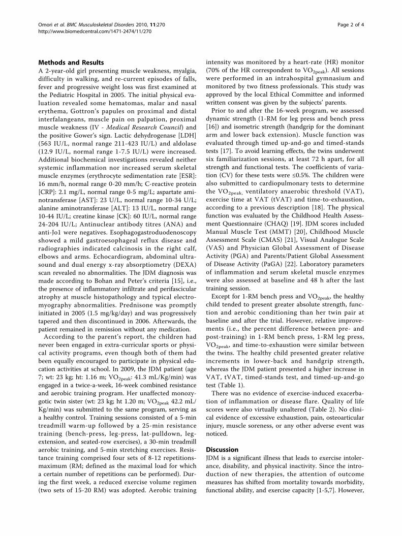
Methods and ResultsA 2-year-old girl presenting muscle weakness, myalgia,difficulty in walking, and re-current episodes of falls,fever and progressive weight loss was first examined atthe Pediatric Hospital in 2005. The initial physical eva-luation revealed some hematomas, malar and nasalerythema, Gottron’s papules on proximal and distalinterfalangeans, muscle pain on palpation, proximalmuscle weakness (IV - Medical Research Council) andthe positive Gower’s sign. Lactic dehydrogenase [LDH](563 IU/L, normal range 211-423 IU/L) and aldolase(12.9 IU/L, normal range 1-7.5 IU/L) were increased.Additional biochemical investigations revealed neithersystemic inflammation nor increased serum skeletalmuscle enzymes (erythrocyte sedimentation rate [ESR]:16 mm/h, normal range 0-20 mm/h; C-reactive protein[CRP]: 2.1 mg/L, normal range 0-5 mg/L; aspartate ami-notransferase [AST]: 23 U/L, normal range 10-34 U/L;alanine aminotransferase [ALT]: 13 IU/L, normal range10-44 IU/L; creatine kinase [CK]: 60 IU/L, normal range24-204 IU/L; Antinuclear antibody titres (ANA) andanti-Jo1 were negatives. Esophagogastroduodenoscopyshowed a mild gastroesophageal reflux disease andradiographies indicated calcinosis in the right calf,elbows and arms. Echocardiogram, abdominal ultra-sound and dual energy x-ray absorptiometry (DEXA)scan revealed no abnormalities. The JDM diagnosis wasmade according to Bohan and Peter’s criteria [15], i.e.,the presence of inflammatory infiltrate and perifascicularatrophy at muscle histopathology and typical electro-myography abnormalities. Prednisone was promptlyinitiated in 2005 (1.5 mg/kg/day) and was progressivelytapered and then discontinued in 2006. Afterwards, thepatient remained in remission without any medication.According to the parent’s report, the children had
never been engaged in extra-curricular sports or physi-cal activity programs, even though both of them hadbeen equally encouraged to participate in physical edu-cation activities at school. In 2009, the JDM patient (age7; wt: 23 kg; ht: 1.16 m; VO2peak: 41.3 mL/Kg/min) wasengaged in a twice-a-week, 16-week combined resistanceand aerobic training program. Her unaffected monozy-gotic twin sister (wt: 23 kg; ht 1.20 m; VO2peak 42.2 mL/Kg/min) was submitted to the same program, serving asa healthy control. Training sessions consisted of a 5-mintreadmill warm-up followed by a 25-min resistancetraining (bench-press, leg-press, lat-pulldown, leg-extension, and seated-row exercises), a 30-min treadmillaerobic training, and 5-min stretching exercises. Resis-tance training comprised four sets of 8-12 repetitions-maximum (RM; defined as the maximal load for whicha certain number of repetitions can be performed). Dur-ing the first week, a reduced exercise volume regimen(two sets of 15-20 RM) was adopted. Aerobic training
intensity was monitored by a heart-rate (HR) monitor(70% of the HR correspondent to VO2peak). All sessionswere performed in an intrahospital gymnasium andmonitored by two fitness professionals. This study wasapproved by the local Ethical Committee and informedwritten consent was given by the subjects’ parents.Prior to and after the 16-week program, we assessed
dynamic strength (1-RM for leg press and bench press[16]) and isometric strength (handgrip for the dominantarm and lower back extension). Muscle function wasevaluated through timed up-and-go and timed-standstests [17]. To avoid learning effects, the twins underwentsix familiarization sessions, at least 72 h apart, for allstrength and functional tests. The coefficients of varia-tion (CV) for these tests were ≤0.5%. The children werealso submitted to cardiopulmonary tests to determinethe VO2peak, ventilatory anaerobic threshold (VAT),exercise time at VAT (tVAT) and time-to-exhaustion,according to a previous description [18]. The physicalfunction was evaluated by the Childhood Health Assess-ment Questionnaire (CHAQ) [19]. JDM scores includedManual Muscle Test (MMT) [20], Childhood MuscleAssessment Scale (CMAS) [21], Visual Analogue Scale(VAS) and Physician Global Assessment of DiseaseActivity (PGA) and Parents/Patient Global Assessmentof Disease Activity (PaGA) [22]. Laboratory parametersof inflammation and serum skeletal muscle enzymeswere also assessed at baseline and 48 h after the lasttraining session.Except for 1-RM bench press and VO2peak, the healthy
child tended to present greater absolute strength, func-tion and aerobic conditioning than her twin pair atbaseline and after the trial. However, relative improve-ments (i.e., the percent difference between pre- andpost-training) in 1-RM bench press, 1-RM leg press,VO2peak, and time-to-exhaustion were similar betweenthe twins. The healthy child presented greater relativeincrements in lower-back and handgrip strength,whereas the JDM patient presented a higher increase inVAT, tVAT, timed-stands test, and timed-up-and-gotest (Table 1).There was no evidence of exercise-induced exacerba-
tion of inflammation or disease flare. Quality of lifescores were also virtually unaltered (Table 2). No clini-cal evidence of excessive exhaustion, pain, osteoarticularinjury, muscle soreness, or any other adverse event wasnoticed.
DiscussionJDM is a significant illness that leads to exercise intoler-ance, disability, and physical inactivity. Since the intro-duction of new therapies, the attention of outcomemeasures has shifted from mortality towards morbidity,functional ability, and exercise capacity [1-5,7]. However,
Omori et al. BMC Musculoskeletal Disorders 2010, 11:270http://www.biomedcentral.com/1471-2474/11/270
Page 2 of 4
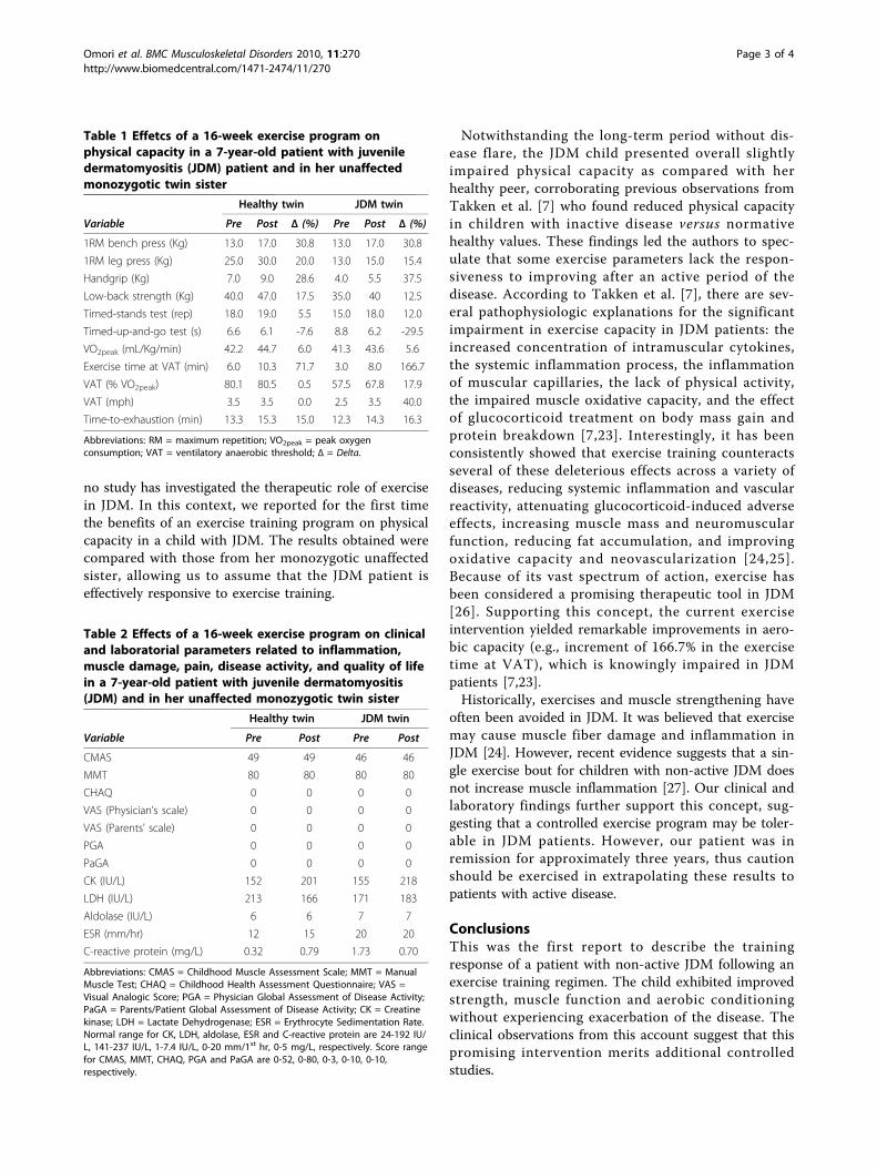
no study has investigated the therapeutic role of exercisein JDM. In this context, we reported for the first timethe benefits of an exercise training program on physicalcapacity in a child with JDM. The results obtained werecompared with those from her monozygotic unaffectedsister, allowing us to assume that the JDM patient iseffectively responsive to exercise training.
Notwithstanding the long-term period without dis-ease flare, the JDM child presented overall slightlyimpaired physical capacity as compared with herhealthy peer, corroborating previous observations fromTakken et al. [7] who found reduced physical capacityin children with inactive disease versus normativehealthy values. These findings led the authors to spec-ulate that some exercise parameters lack the respon-siveness to improving after an active period of thedisease. According to Takken et al. [7], there are sev-eral pathophysiologic explanations for the significantimpairment in exercise capacity in JDM patients: theincreased concentration of intramuscular cytokines,the systemic inflammation process, the inflammationof muscular capillaries, the lack of physical activity,the impaired muscle oxidative capacity, and the effectof glucocorticoid treatment on body mass gain andprotein breakdown [7,23]. Interestingly, it has beenconsistently showed that exercise training counteractsseveral of these deleterious effects across a variety ofdiseases, reducing systemic inflammation and vascularreactivity, attenuating glucocorticoid-induced adverseeffects, increasing muscle mass and neuromuscularfunction, reducing fat accumulation, and improvingoxidative capacity and neovascularization [24,25].Because of its vast spectrum of action, exercise hasbeen considered a promising therapeutic tool in JDM[26]. Supporting this concept, the current exerciseintervention yielded remarkable improvements in aero-bic capacity (e.g., increment of 166.7% in the exercisetime at VAT), which is knowingly impaired in JDMpatients [7,23].Historically, exercises and muscle strengthening have
often been avoided in JDM. It was believed that exercisemay cause muscle fiber damage and inflammation inJDM [24]. However, recent evidence suggests that a sin-gle exercise bout for children with non-active JDM doesnot increase muscle inflammation [27]. Our clinical andlaboratory findings further support this concept, sug-gesting that a controlled exercise program may be toler-able in JDM patients. However, our patient was inremission for approximately three years, thus cautionshould be exercised in extrapolating these results topatients with active disease.
ConclusionsThis was the first report to describe the trainingresponse of a patient with non-active JDM following anexercise training regimen. The child exhibited improvedstrength, muscle function and aerobic conditioningwithout experiencing exacerbation of the disease. Theclinical observations from this account suggest that thispromising intervention merits additional controlledstudies.
Table 1 Effetcs of a 16-week exercise program onphysical capacity in a 7-year-old patient with juveniledermatomyositis (JDM) patient and in her unaffectedmonozygotic twin sister
Healthy twin JDM twin
Variable Pre Post Δ (%) Pre Post Δ (%)
1RM bench press (Kg) 13.0 17.0 30.8 13.0 17.0 30.8
1RM leg press (Kg) 25.0 30.0 20.0 13.0 15.0 15.4
Handgrip (Kg) 7.0 9.0 28.6 4.0 5.5 37.5
Low-back strength (Kg) 40.0 47.0 17.5 35.0 40 12.5
Timed-stands test (rep) 18.0 19.0 5.5 15.0 18.0 12.0
Timed-up-and-go test (s) 6.6 6.1 -7.6 8.8 6.2 -29.5
VO2peak (mL/Kg/min) 42.2 44.7 6.0 41.3 43.6 5.6
Exercise time at VAT (min) 6.0 10.3 71.7 3.0 8.0 166.7
VAT (% VO2peak) 80.1 80.5 0.5 57.5 67.8 17.9
VAT (mph) 3.5 3.5 0.0 2.5 3.5 40.0
Time-to-exhaustion (min) 13.3 15.3 15.0 12.3 14.3 16.3
Abbreviations: RM = maximum repetition; VO2peak = peak oxygenconsumption; VAT = ventilatory anaerobic threshold; Δ = Delta.
Table 2 Effects of a 16-week exercise program on clinicaland laboratorial parameters related to inflammation,muscle damage, pain, disease activity, and quality of lifein a 7-year-old patient with juvenile dermatomyositis(JDM) and in her unaffected monozygotic twin sister
Healthy twin JDM twin
Variable Pre Post Pre Post
CMAS 49 49 46 46
MMT 80 80 80 80
CHAQ 0 0 0 0
VAS (Physician’s scale) 0 0 0 0
VAS (Parents’ scale) 0 0 0 0
PGA 0 0 0 0
PaGA 0 0 0 0
CK (IU/L) 152 201 155 218
LDH (IU/L) 213 166 171 183
Aldolase (IU/L) 6 6 7 7
ESR (mm/hr) 12 15 20 20
C-reactive protein (mg/L) 0.32 0.79 1.73 0.70
Abbreviations: CMAS = Childhood Muscle Assessment Scale; MMT = ManualMuscle Test; CHAQ = Childhood Health Assessment Questionnaire; VAS =Visual Analogic Score; PGA = Physician Global Assessment of Disease Activity;PaGA = Parents/Patient Global Assessment of Disease Activity; CK = Creatinekinase; LDH = Lactate Dehydrogenase; ESR = Erythrocyte Sedimentation Rate.Normal range for CK, LDH, aldolase, ESR and C-reactive protein are 24-192 IU/L, 141-237 IU/L, 1-7.4 IU/L, 0-20 mm/1st hr, 0-5 mg/L, respectively. Score rangefor CMAS, MMT, CHAQ, PGA and PaGA are 0-52, 0-80, 0-3, 0-10, 0-10,respectively.
Omori et al. BMC Musculoskeletal Disorders 2010, 11:270http://www.biomedcentral.com/1471-2474/11/270
Page 3 of 4
ConsentWritten informed consent was obtained from thepatients’ relatives for publication of this case report andany accompanying images. A copy of the written con-sent is available for review by the Editor-in-Chief of thisjournal.
Declaration of competing interestsThe authors declare that they have no competing interests.
AcknowledgementsThis study was supported by Fundação de Amparo à Pesquisa do Estado deSão Paulo - FAPESP, Conselho Nacional de Desenvolvimento Científico eTecnológico - CNPQ, and Federico Foundation.
Author details1Pediatric Rheumatology Unit, Children’s Institute, School of Medicine,University of Sao Paulo (Av. Dr. Arnaldo, 455 - Cerqueira César), Sao Paulo(Postal code: 01246-903), Brazil. 2School of Physical Education and Sports,University of Sao Paulo (Av Mello de Moaraes, 65 - Butantã), Sao Paulo(Postal code: 05508-030), Brazil. 3Division of Rheumatology, School ofMedicine, University of Sao Paulo (Av. Dr. Arnaldo, 455 - Cerqueira César),Sao Paulo (Postal code: 01246-903), Brazil.
Authors’ contributionsCO, BG and CAAS were responsible for the concept and design; CO, DMLPand BG were significant manuscript writers; AMES, ALSP, HR, BP wereresponsible for the data acquisition, analysis and interpretation; CAAS was asignificant manuscript reviewer. All authors read and approved the finalmanuscript.
Received: 16 June 2010 Accepted: 25 November 2010Published: 25 November 2010
References1. Sallum AM, Kiss MH, Sachetti S, Resende MB, Moutinho KC, Carvalho Mde S,
Silva CA, Marie SK: Juvenile dermatomyositis: clinical, laboratorial,histological, therapeutical and evolutive parameters of 35 patients. ArqNeuropsiquiatr 2002, 60(4):889-899.
2. Sallum AM, Pivato FC, Doria-Filho U, Aikawa NE, Liphaus BL, Marie SK,Silva CA: Risk factors associated with calcinosis of juveniledermatomyositis. J Pediatr (Rio J) 2008, 84(1):68-74.
3. Santiago RA, Silva CA, Caparbo VF, Sallum AM, Pereira RM: Bone mineralapparent density in juvenile dermatomyositis: the role of lean bodymass and glucocorticoid use. Scand J Rheumatol 2008, 37(1):40-47.
4. Sato JO, Sallum AM, Ferriani VP, Marini R, Sacchetti SB, Okuda EM,Carvalho JF, Pereira RM, Len CA, Terreri MT, et al: A Brazilian registry ofjuvenile dermatomyositis: onset features and classification of 189 cases.Clin Exp Rheumatol 2009, 27(6):1031-1038.
5. Takken T, Spermon N, Helders PJ, Prakken AB, Van Der Net J: Aerobicexercise capacity in patients with juvenile dermatomyositis. J Rheumatol2003, 30(5):1075-1080.
6. Takken T, van der Net J, Helders PJ: Anaerobic exercise capacity inpatients with juvenile-onset idiopathic inflammatory myopathies. ArthritisRheum 2005, 53(2):173-177.
7. Takken T, van der Net J, Engelbert RH, Pater S, Helders PJ: Responsivenessof exercise parameters in children with inflammatory myositis. ArthritisRheum 2008, 59(1):59-64.
8. Hilario MO, Yamashita H, Lutti D, Len C, Terreri MT, Lederman H: Juvenileidiopathic inflammatory myopathies: the value of magnetic resonanceimaging in the detection of muscle involvement. Sao Paulo Med J 2000,118(2):35-40.
9. Wiesinger GF, Quittan M, Aringer M, Seeber A, Volc-Platzer B, Smolen J,Graninger W: Improvement of physical fitness and muscle strength inpolymyositis/dermatomyositis patients by a training programme. Br JRheumatol 1998, 37(2):196-200.
10. Wiesinger GF, Quittan M, Graninger M, Seeber A, Ebenbichler G, Sturm B,Kerschan K, Smolen J, Graninger W: Benefit of 6 months long-term
physical training in polymyositis/dermatomyositis patients. Br JRheumatol 1998, 37(12):1338-1342.
11. Alexanderson H, Dastmalchi M, Esbjornsson-Liljedahl M, Opava CH,Lundberg IE: Benefits of intensive resistance training in patients withchronic polymyositis or dermatomyositis. Arthritis Rheum 2007,57(5):768-777.
12. Alexanderson H, Stenstrom CH, Jenner G, Lundberg I: The safety of aresistive home exercise program in patients with recent onset activepolymyositis or dermatomyositis. Scand J Rheumatol 2000, 29(5):295-301.
13. Arnardottir S, Alexanderson H, Lundberg IE, Borg K: Sporadic inclusionbody myositis: pilot study on the effects of a home exercise program onmuscle function, histopathology and inflammatory reaction. J RehabilMed 2003, 35(1):31-35.
14. Gualano B, Neves M Jr, Lima FR, Pinto AL, Laurentino G, Borges C,Baptista L, Artioli GG, Aoki MS, Moriscot A, et al: Resistance training withvascular occlusion in inclusion body myositis: a case study. Med SciSports Exerc 42(2):250-254.
15. Bohan A, Peter JB: Polymyositis and dermatomyositis (second of twoparts). N Engl J Med 1975, 292(8):403-407.
16. Brown LE, Weir JP: ASEP procedures recommendation I: accurateassessment of muscular strength and power. Journal of ExercisePhysiologyonline 2001, 4(3):1-21.
17. Newcomer KL, Krug HE, Mahowald ML: Validity and reliability of thetimed-stands test for patients with rheumatoid arthritis and otherchronic diseases. J Rheumatol 1993, 20(1):21-27.
18. Pinto ALS, Oliveira NC, Gualano B, Souza RBC, de Salles VP, Artioli GG,Prado DM, Lima FR: Efficacy and safety of concurrent training in systemicsclerosis. J Strength and Cond Res .
19. Machado CS, Ruperto N, Silva CH, Ferriani VP, Roscoe I, Campos LM,Oliveira SK, Kiss MH, Bica BE, Sztajnbok F, et al: Paediatric RheumatologyInternational Trials Organisation. The Brazilian version of the ChildhoodHealth Assessment Questionnaire (CHAQ) and the Child HealthQuestionnaire (CHQ). Clin Exp Rheumatol 2001, 19(4 Suppl 23):S25-29.
20. Legg AT, Merril JB: Physical therapy in infantili paralysis. Principles andparactice of physical therapy 1932.
21. Lovell DJ, Lindsley CB, Rennebohm RM, Ballinger SH, Bowyer SL,Giannini EH, Hicks JE, Levinson JE, Mier R, Pachman LM, et al: Developmentof validated disease activity and damage indices for the juvenileidiopathic inflammatory myopathies. II. The Childhood MyositisAssessment Scale (CMAS): a quantitative tool for the evaluation ofmuscle function. The Juvenile Dermatomyositis Disease ActivityCollaborative Study Group. Arthritis Rheum 1999, 42(10):2213-2219.
22. Rider LG, Feldman BM, Perez MD, Rennebohm RM, Lindsley CB, Zemel LS,Wallace CA, Ballinger SH, Bowyer SL, Reed AM, et al: Development ofvalidated disease activity and damage indices for the juvenile idiopathicinflammatory myopathies: I. Physician, parent, and patient globalassessments. Juvenile Dermatomyositis Disease Activity CollaborativeStudy Group. Arthritis Rheum 1997, 40(11):1976-1983.
23. Takken T, van der Net J, Helders PJ: The reliability of an aerobic and ananaerobic exercise tolerance test in patients with juvenile onsetdermatomyositis. J Rheumatol 2005, 32(4):734-739.
24. de Salles Painelli V, Gualano B, Artioli GG, de Sa Pinto AL, Bonfa E, LanchaJunior AH, Lima FR: The possible role of physical exercise on thetreatment of idiopathic inflammatory myopathies. Autoimmun Rev 2009,8(5):355-359.
25. Pedersen BK, Saltin B: Evidence for prescribing exercise as therapy inchronic disease. Scand J Med Sci Sports 2006, 16(Suppl 1):3-63.
26. Klepper SE: Exercise in pediatric rheumatic diseases. Curr Opin Rheumatol2008, 20(5):619-624.
27. Maillard SM, Jones R, Owens CM, Pilkington C, Woo PM, Wedderburn LR,Murray KJ: Quantitative assessments of the effects of a single exercisesession on muscles in juvenile dermatomyositis. Arthritis Rheum 2005,53(4):558-564.
Pre-publication historyThe pre-publication history for this paper can be accessed here:http://www.biomedcentral.com/1471-2474/11/270/prepub
doi:10.1186/1471-2474-11-270Cite this article as: Omori et al.: Responsiveness to exercise training injuvenile dermatomyositis: a twin case study. BMC MusculoskeletalDisorders 2010 11:270.
Omori et al. BMC Musculoskeletal Disorders 2010, 11:270http://www.biomedcentral.com/1471-2474/11/270
Page 4 of 4
Autoimmunity Reviews 9 (2010) 569–573
Contents lists available at ScienceDirect
Autoimmunity Reviews
j ourna l homepage: www.e lsev ie r.com/ locate /aut rev
Review
Evidence for prescribing exercise as treatment in pediatric rheumatic diseases
Bruno Gualano a,b,⁎, Ana Lucia Sá Pinto b, Beatriz Perondi b, Danilo Marcelo Leite Prado b,c, Clarissa Omori c,Roberta Tavares Almeida c, Adriana Maluf Elias Sallum c, Clovis Artur Almeida Silva b,c
a Universidade de São Paulo, School of Physical Education and Sports, Brazilb Faculdade de Medicina da Universidade de São Paulo, Division of Rheumatology, Brazilc Faculdade de Medicina da Universidade de São Paulo, Pediatric Rheumatology Unit, Children's Institute, Brazil
⁎ Corresponding author. Av Mello de Moraes, 65-ButaBrazil. Tel.: +55 11 30913096.
E-mail address: [email protected] (B. Gualano).
1568-9972/$ – see front matter © 2010 Elsevier B.V. Aldoi:10.1016/j.autrev.2010.04.001
a b s t r a c t
a r t i c l e i n f oArticle history:Received 28 March 2010Accepted 6 April 2010Available online 11 April 2010
Keywords:Exercise trainingJuvenile idiopathic arthritisJuvenile systemic lupus erythematosusJuvenile dermatomyositis
There has been an increasing recognition of adverse short-, mid-, or long-term effects associated with thetreatment as well as the disease itself that impair the health-related quality of life and functional capacity ofchildren and adolescents with rheumatic diseases. Interestingly, cumulative evidence has suggested thatexercise training may benefit patients with juvenile idiopathic arthritis, juvenile systemic lupuserythematosus, juvenile dermatomyositis and juvenile fibromyalgia, attenuating several clinical symptomsrelated to physical disability. Remarkably, recent evidence also suggests that exercise may have direct effectson the pathogenesis of autoimmune diseases by attenuating chronic low-grade systemic inflammation. It isalso important to emphasize that no exercise-related adverse effects have been reported. This short reviewprovides the evidence for physical training as a treatment of pediatric rheumatic diseases, introducing anovel concept that exercise is a treatment for these populations.
ntã, 05508-030, Sao Paulo, SP,
l rights reserved.
© 2010 Elsevier B.V. All rights reserved.
Contents
1. Introduction . . . . . . . . . . . . . . . . . . . . . . . . . . . . . . . . . . . . . . . . . . . . . . . . . . . . . . . . . . . . . . 5692. Is there a role for exercise training in pediatric rheumatic diseases?. . . . . . . . . . . . . . . . . . . . . . . . . . . . . . . . . . . . 5703. Exploring the rational underlying exercise programs for pediatric rheumatic diseases . . . . . . . . . . . . . . . . . . . . . . . . . . . 570
3.1. Juvenile idiopathic arthritis . . . . . . . . . . . . . . . . . . . . . . . . . . . . . . . . . . . . . . . . . . . . . . . . . . . . .3.2. Juvenile systemic lupus erythematosus . . . . . . . . . . . . . . . . . . . . . . . . . . . . . . . . . . . . . . . . . . . . . . . .3.3. Juvenile dermatomyositis . . . . . . . . . . . . . . . . . . . . . . . . . . . . . . . . . . . . . . . . . . . . . . . . . . . . . .3.4. Juvenile fibromyalgia (JF) . . . . . . . . . . . . . . . . . . . . . . . . . . . . . . . . . . . . . . . . . . . . . . . . . . . . . .
4. Evidence for prescribing exercise training in pediatric rheumatic diseases . . . . . . . . . . . . . . . . . . . . . . . . . . . . . . . . . 5714.1. Juvenile idiopathic arthritis . . . . . . . . . . . . . . . . . . . . . . . . . . . . . . . . . . . . . . . . . . . . . . . . . . . . .4.2. Juvenile systemic lupus erythematosus . . . . . . . . . . . . . . . . . . . . . . . . . . . . . . . . . . . . . . . . . . . . . . . .4.3. Juvenile dermatomyosits . . . . . . . . . . . . . . . . . . . . . . . . . . . . . . . . . . . . . . . . . . . . . . . . . . . . . . .4.4. Juvenile fibromyalgia . . . . . . . . . . . . . . . . . . . . . . . . . . . . . . . . . . . . . . . . . . . . . . . . . . . . . . . .
5. Future perspectives and concluding remarks . . . . . . . . . . . . . . . . . . . . . . . . . . . . . . . . . . . . . . . . . . . . . . . 571Take-home messages . . . . . . . . . . . . . . . . . . . . . . . . . . . . . . . . . . . . . . . . . . . . . . . . . . . . . . . . . . . 572Acknowledgments . . . . . . . . . . . . . . . . . . . . . . . . . . . . . . . . . . . . . . . . . . . . . . . . . . . . . . . . . . . . . 572References . . . . . . . . . . . . . . . . . . . . . . . . . . . . . . . . . . . . . . . . . . . . . . . . . . . . . . . . . . . . . . . . . 572
570570570571
571571571571
1. Introduction
Over the last few decades, the survival rate and the prognosis ofpatients with juvenile idiopathic arthritis (JIA), juvenile systemic lupus
erythematosus (JSLE) and juvenile dermatomyositis (JDM) havesubstantially improved. However, there has been an increasingrecognition of adverse short-, mid-, or long-term effects associatedwith the treatment as well as the disease itself that impair the health-related quality of life (HRQoL) and functional capacity of patients [1–9].In this regard, it has been speculated exercise training may improveexercise capacity, performance of daily activities, and overall HRQoL[10]. Furthermore, recent evidence also suggests that exercisemay havedirect effects on the pathogenesis of autoimmune diseases by
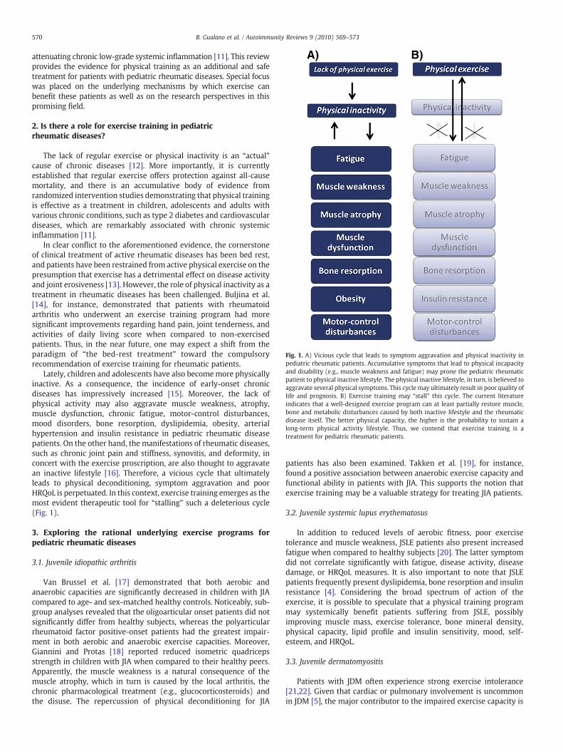
Fig. 1. A) Vicious cycle that leads to symptom aggravation and physical inactivity inpediatric rheumatic patients. Accumulative symptoms that lead to physical incapacityand disability (e.g., muscle weakness and fatigue) may prone the pediatric rheumaticpatient to physical inactive lifestyle. The physical inactive lifestyle, in turn, is believed toaggravate several physical symptoms. This cycle may ultimately result in poor quality oflife and prognosis. B) Exercise training may “stall” this cycle. The current literatureindicates that a well-designed exercise program can at least partially restore muscle,bone and metabolic disturbances caused by both inactive lifestyle and the rheumaticdisease itself. The better physical capacity, the higher is the probability to sustain along-term physical activity lifestyle. Thus, we contend that exercise training is atreatment for pediatric rheumatic patients.
570 B. Gualano et al. / Autoimmunity Reviews 9 (2010) 569–573
attenuating chronic low-grade systemic inflammation [11]. This reviewprovides the evidence for physical training as an additional and safetreatment for patients with pediatric rheumatic diseases. Special focuswas placed on the underlying mechanisms by which exercise canbenefit these patients as well as on the research perspectives in thispromising field.
2. Is there a role for exercise training in pediatricrheumatic diseases?
The lack of regular exercise or physical inactivity is an “actual”cause of chronic diseases [12]. More importantly, it is currentlyestablished that regular exercise offers protection against all-causemortality, and there is an accumulative body of evidence fromrandomized intervention studies demonstrating that physical trainingis effective as a treatment in children, adolescents and adults withvarious chronic conditions, such as type 2 diabetes and cardiovasculardiseases, which are remarkably associated with chronic systemicinflammation [11].
In clear conflict to the aforementioned evidence, the cornerstoneof clinical treatment of active rheumatic diseases has been bed rest,and patients have been restrained from active physical exercise on thepresumption that exercise has a detrimental effect on disease activityand joint erosiveness [13]. However, the role of physical inactivity as atreatment in rheumatic diseases has been challenged. Buljina et al.[14], for instance, demonstrated that patients with rheumatoidarthritis who underwent an exercise training program had moresignificant improvements regarding hand pain, joint tenderness, andactivities of daily living score when compared to non-exercisedpatients. Thus, in the near future, one may expect a shift from theparadigm of “the bed-rest treatment” toward the compulsoryrecommendation of exercise training for rheumatic patients.
Lately, children and adolescents have also becomemore physicallyinactive. As a consequence, the incidence of early-onset chronicdiseases has impressively increased [15]. Moreover, the lack ofphysical activity may also aggravate muscle weakness, atrophy,muscle dysfunction, chronic fatigue, motor-control disturbances,mood disorders, bone resorption, dyslipidemia, obesity, arterialhypertension and insulin resistance in pediatric rheumatic diseasepatients. On the other hand, the manifestations of rheumatic diseases,such as chronic joint pain and stiffness, synovitis, and deformity, inconcert with the exercise proscription, are also thought to aggravatean inactive lifestyle [16]. Therefore, a vicious cycle that ultimatelyleads to physical deconditioning, symptom aggravation and poorHRQoL is perpetuated. In this context, exercise training emerges as themost evident therapeutic tool for “stalling” such a deleterious cycle(Fig. 1).
3. Exploring the rational underlying exercise programs forpediatric rheumatic diseases
3.1. Juvenile idiopathic arthritis
Van Brussel et al. [17] demonstrated that both aerobic andanaerobic capacities are significantly decreased in children with JIAcompared to age- and sex-matched healthy controls. Noticeably, sub-group analyses revealed that the oligoarticular onset patients did notsignificantly differ from healthy subjects, whereas the polyarticularrheumatoid factor positive-onset patients had the greatest impair-ment in both aerobic and anaerobic exercise capacities. Moreover,Giannini and Protas [18] reported reduced isometric quadricepsstrength in children with JIA when compared to their healthy peers.Apparently, the muscle weakness is a natural consequence of themuscle atrophy, which in turn is caused by the local arthritis, thechronic pharmacological treatment (e.g., glucocorticosteroids) andthe disuse. The repercussion of physical deconditioning for JIA
patients has also been examined. Takken et al. [19], for instance,found a positive association between anaerobic exercise capacity andfunctional ability in patients with JIA. This supports the notion thatexercise training may be a valuable strategy for treating JIA patients.
3.2. Juvenile systemic lupus erythematosus
In addition to reduced levels of aerobic fitness, poor exercisetolerance and muscle weakness, JSLE patients also present increasedfatigue when compared to healthy subjects [20]. The latter symptomdid not correlate significantly with fatigue, disease activity, diseasedamage, or HRQoL measures. It is also important to note that JSLEpatients frequently present dyslipidemia, bone resorption and insulinresistance [4]. Considering the broad spectrum of action of theexercise, it is possible to speculate that a physical training programmay systemically benefit patients suffering from JSLE, possiblyimproving muscle mass, exercise tolerance, bone mineral density,physical capacity, lipid profile and insulin sensitivity, mood, self-esteem, and HRQoL.
3.3. Juvenile dermatomyositis
Patients with JDM often experience strong exercise intolerance[21,22]. Given that cardiac or pulmonary involvement is uncommonin JDM [5], the major contributor to the impaired exercise capacity is
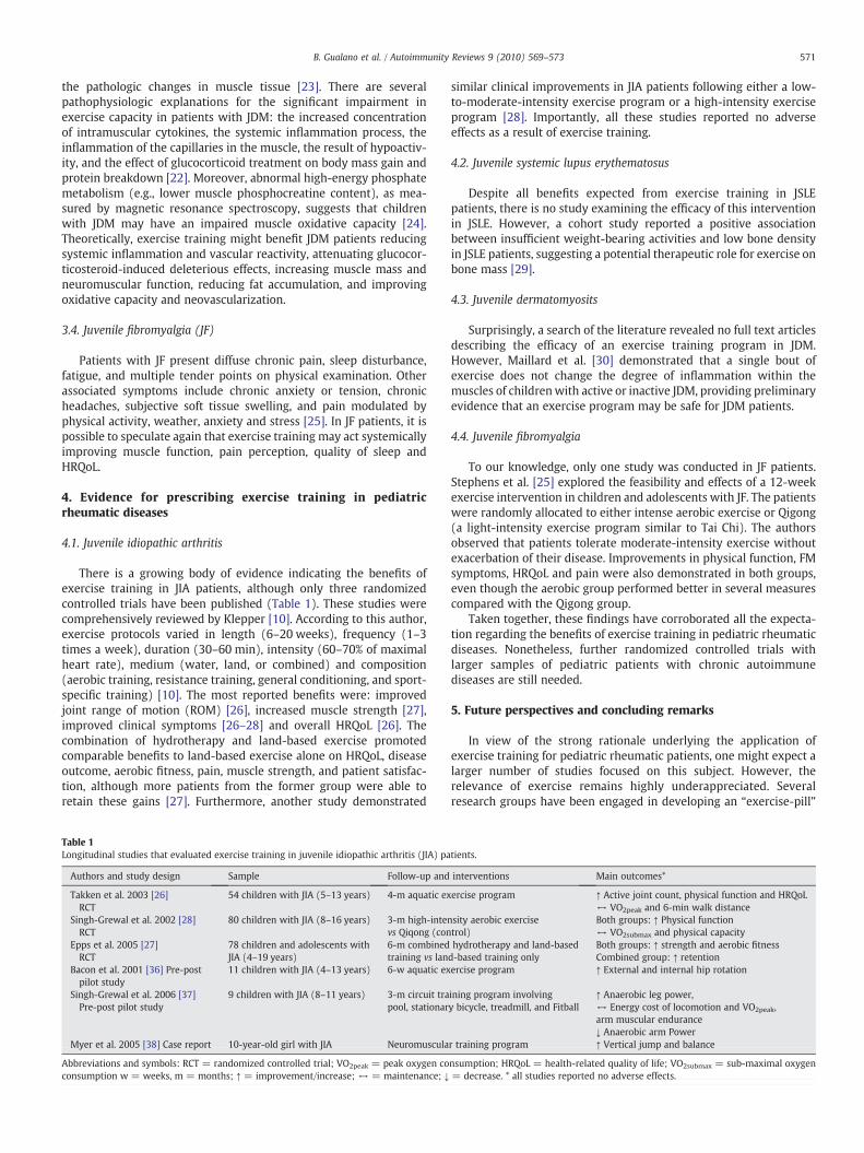
571B. Gualano et al. / Autoimmunity Reviews 9 (2010) 569–573
the pathologic changes in muscle tissue [23]. There are severalpathophysiologic explanations for the significant impairment inexercise capacity in patients with JDM: the increased concentrationof intramuscular cytokines, the systemic inflammation process, theinflammation of the capillaries in the muscle, the result of hypoactiv-ity, and the effect of glucocorticoid treatment on body mass gain andprotein breakdown [22]. Moreover, abnormal high-energy phosphatemetabolism (e.g., lower muscle phosphocreatine content), as mea-sured by magnetic resonance spectroscopy, suggests that childrenwith JDM may have an impaired muscle oxidative capacity [24].Theoretically, exercise training might benefit JDM patients reducingsystemic inflammation and vascular reactivity, attenuating glucocor-ticosteroid-induced deleterious effects, increasing muscle mass andneuromuscular function, reducing fat accumulation, and improvingoxidative capacity and neovascularization.
3.4. Juvenile fibromyalgia (JF)
Patients with JF present diffuse chronic pain, sleep disturbance,fatigue, and multiple tender points on physical examination. Otherassociated symptoms include chronic anxiety or tension, chronicheadaches, subjective soft tissue swelling, and pain modulated byphysical activity, weather, anxiety and stress [25]. In JF patients, it ispossible to speculate again that exercise training may act systemicallyimproving muscle function, pain perception, quality of sleep andHRQoL.
4. Evidence for prescribing exercise training in pediatricrheumatic diseases
4.1. Juvenile idiopathic arthritis
There is a growing body of evidence indicating the benefits ofexercise training in JIA patients, although only three randomizedcontrolled trials have been published (Table 1). These studies werecomprehensively reviewed by Klepper [10]. According to this author,exercise protocols varied in length (6–20 weeks), frequency (1–3times a week), duration (30–60 min), intensity (60–70% of maximalheart rate), medium (water, land, or combined) and composition(aerobic training, resistance training, general conditioning, and sport-specific training) [10]. The most reported benefits were: improvedjoint range of motion (ROM) [26], increased muscle strength [27],improved clinical symptoms [26–28] and overall HRQoL [26]. Thecombination of hydrotherapy and land-based exercise promotedcomparable benefits to land-based exercise alone on HRQoL, diseaseoutcome, aerobic fitness, pain, muscle strength, and patient satisfac-tion, although more patients from the former group were able toretain these gains [27]. Furthermore, another study demonstrated
Table 1Longitudinal studies that evaluated exercise training in juvenile idiopathic arthritis (JIA) pa
Authors and study design Sample Follow-up and
Takken et al. 2003 [26]RCT
54 children with JIA (5–13 years) 4-m aquatic ex
Singh-Grewal et al. 2002 [28]RCT
80 children with JIA (8–16 years) 3-m high-intenvs Qiqong (con
Epps et al. 2005 [27]RCT
78 children and adolescents withJIA (4–19 years)
6-m combinedtraining vs lan
Bacon et al. 2001 [36] Pre-postpilot study
11 children with JIA (4–13 years) 6-w aquatic ex
Singh-Grewal et al. 2006 [37]Pre-post pilot study
9 children with JIA (8–11 years) 3-m circuit trapool, stationar
Myer et al. 2005 [38] Case report 10-year-old girl with JIA Neuromuscula
Abbreviations and symbols: RCT = randomized controlled trial; VO2peak = peak oxygen coconsumption w = weeks, m = months; ↑ = improvement/increase; ↔ = maintenance; ↓
similar clinical improvements in JIA patients following either a low-to-moderate-intensity exercise program or a high-intensity exerciseprogram [28]. Importantly, all these studies reported no adverseeffects as a result of exercise training.
4.2. Juvenile systemic lupus erythematosus
Despite all benefits expected from exercise training in JSLEpatients, there is no study examining the efficacy of this interventionin JSLE. However, a cohort study reported a positive associationbetween insufficient weight-bearing activities and low bone densityin JSLE patients, suggesting a potential therapeutic role for exercise onbone mass [29].
4.3. Juvenile dermatomyosits
Surprisingly, a search of the literature revealed no full text articlesdescribing the efficacy of an exercise training program in JDM.However, Maillard et al. [30] demonstrated that a single bout ofexercise does not change the degree of inflammation within themuscles of childrenwith active or inactive JDM, providing preliminaryevidence that an exercise program may be safe for JDM patients.
4.4. Juvenile fibromyalgia
To our knowledge, only one study was conducted in JF patients.Stephens et al. [25] explored the feasibility and effects of a 12-weekexercise intervention in children and adolescents with JF. The patientswere randomly allocated to either intense aerobic exercise or Qigong(a light-intensity exercise program similar to Tai Chi). The authorsobserved that patients tolerate moderate-intensity exercise withoutexacerbation of their disease. Improvements in physical function, FMsymptoms, HRQoL and pain were also demonstrated in both groups,even though the aerobic group performed better in several measurescompared with the Qigong group.
Taken together, these findings have corroborated all the expecta-tion regarding the benefits of exercise training in pediatric rheumaticdiseases. Nonetheless, further randomized controlled trials withlarger samples of pediatric patients with chronic autoimmunediseases are still needed.
5. Future perspectives and concluding remarks
In view of the strong rationale underlying the application ofexercise training for pediatric rheumatic patients, one might expect alarger number of studies focused on this subject. However, therelevance of exercise remains highly underappreciated. Severalresearch groups have been engaged in developing an “exercise-pill”
tients.
interventions Main outcomes*
ercise program ↑ Active joint count, physical function and HRQoL↔ VO2peak and 6-min walk distance
sity aerobic exercisetrol)
Both groups: ↑ Physical function↔ VO2submax and physical capacity
hydrotherapy and land-basedd-based training only
Both groups: ↑ strength and aerobic fitnessCombined group: ↑ retention
ercise program ↑ External and internal hip rotation
ining program involvingy bicycle, treadmill, and Fitball
↑ Anaerobic leg power,↔ Energy cost of locomotion and VO2peak,arm muscular endurance↓ Anaerobic arm Power
r training program ↑ Vertical jump and balance
nsumption; HRQoL = health-related quality of life; VO2submax = sub-maximal oxygen= decrease. * all studies reported no adverse effects.
572 B. Gualano et al. / Autoimmunity Reviews 9 (2010) 569–573
or “exercise mimetic” capable of mimicking all exercise responses.However, all health benefits of exercise are too complex to be replacedby a single pill. Since exercise produces adaptations in multiple-organs, exercise has natural “builtin” specificity that is unmatched bythe therapeutic limitations of drugs [31]. Furthermore, it is unlikelythat any drug can bemore cost-effective to the health care system andsafer than natural physical activity itself [31]. Thus, we contend thatexercise may be better primary medicine than any current drug forseveral groups, including for pediatric rheumatic patients in the nextfuture. There is a clear window of opportunity to further explore theefficacy of exercise in this population.
In addition to the benefits of exercise training in tertiaryprevention of pediatric rheumatic diseases (i.e., prevention of furtherfunctional loss), exercise might be useful in secondary prevention aswell, ameliorating inflammation. A number of studies suggest thatregular exercise has anti-inflammatory effects [11]. Remarkably,cross-sectional studies have found a positive association betweenphysical inactivity and low-grade systemic inflammation in healthyindividuals [32,33]. Moreover, longitudinal studies have indicatedthat exercise training promotes a reduction in C-reactive proteinconcentration, suggesting that physical activity may suppress sys-temic low-grade inflammation [34,35]. Thus, it is tempting tospeculate whether exercise training can also attenuate chronicinflammation in pediatric rheumatic patients, possibly reducing eitherthe number or the dose of immunosuppressive drugs.
The optimal training protocol for treating patients with pediatricrheumatic diseases remains to be clarified. Considering that childrenand adolescents with rheumatic disease present overall deficits inphysical capacity, further exercise interventions should comprise bothaerobic and strengthening exercises. Although isometric exerciseshave been empirically recommended to avoid joint erosiveness, it ispossible that dynamic exercises may be more effective in improvingdaily-activity functioning. This assumption, however, needs to beexamined for pediatric populations with these chronic illnesses.
Finally, it is important to note that the majority of studies presentquasi-experimental designs. Since some pediatric rheumatic diseaseshave an unstable natural course, the lack of a control group may leadto misinterpretations. Therefore, long-term randomized controlledtrials are necessary to consolidate exercise as a primary treatment forpediatric rheumatic populations. Notwithstanding such literaturegaps, the current evidence itself convincingly supports the notion thatexercise is a treatment for pediatric rheumatic patients.
Take-home messages
• There has been an increasing recognition of adverse short-, mid-, orlong-term effects associated with the treatment as well as thedisease itself that impair the health-related quality of life andfunctional capacity of children and adolescents with rheumaticdiseases.
• Physical inactivitymay aggravatemuscleweakness, atrophy,muscledysfunction, chronic fatigue, motor-control disturbances, mooddisorders, bone resorption, dyslipidemia, obesity and insulinresistance in patients with pediatric rheumatic diseases.
• A growing body of evidence suggests that exercise training canbenefit children and adolescents with juvenile idiopathic arthritis,juvenile systemic lupus erythematosus, juvenile dermatomyositisand juvenile fibromyalgia.
• The most reported benefits include improvements in joint range ofmotion (ROM), muscle strength and function, clinical symptomsand health-related quality of life. No adverse effects have beenreported so far.
• We contend that exercise training is a treatment for patients withpediatric rheumatic diseases.
Acknowledgments
This study was supported by Conselho Nacional de Desenvolvi-mento Científico e Tecnológico— CNPQ (grant 300248/2008-3 to CAS)and by Federico Foundation to CAS.
References
[1] Borchers AT, Selmi C, Cheema G, Keen CL, Shoenfeld Y, Gershwin ME. Juvenileidiopathic arthritis. Autoimmun Rev 2006;5:279–98.
[2] Ruperto N, Lovell DJ, Quartier P, Paz E, Rubio-Perez N, Silva CA, et al. Abatacept inchildren with juvenile idiopathic arthritis: a randomised, double-blind, placebo-controlled withdrawal trial. Lancet 2008;372:383–91.
[3] Kuczynski E, Silva CA, Cristofani LM, Kiss MH, Odone Filho V, Assumpcao Jr FB.Quality of life evaluation in children and adolescents with chronic and/orincapacitating diseases: a Brazilian study. An Pediatr (Barc) 2003;58:550–5.
[4] Avcin T, Cimaz R, Silverman ED, Cervera R, Gattorno M, Garay S, et al. Pediatricantiphospholipid syndrome: clinical and immunologic features of 121 patients inan international registry. Pediatrics 2008;122:e1100–7.
[5] Ravelli A, Trail L, Ferrari C, Ruperto N, Pistorio A, Pilkington C, et al. Long-termoutcome and prognostic factors of juvenile dermatomyositis: a multinational,multicenter study of 490 patients. Arthritis Care Res (Hoboken) 2010;62:63–72.
[6] Sallum AM, Kiss MH, Silva CA, Wakamatsu A, Vianna MA, Sachetti S, et al.Difference in adhesionmolecule expression (ICAM-1 and VCAM-1) in juvenile andadult dermatomyositis, polymyositis and inclusion body myositis. AutoimmunRev 2006;5:93–100.
[7] Lotito AP, Silva CA, Mello SB. Interleukin-18 in chronic joint diseases. AutoimmunRev 2007;6:253–6.
[8] Colombo BM, Cacciapaglia F, Puntoni M, Murdaca G, Rossi E, Rodriguez G, et al.Traditional and non traditional risk factors in accelerated atherosclerosis insystemic lupus erythematosus: role of vascular endothelial growth factor (VEGATSStudy). Autoimmun Rev 2009;8:309–15.
[9] Crispin JC, Tsokos GC. Novel molecular targets in the treatment of systemic lupuserythematosus. Autoimmun Rev 2008;7:256–61.
[10] Klepper SE. Exercise in pediatric rheumatic diseases. Curr Opin Rheumatol 2008;20:619–24.
[11] Mathur N, Pedersen BK. Exercise as a mean to control low-grade systemicinflammation. Mediat Inflamm 2008:109502.
[12] Mokdad AH, Marks JS, Stroup DF, Gerberding JL. Actual causes of death in theUnited States, 2000. JAMA 2004;291:1238–45.
[13] de Salles Painelli V, Gualano B, Artioli GG, de Sa Pinto AL, Bonfa E, Lancha JuniorAH, et al. The possible role of physical exercise on the treatment of idiopathicinflammatory myopathies. Autoimmun Rev 2009;8:355–9.
[14] Buljina AI, Taljanovic MS, Avdic DM, Hunter TB. Physical and exercise therapy fortreatment of the rheumatoid hand. Arthritis Rheum 2001;45:392–7.
[15] Hardy LL, Dobbins TA, Denney-Wilson EA, Okely AD, Booth ML. Sedentariness,small-screen recreation, and fitness in youth. Am J Prev Med 2009;36:120–5.
[16] Takken T, van der Net J, Kuis W, Helders PJ. Physical activity and health relatedphysical fitness in children with juvenile idiopathic arthritis. Ann Rheum Dis2003;62:885–9.
[17] van Brussel M, Lelieveld OT, van der Net J, Engelbert RH, Helders PJ, Takken T.Aerobic and anaerobic exercise capacity in children with juvenile idiopathicarthritis. Arthritis Rheum 2007;57:891–7.
[18] Giannini MJ, Protas EJ. Comparison of peak isometric knee extensor torque inchildren with and without juvenile rheumatoid arthritis. Arthritis Care Res 1993;6:82–8.
[19] Takken T, van der Net J, Helders PJ. Relationship between functional ability andphysical fitness in juvenile idiopathic arthritis patients. Scand J Rheumatol2003;32:174–8.
[20] Houghton KM, Tucker LB, Potts JE, McKenzie DC. Fitness, fatigue, disease activity,and quality of life in pediatric lupus. Arthritis Rheum 2008;59:537–45.
[21] Takken T, Elst E, Spermon N, Helders PJ, Prakken AB, van der Net J. Thephysiological and physical determinants of functional ability measures in childrenwith juvenile dermatomyositis. Rheumatology (Oxford) 2003;42:591–5.
[22] Takken T, van der Net J, Helders PJ. Anaerobic exercise capacity in patients withjuvenile-onset idiopathic inflammatorymyopathies. Arthritis Rheum2005;53:173–7.
[23] Rider LG. The heterogeneity of juvenile myositis. Autoimmun Rev 2007;6:241–7.[24] Park JH, Niermann KJ, Ryder NM, Nelson AE, Das A, Lawton AR, et al. Muscle
abnormalities in juvenile dermatomyositis patients: P-31 magnetic resonancespectroscopy studies. Arthritis Rheum 2000;43:2359–67.
[25] Stephens S, FeldmanBM, Bradley N, Schneiderman J,Wright V, Singh-Grewal D, et al.Feasibility and effectiveness of an aerobic exercise program in children withfibromyalgia: results of a randomized controlled pilot trial. Arthritis Rheum2008;59:1399–406.
[26] Takken T, Van Der Net J, Kuis W, Helders PJ. Aquatic fitness training for childrenwith juvenile idiopathic arthritis. Rheumatology (Oxford) 2003;42:1408–14.
[27] Epps H, Ginnelly L, Utley M, Southwood T, Gallivan S, Sculpher M, et al. Ishydrotherapy cost-effective? A randomised controlled trial of combined hydro-therapy programmes compared with physiotherapy land techniques in childrenwith juvenile idiopathic arthritis. Health Technol Assess 2005;9:1–59.
[28] Singh-Grewal D, Schneiderman-Walker J, Wright V, Bar-Or O, Beyene J, SelvaduraiH, et al. The effects of vigorous exercise training on physical function in childrenwith arthritis: a randomized, controlled, single-blinded trial. Arthritis Rheum2007;57:1202–10.
573B. Gualano et al. / Autoimmunity Reviews 9 (2010) 569–573
[29] Compeyrot-Lacassagne S, Tyrrell PN, Atenafu E, Doria AS, Stephens D, Gilday D,et al. Prevalence and etiology of low bone mineral density in juvenile systemiclupus erythematosus. Arthritis Rheum 2007;56:1966–73.
[30] Maillard SM, Jones R, Owens CM, Pilkington C, Woo PM, Wedderburn LR, et al.Quantitative assessments of the effects of a single exercise session on muscles injuvenile dermatomyositis. Arthritis Rheum 2005;53:558–64.
[31] Booth FW, Laye MJ. Lack of adequate appreciation of physical exercise'scomplexities can pre-empt appropriate design and interpretation in scientificdiscovery. J Physiol 2009;587:5527–39.
[32] Fischer CP, Berntsen A, Perstrup LB, Eskildsen P, Pedersen BK. Plasma levels ofinterleukin-6 and C-reactive protein are associated with physical inactivityindependent of obesity. Scand J Med Sci Sports 2007;17:580–7.
[33] Geffken DF, Cushman M, Burke GL, Polak JF, Sakkinen PA, Tracy RP. Associationbetween physical activity and markers of inflammation in a healthy elderlypopulation. Am J Epidemiol 2001;153:242–50.
Antimalarial treatment may have a time-dependent effect on lupus sur
To evaluate the beneficial effect of antimalarial treatment on lupus survivSocioeconomic and demographic characteristics, clinical manifestationswere examined in patients with systemic lupus erythematosus (SLE) fr(GLADEL) cohort. Shinjo SK. et al. (Arthritis Rheum2010; 62: 855-62). T2 years of cohort entry. Cause of death was classified as active disease,other cause. Patients were subdivided by antimalarial use, grouped accconsecutivemonths (user) and those who had received antimalarial drugdrugs (nonuser). Of the 1480 patients included in the GLADEL cohort, 114drug exposure of 48.5 months (range 6-98months). Death occurred in 89users comparedwith nonusers (4.4% vs 11.5%; p b 0.001). Seventy patientfor 1-2 years, and 925 (81.1%) for N 2 years. Mortality rates among userfollow-up) were 3.85 (95% confidence interval [95% CI] 1.41-8.37), 2.7 (9nonusers, the mortality rate was 3.07 (95% CI 2.18- 4.20) p for trend b 0.model, antimalarial use was associated with a 38% reduction in the moeffect, possibly in a time-dependent manner, on SLE survival. Theserecommended for patients with lupus.
IL-17 contributes to angiogenesis in rheumatoid arthritis
Angiogenesis is an early and a critical event in the pathogenesis of rheumcell activation, migration and proliferation, and inhibition of angiogenePickens SR et al. (J Immunol 2010; 184: 3233-41)document a novel roleankles increases vascularity. The authors demonstrate that IL-17 is angiogplugs in vivo. Additionally, IL-17, in concentrations present in the RAmigration mediated through the PI3K/AKT1pathway. Furthermore, suppformation. They also show that both IL-17-induced HMVEC chemotaxis aNeutralization of either IL-17 inRA synovialfluids or IL-17 receptor C onHsynovialfluid. Finally, RA synovialfluid immunoneutralizedwith anti-IL-1migration beyond the effect detected by immunodepleting each factorangiogenic mediator in RA, supporting IL-17 as a therapeutic target in R
Diet-induced obesity impairs the T cell memory response to influenza v
The Centers for Disease Control and Prevention has suggested that obesifrom H1N1pandemic strain. Memory T cells generated during primary inviruses, making them effective against encounters with heterologous sdemonstrate that inmale, diet-induced obese C57BL/6mice, a secondary25%mortality rate (with no loss of lean controls), 25% increase in lung patiters. Furthermore, mRNA expression for IFN-gamma was N 60% less inspecific CD8+ T cells producing IFN-gamma postsecondary infection vreduction in IFN-gamma production when stimulated with influenza-puspecificmemory T cells is significantly reduced and ineffective in lungs of oyearly influenza outbreaks and the current pandemicmakes it imperativeobese host. Moreover, impairment of memory responses has significant
[34] Stewart LK, Flynn MG, Campbell WW, Craig BA, Robinson JP, Timmerman KL, et al.The influence of exercise training on inflammatory cytokines and C-reactiveprotein. Med Sci Sports Exerc 2007;39:1714–9.
[35] Goldhammer E, Tanchilevitch A, Maor I, Beniamini Y, Rosenschein U, Sagiv M.Exercise training modulates cytokines activity in coronary heart disease patients.Int J Cardiol 2005;100:93–9.
[36] Bacon MC, Nicholson C, Binder H, White PH. Juvenile rheumatoid arthritis. Aquaticexercise and lower-extremity function. Arthritis Care Res 1991;4:102–5.
[37] Singh-Grewal D, Wright V, Bar-Or O, Feldman BM. Pilot study of fitness trainingand exercise testing in polyarticular childhood arthritis. Arthritis Rheum 2006;55:364–72.
[38] Myer GD, Brunner HI, Melson PG, Paterno MV, Ford KR, Hewett TE. Specializedneuromuscular training to improve neuromuscular function and biomechanics in apatient with quiescent juvenile rheumatoid arthritis. Phys Ther 2005;85:791–802.
vival: data from a multinational Latin American inception cohort
al, in a large, multiethnic, international longitudinal inception cohort., classification criteria, laboratory findings, and treatment variablesom the Groupo Latino Americano de Estudio del Lupus Eritematosohe diagnosis of SLE, according to the ACR criteria, was assessedwithininfection, cardiovascular complications, thrombosis, malignancy, orording to those who had received antimalarial drugs for at least 6s for b 6 consecutive months or who had never received antimalarial1 (77%)were considered antimalarial users, with a mean duration ofpatients (6.0%). A lowermortality rate was observed in antimalarial
s (6.1%) had received antimalarial drugs for 6-11months, 146 (12.8%)s by duration of antimalarial treatment (per 1000 person-months of5% CI 1.41-4.76), and 0.54 (95% CI 0.37-0.77), respectively, while for001). After adjustment for potential confounders in a Cox regressionrtality rate. Thus, antimalarial drugs are shown to have a protectiveresults suggest that the use of antimalarial treatment should be
atoid arthritis (RA). Neovascularization is dependent on endothelialsis may provide a novel therapeutic approach in RA. In this study,of IL-17 inmediating angiogenesis. Local expressionof IL-17 inmouseenic by showing its ability to promote blood vessel growth inMatrigeljoint, induces human lung microvascular endothelial cell (HMVEC)ression of the PI3K pathway markedly reduces IL-17-induced tubend tube formation are mediated primarily through IL-17 receptor C.MVECs significantly reduces the induction ofHMVECmigration byRA7 and antivascular endothelial growth factor does not reduceHMVECalone. These observations identify a novel function for IL-17 as anA.
irus infection
ty may be an independent risk factor for increased severity of illnessfluenza infection target internal proteins common among influenzatrains. Here, Karlsson EA. et al. (J Immunol 2010; 184: 3127-33)H1N1 influenza challenge following a primaryH3N2 infection led to athology, failure to regain weight, and 10-to 100-fold higher lung virallungs of obese mice, along with one third the number of influenza-s lean controls. Memory CD8+ T cells from obese mice had a N50%lsed dendritic cells from lean mice. Thus, the function of influenza-besemice. The reality ofworldwide obesity epidemic combinedwithto understandhow influenza virus infection behaves differently in animplications for vaccine efficacy in an obese population.


![DIÁRIO ELETRÔNICO DA JUSTIÇA DO TRABALHO1].pdf · ADVOGADO(A) Rodrigo Campana Fiorot RECDO : Igor Silva da Conceição RECDO : BRAMETAL - Brandão Metalúrgica S.A. - DESPACHO](https://img.pdfslide.tips/doc/110x75/5ac51e157f8b9a333d8daf71/dirio-eletrnico-da-justia-do-1pdfadvogadoa-rodrigo-campana-fiorot-recdo-igor.jpg)