Embed Size (px)
Citation preview
Een rood oog en een rotte tand
Sofie Dekeyzer - Radiologie
Robert Oonk - Urgentiegeneeskunde
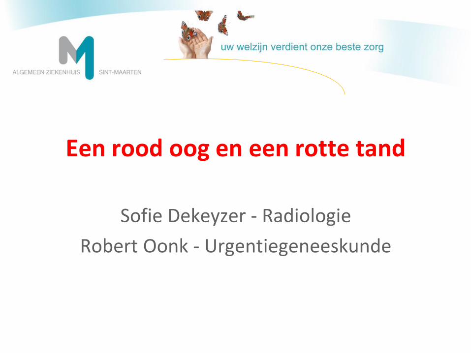
kliniek
• Man, 33 jaar
• Recent tandextractie element 46
• Zwelling rechter onderkaak
• Koorts onder reeds ingestelde antibiotica
• Slikproblemen
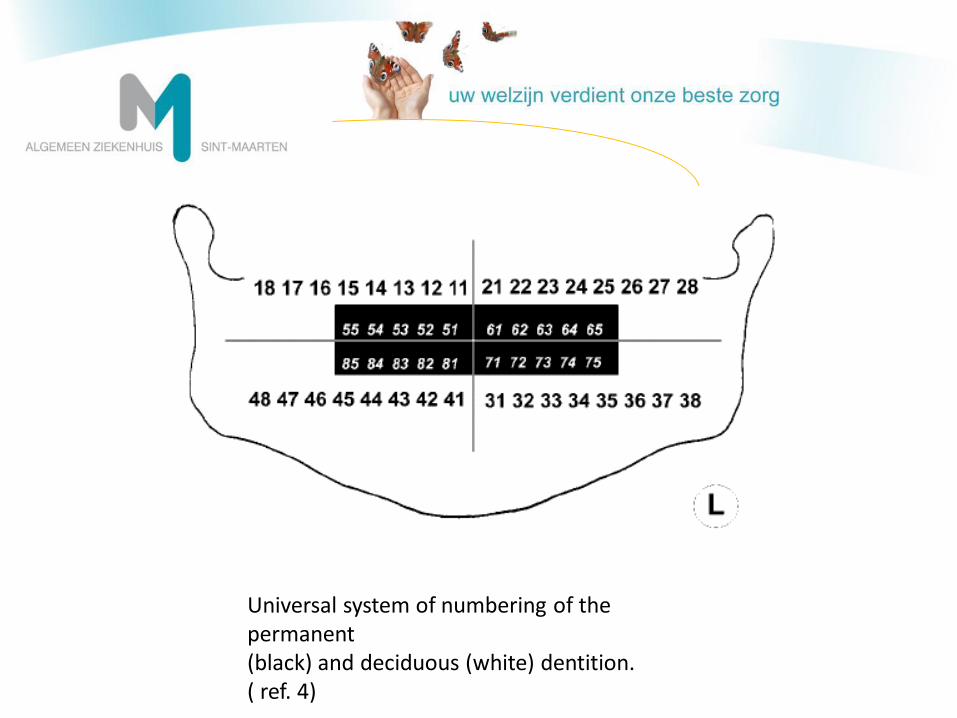
FDI World Dental Federation Notation
Universal system of numbering of the permanent (black) and deciduous (white) dentition. ( ref. 4)
• Bloedname:
Fors verhoogd CRP van 44 mg/dl (<0,5)
Leukocytose 28500/µl met 89% neutrofielen
• Opname op de dienst stomatologie
• Amoxicilline/Clavulanaat IV
• NSAIDs IV
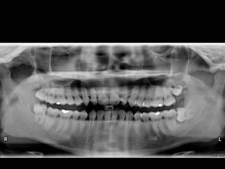
Diagnose:
Necrose van element 47 met abces ter hoogte van de wang
Ontstaan van trismus
Behandeling:
Drainage
Verwijdering van element 47 op D+1
Amoxicilline/Clavulanaat IV en verder PO
Ontslag op D+2
Op D+3 opnieuw op spoedgevallen! ● Toenemende zwelling ter hoogte van de kaak, maar
ook van de submadibulaire regio ● Opnieuw koorts onder amoxicilline/clavulanaat ● Gevoel van een dikke, dubbele tong te hebben Bloedname: ● CRP 16 mg/dl ● Leukocytose 23.400 /µl met 83,6% neutrofielen
Beleid: ● Opname stomatologie ● Amoxicilline/Clavulanaat IV ● Associatie Clindamycine IV ● BEELDVORMING!
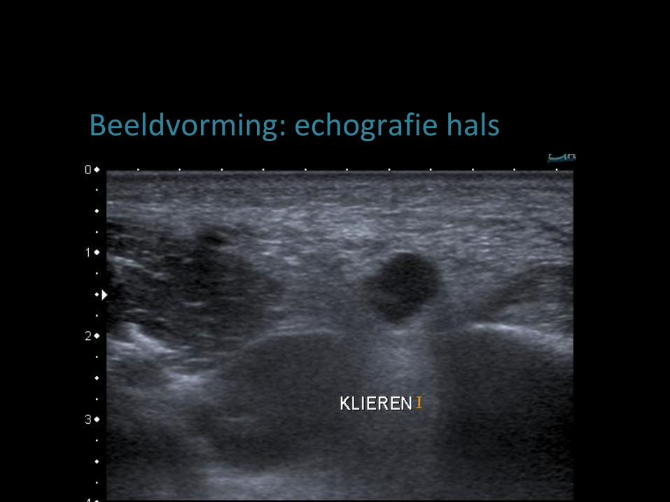
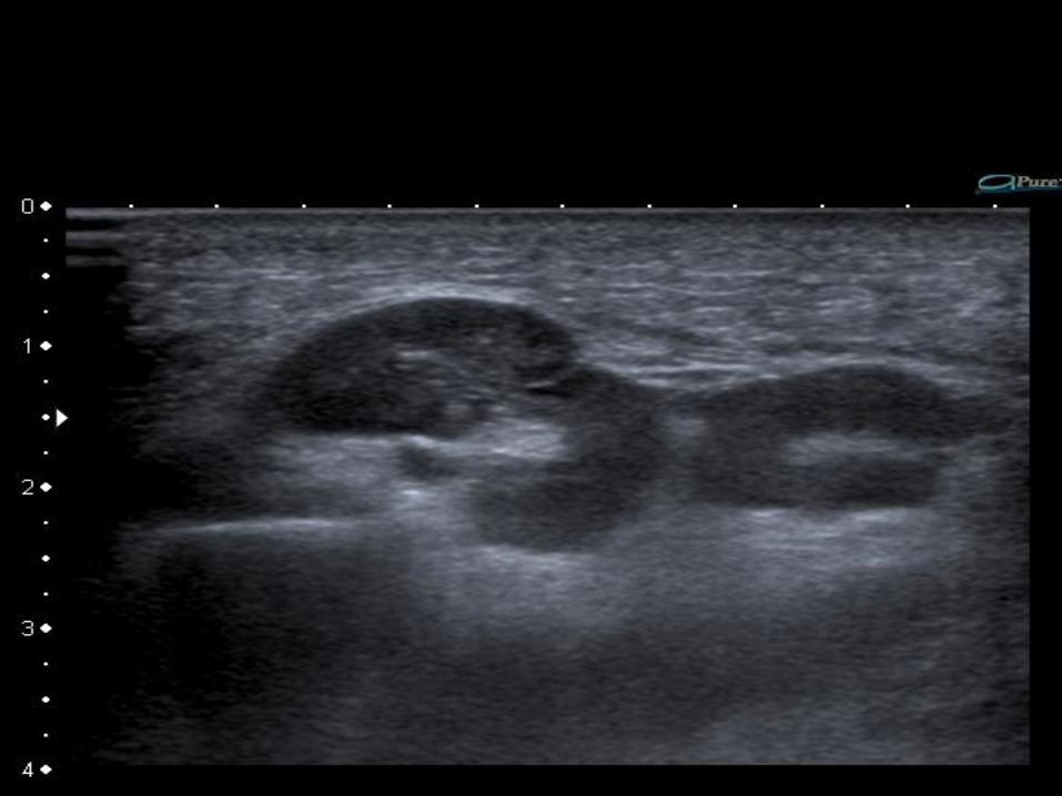
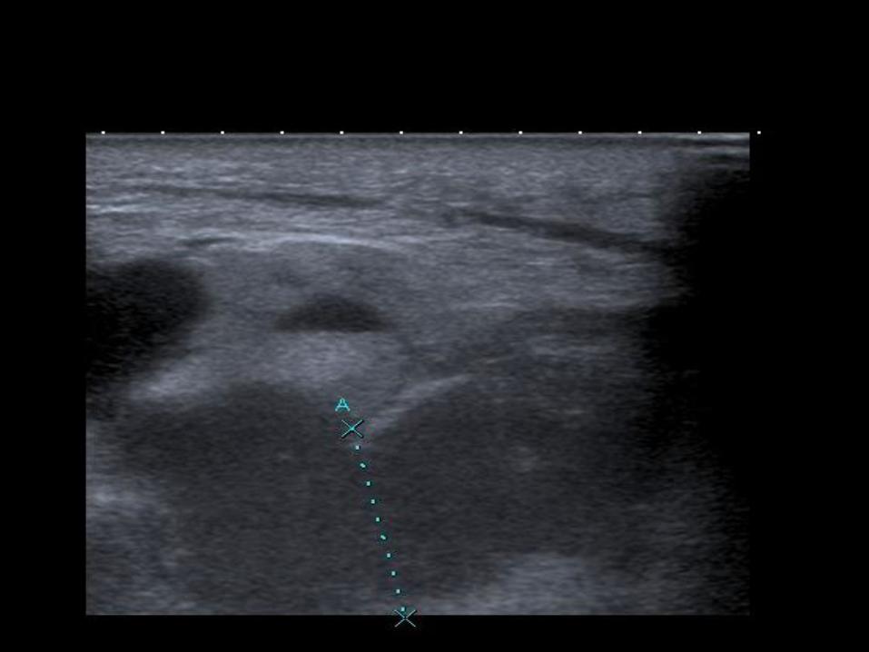
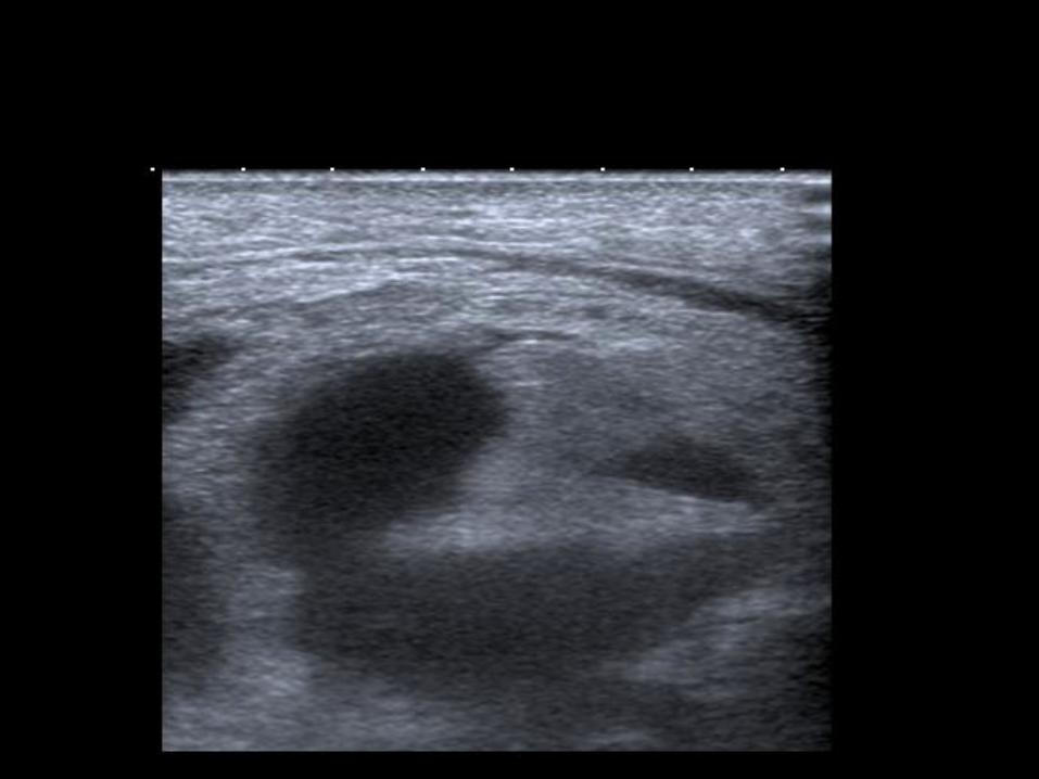
Beeldvorming: echografie hals
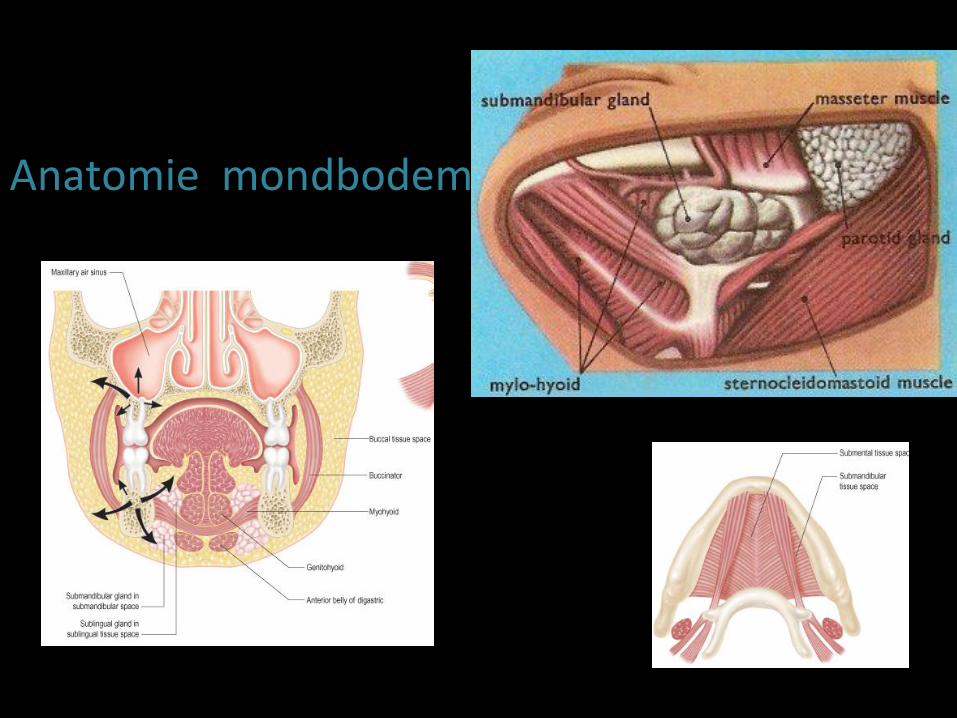
Anatomie mondbodem
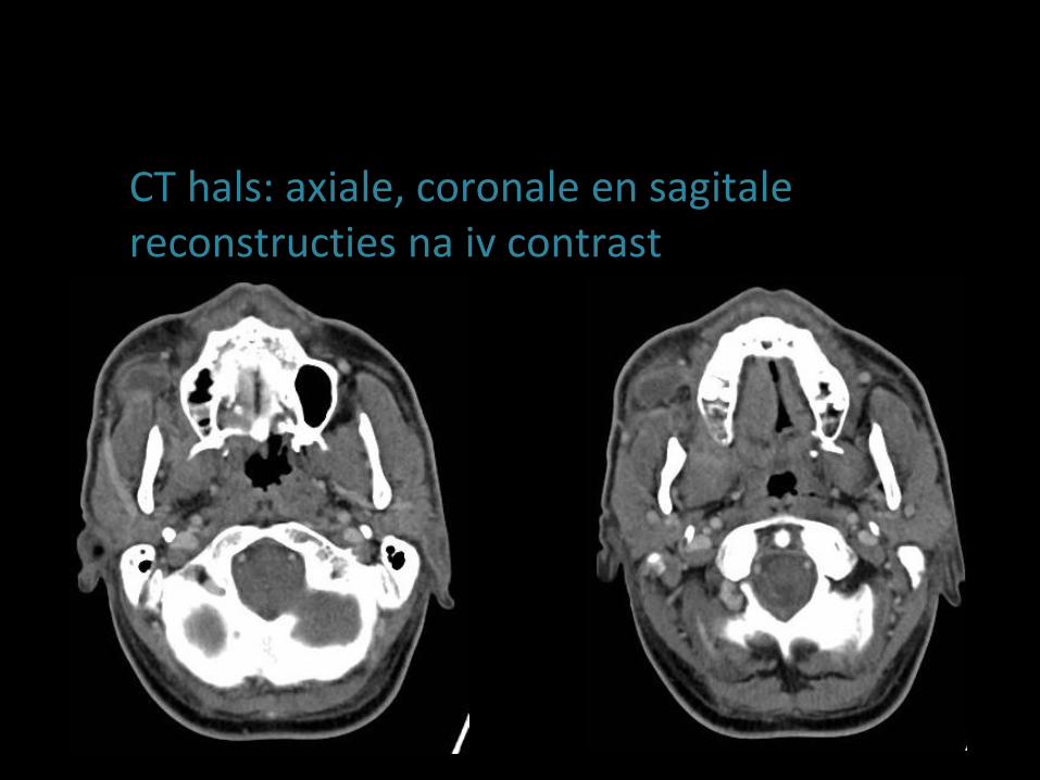
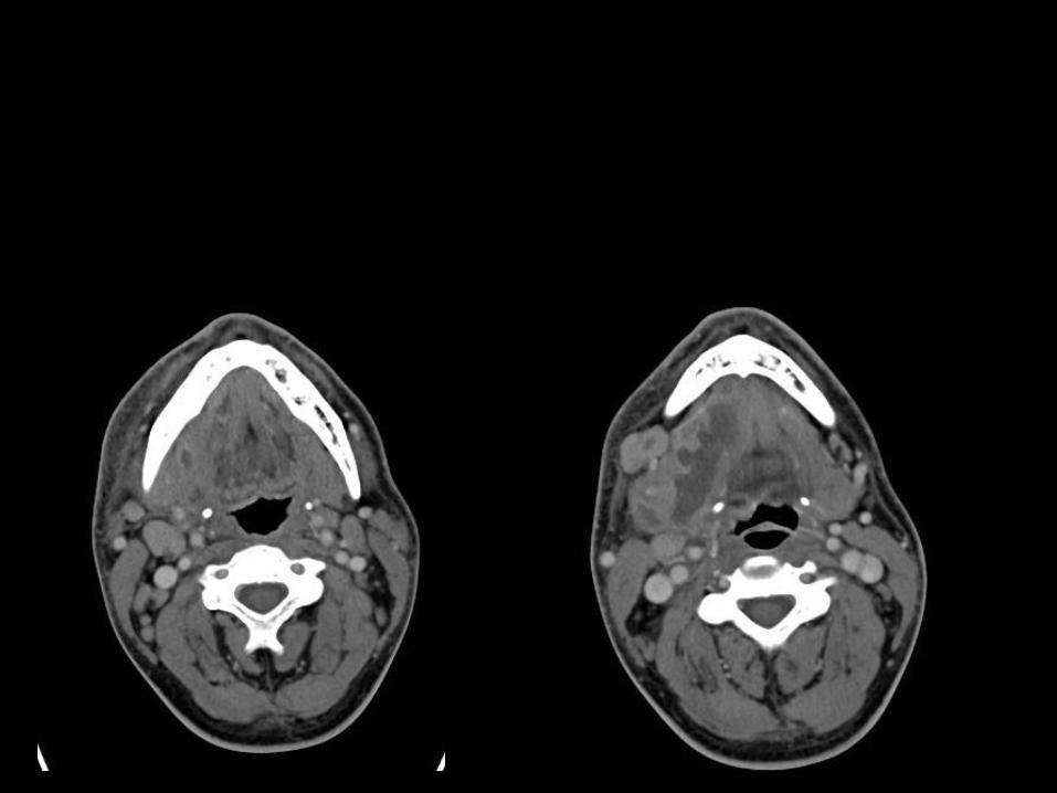
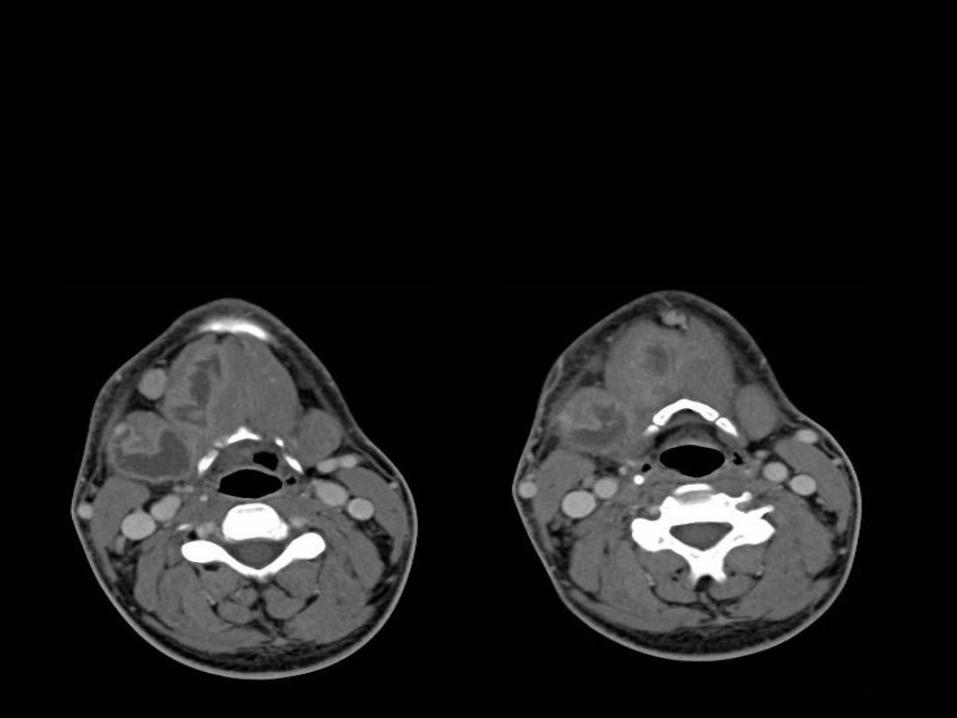
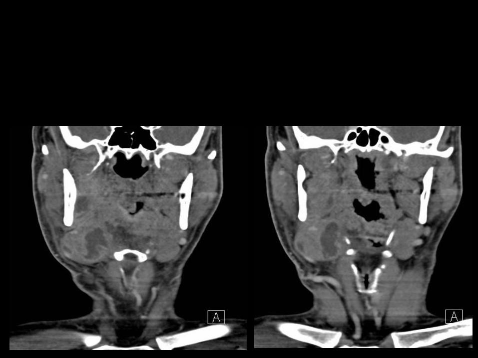
CT hals: axiale, coronale en sagitale reconstructies na iv contrast
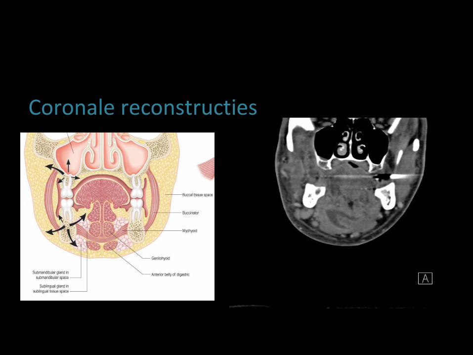
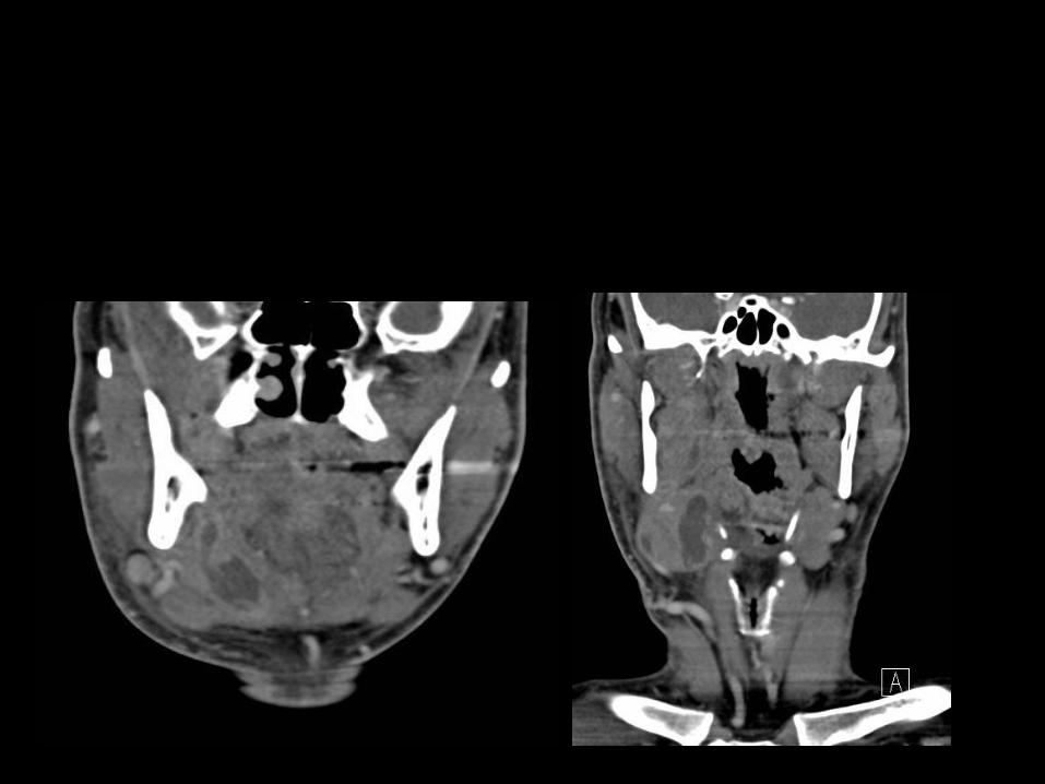
Coronale reconstructies
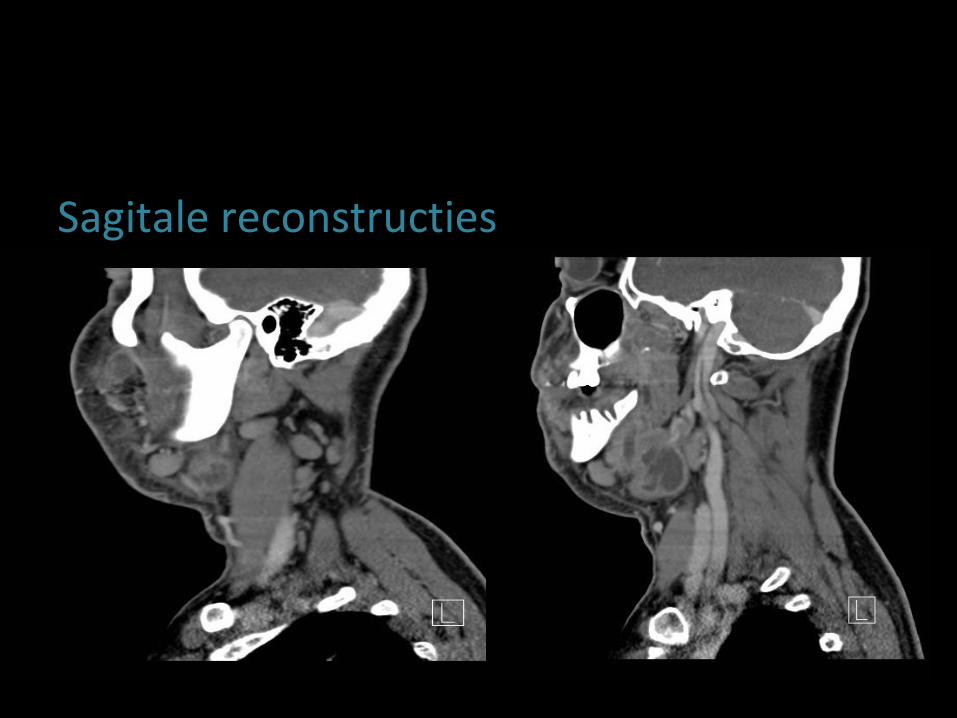
Sagitale reconstructies
Therapie
• Drainage
• Plaatsing drie lamellen
• Doorgedreven antibiotherapie
• Ontslag op D+9 met lamellen in situ
• Verwijdering op D+12 van twee lamellen en op D+14 de sumandibulaire als laatste

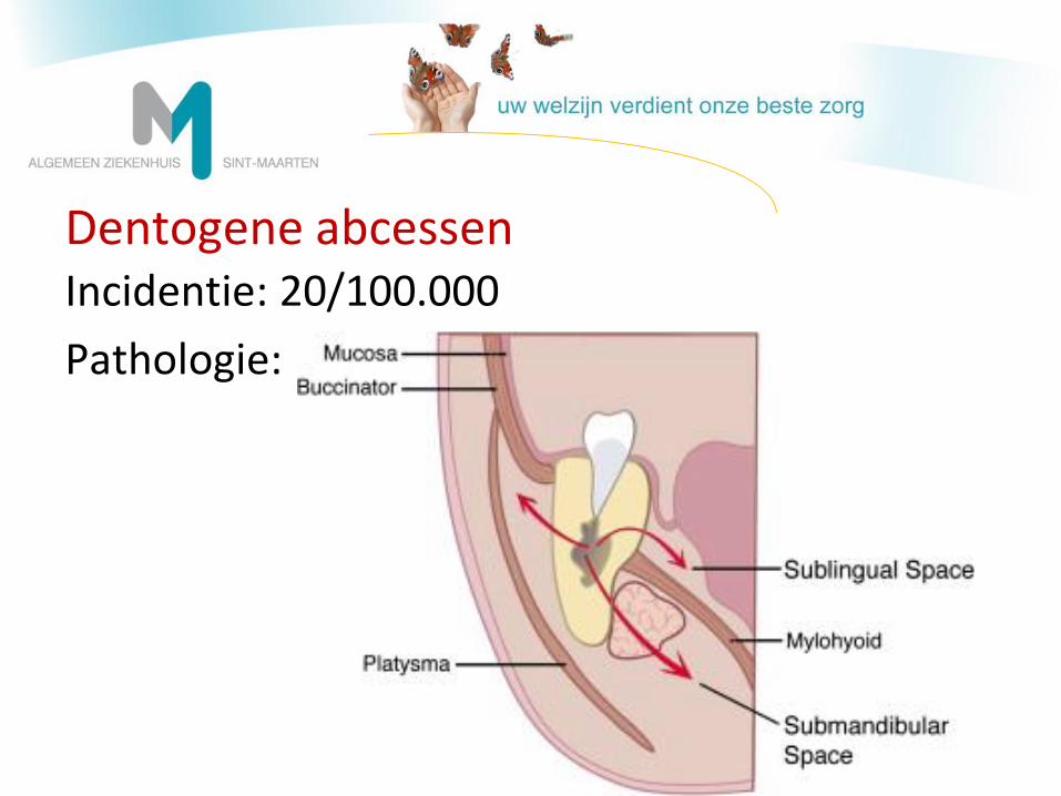
Dentogene abcessen Incidentie: 20/100.000
Pathologie:
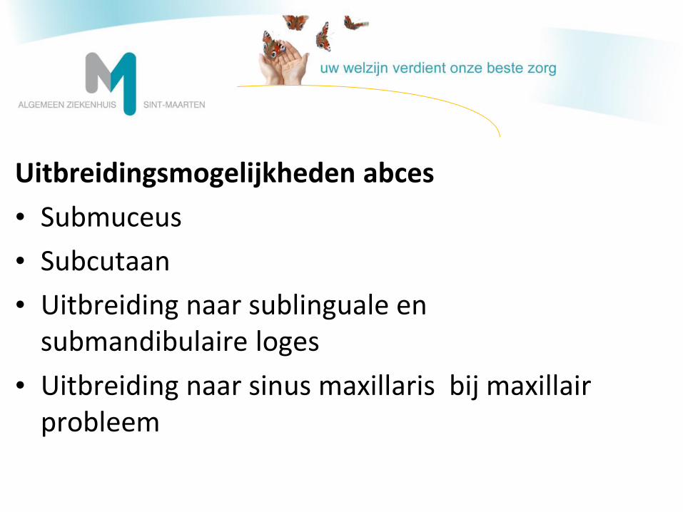
Uitbreidingsmogelijkheden abces
• Submuceus
• Subcutaan
• Uitbreiding naar sublinguale en submandibulaire loges
• Uitbreiding naar sinus maxillaris bij maxillair probleem
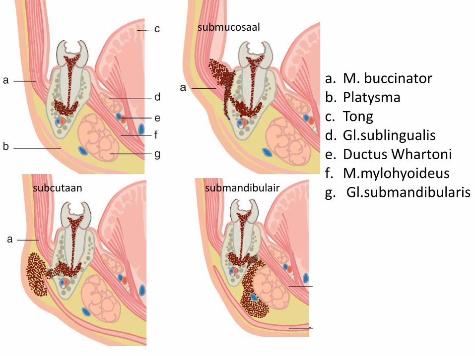
a. M. buccinator b. Platysma c. Tong d. Gl.sublingualis e. Ductus Whartoni f. M.mylohyoideus g. Gl.submandibularis
submucosaal
subcutaan submandibulair
Take home messages
- kliniek!
- Bij vermoeden abces: beeldvorming CT
- AB en drainage abces
Referenties 1.Neurologic/Head and Neck Imaging: Meir H. Scheinfeld, Keivan Shifteh, Laura L. Avery,Harry Dym,and R. Joshua Dym
Continuing Medical Education: Teeth: What Radiologists Should Know Radiographics November-December 2012 32:7 1927-1944; doi:10.1148/rg.327125717
2.Neurologic/Head and Neck Imaging: Sarah J. La'Porte,Jaspal K. Juttla,and Ravi K. Lingam
Continuing Medical Education: Imaging the Floor of the Mouth and the Sublingual Space Radiographics September-October 2011 31:5 1215-1230; doi:10.1148/rg.315105062
3.Education Exhibits: Erin Frankie Capps, James J. Kinsella,Manu Gupta,Amol Madhav Bhatki,and Michael Jeffrey Opatowsky
Continuing Medical Education: Emergency Imaging Assessment of Acute, Nontraumatic Conditions of the Head and Neck Radiographics September 2010 30:5 1335-1352; doi:10.1148/rg.305105040
4. Proceedings of the symposium on MKA beeldvorming of december10-11, 2004, Antwerp-part one conventional dental radiology: what the general radiologist needs to know; A. Bernaerts, F.M. Vanhoenacker, L. Geenen, G. Quisquater, P.M. Parizel; JBR–BTR, 2006, 89: 23-46.