Embed Size (px)
Citation preview
Somatic Mutations of the L12a Gene in V-�1 LightChain Deposition Disease
Potential Effects on Aberrant Protein Conformation andDeposition
Ruben Vidal,* Fernando Goni,*† Fred Stevens,‡Pierre Aucouturier,* Asok Kumar,*Blas Frangione,* Jorge Ghiso,* and Gloria Gallo*From the Department of Pathology,* New York University School
of Medicine, New York, New York; the Facultad de Quımica,†
Universidad de la Republica Oriental del Uruguay, Montevideo,
Uruguay; and the Biosciences Division,‡ Argonne National
Laboratory, Argonne, Illinois
Light chain deposition disease (LCDD) and light chainamyloidosis (AL) are disorders of monoclonal immu-noglobulin deposition in which normally soluble se-rum precursors form insoluble deposits in tissues. Acommon feature in both is the clonal proliferation ofB-cells that produce pathogenic light chains. How-ever, the deposits in LCDD differ from those in AL inthat they are ultrastructurally granular rather thanfibrillar and do not bind Congo red or colocalize withamyloid P component or apolipoprotein E. The rea-son(s) for their differences are unknown but arelikely multifactorial and related to their protein con-formation and their interaction with other moleculesand tissue factors in the microenvironment. Knowl-edge of the primary structure of the light chains inLCDD is very limited. In the present study two new �1
light chains from patients with LCDD are describedand compared to seven other reported �-LCDD pro-teins. The N-terminal amino acid sequences of lightchain GLA extracted from the renal biopsy and lightchain CHO from myocardial tissue were each identi-cal to the respective light chains isolated from theurines and to the V-region amino acid sequencestranslated from the cloned cDNAs obtained frombone marrow cells. The germline V-region sequences,determined from the genomic DNA in both and inMCM, a previously reported �1 LCDD light chain,were identical and related to the L12a germline gene.The expressed light chains in all three exhibit aminoacid substitutions that arise from somatic mutationand result in increased hydrophobicity with the po-tential for protein destabilization and disordered con-formation. (Am J Pathol 1999, 155:2009–2017)
The monoclonal immunoglobulin deposition diseases,which include nonamyloid light chain deposition disease(LCDD) and light chain amyloidosis (AL), have in com-mon monoclonal immunoglobulin synthesis by an ex-panded clone of B cells leading to the deposition ofinsoluble light chains in systemic organs with displace-ment and eventual destruction of parenchymal cells andorgan dysfunction.1 However, LCDD and AL differ inseveral ways. In the case of AL the deposits are congo-philic, have a fibrillar ultrastructure, are more frequentlyderived from � than � light chain, have a patchy distribu-tion within and among systemic organs, and colocalizewith amyloid P component, apolipoprotein-E (apo-E), andglycosaminoglycans (GAGs).2,3 In contrast, the depositsin LCDD are noncongophilic, have a granular rather thanfibrillar ultrastructure, and are more frequently � than �. Incases completely examined at autopsy, deposits are uni-formly distributed in all the basement membranes of sys-temic organs4,5 and do not associate with amyloid Pcomponent or apo-E.2 These differences are of funda-mental importance in determining the mechanisms ofprotein deposition in tissues and the process of fibrillo-genesis possibly relevant to other common types of amy-loidosis, such as Alzheimer’s disease. However, the bio-physical basis for their differences is poorly understood.
Knowledge of the primary structure of the protein de-posits in LCDD is very limited; in only nine cases of LCDDhas the complete light chain variable region (V-region)sequence been published.6–13 Thus, a comparison of thenonfibrillar and fibrillar forms of deposits in LCDD and ALthat could identify differences in their primary structuresand might relate to their dissimilar properties is ham-
Supported in part by a grant from the National Institutes of Health (AR02594, Merit), by the U.S. Department of Energy, Office of Health andEnvironmental Research (contract W-31-109-ENG), and by the U.S. Pub-lic Health Service (grant DKY 3757).
Accepted for publication August 26, 1999.
R. V. and F. G. contributed equally to this work.
P. A.’s current address: Hopital Necker, 161 rue de Sevres, 75743 ParisCEDEX 15, France.
Address reprint requests to Dr. Gloria Gallo, New York University Med-ical Center, 560 First Avenue, New York, NY 10016. E-mail: [email protected].
American Journal of Pathology, Vol. 155, No. 6, December 1999
Copyright © American Society for Investigative Pathology
2009
pered by the apparent scarcity of cases of LCDD to studyand the unavailability of large amounts of tissues frompostmortem examinations for biochemical analysis. Thislimitation is now partially overcome by microextractionmethods to isolate and obtain the amino-terminal se-quence of light chain deposits from milligram amounts ofdiagnostic biopsy tissues,14,15 as well as the applicationof molecular techniques to obtain the light chain V-regionamino acid sequence translated from cloned cDNA ofbone marrow cells. These methods, applied to morereadily available biopsy tissues, allow the opportunity tobuild a primary structure data base for comparison withnonpathogenic Bence-Jones light chains as well as thosein AL disease, with the goal of elucidating the mecha-nism(s) of tissue deposition and fibrillogenesis.
In a previous case of LCDD, we reported the biochem-ical data of a V-region �1 (V-�1) nonamyloidotic immuno-globulin light chain, MCM, obtained by extraction of de-posits from myocardial tissue in which five unique aminoacid substitutions were identified.11 We now report twonew �1 LCDD proteins: GLA and CHO. In both we deter-mined the N-terminal amino acid sequences of the lightchains isolated from the tissues and urines, the completeV-region light chain amino acid sequences deduced fromthe cloned cDNAs, and the nucleotide germline se-quences from genomic DNA. We also determined thegermline sequences from genomic DNA of MCM. Weconclude that the amino acid substitutions identified inGLA and CHO, as well as in MCM, are due to somaticmutations and contribute to protein instability, aggrega-tion, and the deposition of the light chains in the tissues.
Materials and Methods
Patients
The diagnosis of LCDD was made in two patients whopresented with renal disease and whose renal biopsytissues showed monotypic � light chain deposits.
A 61-year-old caucasian female (GLA) presented withincreasing renal functional impairment. Physical exami-nation revealed no edema or other abnormalities. Theserum creatinine rose from 0.8 to 2.6 mg/dl over an8-month period and to 3.7 mg/dl at the ninth month, thetime of admission and renal biopsy. The urine showedmicrohematuria and a urine protein excretion of 0.16–0.37 g/24 hours. The serum cholesterol level was 270mg/dl, C3/C4 was 129/40 mg/dl (normal), and hematocritwas 32%. A diagnosis of � LCDD was made by renalbiopsy. Immunoelectrophoresis revealed no monoclonalprotein in the serum and a mixture of albumin and mono-clonal Bence-Jones � light chain, estimated at 30 mg/dl,in 100-fold concentrated urine. A bone marrow biopsyshowed features of a B-lymphocyte neoplasm. Despitechemotherapy with Melphalan and Prednisone, irrevers-ible renal failure developed, requiring maintenance he-modialysis.
The second patient, a 64-year-old Asian female (CHO),was admitted to the hospital for complaints of fatigue,anorexia, back pain, and acute renal failure that devel-
oped after abdominal computed tomography (CT) withradio contrast. She was known to have normal renalfunction, with a serum creatinine of 0.9 mg/dl, 2 yearsbefore. After CT studies the creatinine was 5.7 mg/dl androse to 9.1 mg/dl 4 days later at the time of admission.The past medical history was unremarkable except forhepatitis B surface antigenemia for many years. Herblood pressure was normal and there was no edema. Thesignificant laboratory findings included urine protein ex-cretion, 1.0 g/24 hour; microhematuria; blood urea nitro-gen, 100 mg/dl; serum Ca, 10.4 mg/dl; Phos, 6.7 mg/dl. Adiagnosis of � LCDD was made by renal biopsy. Theinitial serum and urine immunoelectrophoresis showedno monoclonal Ig, but subsequently Bence-Jones � lightchain was identified in concentrated urine, and lytic le-sions were detected in the skull. Bone marrow examina-tion revealed multiple myeloma. Renal failure worsenedand expiration occurred 2 months after admission. Apostmortem examination was performed.
Pathological and Immunohistological Studies
Paraffin sections of formalin-fixed tissues were stainedwith hematoxylin and eosin, periodic acid silver methen-amine, and Congo red. Standard immunofluorescencemicroscopy examination of frozen sections was per-formed on renal biopsy tissues from both patients and onsystemic tissues obtained at necropsy from CHO. Sec-tions were incubated with a panel of fluorescein-labeledrabbit polyclonal anti-human Ig chain-specific antibodies(�, �, �, �, and �), C3, C1q, fibrin and unlabeled rabbitpolyclonals anti-amyloid P component (Dako, Carpente-ria, CA), and anti-apo-E (Chemicon, Temecula, CA), fol-lowed by fluorescein-conjugated swine anti-rabbit Ig(Dako). Immunoperoxidase examinations of formalin-fixed, paraffin-embedded bone marrow biopsy speci-mens from both patients incubated with polyclonal anti-�and anti-� antibodies (Dako) were performed as de-scribed.16
Electron microscopic studies were carried out on ul-trathin sections of glutaraldehyde-fixed epon-embeddedrenal biopsy tissues and on frozen cardiac tissue storedat �70°C.
Light Chain Isolation from Tissue andUrine Specimens
The GLA residual 1-mm3 frozen renal biopsy tissue waswashed three times in 500 �l of 50 mmol/L phosphate/150 mmol/L NaCl (PBS) (pH 7.2) and centrifuged at 2500rpm. The tissue pellet was placed in dissociating buffer(50 mmol/L Tris, pH 6.8, 2% sodium dodecyl sulfate, 5%glycerol, 0.1% bromophenol blue), incubated at (80°C)for 2 hours with continuous agitation and centrifuged at2500 rpm for 5 minutes. The supernatant was boiled for 5minutes after the addition of dithiothreitol (DTT) to a finalconcentration of 100 mmol/L.
Light chain deposits were extracted from CHO myo-cardial tissue stored at �70°C as previously described.11
Briefly, tissue (5–8 g) was repeatedly homogenized in 10
2010 Vidal et alAJP December 1999, Vol. 155, No. 6
mmol/L phosphate/2.7 mmol/L KCl/137 mmol/L NaCl, pH7.4, with a cocktail of protease inhibitors (Complete, plus1 �mol/L pepstatin and 1 �mol/L leupeptin, all fromBoehringer Mannheim, Indianapolis, IN), and 1 mmol/LEDTA, followed by centrifugation. The pellet was ex-tracted with 6 mol/L guanidine-HCl in 50 mmol/L Tris-HCl,pH 10.2, containing 170 mmol/L DTT under constantstirring for 48 hours at room temperature. The extractedmaterial was centrifuged at 100,000 � g for 1 hour at 4°C,and the crude extract was dialyzed at 4°C in membranetubing with a cutoff of 2 kd against distilled water andlyophilized.
The GLA and CHO 24-hour urine specimens weredialyzed against PBS in tubing with a molecular masscutoff of 2 kd and concentrated 100-fold. A sample di-luted 1:3 in PBS was filtered through a 0.45-�m disk(Millipore, Bedford, MA) and run three times through a1-ml KappaLock sepharose column (Zymed Lab, SanFrancisco, CA) according to the manufacturer’s instruc-tions. The column was washed with 10 column volumes ofPBS before the bound � chains were eluted with 2 columnvolumes of 500 mmol/L propionic acid (Fisher Scientific,Pittsburgh, PA). All of the eluates were lyophilized.
Gel Electrophoresis and WesternBlotting Analysis
The protein extracts from the renal biopsy and myocar-dial tissues and the purified Bence-Jones protein fromthe urines were analyzed by fractionation in 12.5% and/or15% sodium dodecyl sulfate-polyacrylamide gel electro-phoresis (SDS-PAGE) and electrotransferred onto polyvi-nyldifluoride membranes in 3-cyclo-hexylamino-1-pro-panesulfonic acid (Sigma Chemical Co., St. Louis, MO)buffer, pH 11, containing 10% methanol. The membraneswere washed in distilled deionized water. Strips removedfor immunoblotting were blocked for 1 hour inPBS–0.05% Tween 20 containing 5% nonfat dry milk and1% bovine serum albumin, incubated with polyclonal rab-bit anti-human � antibody 1:500 (Dako) for 1 1⁄2 hours atroom temperature, washed three times in PBS–0.1%Tween 20, followed by incubation with horseradish per-oxidase-conjugated donkey anti-rabbit Ig (1:2500; Amer-sham, Arlington Heights, IL) for 2 hours. The reaction wasdeveloped with 4-chloro-1-naphthol peroxidase sub-strate system (Kirkegaard and Perry Laboratories, Gaith-ersburg, MD) or by fluorograms prepared with an ECLWestern blotting kit (Amersham) according to the manu-facturer’s specifications. For sequence purposes, mem-branes were stained with 0.1% Coomassie blue R 250 in40% methanol–1% acetic acid (high-performance liquidchromatography (HPLC) grade), destained with 50%methanol (HPLC grade), and extensively washed indeionized water, and the bands were excised and se-quenced. Automated Edman degradation analysis wascarried out on a 477A microsequencer, and the resultingphenylthiohydantoin derivatives were identified using theon-line 120 A PTH analyzer (Applied Biosystems, FosterCity, CA).
cDNA Cloning
The amino-terminal sequence of the light chain depositsextracted from the renal biopsy and the myocardial tis-sue, as well as the amino acid sequences obtained fromthe light chains isolated from the urines of both subjects,indicated that both light chains belonged to the V-�1
subgroup.17 Accordingly, oligonucleotide forward andreverse primers were synthesized as described.9 TotalRNA isolated from the GLA frozen residual diagnosticbone marrow aspirate and CHO frozen vertebral bonemarrow fragments obtained at autopsy were used in re-verse transcriptase-polymerase chain reaction (RT-PCR)experiments to isolate and clone the cDNAs. Briefly, bonemarrow cells were washed with 10 mmol/L phosphate-buffered saline, pH 7.4, and 150 mmol/L NaCl, and totalRNA was isolated by the guanidine isothiocyanatemethod, using Trizol LS (Gibco BRL, Gaithersburg, MD).Reverse transcription of RNA was performed with a first-strand cDNA synthesis kit (Boehringer Mannheim), usingthe avian myeloblastosis virus reverse transcriptase withthe downstream primer. PCR amplification of the first-strand cDNA produced by reverse transcription was per-formed by introducing the upstream amplimer in 1� PCRbuffer. Each PCR cycle, consisting of a denaturation step(94°C/1 minute), an annealing step (42°C/1 minute), andan elongation step (72°C/2 minute), was repeated 30times. For control purposes RNA samples were pre-treated with RNase A for 30 min at 37°C and subjected toRT-PCR. In these controls as well as in RT-PCR of RNAextracted from other diagnostic bone marrows withoutplasma cell dyscrasia, no specific amplification was ob-served. PCR products were fractionated on 5% polyacryl-amide gels and visualized under UV light. The resultingPCR products were subcloned into the pCR2.1 vector(TA cloning kit; Invitrogen, Carlsbad, CA), and the iso-lated recombinant plasmids were sequenced by thedideoxy chain termination method in both directions.
Germline V-Region Sequences
Genomic DNA was isolated from lymphocytes in freshperipheral blood (GLA) and from stored frozen cardiactissues (CHO, MCM) obtained at necropsy. SpecificPCRs were set up using forward and reverse primers:5�-GTCTTCCYAYAATATGATC-3� and 5�-AGGACCACT-CTCAGCTGATA-3�, respectively, for gene L12. Five hun-dred nanograms of each DNA were added to 25 pg ofeach primer, 200 �mol/L dNTP, and 1.5 units of Taq DNApolymerase in a final volume of 50 �l of appropriate buffer(Pharmacia, Uppsala, Sweden), denatured at 94°C for 5minutes, and amplified through 30 cycles, including 30-second steps at 94°C, 55°C, and 72°C, followed by elon-gation at 72°C for 10 minutes. To rule out possible errorsof Taq polymerase, at least two independent PCR reac-tions were performed on DNA extracted from each spec-imen. Products were ligated to pCR2.1 vector, and aminimum of 10 clones from each case were sequenced inboth directions.
Somatic Mutations in Light Chain Deposition Disease 2011AJP December 1999, Vol. 155, No. 6
Structural Analysis
The spatial locations of the amino acid substitutions of theGLA and CHO light chains were computationally mutatedon the backbone structure of REI, a �1-soluble nonpatho-genic Bence-Jones light chain of known three-dimen-sional structure,18,19 with the computer program Insight II(Biosym, San Diego, CA).
Isoelectric Points of �-Light Chains
The theoretic isoelectric points of the V-region Ig lightchains were determined as previously described.20 Inthis calculation cysteine residues involved in disulfidebond function were assumed to be nonionizable.
Results
Pathological and Immunohistological Studies
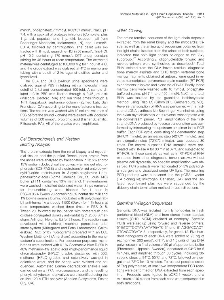
In the GLA renal biopsy tissue there were 29 glomeruli,four of which were obliterated by sclerosis. All of theremaining glomeruli exhibited normal cellularity, andthere were no mesangial nodules. A few had thickenedwrinkled basement membranes but most had minimalabnormalities. There was widespread interstitial edemaand fibrosis, focal lymphocytic infiltration, and focal tubu-lar atrophy. No prominent casts with multinucleated giantcells were seen. Congo red stain for amyloid was nega-tive. Immunofluorescence microscopy demonstratedbright staining of all glomerular, tubular, and vascularbasement membranes for � (Figure 1A), but not � lightchains; �, �, or � heavy chains; C3; C1q; fibrin; amyloidP component; or apoE (not shown). Clustered granulardeposits in the glomerular and tubular basement mem-branes (Figure 1B), typical of LCDD, were found ultra-structurally. A bone marrow aspirate and biopsy exhib-ited increased lymphocytes, dispersed as well as in smallaggregates, some with plasmacytoid differentiation; cell
marker analysis revealed predominantly B cells express-ing surface immunoglobulin of the � isotype with a �:�ratio of 30. A diagnosis of lymphoplasmacytoid lym-phoma was rendered.
In the CHO renal biopsy specimen all of the nineglomeruli present appeared normal; there was moderateinterstitial edema and lymphocytic cell infiltration andmany tubular protein casts, some surrounded by multinu-cleated giant cells, as seen in Bence-Jones cast ne-phropathy. Immunofluorescence microscopy revealedstrong staining of glomerular, tubular, and vascular base-ment membranes for � but not � light chain nor �, �, or �heavy chains; C3; C1q; fibrin; amyloid P component; orapoE. Clustered, granular electron-dense deposits in glo-merular and tubular basement membranes were ultra-structurally typical of LCDD. The bone marrow biopsydisplayed nodules and sheets of �-bearing plasma cellsand only a few scattered �-staining cells, diagnostic ofmyeloma. At autopsy the remarkable findings were a firmenlarged 400-g heart with left ventricular hypertrophy anda liver with nodular cirrhosis. There were no vertebral lyticlesions. Light microscopic examination confirmed the re-nal and bone marrow biopsy findings. The myocardiumexhibited mild interstitial widening by loose eosinophilicmaterial, but no evidence of inflammation or muscle dam-age. Congo red stains of all systemic organs showed noamyloid deposits. Immunofluorescence microscopydemonstrated deposits of � (Figure 1C), but not � lightchain (Figure 1D), around cardiac myocytes, in fibrousseptae and vascular walls, and diffusely in the basementmembranes of all systemic organs.
Western Blotting, Amino Acid Sequencing, andcDNA Cloning
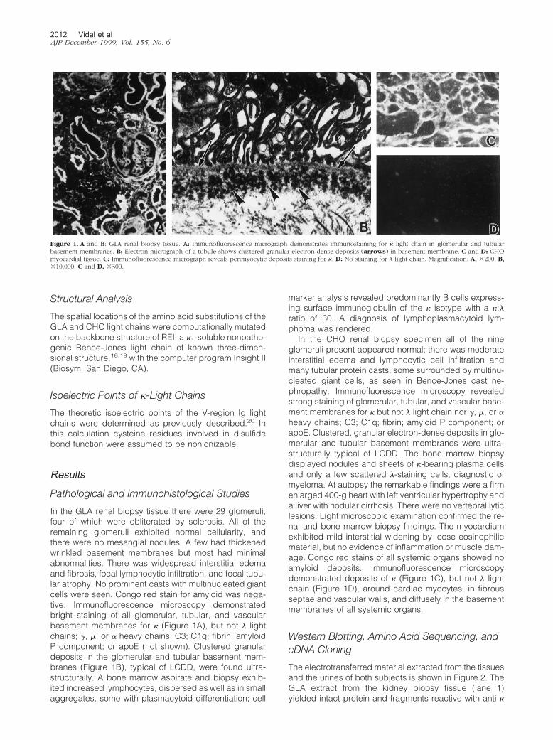
The electrotransferred material extracted from the tissuesand the urines of both subjects is shown in Figure 2. TheGLA extract from the kidney biopsy tissue (lane 1)yielded intact protein and fragments reactive with anti-�
Figure 1. A and B: GLA renal biopsy tissue. A: Immunofluorescence micrograph demonstrates immunostaining for � light chain in glomerular and tubularbasement membranes. B: Electron micrograph of a tubule shows clustered granular electron-dense deposits (arrows) in basement membrane. C and D: CHOmyocardial tissue. C: Immunofluorescence micrograph reveals perimyocytic deposits staining for �. D: No staining for � light chain. Magnification: A, �200; B,�10,000; C and D, �300.
2012 Vidal et alAJP December 1999, Vol. 155, No. 6
(lane 2). The sequences obtained from all bands werehomologous to the intact N-terminus of the �1 light chainsubgroup (Figure 3A); the 13-kd protein yielded the se-quence from residues 1–8 (16 kd, 1–15; 17 and 18 kd,1–10; and the 28-kd protein the sequence from positions1–6). The urine eluate from the KappaLock column was
mainly comprised of a single protein of 28 kd (Figure 2,lane 3) that was reactive with anti-� (Figure 2, lane 4) andrevealed the sequence from positions 1 to 35.
The transferred guanidine extract CHO from cardiactissue (Figure 2, lane 5), also consisting of intact proteinand fragments, demonstrated immunoreactivity with an-ti-� (Figure 2, lane 6); the sequence, homologous with theN-terminus of the �1 light chain subgroup (Figure 3B),was obtained from bands at 6.5 kd yielding residues1–10 (8 kd, 1–16; and 28 kd, 1–39). A 7-kd protein gavethe sequence from residues 51–70. Light chain from theurine eluted from the KappaLock column, composed of a28-kd protein (Figure 2, lane 7), was immunoreactive withanti-� (Figure 2, lane 8) and rendered the sequence fromresidues 1–37.
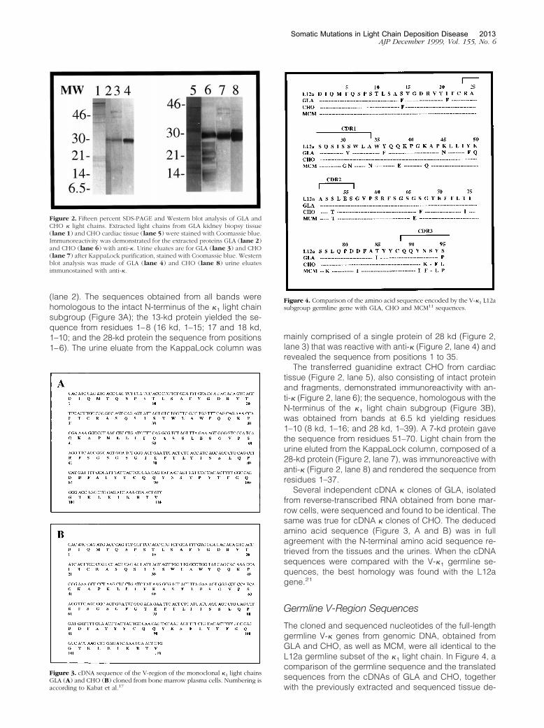
Several independent cDNA � clones of GLA, isolatedfrom reverse-transcribed RNA obtained from bone mar-row cells, were sequenced and found to be identical. Thesame was true for cDNA � clones of CHO. The deducedamino acid sequence (Figure 3, A and B) was in fullagreement with the N-terminal amino acid sequence re-trieved from the tissues and the urines. When the cDNAsequences were compared with the V-�1 germline se-quences, the best homology was found with the L12agene.21
Germline V-Region Sequences
The cloned and sequenced nucleotides of the full-lengthgermline V-� genes from genomic DNA, obtained fromGLA and CHO, as well as MCM, were all identical to theL12a germline subset of the �1 light chain. In Figure 4, acomparison of the germline sequence and the translatedsequences from the cDNAs of GLA and CHO, togetherwith the previously extracted and sequenced tissue de-
Figure 2. Fifteen percent SDS-PAGE and Western blot analysis of GLA andCHO � light chains. Extracted light chains from GLA kidney biopsy tissue(lane 1) and CHO cardiac tissue (lane 5) were stained with Coomassie blue.Immunoreactivity was demonstrated for the extracted proteins GLA (lane 2)and CHO (lane 6) with anti-�. Urine eluates are for GLA (lane 3) and CHO(lane 7) after KappaLock purification, stained with Coomassie blue. Westernblot analysis was made of GLA (lane 4) and CHO (lane 8) urine eluatesimmunostained with anti-�.
Figure 3. cDNA sequence of the V-region of the monoclonal �1 light chainsGLA (A) and CHO (B) cloned from bone marrow plasma cells. Numbering isaccording to Kabat et al.17
Figure 4. Comparison of the amino acid sequence encoded by the V-�1 L12asubgroup germline gene with GLA, CHO and MCM11 sequences.
Somatic Mutations in Light Chain Deposition Disease 2013AJP December 1999, Vol. 155, No. 6
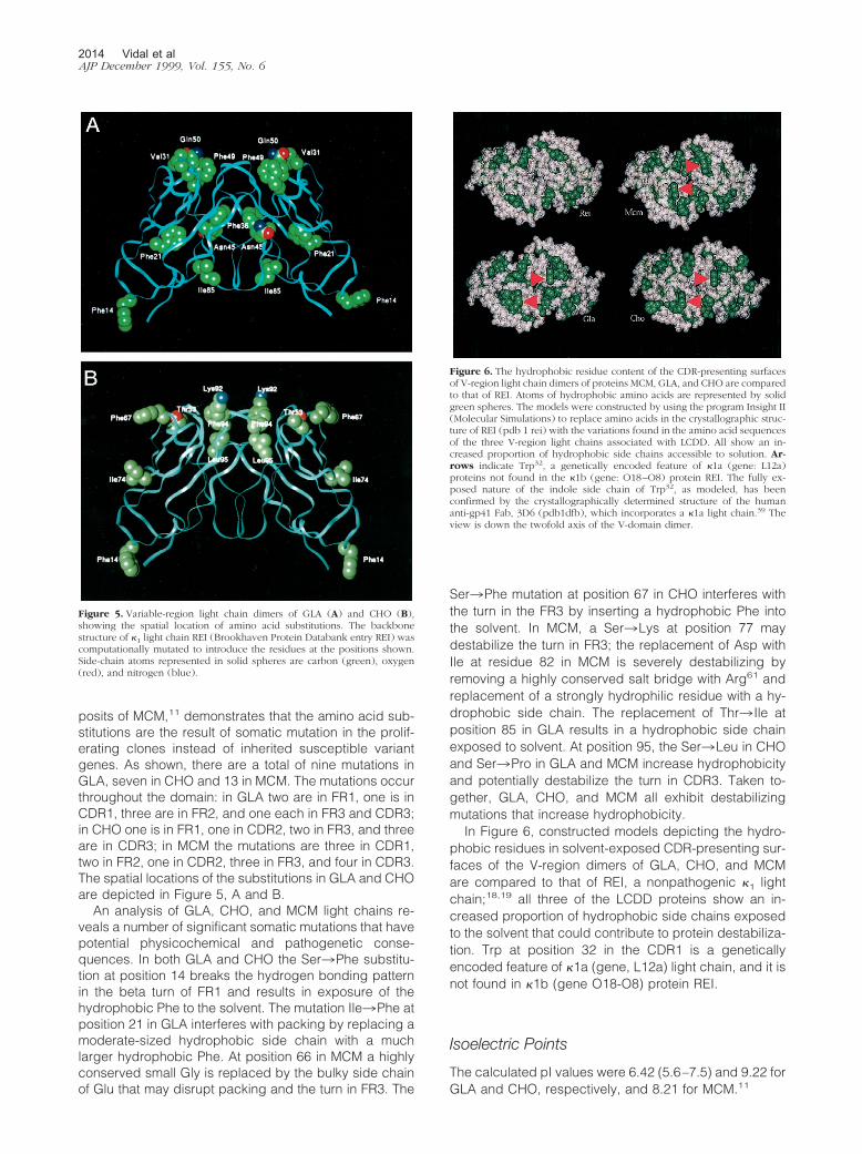
posits of MCM,11 demonstrates that the amino acid sub-stitutions are the result of somatic mutation in the prolif-erating clones instead of inherited susceptible variantgenes. As shown, there are a total of nine mutations inGLA, seven in CHO and 13 in MCM. The mutations occurthroughout the domain: in GLA two are in FR1, one is inCDR1, three are in FR2, and one each in FR3 and CDR3;in CHO one is in FR1, one in CDR2, two in FR3, and threeare in CDR3; in MCM the mutations are three in CDR1,two in FR2, one in CDR2, three in FR3, and four in CDR3.The spatial locations of the substitutions in GLA and CHOare depicted in Figure 5, A and B.
An analysis of GLA, CHO, and MCM light chains re-veals a number of significant somatic mutations that havepotential physicochemical and pathogenetic conse-quences. In both GLA and CHO the Ser3Phe substitu-tion at position 14 breaks the hydrogen bonding patternin the beta turn of FR1 and results in exposure of thehydrophobic Phe to the solvent. The mutation Ile3Phe atposition 21 in GLA interferes with packing by replacing amoderate-sized hydrophobic side chain with a muchlarger hydrophobic Phe. At position 66 in MCM a highlyconserved small Gly is replaced by the bulky side chainof Glu that may disrupt packing and the turn in FR3. The
Ser3Phe mutation at position 67 in CHO interferes withthe turn in the FR3 by inserting a hydrophobic Phe intothe solvent. In MCM, a Ser3Lys at position 77 maydestabilize the turn in FR3; the replacement of Asp withIle at residue 82 in MCM is severely destabilizing byremoving a highly conserved salt bridge with Arg61 andreplacement of a strongly hydrophilic residue with a hy-drophobic side chain. The replacement of Thr3 Ile atposition 85 in GLA results in a hydrophobic side chainexposed to solvent. At position 95, the Ser3Leu in CHOand Ser3Pro in GLA and MCM increase hydrophobicityand potentially destabilize the turn in CDR3. Taken to-gether, GLA, CHO, and MCM all exhibit destabilizingmutations that increase hydrophobicity.
In Figure 6, constructed models depicting the hydro-phobic residues in solvent-exposed CDR-presenting sur-faces of the V-region dimers of GLA, CHO, and MCMare compared to that of REI, a nonpathogenic �1 lightchain;18,19 all three of the LCDD proteins show an in-creased proportion of hydrophobic side chains exposedto the solvent that could contribute to protein destabiliza-tion. Trp at position 32 in the CDR1 is a geneticallyencoded feature of �1a (gene, L12a) light chain, and it isnot found in �1b (gene O18-O8) protein REI.
Isoelectric Points
The calculated pI values were 6.42 (5.6–7.5) and 9.22 forGLA and CHO, respectively, and 8.21 for MCM.11
Figure 5. Variable-region light chain dimers of GLA (A) and CHO (B),showing the spatial location of amino acid substitutions. The backbonestructure of �1 light chain REI (Brookhaven Protein Databank entry REI) wascomputationally mutated to introduce the residues at the positions shown.Side-chain atoms represented in solid spheres are carbon (green), oxygen(red), and nitrogen (blue).
Figure 6. The hydrophobic residue content of the CDR-presenting surfacesof V-region light chain dimers of proteins MCM, GLA, and CHO are comparedto that of REI. Atoms of hydrophobic amino acids are represented by solidgreen spheres. The models were constructed by using the program Insight II(Molecular Simulations) to replace amino acids in the crystallographic struc-ture of REI (pdb 1 rei) with the variations found in the amino acid sequencesof the three V-region light chains associated with LCDD. All show an in-creased proportion of hydrophobic side chains accessible to solution. Ar-rows indicate Trp32, a genetically encoded feature of �1a (gene: L12a)proteins not found in the �1b (gene: O18–O8) protein REI. The fully ex-posed nature of the indole side chain of Trp32, as modeled, has beenconfirmed by the crystallographically determined structure of the humananti-gp41 Fab, 3D6 (pdb1dfb), which incorporates a �1a light chain.39 Theview is down the twofold axis of the V-domain dimer.
2014 Vidal et alAJP December 1999, Vol. 155, No. 6

DiscussionIn the present report the structure of two new �1-LCDDproteins, GLA and CHO, obtained by N-terminal se-quence analysis of light chain deposits extracted fromthe tissues, light chains isolated from the urines, and theV-region amino acid sequences deduced from thecloned and sequenced cDNAs, and MCM,11 are com-pared to the germline sequence determined fromgenomic DNA in each. The findings indicate that the
amino acid substitutions in GLA, CHO, and MCM are theresult of somatic mutations in the proliferating clonesrather than from inherent variant alleles.
Although there are well over 150 published cases ofnonamyloidotic LCDD, the primary structure of the in-volved light chains has been determined in only 11cases. Nine, including GLA and CHO (Table 1), are �light chain, and two are � light chain.12 One (MCM) wasobtained by sequence analysis of light chain depositsextracted from tissue,11 two (SCI, BURN) by sequence
Table 1. Amino Acid Substitutions in V-Region �-Light Chains in LCDD
Genes: L12a L2–L16 B3
Protein: CHO GLA MCM ISE REV SCI BLU BURN FRA
FR-1 1-23 03 V�I* 104 M�L 109 A�G 114 S�F S�F 221 I�F 1
CDR-I 24-34 27 Q�L Q�L 227a S�G 127b V�L 127c L�F 127d Y�F 127e S�N 127f S�P S�T 228 N�R 130 S�G S�N 231 S�V S�N S�I S�T S�Y N�I 632 Y�F Y�C 234 A�N 1
FR-II 35-49 36 Y�F 138 Q�E 142 K�Q 143 P�T 145 K�N K�N K�Q 349 Y�F 1
CDR-II 50-56 50 K�Q G�S 251 A�T 152 S�T 153 S�T S�T S�N T�I T�I T�S 655 E�Q E�G 2
FR-III 57-88 59 P�S 164 G�A 166 G�E 167 S�F 168 G�E 170 D�N 172 T�S 174 T�I T�N 276 S�T S�T 277 S�K S�G S�I S�R S�R 581 E�G 182 D�I 183 F�L 185 T�I V�L V�T 387 Y�F 1
CDR-III 89-97 89 Q�H 190 Q�H 191 Y�I 192 N�K N�F N�D N�G N�E Y�S 693 N�D N�D S�T S�T 494 Y�F Y�L T�A 395 S�L (P)† (P) (P) P�L 2
* Germline � variation.† (Proline) encoded by a germline allele.
Somatic Mutations in Light Chain Deposition Disease 2015AJP December 1999, Vol. 155, No. 6
analysis of the monoclonal light chain from the urine,7,13
and the others (ISE, REV, BLU, FRA, BOU, and RAC)were deduced from cloned cDNAs.6,8–10,12
All of the nine �-LCDD proteins mentioned thus far(Table 1) are V-region light chains that belong to threedifferent light chain subgroups. Four of them (CHO, GLA,MCM, ISE) belong to the �1 subgroup, three (BLU, BURN,FRA) to �IV, and two (REV, SCI) belong to �III. The two�-LCDD proteins (BOU, RAC) belong to the �II subgroup.Among the nine �-LCDD proteins summarized in Table 1,all have amino acid substitutions that occur in all do-mains, but more frequently in the CDRs than in the FRs(51 versus 37), possibly reflecting immunoglobulin anti-body specificity that is antigen driven. Collectively thereare six in the FR1, 21 in the CDR1, eight in the FR2, 12 inthe CDR2, 23 in the FR3, and 18 in the CDR3.
All �-LCDD proteins exhibit one and as many as threemutations that can be expected to be structurally andfunctionally significant in affecting protein destabilization;in each at least one mutation is in a turn/loop exposed tosolvent. All � LCDD proteins characterized to date havecome from the three major germline genes that encode aTrp residue at solvent accessible positions, ie, L12a,Trp32; L2–L16, Trp94; B3, Trp50. Trp, together with theother hydrophobic residues at the surface sufficient toexclude the light chain from the solvent, could causeaggregation and precipitation in tissues. Hydrophobicresidues (Figure 6) are increased in relation to REI, andsuch substitutions in the FR regions (Table 1) could dis-tort the � structure, producing amorphous rather thanfibrillar deposits.
The diffuse organ distribution of nonamyloidotic de-posits in MCM and CHO and in other cases of LCDD4,5 isin contrast to the more patchy distribution of deposits inAL disease. The uniform light chain deposits in systemicbasement membranes of MCM suggested a unique af-finity for some component of the basement membrane,possibly due to an immunological or electrostatic inter-action. The calculated pI values of the V-region aminoacid sequences in MCM (8.21), as well as pI valuescalculated from published sequences of ISE and BLU(8.20, 8.19, respectively), were high.11 This observationraised the possibility that the affinity might be due tocharge interactions between cationic light chains andanionic GAGs in basement membranes,22 a mechanismof immune complex deposition demonstrated in vivo in amurine model.23 The calculated isoelectric point of thecomplete V-region in CHO is also high (9.22). In GLA thepI is lower (5.5–7.5). The high pIs of the light chains inLCDD are in contrast to the acidic pIs of amyloidogenicBence-Jones proteins (mean, 4.8 � 1.1),24 suggesting adifferent mechanism for tissue deposition. Furthermore, ifone compares the pI values determined by the N-terminal50 amino acids encoded by the � germline genes, onefinds acidic pI values for �1b (6.4) and �2 peptides(3.6–4.5). In contrast, all of the other genes encode basic50-mers, with 10.4, 9.9, and 9.8 deduced for �1a, �3a,and �4, respectively, the three gene families for whichLCDD representatives have been identified to date.
Why some Bence-Jones proteins are soluble and non-pathogenic, whereas others form either fibrillar or nonfi-
brillar granular aggregates in AL or LCDD, or even coex-isting fibrillar and nonfibrillar deposits,25 remains elusive.In AL, although the primary protein structure likely playsan important role in fibril formation, amyloidogenic motifshave not been identified in the primary sequence data ofmore than 60 light chains extracted from amyloid tissuedeposits. Moreover, the identity of the complete primarystructure of both the deposited amyloid fibril protein andthe soluble precursor Bence-Jones protein DIA26 and theamino-terminal identity of extracted light chains from co-existing fibrillar deposits of AL and granular deposits ofLCDD25 argue in favor of extrinsic factors and other mol-ecules that might play a part in the processing of sus-ceptible light chains. For example, the detection of amy-loid P component and apo-E in amyloid, but not innonamyloid light chain deposits,2 suggests a function, asyet unclear, for these molecules. On the other hand,spontaneous in vitro fibril formation of amyloid � pep-tides27 and other proteins not known to cause amyloid-osis28,29 relates to a physical-chemical environment fa-voring intermolecular interactions.
It is now apparent that a number of different diseases,including AL and LCDD, involve aberrant protein fold-ing.30,31 Based upon the accumulated data, LCDD andAL are considered to be conformational disorders. Theevidence suggests that somatic mutational effects maybe responsible for destabilization of the normal solubleglobular light chain structure.32 The increased hydropho-bicity of GLA, CHO, and MCM relative to REI shown inFigure 6 and similar observations reported by others33,34
could promote the formation of aggregation-prone, par-tially unfolded intermediates, thereby reducing solubilityand favoring deposition in tissues. In vitro studies haveshown that the Bence-Jones light chains in LCDD and ALwere more unstable than a nonpathogenic Bence-Jonesprotein, and AL light chain was more unstable than theLCDD light chain.35 In the context of current concepts ofprotein folding,36,37 the interesting question posed is therelationship between LCDD and AL, and whether LCDDrepresents a partially unfolded or misfolded intermediatespecies stabilized in a transition state that favors off-pathway aggregation and precipitation in tissues, or al-ternatively is a preamyloid form of disease with restrictionof maturation on pathway to fibril formation. The latterpossibility is supported by both in vivo and in vitro ultra-structural observations showing a transition betweengranular and fibrillar structures in tissue deposits in apatient with coexistent LCDD and AL,38 and the progres-sive formation in solution of Congophilic fibrils withingranular aggregates.29 As new data develop on proteinfolding and protein-protein interactions, it is likely thatnew insights into the different disease expressions exhib-ited in AL and LCDD and, importantly, the mechanisms offibrillogenesis will be achieved.
References
1. Gallo G, Picken M, Buxbaum J, Frangione B: The spectrum of mono-clonal immunoglobulin deposition disease associated with immuno-cytic dyscrasias. Semin Hematol 1989, 3:234–245
2016 Vidal et alAJP December 1999, Vol. 155, No. 6
2. Gallo G, Wisniewski T, Choi-Miura NH, Ghiso J, Frangione B: Potentialrole of apolipoprotein E in fibrillogenesis. Am J Pathol 1994, 145:526–530
3. Lyon AW, Narindrasorasak S, Young ID, Anastassiades T, CouchmanJR, McCarthy KJ, Kisilevsky R: Co-deposition of basement membranecomponents during the induction of murine splenic AA amyloid. LabInvest 1991, 64:785–790
4. Randall RE, Williamson WC Jr, Mulinax F, Tung MY, Still MJ: Manifes-tations of systemic light chain deposition. Am J Med 1976, 60:293–299
5. Ganeval D, Mignon F, Preud’homme JL, Noel L-H, Morel-Maroger L,Droz D, Brouet JC, Mery J, Grunfeld J-P: Visceral deposition ofmonoclonal light chains and immunoglobulins: a study of renal andimmunopathologic abnormalities. Adv Nephrol 1982, 11:25–63
6. Cogne M, Preud’homme JL, Bauwens M, Touchard G, Aucouturier P:Structure of a monoclonal � chain of the V-�IV subgroup in the kidneyand plasma cells in light chain deposition disease. J Clin Invest 1991,87:2186–2190
7. Bellotti V, Stoppini M, Merlini G, Zapponi MC, Meloni ML, Banfi G,Ferri G: Amino acid sequence of � Sci, the Bence Jones proteinisolated from a patient with light chain deposition disease. BiochimBiophys Acta 1991, 1097:177–182
8. Khamlichi AA, Aucouturier P, Silvain C, Bauwens M, Touchard G,Preud’homme J-L, Nau F, Cogne M: Primary structure of a monoclo-nal � chain in myeloma with light chain deposition disease. Clin ExpImmunol 1992, 87:122–126
9. Rocca A, Khamlichi AA, Aucouturier P, Noel LH, Denoroy JL,Preud’homme JL, Cogne M: Primary structure of a variable region ofthe V-�1 subgroup (ISE) in light chain deposition disease. Clin ExpImmunol 1993, 91:506–509
10. Decourt C, Cogne M, Rocca A: Structural peculiarities of a truncatedV�III immunoglobulin light chain in myeloma with light chain deposi-tion disease. Clin Exp Immunol 1996, 106:357–361
11. Gallo G, Goni F, Boctor F, Vidal R, Kumar A, Stevens FJ, Frangione B,Ghiso J: Light chain cardiomyopathy: structural analysis of the lightchain tissue deposits. Am J Pathol 1996, 148:1397–1406
12. Decourt C, Touchard G, Preud’homme J-L, Vidal R, Beaufils H,Diemert M-C, Cogne M: Complete primary sequences of two � im-munoglobulin light chains in myelomas with nonamyloid (Randall-type) light chain deposition disease. Am J Pathol 1998, 153:313–318
13. Stevens FJ, Weiss DT, Solomon A: Structural bases of light chain-related pathology. The Antibodies, vol 5. Edited by M Zanetti and JDCapra. Amsterdam, Harwood Academic Publishers, 1999, pp 175–208
14. Kaplan B, German G, Pras M: Isolation and characterization of amy-loid proteins from milligram amounts of amyloid-containing tissue. JLiq Chromatogr 1993, 16:249–268
15. Kaplan B, Yakar S, Kumar A, Pras M, Gallo G: Immunochemicalcharacterization of amyloid in diagnostic biopsy tissues. Amyloid IntJ Exp Clin Invest 1997, 4:80–86
16. Gallo G, Feiner H, Chuba J, Beneck D, Marion P, Cohen DH: Char-acterization of tissue amyloid by immunofluorescence microscopy.Clin Immunol Immunopathol 1986, 39:479–490
17. Kabat EA, Wu TT, Perry HM, Gottesman KS, Foeller C: Human � LightChain Subgroup I. Sequences of Proteins of Immunological Interest.Bethesda, MD, National Institutes of Health, 1991, pp 103–112
18. Epp O, Lattman EF, Schiffer M, Huber R, Palm W: The molecularstructure of a dimer composed of the variable portions of the Bence-Jones protein REI refined at 2.0-A resolution. Biochemistry 1975,14:4943–4952
19. Choathia C, Lesk AM: Canonical structure for the hypervariable re-gions of the immunoglobulins. J Mol Biol 1987, 196:901–917
20. Skoog B, Wichman A: Calculation of the isoelectric points of polypep-
tides from the amino-acid composition. Trends Anal Chem 1986,82–83
21. Klein R, Jaenichen R, Zachau HG: Expressed human immunoglobulin� genes and their hypermutation. Eur J Immunol 1993, 23:3248–3271
22. Kanwar YS, Farquhar MG: Presence of heparan sulfate in glomerularbasement membrane. Proc Natl Acad Sci USA 1979, 76:1303–1307
23. Gallo GR, Caulin-Glaser T, Lamm ME: Charge of circulating immunecomplexes as a factor in glomerular basement membrane localizationin mice. J Clin Invest 1981, 67:1305–1307
24. Bellotti V, Merlini G, Bucciarelli E, Perfetti V, Quaglini S, Ascari E:Relevance of class, molecular weight and isoelectric point in predict-ing human light chain amyloidogenicity. Br J Haematol 1990, 74:65–69
25. Kaplan B, Vidal R, Kumar A, Ghiso J, Frangione B, Gallo G: Amino-terminal identity of co-existent amyloid and non-amyloid immunoglob-ulin � light chain deposits. A human disease to study alterations ofprotein conformation. Clin Exp Immunol 1997, 110:472–478
26. Klafki H-W, Kratzin HD, Pick AI, Eckart K, Karas M, Hilschmann N:Complete amino acid sequence determinations demonstrate identityof the urinary Bence Jones protein (BJP-DIA) and the amyloid fibrilprotein (AL-DIA) in a case of AL-amyloidosis. Biochemistry 1992,31:3265–3272
27. Soto C, Frangione B: Two conformational states of amyloid �-peptide:implication for the pathogenesis of Alzheimer’s disease. Neurosci Lett1995, 186:115–118
28. Jimenez JL, Guijarro JI, Orlova E, Zurdo J, Dobson CM, Sunde M,Saibil HR: Cryo-electron microscopy structure of an SH3 amyloid fibriland model of the molecular packing. EMBO J 1999, 18:815–821
29. Chiti F, Webster P, Taddei N, Clark A, Stefani M, Ramponi G, DobsonCM: Designing conditions for in vitro formation of amyloid protofila-ments and fibrils. Proc Natl Acad Sci USA 1999, 96:3590–3594
30. Soto C, Ghiso J, Frangione B: Alzheimer’s amyloid � aggregation ismodulated by the interaction of multiple factors. Alzheimer’s Res1997, 3:215–222
31. Carrell RW, Lomas DA: Conformational disease. Lancet 1997, 350:134–138
32. Hurle MR, Helms LR, Li L, Chan W, Wetzel R: A role for destabilizingamino acid replacements in light chain amyloidosis. Proc Natl AcadSci USA 1994, 91:5446–5450
33. Deret S, Chomilier J, Huang DB, Preud’homme J-L, Stevens FJ,Aucouturier P: Molecular modeling of immunoglobulin light chainsimplicates hydrophobic residues in non-amyloid light chain deposi-tion disease. Protein Eng 1997, 10:1191–1197
34. Raffen R, Dieckman LJ, Szpunar M, Wuschl C, Pokkuluri PR, Dave P,Stevens PW, Cai X, Schiffer M, Steven F: Physicochemical conse-quences of amino acid variations that contribute to fibril formation byimmunoglobulin light chains. Protein Sci 1999, 8:509–517
35. Bellotti V, Stoppini M, Mangione PP, Fornasieri A, Min L, Merlini G,Ferri G: Structural and functional characterization of three humanimmunoglobulin kappa light chains with different pathologic implica-tions. Biochim Biophys Acta 1996, 1317:161–167
36. Capaldi AP, Ferguson SJ, Radford S: The Greek key protein apo-pseudoazurin folds through an obligate on-pathway intermediate. JMol Biol 1999, 286:1621–1632
37. Radford SE, Dobson CM: From computer simulations to humandisease: emerging themes in protein folding. Cell 1999, 97:291–298
38. Stokes MB, Jagirdar J, Burchstin O, Kornacki S, Kumar A, Gallo G:Nodular pulmonary immunoglobulin light chain deposits with coexis-tent amyloid and nonamyloid features in an HIV-infected patient. ModPathol 1997, 10:1059–1065
39. He XM, Ruker F, Casale E, Carter DC: Structure of a human mono-clonal antibody Fab fragment against gp41 of human immunodefi-ciency virus type I. Proc Natl Acad Sci USA 1992, 89:7154–7158
Somatic Mutations in Light Chain Deposition Disease 2017AJP December 1999, Vol. 155, No. 6