Embed Size (px)
Citation preview
Research Article
Somatic recombination underlies frequent revertantmosaicism in loricrin keratodermaShotaro Suzuki1, Toshifumi Nomura1 , Toshinari Miyauchi1, Masae Takeda1, Yasuyuki Fujita1, Wataru Nishie1,Masashi Akiyama2, Akemi Ishida-Yamamoto3, Hiroshi Shimizu1
Revertant mosaicism is a phenomenon in which pathogenicmutations are rescued by somatic events, representing a form ofnatural gene therapy. Here, we report on the first evidence forrevertant mosaicism in loricrin keratoderma (LK), an autosomaldominant form of ichthyosis caused by mutations in LOR on1q21.3. We identified two unrelated LK families exhibiting dozensof previously unreported white spots, which increased in bothnumber and size with age. Biopsies of these spots revealed thatthey had normal histology and that causal LOR mutations werelost. Notably, dense single nucleotide polymorphism mappingidentified independent copy-neutral loss-of-heterozygosity eventson chromosome 1q extending from regions centromeric to LOR tothe telomere in all investigated spots, suggesting that somaticrecombination represents a common reversion mechanism in LK.Furthermore, we demonstrated that reversion of LOR mutationsconfers a growth advantage to cells in vitro, but the clinicallylimited size of revertant spots suggests the existence of mecha-nisms constraining revertant clone expansion. Nevertheless, theidentification of revertant mosaicism in LK might pave the way forrevertant therapy for this intractable disease.
DOI 10.26508/lsa.201800284 | Received 18 December 2018 | Revised 25January 2019 | Accepted 28 January 2019 | Published online 4 February 2019
Introduction
The epidermis, the outermost layer of the skin, is a tissue with a highturnover rate that is constantly being replenished by skin-residingstem cells (SCs) (Gonzales & Fuchs, 2017). Epidermal SCs self-renewand generate more-differentiated epidermal cells called kerati-nocytes throughout life, inevitably acquiring mutations that resultfrom both environmental factors and DNA replication errors withage. Although most of these mutations do not have a noticeableeffect, some are deleterious, and the age-related accumulation ofsomatic mutations in SCs potentially contributes to carcinogenesisand the decline of tissue function (Adams et al, 2015; Siudejaet al, 2015; Blokzijl et al, 2016). In theory, such genetic alterations,
including point mutations, gene conversion, and recombination,can also lead to natural correction of pathogenic mutations at thesomatic-cell level in genetic disorders. Somatic reversion of amutant phenotype is referred to as revertant mosaicism and resultsfrom the correction of a disease-causing mutation in a somatic cellfollowed by the survival and clonal expansion of the revertant cell(Jonkman & Pasmooij, 2009). However, this clinically important“natural gene therapy” phenomenon has been documented in only~30 diseases, including seven genodermatoses caused by muta-tions in COL7A1 (recessive dystrophic epidermolysis bullosa [EB,Mendelian inheritance in man (MIM): 226600]), COL17A1 or LAMB3(junctional EB [MIM: 226650]), KRT14 (EB simplex [MIM: 131900]), FERMT1(Kindler syndrome [MIM: 173650]), KRT10 or KRT1 (ichthyosis withconfetti [IWC, MIM: 609165]), and GJB2 (keratitis-ichthyosis-deafnesssyndrome [MIM: 148210]) (Jonkman & Pasmooij, 2009; Gudmundssonet al, 2017; Lim et al, 2017; van den Akker et al, 2018). Althoughmutation-free induced pluripotent SCs established from revertant junctional EBkeratinocytes are already on the way to clinical translation (Umegaki-Arao et al, 2014), the feasibility and generality of this therapeuticapproach appear to be restricted by the currently limited number ofdiseases that are known to exhibit revertant mosaicism, warrantingexpansion of the repertoire of such diseases.
Loricrin keratoderma (LK [MIM: 604117]) is a rare autosomaldominant skin disorder that results from gain-of-function muta-tions in LOR (MIM: 152445), which encodes loricrin, on 1q21.3 (Ishida-Yamamoto et al, 1997; Ishida-Yamamoto, 2003). The late epidermaldifferentiation marker loricrin is the major component of thecornified envelope, a highly insoluble and robust structure that isformed beneath the cell membrane of keratinocytes during ter-minal differentiation and provides the vital physical barrier to theskin (Ishida-Yamamoto, 2003). LK is clinically characterized bydefective skin barrier phenotypes, such as palmoplantar kerato-derma (thickening of the skin on the palms and soles; PPK) andgeneralized ichthyosis (dry, thickened, and scaly skin), with orwithout varying degrees of constricted digits, erythematousplaques, and/or erythroderma (generalized red skin) (Maestriniet al, 1996; Ishida-Yamamoto et al, 1997; Ishida-Yamamoto, 2003;Pohler et al, 2015). Notably, all LOR mutations reported to date
1Department of Dermatology, Hokkaido University Graduate School of Medicine, Sapporo, Japan 2Department of Dermatology, Nagoya University Graduate School ofMedicine, Nagoya, Japan 3Department of Dermatology, Asahikawa Medical University, Asahikawa, Japan
Correspondence: [email protected]
© 2019 Suzuki et al. https://doi.org/10.26508/lsa.201800284 vol 2 | no 1 | e201800284 1 of 10
on 25 December, 2019life-science-alliance.org Downloaded from http://doi.org/10.26508/lsa.201800284Published Online: 4 February, 2019 | Supp Info:
cause a frameshift and replace the glycine-rich C-terminus of the315-amino-acid wild-type loricrin protein with a highly arginine-rich nuclear localization signal sequence (Maestrini et al, 1996;Ishida-Yamamoto et al, 1997; Ishida-Yamamoto, 2003; Pohler et al,2015; Khalil et al, 2017), leading to the accumulation of mutant loricrinin the nucleolus of affected keratinocytes (Ishida-Yamamoto, 2003).Although the precise mode of action of mutant loricrin in thepathogenesis of LK is not fully understood, its elimination is crucialfor curing the disease.
Here, we describe two families with LK exhibiting progressivedevelopment of revertant skin areas in which causal mutations werecorrected. Of particular note, the reversion mechanism in six in-vestigated revertant epidermis samples was somatic recombination.The frequent occurrence of mutation-reversion events via somaticrecombination was a remarkable feature of these LK families.
Results
Identification of revertant skin spots in two unrelated LK patients
In family 1, there were three affected individuals—the proband, a51-yr-old man born to unaffected parents, and his two daughters,aged 12 and 9 (Fig 1A)—exhibiting the hallmark features of LK(i.e., generalized ichthyosis, PPK, constricted digits, and/or well-demarcated erythematous plaques; Figs 1B–D and S1). Histology ofthe lesional skin sampled from the proband revealed hyper-keratosis, parakeratosis (retained nuclei in the stratum corneum[the outermost layer of the epidermis]), prominent keratohyalingranules, and acanthosis, all of which are characteristic features ofLK (Fig 1E and F). Whole-exome and Sanger sequencing in threeaffected and two unaffected individuals in the family revealed thatthe affected individuals were all heterozygous for a 1-bp insertionmutation in LOR, c.545_546insG (p.Gly183ArgfsTer153) (Fig 1G), whichhad previously been reported to cause LK (Song et al, 2008), furtherverifying the diagnosis of LK. By contrast, the unaffected individualswere both wild-type for the mutation. Notably, we found that er-ythematous skin of the proband was interrupted by dozens ofwhitish, normal-appearing skin patches on his trunk and extrem-ities (Fig 1H and I). The patches, which were up to 10mm in diameter,were slightly depressed from the surrounding affected skin (Fig 1I).Dermoscopy, a noninvasive in vivo technique that allows detailedexamination of skin lesions, revealed that the patches exhibited noscaling (Fig 1J), suggesting the possibility that they representedrevertant skin areas.
To test this possibility, we sampled skin from one of the whitishpatches, which was then subjected to histological and geneticanalyses. Notably, the examined patch showed normal histology,with a normal basket-weave stratum corneum (Fig 1K). Immuno-histochemical analysis revealed that mutant loricrin mislocalizedto the nuclei of the affected keratinocytes (Fig 2A–K), which reflectsthe fact that it contains an arginine-rich nuclear localization signalmotif (Fig 2L). In contrast, mutant loricrin was not detected in thekeratinocytes sampled from the normal-appearing patch (Fig 2K).We next examined the LOR genotype in genomic DNA samplesextracted separately from the epidermis and dermis of the normal-
appearing patch. Notably, themutation was absent in the epidermisbut remained in the dermis (Fig 1L). Thus, we confirmed that theclinically and histologically verified reversal of symptoms wasdue to the somatic reversion of the pathogenic mutation in theepidermis.
To clarify whether his other normal-appearing skin patches alsorepresent revertant spots, we analysed three additional patches.Notably, they also showed normal histology and somatic reversionof the pathogenic mutation in the epidermis (Figs S2 and S3). Thus,we confirmed the multiple occurrence of somatic reversion of theLOR mutation in the proband. To the best of our knowledge,however, revertant mosaicism has never been noted in previouslyreported LK cases.
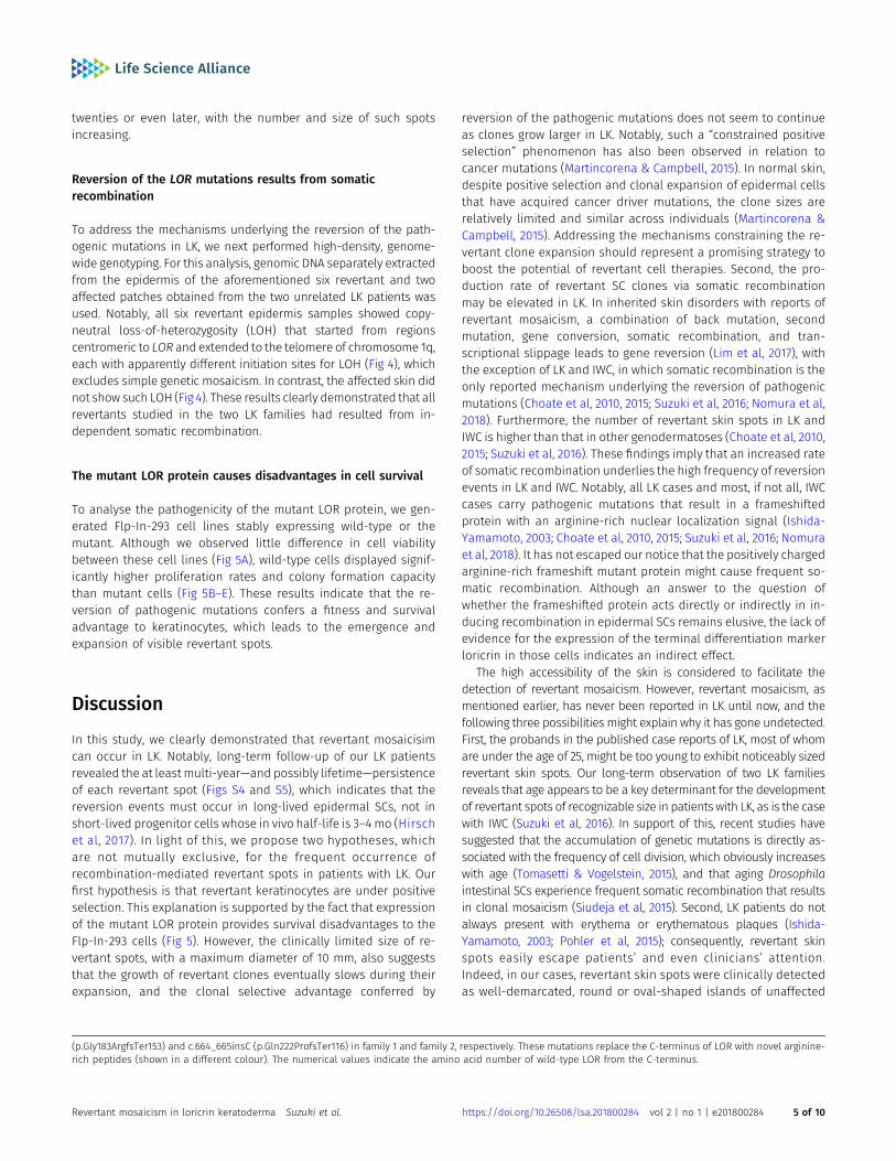
To test the generality of revertant mosaicism in LK, we analyseda second family with the disease. In family 2, there were two aliveaffected individuals whose detailed clinicohistological and ge-netic features had been published elsewhere (Ishida-Yamamotoet al, 1997 and Fig 3A). Briefly, they exhibited generalized ich-thyosis, PPK, constricted fingers, and widespread erythematousplaques (Fig 3B–E) and carried a heterozygous mutation,c.664_665insC, in LOR (Ishida-Yamamoto et al, 1997). The mutationresults in a frameshift and delayed termination (p.Gln222Prof-sTer116), yielding a C-terminally extended arginine-rich mutantloricrin protein (Fig 2L). Of particular note, careful re-evaluation ofthe 58-yr-old proband’s skin phenotypes revealed that her ery-thematous skin was also interrupted by dozens of non-scaly,whitish skin spots, which were up to 10 mm in size and werenoted almost exclusively on the lower extremities (Fig 3D and E).To validate whether they represent revertant spots, two of theclinically normal-appearing patches and the lesional skin of herleft posterior thigh were sampled for histological and geneticanalyses. Histology of the lesional skin showed marked hyper-keratosis with parakeratosis and hypergranulosis due to themutation (Fig 3F–H). In contrast, the two normal-appearingpatches displayed normal epidermal architecture (Fig 3I–L).Sanger sequencing revealed that the disease-causing mutationhad almost reverted in the epidermis, but not in the dermis, of thetwo histologically cured patches (Fig 3M and N). Taken together,we concluded that the clinically and histologically verified re-versal of her skin phenotypes resulted from somatic reversion ofthe disease-causing mutation to wild-type.
Age as a key factor for the development of clinically recognizablerevertant spots
In family 1, according to the proband’s personal statement, thenormal patches had appeared at age 20 and had gradually in-creased in both number and size over decades (Fig S4). In family 2,the photographic record revealed that the proband had notexhibited these spots at the age of 27 (Fig S5), indicating that theyhad become detectable after that age. Furthermore, the proband’stwo affected daughters, aged 12 and 9, in family 1 did not exhibitnormal-appearing patches (Fig S1), whereas the affected father ofthe proband in family 2 showed multiple clinically reverted skinspots when he was 57 (Fig S6). These findings suggest that LKpatients may develop revertant spots for the first time in their
Revertant mosaicism in loricrin keratoderma Suzuki et al. https://doi.org/10.26508/lsa.201800284 vol 2 | no 1 | e201800284 2 of 10
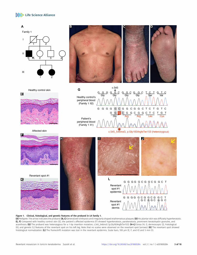
Figure 1. Clinical, histological, and genetic features of the proband in LK family 1.(A) Pedigree. The arrow indicates the proband. (B, C) Generalized ichthyosis and irregularly shaped erythematous plaques. (D)His plantar skin was diffusely hyperkeratotic.(E, F) Compared with healthy control skin (E), the patient’s affected epidermis (F) showed hyperkeratosis, parakeratosis, prominent keratohyalin granules, andacanthosis. (G) The proband was heterozygous for a 1-bp insertion mutation, c.545_546insG (p.Gly183ArgfsTer153). (H–L) Gross (H, I), dermoscopic (J), histological(K), and genetic (L) features of the revertant spot on his left leg. Note that no scales were observed on the revertant spot (arrows). (K) The revertant spot showedhistological normalization. (L) The frameshift mutation was lost in the revertant epidermis. Scale bars, 100 μm (E, F, and K) and 5 mm (I).
Revertant mosaicism in loricrin keratoderma Suzuki et al. https://doi.org/10.26508/lsa.201800284 vol 2 | no 1 | e201800284 3 of 10

Figure 2. Immunolocalization of mutant LOR and its C-terminal amino acid sequence.(A–C) In healthy control epidermis, KRT10 and wild-type LOR were expressed by suprabasal keratinocytes and more-differentiated keratinocytes, respectively. Nomutant LOR was detected. (D–H) Immunolocalization of KRT10 and wild-type LOR revealed thicker stratum spinosum and granulosum in the affected epidermis than in thecontrol. Mutant LOR accumulated in the nuclei of keratinocytes in the upper epidermis. Higher magnification of the square in (F) is shown in (G). Merged image of anti-KRT10 and anti-mutant LOR was also shown (H). (I–K) Immunolocalization of KRT10, wild-type LOR and mutant LOR was normalized in the revertant epidermis.Scale bars, 100 μm. (L) The C-terminal amino acid sequences of the wild-type and mutant LOR. We identified two frameshift mutations in LOR, namely, c.545_546insG
Revertant mosaicism in loricrin keratoderma Suzuki et al. https://doi.org/10.26508/lsa.201800284 vol 2 | no 1 | e201800284 4 of 10
twenties or even later, with the number and size of such spotsincreasing.
Reversion of the LOR mutations results from somaticrecombination
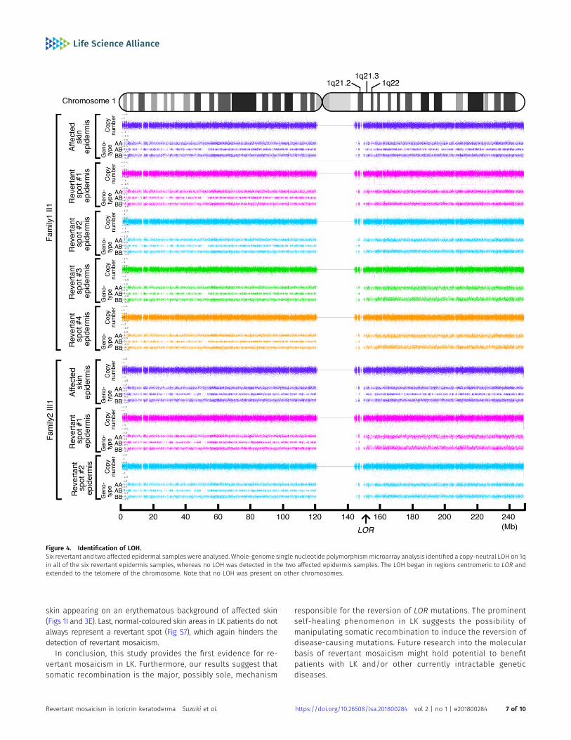
To address the mechanisms underlying the reversion of the path-ogenic mutations in LK, we next performed high-density, genome-wide genotyping. For this analysis, genomic DNA separately extractedfrom the epidermis of the aforementioned six revertant and twoaffected patches obtained from the two unrelated LK patients wasused. Notably, all six revertant epidermis samples showed copy-neutral loss-of-heterozygosity (LOH) that started from regionscentromeric to LOR and extended to the telomere of chromosome 1q,each with apparently different initiation sites for LOH (Fig 4), whichexcludes simple genetic mosaicism. In contrast, the affected skin didnot show such LOH (Fig 4). These results clearly demonstrated that allrevertants studied in the two LK families had resulted from in-dependent somatic recombination.
The mutant LOR protein causes disadvantages in cell survival
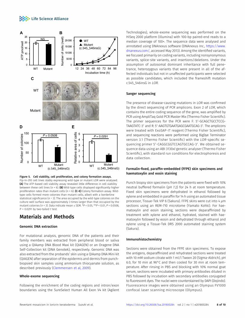
To analyse the pathogenicity of the mutant LOR protein, we gen-erated Flp-In-293 cell lines stably expressing wild-type or themutant. Although we observed little difference in cell viabilitybetween these cell lines (Fig 5A), wild-type cells displayed signif-icantly higher proliferation rates and colony formation capacitythan mutant cells (Fig 5B–E). These results indicate that the re-version of pathogenic mutations confers a fitness and survivaladvantage to keratinocytes, which leads to the emergence andexpansion of visible revertant spots.
Discussion
In this study, we clearly demonstrated that revertant mosaicisimcan occur in LK. Notably, long-term follow-up of our LK patientsrevealed the at least multi-year—and possibly lifetime—persistenceof each revertant spot (Figs S4 and S5), which indicates that thereversion events must occur in long-lived epidermal SCs, not inshort-lived progenitor cells whose in vivo half-life is 3–4mo (Hirschet al, 2017). In light of this, we propose two hypotheses, whichare not mutually exclusive, for the frequent occurrence ofrecombination-mediated revertant spots in patients with LK. Ourfirst hypothesis is that revertant keratinocytes are under positiveselection. This explanation is supported by the fact that expressionof the mutant LOR protein provides survival disadvantages to theFlp-In-293 cells (Fig 5). However, the clinically limited size of re-vertant spots, with a maximum diameter of 10 mm, also suggeststhat the growth of revertant clones eventually slows during theirexpansion, and the clonal selective advantage conferred by
reversion of the pathogenic mutations does not seem to continueas clones grow larger in LK. Notably, such a “constrained positiveselection” phenomenon has also been observed in relation tocancer mutations (Martincorena & Campbell, 2015). In normal skin,despite positive selection and clonal expansion of epidermal cellsthat have acquired cancer driver mutations, the clone sizes arerelatively limited and similar across individuals (Martincorena &Campbell, 2015). Addressing the mechanisms constraining the re-vertant clone expansion should represent a promising strategy toboost the potential of revertant cell therapies. Second, the pro-duction rate of revertant SC clones via somatic recombinationmay be elevated in LK. In inherited skin disorders with reports ofrevertant mosaicism, a combination of back mutation, secondmutation, gene conversion, somatic recombination, and tran-scriptional slippage leads to gene reversion (Lim et al, 2017), withthe exception of LK and IWC, in which somatic recombination is theonly reported mechanism underlying the reversion of pathogenicmutations (Choate et al, 2010, 2015; Suzuki et al, 2016; Nomura et al,2018). Furthermore, the number of revertant skin spots in LK andIWC is higher than that in other genodermatoses (Choate et al, 2010,2015; Suzuki et al, 2016). These findings imply that an increased rateof somatic recombination underlies the high frequency of reversionevents in LK and IWC. Notably, all LK cases and most, if not all, IWCcases carry pathogenic mutations that result in a frameshiftedprotein with an arginine-rich nuclear localization signal (Ishida-Yamamoto, 2003; Choate et al, 2010, 2015; Suzuki et al, 2016; Nomuraet al, 2018). It has not escaped our notice that the positively chargedarginine-rich frameshift mutant protein might cause frequent so-matic recombination. Although an answer to the question ofwhether the frameshifted protein acts directly or indirectly in in-ducing recombination in epidermal SCs remains elusive, the lack ofevidence for the expression of the terminal differentiation markerloricrin in those cells indicates an indirect effect.
The high accessibility of the skin is considered to facilitate thedetection of revertant mosaicism. However, revertant mosaicism, asmentioned earlier, has never been reported in LK until now, and thefollowing three possibilities might explain why it has gone undetected.First, the probands in the published case reports of LK, most of whomare under the age of 25, might be too young to exhibit noticeably sizedrevertant skin spots. Our long-term observation of two LK familiesreveals that age appears to be a key determinant for the developmentof revertant spots of recognizable size in patients with LK, as is the casewith IWC (Suzuki et al, 2016). In support of this, recent studies havesuggested that the accumulation of genetic mutations is directly as-sociated with the frequency of cell division, which obviously increaseswith age (Tomasetti & Vogelstein, 2015), and that aging Drosophilaintestinal SCs experience frequent somatic recombination that resultsin clonal mosaicism (Siudeja et al, 2015). Second, LK patients do notalways present with erythema or erythematous plaques (Ishida-Yamamoto, 2003; Pohler et al, 2015); consequently, revertant skinspots easily escape patients’ and even clinicians’ attention.Indeed, in our cases, revertant skin spots were clinically detectedas well-demarcated, round or oval-shaped islands of unaffected
(p.Gly183ArgfsTer153) and c.664_665insC (p.Gln222ProfsTer116) in family 1 and family 2, respectively. These mutations replace the C-terminus of LOR with novel arginine-rich peptides (shown in a different colour). The numerical values indicate the amino acid number of wild-type LOR from the C-terminus.
Revertant mosaicism in loricrin keratoderma Suzuki et al. https://doi.org/10.26508/lsa.201800284 vol 2 | no 1 | e201800284 5 of 10
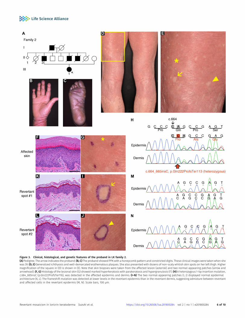
Figure 3. Clinical, histological, and genetic features of the proband in LK family 2.(A) Pedigree. The arrow indicates the proband. (B, C) The proband showed PPK with a honeycomb pattern and constricted digits. These clinical images were taken when shewas 39. (D, E) Generalized ichthyosis and well-demarcated erythematous plaques. She also presented with dozens of non-scaly whitish skin spots on her left thigh. Highermagnification of the square in (D) is shown in (E). Note that skin biopsies were taken from the affected lesion (asterisk) and two normal-appearing patches (arrow andarrowhead). (F, G)Histology of the lesional skin (G) showedmarked hyperkeratosis with parakeratosis and hypergranulosis (F). (H) A heterozygous 1-bp insertion mutation,c.664_665insC (p.Gln222ProfsTer116), was detected in the affected epidermis and dermis. (I–N) The two normal-appearing patches (I, J) displayed normal epidermalarchitecture (K, L). The frameshift mutation was detected at lower levels in the revertant epidermis than in the revertant dermis, suggesting admixture between revertantand affected cells in the revertant epidermis (M, N). Scale bars, 100 μm.
Revertant mosaicism in loricrin keratoderma Suzuki et al. https://doi.org/10.26508/lsa.201800284 vol 2 | no 1 | e201800284 6 of 10
skin appearing on an erythematous background of affected skin(Figs 1I and 3E). Last, normal-coloured skin areas in LK patients do notalways represent a revertant spot (Fig S7), which again hinders thedetection of revertant mosaicism.
In conclusion, this study provides the first evidence for re-vertant mosaicism in LK. Furthermore, our results suggest thatsomatic recombination is the major, possibly sole, mechanism
responsible for the reversion of LOR mutations. The prominentself-healing phenomenon in LK suggests the possibility ofmanipulating somatic recombination to induce the reversion ofdisease-causing mutations. Future research into the molecularbasis of revertant mosaicism might hold potential to benefitpatients with LK and/or other currently intractable geneticdiseases.
Figure 4. Identification of LOH.Six revertant and two affected epidermal samples were analysed. Whole-genome single nucleotide polymorphismmicroarray analysis identified a copy-neutral LOH on 1qin all of the six revertant epidermis samples, whereas no LOH was detected in the two affected epidermis samples. The LOH began in regions centromeric to LOR andextended to the telomere of the chromosome. Note that no LOH was present on other chromosomes.
Revertant mosaicism in loricrin keratoderma Suzuki et al. https://doi.org/10.26508/lsa.201800284 vol 2 | no 1 | e201800284 7 of 10
Materials and Methods
Genomic DNA extraction
For mutational analysis, genomic DNA of the patients and theirfamily members was extracted from peripheral blood or salivausing a QIAamp DNA Blood Maxi kit (QIAGEN) or an Oragene DNASelf-Collection kit (DNA Genotek), respectively. Genomic DNA wasalso extracted from the probands’ skin using a QIAamp DNA Mini kit(QIAGEN) after separation of the epidermis and dermis from punch-biopsied skin samples using ammonium thiocyanate solution, asdescribed previously (Clemmensen et al, 2009).
Whole-exome sequencing
Following the enrichment of the coding regions and intron/exonboundaries using the SureSelect Human All Exon V4 kit (Agilent
Technologies), whole-exome sequencing was performed on theHiSeq 2000 platform (Illumina) with 100-bp paired-end reads to amedian coverage of 100×. The sequence data were analysed andannotated using DNAnexus software (DNAnexus Inc., https://www.dnanexus.com/, accessed May 2013). Among the identified variants,we focused primarily on coding variants, including nonsynonymousvariants, splice-site variants, and insertions/deletions. Under theassumption of autosomal dominant inheritance with full pene-trance, heterozygous variants that were present in all of the af-fected individuals but not in unaffected participants were selectedas possible candidates, which included the frameshift mutationc.545_546insG in LOR.
Sanger sequencing
The presence of disease-causing mutations in LOR was confirmedby the direct sequencing of PCR amplicons. Exon 2 of LOR, whichcontains the entire coding sequence of the gene, was amplified byPCR using AmpliTaq Gold PCR Master Mix (Thermo Fisher Scientific).The primer sequences for the PCR were F: 59-GCAGCTGCCTCCG-TAAGTATC-39 and R: 59-AAGTGTGAATGAGCGAATGCAG-39. The ampliconswere treated with ExoSAP-IT reagent (Thermo Fisher Scientific),and sequencing reactions were performed using BigDye Terminatorversion 3.1 (Thermo Fisher Scientific) with the LOR-specific se-quencing primer 59-CAGGCGGTCCAGTGCCAG-39. We obtained se-quence data using an ABI 3130xl genetic analyser (Thermo FisherScientific), with standard run conditions for electrophoresis anddata collection.
Formalin-fixed, paraffin-embedded (FFPE) skin specimens andhaematoxylin and eosin staining
Punch biopsy skin specimens from the patients were fixed with 10%neutral buffered formalin (pH 7.2) for 24 h at room temperature.Fixed skin specimens were dehydrated in ethanol followed byxylene and embedded in paraffin for 14 h using an automated tissueprocessor, Tissue-Tek VIP 6 (Sakura). FFPE skins were cut into 4 μmsections using an REM-710 microtome (Yamato Kohki). For hae-matoxylin and eosin staining, sections were deparaffinized bytreatment with xylene and ethanol, hydrated, stained with hae-matoxylin followed by eosin and dehydrated through ethanol andxylene using a Tissue-Tek DRS 2000 automated staining system(Sakura).
Immunohistochemistry
Sections were obtained from the FFPE skin specimens. To exposethe antigens, deparaffinized and rehydrated sections were treatedwith 10 mM sodium citrate with 1 ml/l Tween 20 (Sigma-Aldrich), pH6.0, for 10 min at 96°C and then cooled for 30 min at room tem-perature. After rinsing in PBS and blocking with 10% normal goatserum, sections were incubated with primary antibodies diluted inPBS followed by incubation with secondary antibodies conjugatedto fluorescent dyes. The nuclei were counterstained by DAPI (Dojindo).Fluorescence images were obtained using an Olympus FV1000confocal laser scanning microscope (Olympus).
Figure 5. Cell viability, cell proliferation, and colony formation assays.Flp-In-293 cell lines stably expressing wild-type or mutant LOR were analysed.(A) The ATP-based cell viability assay revealed little difference in cell viabilitybetween these cell lines (n = 6). (B) Wild-type cells displayed significantly higherproliferation rates than mutant cells (n = 6). (C–E) Colony formation assay. Wild-type cells formed more colonies than mutant cells, albeit with a borderlinestatistical significance (n = 3). The area occupied by the wild-type colonies on theculture well surface was approximately 3 times larger than that occupied by themutant colonies (n = 3). Data indicate mean ± SEM. *P < 0.05, **P < 0.01, P = 0.0508,P = 0.0291 by two-tailed t test.
Revertant mosaicism in loricrin keratoderma Suzuki et al. https://doi.org/10.26508/lsa.201800284 vol 2 | no 1 | e201800284 8 of 10
Antibodies
The primary antibodies used in this study included a rabbit anti-human LOR antibody (PRB-145P; Covance) for the wild-type LORprotein, a mouse anti-human KRT10 antibody (M7002; DAKO) for thevisualization of differentiated keratinocytes, and a rabbit polyclonalantibody raised against synthetic mutant LOR-specific peptideVQIDPPGYH. The secondary antibodies used included an AlexaFluor 488-conjugated goat anti-rabbit IgG (H + L) antibody (A11008;Thermo Fisher Scientific) and an Alexa Fluor 680-conjugated goatanti-mouse IgG (H + L) antibody (A21057; Thermo Fisher Scientific).
Whole-genome oligo-single nucleotide polymorphism array
An Affymetrix CytoScan HD array (Thermo Fisher Scientific) con-taining more than 2.6 million markers was used to identify copynumber variations and LOH using genomic DNA prepared from theepidermis as described previously (Nomura et al, 2018).
Plasmids
Wild-type and mutant (c.545_546insG) LOR sequences were am-plified from the genomic DNA of the proband in family 1 using PCR,as described above. The amplicons were cloned into the pCR-Bluntvector (Thermo Fisher Scientific) with the addition of the N-terminal3×FLAG tag sequence. The sequence was subsequently confirmedby Sanger sequencing. For the Flp-In expression system (ThermoFisher Scientific), the wild-type and mutant LOR sequences weresubcloned individually into the pcDNA5/FRT mammalian expres-sion vector (Thermo Fisher Scientific).
Cell culture and transfection
Cells were cultured in DMEM (Wako) supplemented with 10%fetal bovine serum (Sigma-Aldrich) under standard cell cultureconditions at 37°C and 5% CO2. Transfection was performedusing Lipofectamine 2000 (Thermo Fisher Scientific) according tothe manufacturer’s instructions.
Flp-In expression system
Vectors expressing the wild-type or mutant LOR were transfectedinto the Flp-In-293 cell line (Thermo Fisher Scientific) with pOG44, arecombinase expression vector. After transfection, cells were cul-tured with 100 μg/ml hygromycin (Wako) for several passages toselect the cells in which the expression cassettes were introducedinto genomic DNA.
Cell viability assay
Flp-In-293 cells expressing wild-type or mutant LOR were seededin a 96-well plate (2.5 × 10³ cells/well) and incubated for 24 h. In-tracellular ATP concentrations (pmol/well) were determined usinga luciferase-based luminescence assay, CellTiter-Glo 2.0 (Promega).Standard curves using known concentrations of ATP (Wako) weregenerated to calculate the ATP concentration in the cells.
Cell proliferation assay
Flp-In-293 cells expressing wild-type or mutant LOR were seeded ina 96-well plate (2.5 × 10³ cells/well) and incubated for 12, 24, 36, 48,60, 72, 84, or 96 h. The cells were stained by adding 0.5 μg Hoechst33342 (Dojindo) in 100 μl of 10% neutral buffered formalin (pH 7.2) toeach well. The average cell number at each time point was mea-sured using a BZ-700 fluorescence microscope (Keyence).
Colony formation assay
Flp-In-293 cells expressing wild-type or mutant LOR were seeded ina six-well plate (5.0 × 10² cells/well) and incubated for 2 wk. Thecolonies were visualized by staining the cells with 0.5% crystal violetand 6% glutaraldehyde in PBS. The number and size of the colonieswere measured using ImageQuant LAS 4000 (Fujifilm). Colony im-ages were obtained using a common digital camera.
Statistical analysis
Results of at least three biological replicates were represented asmean ± SEM. The significance of difference between two groups wasanalysed by t test using Statplus Pro statistical analysis software(Analystsoft).
Study approval
The patients and their family members provided written informedconsent to participate in this study, in compliance with the Dec-laration of Helsinki. The Institutional Review Board at the HokkaidoUniversity Graduate School of Medicine approved this study(project No. 14-063). Note that we do not have consent to share theraw whole-exome sequencing data.
Supplementary Information
Supplementary Information is available at https://doi.org/10.26508/lsa.201800284.
Acknowledgements
We are most indebted to the patients and their family members for theirparticipation in this study. This work was supported by the JSPS KAKENHI (grantsJP15K09738 and JP17H06271 to T Nomura andH Shimizu, respectively), the TerumoFoundation for Life Sciences and Arts (grants 16-II 330 to T Nomura), the RohtoDermatology Research Award (to T Nomura), the Akiyama Life Science Foun-dation (to T Nomura), the Nakatomi Foundation (to T Nomura), the IchiroKanehara Foundation (to T Nomura), and the Northern Advancement Center forScience & Technology Foundation (grant no. H28 T-1-42 to T Nomura).
Author Contributions
S Suzuki: conceptualization, data curation, formal analysis, meth-odology, investigation, validation, writing—review and editing.T Nomura: conceptualization, data curation, funding acquisition,project administration, resources, supervision, writing—originaldraft, review and editing.
Revertant mosaicism in loricrin keratoderma Suzuki et al. https://doi.org/10.26508/lsa.201800284 vol 2 | no 1 | e201800284 9 of 10
T Miyauchi: resources, writing—review and editing.M Takeda: methodology, writing—review and editing.Y Fujita: resources, writing—review and editing.W Nishie: resources, writing—review and editing.M Akiyama: resources, writing—review and editing.A Ishida-Yamamoto: resources, writing—review and editing.H Shimizu: funding acquisition, writing—review and editing.
Conflict of Interest Statement
The authors declare that they have no conflict of interest.
References
Adams PD, Jasper H, Rudolph KL (2015) Aging-induced stem cell mutations asdrivers for disease and cancer. Cell Stem Cell 16: 601–612. doi:10.1016/j.stem.2015.05.002
Blokzijl F, de Ligt J, JagerM, Sasselli V, Roerink S, Sasaki N, HuchM, Boymans S, KuijkE, Prins P, et al (2016) Tissue-specific mutation accumulation in humanadult stem cells during life. Nature 538: 260–264. doi:10.1038/nature19768
Choate KA, Lu Y, Zhou J, Choi M, Elias PM, Farhi A, Nelson-Williams C, CrumrineD, Williams ML, Nopper AJ, et al (2010) Mitotic recombination inpatients with ichthyosis causes reversion of dominant mutations inKRT10. Science 330: 94–97. doi:10.1126/science.1192280
Choate KA, Lu Y, Zhou J, Elias PM, Zaidi S, Paller AS, Farhi A, Nelson-Williams C,Crumrine D, Milstone LM, et al (2015) Frequent somatic reversion ofKRT1mutations in ichthyosis with confetti. J Clin Invest 125: 1703–1707.doi:10.1172/jci64415
Clemmensen A, Thomassen M, Clemmensen O, Tan Q, Kruse TA, Petersen TK,Andersen F, Andersen KE (2009) Extraction of high-quality epidermalRNA after ammonium thiocyanate-induced dermo-epidermalseparation of 4 mm human skin biopsies. Exp Dermatol 18: 979–984.doi:10.1111/j.1600-0625.2009.00921.x
Gonzales KAU, Fuchs E (2017) Skin and its regenerative powers: An alliancebetween stem cells and their niche. Dev Cell 43: 387–401. doi:10.1016/j.devcel.2017.10.001
Gudmundsson S, Wilbe M, Ekvall S, Ameur A, Cahill N, Alexandrov LB, VirtanenM, Hellstrom Pigg M, Vahlquist A, Torma H, et al (2017) Revertantmosaicism repairs skin lesions in a patient with keratitis-ichthyosis-deafness syndrome by second-site mutations in connexin 26. HumMol Genet 26: 1070–1077. doi:10.1093/hmg/ddx017
Hirsch T, Rothoeft T, Teig N, Bauer JW, Pellegrini G, De Rosa L, Scaglione D,Reichelt J, Klausegger A, Kneisz D, et al (2017) Regeneration of thehuman entire epidermis using transgenic stem cells. Nature 551:327–332. doi:10.1038/nature24487
Ishida-Yamamoto A, McGrath JA, Lam H, Iizuka H, Friedman RA, Christiano AM(1997) The molecular pathology of progressive symmetricerythrokeratoderma: A frameshift mutation in the loricrin gene andperturbations in the cornified cell envelope. Am J Hum Genet 61:581–589. doi:10.1086/515518
Ishida-Yamamoto A (2003) Loricrin keratoderma: A novel disease entitycharacterized by nuclear accumulation of mutant loricrin. J DermatolSci 31: 3–8. doi:10.1016/s0923-1811(02)00143-3
Jonkman MF, Pasmooij AM (2009) Revertant mosaicism: Patchwork in theskin. N Engl J Med 309: 1680–1682. doi:10.1056/nejmc0809896
Khalil S, Daou L, Hayashi R, Abbas O, Nemer G, Saadeh D, Shimomura Y,Kurban M (2017) Identification of a novel mutation in the LOR gene inan Iraqi patient with loricrin keratoderma resembling epidermolytichyperkeratosis. J Eur Acad Dermatol Venereol 31: e142-e144.doi:10.1111/jdv.13882
Lim YH, Fisher JM, Choate KA (2017) Revertant mosaicism in genodermatoses.Cell Mol Life Sci 74: 2229–2238. doi:10.1007/s00018-017-2468-2
Maestrini E, Monaco AP, McGrath JA, Ishida-Yamamoto A, Camisa C,Hovnanian A, Weeks DE, Lanthrop M, Uitto J, Christiano AM (1996) Amolecular defect in loricrin, the major component of the cornifiedenvelope, underlies Vohwinkel’s syndrome. Nat Genet 13: 70–77.doi:10.1038/ng0596-70
Martincorena I, Campbell PJ (2015) Somatic mutation in cancer and normalcells. Science 349: 1483–1489. doi:10.1126/science.aab4082
Nomura T, Suzuki S, Miyauchi T, Takeda M, Shinkuma S, Fujita Y, Nishie W,Akiyama M, Shimizu H (2018) Chromosomal inversions as a hiddendisease-modifying factor for somatic recombination phenotypes. JCIInsight 3: e97595. doi:10.1172/jci.insight.97595
Pohler E, Cunningham F, Sandilands A, Cole C, Digby S, McMillan JR,Aristodemou S, McGrath JA, Smith FJ, McLean WH, et al (2015)Novel autosomal dominant mutation in loricrin presenting asprominent ichthyosis. Br J Dermatol 173: 1291–1294. doi:10.1111/bjd.13895
Siudeja K, Nassari S, Gervais L, Skorski P, Lameiras S, Stolfa D, Zande M,Bernard V, Frio TR, Bardin AJ (2015) Frequent somatic mutationin adult intestinal stem cells drives neoplasia and geneticmosaicism during aging. Cell Stem Cell 17: 663–674. doi:10.1016/j.stem.2015.09.016
Song S, Shen C, Song G, Mao X, Yan G, Wang X, Yan M, Zhong N (2008) A novelc.545-546insG mutation in the loricrin gene correlates with aheterozygous phenotype of loricrin keratoderma. Br J Dermatol 159:714–719. doi:10.1111/j.1365-2133.2008.08657.x
Suzuki S, Nomura T, Miyauchi T, Takeda M, Nakamura H, Shinkuma S, Fujita Y,Akiyama M, Shimizu H (2016) Revertant mosaicism in ichthyosis withconfetti caused by a frameshift mutation in KRT1. J Invest Dermatol136: 2093–2095. doi:10.1016/j.jid.2016.05.109
Tomasetti C, Vogelstein B (2015) Cancer etiology. Variation in cancer riskamong tissues can be explained by the number of stem cell divisions.Science 347: 78–81. doi:10.1126/science.1260825
Umegaki-Arao N, Pasmooij AM, Itoh M, Cerise JE, Guo Z, Levy B, Gostynski A,Rothman LR, Jonkman MF, Christiano AM (2014) Induced pluripotentstem cells from human revertant keratinocytes for the treatment ofepidermolysis bullosa. Sci Transl Med 6: 264ra164. doi:10.1126/scitranslmed.3009342
van den Akker PC, Pasmooij AMG, Joenje H, Hofstra RMW, Te Meerman GJ,Jonkman MF (2018) A “late-but-fitter revertant cell” explains the highfrequency of revertant mosaicism in epidermolysis bullosa. PLoS One13: e0192994. doi:10.1371/journal.pone.0192994
License: This article is available under a CreativeCommons License (Attribution 4.0 International, asdescribed at https://creativecommons.org/licenses/by/4.0/).
Revertant mosaicism in loricrin keratoderma Suzuki et al. https://doi.org/10.26508/lsa.201800284 vol 2 | no 1 | e201800284 10 of 10