Embed Size (px)
Citation preview
Spatial Distribution of Viruses Associated with Planktonic andAttached Microbial Communities in Hydrothermal Environments
Yukari Yoshida-Takashima,a Takuro Nunoura,a Hiromi Kazama,a Takuroh Noguchi,b Kazuhiro Inoue,c Hironori Akashi,d
Toshiro Yamanaka,d Tomohiro Toki,e Masahiro Yamamoto,a Yasuo Furushima,f Yuichiro Ueno,g Hiroyuki Yamamoto,h and Ken Takaia
Subsurface Geobiology Advanced Research (SUGAR) Team, Extremobiosphere Research Program, Institute of Biogeosciences, Japan Agency for Marine-Earth Science andTechnology (JAMSTEC), Kanagawa, Japana; Center for Advanced Marine Core Research, Kochi University, Kochi, Japanb; Department of Environmental Science andTechnology, Tokyo Institute of Technology, Tokyo, Japanc; Department of Earth Science, Graduate School of Natural Science and Technology, Okayama University,Okayama, Japand; Faculty of Science, University of the Ryukyus, Okinawa, Japane; Deep-Sea Ecosystem Research Team, Extremobiosphere Research Program, Institute ofBiogeosciences, Japan Agency for Marine-Earth Science and Technology (JAMSTEC), Kanagawa, Japanf; Global Edge Institute, Department of Earth and PlanetarySciences, Tokyo Institute of Technology, Tokyo, Japang; and Extremobiosphere Research Program, Institute of Biogeosciences, Japan Agency for Marine-Earth Science andTechnology (JAMSTEC), Kanagawa, Japanh
Viruses play important roles in marine surface ecosystems, but little is known about viral ecology and virus-mediated processesin deep-sea hydrothermal microbial communities. In this study, we examined virus-like particle (VLP) abundances in plank-tonic and attached microbial communities, which occur in physical and chemical gradients in both deep and shallow submarinehydrothermal environments (mixing waters between hydrothermal fluids and ambient seawater and dense microbial communi-ties attached to chimney surface areas or macrofaunal bodies and colonies). We found that viruses were widely distributed in avariety of hydrothermal microbial habitats, with the exception of the interior parts of hydrothermal chimney structures. TheVLP abundance and VLP-to-prokaryote ratio (VPR) in the planktonic habitats increased as the ratio of hydrothermal fluid tomixing water increased. On the other hand, the VLP abundance in attached microbial communities was significantly and posi-tively correlated with the whole prokaryotic abundance; however, the VPRs were always much lower than those for the sur-rounding hydrothermal waters. This is the first report to show VLP abundance in the attached microbial communities of subma-rine hydrothermal environments, which presented VPR values significantly lower than those in planktonic microbialcommunities reported before. These results suggested that viral lifestyles (e.g., lysogenic prevalence) and virus interactions withprokaryotes are significantly different among the planktonic and attached microbial communities that are developing in the sub-marine hydrothermal environments.
In deep-sea and shallow submarine hydrothermal environments,a diversity of microbial communities associated with steep phys-
ical and chemical gradients has been intensively investigated usingboth culture-dependent and culture-independent microbiologi-cal techniques (3, 27, 42, 44, 46, 51, 63). These chemosynthesis-dominated ecosystems are primarily sustained by chemolithoau-totrophic and methanotrophic prokaryotes that are planktonic,attached to vent surfaces, or symbiotically associated with macro-fauna endemic to the vent (28, 40).
Viruses are now recognized to be significant components ofmarine surface ecosystems (58, 75). It has been suggested thatthey regulate microbial cellular and functional abundancesand, consequently, affect global nutrient and energy cycles(13). Viruses can also mediate lateral gene transfers and drivethe coevolution between viruses and hosts (55, 72). However,in contrast to the extensively studied marine surface environ-ments, viral functions and ecology in deep-sea hydrothermalenvironments remain poorly characterized. To our knowledge,only a few studies on viral abundance and distribution in deep-sea hydrothermal vents have been reported (19, 29, 48, 76).Additionally, several viruses have been isolated from prokary-otic hosts obtained from deep-sea vents, and their molecularbiological and genomic traits have been characterized (17, 18,31, 32, 69, 71, 77). Viral abundance and virus-host interactionsin shallow marine hydrothermal environments are not as wellunderstood as those in deep-sea vents (35, 37).
Based on the abundances of viruses in the hydrothermal vent
fluids, diffusing-flow fluids, plume waters, and vent waters wheremacrofaunal colonies are endemic (29, 48, 76), Ortmann andSuttle (48) hypothesized the potentially high, virus-mediatedmortality of the microbial populations in the vicinity of hydro-thermal vent fluids and the low viral production in the plumewater. However, using induction assays, Williamson et al. (74)demonstrated that lysogenic virus-host interactions dominated inthe diffusing-flow fluids rather than in the ambient seawater. Ad-ditionally, a number of novel genes were identified in the induc-ible prophage communities using a metagenomic analysis. Al-though these studies examined the distribution of viral abundancein the various planktonic microbial habitats in deep-sea hydro-thermal environments, the relationships among viral abundance,the host prokaryotic community composition, and geochemicalconditions remain unclear. Additionally, viral abundance andproduction in attached microbial communities have not yet beenexplored. These communities potentially represent those with the
Received 9 August 2011 Accepted 12 December 2011
Published ahead of print 30 December 2011
Address correspondence to Yukari Yoshida-Takashima, [email protected].
Supplemental material for this article may be found at http://aem.asm.org/.
Copyright © 2012, American Society for Microbiology. All Rights Reserved.
doi:10.1128/AEM.06491-11
0099-2240/12/$12.00 Applied and Environmental Microbiology p. 1311–1320 aem.asm.org 1311
on May 13, 2020 by guest
http://aem.asm
.org/D
ownloaded from
highest biomass and productivity of the chemosynthetic prokary-otic communities in deep-sea hydrothermal environments.
In this study, the viral and prokaryotic cellular abundances invarious planktonic and attached microbial habitats of deep-seaand shallow submarine hydrothermal environments were investi-gated. The geochemical properties and prokaryotic communitycompositions were characterized by fluid chemistry analysis and16S rRNA gene clone analysis, respectively. The ratio between theabundances of virus-like particles (VLPs) and prokaryotic cells(the VLP-to-prokaryote ratio [VPR]) provides important insightinto viral productivity and the various ecological roles of viruses inthe different microbial habitats of submarine hydrothermal envi-ronments.
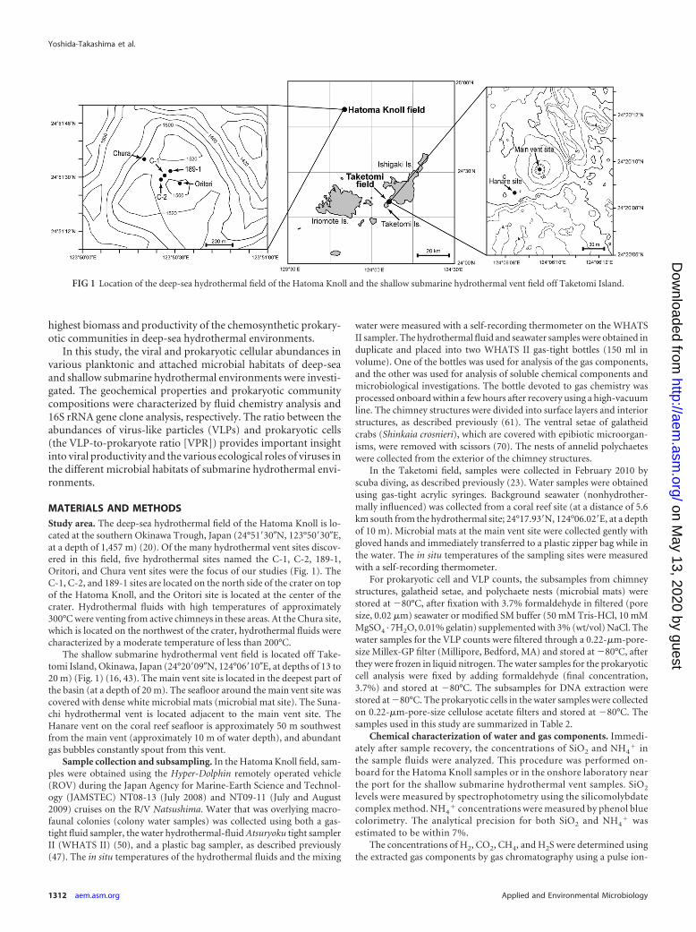
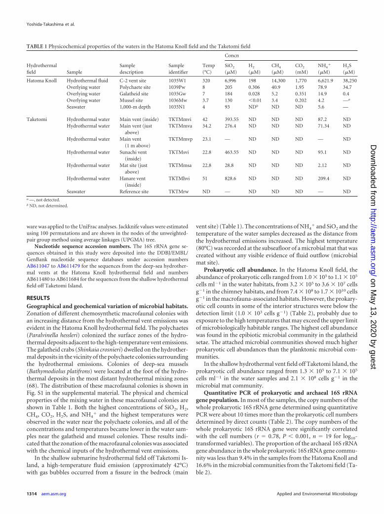
MATERIALS AND METHODSStudy area. The deep-sea hydrothermal field of the Hatoma Knoll is lo-cated at the southern Okinawa Trough, Japan (24°51=30�N, 123°50=30�E,at a depth of 1,457 m) (20). Of the many hydrothermal vent sites discov-ered in this field, five hydrothermal sites named the C-1, C-2, 189-1,Oritori, and Chura vent sites were the focus of our studies (Fig. 1). TheC-1, C-2, and 189-1 sites are located on the north side of the crater on topof the Hatoma Knoll, and the Oritori site is located at the center of thecrater. Hydrothermal fluids with high temperatures of approximately300°C were venting from active chimneys in these areas. At the Chura site,which is located on the northwest of the crater, hydrothermal fluids werecharacterized by a moderate temperature of less than 200°C.
The shallow submarine hydrothermal vent field is located off Take-tomi Island, Okinawa, Japan (24°20=09�N, 124°06=10�E, at depths of 13 to20 m) (Fig. 1) (16, 43). The main vent site is located in the deepest part ofthe basin (at a depth of 20 m). The seafloor around the main vent site wascovered with dense white microbial mats (microbial mat site). The Suna-chi hydrothermal vent is located adjacent to the main vent site. TheHanare vent on the coral reef seafloor is approximately 50 m southwestfrom the main vent (approximately 10 m of water depth), and abundantgas bubbles constantly spout from this vent.
Sample collection and subsampling. In the Hatoma Knoll field, sam-ples were obtained using the Hyper-Dolphin remotely operated vehicle(ROV) during the Japan Agency for Marine-Earth Science and Technol-ogy (JAMSTEC) NT08-13 (July 2008) and NT09-11 (July and August2009) cruises on the R/V Natsushima. Water that was overlying macro-faunal colonies (colony water samples) was collected using both a gas-tight fluid sampler, the water hydrothermal-fluid Atsuryoku tight samplerII (WHATS II) (50), and a plastic bag sampler, as described previously(47). The in situ temperatures of the hydrothermal fluids and the mixing
water were measured with a self-recording thermometer on the WHATSII sampler. The hydrothermal fluid and seawater samples were obtained induplicate and placed into two WHATS II gas-tight bottles (150 ml involume). One of the bottles was used for analysis of the gas components,and the other was used for analysis of soluble chemical components andmicrobiological investigations. The bottle devoted to gas chemistry wasprocessed onboard within a few hours after recovery using a high-vacuumline. The chimney structures were divided into surface layers and interiorstructures, as described previously (61). The ventral setae of galatheidcrabs (Shinkaia crosnieri), which are covered with epibiotic microorgan-isms, were removed with scissors (70). The nests of annelid polychaeteswere collected from the exterior of the chimney structures.
In the Taketomi field, samples were collected in February 2010 byscuba diving, as described previously (23). Water samples were obtainedusing gas-tight acrylic syringes. Background seawater (nonhydrother-mally influenced) was collected from a coral reef site (at a distance of 5.6km south from the hydrothermal site; 24°17.93=N, 124°06.02=E, at a depthof 10 m). Microbial mats at the main vent site were collected gently withgloved hands and immediately transferred to a plastic zipper bag while inthe water. The in situ temperatures of the sampling sites were measuredwith a self-recording thermometer.
For prokaryotic cell and VLP counts, the subsamples from chimneystructures, galatheid setae, and polychaete nests (microbial mats) werestored at �80°C, after fixation with 3.7% formaldehyde in filtered (poresize, 0.02 �m) seawater or modified SM buffer (50 mM Tris-HCl, 10 mMMgSO4 · 7H2O, 0.01% gelatin) supplemented with 3% (wt/vol) NaCl. Thewater samples for the VLP counts were filtered through a 0.22-�m-pore-size Millex-GP filter (Millipore, Bedford, MA) and stored at �80°C, afterthey were frozen in liquid nitrogen. The water samples for the prokaryoticcell analysis were fixed by adding formaldehyde (final concentration,3.7%) and stored at �80°C. The subsamples for DNA extraction werestored at �80°C. The prokaryotic cells in the water samples were collectedon 0.22-�m-pore-size cellulose acetate filters and stored at �80°C. Thesamples used in this study are summarized in Table 2.
Chemical characterization of water and gas components. Immedi-ately after sample recovery, the concentrations of SiO2 and NH4
� inthe sample fluids were analyzed. This procedure was performed on-board for the Hatoma Knoll samples or in the onshore laboratory nearthe port for the shallow submarine hydrothermal vent samples. SiO2
levels were measured by spectrophotometry using the silicomolybdatecomplex method. NH4
� concentrations were measured by phenol bluecolorimetry. The analytical precision for both SiO2 and NH4
� wasestimated to be within 7%.
The concentrations of H2, CO2, CH4, and H2S were determined usingthe extracted gas components by gas chromatography using a pulse ion-
FIG 1 Location of the deep-sea hydrothermal field of the Hatoma Knoll and the shallow submarine hydrothermal vent field off Taketomi Island.
Yoshida-Takashima et al.
1312 aem.asm.org Applied and Environmental Microbiology
on May 13, 2020 by guest
http://aem.asm
.org/D
ownloaded from
ization detector (PID; GL Science, Tokyo, Japan). The concentrationswere determined within a 5% error margin.
Prokaryotic cell and VLP abundances. Detachment of cells and VLPsfrom chimney structures, microbial mats, and macrofaunal tissues wasperformed as follows: subsample suspensions fixed in seawater or modi-fied SM buffer were shaken at maximum speed using a ShakeMaster BMS-A15 apparatus (Bio Medical Science, Tokyo, Japan) for 1 min and centri-fuged for 1 min at 800 � g. The supernatants were then used forprokaryote and virus counts.
For the prokaryotic cell counts, each sample was filtered through a0.2-�m-pore-size Isopore membrane filter (Millipore, Bedford, MA), af-ter staining with 4=,6-diamidino-2-phenylindole (DAPI). VLP countswere performed as described previously (66). One milliliter of each envi-ronmental sample was stained with 25 �l of 100� SYBR gold (Invitrogen,Carlsbad, CA) for 15 min and filtered onto a 0.02-�m-pore-size Anodiscfilter (GE Healthcare, Piscataway, NJ). The cells and VLPs on these filterswere counted with an Olympus BX51 fluorescence microscope (Olympus,Tokyo, Japan) at a magnification of �1,500. At least 400 viral particles persample were counted in more than 20 randomly chosen fields in triplicatefor both the prokaryotic cell and VLP counts.
DNA extraction. Microbial DNA was extracted from chimney struc-tures, polychaete nests, galatheid setae, microbial mats, and microbialcells filtered from the water samples using an UltraClean MegaPrep soilDNA isolation kit or UltraClean soil DNA isolation kit (Mo Bio Labora-tories Inc., Carlsbad, CA) according to the manufacturer’s instructions.
Quantitative PCR for small-subunit (SSU) rRNA genes. Quantifica-tion of the archaeal and whole prokaryotic 16S rRNA genes using primerand probe sets Arch349F-Arch806R-Arch516F and Uni340F-Uni806R-Uni516F, respectively, was performed according to a previously publishedmethod (60) with some modifications, as follows. As described previ-ously, we used a prepared mixture of qPCR Quick GoldStar MastermixPlus (Eurogentec, Seraing, Belgium), and concentrations for primers and
probes were modified following the manufacturer’s instructions (45).These primers have been applied in many studies targeting hydrothermalenvironments as well as sedimentary habitats (41, 46, 47).
Construction of 16S rRNA clone libraries and sequence analysis.Bacterial and archaeal 16S rRNA genes were amplified by LA Taq poly-merase (Takara Bio, Otsu, Japan) with primer sets Bac27F-Bac927R (30)and Arch21F-Arch958R (11) for the bacterial and archaeal SSU rRNAgenes, respectively. For the bacterial 16S rRNA gene amplification, thePCR program consisted of an initial preheating step for 1 min at 96°C,followed by 25 to 35 cycles of 96°C for 25 s, 53°C for 45 s, and 72°C for 90s and a final extension at 72°C for 7 min. For the archaeal 16S rRNA geneamplification, 25 to 45 cycles of PCR amplification, after preheating for 1min at 96°C, were performed using the following conditions: 96°C for 25s, 50°C for 45 s, and 72°C for 90 s and a final extension at 72°C for 7 min.The amplified PCR products were cloned into the pCR2.1 vector using theTA cloning kit (Invitrogen, Carlsbad, CA) according to the manufactur-er’s instructions. The inserts in the vectors were directly sequenced usingM13 primers with a BigDye (version 3.1) sequencing kit (Applied Biosys-tems, Carlsbad, CA). Approximately 900 bp of 16S rRNA gene fragmentswas assembled and analyzed using Genetyx-MAC/ATSQ software(Genetyx Co. Ltd., Tokyo, Japan). The number of sequenced 16S rRNAclones per sample is shown in Fig. 2. Sequences showing �97% identitywere assigned to the same phylogenetic clone type (phylotype). Each ofthe 16S rRNA gene phylotypes was inserted into the reference tree usingthe parsimony insertion algorithm in ARB software (34) and phylogeneti-cally classified into specific taxonomic units using Hugenholtz’s small-subunit rRNA sequence database and phylogenetic classification, withminor modifications (24). To assess similarities and differences among16S rRNA gene communities, we used Jackknife environment clusteranalysis in the UniFrac program (http://bmf.colorado.edu/unifrac/) (33).The phylogenetic tree constructed from the representative phylotypes inthe clone libraries using the neighbor-joining method with the ARB soft-
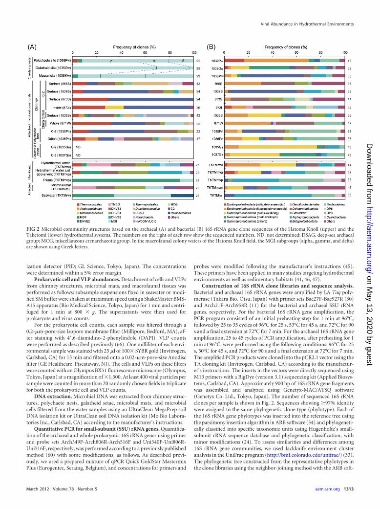
FIG 2 Microbial community structures based on the archaeal (A) and bacterial (B) 16S rRNA gene clone sequences of the Hatoma Knoll (upper) and theTaketomi (lower) hydrothermal systems. The numbers on the right of each row show the sequenced numbers. ND, not determined; DSAG, deep-sea archaealgroup; MCG, miscellaneous crenarchaeotic group. In the macrofaunal colony waters of the Hatoma Knoll field, the MGI subgroups (alpha, gamma, and delta)are shown using Greek letters.
Viral Abundance in Hydrothermal Environments
March 2012 Volume 78 Number 5 aem.asm.org 1313
on May 13, 2020 by guest
http://aem.asm
.org/D
ownloaded from
ware was applied to the UniFrac analyses. Jackknife values were estimatedusing 100 permutations and are shown in the nodes of the unweighted-pair group method using average linkages (UPGMA) tree.
Nucleotide sequence accession numbers. The 16S rRNA gene se-quences obtained in this study were deposited into the DDBJ/EMBL/GenBank nucleotide sequence databases under accession numbersAB611047 to AB611479 for the sequences from the deep-sea hydrother-mal vents at the Hatoma Knoll hydrothermal field and numbersAB611480 to AB611684 for the sequences from the shallow hydrothermalfield off Taketomi Island.
RESULTSGeographical and geochemical variation of microbial habitats.Zonation of different chemosynthetic macrofaunal colonies withan increasing distance from the hydrothermal vent emissions wasevident in the Hatoma Knoll hydrothermal field. The polychaetes(Paralvinella hessleri) colonized the surface zones of the hydro-thermal deposits adjacent to the high-temperature vent emissions.The galatheid crabs (Shinkaia crosnieri) dwelled on the hydrother-mal deposits in the vicinity of the polychaete colonies surroundingthe hydrothermal emissions. Colonies of deep-sea mussels(Bathymodiolus platifrons) were located at the foot of the hydro-thermal deposits in the most distant hydrothermal mixing zones(68). The distribution of these macrofaunal colonies is shown inFig. S1 in the supplemental material. The physical and chemicalproperties of the mixing water in these macrofaunal colonies areshown in Table 1. Both the highest concentrations of SiO2, H2,CH4, CO2, H2S, and NH4
� and the highest temperatures wereobserved in the water near the polychaete colonies, and all of theconcentrations and temperatures became lower in the water sam-ples near the galatheid and mussel colonies. These results indi-cated that the zonation of the macrofaunal colonies was associatedwith the chemical inputs of the hydrothermal vent emissions.
In the shallow submarine hydrothermal field off Taketomi Is-land, a high-temperature fluid emission (approximately 42°C)with gas bubbles occurred from a fissure in the bedrock (main
vent site) (Table 1). The concentrations of NH4� and SiO2 and the
temperature of the water samples decreased as the distance fromthe hydrothermal emissions increased. The highest temperature(80°C) was recorded at the subseafloor of a microbial mat that wascreated without any visible evidence of fluid outflow (microbialmat site).
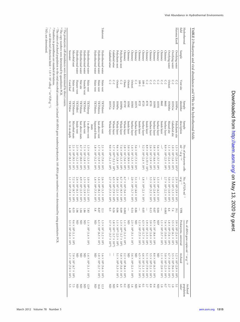
Prokaryotic cell abundance. In the Hatoma Knoll field, theabundance of prokaryotic cells ranged from 1.0 � 105 to 1.1 � 105
cells ml�1 in the water habitats, from 3.2 � 105 to 3.6 � 107 cellsg�1 in the chimney habitats, and from 7.4 � 108 to 1.7 � 1010 cellsg�1 in the macrofauna-associated habitats. However, the prokary-otic cell counts in some of the interior structures were below thedetection limit (1.0 � 103 cells g�1) (Table 2), probably due toexposure to the high temperatures that may exceed the upper limitof microbiologically habitable ranges. The highest cell abundancewas found in the epibiotic microbial community in the galatheidsetae. The attached microbial communities showed much higherprokaryotic cell abundances than the planktonic microbial com-munities.
In the shallow hydrothermal vent field off Taketomi Island, theprokaryotic cell abundance ranged from 1.3 � 105 to 7.1 � 105
cells ml�1 in the water samples and 2.1 � 108 cells g�1 in themicrobial mat community.
Quantitative PCR of prokaryotic and archaeal 16S rRNAgene population. In most of the samples, the copy numbers of thewhole prokaryotic 16S rRNA gene determined using quantitativePCR were about 10 times more than the prokaryotic cell numbersdetermined by direct counts (Table 2). The copy numbers of thewhole prokaryotic 16S rRNA gene were significantly correlatedwith the cell numbers (r � 0.78, P � 0.001, n � 19 for log10-transformed variables). The proportion of the archaeal 16S rRNAgene abundance in the whole prokaryotic 16S rRNA gene commu-nity was less than 9.4% in the samples from the Hatoma Knoll and16.6% in the microbial communities from the Taketomi field (Ta-ble 2).
TABLE 1 Physicochemical properties of the waters in the Hatoma Knoll field and the Taketomi field
Hydrothermalfield Sample
Sampledescription
Sampleidentifier
Temp(°C)
Concn
SiO2
(�M)H2
(�M)CH4
(�M)CO2
(mM)NH4
�
(�M)H2S(�M)
Hatoma Knoll Hydrothermal fluid C-2 vent site 1035W1 320 6,996 198 14,300 1,770 6,621.9 38,250Overlying water Polychaete site 1039Pw 8 205 0.306 40.9 1.95 78.9 34.7Overlying water Galatheid site 1035Gw 7 184 0.028 5.2 0.351 14.9 0.4Overlying water Mussel site 1036Mw 3.7 130 �0.01 3.4 0.202 4.2 —a
Seawater 1,000-m depth 1035N1 4 93 NDb ND ND 5.6 —
Taketomi Hydrothermal water Main vent (inside) TKTMmvi 42 393.55 ND ND ND 87.2 NDHydrothermal water Main vent (just
above)TKTMmva 34.2 276.4 ND ND ND 71.34 ND
Hydrothermal water Main vent(1 m above)
TKTMmvp 23.1 — ND ND ND — ND
Hydrothermal water Sunachi vent(inside)
TKTMsvi 22.8 463.55 ND ND ND 93.1 ND
Hydrothermal water Mat site (justabove)
TKTMmsa 22.8 28.8 ND ND ND 2.12 ND
Hydrothermal water Hanare vent(inside)
TKTMhvi 51 828.6 ND ND ND 209.4 ND
Seawater Reference site TKTMrw ND — ND ND ND — NDa —, not detected.b ND, not determined.
Yoshida-Takashima et al.
1314 aem.asm.org Applied and Environmental Microbiology
on May 13, 2020 by guest
http://aem.asm
.org/D
ownloaded from
TA
BLE
2P
rokaryotean
dviralabu
ndan
cesan
dV
PR
sin
the
hydroth
ermalfi
elds
Hydroth
ermal
field
Sample
Ven
tsite
Sample
identifi
erSam
pledescription
No.ofprokaryote
cellsm
l �1
org
�1
a
No.ofV
LPs
ml �
1
org
�1
VP
R
No.ofrR
NA
gene
copiesm
l �1
org
�1
bA
rchaeal
population
ratioc
Prokaryotic,u
niversal
Arch
aeal
Hatom
aK
noll
Overlyin
gw
aterC
-21039P
wP
olychaete
site1.5
�10
5(2.6
�10
4)d
2.9�
106
(3.9�
105)
19.13.9
�10
6(2.4
�10
6)1.2
�10
5(2.9
�10
4)3.1
Overlyin
gw
aterC
-21035G
wG
alatheid
site1.0
�10
5(1.6
�10
4)7.5
�10
5(1.3
�10
5)7.4
3.5�
106
(6.1�
105)
3.5�
104
(9.6�
103)
1.0O
verlying
water
Oritori
1036Mw
Mu
sselsite1.1
�10
5(2.4
�10
4)5.0
�10
5(1.0
�10
5)4.6
1.8�
106
(7.6�
104)
4.7�
104
(1.0�
103)
2.7C
him
ney
C-2
866SSu
rfacelayer
8.9�
106
(2.5�
106)
2.7�
104
(2.2�
103)
0.0037.6
�10
7(5.0
�10
6)5.0
�10
5(2.3
�10
3)0.7
Ch
imn
eyC
-2866I
Inside
layer—
e—
—1.1
�10
4(3.7
�10
3)1.1
�10
2(2.0
�10
1)1.0
Ch
imn
eyC
-21035S
Surface
layer3.6
�10
7(3.7
�10
6)1.5
�10
6(4.2
�10
5)0.04
ND
fN
DN
DC
him
ney
C-2
1039SSu
rfacelayer
3.8�
106
(4.6�
105)
2.6�
105
(2.4�
104)
0.078.5
�10
7(8.6
�10
6)8.0
�10
6(6.3
�10
5)9.4
Ch
imn
eyC
-2873S
Surface
layer2.6
�10
7(1.9
�10
6)3.3
�10
6(3.2
�10
5)0.13
3.9�
109
(1.0�
107)
3.5�
108
(2.1�
107)
8.9C
him
ney
C-2
873IIn
sidelayer
8.9�
105
(1.53�
105)
——
1.9�
107
(1.5�
106)
5.1�
105
(7.9�
103)
2.7C
him
ney
189-1872S
Surface
layer3.2
�10
5(2.4
�10
4)1.8
�10
4(2.1
�10
3)0.05
4.3�
106
(1.7�
105)
4.6�
103
(1.1�
103)
0.1C
him
ney
189-1872I
Inside
layer—
——
1.2�
104
(7.5�
102)
4.5�
102
(1.7�
101)
3.8C
him
ney
C-1
1037SSu
rfacelayer
3.6�
105
(7.3�
104)
3.1�
105
(4.6�
104)
0.86N
DN
DN
DC
him
ney
Oritori
1036SSu
rfacelayer
1.4�
107
(1.3�
106)
2.0�
106
(1.7�
105)
0.142.3
�10
8(3.1
�10
7)3.6
�10
6(2.1
�10
5)1.6
Ch
imn
eyO
ritori1036I
Inside
layer3.0
�10
6(9.9
�10
5)—
—N
DN
DN
DC
him
ney
Ch
ura
871WW
hole
layer2.8
�10
7(3.6
�10
6)6.7
�10
4(5.6
�10
3)0.002
1.1�
108
(1.2�
106)
2.2�
105
(8.4�
104)
0.2P
olychaete
nest
C-2
1039Pn
Wh
olelayer
7.4�
108
(1.5�
108)
6.7�
107
(9.3�
106)
0.091.3
�10
10
(1.7�
109)
5.8�
108
(5.0�
107)
4.5P
olychaete
nest
Oritori
1036Pn
Wh
olelayer
9.6�
108
(1.8�
108)
5.8�
107
(2.1�
106)
0.061.1
�10
10
(2.5�
108)
5.1�
108
(1.5�
107)
4.6G
alatheid
setaeC
-21035G
sW
hole
layer1.7
�10
10
(3.2�
109)
4.5�
109
(7.9�
108)
0.261.8
�10
11
(1.7�
101
0)—
ND
Galath
eidsetae
C-2
1037Gs
Wh
olelayer
9.0�
109
(1.2�
109)
2.7�
109
(7.3�
108)
0.33.7
�10
11
(3.7�
101
0)—
ND
Taketom
iH
ydrotherm
alwater
Main
vent
TK
TM
mvi
Inside
vent
2.1�
105
(7.0�
104)
2.6�
106
(1.3�
106)
12.11.3
�10
7(1.2
�10
6)1.6
�10
6(5.6
�10
5)12.3
Hydroth
ermalw
aterM
ainven
tT
KT
Mm
vaJu
stabove
vent
1.7�
105
(1.9�
104)
1.4�
106
(8.2�
105)
8.076.3
�10
4(6.3
�10
3)1.0
�10
4(2.7
�10
2)16.6
Hydroth
ermalw
aterM
ainven
tT
KT
Mm
vsSeparated
fromm
ats1.8
�10
5(5.2
�10
4)1.4
�10
6(7.3
�10
5)5.8
ND
ND
ND
Hydroth
ermalw
aterM
ainven
tT
KT
Mm
vp1
mabove
vent
1.3�
105
(1.5�
104)
1.0�
106
(2.2�
105)
7.831.1
�10
5(1.4
�10
4)1.4
�10
4(1.1
�10
3)12.9
Hydroth
ermalw
aterSu
nach
ivent
TK
TM
sviIn
sideven
t7.1
�10
5(1.8
�10
5)1.3
�10
6(1.9
�10
5)1.86
ND
ND
ND
Hydroth
ermalw
aterM
atsite
TK
TM
msa
Just
abovem
ats4.7
�10
5(8.0
�10
4)4.9
�10
5(2.5
�10
5)1.04
ND
ND
ND
Hydroth
ermalw
aterH
anare
vent
TK
TM
hvi
Inside
vent
2.3�
105
(1.2�
105)
2.4�
105
(6.1�
104)
1.06N
DN
DN
DM
icrobialmat
Main
vent
TK
TM
mvm
Microbialm
ats2.1
�10
8(9.5
�10
7)7.2
�10
7(8.7
�10
6)0.35
9.4�
108
(1.1�
108)
7.0�
107
(4.7�
106)
7.5Seaw
aterC
oralleafT
KT
Mrw
At
10-mdepth
2.6�
105
(8.3�
104)
1.1�
106
(7.8�
105)
4.283.3
�10
5(5.0
�10
4)1.7
�10
4(7.0
�10
2)5.3
aT
he
prokaryoticcellabu
ndan
cesw
eredeterm
ined
bydirect
coun
ts.b
Th
ecopy
nu
mbers
were
determin
edby
quan
titativeP
CR
.cT
he
ratiosofarch
aealpopulation
toth
etotalm
icrobialcomm
un
ity(arch
aeal16SrR
NA
gene
nu
mbers/prokaryotic
16SrR
NA
gene
nu
mbers)
were
determin
edby
usin
gqu
antitative
PC
R.
dN
um
bersin
parenth
esesare
standard
deviations.
e—
,not
detected(detection
limit
�1.0
�10
3cells
g�
1or
VLP
sg
�1).
fND
,not
determin
ed.
Viral Abundance in Hydrothermal Environments
March 2012 Volume 78 Number 5 aem.asm.org 1315
on May 13, 2020 by guest
http://aem.asm
.org/D
ownloaded from
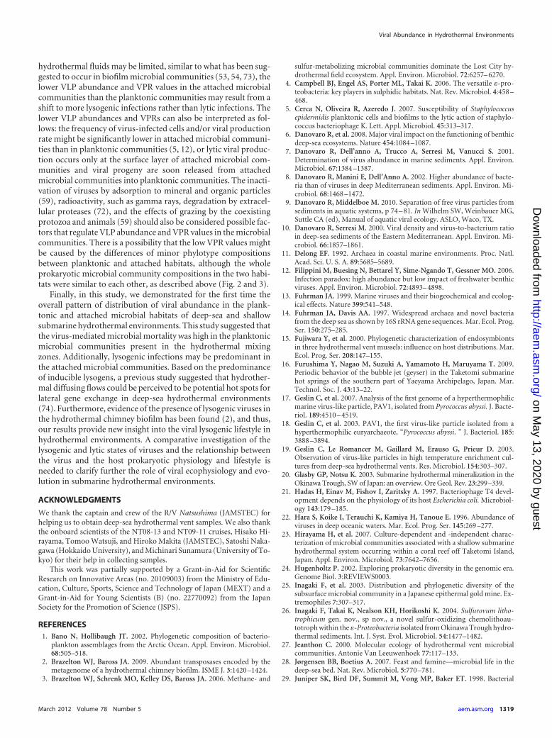
Prokaryotic 16S rRNA gene community structures. The 16SrRNA phylotype compositions of planktonic prokaryotes and thegeographical variation were similar between the deep-sea andshallow submarine hydrothermal environments (Fig. 2). In thedeep-sea macrofaunal colony waters, the archaeal phylotype com-positions were similar to each other; the phylotypes of marinegroup I (MGI) archaea were dominant in the archaeal 16S rRNAgene population and were similar in composition in the MGI sub-groups (Fig. 2A; see Fig. S2 in the supplemental material). In contrast,the dominant bacterial 16S rRNA gene compositions varied amongthe different macrofaunal colony waters. The most predominant bac-terial phylotypes in the polychaete colony habitats were affiliated withthe epsilonproteobacterial family Thiovulgaceae, which includes thefacultatively anaerobic, mesophilic, and thiotrophic chemolithoau-totrophs, represented by the genus Sulfurovum (group F) (4, 26).Thiotrophic and methanotrophic gammaproteobacterial groups(15) predominated in the galatheid colony habitats. Finally, the deep-sea-pervasive, heterotrophic gammaproteobacterial genera (1, 14)predominated in the mussel colony habitats (Fig. 2B).
In most of the chimney habitats and the polychaete nests,the archaeal 16S rRNA gene composition was characterized bya dominating clonal frequency of the Thermococcales and thedeep-sea hydrothermal vent euryarchaeotic group 2 (DHVE2),which is represented by the genus Aciduliprofundum (49), andthe occasional abundance of clones within the Desulfurococca-les, Archaeoglobales, Methanococcales, the deep-sea hydrother-mal vent euryarchaeotic group 1 (DHVE1), and hot water cre-narchaeotic group IV (HWCGIV; also known as unculturedcrenarchaea group II [UCII]) (25, 51) (Fig. 2A). Additionally,the bacterial 16S rRNA gene communities in the chimney hab-itats were always dominated by the phylotypes of Deltaproteo-bacteria and the Thiovulgaceae, which are members of the Ep-silonproteobacteria. Some of these communities were alsocomposed of the thiotrophic and methanotrophic gammapro-
teobacterial groups and the heterotrophic gammaproteobacte-rial and Bacteroidetes members (Fig. 2B). The prokaryotic 16SrRNA gene compositions in the galatheid setae were dominatedmainly by thiotrophic and methanotrophic gammaproteobac-terial groups (Fig. 2B).
In the planktonic microbial habitats of the shallow submarinehydrothermal field off Taketomi Island, the Thiovulgaceae andCampylobacteraceae phylotypes of the Epsilonproteobacteria andthe thiotrophic phylotypes of the Gammaproteobacteria were pre-dominant in the bacterial 16S rRNA gene compositions in thehydrothermal fluids (Fig. 2B). A similar but relatively more diversebacterial 16S rRNA phylotype composition was obtained from themicrobial mat communities that colonized the vent surfaces adjacentto the hydrothermal emissions (Fig. 2B). The archaeal 16S rRNA genecompositions in the hydrothermal fluids and the microbial mat weredominated not only by the DHVE1 and DHVE4 phylotypes but alsoby the terrestrial miscellaneous euryarchaeotal group (TMEG) (62,67) (Fig. 2A). In the planktonic microbial communities of the mixedemissions and ambient seawater, the bacterial and archaeal rRNAphylotype compositions were considerably changed. In this area, thepredominant phylotypes were affiliated with the Alphaproteobacteria,Bacteroidetes, and Cyanobacteria in the domain Bacteria and the MGIand marine group II (MGII) in the domain Archaea (Fig. 2). Theobligately anaerobic Epsilonproteobacteria, Desulfurobacteriales, Ther-mococcales, and DHVE8 were found only in hydrothermal waters.
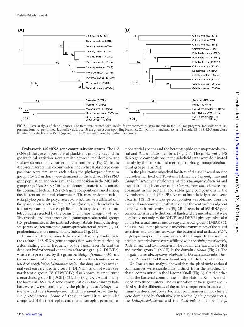
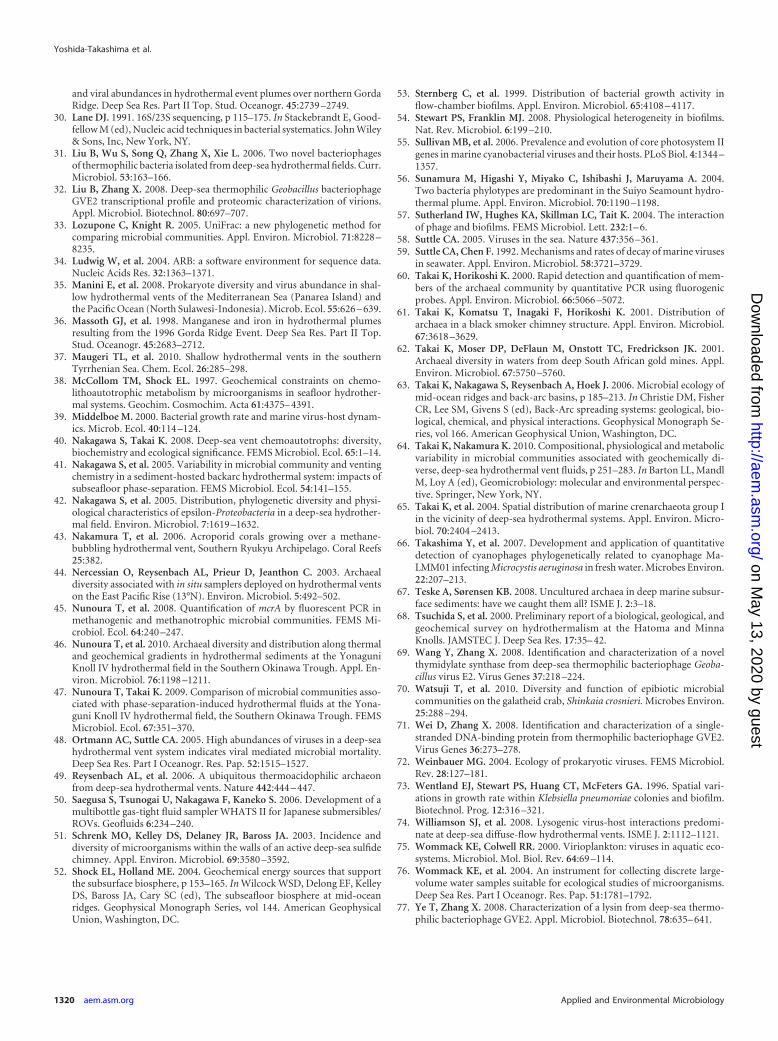
UniFrac cluster analysis showed that the planktonic archaealcommunities were significantly distinct from the attached ar-chaeal communities in the Hatoma Knoll (Fig. 3). On the otherhand, the bacterial communities in the Hatoma Knoll were di-vided into three clusters. The classification of these groups coin-cided with the differences of the major components in each com-munity as described above: bacterial communities in two clusterswere dominated by facultatively anaerobic Epsilonproteobacteria,the Deltaproteobacteria, and the Bacteroidetes members (e.g.,
FIG 3 Cluster analysis of clone libraries. The trees were created with Jackknife environment clusters analysis in the UniFrac program. Jackknife with 100permutations was performed. Jackknife values over 50 are given at corresponding branches. Comparison of archaeal (A) and bacterial (B) 16S rRNA gene clonelibraries from the Hatoma Knoll (upper) and the Taketomi (lower) hydrothermal systems.
Yoshida-Takashima et al.
1316 aem.asm.org Applied and Environmental Microbiology
on May 13, 2020 by guest
http://aem.asm
.org/D
ownloaded from
1036Pn and 871W), and those in the other cluster mainly con-sisted of the thiotrophic, methanotrophic, and heterotrophicGammaproteobacteria groups (e.g., 1035Gs and 1039S). The resultsuggests that temperature and geochemical conditions but notattachment matrices (rocks or animal bodies) or lifestyles (attach-ment or planktonic) influence bacterial community structures inthe Hatoma Knoll hydrothermal field. Similarly, both the archaealand bacterial communities in the Taketomi field were divided intotwo groups, and their classifications were highly related to themajor community components.
VLP abundance. The VLP abundance ranged from 5.0 � 105
to 2.9 � 106 and from 2.4 � 105 to 2.6 � 106 VLPs ml�1 in theplanktonic microbial habitats of the Hatoma Knoll field and theTaketomi field, respectively (Table 2). Using both the prokaryoticcell and VLP abundances, the VPRs in the planktonic habitatswere estimated to be 4.6 to 19 for the Hatoma Knoll field and 1.0 to12.1 for the Taketomi field (Table 2). There were no correlationsbetween the log10-transformed VLP abundances and prokaryoticcell abundances (Pearson correlation; r � 0.155, P � 0.05, n � 9).
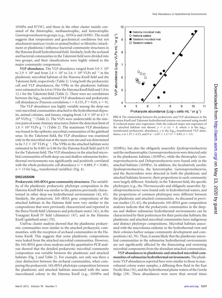
The VLP abundance was highly variable among the deep-seavent microbial communities attached to the hydrothermal depos-its, animal colonies, and tissues, ranging from 1.8 � 104 to 4.5 �109 VLPs g�1 (Table 2). The VLPs were undetectable in the inte-rior parts of some chimney structures (below the detection limit of1.0 � 103 VLPs g�1) (Table 2). The maximum VLP abundancewas found in the epibiotic microbial communities of the galatheidsetae. In the Taketomi field, the VLP abundance was examinedonly in the microbial mat at the main vent site and was determinedto be 7.2 � 107 VLPs g�1. The VPRs in the attached habitats wereestimated to be 0.001 to 0.86 for the Hatoma Knoll field and 0.35for the Taketomi field. The VLP abundance in the attached micro-bial communities of both deep-sea and shallow submarine hydro-thermal environments was significantly and positively correlatedwith the whole prokaryotic cell abundance (r � 0.889, P � 0.001,n � 13 for log10-transformed variables) (Fig. 4).
DISCUSSIONProkaryotic 16S rRNA gene community structures. The variabil-ity of the planktonic prokaryotic phylotype composition in theHatoma Knoll field was similar to the patterns previously charac-terized in other deep-sea hydrothermal environments (42, 65).Similarly, the prokaryotic 16S rRNA gene compositions of theattached habitats in the Hatoma field were very similar to thecompositions that were previously characterized and reported inthe Iheya North field (chimneys and polychaete nests) (42), in theYonaguni Knoll IV field (chimneys) (47), and in the HatomaKnoll (galatheid setae) (70).
UniFrac cluster analysis showed that the planktonic prokary-otic communities were similar to the attached prokaryotic com-munities, with the exception of archaeal communities in the Ha-toma Knoll. This suggests that some planktonic compositionswere leaked from the attached microbial communities. However,the 16S rRNA gene clone analyses and the quantitative PCR anal-ysis showed that the detailed prokaryotic microbial communitycomposition was variable between the planktonic and attachedhabitats (Fig. 2 and Table 2). For example, not only was there aclear distinction between the archaeal communities, when com-paring the prokaryotic 16S rRNA phylotype composition betweenthe planktonic and attached habitats associated with the samemacrofaunal colony in the Hatoma Knoll (e.g., 1039Pw and
1039Pn), but also the obligately anaerobic Epsilonproteobacteriaand the methanotrophic Gammaproteobacteria were detected onlyin the planktonic habitats (1039Pw), while the thiotrophic Gam-maproteobacteria and Deltaproteobacteria were found only in theattached habitats (1039Pn). In addition, the facultatively aerobicEpsilonproteobacteria, the heterotrophic Gammaproteobacteria,and the Bacteroidetes were detected in both the planktonic andattached habitats; however, their proportions in each communitywere largely different. Similarly, in the Taketomi field, the specificphylotypes (e.g., the Thermococcales and obligately anaerobic Ep-silonproteobacteria) were found only in hydrothermal waters, andthe proportions of common phylotypes were different betweenthe planktonic and attached communities. As discussed in previ-ous studies (23, 42), the prokaryotic 16S rRNA gene compositionanalyses indicate that the prokaryotic communities in the deep-sea and shallow submarine hydrothermal environments can becharacterized by their preferences for their particular habitats; theplanktonic and attached microbial communities have indigenousand distinct phylotype compositions. Microbial habitats associ-ated with the macrofauna endemic to the hydrothermal vent andtheir colonies harbor unique community development and com-positions (42, 70). Thus, it seems likely that the planktonic micro-bial communities in the submarine hydrothermal environmentsare not significantly affected by the dissociating and swarmingmicrobial components from the abundant attached communities.
VLP abundances in planktonic and attached microbial com-munities of submarine hydrothermal environments. The plank-tonic VLP abundances reported here were similar to those in mac-rofaunal colony water samples, a diffusing-flow fluid in the EastPacific Rise (76), and the hydrothermal plume waters of the GordaRidge (29). These abundances were more than several times
FIG 4 The relationship between the prokaryotic and VLP abundances in theHatoma Knoll and Taketomi hydrothermal systems was assessed using modelII (reduced major axis) regression. Only the reduced major axis regression ofthe attached habitats was shown: y � a (x) � b, where x is the log10-transformed prokaryotic abundance, y is the log10-transformed VLP abun-dance, a is 1.19 � 0.15, and b is �2.65 � 1.17 (r2 � 0.83, n � 13).
Viral Abundance in Hydrothermal Environments
March 2012 Volume 78 Number 5 aem.asm.org 1317
on May 13, 2020 by guest
http://aem.asm
.org/D
ownloaded from
higher than what was measured in nonhydrothermal deep seawa-ter (22, 29, 76). The VLP abundances in the deep-sea vent envi-ronments represented 1/100 to 1/10 of the abundances obtainedfrom the hydrothermal vent, plume, or diffusing-flow waters ofthe Endeavor Ridge (48). Because the VLP enumeration tech-niques were different among these studies, the results may havecertain deviations due to biases in the methodologies used.
We found that viruses were widely distributed in a variety ofattached hydrothermal microbial habitats: chimney surface areasor macrofaunal bodies and colonies. This is the first report to showVLP abundance in the attached microbial communities of subma-rine hydrothermal environments. However, the VLP abundancesand VPR values in the hydrothermal attached microbial commu-nities were significantly lower than expected, and all VPRs werebelow 1.0. These VPRs were also much lower than the VPR level innonhydrothermal deep-sea benthic microbial communities in thesediment (6, 10). The efficiency of separation of viruses from sed-iment samples has been reported to be significantly increased bychemical treatments with surfactants and by mechanical shaking(7, 9). Using some attached microbial samples, we observed nosignificant effects of these treatments on VLP abundance (data notshown). However, some attached microbial samples in this studywere fixed with formaldehyde, which has been reported by severalauthors to cause rapid viral decay after fixation (7). Thus, the viralabundance obtained in this study might underestimate the actualviral abundance because of differences in methodological ap-proaches.
The unexpected VLP abundances may be due to the possiblerapid diffusion of VLPs from the attached microbial communitiesduring sampling and processing. To test this possibility, multiplesamplings and procedures were examined for the microbial matand the surrounding water at the main vent site in the Taketomifield. The VLP abundances in the interstitial seawater of the in situmicrobial mat, which was carefully obtained from the seafloor bya plastic syringe (sample TKTMmva), and those in the residualseawater, which was taken from a plastic bag after the microbialmat sample was transferred into another plastic bag onboard(sample TKTMmvs), were compared. The VLP abundance ofsample TKTMmva indicates the naturally diffusing VLP abun-dance from the in situ microbial mat, and the VLP abundance ofsample TKTMmvs represents the diffusing VLP abundance dur-ing sampling and recovery. The VLP abundances were very similaramong the in situ interstitial seawater, the residual seawater in thesampling bag, and the experimentally extracted water (Table 2).Thus, the low VLP abundances and VPR values in the hydrother-mal attached microbial communities, at least in the microbial matin the Taketomi field, were not highly affected by sampling biases.
VLP distribution in planktonic microbial communities ofsubmarine hydrothermal environments. Although the statisti-cally significant correlations between the prokaryotic cell and VLPabundances were not evident in the planktonic microbial commu-nities, there was a trend for the VLP abundance in the planktonichabitats to decrease with increasing distance from the hydrother-mal fluid sources (Tables 1 and 2). For instance, the abundancewas the highest in the polychaete colony waters and became lowerin the galatheid and mussel colony waters in the Hatoma Knollfield. Similarly, at the main vent site in the Taketomi field, the VLPabundance was the highest in the hydrothermal fluid (sampleTKTMmvi) and became lower in the mixing waters between hy-drothermal fluids and ambient seawater (samples TKTMmvs and
TKTMmvp) (Tables 1 and 2). Similar patterns of distribution inviral abundance were also revealed in other deep-sea hydrother-mal environments (29, 36, 48). In the hydrothermal plume waters,the viral abundance increased from the periphery to the center ofplumes and was positively correlated with the increasing input ofhydrothermal fluids, such as temperature and chemical composi-tion (29, 36). Another study showed that the VLP abundances inthe high-temperature hydrothermal fluids and diffusing flowswere higher than those in the plume waters (48). Thus, there is atendency for the abundances of the prokaryotic biomass and thechemolithoautotrophic population to be enhanced in the plank-tonic habitats of the hydrothermal mixing zones with higher in-puts of hydrothermal fluids (42, 56). The higher inputs of thereductive substances, such as H2, H2S, CH4, Fe(II), and Mn(IV),from the hydrothermal fluids provide greater energy to the che-mosynthetic microbial communities. Indeed, it has been theoret-ically and empirically verified that the productivity and the com-munity composition of the microbial communities are regulatedby the geochemical energy state of the habitats (38, 52, 64). Addi-tionally, viral production and propagation are highly associatedwith the productivity and nutrition states of the host organisms(21, 39). Thus, the viral abundance and productivity in the plank-tonic microbial communities of the hydrothermal mixing zonesare likely associated with the geochemical energy state of the hab-itats, which is primarily driven by the chemical input of the hy-drothermal fluid.
When comparing the prokaryotic cell and VLP abundances inthe hydrothermal fluids from various vents in the Taketomi field,neither the temperatures nor the chemical components of the hy-drothermal fluids correlated with the sizes of the prokaryotic celland VLP populations (Tables 1 and 2). This result may reveal thatprokaryote-virus productivity and their interactions are influ-enced not only by the geochemical features of the hydrothermalwaters that sustain chemosynthetic microbial community devel-opment but also by the physical aspects of hydrothermal fluidemissions, such as the flow rate and the physical properties of thesubseafloor hydrothermal fluid passages that establish the habitatconditions for each microbial community.
VLP distribution in attached microbial communities of sub-marine hydrothermal environments. The formation of a biofilmby the microorganisms entails a dynamic change in the physical andchemical conditions of the habitats and in the physiological state ofthe constitutive microorganisms (54). Thus, virus-host interactionscan be highly variable (57). However, a statistically significant positiverelationship between the prokaryotic biomass and VLP abundance inthe attached microbial communities was observed. This suggests thatin the attached habitats there was a specific virus-host interactionassociated with hydrothermal activity.
Because the VPR values were quite different between theplanktonic and attached microbial communities in the submarinehydrothermal environments, it is suggested that the viral produc-tivity and propagation associated with the host prokaryotic com-munities were also different. Previous study has shown that theratios of virus to bacteria were always �1.0 in the deep-sea sedi-ments, and the values were significantly related to a low bacterialgrowth rate (8). Thus, the low VPRs in the attached habitats mightbe caused by low prokaryotic productivity. Furthermore, lysogenyis proposed to be a strategy for virus propagation in prokaryotichosts under unfavorable growth conditions (13, 72). Since theaccessibility of the chemolithotrophic energy sources from the
Yoshida-Takashima et al.
1318 aem.asm.org Applied and Environmental Microbiology
on May 13, 2020 by guest
http://aem.asm
.org/D
ownloaded from
hydrothermal fluids may be limited, similar to what has been sug-gested to occur in biofilm microbial communities (53, 54, 73), thelower VLP abundance and VPR values in the attached microbialcommunities than the planktonic communities may result from ashift to more lysogenic infections rather than lytic infections. Thelower VLP abundances and VPRs can also be interpreted as fol-lows: the frequency of virus-infected cells and/or viral productionrate might be significantly lower in attached microbial communi-ties than in planktonic communities (5, 12), or lytic viral produc-tion occurs only at the surface layer of attached microbial com-munities and viral progeny are soon released from attachedmicrobial communities into planktonic communities. The inacti-vation of viruses by adsorption to mineral and organic particles(59), radioactivity, such as gamma rays, degradation by extracel-lular proteases (72), and the effects of grazing by the coexistingprotozoa and animals (59) should also be considered possible fac-tors that regulate VLP abundance and VPR values in the microbialcommunities. There is a possibility that the low VPR values mightbe caused by the differences of minor phylotype compositionsbetween planktonic and attached habitats, although the wholeprokaryotic microbial community compositions in the two habi-tats were similar to each other, as described above (Fig. 2 and 3).
Finally, in this study, we demonstrated for the first time theoverall pattern of distribution of viral abundance in the plank-tonic and attached microbial habitats of deep-sea and shallowsubmarine hydrothermal environments. This study suggested thatthe virus-mediated microbial mortality was high in the planktonicmicrobial communities present in the hydrothermal mixingzones. Additionally, lysogenic infections may be predominant inthe attached microbial communities. Based on the predominanceof inducible lysogens, a previous study suggested that hydrother-mal diffusing flows could be perceived to be potential hot spots forlateral gene exchange in deep-sea hydrothermal environments(74). Furthermore, evidence of the presence of lysogenic viruses inthe hydrothermal chimney biofilm has been found (2), and thus,our results provide new insight into the viral lysogenic lifestyle inhydrothermal environments. A comparative investigation of thelysogenic and lytic states of viruses and the relationship betweenthe virus and the host prokaryotic physiology and lifestyle isneeded to clarify further the role of viral ecophysiology and evo-lution in submarine hydrothermal environments.
ACKNOWLEDGMENTS
We thank the captain and crew of the R/V Natsushima (JAMSTEC) forhelping us to obtain deep-sea hydrothermal vent samples. We also thankthe onboard scientists of the NT08-13 and NT09-11 cruises, Hisako Hi-rayama, Tomoo Watsuji, and Hiroko Makita (JAMSTEC), Satoshi Naka-gawa (Hokkaido University), and Michinari Sunamura (University of To-kyo) for their help in collecting samples.
This work was partially supported by a Grant-in-Aid for ScientificResearch on Innovative Areas (no. 20109003) from the Ministry of Edu-cation, Culture, Sports, Science and Technology of Japan (MEXT) and aGrant-in-Aid for Young Scientists (B) (no. 22770092) from the JapanSociety for the Promotion of Science (JSPS).
REFERENCES1. Bano N, Hollibaugh JT. 2002. Phylogenetic composition of bacterio-
plankton assemblages from the Arctic Ocean. Appl. Environ. Microbiol.68:505–518.
2. Brazelton WJ, Baross JA. 2009. Abundant transposases encoded by themetagenome of a hydrothermal chimney biofilm. ISME J. 3:1420 –1424.
3. Brazelton WJ, Schrenk MO, Kelley DS, Baross JA. 2006. Methane- and
sulfur-metabolizing microbial communities dominate the Lost City hy-drothermal field ecosystem. Appl. Environ. Microbiol. 72:6257– 6270.
4. Campbell BJ, Engel AS, Porter ML, Takai K. 2006. The versatile �-pro-teobacteria: key players in sulphidic habitats. Nat. Rev. Microbiol. 4:458 –468.
5. Cerca N, Oliveira R, Azeredo J. 2007. Susceptibility of Staphylococcusepidermidis planktonic cells and biofilms to the lytic action of staphylo-coccus bacteriophage K. Lett. Appl. Microbiol. 45:313–317.
6. Danovaro R, et al. 2008. Major viral impact on the functioning of benthicdeep-sea ecosystems. Nature 454:1084 –1087.
7. Danovaro R, Dell’anno A, Trucco A, Serresi M, Vanucci S. 2001.Determination of virus abundance in marine sediments. Appl. Environ.Microbiol. 67:1384 –1387.
8. Danovaro R, Manini E, Dell’Anno A. 2002. Higher abundance of bacte-ria than of viruses in deep Mediterranean sediments. Appl. Environ. Mi-crobiol. 68:1468 –1472.
9. Danovaro R, Middelboe M. 2010. Separation of free virus particles fromsediments in aquatic systems, p 74 – 81. In Wilhelm SW, Weinbauer MG,Suttle CA (ed), Manual of aquatic viral ecology. ASLO, Waco, TX.
10. Danovaro R, Serresi M. 2000. Viral density and virus-to-bacterium ratioin deep-sea sediments of the Eastern Mediterranean. Appl. Environ. Mi-crobiol. 66:1857–1861.
11. Delong EF. 1992. Archaea in coastal marine environments. Proc. Natl.Acad. Sci. U. S. A. 89:5685–5689.
12. Filippini M, Buesing N, Bettarel Y, Sime-Ngando T, Gessner MO. 2006.Infection paradox: high abundance but low impact of freshwater benthicviruses. Appl. Environ. Microbiol. 72:4893– 4898.
13. Fuhrman JA. 1999. Marine viruses and their biogeochemical and ecolog-ical effects. Nature 399:541–548.
14. Fuhrman JA, Davis AA. 1997. Widespread archaea and novel bacteriafrom the deep sea as shown by 16S rRNA gene sequences. Mar. Ecol. Prog.Ser. 150:275–285.
15. Fujiwara Y, et al. 2000. Phylogenetic characterization of endosymbiontsin three hydrothermal vent mussels: influence on host distributions. Mar.Ecol. Prog. Ser. 208:147–155.
16. Furushima Y, Nagao M, Suzuki A, Yamamoto H, Maruyama T. 2009.Periodic behavior of the bubble jet (geyser) in the Taketomi submarinehot springs of the southern part of Yaeyama Archipelago, Japan. Mar.Technol. Soc. J. 43:13–22.
17. Geslin C, et al. 2007. Analysis of the first genome of a hyperthermophilicmarine virus-like particle, PAV1, isolated from Pyrococcus abyssi. J. Bacte-riol. 189:4510 – 4519.
18. Geslin C, et al. 2003. PAV1, the first virus-like particle isolated from ahyperthermophilic euryarchaeote, “Pyrococcus abyssi. ” J. Bacteriol. 185:3888 –3894.
19. Geslin C, Le Romancer M, Gaillard M, Erauso G, Prieur D. 2003.Observation of virus-like particles in high temperature enrichment cul-tures from deep-sea hydrothermal vents. Res. Microbiol. 154:303–307.
20. Glasby GP, Notsu K. 2003. Submarine hydrothermal mineralization in theOkinawa Trough, SW of Japan: an overview. Ore Geol. Rev. 23:299–339.
21. Hadas H, Einav M, Fishov I, Zaritsky A. 1997. Bacteriophage T4 devel-opment depends on the physiology of its host Escherichia coli. Microbiol-ogy 143:179 –185.
22. Hara S, Koike I, Terauchi K, Kamiya H, Tanoue E. 1996. Abundance ofviruses in deep oceanic waters. Mar. Ecol. Prog. Ser. 145:269 –277.
23. Hirayama H, et al. 2007. Culture-dependent and -independent charac-terization of microbial communities associated with a shallow submarinehydrothermal system occurring within a coral reef off Taketomi Island,Japan. Appl. Environ. Microbiol. 73:7642–7656.
24. Hugenholtz P. 2002. Exploring prokaryotic diversity in the genomic era.Genome Biol. 3:REVIEWS0003.
25. Inagaki F, et al. 2003. Distribution and phylogenetic diversity of thesubsurface microbial community in a Japanese epithermal gold mine. Ex-tremophiles 7:307–317.
26. Inagaki F, Takai K, Nealson KH, Horikoshi K. 2004. Sulfurovum litho-trophicum gen. nov., sp nov., a novel sulfur-oxidizing chemolithoau-totroph within the �-Proteobacteria isolated from Okinawa Trough hydro-thermal sediments. Int. J. Syst. Evol. Microbiol. 54:1477–1482.
27. Jeanthon C. 2000. Molecular ecology of hydrothermal vent microbialcommunities. Antonie Van Leeuwenhoek 77:117–133.
28. Jørgensen BB, Boetius A. 2007. Feast and famine—microbial life in thedeep-sea bed. Nat. Rev. Microbiol. 5:770 –781.
29. Juniper SK, Bird DF, Summit M, Vong MP, Baker ET. 1998. Bacterial
Viral Abundance in Hydrothermal Environments
March 2012 Volume 78 Number 5 aem.asm.org 1319
on May 13, 2020 by guest
http://aem.asm
.org/D
ownloaded from
and viral abundances in hydrothermal event plumes over northern GordaRidge. Deep Sea Res. Part II Top. Stud. Oceanogr. 45:2739 –2749.
30. Lane DJ. 1991. 16S/23S sequencing, p 115–175. In Stackebrandt E, Good-fellow M (ed), Nucleic acid techniques in bacterial systematics. John Wiley& Sons, Inc, New York, NY.
31. Liu B, Wu S, Song Q, Zhang X, Xie L. 2006. Two novel bacteriophagesof thermophilic bacteria isolated from deep-sea hydrothermal fields. Curr.Microbiol. 53:163–166.
32. Liu B, Zhang X. 2008. Deep-sea thermophilic Geobacillus bacteriophageGVE2 transcriptional profile and proteomic characterization of virions.Appl. Microbiol. Biotechnol. 80:697–707.
33. Lozupone C, Knight R. 2005. UniFrac: a new phylogenetic method forcomparing microbial communities. Appl. Environ. Microbiol. 71:8228 –8235.
34. Ludwig W, et al. 2004. ARB: a software environment for sequence data.Nucleic Acids Res. 32:1363–1371.
35. Manini E, et al. 2008. Prokaryote diversity and virus abundance in shal-low hydrothermal vents of the Mediterranean Sea (Panarea Island) andthe Pacific Ocean (North Sulawesi-Indonesia). Microb. Ecol. 55:626 – 639.
36. Massoth GJ, et al. 1998. Manganese and iron in hydrothermal plumesresulting from the 1996 Gorda Ridge Event. Deep Sea Res. Part II Top.Stud. Oceanogr. 45:2683–2712.
37. Maugeri TL, et al. 2010. Shallow hydrothermal vents in the southernTyrrhenian Sea. Chem. Ecol. 26:285–298.
38. McCollom TM, Shock EL. 1997. Geochemical constraints on chemo-lithoautotrophic metabolism by microorganisms in seafloor hydrother-mal systems. Geochim. Cosmochim. Acta 61:4375– 4391.
39. Middelboe M. 2000. Bacterial growth rate and marine virus-host dynam-ics. Microb. Ecol. 40:114 –124.
40. Nakagawa S, Takai K. 2008. Deep-sea vent chemoautotrophs: diversity,biochemistry and ecological significance. FEMS Microbiol. Ecol. 65:1–14.
41. Nakagawa S, et al. 2005. Variability in microbial community and ventingchemistry in a sediment-hosted backarc hydrothermal system: impacts ofsubseafloor phase-separation. FEMS Microbiol. Ecol. 54:141–155.
42. Nakagawa S, et al. 2005. Distribution, phylogenetic diversity and physi-ological characteristics of epsilon-Proteobacteria in a deep-sea hydrother-mal field. Environ. Microbiol. 7:1619 –1632.
43. Nakamura T, et al. 2006. Acroporid corals growing over a methane-bubbling hydrothermal vent, Southern Ryukyu Archipelago. Coral Reefs25:382.
44. Nercessian O, Reysenbach AL, Prieur D, Jeanthon C. 2003. Archaealdiversity associated with in situ samplers deployed on hydrothermal ventson the East Pacific Rise (13°N). Environ. Microbiol. 5:492–502.
45. Nunoura T, et al. 2008. Quantification of mcrA by fluorescent PCR inmethanogenic and methanotrophic microbial communities. FEMS Mi-crobiol. Ecol. 64:240 –247.
46. Nunoura T, et al. 2010. Archaeal diversity and distribution along thermaland geochemical gradients in hydrothermal sediments at the YonaguniKnoll IV hydrothermal field in the Southern Okinawa Trough. Appl. En-viron. Microbiol. 76:1198 –1211.
47. Nunoura T, Takai K. 2009. Comparison of microbial communities asso-ciated with phase-separation-induced hydrothermal fluids at the Yona-guni Knoll IV hydrothermal field, the Southern Okinawa Trough. FEMSMicrobiol. Ecol. 67:351–370.
48. Ortmann AC, Suttle CA. 2005. High abundances of viruses in a deep-seahydrothermal vent system indicates viral mediated microbial mortality.Deep Sea Res. Part I Oceanogr. Res. Pap. 52:1515–1527.
49. Reysenbach AL, et al. 2006. A ubiquitous thermoacidophilic archaeonfrom deep-sea hydrothermal vents. Nature 442:444 – 447.
50. Saegusa S, Tsunogai U, Nakagawa F, Kaneko S. 2006. Development of amultibottle gas-tight fluid sampler WHATS II for Japanese submersibles/ROVs. Geofluids 6:234 –240.
51. Schrenk MO, Kelley DS, Delaney JR, Baross JA. 2003. Incidence anddiversity of microorganisms within the walls of an active deep-sea sulfidechimney. Appl. Environ. Microbiol. 69:3580 –3592.
52. Shock EL, Holland ME. 2004. Geochemical energy sources that supportthe subsurface biosphere, p 153–165. In Wilcock WSD, Delong EF, KelleyDS, Baross JA, Cary SC (ed), The subseafloor biosphere at mid-oceanridges. Geophysical Monograph Series, vol 144. American GeophysicalUnion, Washington, DC.
53. Sternberg C, et al. 1999. Distribution of bacterial growth activity inflow-chamber biofilms. Appl. Environ. Microbiol. 65:4108 – 4117.
54. Stewart PS, Franklin MJ. 2008. Physiological heterogeneity in biofilms.Nat. Rev. Microbiol. 6:199 –210.
55. Sullivan MB, et al. 2006. Prevalence and evolution of core photosystem IIgenes in marine cyanobacterial viruses and their hosts. PLoS Biol. 4:1344 –1357.
56. Sunamura M, Higashi Y, Miyako C, Ishibashi J, Maruyama A. 2004.Two bacteria phylotypes are predominant in the Suiyo Seamount hydro-thermal plume. Appl. Environ. Microbiol. 70:1190 –1198.
57. Sutherland IW, Hughes KA, Skillman LC, Tait K. 2004. The interactionof phage and biofilms. FEMS Microbiol. Lett. 232:1– 6.
58. Suttle CA. 2005. Viruses in the sea. Nature 437:356 –361.59. Suttle CA, Chen F. 1992. Mechanisms and rates of decay of marine viruses
in seawater. Appl. Environ. Microbiol. 58:3721–3729.60. Takai K, Horikoshi K. 2000. Rapid detection and quantification of mem-
bers of the archaeal community by quantitative PCR using fluorogenicprobes. Appl. Environ. Microbiol. 66:5066 –5072.
61. Takai K, Komatsu T, Inagaki F, Horikoshi K. 2001. Distribution ofarchaea in a black smoker chimney structure. Appl. Environ. Microbiol.67:3618 –3629.
62. Takai K, Moser DP, DeFlaun M, Onstott TC, Fredrickson JK. 2001.Archaeal diversity in waters from deep South African gold mines. Appl.Environ. Microbiol. 67:5750 –5760.
63. Takai K, Nakagawa S, Reysenbach A, Hoek J. 2006. Microbial ecology ofmid-ocean ridges and back-arc basins, p 185–213. In Christie DM, FisherCR, Lee SM, Givens S (ed), Back-Arc spreading systems: geological, bio-logical, chemical, and physical interactions. Geophysical Monograph Se-ries, vol 166. American Geophysical Union, Washington, DC.
64. Takai K, Nakamura K. 2010. Compositional, physiological and metabolicvariability in microbial communities associated with geochemically di-verse, deep-sea hydrothermal vent fluids, p 251–283. In Barton LL, MandlM, Loy A (ed), Geomicrobiology: molecular and environmental perspec-tive. Springer, New York, NY.
65. Takai K, et al. 2004. Spatial distribution of marine crenarchaeota group Iin the vicinity of deep-sea hydrothermal systems. Appl. Environ. Micro-biol. 70:2404 –2413.
66. Takashima Y, et al. 2007. Development and application of quantitativedetection of cyanophages phylogenetically related to cyanophage Ma-LMM01 infecting Microcystis aeruginosa in fresh water. Microbes Environ.22:207–213.
67. Teske A, Sørensen KB. 2008. Uncultured archaea in deep marine subsur-face sediments: have we caught them all? ISME J. 2:3–18.
68. Tsuchida S, et al. 2000. Preliminary report of a biological, geological, andgeochemical survey on hydrothermalism at the Hatoma and MinnaKnolls. JAMSTEC J. Deep Sea Res. 17:35– 42.
69. Wang Y, Zhang X. 2008. Identification and characterization of a novelthymidylate synthase from deep-sea thermophilic bacteriophage Geoba-cillus virus E2. Virus Genes 37:218 –224.
70. Watsuji T, et al. 2010. Diversity and function of epibiotic microbialcommunities on the galatheid crab, Shinkaia crosnieri. Microbes Environ.25:288 –294.
71. Wei D, Zhang X. 2008. Identification and characterization of a single-stranded DNA-binding protein from thermophilic bacteriophage GVE2.Virus Genes 36:273–278.
72. Weinbauer MG. 2004. Ecology of prokaryotic viruses. FEMS Microbiol.Rev. 28:127–181.
73. Wentland EJ, Stewart PS, Huang CT, McFeters GA. 1996. Spatial vari-ations in growth rate within Klebsiella pneumoniae colonies and biofilm.Biotechnol. Prog. 12:316 –321.
74. Williamson SJ, et al. 2008. Lysogenic virus-host interactions predomi-nate at deep-sea diffuse-flow hydrothermal vents. ISME J. 2:1112–1121.
75. Wommack KE, Colwell RR. 2000. Virioplankton: viruses in aquatic eco-systems. Microbiol. Mol. Biol. Rev. 64:69 –114.
76. Wommack KE, et al. 2004. An instrument for collecting discrete large-volume water samples suitable for ecological studies of microorganisms.Deep Sea Res. Part I Oceanogr. Res. Pap. 51:1781–1792.
77. Ye T, Zhang X. 2008. Characterization of a lysin from deep-sea thermo-philic bacteriophage GVE2. Appl. Microbiol. Biotechnol. 78:635– 641.
Yoshida-Takashima et al.
1320 aem.asm.org Applied and Environmental Microbiology
on May 13, 2020 by guest
http://aem.asm
.org/D
ownloaded from