Embed Size (px)
Citation preview

การตรวจพเิศษทางรังสีSpecial Radiographic
Procedure
SOMPONG SRIBUREEDepartment of Radiologic Technology Faculty of Associated Medical Sciences
Chiang Mai University
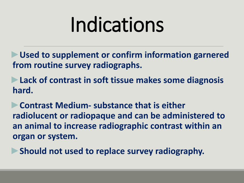
Indications►Used to supplement or confirm information garnered from routine survey radiographs.
►Lack of contrast in soft tissue makes some diagnosis hard.
►Contrast Medium- substance that is either radiolucent or radiopaque and can be administered to an animal to increase radiographic contrast within an organ or system.
►Should not used to replace survey radiography.
Contrast Media
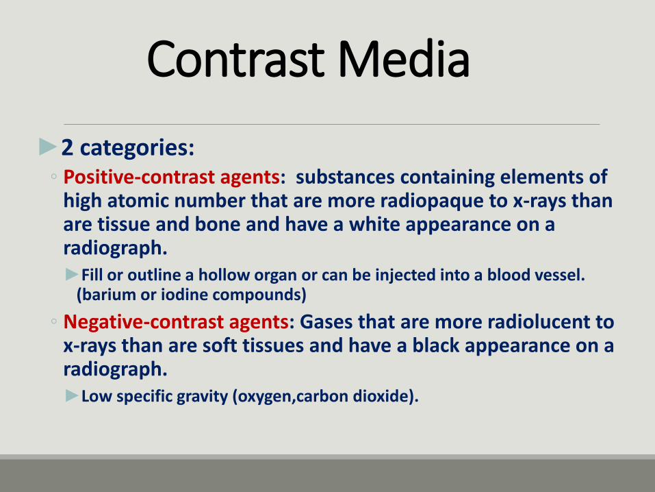
►2 categories:◦ Positive-contrast agents: substances containing elements of
high atomic number that are more radiopaque to x-rays than are tissue and bone and have a white appearance on a radiograph.►Fill or outline a hollow organ or can be injected into a blood vessel.
(barium or iodine compounds)
◦ Negative-contrast agents: Gases that are more radiolucent to x-rays than are soft tissues and have a black appearance on a radiograph.►Low specific gravity (oxygen,carbon dioxide).
Contrast Media- Continued
►3 General Categories:◦1. Positive-contrast iodinated preparations
◦2. Positive-contrast barium sulfate preparations
◦3. Negative-contrast gases.
Iodine Preparations
►2 Categories:◦ 1. Water-Soluble agents:►Make up the largest group of contrast agents.
►Most are opaque to x-rays, pharmacologically inert, low in viscosity for rapid intravenous injection, low in toxicity, rapidly excreted by the kidneys, and chemically stable so that no iodine is released in the body.
►Triiodinated compounds- a common component of iodinated positive-contrast media that contains three atoms of iodine per molecule. (well tolerated by body and provide excellent contrast).
►Can be injected into a vascular system for immediate visualization or infused into the bladder.
►Contraindicated for myelography and arthrography.

Iodine Preparations - Continued
◦2. Viscous/oily agents:►Have little application in veterinary radiography.
►Limited to lymphography.
►Consist of iodized oils, not resorbed in the body and produce fat embolism.
►Cannot be administered intravascularly.
►Does not mix with cerebrospinal fluid during myelography.
►Absorption rate is estimated at approximately 1 mL/year.
Possible toxicity
Local irritant effect
Mild discomfort
Nausia
Cardiac arrest
Anaphylaxis
Osmolality ; milliosmol/kg water (high osmol=strong toxicity)
Barium Preparations
►Barium sulfate: A common positive-contrast medium that is available in various forms and is often used as a suspension in gastrointestinal evaluations.
►Completely insoluble, not diluted or absorbed through the intestines.
►Available in liquid, paste, and powder that can be reconstituted with water.
►Disadvantage: If a perforation is present, barium may pass through and be in body indefinitely.
Negative-Contrast Agents: Gases
►Gases used include air, oxygen, nitrogen, nitrous oxide, and carbon dioxide.
►Carbon dioxide has an advantage over room air because it is better absorbed into the body when administered into a hollow organ; room air can cause air emboli.
►Are inexpensive, relatively safe and easy to administer.
►Double contrast: a radiographic contrast technique that uses a combination of positive and negative-contrast media simultaneously.
Patient Preparation
►Proper patient preparation is vital to radiographic study.
►Food should be withheld for 12-24 hours and, if necessary, administering an enema.
►Sedation may be needed, but should avoid drugs with anticholinergic effects.
Contrast Studies of the Gastrointestinal Tract
►Consists of introduction of contrast media either by oral administration or via orogastric tube.
►Radiographs are then taken at intervals to evaluate changes in morphology and the rate of gastric emptying as well as small bowel transit time.

ขอ้ควรระวงัขอ้ควรระวงั
• Perforation in the alimentary tract into the thorax or abdomen
*water- soluble contrast medium is prefer
• Aspiration the agent into the lungs
FluoroscopyFluoroscopy
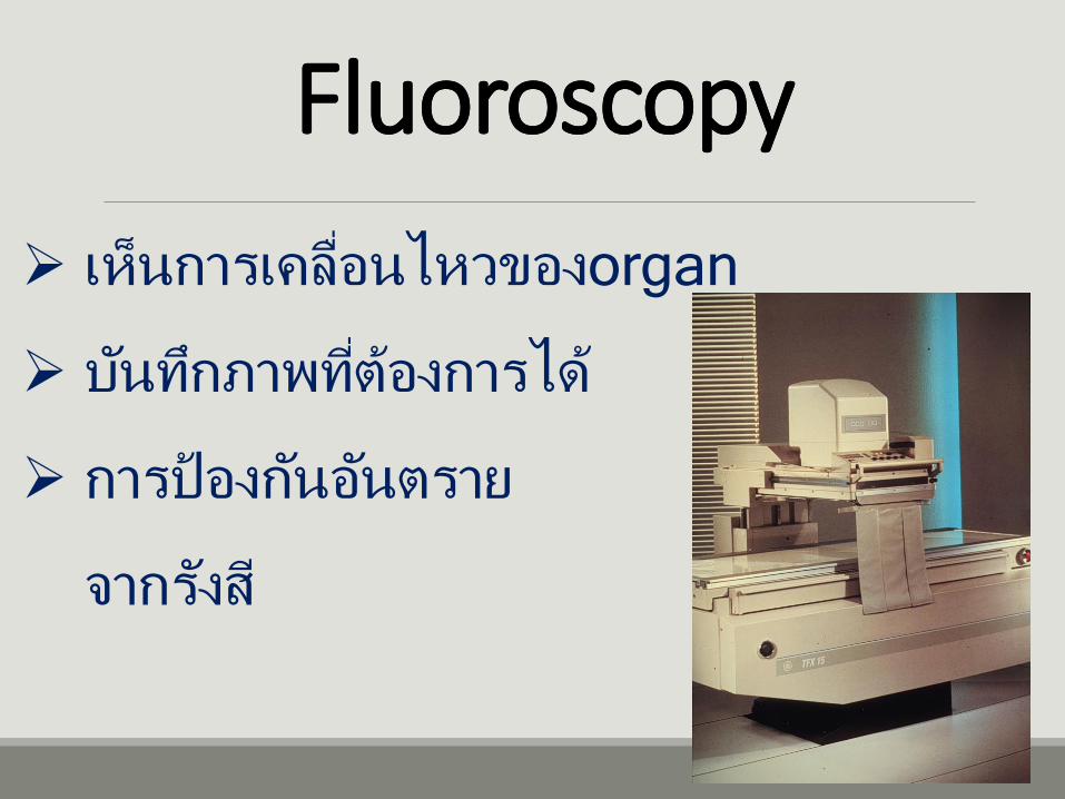
เห็นการเคลือ่นไหวของorgan
บนัทกึภาพทีต่อ้งการได้
การป้องกนัอนัตราย
จากรงัสี
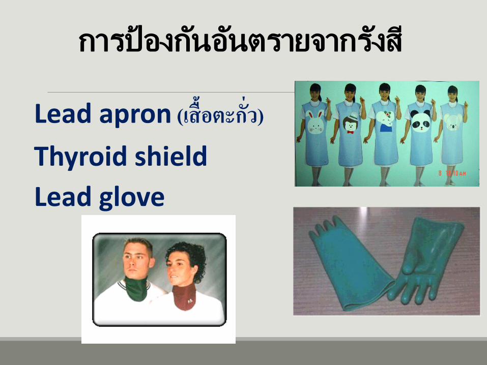
การป้องกนัอนัตรายจากรงัสี
Lead apron (เส้ือตะกัว่)
Thyroid shield
Lead glove
Esophagography
►A radiographic contrast study performed to evaluate esophageal function and morphology.
►Indicated for patients with a history or regurgitation of undigested food, acute gagging, or dysphagia.
►Liquid barium sulfate is usually contrast medium of choice.
►Precautions:◦ Make sure not able to aspirate barium.◦ If concerned about perforation or rupture, iodinated agent
should be used instead.
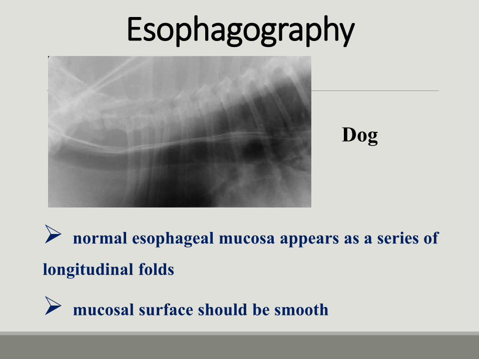
normal esophageal mucosa appears as a series of longitudinal folds
mucosal surface should be smooth
Esophagography
Dog
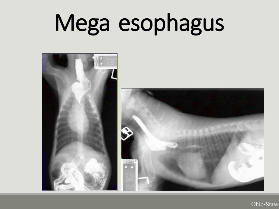
Mega esophagus
Ohio-State

Esophageal foreign bodies (intraluminal lesion)
a filling defect within the contrast, or by retention of contrast around the margins
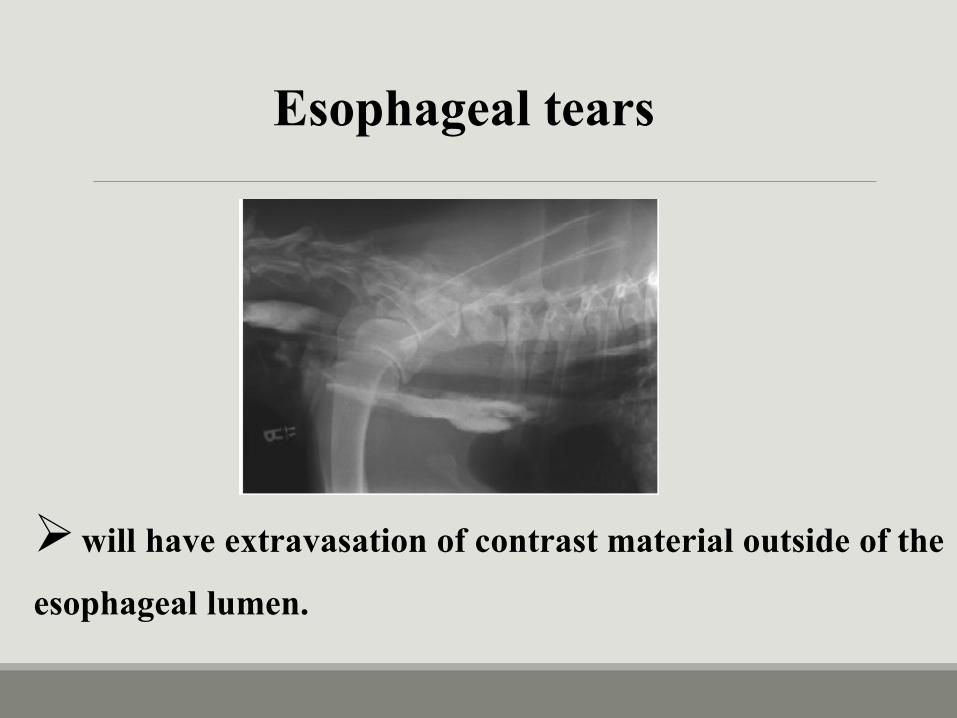
will have extravasation of contrast material outside of theesophageal lumen.
Esophageal tears
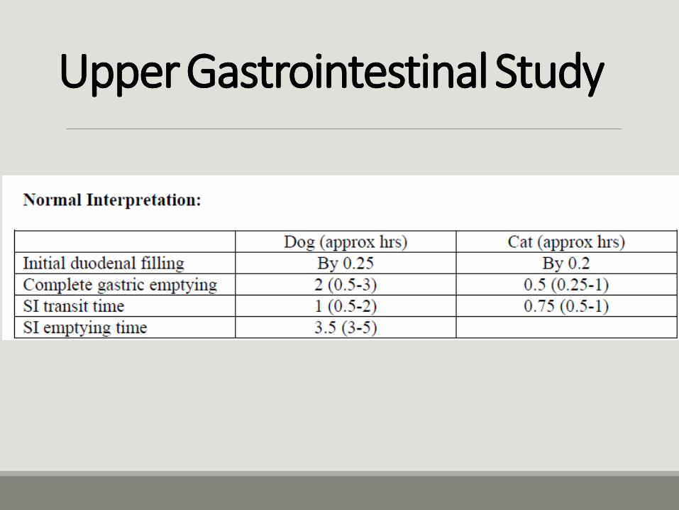
Upper Gastrointestinal Study
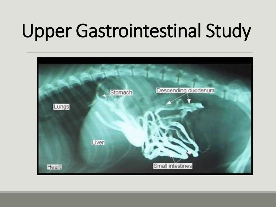
►(UGI)- A radiographic contrast study evaluating the stomach and small intestines.
►Indicated in cases of recurrent and unresponsive vomiting, abnormal bowel movements, suspected foreign body or obstruction, chronic weight loss, or persistent abdominal pain.
►Contrast medium is administered orally, and radiographs are taken during the passage of the agent.
►Precautions:◦ If perforation or rupture is suspected, barium should not be
used.
Upper Gastrointestinal Study
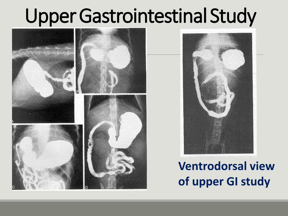
Ventrodorsal viewof upper GI study
UpperGastrointestinal StudyUpperGastrointestinal Study
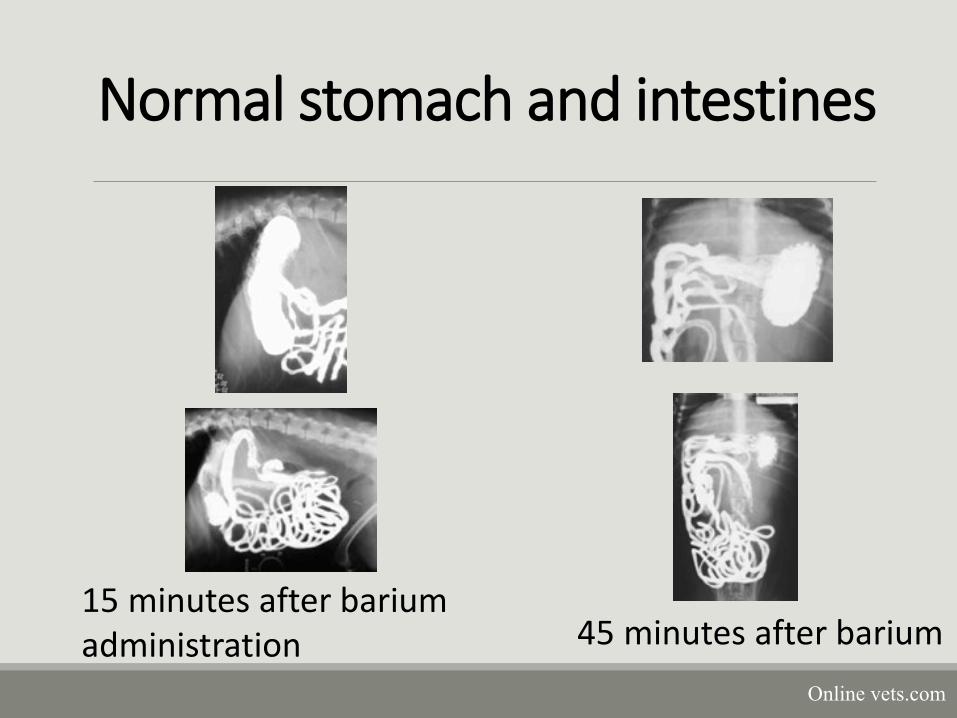
Normal stomach and intestines
15 minutes after barium administration 45 minutes after barium
Online vets.com
UpperGastrointestinal StudyUpperGastrointestinal Study
Gastrography►A radiographic contrast study performed to evaluate the size, shape, position, and morphology of the stomach.
►Indicated for patients that are experiencing acute or chronic vomiting, blood in the vomitus, or cranial abdominal pain.
►Contrast is administered orally and subsequent radiographs are exposed with the animal in various positions.
►Can use positive, negative or double contrast studies.
►Precautions:◦ Double contrast not recommended in animals with a history of gastric distension
or volvulus.
◦ If perforation suspected, the oral iodine preparation should be used.
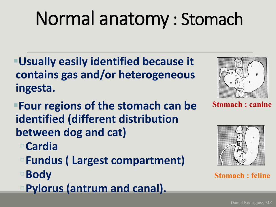
Normal anatomy : StomachNormal anatomy : Stomach
Usually easily identified because it contains gas and/or heterogeneous ingesta.
Four regions of the stomach can be identified (different distribution between dog and cat) CardiaFundus ( Largest compartment) BodyPylorus (antrum and canal).
Stomach : canine
Stomach : feline
Daniel Rodriguez, MZ
Stomach
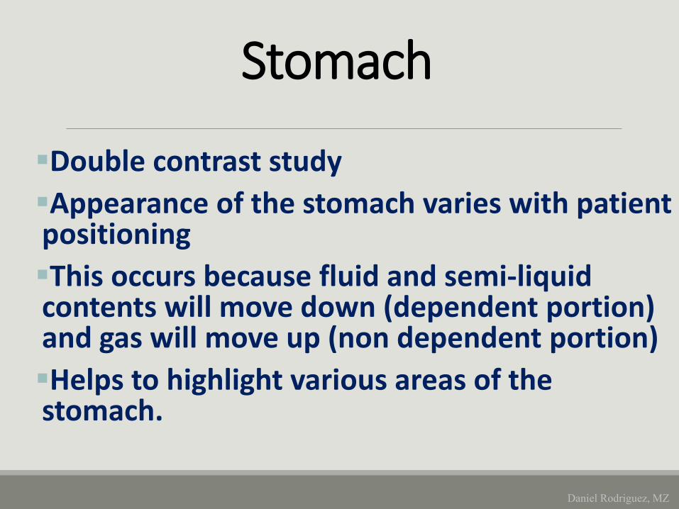
Double contrast study
Appearance of the stomach varies with patient positioning
This occurs because fluid and semi-liquid contents will move down (dependent portion) and gas will move up (non dependent portion)
Helps to highlight various areas of the stomach.
Daniel Rodriguez, MZ
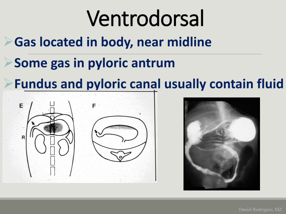
VentrodorsalGas located in body, near midline
Some gas in pyloric antrum
Fundus and pyloric canal usually contain fluid
Daniel Rodriguez, MZ
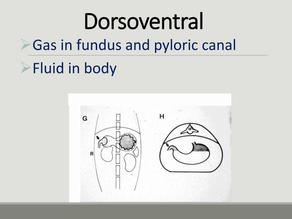
DorsoventralGas in fundus and pyloric canal
Fluid in body
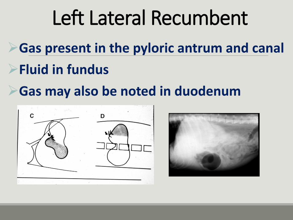
Left Lateral Recumbent
Gas present in the pyloric antrum and canal
Fluid in fundus
Gas may also be noted in duodenum
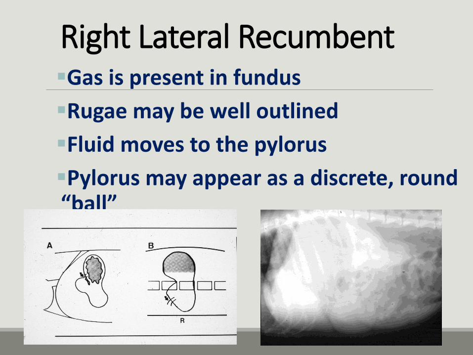
Right Lateral RecumbentGas is present in fundus
Rugae may be well outlined
Fluid moves to the pylorus
Pylorus may appear as a discrete, round “ball”

may present as a mass lesion that projects into the
lumen as a filling defect, or as a diffuse, infiltrative mural lesion.
Gastric neoplasia
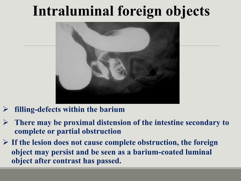
Intraluminal foreign objects
filling-defects within the barium
There may be proximal distension of the intestine secondary to complete or partial obstruction
If the lesion does not cause complete obstruction, the foreign
object may persist and be seen as a barium-coated luminal object after contrast has passed.
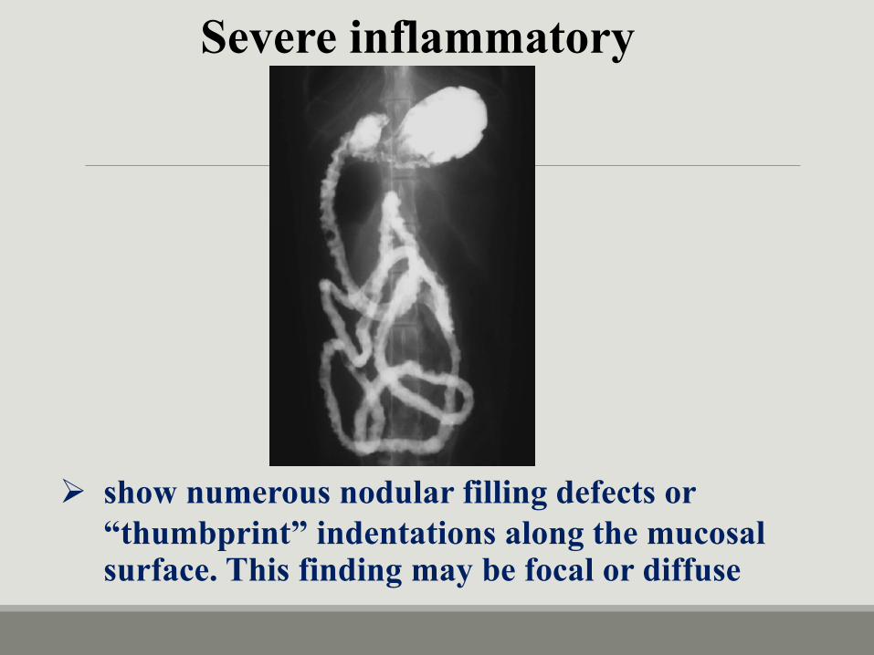
Severe inflammatory
show numerous nodular filling defects or
“thumbprint” indentations along the mucosal surface. This finding may be focal or diffuse
Lower Gastrointestinal Study
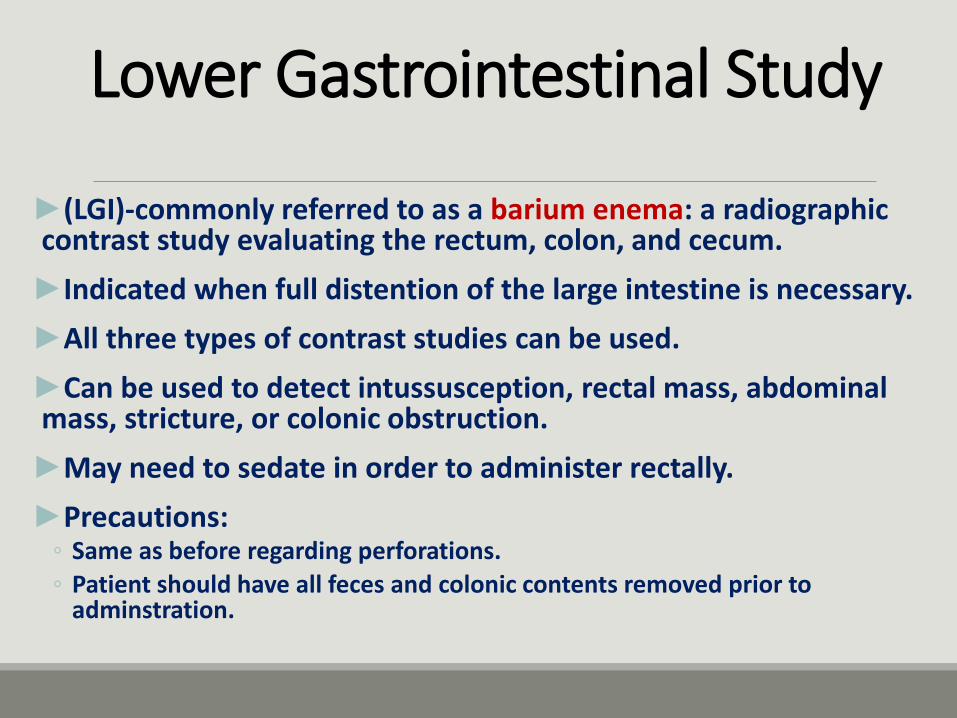
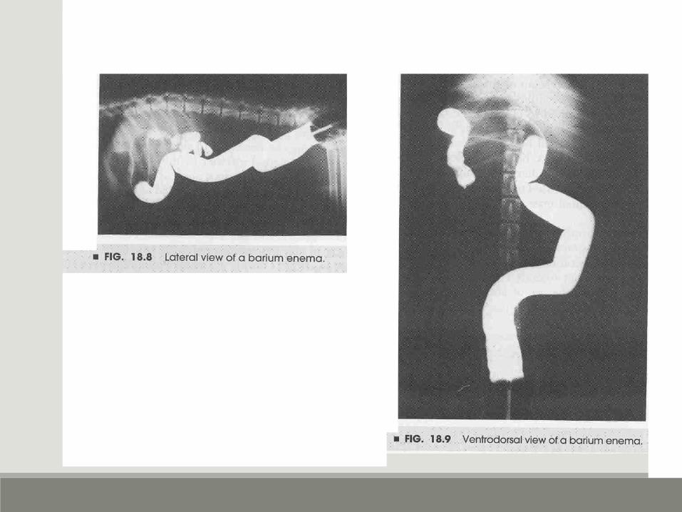
►(LGI)-commonly referred to as a barium enema: a radiographic contrast study evaluating the rectum, colon, and cecum.
►Indicated when full distention of the large intestine is necessary.
►All three types of contrast studies can be used.
►Can be used to detect intussusception, rectal mass, abdominal mass, stricture, or colonic obstruction.
►May need to sedate in order to administer rectally.
►Precautions:◦ Same as before regarding perforations.
◦ Patient should have all feces and colonic contents removed prior to adminstration.

Dog Cat
Large Intestine Cecum Colon - Consists of ascending, transverse, and descending portions Rectum
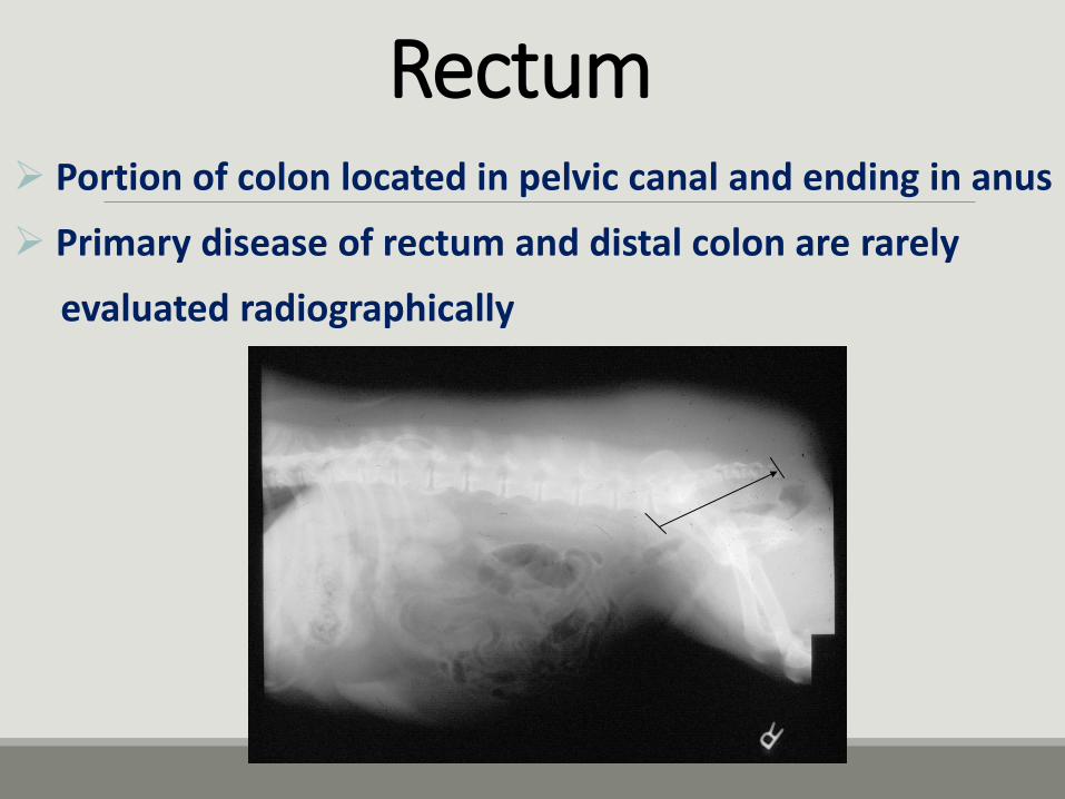
Rectum Portion of colon located in pelvic canal and ending in anus
Primary disease of rectum and distal colon are rarely
evaluated radiographically
Contrast Studies of the Urinary System
►Evaluate the kidneys, ureters, bladder and urethra.
►Relatively inexpensive and highly diagnostic.
►May be indicated in patients with hematuria, proteinuria, crytalluria, polyuria, isothenuria, or dysuria.

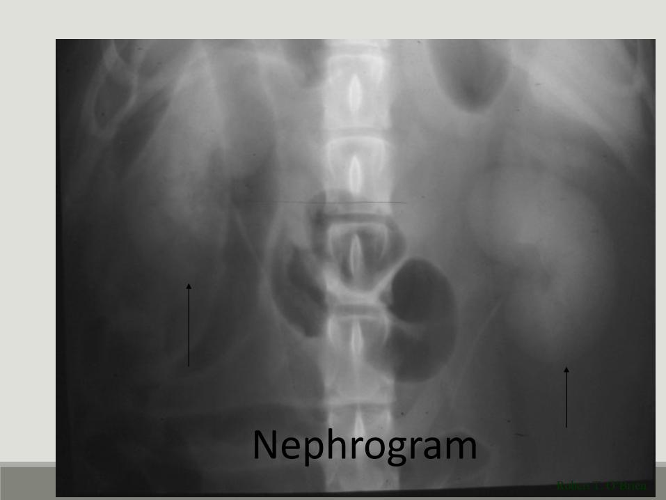
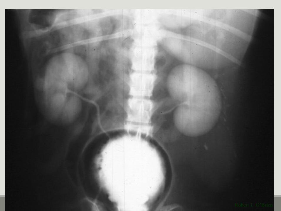
Excretory Urography
►An intravenous radiographic contrast study of the kidneys and ureters.
►Also called Intravenous urogram (IVU) or Intravenous pyelogram (IVP).
►Iodinated contrast medium circulates through the blood, is filtered out by the blood, and collects in the kidneys.
►Divided into nephrogram and pyelogram.
►Precautions:◦ Any urinary samples for diagnostic purposes should be taken prior to injection of
contrast.
◦ Contrast media may induce a false-positive reaction for protein detected by sulfosalicylic acid.
◦ May need to place indwelling catheter.
◦ May produce a reaction, so preparations must be taken for potential reaction.
Robert T. O’Brien
Nephrogram
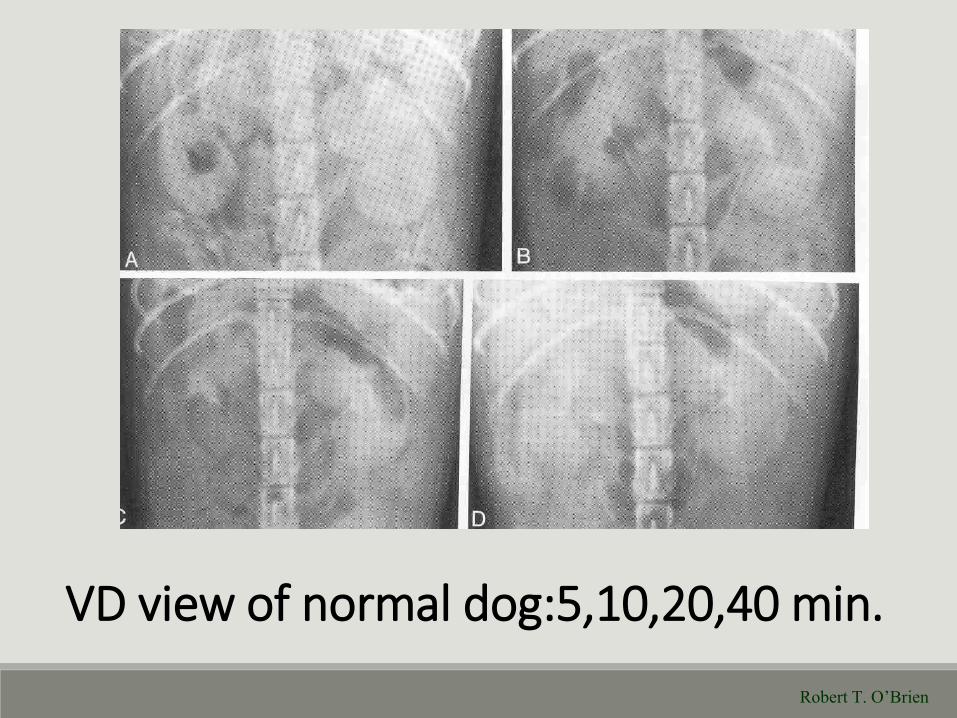
VD view of normal dog:5,10,20,40 min.
Robert T. O’Brien
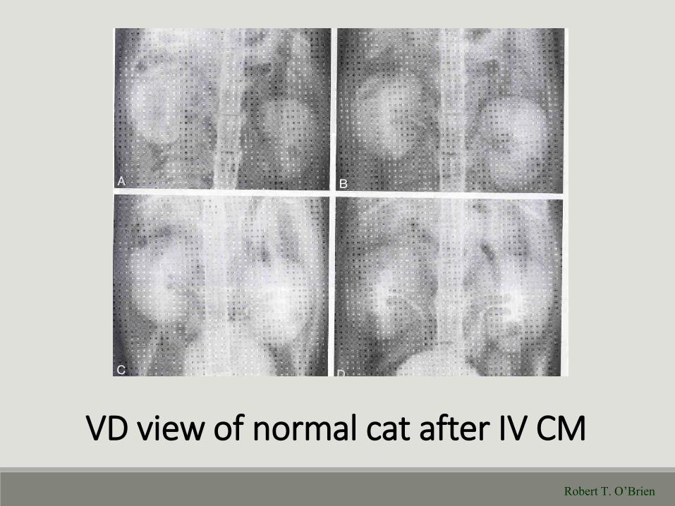
VD view of normal cat after IV CM
Robert T. O’Brien
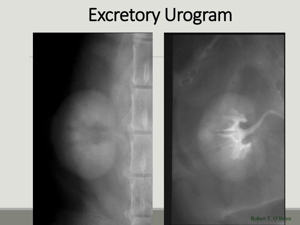
Excretory Urogram
Robert T. O’Brien
Robert T. O’Brien
Robert T. O’Brien
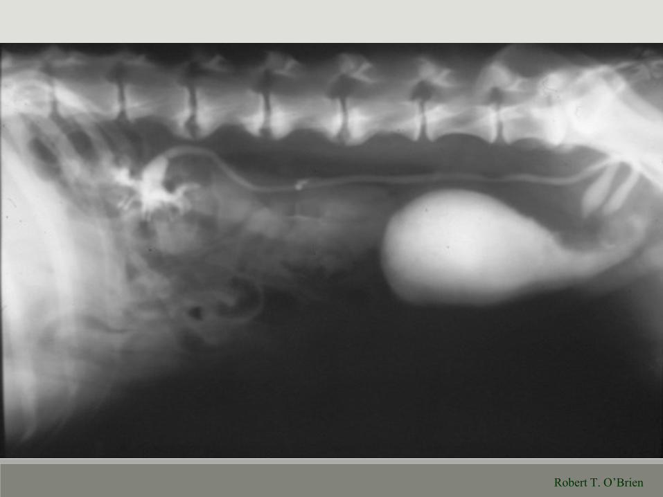
Cystography
►Radiographic contrast studies involving the urinary bladder.
►Usually introduced through a bladder catheter.
►All three types of contrast studies can be used.
►Evaluates for cystic calculi, mural lesions, bladder rupture, and other bladder wall abnormalities.
►Indicated for unresponsive hematuria, crystalluria, bacturia, dysuria, anuria, and incontinence.
►Sedation is recommended.

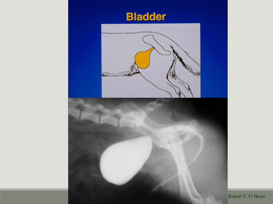
Urinary Bladder
Robert T. O’Brien
Robert T. O’Brien
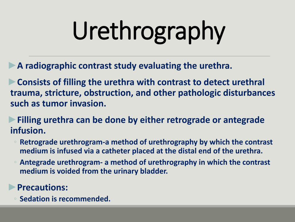
Urethrography►A radiographic contrast study evaluating the urethra.
►Consists of filling the urethra with contrast to detect urethral trauma, stricture, obstruction, and other pathologic disturbances such as tumor invasion.
►Filling urethra can be done by either retrograde or antegrade infusion.◦ Retrograde urethrogram-a method of urethrography by which the contrast
medium is infused via a catheter placed at the distal end of the urethra.
◦ Antegrade urethrogram- a method of urethrography in which the contrast medium is voided from the urinary bladder.
►Precautions:◦ Sedation is recommended.
Robert T. O’Brien
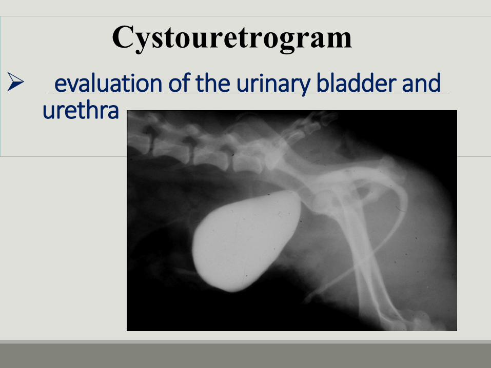
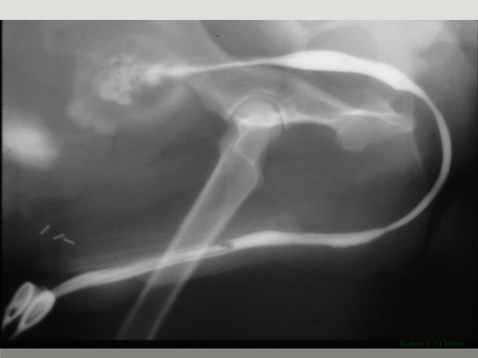
evaluation of the urinary bladder and urethra
Cystouretrogram
Arthrography►A radiographic contrast technique evaluating the articular cartilage, joint space, and joint capsule.
►Indicated in patients that are lame or have pain associated with a joint.
►Can be used to evaluate a ruptured joint capsule, the presence of a cartilaginous flap, meniscal injuries, or the necessity for surgery.
►Can be performed with water soluble iodine compound or carbon dioxide or nitrous oxide.
►Contraindicated if there is infection of soft tissues surrounding the joint.

Angiography and Angiocardiography
►Angiography: An intravenous radiographic contrast study evaluating the vascular system.
►Angiocardiography: An intravenous radiographic contrast study evaluating the vascular system and chambers of the heart.
►Due to how quickly blood vessels carry contrast, images must be taken during or immediately after injection.
►Water-soluble iodine compound is contrast medium of choice.
Cholecystography►An oral or intravenous radiographic contrast study evaluating the bile ducts and gallbladder.
►Can indicate possible gallbladder disease, biliary obstruction, gallstones, or hepatocellular dysfunction.
Fistulography►A positive or negative radiographic contrast study used to determine the depth and origin of a fistulous tract.
►Fistula-any abnormal tubelike passage within the body tissue.
►Contrast agent of choice is water-soluble iodinated agent.
Lymphography
►A radiographic contrast study evaluating lymphatic vessels and lymph nodes.
►Usually limited to areas of the extremities, head, and cervical regions.
►Expose lymphatic duct and introduce contrast medium into that duct.
►Radiographs are obtained immediately after injection.
►Water-soluble or oily iodinated contrast agents are used.
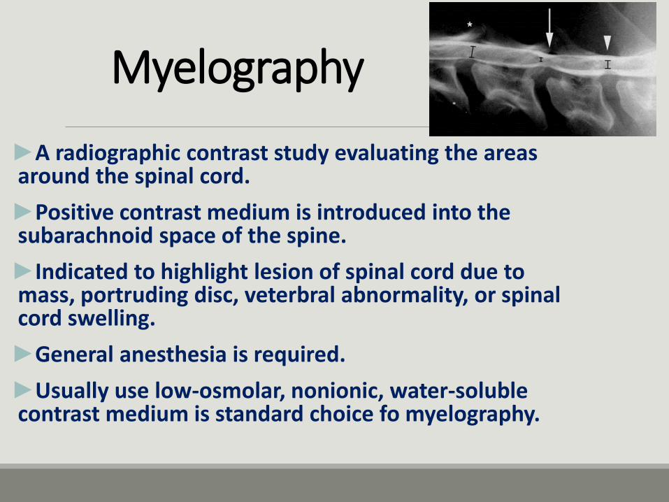
Myelography
►A radiographic contrast study evaluating the areas around the spinal cord.
►Positive contrast medium is introduced into the subarachnoid space of the spine.
►Indicated to highlight lesion of spinal cord due to mass, portruding disc, veterbral abnormality, or spinal cord swelling.
►General anesthesia is required.
►Usually use low-osmolar, nonionic, water-soluble contrast medium is standard choice fo myelography.
Pneumoperitoneography
►A negative-contrast radiographic study consisting of the introduction of a gas into the peritoneal cavity.
►Evaluates the liver, spleen, stomach, distal colon, kidneys, urinary bladder, uterus, and abdominal wall.
►Carbon dioxide and nitrous oxide are preferred gases due to rapid absorption in the body.
►Requires sedation.
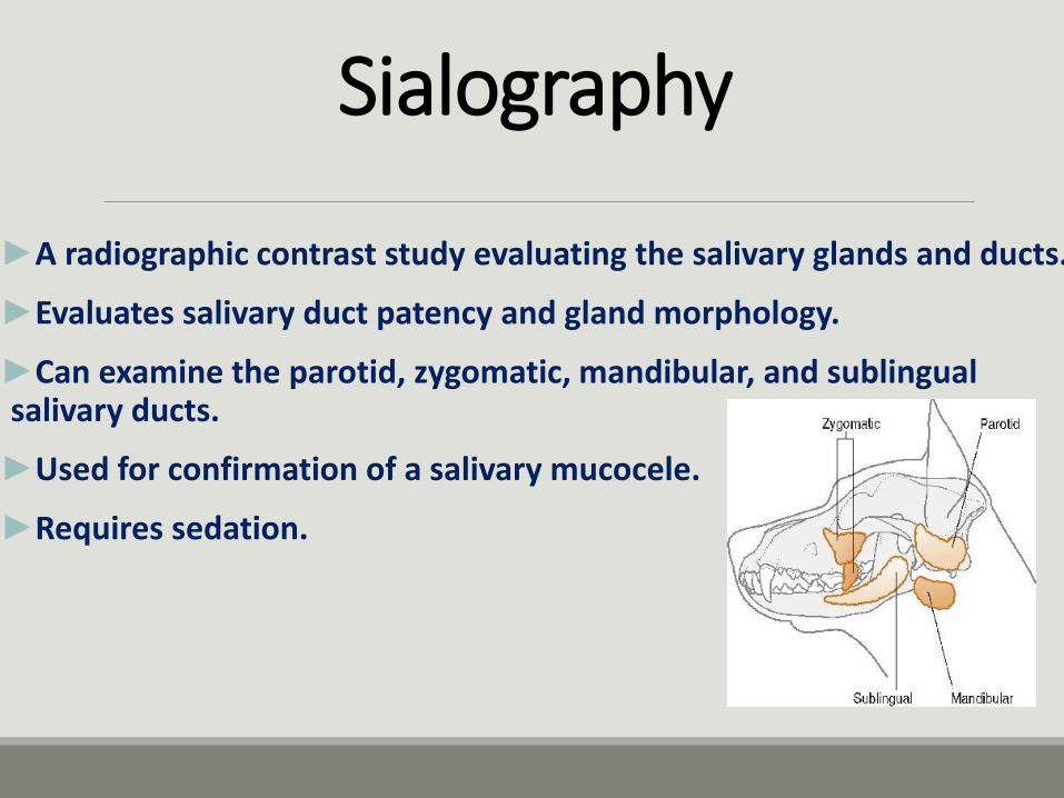
Sialography
►A radiographic contrast study evaluating the salivary glands and ducts.
►Evaluates salivary duct patency and gland morphology.
►Can examine the parotid, zygomatic, mandibular, and sublingual salivary ducts.
►Used for confirmation of a salivary mucocele.
►Requires sedation.
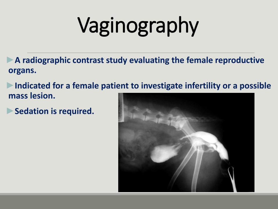
Vaginography
►A radiographic contrast study evaluating the female reproductive organs.
►Indicated for a female patient to investigate infertility or a possible mass lesion.
►Sedation is required.
Q&Ahttp://www.cvm.umn.edu/vetrad/


![[PPT]PowerPoint Presentation - دانشگاه علوم پزشکی بوشهرdnt.bpums.ac.ir/UploadedFiles/CourseFiles/bone1__27a80f... · Web viewTransient osteopetrosis Radiographic](https://img.pdfslide.tips/doc/110x75/5aab48e97f8b9a2e088ba039/pptpowerpoint-presentation-dntbpumsaciruploadedfilescoursefilesbone127a80fweb.jpg)