Embed Size (px)
Citation preview
Copyrights © 2018 The Korean Society of Radiology 191
Case Report
서론
췌장의 편평세포암은 전체 췌장암 중에서 0.5~2%로 매우
낮은 빈도를 보인다(1). 췌장의 편평세포암은 국내외에서 드물
게 보고가 되고 있으나 저자들이 아는 바로는 확산강조영상
(diffusion weighted image; 이하 DWI)을 포함한 자기공명영
상(magnetic resonance imaging; 이하 MRI)과 과혈관성 소견
을 보이지 않는 컴퓨터단층촬영(Computed Tomography; 이하
CT)은 국내외적으로 보고된 바가 없다. 저자들은 특별한 기저
질환 없는 건강한 63세 남자 환자에서 췌장의 편평세포암을
경험하였기에 문헌고찰과 함께 CT와 MRI 소견을 보고하는 바
이다.
증례 보고
63세 남자 환자가 발열(fever)을 주소로 내원하였다. 특별한
기저질환 없이 건강하게 지내던 환자로서 호흡기 증상이나 복
부 증상은 없었다. 내원 당시 시행한 혈액 검사상에 white blood
cell 17960/mm3, alkaline phosphatase 134 U/L, r-glutamyl
transpeptidase 111 IU/L, C-reactive protein 10.39 mg/dL로
증가한 소견을 보였다.
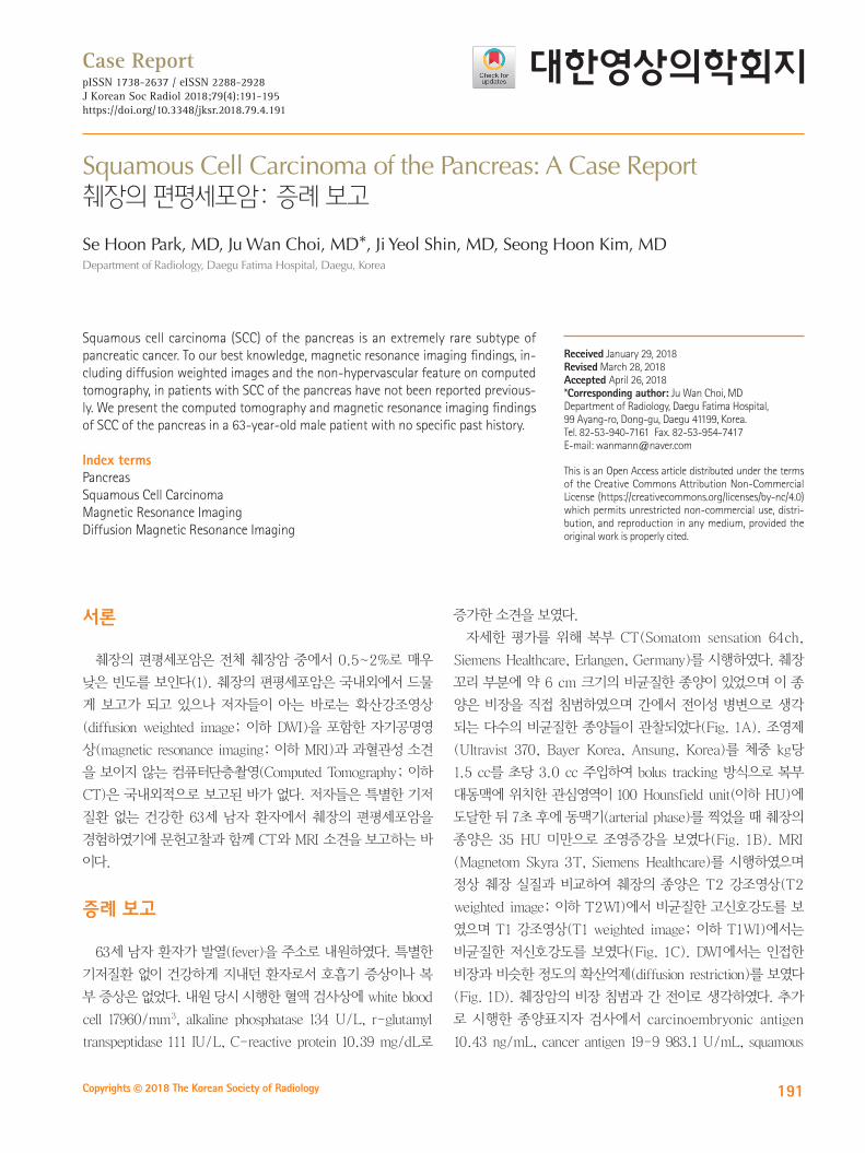
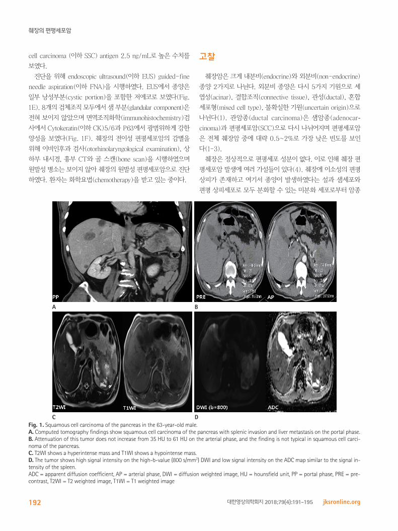
자세한 평가를 위해 복부 CT(Somatom sensation 64ch,
Siemens Healthcare, Erlangen, Germany)를 시행하였다. 췌장
꼬리 부분에 약 6 cm 크기의 비균질한 종양이 있었으며 이 종
양은 비장을 직접 침범하였으며 간에서 전이성 병변으로 생각
되는 다수의 비균질한 종양들이 관찰되었다(Fig. 1A). 조영제
(Ultravist 370, Bayer Korea, Ansung, Korea)를 체중 kg당
1.5 cc를 초당 3.0 cc 주입하여 bolus tracking 방식으로 복부
대동맥에 위치한 관심영역이 100 Hounsfield unit(이하 HU)에
도달한 뒤 7초 후에 동맥기(arterial phase)를 찍었을 때 췌장의
종양은 35 HU 미만으로 조영증강을 보였다(Fig. 1B). MRI
(Magnetom Skyra 3T, Siemens Healthcare)를 시행하였으며
정상 췌장 실질과 비교하여 췌장의 종양은 T2 강조영상(T2
weighted image; 이하 T2WI)에서 비균질한 고신호강도를 보
였으며 T1 강조영상(T1 weighted image; 이하 T1WI)에서는
비균질한 저신호강도를 보였다(Fig. 1C). DWI에서는 인접한
비장과 비슷한 정도의 확산억제(diffusion restriction)를 보였다
(Fig. 1D). 췌장암의 비장 침범과 간 전이로 생각하였다. 추가
로 시행한 종양표지자 검사에서 carcinoembryonic antigen
10.43 ng/mL, cancer antigen 19-9 983.1 U/mL, squamous
Squamous Cell Carcinoma of the Pancreas: A Case Report췌장의 편평세포암: 증례 보고
Se Hoon Park, MD, Ju Wan Choi, MD*, Ji Yeol Shin, MD, Seong Hoon Kim, MDDepartment of Radiology, Daegu Fatima Hospital, Daegu, Korea
Squamous cell carcinoma (SCC) of the pancreas is an extremely rare subtype of pancreatic cancer. To our best knowledge, magnetic resonance imaging findings, in-cluding diffusion weighted images and the non-hypervascular feature on computed tomography, in patients with SCC of the pancreas have not been reported previous-ly. We present the computed tomography and magnetic resonance imaging findings of SCC of the pancreas in a 63-year-old male patient with no specific past history.
Index termsPancreasSquamous Cell CarcinomaMagnetic Resonance ImagingDiffusion Magnetic Resonance Imaging
Received January 29, 2018Revised March 28, 2018Accepted April 26, 2018*Corresponding author: Ju Wan Choi, MDDepartment of Radiology, Daegu Fatima Hospital, 99 Ayang-ro, Dong-gu, Daegu 41199, Korea.Tel. 82-53-940-7161 Fax. 82-53-954-7417E-mail: [email protected]
This is an Open Access article distributed under the terms of the Creative Commons Attribution Non-Commercial License (https://creativecommons.org/licenses/by-nc/4.0) which permits unrestricted non-commercial use, distri-bution, and reproduction in any medium, provided the original work is properly cited.
pISSN 1738-2637 / eISSN 2288-2928J Korean Soc Radiol 2018;79(4):191-195https://doi.org/10.3348/jksr.2018.79.4.191
췌장의 편평세포암
192 jksronline.org대한영상의학회지 2018;79(4):191-195
cell carcinoma (이하 SSC) antigen 2.5 ng/mL로 높은 수치를
보였다.
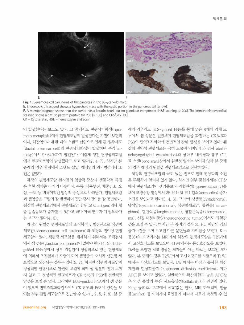
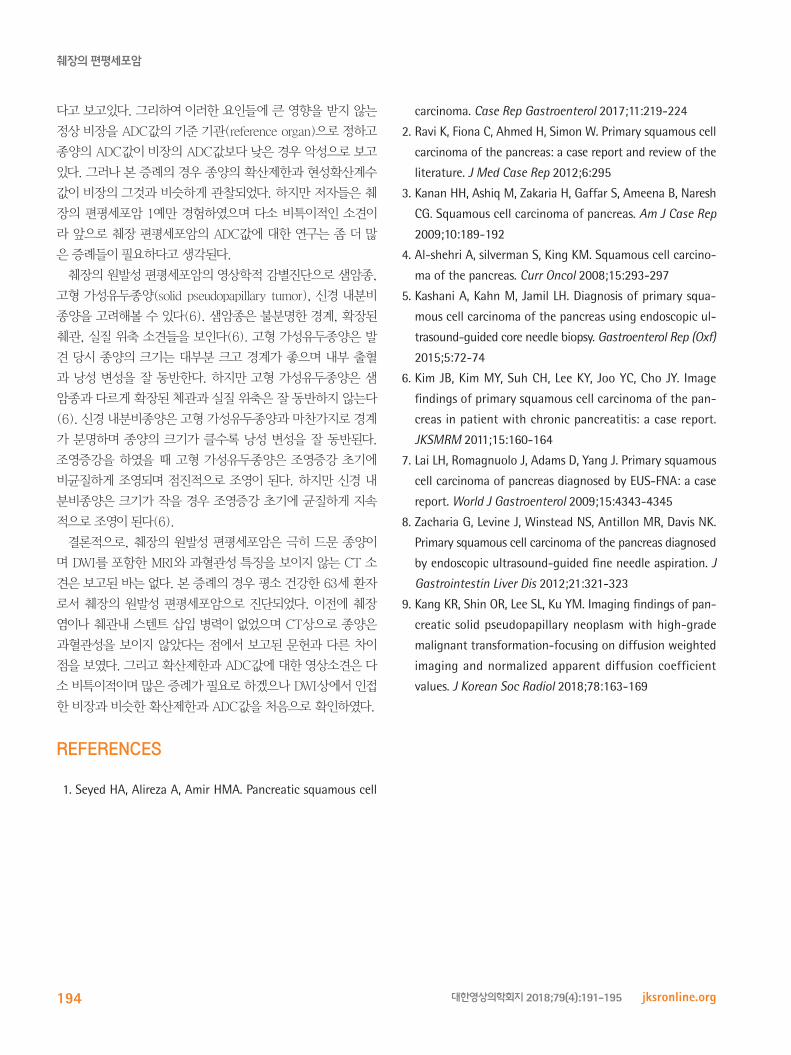
진단을 위해 endoscopic ultrasound(이하 EUS) guided-fine
needle aspiration(이하 FNA)을 시행하였다. EUS에서 종양은
일부 낭성부분(cystic portion)을 포함한 저에코로 보였다(Fig.
1E). 8개의 검체조직 모두에서 샘 부분(glandular component)은
전혀 보이지 않았으며 면역조직화학(immunohistochemistry)검
사에서 Cytokeratin(이하 CK)5/6과 P63에서 광범위하게 강한
양성을 보였다(Fig. 1F). 췌장의 전이성 편평세포암의 감별을
위해 이비인후과 검사(otorhinolaryngological examination), 상
하부 내시경, 흉부 CT와 골 스캔(bone scan)을 시행하였으며
원발성 병소는 보이지 않아 췌장의 원발성 편평세포암으로 진단
하였다. 환자는 화학요법(chemotherapy)을 받고 있는 중이다.
고찰
췌장암은 크게 내분비(endocrine)와 외분비(non-endocrine)
종양 2가지로 나뉜다. 외분비 종양은 다시 5가지 기원으로 세
엽성(acinar), 결합조직(connective tissue), 관성(ductal), 혼합
세포형(mixed cell type), 불확실한 기원(uncertain origin)으로
나뉜다(1). 관암종(ductal carcinoma)은 샘암종(adenocar-cinoma)과 편평세포암(SCC)으로 다시 나뉘어지며 편평세포암
은 전체 췌장암 중에 대략 0.5~2%로 가장 낮은 빈도를 보인
다(1-3).
췌장은 정상적으로 편평세포 성분이 없다. 이로 인해 췌장 편
평세포암 발생에 여러 가설들이 있다(4). 췌장에 이소성의 편평
상피가 존재하고 여기서 종양이 발생하였다는 설과 샘세포와
편평 상피세포로 모두 분화할 수 있는 미분화 세포로부터 암종
Fig. 1. Squamous cell carcinoma of the pancreas in the 63-year-old male.A. Computed tomography findings show squamous cell carcinoma of the pancreas with splenic invasion and liver metastasis on the portal phase.B. Attenuation of this tumor does not increase from 35 HU to 61 HU on the arterial phase, and the finding is not typical in squamous cell carci-noma of the pancreas.C. T2WI shows a hyperintense mass and T1WI shows a hypointense mass.D. The tumor shows high signal intensity on the high-b-value (800 s/mm2) DWI and low signal intensity on the ADC map similar to the signal in-tensity of the spleen. ADC = apparent diffusion coefficient, AP = arterial phase, DWI = diffusion weighted image, HU = hounsfield unit, PP = portal phase, PRE = pre-contrast, T2WI = T2 weighted image, T1WI = T1 weighted image
C D
A B
박세훈 외
193jksronline.org 대한영상의학회지 2018;79(4):191-195
이 발생한다는 보고도 있다. 그 중에서도 편평상피화생(squa-mous metaplasia)에서 편평세포암이 발생했다는 기전이 보편적
이다. 췌장염이나 췌관 내의 스텐트 삽입으로 인해 관 원주세포
(ductal columnar cell)의 편평상피화생이 발생하며 부검(au-topsy)에서 9~64%까지 발견된다. 이렇게 생긴 편평상피회생
에서 편평세포암이 발생했다고 보고 있다(2, 4-7). 하지만 본
증례의 경우 환자에서 스텐트 삽입, 췌장염의 과거병력이나 소
견은 없었다.
췌장의 편평세포암 환자들의 임상적 증상과 생물학적 특징
은 흔한 샘암종과 거의 비슷하다. 복통, 식욕부진, 체중감소, 오
심, 구토 등 비특이적인 임상적 증상으로 나타난다. 편평세포암
과 샘암종은 고령에 잘 발생하며 진단 당시 전이를 잘 동반한다.
췌장의 편평세포암에서 편평세포암 항원(SCC antigen)이나 혈
중 칼슘농도가 증가할 수 있다고 하나 아직 연구가 더 필요하다
는 보고가 있다(4, 6).
췌장의 원발성 편평세포암의 조직학적 감별진단으로 샘편평
세포암(adenosquamous cell carcinoma)과 췌장의 전이성 편평
세포암이 있다. 샘편평 세포암을 배제하기 위해서는 조직검사
에서 샘 성분(glandular component)이 없어야 한다(4, 5). EUS-
guided FNA상에서 상부 위장관에 정상적으로 있는 편평세포
에 의해서 조직검체가 오염이 되어 샘암종이 오히려 샘편평 세
포암으로 오진되는 경우는 있다(5, 7). 하지만 샘편평 세포암이
정상적인 편평세포로 완전히 오염이 되어 샘 성분이 전혀 보이
지 않고 그 정상적인 편평세포가 CK 5/6과 P63에 전반적인
양성을 보일 순 없다. 그리하여 EUS-guided FNA에서 샘 성분
이 없으며 면역조직화학검사에서 CK 5/6과 P63에 양성을 보
이는 경우 편평 세포암으로 진단할 수 있다(1, 2, 5, 7, 8). 본 증
례의 경우에도 EUS-guided FNA를 통해 얻은 8개의 검체 모
두에서 샘 성분은 없었으며 편평세포암을 확진하는 CK5/6과
P63의 면역조직화학에 전반적인 강한 양성을 보이고 있다. 췌
장의 전이성 편평세포는 극히 드물며 이비인후과 검사(otorhi-nolaryngological examination)와 상하부 내시경과 흉부 CT,
골 스캔(bone scan)상에서 원발성 병소는 보이지 않아 본 증례
의 경우 췌장의 원발성 편평세포암으로 진단하였다.
췌장의 편평세포암의 극히 낮은 빈도로 인해 영상학적 소견
은 뚜렷하게 알려져 있지 않다. 하지만 일부 문헌에서는 CT상
에서 편평세포암이 샘암종보다 과혈관성(hyperevascularity)을
보여 조영증강 영상에서 35 HU-61 HU 감쇠(attenuation) 증가
소견을 보인다고 한다(3, 4, 6). 그 밖에 낭샘종(cystadenoma),
낭샘암(cystadenocarcinoma), 샘편평세포암, 혈관종(heman-gioma), 혈관육종(angiosarcoma), 평활근육종(leiomyosarco-ma), 신경 내분비종양(neuroendocrine tumor)에서도 과혈관
성을 보일 수 있다. 하지만 본 증례의 경우 35 HU 미만의 감쇠
증가소견을 보여 보고된 다른 문헌들과 차이점을 보였다. Kim
등(6)의 보고에서는 MRI에서 췌장의 편평세포암은 T2WI에
서 고신호강도를 보였으며 T1WI에서는 동신호강도를 보였다.
DWI를 포함한 MRI 영상은 저자들이 아는 바로는 보고된 바가
없다. 본 증례의 경우 T2WI에서 고신호강도를 보였으며 T1WI
에서는 저신호강도를 보였다. DWI에서는 비장과 유사한 확산
제한과 현성확산계수(apparent diffusion coefficient; 이하
ADC)를 보이고 있었다. 일반적으로 확산제한과 낮은 ADC값
은 악성 종양의 높은 세포충실성(cellularity)과 관련이 있다.
Kang 등(9)의 보고에서 ADC값은 환자, MRI 하드웨어, 인공
물(artifact) 등 여러가지 요인들에 따라서 다르게 측정될 수 있
Fig. 1. Squamous cell carcinoma of the pancreas in the 63-year-old male.E. Endoscopic ultrasound shows a hypoechoic mass with the cystic portion in the pancreas tail (arrow).F. A microphotograph shows that the tumor has a keratin pearl, but no glandular component (H&E staining, × 200). The immunohistochemical staining shows a diffuse pattern positive for P63 (× 100) and CK5/6 (× 100).CK = Cytokeratin, H&E = hematoxylin and eosin
E F
췌장의 편평세포암
194 jksronline.org대한영상의학회지 2018;79(4):191-195
다고 보고있다. 그리하여 이러한 요인들에 큰 영향을 받지 않는
정상 비장을 ADC값의 기준 기관(reference organ)으로 정하고
종양의 ADC값이 비장의 ADC값보다 낮은 경우 악성으로 보고
있다. 그러나 본 증례의 경우 종양의 확산제한과 현성확산계수
값이 비장의 그것과 비슷하게 관찰되었다. 하지만 저자들은 췌
장의 편평세포암 1예만 경험하였으며 다소 비특이적인 소견이
라 앞으로 췌장 편평세포암의 ADC값에 대한 연구는 좀 더 많
은 증례들이 필요하다고 생각된다.
췌장의 원발성 편평세포암의 영상학적 감별진단으로 샘암종,
고형 가성유두종양(solid pseudopapillary tumor), 신경 내분비
종양을 고려해볼 수 있다(6). 샘암종은 불분명한 경계, 확장된
췌관, 실질 위축 소견들을 보인다(6). 고형 가성유두종양은 발
견 당시 종양의 크기는 대부분 크고 경계가 좋으며 내부 출혈
과 낭성 변성을 잘 동반한다. 하지만 고형 가성유두종양은 샘
암종과 다르게 확장된 체관과 실질 위축은 잘 동반하지 않는다
(6). 신경 내분비종양은 고형 가성유두종양과 마찬가지로 경계
가 분명하며 종양의 크기가 클수록 낭성 변성을 잘 동반된다.
조영증강을 하였을 때 고형 가성유두종양은 조영증강 초기에
비균질하게 조영되며 점진적으로 조영이 된다. 하지만 신경 내
분비종양은 크기가 작을 경우 조영증강 초기에 균질하게 지속
적으로 조영이 된다(6).
결론적으로, 췌장의 원발성 편평세포암은 극히 드문 종양이
며 DWI를 포함한 MRI와 과혈관성 특징을 보이지 않는 CT 소
견은 보고된 바는 없다. 본 증례의 경우 평소 건강한 63세 환자
로서 췌장의 원발성 편평세포암으로 진단되었다. 이전에 췌장
염이나 췌관내 스텐트 삽입 병력이 없었으며 CT상으로 종양은
과혈관성을 보이지 않았다는 점에서 보고된 문헌과 다른 차이
점을 보였다. 그리고 확산제한과 ADC값에 대한 영상소견은 다
소 비특이적이며 많은 증례가 필요로 하겠으나 DWI상에서 인접
한 비장과 비슷한 확산제한과 ADC값을 처음으로 확인하였다.
RefeRences
1. Seyed HA, Alireza A, Amir HMA. Pancreatic squamous cell
carcinoma. Case Rep Gastroenterol 2017;11:219-224
2. Ravi K, Fiona C, Ahmed H, Simon W. Primary squamous cell
carcinoma of the pancreas: a case report and review of the
literature. J Med Case Rep 2012;6:295
3. Kanan HH, Ashiq M, Zakaria H, Gaffar S, Ameena B, Naresh
CG. Squamous cell carcinoma of pancreas. Am J Case Rep
2009;10:189-192
4. Al-shehri A, silverman S, King KM. Squamous cell carcino-
ma of the pancreas. Curr Oncol 2008;15:293-297
5. Kashani A, Kahn M, Jamil LH. Diagnosis of primary squa-
mous cell carcinoma of the pancreas using endoscopic ul-
trasound-guided core needle biopsy. Gastroenterol Rep (Oxf)
2015;5:72-74
6. Kim JB, Kim MY, Suh CH, Lee KY, Joo YC, Cho JY. Image
findings of primary squamous cell carcinoma of the pan-
creas in patient with chronic pancreatitis: a case report.
JKSMRM 2011;15:160-164
7. Lai LH, Romagnuolo J, Adams D, Yang J. Primary squamous
cell carcinoma of pancreas diagnosed by EUS-FNA: a case
report. World J Gastroenterol 2009;15:4343-4345
8. Zacharia G, Levine J, Winstead NS, Antillon MR, Davis NK.
Primary squamous cell carcinoma of the pancreas diagnosed
by endoscopic ultrasound-guided fine needle aspiration. J
Gastrointestin Liver Dis 2012;21:321-323
9. Kang KR, Shin OR, Lee SL, Ku YM. Imaging findings of pan-
creatic solid pseudopapillary neoplasm with high-grade
malignant transformation-focusing on diffusion weighted
imaging and normalized apparent diffusion coefficient
values. J Korean Soc Radiol 2018;78:163-169
박세훈 외
195jksronline.org 대한영상의학회지 2018;79(4):191-195
췌장의 편평세포암: 증례 보고
박세훈 · 최주완* · 신지열 · 김성훈
췌장의 편평세포암(squamous cell carcinoma of the pancreas)은 아주 드문 질환이며 저자들이 아는 바로는 확산강조영
상을 포함한 자기공명영상과 과혈관성 소견을 보이지 않는 컴퓨터단층촬영 소견은 국내외적으로 보고된 바가 없다. 특별
한 과거력이 없는 63세 남자 환자에서 췌장의 편평세포암 1예를 경험하였기에 컴퓨터단층촬영과 자기공명영상 소견을 중
심으로 보고하는 바이다.
대구파티마병원 영상의학과