Embed Size (px)
Citation preview
STING inhibitors target the cyclic dinucleotidebinding pocketZe Honga,1
, Jiahao Meia,1, Chenhui Lia,1, Guohui Baib, Munire Maimaitia, Haiyang Hua, Wenying Yuc, Li Sund,Lele Zhange
, Dan Chenga, Yixian Liaoc, Senlin Lie, Yanping Youd, Hongbin Sunc, Jing Huangb, Xing Liuf
,Judy Liebermang,2
, and Chen Wanga,2
aState Key Laboratory of Natural Medicines, Department of Life Science and Technology, China Pharmaceutical University, 211198 Nanjing, China;bShanghai Institute of Precision Medicine, Ninth People’s Hospital, Shanghai Jiao Tong University School of Medicine, 200025 Shanghai, China; cState KeyLaboratory of Natural Medicines, Department of Natural Medicinal Chemistry, China Pharmaceutical University, 211198 Nanjing, China; dState KeyLaboratory of Natural Medicines, Center for Drug Discovery, China Pharmaceutical University, 211198 Nanjing, China; eState Key Laboratory of Cell Biology,Institute of Biochemistry and Cell Biology, Shanghai Institutes for Biological Sciences, Chinese Academy of Sciences, 200031 Shanghai, China; fCenter forMicrobes, Development and Health, Key Laboratory of Molecular Virology and Immunology, Institut Pasteur of Shanghai, Chinese Academy of Sciences,200031 Shanghai, China; and gProgram in Cellular and Molecular Medicine, Boston Children’s Hospital, Boston, MA 02115
Contributed by Judy Lieberman, May 6, 2021 (sent for review March 22, 2021; reviewed by Hong-Bing Shu and Nan Yan)
Cytosolic DNA activates cGAS (cytosolic DNA sensor cyclic AMP-GMP synthase)-STING (stimulator of interferon genes) signaling,which triggers interferon and inflammatory responses that helpdefend against microbial infection and cancer. However, aberrantcytosolic self-DNA in Aicardi–Goutière’s syndrome and constituentlyactive gain-of-function mutations in STING in STING-associatedvasculopathy with onset in infancy (SAVI) patients lead to excessivetype I interferons and proinflammatory cytokines, which causedifficult-to-treat and sometimes fatal autoimmune disease. Here,in silico docking identified a potent STING antagonist SN-011 thatbinds with higher affinity to the cyclic dinucleotide (CDN)-bindingpocket of STING than endogenous 2′3′-cGAMP. SN-011 locks STINGin an open inactive conformation, which inhibits interferon and in-flammatory cytokine induction activated by 2′3′-cGAMP, herpes sim-plex virus type 1 infection, Trex1 deficiency, overexpression ofcGAS-STING, or SAVI STING mutants. In Trex1−/− mice, SN-011 waswell tolerated, strongly inhibited hallmarks of inflammation andautoimmunity disease, and prevented death. Thus, a specific STINGinhibitor that binds to the STING CDN-binding pocket is a promisinglead compound for STING-driven disease.
STING | type I interferons | antagonist | Aicardi–Goutières syndrome | SAVI
Cytoplasmic microbial and host DNAs act as danger signals (1).The cytosolic DNA sensor cyclic AMP-GMP synthase (cGAS,
also known as MB21D1 or C6orf150) catalyzes the production of anoncanonical cyclic dinucleotide (CDN) c[G (2′,5′)pA (3′,5′)p](2′3′-cGAMP) from ATP and GTP (2–5). The 2′3′-cGAMP andbacterial CDNs c-di-GMP and c-di-AMP bind to and activate theendoplasmic reticulum (ER)-associated stimulator of interferon(IFN) genes (STING, also known as MITA, ERIS, or MPYS)(6–9). CDNs bind to the CDN-binding domain (CBD) in a pocketat the interface of the STING dimer (10, 11). CDN binding inducesa conformational change that closes the dimer pocket and causeshigher-order multimerization and activation of STING (12–15).Activated STING translocates from the ER, through the Golgiapparatus, to perinuclear vesicles (16). During this process, STINGrecruits and activates the kinase TBK1, which in turn phosphory-lates the transcription factor IRF3 (17–20). Dimerization and nu-clear translocation of phosphorylated IRF3 potently induces typeI IFNs. STING activation also initiates NF-κB signaling throughTBK1 and TRAF6, which leads to proinflammatory cytokineexpression (21).The cGAS-STING axis plays an essential role in initiating host
immune defense against microbial invasion. STING-deficient miceare more susceptible to some viral infections (22). Genomic DNAextruded into the cytosol because of genomic instability in canceralso stimulates cGAS and STING to enhance antitumor immunity(23, 24). However, inappropriate activation of this signaling path-way by aberrant self-DNAs leads to chronic IFN expression, which
has been implicated in the development of some autoimmunedisorders (25). Genetic mutations of genes that cause cytosolic accu-mulation of nucleic acids, including the DNase TREX1, RNASEH2A-C, SAMHD1, ADAR, and MDA5, cause a broad spectrum ofinflammatory and autoimmune phenotypes, including Aicardi–Goutières syndrome (AGS), familial chilblain lupus, and systemiclupus erythematosus (SLE) (26–28). Trex1 deficiency in mice causesinflammatory myocarditis, progressive cardiomyopathy, and circu-latory failure. Trex1-deficient mice lacking STING are completelyprotected from otherwise lethal inflammatory diseases, indicatingthe central role of STING in disease pathogenesis. Moreover,gain-of-function (GOF) mutations in STING cause early-onsetvasculopathy and pulmonary inflammation in patients, known asSTING-associated vasculopathy with onset in infancy (SAVI)(29, 30). In addition, in some IFN or inflammation-driven disorders,including senescence, lethal sepsis, acute pancreatitis, Parkinson’s
Significance
cGAS (cytosolic DNA sensor cyclic AMP-GMP synthase)-STING(stimulator of interferon genes) signaling is critical for sensingcytosolic DNA to initiate host immune responses against in-vading pathogens and cancer. However, inappropriate activationof STING signaling causes severe and often fatal autoimmune orautoinflammatory diseases. Hence, STING is an attractive drugtarget for the treatment of STING-driven autoimmune and in-flammatory disorders. Therefore, there is a need to identify leadcompounds that effectively inhibit human STING for furtherdrug development. Here, we identified and characterized aSTING-specific inhibitor SN-011 with high efficiency, specificity,and safety, paving the way for therapeutically manipulatingSTING-mediated clinical diseases.
Author contributions: Z.H., J.M., C.L., J.L., and C.W. designed research; Z.H., J.M., C.L., J.L.,and C.W. performed research; Z.H. and J.M. conducted surface plasmon resonance exper-iments; Z.H., J.M., and M.M. performed in vivo animal experiments; Z.H. and C.W. de-signed the compounds; L.S., Y.Y., and H.S. participated in chemical synthesis; Z.H., J.M.,and H.H. performed RNA-sequencing experiments and analysis; W.Y. and Y.L. performedthe docking screen; G.B., L.Z., D.C., S.L., and J.H. assisted in experiments; J.L. analyzeddata; and Z.H., J.M., X.L., J.L., and C.W. wrote the paper.
Reviewers: H.-B.S., Wuhan University; and N.Y., University of Texas SouthwesternMedical Center.
The authors declare no competing interest.
This open access article is distributed under Creative Commons Attribution-NonCommercial-NoDerivatives License 4.0 (CC BY-NC-ND).1Z.H., J.M., and C.L. contributed equally to this work.2To whom correspondence may be addressed. Email: [email protected] or [email protected].
This article contains supporting information online at https://www.pnas.org/lookup/suppl/doi:10.1073/pnas.2105465118/-/DCSupplemental.
Published June 7, 2021.
PNAS 2021 Vol. 118 No. 24 e2105465118 https://doi.org/10.1073/pnas.2105465118 | 1 of 10
IMMUNOLO
GYAND
INFLAMMATION
Dow
nloa
ded
by g
uest
on
Oct
ober
14,
202
1
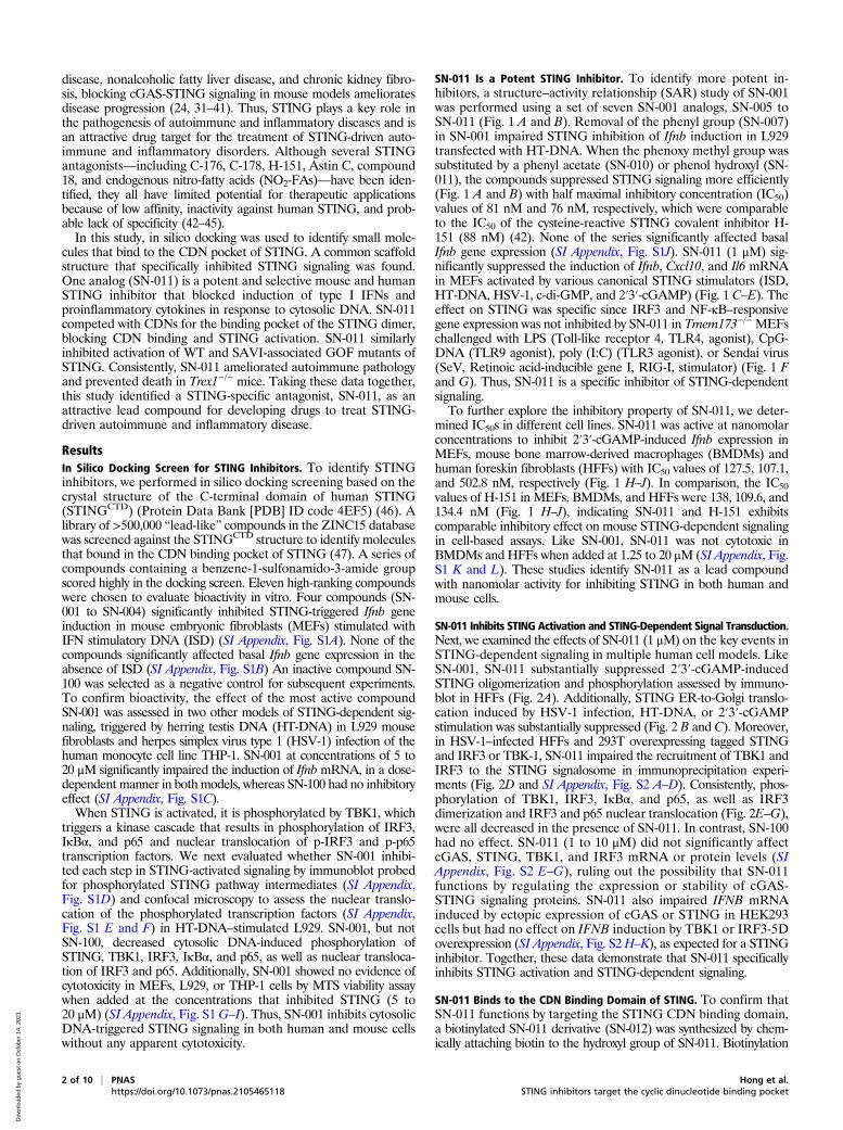
disease, nonalcoholic fatty liver disease, and chronic kidney fibro-sis, blocking cGAS-STING signaling in mouse models amelioratesdisease progression (24, 31–41). Thus, STING plays a key role inthe pathogenesis of autoimmune and inflammatory diseases and isan attractive drug target for the treatment of STING-driven auto-immune and inflammatory disorders. Although several STINGantagonists—including C-176, C-178, H-151, Astin C, compound18, and endogenous nitro-fatty acids (NO2-FAs)—have been iden-tified, they all have limited potential for therapeutic applicationsbecause of low affinity, inactivity against human STING, and prob-able lack of specificity (42–45).In this study, in silico docking was used to identify small mole-
cules that bind to the CDN pocket of STING. A common scaffoldstructure that specifically inhibited STING signaling was found.One analog (SN-011) is a potent and selective mouse and humanSTING inhibitor that blocked induction of type I IFNs andproinflammatory cytokines in response to cytosolic DNA. SN-011competed with CDNs for the binding pocket of the STING dimer,blocking CDN binding and STING activation. SN-011 similarlyinhibited activation of WT and SAVI-associated GOF mutants ofSTING. Consistently, SN-011 ameliorated autoimmune pathologyand prevented death in Trex1−/− mice. Taking these data together,this study identified a STING-specific antagonist, SN-011, as anattractive lead compound for developing drugs to treat STING-driven autoimmune and inflammatory disease.
ResultsIn Silico Docking Screen for STING Inhibitors. To identify STINGinhibitors, we performed in silico docking screening based on thecrystal structure of the C-terminal domain of human STING(STINGCTD) (Protein Data Bank [PDB] ID code 4EF5) (46). Alibrary of >500,000 “lead-like” compounds in the ZINC15 databasewas screened against the STINGCTD structure to identify moleculesthat bound in the CDN binding pocket of STING (47). A series ofcompounds containing a benzene-1-sulfonamido-3-amide groupscored highly in the docking screen. Eleven high-ranking compoundswere chosen to evaluate bioactivity in vitro. Four compounds (SN-001 to SN-004) significantly inhibited STING-triggered Ifnb geneinduction in mouse embryonic fibroblasts (MEFs) stimulated withIFN stimulatory DNA (ISD) (SI Appendix, Fig. S1A). None of thecompounds significantly affected basal Ifnb gene expression in theabsence of ISD (SI Appendix, Fig. S1B) An inactive compound SN-100 was selected as a negative control for subsequent experiments.To confirm bioactivity, the effect of the most active compoundSN-001 was assessed in two other models of STING-dependent sig-naling, triggered by herring testis DNA (HT-DNA) in L929 mousefibroblasts and herpes simplex virus type 1 (HSV-1) infection of thehuman monocyte cell line THP-1. SN-001 at concentrations of 5 to20 μM significantly impaired the induction of IfnbmRNA, in a dose-dependent manner in both models, whereas SN-100 had no inhibitoryeffect (SI Appendix, Fig. S1C).When STING is activated, it is phosphorylated by TBK1, which
triggers a kinase cascade that results in phosphorylation of IRF3,IκBα, and p65 and nuclear translocation of p-IRF3 and p-p65transcription factors. We next evaluated whether SN-001 inhibi-ted each step in STING-activated signaling by immunoblot probedfor phosphorylated STING pathway intermediates (SI Appendix,Fig. S1D) and confocal microscopy to assess the nuclear translo-cation of the phosphorylated transcription factors (SI Appendix,Fig. S1 E and F) in HT-DNA–stimulated L929. SN-001, but notSN-100, decreased cytosolic DNA-induced phosphorylation ofSTING, TBK1, IRF3, IκBα, and p65, as well as nuclear transloca-tion of IRF3 and p65. Additionally, SN-001 showed no evidence ofcytotoxicity in MEFs, L929, or THP-1 cells by MTS viability assaywhen added at the concentrations that inhibited STING (5 to20 μM) (SI Appendix, Fig. S1G–I). Thus, SN-001 inhibits cytosolicDNA-triggered STING signaling in both human and mouse cellswithout any apparent cytotoxicity.
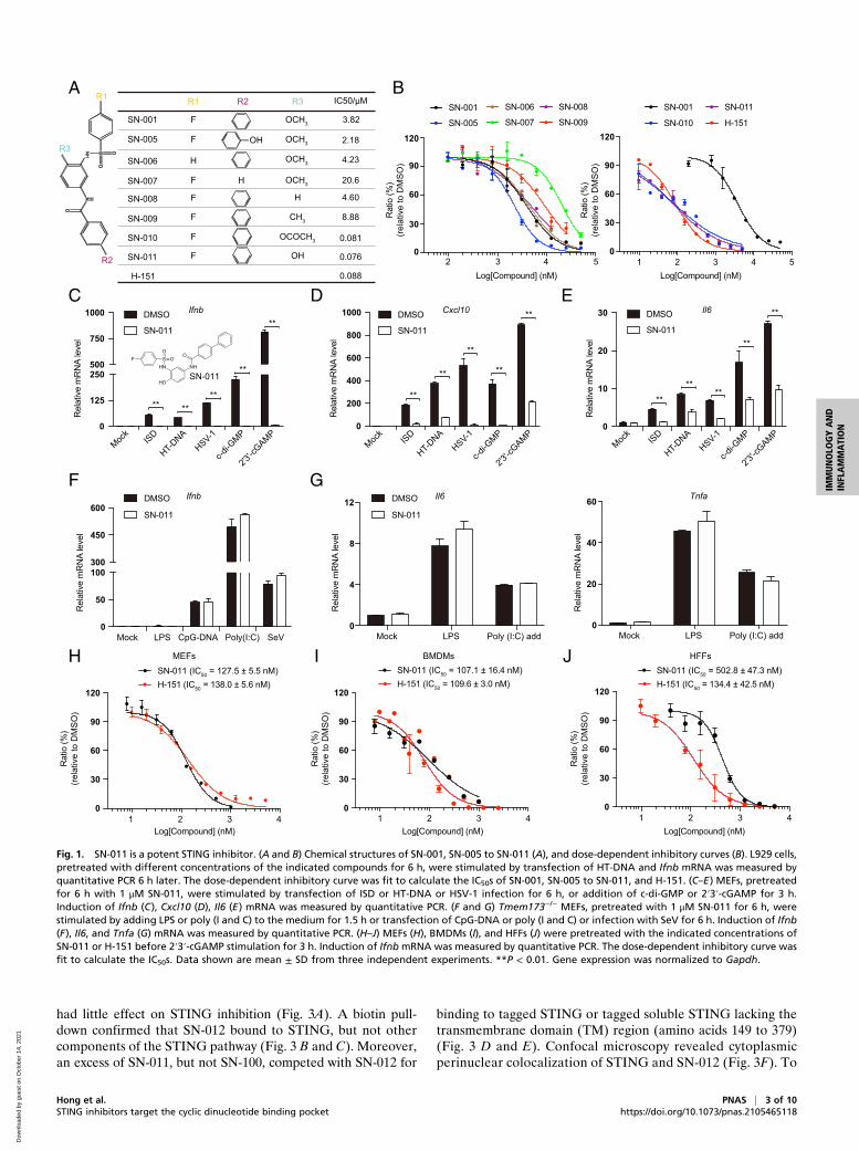
SN-011 Is a Potent STING Inhibitor. To identify more potent in-hibitors, a structure–activity relationship (SAR) study of SN-001was performed using a set of seven SN-001 analogs, SN-005 toSN-011 (Fig. 1 A and B). Removal of the phenyl group (SN-007)in SN-001 impaired STING inhibition of Ifnb induction in L929transfected with HT-DNA. When the phenoxy methyl group wassubstituted by a phenyl acetate (SN-010) or phenol hydroxyl (SN-011), the compounds suppressed STING signaling more efficiently(Fig. 1 A and B) with half maximal inhibitory concentration (IC50)values of 81 nM and 76 nM, respectively, which were comparableto the IC50 of the cysteine-reactive STING covalent inhibitor H-151 (88 nM) (42). None of the series significantly affected basalIfnb gene expression (SI Appendix, Fig. S1J). SN-011 (1 μM) sig-nificantly suppressed the induction of Ifnb, Cxcl10, and Il6 mRNAin MEFs activated by various canonical STING stimulators (ISD,HT-DNA, HSV-1, c-di-GMP, and 2′3′-cGAMP) (Fig. 1 C–E). Theeffect on STING was specific since IRF3 and NF-κB–responsivegene expression was not inhibited by SN-011 in Tmem173−/− MEFschallenged with LPS (Toll-like receptor 4, TLR4, agonist), CpG-DNA (TLR9 agonist), poly (I:C) (TLR3 agonist), or Sendai virus(SeV, Retinoic acid-inducible gene I, RIG-I, stimulator) (Fig. 1 Fand G). Thus, SN-011 is a specific inhibitor of STING-dependentsignaling.To further explore the inhibitory property of SN-011, we deter-
mined IC50s in different cell lines. SN-011 was active at nanomolarconcentrations to inhibit 2′3′-cGAMP-induced Ifnb expression inMEFs, mouse bone marrow-derived macrophages (BMDMs) andhuman foreskin fibroblasts (HFFs) with IC50 values of 127.5, 107.1,and 502.8 nM, respectively (Fig. 1 H–J). In comparison, the IC50values of H-151 in MEFs, BMDMs, and HFFs were 138, 109.6, and134.4 nM (Fig. 1 H–J), indicating SN-011 and H-151 exhibitscomparable inhibitory effect on mouse STING-dependent signalingin cell-based assays. Like SN-001, SN-011 was not cytotoxic inBMDMs and HFFs when added at 1.25 to 20 μM (SI Appendix, Fig.S1 K and L). These studies identify SN-011 as a lead compoundwith nanomolar activity for inhibiting STING in both human andmouse cells.
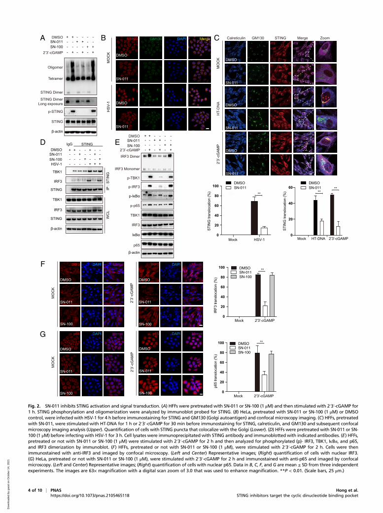
SN-011 Inhibits STING Activation and STING-Dependent Signal Transduction.Next, we examined the effects of SN-011 (1 μM) on the key events inSTING-dependent signaling in multiple human cell models. LikeSN-001, SN-011 substantially suppressed 2′3′-cGAMP-inducedSTING oligomerization and phosphorylation assessed by immuno-blot in HFFs (Fig. 2A). Additionally, STING ER-to-Golgi translo-cation induced by HSV-1 infection, HT-DNA, or 2′3′-cGAMPstimulation was substantially suppressed (Fig. 2 B and C). Moreover,in HSV-1–infected HFFs and 293T overexpressing tagged STINGand IRF3 or TBK-1, SN-011 impaired the recruitment of TBK1 andIRF3 to the STING signalosome in immunoprecipitation experi-ments (Fig. 2D and SI Appendix, Fig. S2 A–D). Consistently, phos-phorylation of TBK1, IRF3, IκBα, and p65, as well as IRF3dimerization and IRF3 and p65 nuclear translocation (Fig. 2E–G),were all decreased in the presence of SN-011. In contrast, SN-100had no effect. SN-011 (1 to 10 μM) did not significantly affectcGAS, STING, TBK1, and IRF3 mRNA or protein levels (SIAppendix, Fig. S2 E–G), ruling out the possibility that SN-011functions by regulating the expression or stability of cGAS-STING signaling proteins. SN-011 also impaired IFNB mRNAinduced by ectopic expression of cGAS or STING in HEK293cells but had no effect on IFNB induction by TBK1 or IRF3-5Doverexpression (SI Appendix, Fig. S2H–K), as expected for a STINGinhibitor. Together, these data demonstrate that SN-011 specificallyinhibits STING activation and STING-dependent signaling.
SN-011 Binds to the CDN Binding Domain of STING. To confirm thatSN-011 functions by targeting the STING CDN binding domain,a biotinylated SN-011 derivative (SN-012) was synthesized by chem-ically attaching biotin to the hydroxyl group of SN-011. Biotinylation
2 of 10 | PNAS Hong et al.https://doi.org/10.1073/pnas.2105465118 STING inhibitors target the cyclic dinucleotide binding pocket
Dow
nloa
ded
by g
uest
on
Oct
ober
14,
202
1
had little effect on STING inhibition (Fig. 3A). A biotin pull-down confirmed that SN-012 bound to STING, but not othercomponents of the STING pathway (Fig. 3 B and C). Moreover,an excess of SN-011, but not SN-100, competed with SN-012 for
binding to tagged STING or tagged soluble STING lacking thetransmembrane domain (TM) region (amino acids 149 to 379)(Fig. 3 D and E). Confocal microscopy revealed cytoplasmicperinuclear colocalization of STING and SN-012 (Fig. 3F). To
A B
C D E
F G
H I J
Log[Compound] (nM)
Ifnb
Mock LPS CpG-DNA Poly(I:C) SeV0
50
100300
450
600
Rel
ativ
e m
RN
A le
vel
DMSO
SN-011
DMSO
SN-011
Il6 Tnfa
** ****
**
**
Mock
ISD
HT-DNA
HSV-1
c-di-G
MP
2'3'-c
GAMPMoc
kISD
HT-DNA
HSV-1
c-di-G
MP
2'3'-c
GAMPMoc
kISD
HT-DNA
HSV-1
c-di-G
MP
2'3'-c
GAMP
Rel
ativ
e m
RN
A le
vel
IfnbDMSO
SN-011
0
125
250500
750
1000
**
**
** **
**
Rel
ativ
e m
RN
A le
vel
Cxcl10DMSO
SN-011
0
200
400
600
800
1000
**
****
**
**
Rel
ativ
e m
RN
A le
vel
Il6DMSO
SN-011
0
10
20
30
Rat
io (%
)(re
lativ
e to
DM
SO)
Rat
io (%
)(re
lativ
e to
DM
SO)
Rat
io (%
)(re
lativ
e to
DM
SO)
Rat
io (%
)(re
lativ
e to
DM
SO)
Rat
io (%
)(re
lativ
e to
DM
SO)
Mock LPS Poly (I:C) add0
4
8
12
Rel
ativ
e m
RN
A le
vel
Mock LPS Poly (I:C) add0
20
40
60
Rel
ativ
e m
RN
A le
vel
R1 R2 R3 IC50/μM
SN-001
SN-005
SN-006
SN-007
SN-008
SN-009
SN-010
SN-011
H-151
F
F
F
OCH3 3.82
2.18
4.23
20.6
0.081
0.076
0.088
F CH3
F OCOCH3
OH
F OH
F OCH3
OCH3
OCH3
H
H
H 4.60
8.88
SO
NH
HN
O
O
R1
R3
R2
Log[Compound] (nM) Log[Compound] (nM)Log[Compound] (nM)
2 3 4 50
30
60
90
120
SN-001
SN-005
SN-006
SN-007
SN-008
SN-009
Log[Compound] (nM)
SN-001
SN-010
1 2 3 4 50
30
60
90
120
SN-011
H-151
1 2 3 40
30
60
90
120
MEFs
H-151 (IC50 = 138.0 ± 5.6 nM)SN-011 (IC50 = 127.5 ± 5.5 nM)
1 2 3 40
30
60
90
120
HFFs
H-151 (IC50 = 134.4 ± 42.5 nM)SN-011 (IC50 = 502.8 ± 47.3 nM)
BMDMs
H-151 (IC50 = 109.6 ± 3.0 nM)SN-011 (IC50 = 107.1 ± 16.4 nM)
1 2 3 40
30
60
90
120
SN-011
Fig. 1. SN-011 is a potent STING inhibitor. (A and B) Chemical structures of SN-001, SN-005 to SN-011 (A), and dose-dependent inhibitory curves (B). L929 cells,pretreated with different concentrations of the indicated compounds for 6 h, were stimulated by transfection of HT-DNA and Ifnb mRNA was measured byquantitative PCR 6 h later. The dose-dependent inhibitory curve was fit to calculate the IC50s of SN-001, SN-005 to SN-011, and H-151. (C–E) MEFs, pretreatedfor 6 h with 1 μM SN-011, were stimulated by transfection of ISD or HT-DNA or HSV-1 infection for 6 h, or addition of c-di-GMP or 2′3′-cGAMP for 3 h.Induction of Ifnb (C), Cxcl10 (D), Il6 (E) mRNA was measured by quantitative PCR. (F and G) Tmem173−/− MEFs, pretreated with 1 μM SN-011 for 6 h, werestimulated by adding LPS or poly (I and C) to the medium for 1.5 h or transfection of CpG-DNA or poly (I and C) or infection with SeV for 6 h. Induction of Ifnb(F), Il6, and Tnfa (G) mRNA was measured by quantitative PCR. (H–J) MEFs (H), BMDMs (I), and HFFs (J) were pretreated with the indicated concentrations ofSN-011 or H-151 before 2′3′-cGAMP stimulation for 3 h. Induction of Ifnb mRNA was measured by quantitative PCR. The dose-dependent inhibitory curve wasfit to calculate the IC50s. Data shown are mean ± SD from three independent experiments. **P < 0.01. Gene expression was normalized to Gapdh.
Hong et al. PNAS | 3 of 10STING inhibitors target the cyclic dinucleotide binding pocket https://doi.org/10.1073/pnas.2105465118
IMMUNOLO
GYAND
INFLAMMATION
Dow
nloa
ded
by g
uest
on
Oct
ober
14,
202
1
E
A
D
HSV-1
STING
IRF3
β-actin
IRF3
TBK1
DMSO+ +
++
IgG STING
- -
-
-
- -- -+ +- - -- -
+-
+ + - - + - -
TBK1
WC
LIP
: ST
ING
STING
p-p65
p65
IκBα
p-IκBα
IRF3 Monomer
IRF3 Dimer
TBK1
IRF3
β-actin
p-IRF3
p-TBK1
-+ -
- -- ++ +
- +-DMSO + --+ - -
- - +- - +2’3’-cGAMP
B STING GM130 DAPI Merge
F
MO
CK
2’3’
-cG
AMP
MO
CK
HSV
-1
MO
CK
2’3’
-cG
AMP
T5
Z5
T5
Z5
DMSO
SN-011
DMSO
SN-011
DMSO
SN-011
SN-100
DMSO
SN-011
SN-100
IRF3 DAPI Merge IRF3 DAPI Merge
DMSO
SN-011
SN-100
DMSO
SN-011
SN-100
SN-011SN-100
SN-011SN-100
p-STING
STING
STING Dimer
Oligomer
SN-011SN-100
-+ -
- -- ++ +
- +-DMSO + --+ - -
- - +- - +
β-actin
Tetramer
STING DimerLong exposure
2’3’-cGAMP
G p65 DAPI Merge p65 DAPI Merge
Mock HSV-10
20
40
60
80
100
STIN
G tr
ansl
ocat
ion
(%) **
**
**
Mock 2'3'-cGAMP
Mock 2'3'-cGAMP
0
20
40
60
80
100
IRF3
tran
sloc
atio
n (%
)
0
20
40
60
80
100
p65
trans
loca
tion
(%)
DMSOSN-011SN-100
DMSOSN-011SN-100
MO
CK
HT-
DN
A
C GM130Calreticulin Merge ZoomSTING
****
0
20
40
60
STIN
G tr
ansl
ocat
ion
(%)
DMSOSN-011
Mock HT-DNA 2’3’-cGAMP
DMSOSN-011
DMSO
SN-011
DMSO
SN-011
2’3’
-cG
AMP
SN-011
DMSO
Fig. 2. SN-011 inhibits STING activation and signal transduction. (A) HFFs were pretreated with SN-011 or SN-100 (1 μM) and then stimulated with 2′3′-cGAMP for1 h. STING phosphorylation and oligomerization were analyzed by immunoblot probed for STING. (B) HeLa, pretreated with SN-011 or SN-100 (1 μM) or DMSOcontrol, were infected with HSV-1 for 4 h before immunostaining for STING and GM130 (Golgi autoantigen) and confocal microscopy imaging. (C) HFFs, pretreatedwith SN-011, were stimulated with HT-DNA for 1 h or 2′3′-cGAMP for 30 min before immunostaining for STING, calreticulin, and GM130 and subsequent confocalmicroscopy imaging analysis (Upper). Quantification of cells with STING puncta that colocalize with the Golgi (Lower). (D) HFFs were pretreated with SN-011 or SN-100 (1 μM) before infecting with HSV-1 for 3 h. Cell lysates were immunoprecipitated with STING antibody and immunoblotted with indicated antibodies. (E) HFFs,pretreated or not with SN-011 or SN-100 (1 μM) were stimulated with 2′3′-cGAMP for 2 h and then analyzed for phosphorylated (p)- IRF3, TBK1, IκBα, and p65,and IRF3 dimerization by immunoblot. (F) HFFs, pretreated or not with SN-011 or SN-100 (1 μM), were stimulated with 2′3′-cGAMP for 2 h. Cells were thenimmunostained with anti-IRF3 and imaged by confocal microscopy. (Left and Center) Representative images; (Right) quantification of cells with nuclear IRF3.(G) HeLa, pretreated or not with SN-011 or SN-100 (1 μM), were stimulated with 2′3′-cGAMP for 2 h and immunostained with anti-p65 and imaged by confocalmicroscopy. (Left and Center) Representative images; (Right) quantification of cells with nuclear p65. Data in B, C, F, and G are mean ± SD from three independentexperiments. The images are 63× magnification with a digital scan zoom of 3.0 that was used to enhance magnification. **P < 0.01. (Scale bars, 25 μm.)
4 of 10 | PNAS Hong et al.https://doi.org/10.1073/pnas.2105465118 STING inhibitors target the cyclic dinucleotide binding pocket
Dow
nloa
ded
by g
uest
on
Oct
ober
14,
202
1
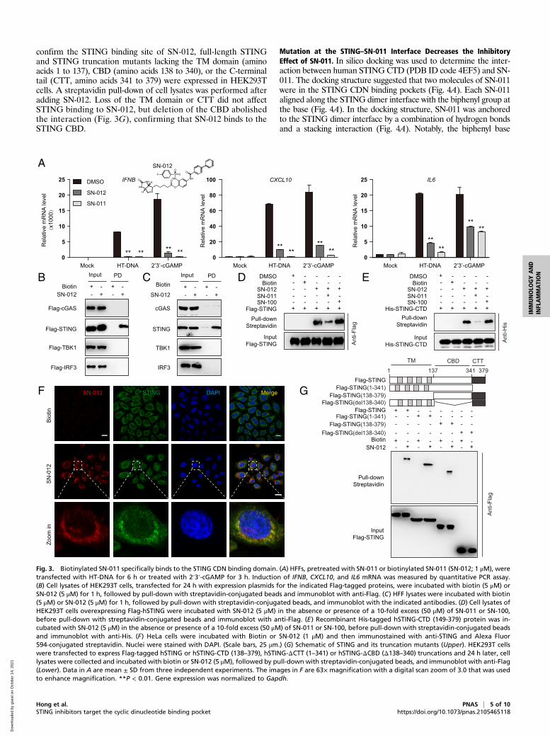
confirm the STING binding site of SN-012, full-length STINGand STING truncation mutants lacking the TM domain (aminoacids 1 to 137), CBD (amino acids 138 to 340), or the C-terminaltail (CTT, amino acids 341 to 379) were expressed in HEK293Tcells. A streptavidin pull-down of cell lysates was performed afteradding SN-012. Loss of the TM domain or CTT did not affectSTING binding to SN-012, but deletion of the CBD abolishedthe interaction (Fig. 3G), confirming that SN-012 binds to theSTING CBD.
Mutation at the STING–SN-011 Interface Decreases the InhibitoryEffect of SN-011. In silico docking was used to determine the inter-action between human STING CTD (PDB ID code 4EF5) and SN-011. The docking structure suggested that two molecules of SN-011were in the STING CDN binding pockets (Fig. 4A). Each SN-011aligned along the STING dimer interface with the biphenyl group atthe base (Fig. 4A). In the docking structure, SN-011 was anchoredto the STING dimer interface by a combination of hydrogen bondsand a stacking interaction (Fig. 4A). Notably, the biphenyl base
SN-012
Input PDBiotin
++ +
+- -
- -
cGAS
IRF3
TBK1
STING
B
A
SN-012
Input PD
Biotin+
+ ++
- -- -
Flag-cGAS
Flag-IRF3
Flag-TBK1
Flag-STING
E
F G
DCSN-012
Biotin +
SN-011 ++
SN-100
+- - --
-- - -- -
+
+-
-
DMSO + - - - -
-
-
SN-012Biotin +
SN-011 ++
SN-100
+- - --
-- - -- -
+
+-
-
DMSO + - - - -
-
-Flag-STING + + ++ +
InputFlag-STING
InputFlag-STING
Pull-downStreptavidin
Pull-downStreptavidin
InputHis-STING-CTD
His-STING-CTD + + ++ +Pull-down
Streptavidin
Biot
inSN
-012
Zoom
in
SN-012 STING DAPI Merge
DMSO
SN-012
SN-011
IFNB
Mock HT-DNA 2’3’-cGAMP0
5
10
15
20
25
Rel
ativ
e m
RN
A le
vel
(x10
00)
** ** ** **
Mock HT-DNA 2’3’-cGAMP0
20
40
60
80
100
Rel
ativ
e m
RN
A le
vel
CXCL10
****
****
Mock HT-DNA 2’3’-cGAMP0
5
10
15
20
25
Rel
ativ
e m
RN
A le
vel
IL6
****
****
Biotin
+ ++ +
+ +
+ + ++ + +
- - - - - -- - - - - -
- -- - - -+ +- - - - - -
- - -- - -
++-
-SN-012
Flag-STING
Flag-STING(138-379)Flag-STING(1-341)
Flag-STING(del138-340)Flag-STING
Flag-STING(138-379)Flag-STING(1-341)
Flag-STING(del138-340)
1 137 341 379CBDTM CTT
Anti-
Flag
Anti-
Flag
Anti-
His
SN-012
Fig. 3. Biotinylated SN-011 specifically binds to the STING CDN binding domain. (A) HFFs, pretreated with SN-011 or biotinylated SN-011 (SN-012; 1 μM), weretransfected with HT-DNA for 6 h or treated with 2′3′-cGAMP for 3 h. Induction of IFNB, CXCL10, and IL6 mRNA was measured by quantitative PCR assay.(B) Cell lysates of HEK293T cells, transfected for 24 h with expression plasmids for the indicated Flag-tagged proteins, were incubated with biotin (5 μM) orSN-012 (5 μM) for 1 h, followed by pull-down with streptavidin-conjugated beads and immunoblot with anti-Flag. (C) HFF lysates were incubated with biotin(5 μM) or SN-012 (5 μM) for 1 h, followed by pull-down with streptavidin-conjugated beads, and immunoblot with the indicated antibodies. (D) Cell lysates ofHEK293T cells overexpressing Flag-hSTING were incubated with SN-012 (5 μM) in the absence or presence of a 10-fold excess (50 μM) of SN-011 or SN-100,before pull-down with streptavidin-conjugated beads and immunoblot with anti-Flag. (E) Recombinant His-tagged hSTING-CTD (149-379) protein was in-cubated with SN-012 (5 μM) in the absence or presence of a 10-fold excess (50 μM) of SN-011 or SN-100, before pull-down with streptavidin-conjugated beadsand immunoblot with anti-His. (F) HeLa cells were incubated with Biotin or SN-012 (1 μM) and then immunostained with anti-STING and Alexa Fluor594-conjugated streptavidin. Nuclei were stained with DAPI. (Scale bars, 25 μm.) (G) Schematic of STING and its truncation mutants (Upper). HEK293T cellswere transfected to express Flag-tagged hSTING or hSTING-CTD (138–379), hSTING-ΔCTT (1–341) or hSTING-ΔCBD (Δ138–340) truncations and 24 h later, celllysates were collected and incubated with biotin or SN-012 (5 μM), followed by pull-down with streptavidin-conjugated beads, and immunoblot with anti-Flag(Lower). Data in A are mean ± SD from three independent experiments. The images in F are 63× magnification with a digital scan zoom of 3.0 that was usedto enhance magnification. **P < 0.01. Gene expression was normalized to Gapdh.
Hong et al. PNAS | 5 of 10STING inhibitors target the cyclic dinucleotide binding pocket https://doi.org/10.1073/pnas.2105465118
IMMUNOLO
GYAND
INFLAMMATION
Dow
nloa
ded
by g
uest
on
Oct
ober
14,
202
1
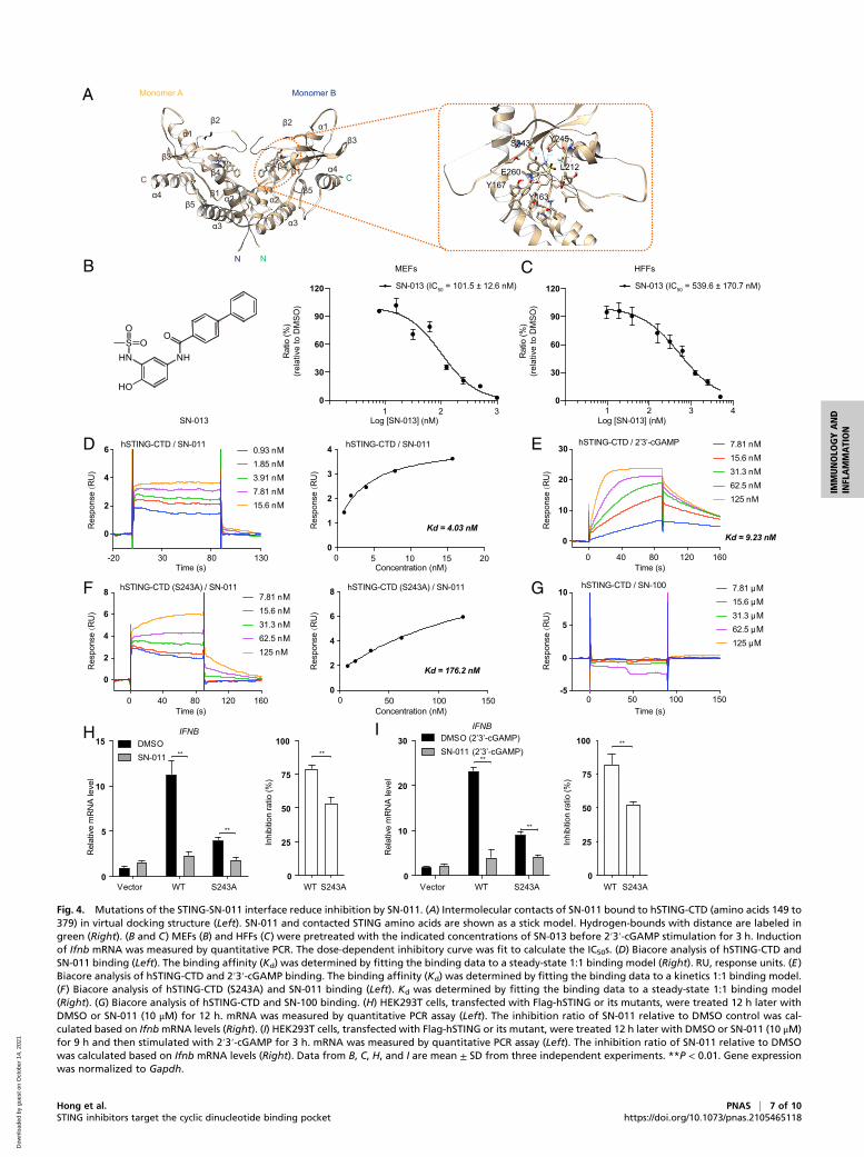
group of SN-011 stacked against the aromatic ring of Tyr167 andwas further stabilized by Glu-260. The phenolic hydroxyl and sul-fonamide groups of SN-011 formed several hydrogen bonds withLeu212, Ser243, and Tyr245. Consistently, substitution of the hy-droxyl group, which interacted with Ser243 via hydrogen bonding,with hydrogen (SN-007) or a methyl group (SN-008), significantlydecreased inhibitory activity (Fig. 1 A and B). As the fluorobenzenegroup in SN-011 did not interact with soluble human STING in thestructure, we synthesized an analog of SN-011, named SN-013,without the fluorobenzene group. SN-013 inhibited 2′3′-cGAMP-induced Ifnb gene expression with IC50 values of 101.5 nM, and539.6 nM in MEF and HFF cells, respectively, which were com-parable to those of SN-011 (Fig. 4 B and C).To confirm that the key STING residues identified by structural
analysis mediate SN-011 binding, surface plasmon resonance (SPR)was used to measure the binding affinity and kinetics between SN-011 and soluble human STING. Soluble human STING bound SN-011 with an affinity (Kd) of 4.03 nM (Fig. 4D), which is lower thanthat of the natural 2′3′-cGAMP ligand (Kd = 9.23 nM) (Fig. 4E).Notably, the binding kinetics of SN-011 (fast association anddissociation) and 2′3′-cGAMP (slow association and dissociation)were markedly different. In contrast to WT STING, the S243Amutant had lower binding affinities to SN-011 (Kd = 176 nM)(Fig. 4F) and the control compound SN-100 did not bind (Fig. 4G).Next, we examined the effect of the mutants on STING signalingand SN-011 inhibition. Overexpression of S243A mutant inducedless IFNB than WT STING both basally and in the presence of2′3′-cGAMP and SN-011 was less active at inhibiting what IFNBwas produced (Fig. 4 H and I). Taken together, these data confirmthat the identified residues in the STING dimer interface mediatebinding of SN-011 and its inhibitory activity.
SN-011 Abrogates SAVI-Associated Mutant STING Signal Activation.To evaluate the therapeutic potential of SN-011, we tested its in vitroability to inhibit the autoactivation of human STING carrying GOFmutations N154S, V155M, G166E, C206Y, R281Q, and R284Glinked to SAVI, an autoinflammatory disease caused by GOF mu-tations in TMEM173, the gene encoding STING (29, 30, 48, 49).WT and SAVI-linked mutated human STING were expressed inHEK293T. Cells expressing the GOF human STING mutantsexhibited high levels of IFNB, CXCL10, and TNFA mRNA,which was strongly inhibited by incubation with SN-011 (SI Ap-pendix, Fig. S3A). SN-011 incubation also markedly decreasedthe recruitment of TBK1 and IRF3 to the STING signalosome,assessed by STING pull-down (SI Appendix, Fig. S3B), and alsoreduced TBK1 and IRF3 phosphorylation induced by STINGSAVI mutants (SI Appendix, Fig. S3C). These GOF mutants spon-taneously oligomerize more than WT STING when expressed inHEK293T cells (13, 14). SN-011 disrupted oligomerization byWT aswell as mutant STING (SI Appendix, Fig. S3D). Notably, SN-011significantly inhibited the spontaneous accumulation of STINGGOF mutants with the Golgi apparatus, suggesting that SN-011locks STING GOF mutants into a conformation that is incom-patible with ER-to-Golgi translocation (SI Appendix, Fig. S4 Aand B). Together, these data suggest that SN-011 could be usedto inhibit STING activation caused by either WT STING ormutant STING in SAVI patients.
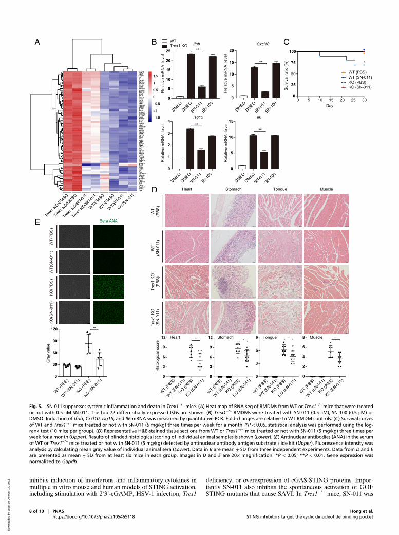
SN-011 Suppresses Systemic Inflammation in Trex1−/− Mice. Accu-mulation of cytosolic self-DNA causes severe and fatal STING-dependent IFN-mediated autoinflammatory disease especially inthe heart and other muscles in Trex1−/− mice (26, 50, 51). To beginto evaluate whether SN-011 could be used therapeutically to inhibitSTING signaling, BMDMs, harvested fromWT and Trex1−/− mice,were treated for 12 h with 500 nM SN-011 or DMSO and thenanalyzed by RNA-sequencing (RNA-seq) (Fig. 5A). SN-011 treatmentsignificantly reduced the IFN signature of Trex1−/− BMDMs, whichwas confirmed by measuring expression of Ifnb and representative
IFN-stimulated genes (ISGs) by quantitative PCR (Cxcl10, Isg15,and Il6) (Fig. 5B). To test the ability of SN-011 to protect Trex1−/−
mice, SN-011 (5 mg/kg) or medium was injected intraperitoneallythree times per week for a month into 4-wk-old WT and Trex1−/−
mice. During the course of treatment, 3 of 10 untreated Trex1−/−
mice died, while none of the 10 mice that received SN-011 died(P = 0.018) (Fig. 5C). At the end of the month, surviving mice werekilled and heart, stomach, tongue, and muscle were analyzed byhematoxylin and eosin (H&E) staining, which showed severemultiorgan inflammation in untreated Trex1−/− mice, which wasreduced by SN-011 treatment (Fig. 5D). Moreover, Ifnb and rep-resentative ISG mRNA levels assessed by quantitative PCR ofRNA isolated from whole tissues at the time of killing were alsosignificantly reduced (SI Appendix, Fig. S5A). Moreover, serumantinuclear antibody was markedly reduced by SN-011 treatment(Fig. 5E). SN-011 also significantly reduced the number of ac-tivated CD69+ CD8 T cells and memory CD44highCD62Llow
CD4 and CD8 T cells to near normal levels in the spleens oftreated Trex1−/− mice (SI Appendix, Fig. S5 B and C). SN-011had no significant effect on the number of splenic activated CD4T cells. Thus, SN-011 strongly reduced inflammation and pro-tected Trex1−/− mice from death.
SN-011 Exhibits Low Cytotoxicity and High Specificity in Comparisonwith H-151. To compare potential cytotoxicity of SN-011 and H-151,cell viability and cell death were examined after adding differentconcentrations of each compound (1 to 10 μM) for 12 to 36 h toMEFs, Tmem173−/−MEFs, and 3T3 cells (SI Appendix, Fig. S6 A–F).SN-011 had no significant effect on cell viability, whereas H-151significantly impaired cell viability and caused cell death (SI Appendix,Fig. S6 A–F). Moreover, H-151 was less specific for STING signalingsince it more potently than SN-011 suppressed Ifnb and inflammatorycytokine (Tnfa and Il6) mRNA induction triggered by STING-independent stimuli, LPS, poly (I:C) and hp-RNA, in L929, HFFs,and Tmem173−/− MEFs (SI Appendix, Fig. S6 G–I). These data in-dicate that SN-011 is a more specific inhibitor of STING-dependentsignaling than H-151 in vitro.Next, we compared the inhibitory effects of SN-011 and H-151
on self-DNA–triggered inflammatory responses in Trex1−/− BMDMsby RNA-seq analysis. Both SN-011 and H-151 suppressed differen-tially expressed (DE) ISG genes to a comparable extent indepen-dently of the false-discovery rate (FDR) cutoff used for the analysis(0.05, 0.01, 0.001) (SI Appendix, Fig. S7A). The number, overlap andmagnitude of significantly down-regulated and up-regulated DE ISGgenes after SN-011 or H-151 treatment were not significantly dif-ferent (SI Appendix, Fig. S7 B–D). To further assess and comparethe efficacy of the two compounds in vivo, 10 mg/kg SN-011 or H-151 was injected intraperitoneally into 6-wk-old Trex1−/− mice dailyfor 2 wk. Ifnb and ISGs expression in the affected tissues of Trex1−/−
mice were comparably suppressed by SN-011 and H-151 (SI Ap-pendix, Fig. S7E). H&E staining also showed comparable amelio-ration of heart inflammation by both compounds (SI Appendix, Fig.S7 F and G). Taken together, these data show that SN-011 and H-151 comparably inhibit STING-mediated inflammation in both cell-based and Trex1−/− mouse models.
DiscussionIn this study, we performed a large-scale virtual screen againstthe STING CDN-binding pocket to identify STING antagonists.Structural optimization identified SN-011 as a potent STINGinhibitor that impedes STING oligomerization, trafficking and ac-tivation in response to cytosolic DNA and markedly decreasesSTING-driven expression of type I IFNs and proinflammatory cy-tokines. SN-011 binds to the CDN-binding pocket with higher af-finity than the endogenous cGAS product 2′3′-cGAMP and locksthe STING dimer in an open, inactive conformation. SN-011inhibits STING-dependent signaling, with IC50 values of ∼100 nMand ∼500 nM for mouse and human cells, respectively. SN-011
6 of 10 | PNAS Hong et al.https://doi.org/10.1073/pnas.2105465118 STING inhibitors target the cyclic dinucleotide binding pocket
Dow
nloa
ded
by g
uest
on
Oct
ober
14,
202
1
A
B C
SN-0131 2 3 4
0
30
60
90
120
Rat
io (%
)(re
lativ
e to
DM
SO)
Rat
io (%
)(re
lativ
e to
DM
SO)
Log [SN-013] (nM)Log [SN-013] (nM)
HFFs
SN-013 (IC50 = 539.6 ± 170.7 nM)
1 2 30
30
60
90
120
MEFs
SN-013 (IC50 = 101.5 ± 12.6 nM)
D
F
E
G
H I
0 5 10 15 200
1
2
3
4
Concentration (nM)
Kd = 4.03 nMKd = 9.23 nM
hSTING-CTD / SN-011 hSTING-CTD / SN-011
hSTING-CTD / SN-100
hSTING-CTD / 2’3’-cGAMP
hSTING-CTD (S243A) / SN-011
Res
pons
e(R
U)
Re s
pons
e( R
U)
Res
pons
e(R
U)
Res
pons
e(R
U)
Res
pons
e(R
U)
Res
pons
e(R
U)
0.93 nM1.85 nM3.91 nM7.81 nM15.6 nM
Time (s) Time (s)
Concentration (nM)Time (s) Time (s)
-20 30 80 130
0
2
4
6
7.81 nM15.6 nM31.3 nM62.5 nM125 nM
hSTING-CTD (S243A) / SN-011
Kd = 176.2 nM
0 50 100 1500
2
4
6
8
0 50 100 150-5
0
5
10 7.81 μM15.6 μM31.3 μM62.5 μM125 μM
0 40 80 120 160
0
10
20
30 7.81 nM15.6 nM31.3 nM62.5 nM125 nM
0 40 80 120 160
0
2
4
6
8
DMSOSN-011
IFNB
****
**
**
Rel
ativ
em
RN
A le
vel
Rel
ativ
em
RN
A le
vel
I nhi
bitio
nra
t io( %
)
Inhi
bitio
nra
tio(%
)
Vector WT S243A0
5
10
15
WT S243A WT S243A0
25
50
75
100
0
25
50
75
DMSO (2’3’-cGAMP)SN-011 (2’3’-cGAMP)
IFNB
**
**
Vector WT S243A0
10
20
30 100
N
Monomer A Monomer B
C
α1
α2 α2
α3α3
α4
α4
β1
β2
β3
β4
β5β5
β4
β3
β2
β1
α1
N
CL212E260
Y245S243
Y167Y163
Fig. 4. Mutations of the STING-SN-011 interface reduce inhibition by SN-011. (A) Intermolecular contacts of SN-011 bound to hSTING-CTD (amino acids 149 to379) in virtual docking structure (Left). SN-011 and contacted STING amino acids are shown as a stick model. Hydrogen-bounds with distance are labeled ingreen (Right). (B and C) MEFs (B) and HFFs (C) were pretreated with the indicated concentrations of SN-013 before 2′3′-cGAMP stimulation for 3 h. Inductionof Ifnb mRNA was measured by quantitative PCR. The dose-dependent inhibitory curve was fit to calculate the IC50s. (D) Biacore analysis of hSTING-CTD andSN-011 binding (Left). The binding affinity (Kd) was determined by fitting the binding data to a steady-state 1:1 binding model (Right). RU, response units. (E)Biacore analysis of hSTING-CTD and 2′3′-cGAMP binding. The binding affinity (Kd) was determined by fitting the binding data to a kinetics 1:1 binding model.(F) Biacore analysis of hSTING-CTD (S243A) and SN-011 binding (Left). Kd was determined by fitting the binding data to a steady-state 1:1 binding model(Right). (G) Biacore analysis of hSTING-CTD and SN-100 binding. (H) HEK293T cells, transfected with Flag-hSTING or its mutants, were treated 12 h later withDMSO or SN-011 (10 μM) for 12 h. mRNA was measured by quantitative PCR assay (Left). The inhibition ratio of SN-011 relative to DMSO control was cal-culated based on IfnbmRNA levels (Right). (I) HEK293T cells, transfected with Flag-hSTING or its mutant, were treated 12 h later with DMSO or SN-011 (10 μM)for 9 h and then stimulated with 2′3′-cGAMP for 3 h. mRNA was measured by quantitative PCR assay (Left). The inhibition ratio of SN-011 relative to DMSOwas calculated based on Ifnb mRNA levels (Right). Data from B, C, H, and I are mean ± SD from three independent experiments. **P < 0.01. Gene expressionwas normalized to Gapdh.
Hong et al. PNAS | 7 of 10STING inhibitors target the cyclic dinucleotide binding pocket https://doi.org/10.1073/pnas.2105465118
IMMUNOLO
GYAND
INFLAMMATION
Dow
nloa
ded
by g
uest
on
Oct
ober
14,
202
1
inhibits induction of interferons and inflammatory cytokines inmultiple in vitro mouse and human models of STING activation,including stimulation with 2′3′-cGAMP, HSV-1 infection, Trex1
deficiency, or overexpression of cGAS-STING proteins. Impor-tantly SN-011 also inhibits the spontaneous activation of GOFSTING mutants that cause SAVI. In Trex1−/− mice, SN-011 was
0 5 10 15 20 25 300
25
50
75
100
Day
Surv
ival
ratio
(%)
KO (PBS)KO (SN-011)
WT (PBS)WT (SN-011)
Cxcl10
Isg15 Il6
B
D
C
E
WTTrex1 KOA
**
****
*
WT
(PBS
)Tr
ex1
KO(P
BS)
Trex
1 KO
(SN
-011
)
Heart
Heart
Stomach
Stomach
Tongue
Tongue
Muscle
Muscle
WT
(SN
-011
)
B
D
A
)
WT/SN-011
WT/SN-011
WT/DMSO
WT/DMSO
Trex1
KO/SN-011
Trex1
KO/SN-011
Trex1
KO/DMSO
Trex1
KO/DMSO
DMSO
SN-011
DMSO
SN-100
DMSO
SN-011
DMSO
SN-100
DMSO
SN-011
DMSO
SN-100
DMSO
SN-011
DMSO
SN-100
Rel
ativ
e m
RN
A le
vel
Rel
ativ
e m
RN
A le
vel
Rel
ativ
e m
RN
A le
vel
Rel
ativ
e m
RN
A le
vel
Ifnb**
0
5
10
15
20
25
0
5
10
15
20
0
1
2
3
4
0
5
10
15
WT (PBS)
WT (SN-01
1)
KO (PBS)
KO (SN-01
1)0
30
60
90
120
Gra
yva
lue
**
WT (PBS)
WT (SN-01
1)
KO (PBS)
KO (SN-01
1)
WT (PBS)
WT (SN-01
1)
KO (PBS)
KO (SN-01
1)
WT (PBS)
WT (SN-01
1)
KO (PBS)
KO (SN-01
1)
WT (PBS)
WT (SN-01
1)
KO (PBS)
KO (SN-01
1)
His
tolo
gica
lsco
re
* ** *
0
3
6
9
12
0
3
6
9
12
0
3
6
9
0
2
4
6
8
Sera ANA
WT(
PBS)
WT(
SN-0
11)
KO(S
N-0
11)
KO(P
BS)
Fig. 5. SN-011 suppresses systemic inflammation and death in Trex1−/− mice. (A) Heat map of RNA-seq of BMDMs fromWT or Trex1−/− mice that were treatedor not with 0.5 μM SN-011. The top 72 differentially expressed ISGs are shown. (B) Trex1−/− BMDMs were treated with SN-011 (0.5 μM), SN-100 (0.5 μM) orDMSO. Induction of Ifnb, Cxcl10, Isg15, and Il6 mRNA was measured by quantitative PCR. Fold-changes are relative to WT BMDM controls. (C) Survival curvesof WT and Trex1−/− mice treated or not with SN-011 (5 mg/kg) three times per week for a month. *P < 0.05, statistical analysis was performed using the log-rank test (10 mice per group). (D) Representative H&E-stained tissue sections from WT or Trex1−/− mice treated or not with SN-011 (5 mg/kg) three times perweek for a month (Upper). Results of blinded histological scoring of individual animal samples is shown (Lower). (E) Antinuclear antibodies (ANA) in the serumof WT or Trex1−/− mice treated or not with SN-011 (5 mg/kg) detected by antinuclear antibody antigen substrate slide kit (Upper). Fluorescence intensity wasanalysis by calculating mean gray value of individual animal sera (Lower). Data in B are mean ± SD from three independent experiments. Data from D and Eare presented as mean ± SD from at least six mice in each group. Images in D and E are 20× magnification. *P < 0.05; **P < 0.01. Gene expression wasnormalized to Gapdh.
8 of 10 | PNAS Hong et al.https://doi.org/10.1073/pnas.2105465118 STING inhibitors target the cyclic dinucleotide binding pocket
Dow
nloa
ded
by g
uest
on
Oct
ober
14,
202
1
well tolerated, strongly inhibited hallmarks of inflammation andautoimmunity, and prevented death. Collectively, these studiessuggest that SN-011 is an attractive lead compound for devel-oping drugs to inhibit STING-dependent signaling.In comparison, other recently identified STING antagonists
are either inactive (C-176, C-178) or have low bioactivity againsthuman STING (NO2-FAs, Astin C, compound 18) (42–45). Com-pound 18 binds deep in the cleft of the human STING dimer, butwith an IC50 of ∼11 μM, about 20-fold higher than SN-011, evenafter optimization by analyzing SARs (44). The most promisingreported STING antagonist, H-151, is a covalent inhibitor thatbinds to a reactive Cys91 in the transmembrane domain of STINGto inhibit activation-induced palmitoylation (42). SN-011 andH-151 equivalently inhibit STING in mouse cell lines, with an IC50value of ∼100 nM. Elevated ISG expression in Trex1−/− BMDMswere similarly ameliorated by in vitro exposure to SN-011 or H-151.When SN-011 and H-151 were administered at the same dose(10 mg/kg), both compounds comparably suppressed inflammationin the affected tissues (heart, stomach, tongue, and muscle) inTrex1−/− mice. Future preclinical experiments to compare phar-macokinetics and dynamics, efficacy, and safety of both drugsadministered back-to-back at different doses in mouse AGS andSAVI models will be needed to further explore the relative ad-vantages of each compound. Like other Cys-reactive drugs, whichare notoriously nonspecific and covalently bind to unpaired Cys inmany proteins, H-151 likely has more off-target activities than SN-011 (52, 53). H-151 significantly impaired cell viability and inducedcell death while SN-011 was not cytotoxic. In addition, H-151showed potent inhibitory effects on TLR- or RIG-I-mediated sig-naling in cell-based assays compared to SN-011. All of these dataindicate that SN-011 shows better specificity and safety than H-151.The ability to modify these lead compounds to optimize STINGinhibition and their pharmacological properties will ultimatelydetermine which compounds can lead to effective new treatmentsof STING-mediated diseases.Insights into the structural basis for SN-011 activity was provided
by docking analysis. In the docking structure, SN-011 bound to theSTING dimer interface in the CDN-binding pocket to lock STINGin an inactive open conformation. One molecule of SN-011 boundto each STING monomer with an extended binding site along thepocket. SPR confirmed the stoichiometry of STING monomer toSN-011 as 1:1 with a binding dissociation constant of 4.03 nM. Bycomparison, compound 18 lies in the bottom of the cleft of theSTING dimer (PDB ID code 6MXE) in a 1:2 binding stoichiom-etry with an affinity of ∼50 μM for the binding of the first moleculeand an affinity of ∼2 nM for the binding of the second molecule(44). This spatial and affinity difference may explain the differentinhibitory activities between SN-011 and compound 18. Moreover,the interaction between STING and SN-011 over an extendedbinding site is mediated by residues Tyr167, Leu212, Ser243, Tyr245,and Glu260 in STING, all of which are evolutionarily conservedbetween species. Tyr167 and Glu260 are essential for CDN bindingand Ser243 and Tyr245 are in the lid region, which involves lid for-mation upon 2′3′-cGAMP binding (11, 54, 55). Thus, association ofSN-011 with STING leads to the occupation of the binding pocketand steric hindrance that inhibits the approach of CDNs to STING.Our SAR analysis of SN-001 to SN-011 revealed that the phe-
nolic hydroxyl group is essential for bioactivity and substitutionwith a hydrogen, methyl, or oxymethyl group impairs inhibitoryactivity, but the fluorobenzene group is dispensable. The virtualdocking structure of SN-011 with the STING dimer showed bi-phenyl group stacking at the bottom of the STING dimer pocket,which may allow us to improve SN-011 by introducing a linker toconnect benzene rings of two molecules of SN-011 (to yield asingle dimeric SN-011, di-SN-011). di-SN-011 may enhance bind-ing affinity and bioactivity as was shown for the STING agonistsdiABZI and MSA-2 (56, 57). Future work will seek to develop
more potent SN-011 analogs and define their pharmacokineticsand safety as a prerequisite for clinical development.Aberrant activation of cGAS-STING signaling by self-DNA
causes severe autoimmune diseases, such as AGS, SLE, andother lupus-like diseases, called interferonopathies because theyare largely caused by chronic overexpression of type I IFNs (58,59). Trex1−/− mice are an experimental AGS and SLE model.SN-011 treatment inhibited the interferon signature in Trex1−/−
BMDMs and alleviated the systemic autoinflammatory pheno-type, extending mouse survival. Future studies will examine theinhibitory effect of SN-011 on peripheral blood mononuclear cellsfrom AGS and SLE patients to evaluate the therapeutic potentialof SN-011 and its derivatives on human diseases that require bonemarrow transplantation or are inadequately treated with currenttherapies. SN-011 also blocked the type I IFN and proinflammatorygene expression induced by SAVI-associated STING mutants bypreventing spontaneous STING auto-oligomerization and activa-tion. Future structural studies will further probe how SN-011 in-hibits GOF mutants of STING. It will also be worth testing thein vivo and ex vivo efficacy of SN-011 in SAVI-related mousemodels (V154M or N153S) and primary cells from SAVI patients.As of now, no standard immunosuppressive and curative approachhas been identified to treat SAVI. Inhibiting type I IFN signalingwith Janus kinase (JAK) inhibitors (e.g., tofacitinib, ruxolitinib, andbaricitinib) controls disease progression in a subset of SAVI patients(29, 49, 60). However, lung inflammation, myeloid cell expansion,and T cell cytopenia developed in STINGN153S or V154M knockinmouse models of SAVI do not depend on IRF3 or IFN signaling,suggesting that NF-κB–regulated induction of inflammatory genes isalso important in disease pathogenesis (61–63). A STING inhibitor,such as SN-011, that blocks the first step of STING activation is anespecially attractive drug candidate for treating SAVI, in comparisonto JAK inhibitors. Previous studies showed that STING inhibitortool compounds control tissue inflammation by blocking both IFNand NF-κB signaling, inhibit STING-triggered apoptosis of T cells,monocytes and endothelial cells of SAVI patients and reduce therisk of opportunistic infection by not inhibiting other pattern rec-ognition receptor pathways (59, 64). SN-011 specifically inhibitedboth IFN and inflammatory downstream consequences of STINGactivation without interfering with other innate immune pathways.Our findings support drug development of STING inhibitors to treatSTING-associated autoimmune diseases.
Materials and MethodsDetails of the materials and methods, including immunoblot analysis, immuno-fluorescence, real-time RT-PCR, animal experiments, flow cytometry, biotinpull-down assay, molecular docking and virtual screening, SPR, RNA-seq ex-periments, and chemical synthesis are presented in SI Appendix, SI Materialsand Methods. All animal experiments were performed in accordance with theNIH Guide for the Care and Use of Laboratory Animals (65), with the ap-proval of the Center for New Drug Safety Evaluation and Research, ChinaPharmaceutical University.
Data Availability. All original RNA-seq data are deposited in the Gene ExpressionOmnibus database, https://www.ncbi.nlm.nih.gov/geo (accession no. GSE143830).All other study data are included in the article and SI Appendix.
ACKNOWLEDGMENTS. We thank Dr. Tomas Lindahl and Dr. Deborah Barnes(Cancer Research UK, London) and Dr. Nan Yan (University of Texas South-western Medical Center) for kindly sharing Trex1+/− mice; Dr. Min-Hua Luo(Wuhan Institute of Virology) for kindly sharing the human foreskin fibroblastcell line; Dr. Wentao Qiao (Nankai University), Dr. Chunfu Zheng (Suzhou Uni-versity), Dr. Youcun Qian (Institute of Health Sciences), and Dr. Yong Yang(China Pharmaceutical University) for providing reagents; Xiaonan Ma fromthe Cellular and Molecular Biology Center of China Pharmaceutical Universityfor providing technical assistance for using the Carl Zeiss LSM700; andWei Jiangfrom the State Key Laboratory of Natural Medicines of China PharmaceuticalUniversity for providing technical assistance for surface plasmon resonance datacollection. This study was supported by the National Key R&D Program of China(2016YFA0501800), the National Natural Science Foundation of China(31730018, 81672029, 3173000227, 31470428), Project Program of StateKey Laboratory of Natural Medicines, China Pharmaceutical University No.
Hong et al. PNAS | 9 of 10STING inhibitors target the cyclic dinucleotide binding pocket https://doi.org/10.1073/pnas.2105465118
IMMUNOLO
GYAND
INFLAMMATION
Dow
nloa
ded
by g
uest
on
Oct
ober
14,
202
1
SKLNMZZ202002), the Jiangsu Innovative and Entrepreneurial Talents Program,the National New Drug Innovation Major Project of China (2017ZX09309027),and the Fund of Chinese Academy of Sciences (XDA09030301-4, Hundred
Talents Program). Z.H. is sponsored by China Postdoctoral Research Program(2019TQ0356). C.L. is sponsored by National Natural Science Foundation ofChina for the Youth (31800724).
1. C. Soni, B. Reizis, DNA as a self-antigen: Nature and regulation. Curr. Opin. Immunol.55, 31–37 (2018).
2. L. Sun, J. Wu, F. Du, X. Chen, Z. J. Chen, Cyclic GMP-AMP synthase is a cytosolic DNAsensor that activates the type I interferon pathway. Science 339, 786–791 (2013).
3. A. Ablasser et al., cGAS produces a 2′-5′-linked cyclic dinucleotide second messengerthat activates STING. Nature 498, 380–384 (2013).
4. P. Gao et al., Cyclic [G(2′,5′)pA(3′,5′)p] is the metazoan second messenger produced byDNA-activated cyclic GMP-AMP synthase. Cell 153, 1094–1107 (2013).
5. J. Wu et al., Cyclic GMP-AMP is an endogenous second messenger in innate immunesignaling by cytosolic DNA. Science 339, 826–830 (2013).
6. H. Ishikawa, G. N. Barber, STING is an endoplasmic reticulum adaptor that facilitatesinnate immune signalling. Nature 455, 674–678 (2008).
7. L. Jin et al., MPYS, a novel membrane tetraspanner, is associated with major histo-compatibility complex class II and mediates transduction of apoptotic signals. Mol.Cell. Biol. 28, 5014–5026 (2008).
8. B. Zhong et al., The adaptor protein MITA links virus-sensing receptors to IRF3 tran-scription factor activation. Immunity 29, 538–550 (2008).
9. W. Sun et al., ERIS, an endoplasmic reticulum IFN stimulator, activates innate immunesignaling through dimerization. Proc. Natl. Acad. Sci. U.S.A. 106, 8653–8658 (2009).
10. D. L. Burdette et al., STING is a direct innate immune sensor of cyclic di-GMP. Nature478, 515–518 (2011).
11. X. Zhang et al., Cyclic GMP-AMP containing mixed phosphodiester linkages is anendogenous high-affinity ligand for STING. Mol. Cell 51, 226–235 (2013).
12. P. Gao et al., Structure-function analysis of STING activation by c[G(2′,5′)pA(3′,5′)p]and targeting by antiviral DMXAA. Cell 154, 748–762 (2013).
13. S. L. Ergun, D. Fernandez, T. M. Weiss, L. Li, STING polymer structure reveals mech-anisms for activation, hyperactivation, and inhibition. Cell 178, 290–301.e10 (2019).
14. G. Shang, C. Zhang, Z. J. Chen, X. C. Bai, X. Zhang, Cryo-EM structures of STING revealits mechanism of activation by cyclic GMP-AMP. Nature 567, 389–393 (2019).
15. Y. H. Huang, X. Y. Liu, X. X. Du, Z. F. Jiang, X. D. Su, The structural basis for the sensingand binding of cyclic di-GMP by STING. Nat. Struct. Mol. Biol. 19, 728–730 (2012).
16. K. Mukai et al., Activation of STING requires palmitoylation at the Golgi. Nat. Com-mun. 7, 11932 (2016).
17. C. Zhang et al., Structural basis of STING binding with and phosphorylation by TBK1.Nature 567, 394–398 (2019).
18. B. Zhao et al., A conserved PLPLRT/SD motif of STING mediates the recruitment andactivation of TBK1. Nature 569, 718–722 (2019).
19. S. Liu et al., Phosphorylation of innate immune adaptor proteins MAVS, STING, andTRIF induces IRF3 activation. Science 347, aaa2630 (2015).
20. B. Zhao et al., Structural basis for concerted recruitment and activation of IRF-3 byinnate immune adaptor proteins. Proc. Natl. Acad. Sci. U.S.A. 113, E3403–E3412(2016).
21. T. Abe, G. N. Barber, Cytosolic-DNA-mediated, STING-dependent proinflammatorygene induction necessitates canonical NF-κB activation through TBK1. J. Virol. 88,5328–5341 (2014).
22. Z. Ma, B. Damania, The cGAS-STING defense pathway and its counteraction by viruses.Cell Host Microbe 19, 150–158 (2016).
23. K. J. Mackenzie et al., cGAS surveillance of micronuclei links genome instability toinnate immunity. Nature 548, 461–465 (2017).
24. S. F. Bakhoum et al., Chromosomal instability drives metastasis through a cytosolicDNA response. Nature 553, 467–472 (2018).
25. A. N. Theofilopoulos, D. H. Kono, R. Baccala, The multiple pathways to autoimmunity.Nat. Immunol. 18, 716–724 (2017).
26. A. Gall et al., Autoimmunity initiates in nonhematopoietic cells and progresses vialymphocytes in an interferon-dependent autoimmune disease. Immunity 36, 120–131(2012).
27. D. Gao et al., Activation of cyclic GMP-AMP synthase by self-DNA causes autoimmunediseases. Proc. Natl. Acad. Sci. U.S.A. 112, E5699–E5705 (2015).
28. Y. J. Crow, N. Manel, Aicardi-Goutières syndrome and the type I interferonopathies.Nat. Rev. Immunol. 15, 429–440 (2015).
29. Y. Liu et al., Activated STING in a vascular and pulmonary syndrome. N. Engl. J. Med.371, 507–518 (2014).
30. N. Jeremiah et al., Inherited STING-activating mutation underlies a familial inflam-matory syndrome with lupus-like manifestations. J. Clin. Invest. 124, 5516–5520(2014).
31. Z. Dou et al., Cytoplasmic chromatin triggers inflammation in senescence and cancer.Nature 550, 402–406 (2017).
32. S. Glück et al., Innate immune sensing of cytosolic chromatin fragments through cGASpromotes senescence. Nat. Cell Biol. 19, 1061–1070 (2017).
33. E. L. Heipertz, J. Harper, W. E. Walker, STING and TRIF contribute to mouse sepsis,depending on severity of the disease model. Shock 47, 621–631 (2017).
34. L. Zeng et al., ALK is a therapeutic target for lethal sepsis. Sci. Transl. Med. 9,eaan5689 (2017).
35. D. A. Sliter et al., Parkin and PINK1 mitigate STING-induced inflammation. Nature 561,258–262 (2018).
36. Q. Zhao, Y. Wei, S. J. Pandol, L. Li, A. Habtezion, STING signaling promotes inflam-mation in experimental acute pancreatitis. Gastroenterology 154, 1822–1835.e2(2018).
37. J. Bai et al., DsbA-L prevents obesity-induced inflammation and insulin resistance bysuppressing the mtDNA release-activated cGAS-cGAMP-STING pathway. Proc. Natl.Acad. Sci. U.S.A. 114, 12196–12201 (2017).
38. X. Luo et al., Expression of STING is increased in liver tissues from patients with NAFLDand promotes macrophage-mediated hepatic inflammation and fibrosis in mice.Gastroenterology 155, 1971–1984.e4 (2018).
39. S. Benmerzoug et al., STING-dependent sensing of self-DNA drives silica-induced lunginflammation. Nat. Commun. 9, 5226 (2018).
40. Q. Chen et al., Carcinoma-astrocyte gap junctions promote brain metastasis bycGAMP transfer. Nature 533, 493–498 (2016).
41. K. W. Chung et al., Mitochondrial damage and activation of the STING pathway leadto renal inflammation and fibrosis. Cell Metab. 30, 784–799.e5 (2019).
42. S. M. Haag et al., Targeting STING with covalent small-molecule inhibitors. Nature559, 269–273 (2018).
43. S. Li et al., The cyclopeptide Astin C specifically inhibits the innate immune CDN sensorSTING. Cell Rep. 25, 3405–3421.e7 (2018).
44. T. Siu et al., Discovery of a novel cGAMP competitive ligand of the inactive form ofSTING. ACS Med. Chem. Lett. 10, 92–97 (2018).
45. A. L. Hansen et al., Nitro-fatty acids are formed in response to virus infection and arepotent inhibitors of STING palmitoylation and signaling. Proc. Natl. Acad. Sci. U.S.A.115, E7768–E7775 (2018).
46. S. Ouyang et al., Structural analysis of the STING adaptor protein reveals a hydro-phobic dimer interface and mode of cyclic di-GMP binding. Immunity 36, 1073–1086(2012).
47. T. Sterling, J. J. Irwin, ZINC 15—Ligand discovery for everyone. J. Chem. Inf. Model. 55,2324–2337 (2015).
48. I. Melki et al., Disease-associated mutations identify a novel region in human STINGnecessary for the control of type I interferon signaling. J. Allergy Clin. Immunol. 140,543–552.e5 (2017).
49. N. König et al., Familial chilblain lupus due to a gain-of-function mutation in STING.Ann. Rheum. Dis. 76, 468–472 (2017).
50. M. A. Lee-Kirsch et al., Mutations in the gene encoding the 3′-5′ DNA exonucleaseTREX1 are associated with systemic lupus erythematosus. Nat. Genet. 39, 1065–1067(2007).
51. D. B. Stetson, J. S. Ko, T. Heidmann, R. Medzhitov, Trex1 prevents cell-intrinsic initi-ation of autoimmunity. Cell 134, 587–598 (2008).
52. E. Weerapana et al., Quantitative reactivity profiling predicts functional cysteines inproteomes. Nature 468, 790–795 (2010).
53. J. Singh, R. C. Petter, T. A. Baillie, A. Whitty, The resurgence of covalent drugs. Nat.Rev. Drug Discov. 10, 307–317 (2011).
54. G. Shang et al., Crystal structures of STING protein reveal basis for recognition of cyclicdi-GMP. Nat. Struct. Mol. Biol. 19, 725–727 (2012).
55. C. Shu, G. Yi, T. Watts, C. C. Kao, P. Li, Structure of STING bound to cyclic di-GMPreveals the mechanism of cyclic dinucleotide recognition by the immune system. Nat.Struct. Mol. Biol. 19, 722–724 (2012).
56. J. M. Ramanjulu et al., Design of amidobenzimidazole STING receptor agonists withsystemic activity. Nature 564, 439–443 (2018).
57. B. S. Pan et al., An orally available non-nucleotide STING agonist with antitumoractivity. Science 369, eaba6098 (2020).
58. J. T. Crowl, E. E. Gray, K. Pestal, H. E. Volkman, D. B. Stetson, Intracellular nucleic aciddetection in autoimmunity. Annu. Rev. Immunol. 35, 313–336 (2017).
59. A. Ablasser, Z. J. Chen, cGAS in action: Expanding roles in immunity and inflamma-tion. Science 363, eaat8657 (2019).
60. M. L. Frémond et al., Efficacy of the Janus kinase 1/2 inhibitor ruxolitinib in thetreatment of vasculopathy associated with TMEM173-activating mutations in 3 chil-dren. J. Allergy Clin. Immunol. 138, 1752–1755 (2016).
61. J. D. Warner et al., STING-associated vasculopathy develops independently of IRF3 inmice. J. Exp. Med. 214, 3279–3292 (2017).
62. D. Bouis et al., Severe combined immunodeficiency in stimulator of interferon genes(STING) V154M/wild-type mice. J. Allergy Clin. Immunol. 143, 712–725.e5 (2019).
63. M. Motwani et al., Hierarchy of clinical manifestations in SAVI N153S and V154Mmouse models. Proc. Natl. Acad. Sci. U.S.A. 116, 7941–7950 (2019).
64. M. F. Gulen et al., Signalling strength determines proapoptotic functions of STING.Nat. Commun. 8, 427 (2017).
65. National Research Council, Guide for the Care and Use of Laboratory Animals(National Academies Press, Washington, DC, ed. 8, 2011).
10 of 10 | PNAS Hong et al.https://doi.org/10.1073/pnas.2105465118 STING inhibitors target the cyclic dinucleotide binding pocket
Dow
nloa
ded
by g
uest
on
Oct
ober
14,
202
1