Embed Size (px)
Citation preview
Structure and Function of Skeletal Muscle
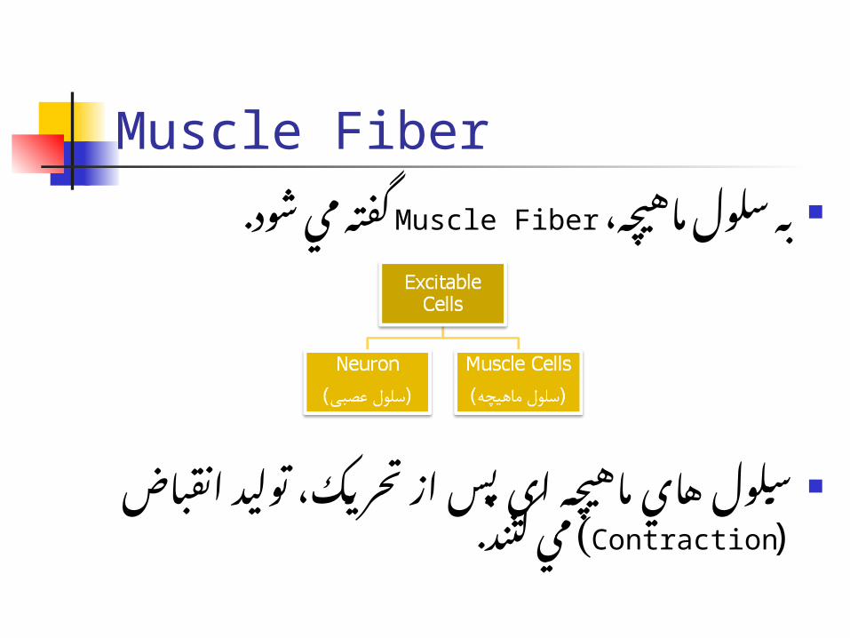
Muscle Fiber ،ماهيچ�ه س�لول مي Muscle Fiberبه گفت�ه
شود.
سلول ه�اي ماهيچ�ه اي پس از تحري�ک، تولي�د( مي کنند. Contractionانقباض )
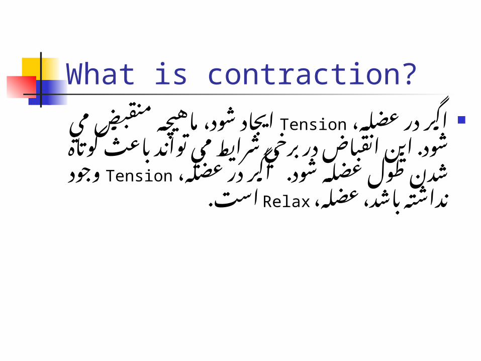
What is contraction? ،اگ�ر در عض�لهTension ايج�اد ش�ود، ماهيچ�ه
ب�رخي در انقب�اض اين ش�ود. مي منقبض ب�اعث کوت�اه ش�دن ط�ول توان�د ش�رايط مي
ش�ود. عض�له، عض�له در وج�ود Tensionاگ�ر است.Relaxنداشته باشد، عضله،
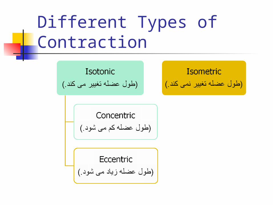
Different Types of Contraction
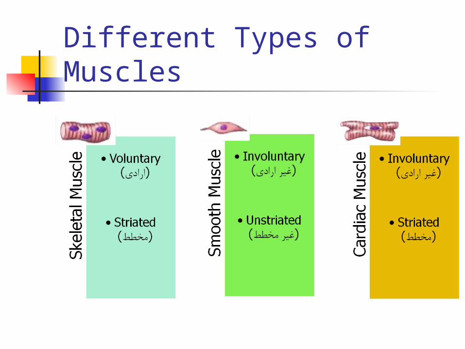
Different Types of Muscles
Skeletal Muscle Human body contains over 400
skeletal muscles 40-50% of total body weight
Functions of skeletal muscle Force production for locomotion and
breathing Force production for postural support Heat production during cold stress
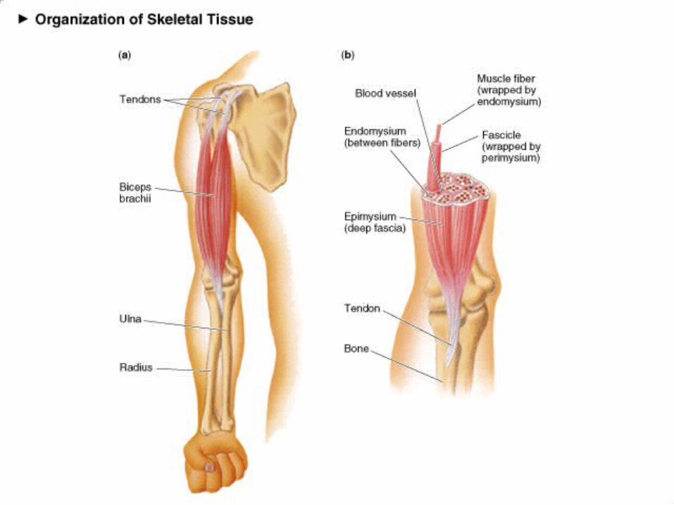
Structure of Skeletal Muscle:Connective Tissue Covering Epimysium
Surrounds entire muscle Perimysium
Surrounds bundles of muscle fibers Fascicles
Endomysium Surrounds individual muscle fibers
Structure of Skeletal Muscle:Microstructure Sarcolemma
Muscle cell membrane Myofibrils
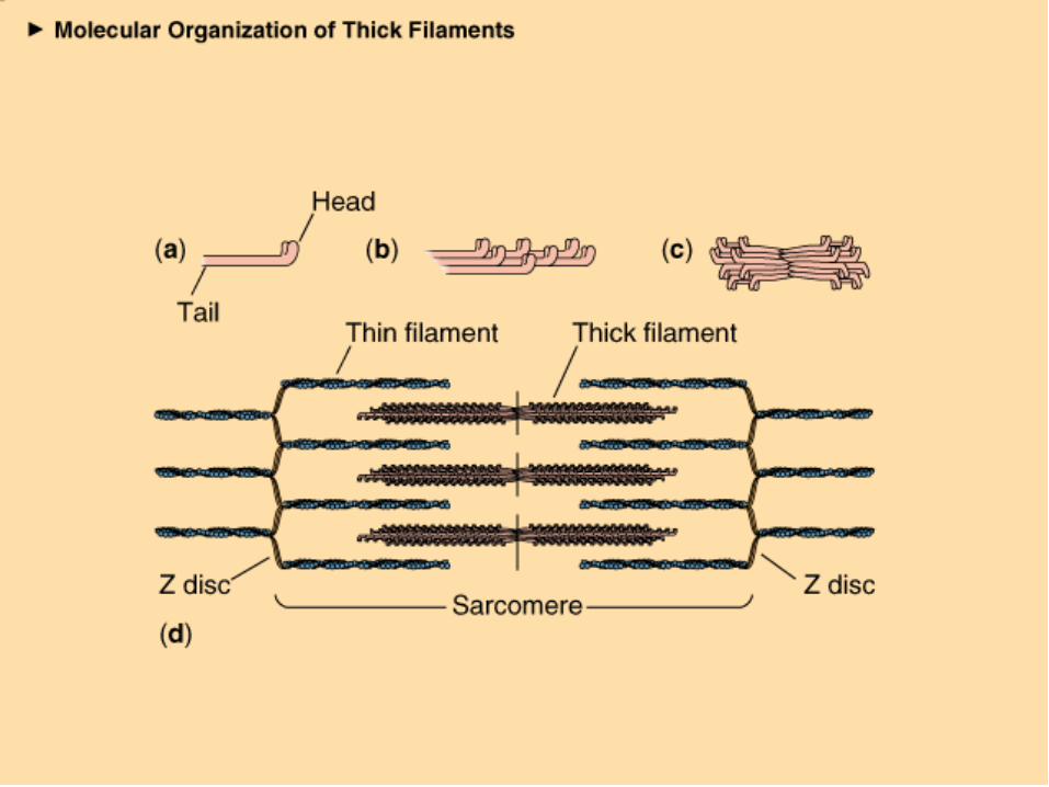
Threadlike strands within muscle fibers Actin )thin filament(
Troponin Tropomyosin
Myosin )thick filament(
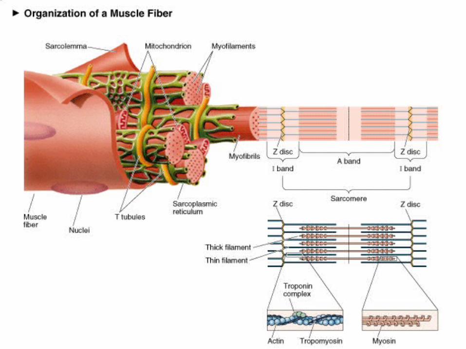
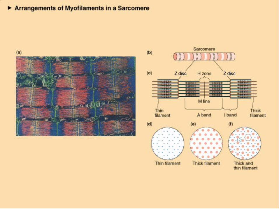
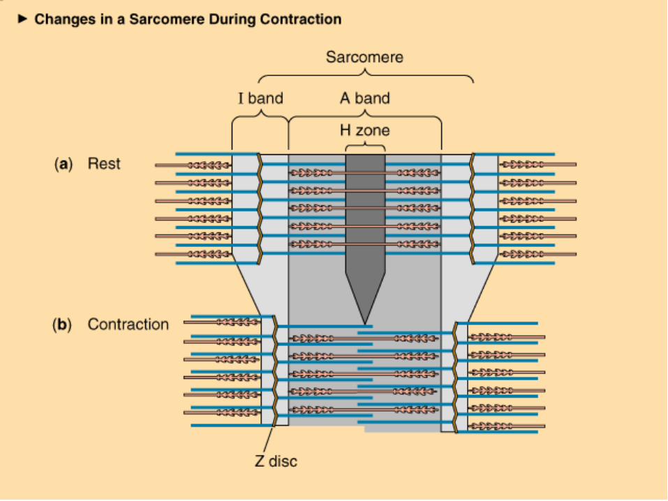
Structure of Skeletal Muscle:The Sarcomere Further divisions of myofibrils
Z-line A-band I-band
Within the sarcoplasm Sarcoplasmic reticulum
Storage sites for calcium Transverse tubules Terminal cisternae
The Neuromuscular Junction Site where motor neuron meets the
muscle fiber Separated by gap called the neuromuscular
cleft Motor end plate
Pocket formed around motor neuron by sarcolemma
Acetylcholine is released from the motor neuron Causes an end-plate potential )EPP(
Depolarization of muscle fiber
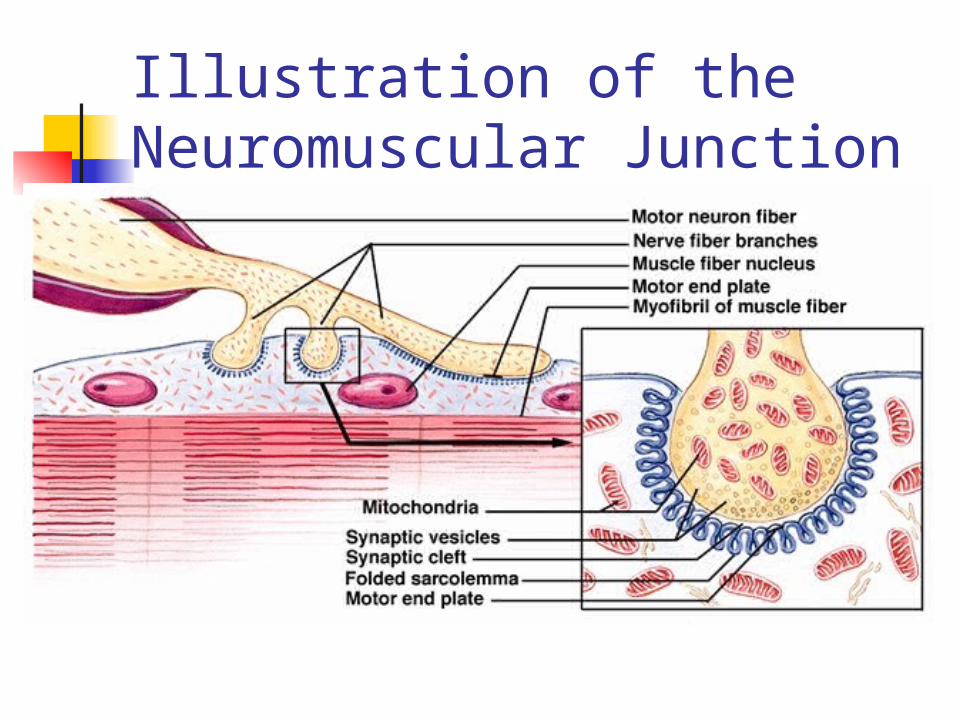
Illustration of the Neuromuscular Junction
Motor Unit Single motorneuron & muscle
fibers it innervates Eye muscles – 1:1 muscle/nerve
ratio Hamstrings – 300:1 muscle/nerve
ratio
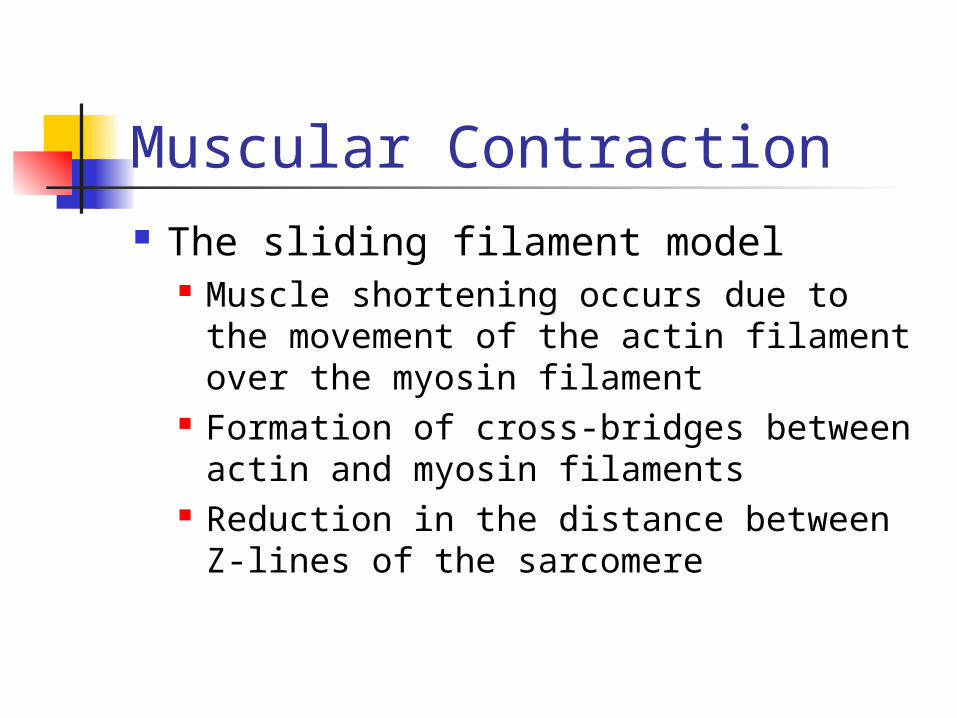
Muscular Contraction The sliding filament model
Muscle shortening occurs due to the movement of the actin filament over the myosin filament
Formation of cross-bridges between actin and myosin filaments
Reduction in the distance between Z-lines of the sarcomere
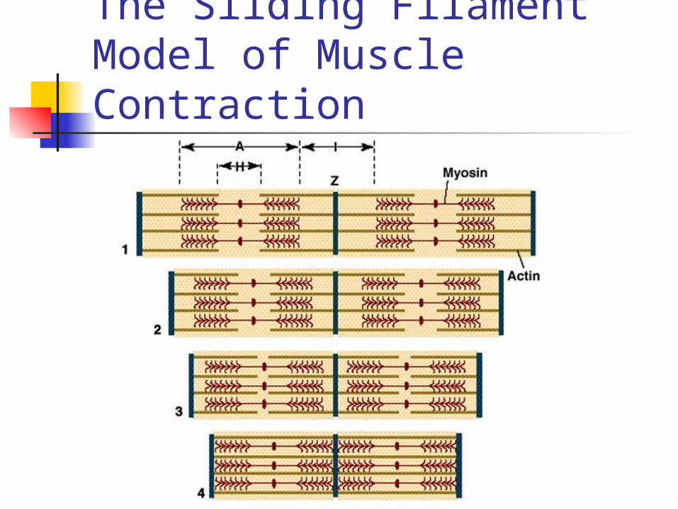
The Sliding Filament Model of Muscle Contraction
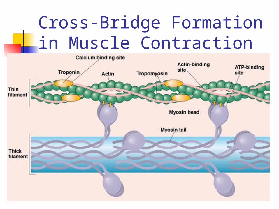
Cross-Bridge Formation in Muscle Contraction
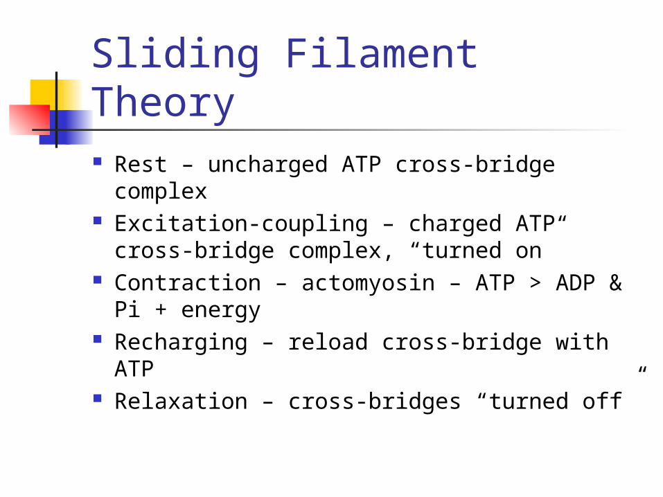
Sliding Filament Theory Rest – uncharged ATP cross-bridge
complex Excitation-coupling – charged ATP cross-
bridge complex, “turned on” Contraction – actomyosin – ATP > ADP
& Pi + energy Recharging – reload cross-bridge with
ATP Relaxation – cross-bridges “turned off”
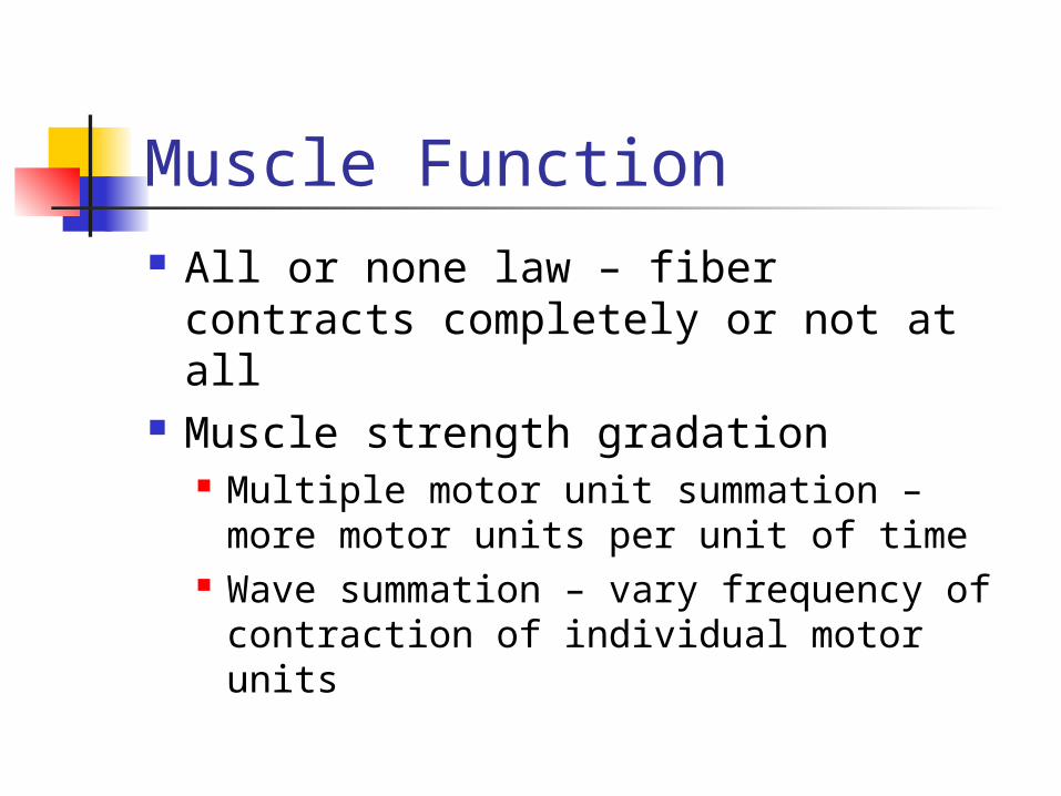
Muscle Function All or none law – fiber contracts
completely or not at all Muscle strength gradation
Multiple motor unit summation – more motor units per unit of time
Wave summation – vary frequency of contraction of individual motor units
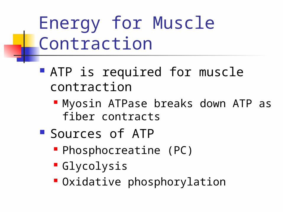
Energy for Muscle Contraction ATP is required for muscle
contraction Myosin ATPase breaks down ATP as
fiber contracts Sources of ATP
Phosphocreatine )PC( Glycolysis Oxidative phosphorylation

Sources of ATP for Muscle Contraction
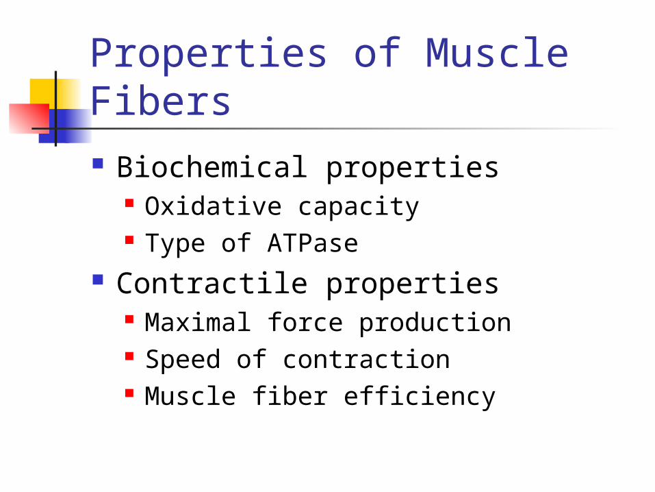
Properties of Muscle Fibers Biochemical properties
Oxidative capacity Type of ATPase
Contractile properties Maximal force production Speed of contraction Muscle fiber efficiency
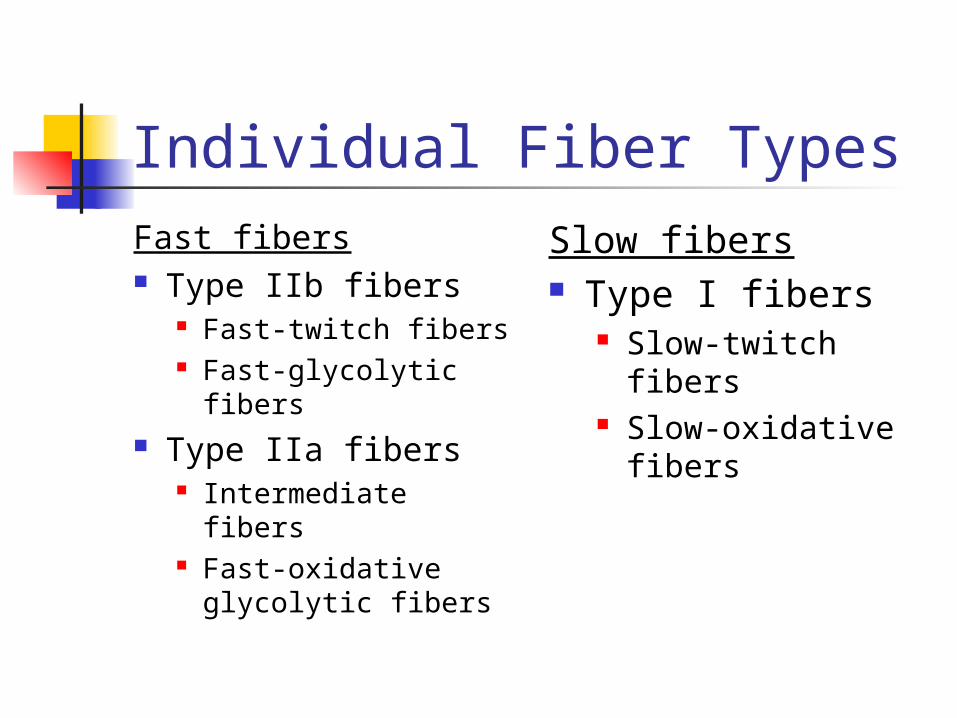
Individual Fiber TypesFast fibers Type IIb fibers
Fast-twitch fibers Fast-glycolytic
fibers Type IIa fibers
Intermediate fibers Fast-oxidative
glycolytic fibers
Slow fibers Type I fibers
Slow-twitch fibers Slow-oxidative
fibers
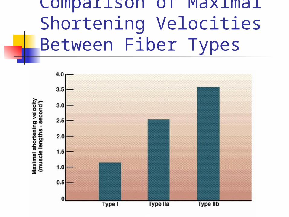
Comparison of Maximal Shortening Velocities Between Fiber Types
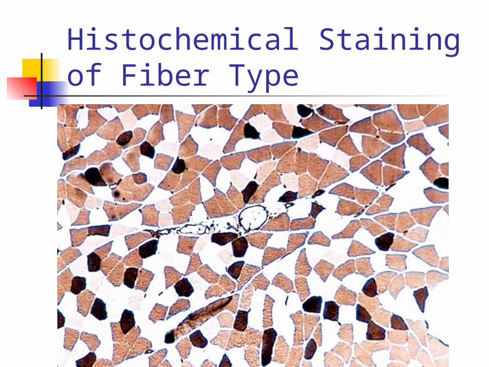
Histochemical Staining of Fiber Type
Fiber Types and Performance Power athletes
Sprinters Possess high percentage of fast fibers
Endurance athletes Distance runners Have high percentage of slow fibers
Others Weight lifters and nonathletes Have about 50% slow and 50% fast fibers
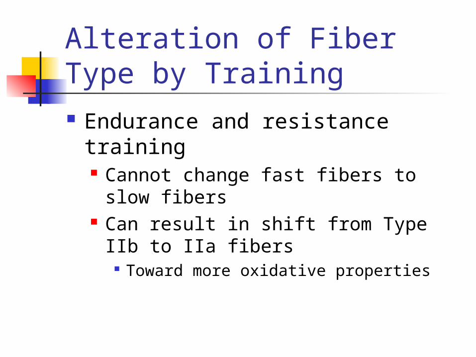
Alteration of Fiber Type by Training Endurance and resistance training
Cannot change fast fibers to slow fibers
Can result in shift from Type IIb to IIa fibers
Toward more oxidative properties
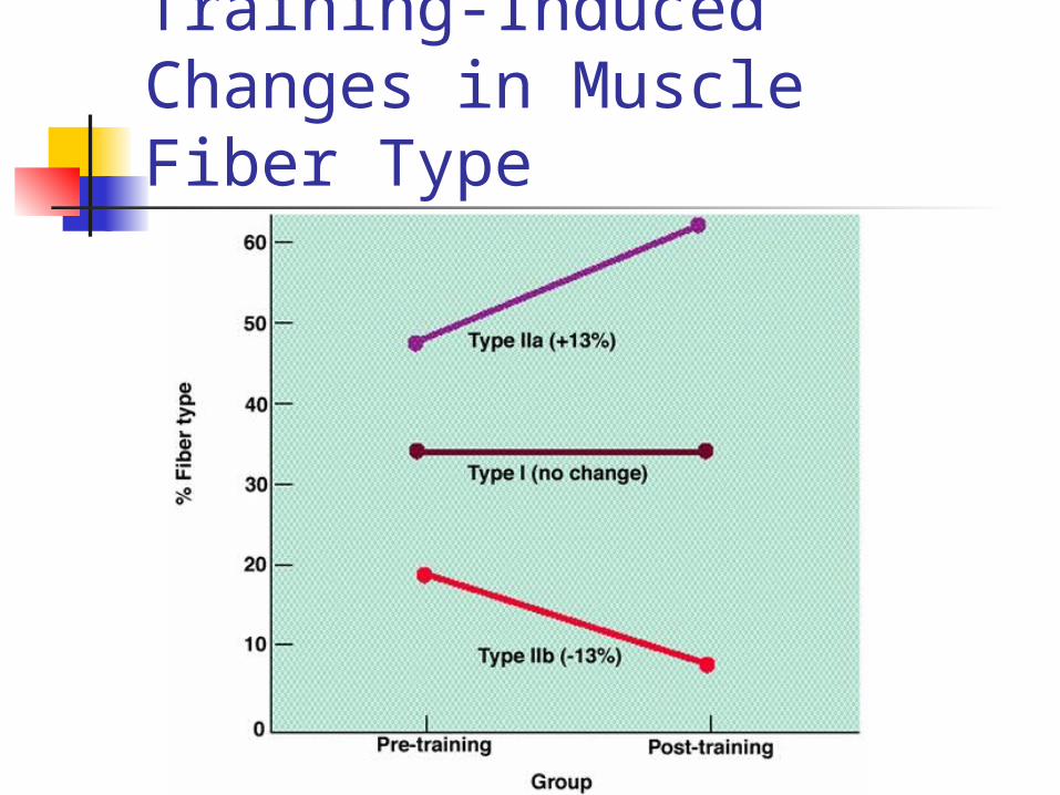
Training-Induced Changes in Muscle Fiber Type
Hypertrophy and Hyperplasia Increase in size Increase in
number
Age-Related Changes in Skeletal Muscle Aging is associated with a loss of
muscle mass Rate increases after 50 years of age
Regular exercise training can improve strength and endurance Cannot completely eliminate the age-
related loss in muscle mass
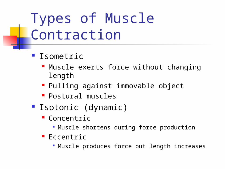
Types of Muscle Contraction Isometric
Muscle exerts force without changing length Pulling against immovable object Postural muscles
Isotonic )dynamic( Concentric
Muscle shortens during force production Eccentric
Muscle produces force but length increases
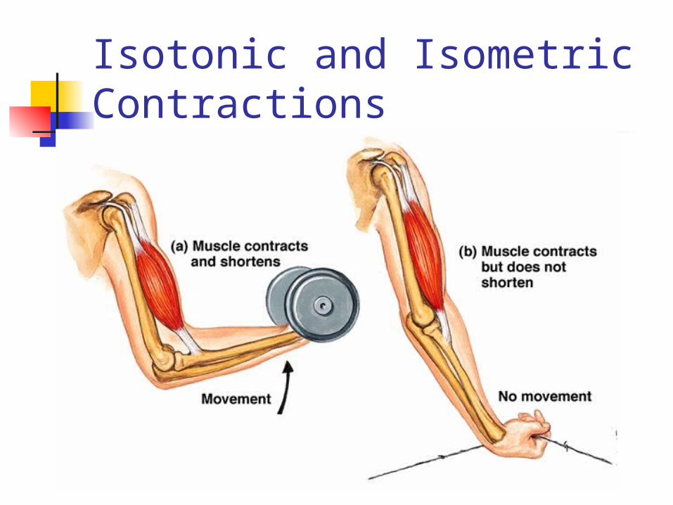
Isotonic and Isometric Contractions
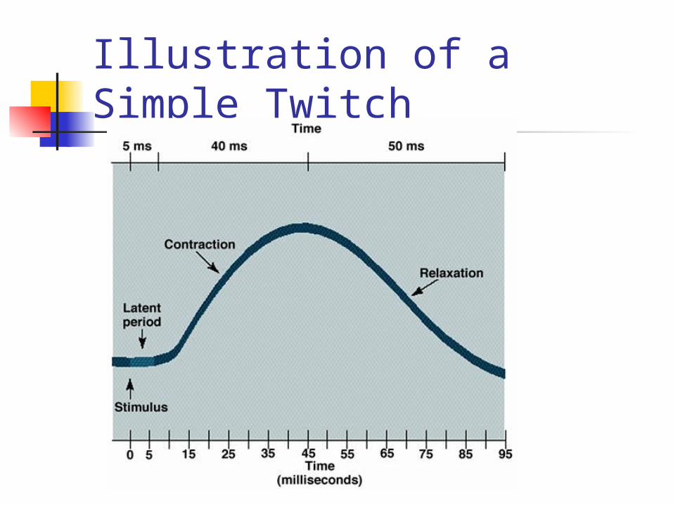
Illustration of a Simple Twitch
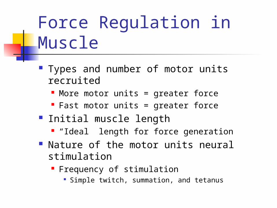
Force Regulation in Muscle Types and number of motor units recruited
More motor units = greater force Fast motor units = greater force
Initial muscle length “Ideal” length for force generation
Nature of the motor units neural stimulation Frequency of stimulation
Simple twitch, summation, and tetanus
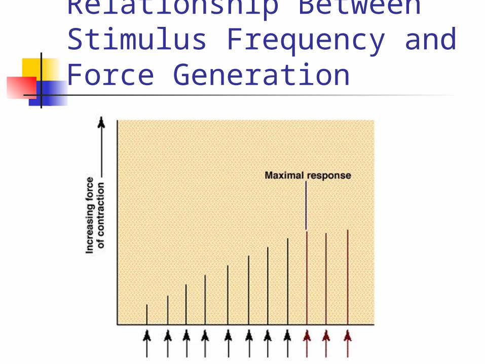
Relationship Between Stimulus Frequency and Force Generation
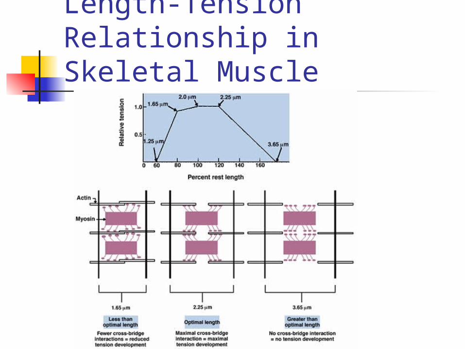
Length-Tension Relationship in Skeletal Muscle
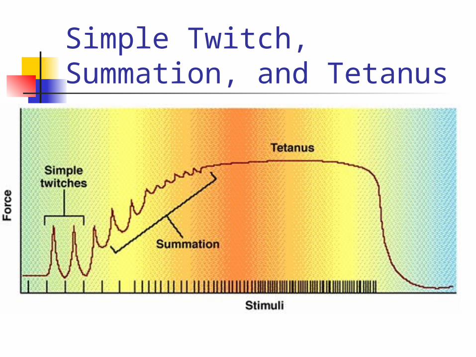
Simple Twitch, Summation, and Tetanus
Force-Velocity Relationship At any absolute force the speed of
movement is greater in muscle with higher percent of fast-twitch fibers
The maximum velocity of shortening is greatest at the lowest force True for both slow and fast-twitch fibers
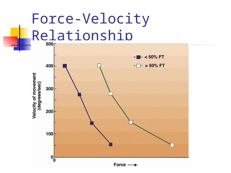
Force-Velocity Relationship
Force-Power Relationship At any given velocity of movement
the power generated is greater in a muscle with a higher percent of fast-twitch fibers
The peak power increases with velocity up to movement speed of 200-300 degrees•second-1
Force decreases with increasing movement speed beyond this velocity
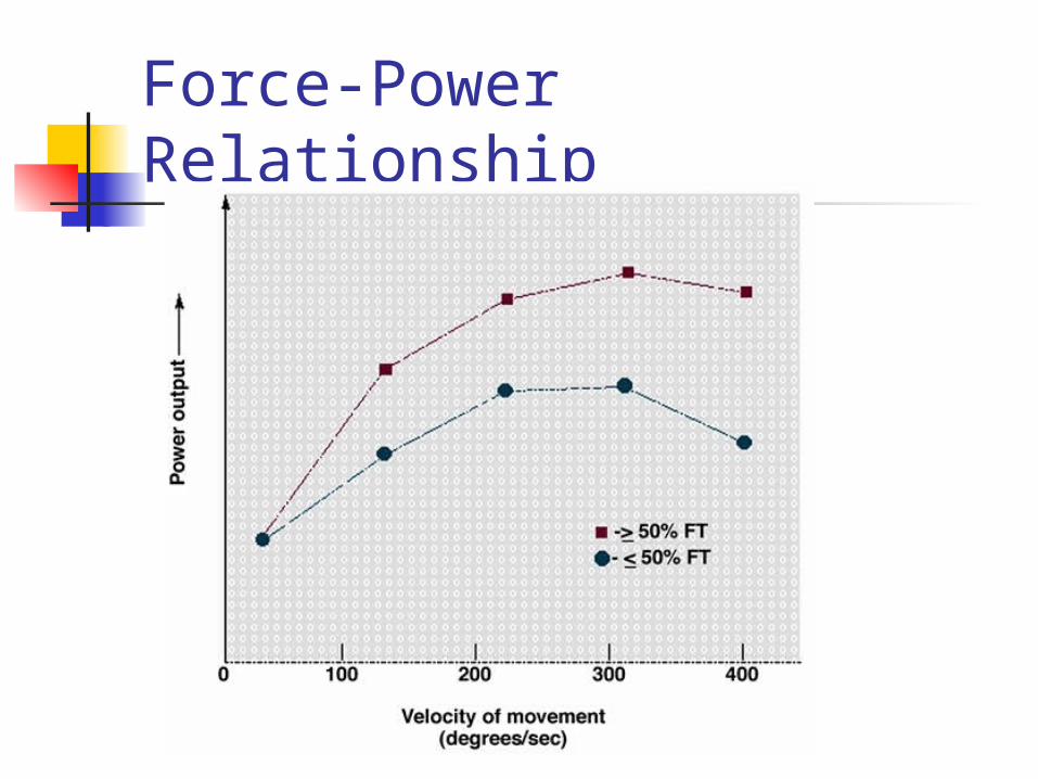
Force-Power Relationship
Receptors in Muscle Muscle spindle
Detect dynamic and static changes in muscle length
Stretch reflex Stretch on muscle causes reflex contraction
Golgi tendon organ )GTO( Monitor tension developed in muscle Prevents damage during excessive force
generation Stimulation results in reflex relaxation of muscle
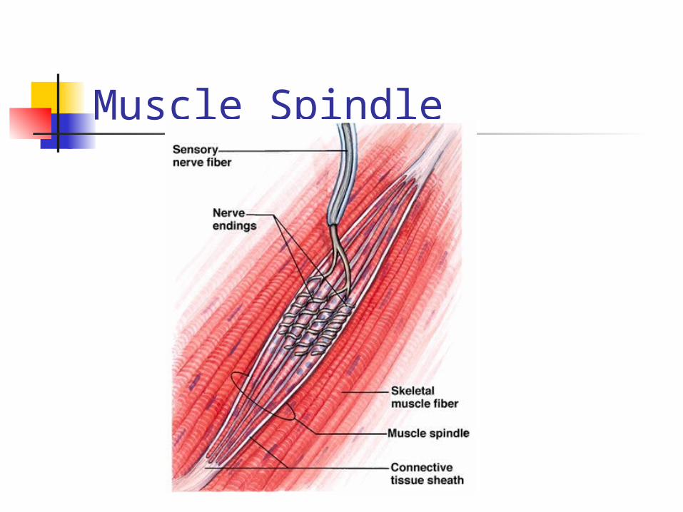
Muscle Spindle
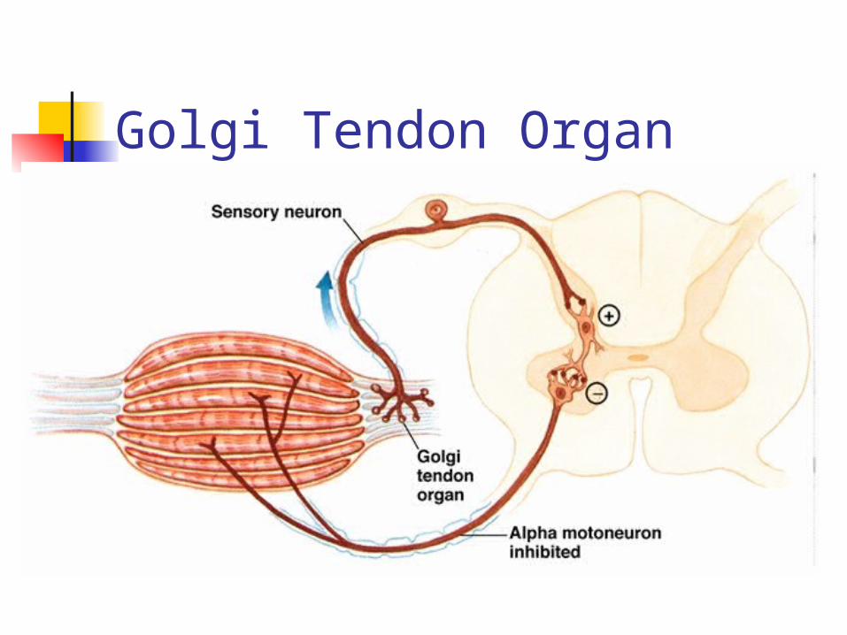
Golgi Tendon Organ