Embed Size (px)
Citation preview
..............................................................
Structure of the inositol 1,4,5-trisphosphate receptor bindingcore in complex with its ligandIvan Bosanac*, Jean-Rene Alattia*, Tapas K. Mal*, Jenny Chan*,Susanna Talarico*, Frances K. Tong*, Kit I. Tong*, Fumio Yoshikawa†,Teiichi Furuichi†, Miwako Iwai‡, Takayuki Michikawa‡§,Katsuhiko Mikoshiba†‡§ & Mitsuhiko Ikura*
* Division of Molecular and Structural Biology, Ontario Cancer Institute andDepartment of Medical Biophysics, University of Toronto, 610 University Avenue,Toronto, Ontario, Canada M5G 2M9† Laboratory for Developmental Neurobiology and Molecular Neurogenesis, BrainScience Institute, RIKEN, Saitama 351-0198, Japan‡ Department of Molecular Neurobiology, Institute of Medical Science, Universityof Tokyo, Tokyo 108-8639, Japan§ Calcium Oscillation Project, ICORP, Japan Science and Technology Corporation(JST), Tokyo, 108-0071, Japan.............................................................................................................................................................................
In a variety of cells, the Ca21 signalling process is mediated by theendoplasmic-reticulum-membrane-associated Ca21 releasechannel, inositol 1,4,5-trisphosphate (InsP3) receptor (InsP3R)1.Being ubiquitous and present in organisms ranging fromhumans to Caenorhabditis elegans, InsP3R has a vital role inthe control of cellular and physiological processes as diverse ascell division, cell proliferation, apoptosis, fertilization, develop-ment, behaviour, memory and learning2. Mouse type I InsP3R(InsP3R1), found in high abundance in cerebellar Purkinje cells,
is a polypeptide with three major functionally distinct regions:the amino-terminal InsP3-binding region, the central modula-tory region and the carboxy-terminal channel region2. Here wepresent a 2.2-A crystal structure of the InsP3-binding core ofmouse InsP3R1 in complex with InsP3. The asymmetric, boom-erang-like structure consists of an N-terminal b-trefoil domainand a C-terminal a-helical domain containing an ‘armadillorepeat’-like fold. The cleft formed by the two domains exposesa cluster of arginine and lysine residues that coordinate the threephosphoryl groups of InsP3. Putative Ca21-binding sites areidentified in two separate locations within the InsP3-bindingcore.
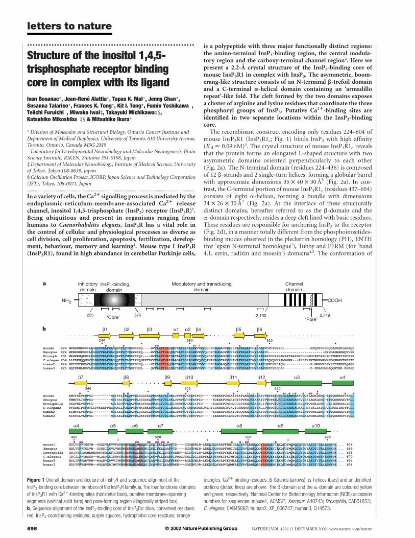
The recombinant construct encoding only residues 224–604 ofmouse InsP3R1 (InsP3R1c; Fig. 1) binds InsP3 with high affinity(K d < 0.09 nM)3. The crystal structure of mouse InsP3R1c revealsthat the protein forms an elongated L-shaped structure with twoasymmetric domains oriented perpendicularly to each other(Fig. 2a). The N-terminal domain (residues 224–436) is composedof 12 b-strands and 2 single-turn helices, forming a globular barrelwith approximate dimensions 35 £ 40 £ 30 A3 (Fig. 2a). In con-trast, the C-terminal portion of mouse InsP3R1c (residues 437–604)consists of eight a-helices, forming a bundle with dimensions34 £ 26 £ 30 A3 (Fig. 2a). At the interface of these structurallydistinct domains, hereafter referred to as the b-domain and thea-domain respectively, resides a deep cleft lined with basic residues.These residues are responsible for anchoring InsP3 to the receptor(Fig. 2d), in a manner totally different from the phosphoinositides-binding modes observed in the pleckstrin homology (PH), ENTH(for ‘epsin N-terminal homologue’), Tubby and FERM (for ‘band4.1, ezrin, radixin and moesin’) domains4,5. The conformation of
Figure 1 Overall domain architecture of InsP3R and sequence alignment of the
InsP3-binding core between members of the InsP3R family. a, The four functional domains
of InsP3R1 with Ca2þ-binding sites (horizontal bars), putative membrane-spanning
segments (vertical solid bars) and pore-forming region (diagonally striped box).
b, Sequence alignment of the InsP3-binding core of InsP3Rs: blue, conserved residues;
red, InsP3-coordinating residues; purple squares, hydrophobic core residues; orange
triangles, Ca2þ-binding residues. b-Strands (arrows), a-helices (bars) and unidentified
portions (dotted lines) are shown. The b-domain and the a-domain are coloured yellow
and green, respectively. National Center for Biotechnology Information (NCBI) accession
numbers for sequences: mouse1, ACMSIT; Xenopus, A40743; Drosophila, CAB51853;
C. elegans, CAB45862; human2, XP_006747; human3, Q14573.
letters to nature
NATURE | VOL 420 | 12 DECEMBER 2002 | www.nature.com/nature696 © 2002 Nature Publishing Group
the receptor-bound InsP3 molecule is similar to that found in thecrystal structures of isolated InsP3 and InsP3 bound to a PHdomain6,7. However, the phosphorus–phosphorus distances differbetween this study (8.4, 7.2 and 5.0 A for P1/P4, P1/P5 and P4/P5pairs) and others (8.1, 6.9 and 4.1 A for isolated InsP3, and 8.2, 7.2and 4.3 A for InsP3 bound to the PH domain)8, with P4 and P5separation being significant.
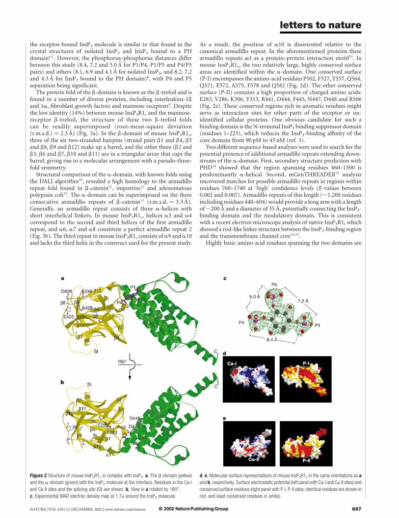
The protein fold of the b-domain is known as the b-trefoil and isfound in a number of diverse proteins, including interleukins-1band 1a, fibroblast growth factors and mannose receptors9. Despitethe low identity (14%) between mouse InsP3R1c and the mannose-receptor b-trefoil, the structure of these two b-trefoil foldscan be readily superimposed (root-mean-square deviation(r.m.s.d.) ¼ 2.3 A) (Fig. 3a). In the b-domain of mouse InsP3R1c,three of the six two-stranded hairpins (strand pairs b1 and b4, b5and b8, b9 and b12) make up a barrel, and the other three (b2 andb3, b6 and b7, b10 and b11) are in a triangular array that caps thebarrel, giving rise to a molecular arrangement with a pseudo-three-fold symmetry.
Structural comparison of the a-domain, with known folds usingthe DALI algorithm10, revealed a high homology to the armadillorepeat fold found in b-catenin11, importins12 and adenomatouspolyposis coli13. The a-domain can be superimposed on the threeconsecutive armadillo repeats of b-catenin11 (r.m.s.d. ¼ 3.3 A).Generally, an armadillo repeat consists of three a-helices withshort interhelical linkers. In mouse InsP3R1c, helices a3 and a4correspond to the second and third helices of the first armadillorepeat, and a6, a7 and a8 constitute a perfect armadillo repeat 2(Fig. 3b). The third repeat in mouse InsP3R1c consists of a9 and a10and lacks the third helix in the construct used for the present study.
As a result, the position of a10 is disoriented relative to thecanonical armadillo repeat. In the aforementioned proteins thesearmadillo repeats act as a protein–protein interaction motif13. Inmouse InsP3R1c, the two relatively large, highly conserved surfaceareas are identified within the a-domain. One conserved surface(P-I) encompasses the amino-acid residues P502, F527, Y557, Q564,Q571, E572, A575, F578 and Q582 (Fig. 2d). The other conservedsurface (P-II) contains a high proportion of charged amino acids:E283, V286, K306, Y313, R441, D444, F445, N447, D448 and R506(Fig. 2e). These conserved regions rich in aromatic residues mightserve as interaction sites for other parts of the receptor or un-identified cellular proteins. One obvious candidate for such abinding domain is the N-terminal InsP3 binding suppressor domain(residues 1–225), which reduces the InsP3-binding affinity of thecore domain from 90 pM to 45 nM (ref. 3).
Two different sequence-based analyses were used to search for thepotential presence of additional armadillo repeats extending down-stream of the a-domain. First, secondary structure prediction withPHD14 showed that the region spanning residues 460–1500 ispredominantly a-helical. Second, mGenTHREADER15 analysisuncovered matches for possible armadillo repeats in regions withinresidues 760–1740 at ‘high’ confidence levels (E-values between0.002 and 0.007). Armadillo repeats of this length (,1,200 residuesincluding residues 440–604) would provide a long arm with a lengthof ,200 A and a diameter of 35 A, potentially connecting the InsP3-binding domain and the modulatory domain. This is consistentwith a recent electron microscope analysis of native InsP3R1, whichshowed a rod-like linker structure between the InsP3-binding regionand the transmembrane channel core16,17.
Highly basic amino acid residues spanning the two domains are
Figure 2 Structure of mouse InsP3R1c in complex with InsP3. a, The b-domain (yellow)
and the a-domain (green) with the InsP3 molecule at the interface. Residues in the Ca-I
and Ca-II sites and the splicing site (SI) are shown. b, View in a rotated by 1808.
c, Experimental MAD electron density map at 1.7j around the InsP3 molecule.
d, e, Molecular surface representations of mouse InsP3R1c in the same orientations as a
and b, respectively. Surface electrostatic potential (left panel with Ca-I and Ca-II sites) and
conserved surface residues (right panel with P-I, P-II sites; identical residues are shown in
red, and least-conserved residues in white).
letters to nature
NATURE | VOL 420 | 12 DECEMBER 2002 | www.nature.com/nature 697© 2002 Nature Publishing Group
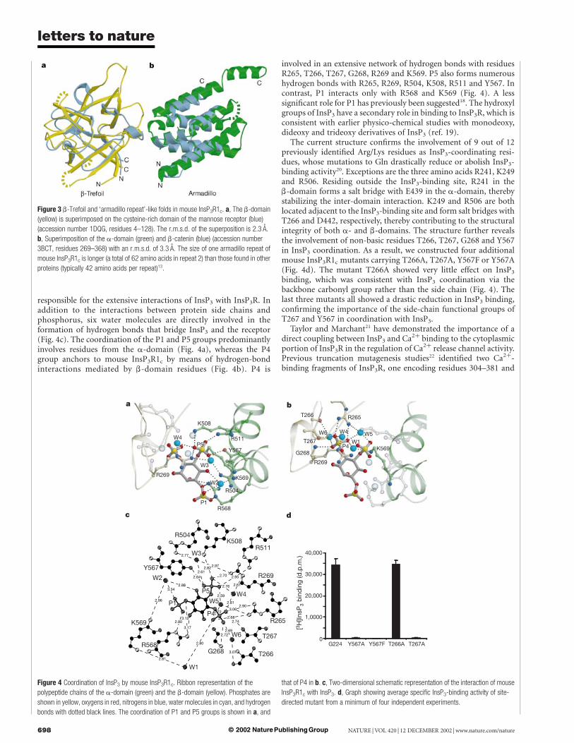
responsible for the extensive interactions of InsP3 with InsP3R. Inaddition to the interactions between protein side chains andphosphorus, six water molecules are directly involved in theformation of hydrogen bonds that bridge InsP3 and the receptor(Fig. 4c). The coordination of the P1 and P5 groups predominantlyinvolves residues from the a-domain (Fig. 4a), whereas the P4group anchors to mouse InsP3R1c by means of hydrogen-bondinteractions mediated by b-domain residues (Fig. 4b). P4 is
involved in an extensive network of hydrogen bonds with residuesR265, T266, T267, G268, R269 and K569. P5 also forms numeroushydrogen bonds with R265, R269, R504, K508, R511 and Y567. Incontrast, P1 interacts only with R568 and K569 (Fig. 4). A lesssignificant role for P1 has previously been suggested18. The hydroxylgroups of InsP3 have a secondary role in binding to InsP3R, which isconsistent with earlier physico-chemical studies with monodeoxy,dideoxy and trideoxy derivatives of InsP3 (ref. 19).
The current structure confirms the involvement of 9 out of 12previously identified Arg/Lys residues as InsP3-coordinating resi-dues, whose mutations to Gln drastically reduce or abolish InsP3-binding activity20. Exceptions are the three amino acids R241, K249and R506. Residing outside the InsP3-binding site, R241 in theb-domain forms a salt bridge with E439 in the a-domain, therebystabilizing the inter-domain interaction. K249 and R506 are bothlocated adjacent to the InsP3-binding site and form salt bridges withT266 and D442, respectively, thereby contributing to the structuralintegrity of both a- and b-domains. The structure further revealsthe involvement of non-basic residues T266, T267, G268 and Y567in InsP3 coordination. As a result, we constructed four additionalmouse InsP3R1c mutants carrying T266A, T267A, Y567F or Y567A(Fig. 4d). The mutant T266A showed very little effect on InsP3
binding, which was consistent with InsP3 coordination via thebackbone carbonyl group rather than the side chain (Fig. 4). Thelast three mutants all showed a drastic reduction in InsP3 binding,confirming the importance of the side-chain functional groups ofT267 and Y567 in coordination with InsP3.
Taylor and Marchant21 have demonstrated the importance of adirect coupling between InsP3 and Ca2þ binding to the cytoplasmicportion of InsP3R in the regulation of Ca2þ release channel activity.Previous truncation mutagenesis studies22 identified two Ca2þ-binding fragments of InsP3R, one encoding residues 304–381 and
Figure 3 b-Trefoil and ‘armadillo repeat’-like folds in mouse InsP3R1c. a, The b-domain
(yellow) is superimposed on the cysteine-rich domain of the mannose receptor (blue)
(accession number 1DQG, residues 4–128). The r.m.s.d. of the superposition is 2.3 A.
b, Superimposition of the a-domain (green) and b-catenin (blue) (accession number
3BCT, residues 269–368) with an r.m.s.d. of 3.3 A. The size of one armadillo repeat of
mouse InsP3R1c is longer (a total of 62 amino acids in repeat 2) than those found in other
proteins (typically 42 amino acids per repeat)13.
Figure 4 Coordination of InsP3 by mouse InsP3R1c. Ribbon representation of the
polypeptide chains of the a-domain (green) and the b-domain (yellow). Phosphates are
shown in yellow, oxygens in red, nitrogens in blue, water molecules in cyan, and hydrogen
bonds with dotted black lines. The coordination of P1 and P5 groups is shown in a, and
that of P4 in b. c, Two-dimensional schematic representation of the interaction of mouse
InsP3R1c with InsP3. d, Graph showing average specific InsP3-binding activity of site-
directed mutant from a minimum of four independent experiments.
letters to nature
NATURE | VOL 420 | 12 DECEMBER 2002 | www.nature.com/nature698 © 2002 Nature Publishing Group
the other corresponding to residues 378–450. More recently, resi-dues E425, D426, E428, D442 and D444 have been shown to beessential for Ca2þ coordination23. These residues are part of the twosurface acidic clusters identified in the present crystal structure ofmouse InsP3R1c. The first site, Ca-I, is located in the b-domain andconsists of residues E246, E425, D426 and E428 (Fig. 2d). Thesecond site, Ca-II, located across the two domains, is composed ofresidues E283, E285, D444 and D448 (Fig. 2e). Interestingly, Ca2þ-binding site Ca-II overlaps with the conserved region P-II,suggesting that the binding of Ca2þ to this site is conformationallycoupled with the aforementioned protein–protein interactioninvolving other protein domain(s). This finding, together withrecent electron microscope studies of InsP3R16,17 and previousbiochemical studies21, leads to a tempting speculation on theCa2þ–InsP3 coupling mechanism required for channel activation.The role of binding of InsP3 to the core domain (residues 226–576)might include the release of a conformational constraint thatprevents Ca2þ from binding to the receptor. The N-terminalInsP3 binding suppressor region (residues 1–225) might be directlyinvolved in this negative regulation of Ca2þ binding to the receptor,in addition to the modulation of InsP3 binding affinity20. It isequally possible that some other part of the InsP3R or an unidenti-fied cellular protein is involved in this InsP3–Ca2þ couplingmechanism. A
MethodsOverexpression and purification of InsP3Rc
The mouse type 1 InsP3R protein fragment encompassing the ligand-binding core,residues 224–604, was expressed as a glutathione S-transferase (GST) fusion protein(GST–mouse InsP3R1c) by polymerase chain reaction, cloning and sequencing asdescribed previously20,24. The protein was expressed in Escherichia coli strain BL21-CodonPlus-RIL (Stratagene) at 15 8C for ,20 h by induction with 0.5 mM isopropyl b-D-thiogalactopyranoside24. GST–mouse InsP3R1c was first purified with glutathione–Sepharose (Pharmacia) followed by digestion overnight with thrombin to remove GSTfrom the cutting buffer (20 mM Tris-HCl pH 8.4, 200 mM NaCl, 10% (v/v) glycerol and20 mM dithiothreitol) at 4 8C with constant mixing. Cation-exchange chromatography(Fractogel EMD SO3
2 resin; EM Industries Inc.) and size-exclusion chromatography(Superdex 75; Pharmacia) were then performed to remove thrombin and any otherimpurities. Selenomethionine-labelled protein was produced by the technique involvingthe inhibition of methionine biosynthesis25 and was confirmed by electrospray massspectrometry. The purified protein was concentrated to 20 mg ml21 in a crystallizationbuffer (15 mM Tris-HCl pH 8.0, 300 mM Na2SO4, 2 mM tris-(2-carboxyethyl)phosphineand 50% molar excess InsP3).
Crystallization and data collectionInitial crystals of mouse InsP3R1c were produced at 295 K by the hanging-drop method byadding 2 ml of protein complex to 2 ml of well solution (100 mM MES pH 6.0, 20%polyethylene glycol (PEG) 3350, 0.2 M CH5NO3 and 2–5% dioxane). Crystals of thecomplex grew as extensive clusters of rods within 2 weeks. Series of microseedings wererequired to obtain single rod-type crystals with dimensions of 0.05 £ 0.1 £ 0.4 mm3.Crystals were flash-cooled in crystallization buffer supplemented with 25% PEG 400.Multiwavelength anomalous dispersion (MAD) and native data were collected at 100 K ona BL44XU beam line at the SPring-8 Synchrotron facility and were processed withd*TREK26. Crystals belonged to space group C2221 with cell dimensions a ¼ 44.2 A,b ¼ 90.3 A and c ¼ 207.9 A, and contained one complex in the asymmetric unit.Radiation damage to the crystal prevented completion of the data set at l3 (remotewavelength) (Supplementary Table 1).
Structure determination and refinementThe initial positions of four out of six expected selenium atoms were determined by Solve27
and refined with SHARP28 to a figure of merit of 0.42. The position of the two missingselenomethionine residues (M224 and M535) were never identified because one is locatedat the N terminus of the protein fragment and the other in the a7/a8 loop region, which isnot visible in the present model. Density modification and solvent flattening with theprogram CNS29 increased the overall figure of merit to 0.93. The experimental map hadcontinuous electron density for most of mouse InsP3R1c; the InsP3 ligand was easilyidentifiable. Model building was performed with the program O30 and refinement withCNS29. The final model contained residues 236–288, 302–319, 351–372, 387–528 and 546–602, with 92.7% of residues in the most favoured regions of the Ramachandran plot and noresidues in the disallowed region. The b-domain contains three crystallographicallyinvisible loops, between strands b4/b5, b6/b7 and b8/b9 with the loop encompassingresidues 320 to 350 (b6/b7) having a splicing site I. In total, 2,522 atoms with a meantemperature factor at 34.1 A2 were observed: 2,356 protein atoms ðkBl¼ 35:2 �A2Þ; 24ligand atoms ðkBl¼ 28:6 �A2Þ and 142 solvent atoms ðkBl¼ 36:2 �A2Þ: The R cryst and R free ofthe final model were 22.4 and 25.0, respectively.
InsP3 binding assaySite-directed mutagenesis of Y567A, Y567F, T266A and T267A was introduced into mouseInsP3R1c, and protein fragments were expressed in E. coli. Preparation of soluble proteinfrom the cell pellets was performed as described elsewhere24. Soluble protein (30 mg) wasdiluted to 100 ml with the binding buffer and incubated with 9.6 nM [3H]InsP3 for 10 minat 4 8C in the absence and presence of 10 mM InsP3 to measure total and nonspecificbinding, respectively. After the addition of 4 ml of 50 mg ml21 g-globulin and 100 ml of asolution containing 30% (w/v) PEG 6000, 1 mM EDTA and 50 mM Tris-HCl (pH 8.0 at4 8C), the InsP3–protein mixture was incubated for a further 5 min at 4 8C. The protein–PEG complex was pelleted by centrifugation at 18,000g for 5 min at 4 8C. The resultantpellet was solubilized with 180 ml of Solvable (Packard), then neutralized by the addition of18 ml of acetic acid. After mixing 5 ml of Atomlight (Packard), radioactivity of the sampleswas measured with a liquid-scintillation counter (Beckman). Specific binding activity wasdefined by subtracting nonspecific binding from total binding.
Received 15 August; accepted 29 October 2002; doi:10.1038/nature01268.
Published online 17 November 2002.
1. Berridge, M. J., Lipp, P. & Bootman, M. D. The versatility and universality of calcium signalling.
Nature Rev. Mol. Cell Biol. 1, 11–21 (2000).
2. Furuichi, T. & Mikoshiba, K. Inositol 1,4,5-trisphosphate receptor-mediated Ca2þ signaling in the
brain. J. Neurochem. 64, 953–960 (1995).
3. Uchiyama, T., Yoshikawa, F., Hishida, A., Furuichi, T. & Mikoshiba, K. A novel recombinant
hyperaffinity inositol 1,4,5-trisphosphate (IP3) absorbent traps IP3, resulting in specific inhibition of
IP3-mediated calcium signaling. J. Biol. Chem. 277, 8106–8113 (2002).
4. Overduin, M., Cheever, M. L. & Kutateladze, T. G. Signaling with phoshoinositides: better than binary.
Mol. Interventions 1, 151–159 (2001).
5. Hamada, K., Shimizu, T., Matsui, T., Tsukita, S. & Hakoshima, T. Structural basis of the
membrane-targeting and unmasking mechanisms of the radixin FERM domain. EMBO J. 19,
4449–4462 (2000).
6. Ferguson, K. M., Lemmon, M. A., Schlessinger, J. & Sigler, P. B. Structure of the high affinity complex
of inositol trisphosphate with a phospholipase C pleckstrin homology domain. Cell 83, 1037–1046
(1995).
7. Hyvonen, M. et al. Structure of the binding site for inositol phosphates in a PH domain. EMBO J. 14,
4676–4685 (1995).
8. Hotoda, H. et al. Molecular recognition of adenophostin, a very potent Ca2þ inducer, at the D-myo-
inositol 1,4,5-trisphosphate receptor. Biochemistry 38, 9234–9241 (1999).
9. Murzin, A. G., Lesk, A. M. & Chothia, C. beta-Trefoil fold. Patterns of structure and sequence in the
Kunitz inhibitors interleukins-1b and 1a and fibroblast growth factors. J. Mol. Biol. 223, 531–543
(1992).
10. Holm, L. & Sander, C. Protein structure comparison by alignment of distance matrices. J. Mol. Biol.
233, 123–138 (1993).
11. Huber, A. H., Nelson, W. J. & Weis, W. I. Three-dimensional structure of the armadillo repeat region of
b-catenin. Cell 90, 871–882 (1997).
12. Cingolani, G., Petosa, C., Weis, K. & Muller, C. W. Structure of importin-b bound to the IBB domain
of importin-a. Nature 399, 221–229 (1999).
13. Peifer, M., Berg, S. & Reynolds, A. B. A repeating amino acid motif shared by proteins with diverse
cellular roles. Cell 76, 789–791 (1994).
14. Rost, B., Sander, C. & Schneider, R. PHD—an automatic mail server for protein secondary structure
prediction. Comput. Appl. Biosci. 10, 53–60 (1994).
15. Jones, D. T. GenTHREADER: an efficient and reliable protein fold recognition method for genomic
sequences. J. Mol. Biol. 287, 797–815 (1999).
16. Hamada, K., Miyata, T., Mayanagi, K., Hirota, J. & Mikoshiba, K. Two-state conformational changes
in inositol 1,4,5-trisphosphate receptor regulated by calcium. J. Biol. Chem. 277, 21115–21118 (2002).
17. Jiang, Q. X., Thrower, E. C., Chester, D. W., Ehrlich, B. E. & Sigworth, F. J. Three-dimensional
structure of the type 1 inositol 1,4,5-trisphosphate receptor at 24 A resolution. EMBO J. 21,
3575–3581 (2002).
18. Wilcox, R. A., Primrose, W. U., Nahorski, S. R. & Challiss, R. A. New developments in the molecular
pharmacology of the myo-inositol 1,4,5-trisphosphate receptor. Trends Pharmacol. Sci. 19, 467–475
(1998).
19. Kozikowski, A. P., Ognyanov, V. I., Fauq, A. H., Nahorski, A. R. & Wilcox, R. A. Synthesis of 1D-3-
deoxy-, 1D-2,3-dideoxy-, and 1D-2,3,6-trideoxy-myo-inositol 1,4,5-trisphosphate from quebrachitol,
their binding affinities, and calcium release activity. J. Am. Chem. Soc. 115, 4429–4434 (1993).
20. Yoshikawa, F. et al. Mutational analysis of the ligand binding site of the inositol 1,4,5-trisphosphate
receptor. J. Biol. Chem. 271, 18277–18284 (1996).
21. Marchant, J. S. & Taylor, C. W. Cooperative activation of IP3 receptors by sequential binding of IP3 and
Ca2þ safeguards against spontaneous activity. Curr. Biol. 7, 510–518 (1997).
22. Sienaert, I. et al. Molecular and functional evidence for multiple Ca2þ-binding domains in the type 1
inositol 1,4,5-trisphosphate receptor. J. Biol. Chem. 272, 25899–25906 (1997).
23. Sienaert, I. et al. Localization and function of a calmodulin/apocalmodulin binding domain in the N-
terminal part of the type 1 inositol 1,4,5-trisphosphate receptor. Biochem. J. 365, 269–277 (2002).
24. Yoshikawa, F. et al. High efficient expression of the functional ligand binding site of the inositol 1,4,5-
triphosphate receptor in Escherichia coli. Biochem. Biophys. Res. Commun. 257, 792–797 (1999).
25. Van Duyne, G. D., Standaert, R. F., Karplus, P. A., Schreiber, S. L. & Clardy, J. Atomic structures of the
human immunophilin FKBP-12 complexes with FK506 and rapamycin. J. Mol. Biol. 229, 105–124
(1993).
26. Pflugrath, J. W. The finer things in X-ray diffraction data collection. Acta Crystallogr. D 55, 1718–1725
(1999).
27. Terwillinger, T. C., Kim, S. H. & Eisenberg, J. Generalized method of determining heavy-atom
positions using the difference Patterson function. Acta Crystallogr. A 43, 1–5 (1987).
28. La Fortelle, E. D. & Bricogne, G. Maximum-likelihood heavy atom parameter refinement in the MIR
and MAD methods. Methods Enzymol. 276, 472–494 (1997).
29. Brunger, A. T. et al. Crystallography and NMR system: A new software suite for macromolecular
structure determination. Acta Crystallogr. D 54, 905–921 (1998).
30. Jones, T. A., Zou, J. Y., Cowan, S. W. & Kjeldgaard, M. Improved methods for binding protein models
letters to nature
NATURE | VOL 420 | 12 DECEMBER 2002 | www.nature.com/nature 699© 2002 Nature Publishing Group
in electron density maps and the location of errors in these models. Acta Crystallogr. A 47, 110–119
(1991).
Supplementary Information accompanies the paper on Nature’s website
(ç http://www.nature.com/nature).
Acknowledgements We thank A. Nakagawa, A. Miyazaki and K.T. Chong for their assistance
at the beamline BL44XU at SPring-8, Japan; the staff at the X8-C and X25 beamline of
Brookhaven National Laboratory and the BioCars beamline at Advanced Photon Source for their
assistance; D. Jones and M. Swindells for mGenTHREADER analysis; and members of our
division for discussions. This work was supported by grants from the Canadian Institutes of
Health Research (CIHR) to I.B. and M.I., and by grants from RIKEN (to K.M.) and the Ministry
of Education, Science, Sports, and Culture of Japan (to K.M. and T.M.). M.I. is a CIHR
Investigator.
Competing interests statement The authors declare that they have no competing financial
interests.
Correspondence and requests for materials should be addressed to M.I.
(e-mail: [email protected]). The atomic coordinates for InsP3-bound mouse InsP3R1c
have been deposited in the Protein Data Bank under accession code 1N4K.Macmillian Magazines
Ltd., 2003Nature Publishing GroupLondon, UK0028-083659364700700
..............................................................
erratum
The strength of Mg0.9Fe0.1Si03
perovskite at high pressure andtemperature
Jiuhua Chen, Donald J. Weldner & Michael T. Vaughan
Nature 419, 824–826 (2002)..............................................................................................................................................................................
On page 825 of this Letter, the equation should not have contained‘kern þ1’ in the second term. The equation should read:
FWHM2 ¼ ð21EÞ3þ ðKhc=2P sinv0Þ2
A
letters to nature
NATURE | VOL 420 | 12 DECEMBER 2002 | www.nature.com/nature700 © 2002 Nature Publishing Group