Embed Size (px)
Citation preview
大学院 自然科学研究科 | Graduate School of Natural Science
Studies on iPSCs-converted pancreatic Cancer Stem Cells
博士課程 | Doctoral course
Anna Sanchez Calle March-2017

1
2
Index
Summary…………………………………………………………………………….. 4-6
Chapter 1. Introduction to Pancreatic Cancer……………………………………………...... 7-20
Epidemiology……………………………………………………………………….. 8
Risk Factors for PDAC……………………………………………………………... 8-10
Histology of Pancreatic cancer……………………………………………………... 10-13
Key regulators in PDAC carcinogenesis…………………………………………… 13-17
References……………………………………………………………………...….. 18-20
Chapter 2. Cancer Stem Cells and Tumour Microenvironment……………………………. 21-31
Cancer Stem Cells…………………………………………………………………. 22-25
Tumour Microenvironment………………………………………………………... 25-29
References…………………………………………………………………………. 30-31
Chapter 3. A new PDAC mouse model originated from iPSCs-converted pancreatic Cancer
Stem Cells (CSCcm)……………………………………………………………………...… 32-74
Abstract…………………………………………………………………………….. 33
Introduction………………………………………………………………………... 34-35
Results………………………………………………………………………………36-58
iPSCs-converted CSCs (CSCcm) display CSCs features…………………... 36-41
Serial Transplantation leads to a more established PDAC phenotype……. 41-46
Characterization of CSCcm lines and primary tumours by PDAC
hallmarks………………………………………………………………….. 46-48
A possible model for lineage tracing ADM events………………………… 49-52

3
Molecular characterization confirms the acquisition of CSC features and a
subsequent established PDAC pattern…………………………………….. 52-57
“Mutation or not mutation-that is the question” …………………………. 57-58
Discussion………………………………………………………………………….. 59-63
Material and Methods……………………………………………………………… 64-68
References…………………………………………………………………………. 69-74
Acknowledgment………………………………………………………………….. 75-77

4
To all the warriors and their beloved relatives that have had faced this disease.
All my support and affection.
A tots els lluitadors i lluitadores i als seus estimats familiars que han hagut d’enfrontar aquesta malaltia.
Tot el meu suport i afecte.
A todos los luchadores y luchadoras, y a sus queridos familiares que han tenido que afrontar esta enfermedad.
Todo mi apoyo y afecto.
5
Summary
Pancreatic ductal adenocarcinoma (PDAC) represents the 90% of the forms of pancreatic
cancers. The mortality rate is high due to its rapid dissemination, the strong resistance to the radio
and chemotherapy, along with the lack of prognostic approaches. Unfortunately, a merely 20%
out of the total diagnosed patients may go under treatment and the 5-year survival rate is less than
5%. Therefore, this highlights the urgent need to find novel therapies along with new pancreatic
cancer markers towards the early detection of the primary stages of the PDAC.
PDAC solid tumours are constituted by heterogeneous populations. Cancer stem cells
(CSC) are a unique subpopulation of cells within the tumour that possess stem properties such as
self-renew, but also are able to give rise a diverse progeny with self-limited proliferative capacity
which perpetuates the malignant growth and even promotes the invasion towards new tissues.
These particular characteristics place them at the apex of an organized hierarchical system where
CSC are responsible for the new generation of more differentiated progenies (non-CSC) and are
accompanied by desmoplastic reactive stroma and immune cells.
The identification and consequent isolation of pancreatic CSCs facilitated the generation
of genetically engineered murine models. Nonetheless, the current models may not be
representative for the spontaneous tumour occurrence.
The present study show the generation of a novel pancreatic iPSC-converted cancer stem
cell lines (CSCcm) as a cutting-edge model for the study of PDAC. Thus, CSCcm lines were
exclusively achieved by means of the conversion of iPSCs using the influence of pancreatic
cancer cell line PK-8 and KLM-1 conditioned medium (CM) and were not subjected to any
genetic manipulation.
The xenografts tumours resulting from both the subcutaneous and the orthotopic
transplantation of CSCcm PK8 and CSCcm KLM-1 cell lines displayed histopathological
adenocarcinoma-like morphologies and showed pancreatic cancer specific hallmarks as PanIN
lesions and PDAC features. In addition, when CSCcm cell lines were orthotopically implanted

6
not only recapitulated the histological features of pancreatic cancer in the primary tumours but
also displayed a preferential metastasis towards the liver as the current disease. Consistently,
immunohistochemistry analysis for the lineage tracing marker GFP demonstrated that either the
pancreatic lesions or the metastatic nodes were originated from CSCcm cell lines.
A preliminary screening for the expression of the transcript levels for the most
representative CSC markers revealed a strong enhancement for the primary culture CSCcm cell
lines with a slight decrease in the subsequent generations, suggesting a possible establishment of
a more differentiated phenotype. This was further confirmed by the molecular characterization
from RNA-sequencing analysis which indeed highlighted primary culture cell lines (1st CSCcm)
as potential candidates to represent the pancreatic CSCs and indicated the establishment of a more
differentiated pancreatic cancer molecular pattern in their subsequent progenies 2nd CSCcm and
3rd CSCcm.
It is noteworthy that RNA-seq analysis revealed that the distinct CSCcm lines did not
harbour single point mutations for the oncogene Kras codon 12 or 13 and that DESeq analysis
showed increased expression of Myc in the 2nd CSCcm generation whilst it was remarkably
downregulated in 1st CSCcm. The latter correlated with the recent findings on genome-wide DNA
methylation profiling which determined CpG sites annotated to Myc to be highly methylated in
pancreatic CSCs rather than in more differentiated pancreatic cancer cells. Furthermore, the
overexpression of the Dnmt3a and Dntm3b observed in 1st CSCcm and the subsequent
downregulation in 2nd CSCcm strongly suggested that the activation of Kras and Myc are required
but not sufficient to originate a PDAC tumour and their turnover between 1st CSCcm and more
differentiated 2nd CSCcm may be tightly related to epigenetic alterations.
Even though the conditioned medium from cancer cell lines provides an appropriate
microenvironment able to initiate a malignant transformation it is not until CSCcm get in touch
with an in vivo system that the CSCs features become robust at the level of transcriptome.
Therefore, the intervention of the organisms is still essential. Certainly, this reliance on in vivo
systems is an unresolved matter that must eventually be overcome.

7
Overall, we conclude that PDAC-CSCcm model may provide new insights about the
actual occurrence of the pancreatic cancer leading to develop different approaches to target CSCs
and abrogate the progression of this fatidic disease.

8
Chapter 1
Introduction to Pancreatic Cancer
9
Pancreatic cancer
Epidemiology
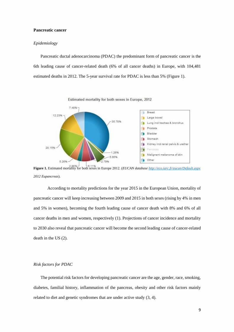
Pancreatic ductal adenocarcinoma (PDAC) the predominant form of pancreatic cancer is the
6th leading cause of cancer-related death (6% of all cancer deaths) in Europe, with 104,481
estimated deaths in 2012. The 5-year survival rate for PDAC is less than 5% (Figure 1).
Figure 1. Estimated mortality for both sexes in Europe 2012. (EUCAN database http://eco.iarc.fr/eucan/Default.aspx
2012 Eupancreas).
According to mortality predictions for the year 2015 in the European Union, mortality of
pancreatic cancer will keep increasing between 2009 and 2015 in both sexes (rising by 4% in men
and 5% in women), becoming the fourth leading cause of cancer death with 8% and 6% of all
cancer deaths in men and women, respectively (1). Projections of cancer incidence and mortality
to 2030 also reveal that pancreatic cancer will become the second leading cause of cancer-related
death in the US (2).
Risk factors for PDAC
The potential risk factors for developing pancreatic cancer are the age, gender, race, smoking,
diabetes, familial history, inflammation of the pancreas, obesity and other risk factors mainly
related to diet and genetic syndromes that are under active study (3, 4).
10
Age: The risk of developing pancreatic cancer goes up as people age. Almost all patients
are older than 45. About two-thirds are at least 65 years old. The average age at the time
of diagnosis is 71.
Gender: Men are slightly more likely to develop pancreatic cancer than women. This may
be due, at least in part, to higher tobacco use in men, which raises pancreatic cancer risk
(see above). The difference in pancreatic cancer risk was larger in the past (when tobacco
use was much more common among men than women), but the gap has closed in recent
years.
Race: African Americans are slightly more likely to develop pancreatic cancer than
whites. The reasons for this aren’t clear, but it may be due in part to having higher rates
of some other risk factors for pancreatic cancer, such as diabetes, smoking in men, and
being overweight in women.
Smoking: Pancreatic cancer risk is 2.2 times higher in current smokers compared with
never-smokers, a pooled analysis showed. Risk increases with the number of cigarettes
smoked per day, and duration of cigarette smoking, meta- and pooled analyses have
shown.
Diabetes: Pancreatic cancer is more common in people with diabetes. The reason for this
is not known. Most of the risk is found in people with type 2 diabetes. This type of
diabetes most often starts in adulthood and is often related to being overweight or obese.
It’s not clear if people with type 1 (juvenile) diabetes have a higher risk.
Family history: Pancreatic cancer seems to run in some families. In some of these families,
the high risk is due to an inherited syndrome (explained below). In other families, the
gene causing the increased risk is not known. Although family history is a risk factor,
most people who get pancreatic cancer do not have a family history of it.
Inflammation of the pancreas: Chronic pancreatitis, a long-term inflammation of the
pancreas, is linked with an increased risk of pancreatic cancer (especially in smokers),
but most people with pancreatitis never develop pancreatic cancer. Chronic pancreatitis

11
is sometimes due to an inherited gene mutation. People with this inherited (familial) form
of pancreatitis have a high lifetime risk of pancreatic cancer.
Obesity: Body fatness is classified by the International Agency for Research on Cancer
(IARC) and the World Cancer Research Fund/American Institute for Cancer Research
(WCRF/AICR) as a cause of pancreatic cancer. Abdominal fatness is classified by
WCRF/AICR as a probable cause of pancreatic cancer. Pancreatic cancer risk increases
by 10% per 5-unit body mass index (BMI) increase, a meta-analysis showed. Pancreatic
cancer risk increases by 11% per 10cm waist circumference increase, and by 19% per
0.1-unit waist-to-hip ratio increment, this meta-analysis showed.
Genetic syndromes: Inherited gene changes (mutations) can be passed from parent to
child. These gene changes may cause as many as 10% of pancreatic cancers. Sometimes
these changes result in syndromes that include increased risks of other cancers (or other
health problems).
Histology of Pancreatic cancer
The pancreas is an organ located behind the stomach in the upper left abdomen. It is
surrounded by other organs including the small intestine, liver, and spleen. It is spongy, about six
to ten inches long, and is shaped like a flat pear or a fish extended horizontally across the abdomen.
The wide part, called the head of the pancreas, is positioned toward the center of the abdomen at
the juncture where the stomach meets the first part of the small intestine (Figure 2).
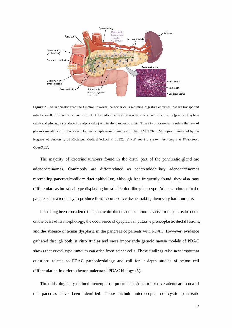
It has two main functions: the exocrine which involves the acinar cells secreting enzymes that
are transported into the small intestine by the pancreatic duct and are important to digestion and
the endocrine that consists of islet cells (islets of Langerhans) that create and secrete important
hormones such as glucagon, insulin, somatostatin, and pancreatic polypeptide (PP) directly into
the bloodstream.
12
Figure 2. The pancreatic exocrine function involves the acinar cells secreting digestive enzymes that are transported
into the small intestine by the pancreatic duct. Its endocrine function involves the secretion of insulin (produced by beta
cells) and glucagon (produced by alpha cells) within the pancreatic islets. These two hormones regulate the rate of
glucose metabolism in the body. The micrograph reveals pancreatic islets. LM × 760. (Micrograph provided by the
Regents of University of Michigan Medical School © 2012). (The Endocrine System. Anatomy and Physiology.
OpenStax).
The majority of exocrine tumours found in the distal part of the pancreatic gland are
adenocarcinomas. Commonly are differentiated as pancreaticobiliary adenocarcinomas
resembling pancreaticobiliary duct epithelium, although less frequently found, they also may
differentiate as intestinal type displaying intestinal/colon-like phenotype. Adenocarcinoma in the
pancreas has a tendency to produce fibrous connective tissue making them very hard tumours.
It has long been considered that pancreatic ductal adenocarcinoma arise from pancreatic ducts
on the basis of its morphology, the occurrence of dysplasia in putative preneoplastic ductal lesions,
and the absence of acinar dysplasia in the pancreas of patients with PDAC. However, evidence
gathered through both in vitro studies and more importantly genetic mouse models of PDAC
shows that ductal-type tumours can arise from acinar cells. These findings raise new important
questions related to PDAC pathophysiology and call for in-depth studies of acinar cell
differentiation in order to better understand PDAC biology (5).
Three histologically defined preneoplastic precursor lesions to invasive adenocarcinoma of
the pancreas have been identified. These include microscopic, non-cystic pancreatic
13
intraepithelial neoplasia (PanIN) and macroscopic, cystic intraductal papillary mucinous
neoplasms (IPMN) and mucinous cystic neoplasms (MCN) (6, 7).
Based on the international nomenclature and classification established by the assembly of
Pancreatic Cancer Think Tank held in Park City, Utah in September 1999, we can currently define
the preneoplastic lesions as following (7-10):
PanIN-1A: (Pancreatic Intraepithelial Neoplasia 1-A): These are flat epithelial lesions
composed of tall columnar cells with basally located nuclei and abundant supranuclear
mucin. The nuclei are small and round to oval in shape. When oval the nuclei are oriented
perpendicular to the basement membrane. It is recognized that there is considerable
histologic overlap between non-neoplastic flat hyperplastic lesions and flat neoplastic
lesions without atypia. Therefore, some may choose to designate these lesions with the
modifier lesion ("PanIN/[L]-1A") to reflect the fact that the neoplastic nature of many
cases of PanIn-1A has not been established.
PanIN-1B: (Pancreatic Intraepithelial Neoplasia 1-B): These epithelial lesions have a
papillary, micropapillary or basally pseudostratified architecture, but are otherwise
identical to PanIN-1A.
PanIN-2: (Pancreatic Intraepithelial Neoplasia 2): Architecturally these mucinous
epithelial lesions may be flat or papillary. Cytologically, by definition, these lesions must
have some nuclear abnormalities. These abnormalities may include some loss of polarity,
nuclear crowding, enlarged nuclei, pseudo-stratification and hyperchromatism. These
nuclear abnormalities fall short of those seen in PanIN-3. Mitoses are rare, but when
present are non-luminal (not apical) and not atypical. True cribriforming luminal necrosis
and marked cytologic abnormalities are generally not seen, and when present should
suggest the diagnosis of PanIN-3.
PanIN-3: (Pancreatic Intraepithelial Neoplasia 3): Architecturally, these lesions are
usually papillary or micropapillary, however, they may rarely be flat. True cribriforming,
budding off of small clusters of epithelial cells into the lumen and luminal necroses

14
should all suggest the diagnosis of PanIN-3. Cytologically, these lesions are characterized
by a loss of nuclear polarity, dystrophic goblet cells (goblet cells with nuclei oriented
towards the lumen and mucinous cytoplasm oriented toward the basement membrane),
mitoses which may occasionally be abnormal, nuclear irregularities and prominent
(macro) nucleoli.
Intraductal Papillary Mucinous Neoplasms (IPMNs): IPMNs are mucinous epithelial
neoplasms which involve the main pancreatic duct or its major branches. They are larger
than PanINs and therefore usually visible grossly or by radiologic imaging. IPMNs may
extend into small ducts. In these cases serial (step) sections may be helpful in defining
the relationship of the two lesions.
Mucinous cystic neoplasms (MCN): Mucinous cystic neoplasms are characterized by the
presence of ovarian stroma and the absence of a connection to the duct system. These
features and the larger size of mucinous cystic neoplasms help distinguish mucinous
cystic neoplasms from PanINs.
Key regulators in PDAC carcinogenesis
The accumulation of somatic mutations, epigenetic modifications and changes in the
micro-environment leads to the development of pancreatic cancer.
Somatic mutations together with genomic rearrangements have been identified to drive
tumorigenesis in pancreatic cancer (11, 12). The mutation in KRAS codon 12 is the most
frequently found with a prevalence of ~30% in PanIN neoplasms and nearly 100% of advanced
PDAC cases (13-15). In addition, somatic mutations in CDKN2A, TP53 and DPC4/SMAD4 are
also observed in patients, although none as frequent as KRAS.
The proto-oncogene KRAS encodes a small guanine nucleotide transferase (GTPase)
which cycles between GTP-bound active and GDP-bound inactive states. Thus, while its
activation is driven first by the dissociation of GDP followed by the later binding of the GTP
15
mediated by the guanine nucleotide exchange factors (GEFs), its inactivation is induced by the
hydrolysis of GTP therefore returning to the GDP-bound promoted by GTPase-activating proteins
(GAPs). Activating mutations of KRAS found in PDAC (point mutations at codon G12, G13 and
Q61) impair its intrinsic GTPase activity by blocking the interaction with GAPs leading to its
constitutive activation and therefore a persistent stimulation of downstream signalling pathways
that drive many of the hallmarks of cancer, sustained proliferation, metabolic reprogramming,
anti-apoptosis, remodelling of the tumour microenvironment, evasion of the immune response,
cell migration and metastasis (15).
The cell signalling pathways linked to KRAS are considerably complex and dynamic,
since involve distinct downstream effectors such as the canonical Raf/Mek/Erk,
phosphatidylinositol 3-kinase (PI3K)/3-phosphoinositide-dependent protein kinase-1 (Pdk1)/Akt,
RalGDS/p38MAPK, Rac and Rho, Rassf1, NF1, p120GAP and PLC-ε (16, 17). However in the
context of PDAC it is believed that oncogenic KRAS signalling passes through three main
pathways: Raf/Mek/Erk, PI3K/Pdk1/Akt and the Ral guanine nucleotide exchange factor pathway.
The inhibitory crosstalk between the PI3K/AKT and MAPK pathways at the level of AKT
and RAF modulates proliferation of the cancer cells (18). This interaction depends on several
parameters such as the cell type, the RTK input and even the time course of RTK activation what
indeed does not make simple the elucidation of the involved mechanisms (19). Nonetheless
Zmajkovicova et al (2013) found a novel crosstalk between PI3K/AKT and MAPK independent
of the RTK receptors, which negatively regulates the PI3K signalling by means of the
phosphorylation of Mek1 at T292 by Erk resulting in the activation of the complex
MAGI1/Mek1/PTEN. Therefore, the inhibition of the MAPK pathway likely interferes PTEN
tumour suppressor function leading to PI3K pathway activation. The loss of PTEN and the
subsequent activation of the PI3K pathway has been found in PDAC and shown to accelerate
tumour formation. This has been attributed to increase the NFκB survival signalling and pro-
tumourigenic changes in the tumour microenvironment (20).

16
The oncogenic KRAS signalling is the main driving force behind PDAC. The signalling
networks are characterised by the activation of several effector pathways and these are
interconnected at various levels by cross-signalling and feedback loops (Figure 3).
Figure 3. An overview of oncogenic KRAS-driven RAF/MEK/ERK and PI3K/PDK1/AKT signalling networks in
pancreatic cancer. (Eser, S. et al. Oncogenic KRAS signalling in pancreatic cancer. British Journal of Cancer 2014
111, 817–822).
As it has been aforementioned approximately 5–10% of patients have a family history of
the disease (15). Penetrant germline mutations have been found in the tumour suppressors
CDKN2A, BRCA2, PALB2, ATM and STK11, the DNA mismatch repair gene MLH1, as well
as in the hereditary pancreatitis associated genes PRSS1and SPINK1, and the cystic fibrosis gene
CFTR (21-23). Recent genome wide association studies (GWAS) identified multiple loci that
harbor common germline susceptibility variants with small effect sizes located in intergenic or
intronic regions on chromosomes 1q32.1 (NR5A2) 5p15.33 (TERT/CLPTM1L), 9q34.2 (ABO),
13q22.1 (nongenic) in populations of European descent, and on 3q29 (TFRC), 5p13.1 (DAB2),
6p25.3 (FOXQ1), 7q36.2 (DPP6), 10q26.11 (PRLHR), 12p11.21 (BICD1), 21q21.3 (BACH1),
21q22.3 (TFF1) and 22q13.32 (FAM19A5), in Asian populations (15). Furthermore, mutations
17
in HNF1A, HNF1B, HNF4A and PDX1 can each cause maturity onset diabetes of the young, and
SNPs localizing within the NR5A2, HNF1A, PDX1 and HNF1B gene regions have been
identified as markers of pancreatic cancer risk loci through GWAS or pathway based GWAS
analyses. Interestingly, unlike it has been reported so far recent transcriptome analysis in PDAC
described HNF1A and HNF1B, PDX1 and PTF1A to be among the top five most consistently
dysregulated sub-networks (15). Nonetheless, new integrated genomic analysis identified 32
recurrently mutated genes that were aggregated into 10 pathways: KRAS, TGF-β, WNT, NOTCH,
ROBO/SLIT signalling, G1/S transition, SWI-SNF, chromatin modification, DNA repair and
RNA processing. The subsequent expression analysis defined 4 new subtypes of PDAC: (1) the
squamous which are enriched for TP53 and KDM6A mutations, the upregulation of the TP63ΔN
transcriptional network together with the hypermethylation of pancreatic endodermal cell-fate
determining genes and have a poor prognosis; (2) the pancreatic progenitor that preferentially
express FOXA2/3, PDX1 and MNX1 which are involved in early pancreatic development; (3) the
immunogenic subtype containing upregulated immune networks including pathways involved in
acquired immune suppression; and (4) the aberrantly differentiated endocrine exocrine (ADEX)
which displayed the upregulation of genes that regulate networks involved in KRAS activation,
exocrine (NR5A2 and RBPJL), and endocrine differentiation (NEUROD1 and NKX2-2 (26, 27).
Studies of Genome-wide analysis of promoter methylation associated with gene
expression attributed aberrant gene hyper- and hypo- methylation to contribute to the
development and progression of pancreatic cancer (28, 29). Consistently, aberrant methylation
increases during neoplastic development among the PanINs and IPMNs (29, 30). Among the list
of 1206 candidate BAI1, KCNV1, EYA4, BNC1, HOXA5, PAX7, SOX14, TLX3, NRXN1,
CNTNAP2, PKP1, ACTA1, MDFI, EVC2, LIN28, NRN1, PENK, m FAM84A, and ZNF415
were found to be hypermethylated. Whereas within the differential methylation profiles of genes
whose epigenetic was silenced included NPTX2, CLDN5, LHX1, WNT7A, FOXE1, PAX6,
BNIP3, GADD45B, HIC1, HS3ST2, TWIST1, IRF7, CCNA1, ALPP, CEBPA, CACNA1G,
CCND2, and TFPI-2. Interestingly, DNA hypermethylation of genes involved in stem cell
18
pluripotency, such as the intestinal stem cell marker and chromatin structure regulator BMI1, the
genes encoding bone morphogenetic proteins BMP3, BMP6, the transcription factors FOXD3,
CDX2, UTF1, and T, as well as NR5A1, NR5A2, NR2F1, NTRK1, NTRK2, NTRK3, NODAL,
SALL4, and SPHK1 genes were also identified (31).
Overall, the identification of the accumulated somatic mutations, the aberrantly
hypermethylated and silenced genes with the elucidation of the epigenetic mechanisms are crucial
for the improvement of diagnosis, prognostic and therapeutic applications.
19
References
1 De Angelis, R. et al. EUROCARE-5 Working Group. Cancer survival in Europe 1999-
2007 by country and age: results of EUROCARE–5-a population-based study. Lancet Oncol.
2014; 15(1):23-34.
2 Ferlay, J. et al. Cancer incidence and mortality patterns in Europe: estimates for 40
countries in 2012. Eur J Cancer. 2013;49(6):1374-403.
3 Malvezzi, M. et al. European cancer mortality predictions for the year 2015: does lung
cancer have the highest death rate in EU women? Ann Oncol. 2015;26(4):779-86.
4 Rahib, L. et al. Projecting cancer incidence and deaths to 2030: the unexpected burden of
thyroid, liver, and pancreas cancers in the United States. Cancer Res. 2014; 74(11):2913-21
5 Rooman, I. et al. Pancreatic ductal adenocarcinoma and acinar cells: a matter of
differentiation and development? Gut. 2012; 61(3):449-58.
6 Matthaei, H. et al. Cystic precursors to invasive pancreatic cancer. Nat Rev Gastroenterol
Hepatol. 2011; 8(3): 141–150.
7 Hurban, RH. et al. Precursors to Pancreatic Cancer. Gastroenterol Clin North Am. 2007;
36(4): 831–vi.
8 Distler, M. et al. Precursor Lesions for Sporadic Pancreatic Cancer: PanIN, IPMN, and
MCN. Biomed Res Int. 2014; 2014: 474905.
9 Hruban, RH. et al. Pancreatic adenocarcinoma: update on the surgical pathology of
carcinomas of ductal origin and PanINs. Modern Pathology 2007; 20:S61–S70.
10 Hruban, RH. et al. Pancreatic intraepithelial neoplasia (PanIN): A New nomenclature and
classification system for pancreatic duct lesions. Am J Surg Pathol. 2001; 25(5):579-86.
11 Eser, S. et al. Oncogenic KRAS signalling in pancreatic cancer. Br J Cancer. 2014;
111(5): 817–822.
20
12 Jones,S. et al. Core signaling pathways in human pancreatic cancers revealed by global
genomic analyses. Science 2008, 321:1801–1806.
13 Campbell, P.J. et al. The patterns and dynamics of genomic instability in metastatic
pancreatic cancer. Nature 2010, 467:1109–1113.
14 Hezel, A.F. et al. Genetics and biology of pancreatic ductal adenocarcinoma.
Genes Dev. 2006, 20:1218–1249.
15 Hoskins JW et al. Transcriptome analysis of pancreatic cancer reveals a tumor suppressor
function for HNF1A. Carcinogenesis. 2014; 35(12):2670-8.
16 Audrey Vincent et al. Genome-wide analysis of promoter methylation associated with
gene expression profile in pancreatic adenocarcinoma. Clin Cancer Res. 2011; 17(13): 4341–4354.
17 Kanda, M. et al. Presence of somatic mutations in most early-stage pancreatic
intraepithelial neoplasia. Gastroenterology 2012, 142:730–733.e9.
18 Castellano, E. et al. RAS Interaction with PI3K: more than just another effector pathway.
Genes Cancer 2011; 2: 261–274.
19 Pylayeva-Gupta, Y. et al. RAS oncogenes: weaving a tumorigenic web. Nat Rev Cancer
2011; 11: 761–774.
20 Zimmermann, S. et al, Phosphorylation and regulation of Raf by Akt (protein kinase B).
Science 1999; 286: 1741–1744.
21 Moelling, K. et al. Regulation of Raf-Akt Cross-talk. J Biol Chem 2002; 277: 31099–
31106.
22 Ying, H. et al. PTEN is a major tumor suppressor in pancreatic ductal adenocarcinoma
and regulates an NF-kappaB-cytokine network. Cancer Discov 2011; 1: 158–169.
23 Klein, A.P. Genetic susceptibility to pancreatic cancer. Mol. Carcinog. 2012; 51:14–24.
21
24 Roberts, N.J. et al. ATM mutations in patients with hereditary pancreatic cancer. Cancer
Discov. 2012; 2:41–46.
25 Petersen, G.M. et al. A genome-wide association study identifies pancreatic cancer
susceptibility loci on chromosomes 13q22.1, 1q32.1 and 5p15.33. Nat. Genet. 2010; 42:224–228.
26 Li, D. et al. Pathway analysis of genome-wide association study data highlights pancreatic
development genes as susceptibility factors for pancreatic cancer. Carcinogenesis 2012; 33:1384–
1390.
27 Bailey, P. et al. Genomic analyses identify molecular subtypes of pancreatic cancer.
Nature. 2016 3;531(7592):47-52.
28 Fukushima, N. et al. Aberrant methylation of preproenkephalin and p16 genes in
pancreatic intraepithelial neoplasia and pancreatic ductal adenocarcinoma. Am J Pathol. 2002;
160:1573–81.
29 Sato, N. et al. Aberrant methylation of CpG islands in intraductal papillary mucinous
neoplasms of the pancreas. Gastroenterology. 2002; 123:365–72.
30 Hong, SM. et al. Multiple genes are hypermethylated in intraductal papillary mucinous
neoplasms of the pancreas. Mod Pathol. 2008; 21:1499–507.
31 Vincent, A. et al. Genome-wide analysis of promoter methylation associated with gene
expression profile in pancreatic adenocarcinoma. Clin Cancer Res. 2011; 17(13): 4341–4354.
22
Chapter 2
Cancer Stem Cells and Tumour Microenvironment
23
Cancer Stem Cells
The distinctive characteristic of cancer is the heterogeneity. The major evidence is the
diversity present in the subpopulations of the neoplastic cells within the same tumours which
strongly describes the co-existence of cancer cells in a multiple levels of differentiation. The
diversification give rise to different functional properties throughout the cancer progression. This
might be due to the genome instability along with the epigenetic modifications. The heterogeneity
may be defined as the intratumoral heterogeneity and intertumoral heterogeneity referring to the
molecular differences conferred by tumours initiated in the same organ which allows the
classification of these tumours and even representing biologically distinct disease entities. Thus,
in the context of the intratumoral heterogeneity the categories that are comprehended are at the
level of functionality, phenotype and indeed genetically. Consequently, distinct members co-exist
and interact within the same cohort of cells and these interactions may represent one of the major
causes responsible of modulating their nature (1, 2).
A unique subpopulation of cells that possess stem properties such as self-renew but also,
are able to give rise a diverse progeny with self-limited proliferative capacity thereby perpetuating
the malignant growth and even promoting the invasion towards new tissues, receive the name of
cancer stem cells (CSC) and their characteristics place them at the apex of an organized
hierarchical system within the tumour in where CSC are responsible for the new generation of
more differentiated progenies (non-CSC) (1-3).
The origin of the CSC is a question that remains to be elucidated and several studies are
emerging in order to address this point. Different origins have been hypothesized however the
controversy is mainly serve in a dichotomy manner between the clonal evolution and the CSC
paradigm model. The popular theory of Peter Nowell described almost four decades ago about
the clonal evolution wherein a single cell undergoes to a sequential cloning leading up to the
tumour progression by means of the selection of the most aggressive subclones due to the stepwise
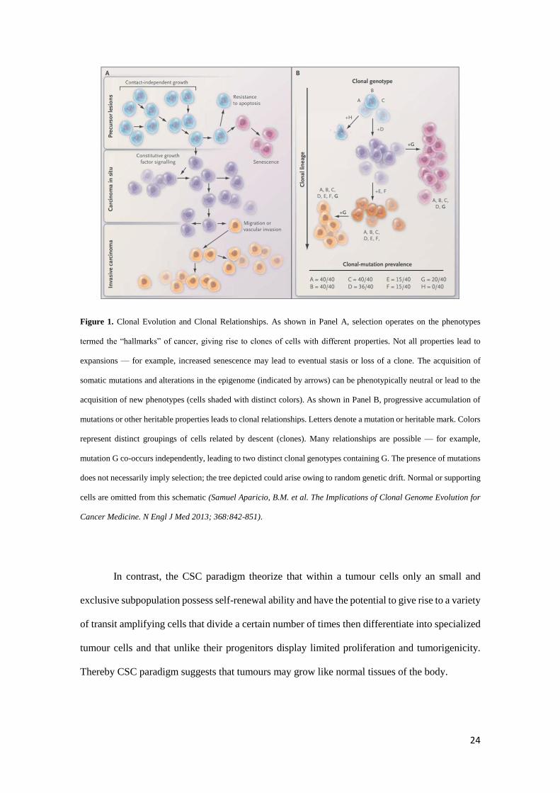
acquisition of mutations, has been preserved until nowadays (Figure 1).
24
Figure 1. Clonal Evolution and Clonal Relationships. As shown in Panel A, selection operates on the phenotypes
termed the “hallmarks” of cancer, giving rise to clones of cells with different properties. Not all properties lead to
expansions — for example, increased senescence may lead to eventual stasis or loss of a clone. The acquisition of
somatic mutations and alterations in the epigenome (indicated by arrows) can be phenotypically neutral or lead to the
acquisition of new phenotypes (cells shaded with distinct colors). As shown in Panel B, progressive accumulation of
mutations or other heritable properties leads to clonal relationships. Letters denote a mutation or heritable mark. Colors
represent distinct groupings of cells related by descent (clones). Many relationships are possible — for example,
mutation G co-occurs independently, leading to two distinct clonal genotypes containing G. The presence of mutations
does not necessarily imply selection; the tree depicted could arise owing to random genetic drift. Normal or supporting
cells are omitted from this schematic (Samuel Aparicio, B.M. et al. The Implications of Clonal Genome Evolution for
Cancer Medicine. N Engl J Med 2013; 368:842-851).
In contrast, the CSC paradigm theorize that within a tumour cells only an small and
exclusive subpopulation possess self-renewal ability and have the potential to give rise to a variety
of transit amplifying cells that divide a certain number of times then differentiate into specialized
tumour cells and that unlike their progenitors display limited proliferation and tumorigenicity.
Thereby CSC paradigm suggests that tumours may grow like normal tissues of the body.

25
Figure 2. Cancer theories: The cancer stem cell theory suggests a clear hierarchy of cells within a tumour. The
stochastic model says that tumour growth is a random process to which all cells can contribute. (Blanpain, C. et al.
EuroStemCell. 2015).
According to this, the cells within the tumour are in a strictly organised system wherein
CSC are at the top of the hierarchy giving rise to more differentiated cancer cells.
On the other hand, inside the CSC paradigm has been proposed a stochastic model of
cancer growth. This model explains the tumour growth from different and new approach. This
theory argues that all cancer cells have the same potential to grow and divide but each cell is
attributed randomly between self-renewal and differentiation. The cells in a tumour are not in an
organised system – any cell has the same intrinsic potential to contribute to tumour growth. In
fact, different types of cancer may work in different ways, so it is possible that both of these
theories are right. Perhaps they apply to different cancers or different stages of tumour
development (Figure 2).
The clonal evolution and the two CSC paradigm models are not mutually exclusive, the
difficulties in finding clear evidences to discern the main origin are notorious and recent reports
suggest that even might exist differences in the origin depending on the type of disease (4-6).

26
However, our recent data provided new insights that will change the current picture of the CSC
paradigm and the clonal evolution theories.
Tumour Cell Plasticity
The tumour cell plasticity comprised the possibility that a non-CSC undergoes to
differentiation which ends up turning into a cancer cell endowed of stem-like properties. This
bidirectional transformation from non-CSC to CSC has been found in different tumours such as
glioma, intestinal tumours and certain types of breast cancers, wherein the common mechanisms
are the dysregulation of specific genetic pathways which dictates the emergence and phenotype
of the disease.
The process of dedifferentiation refers to the loss of mature functional features and the
subsequent possible re-acquisition of the embryonic properties. This concept has been
controversial but latest reports reinforcing its existence suggested that could be involved in the
transition for tumorigenesis. Thus, the reversion of the differentiation to a stem cell phenotype
may suppose an oncogenic transformation.
Despite the apparent differences, every time more the importance of the balance of the
tissue homeostasis is becoming more relevant and appears to be the common fact throughout all
cancer types. Thus the consequences because of the changes in the tissue homeostasis, known as
a tumour microenvironment, exert a major influence in the cell fate and may be decisive for the
occurrence of the disease (1-6).
Tumour Microenvironment (TME)
It has been appreciated for some time that the tumour microenvironment (TME) plays a
significant role in disease progression, but the precise function of each constituent remains
unknown. The TME is constitute by stroma cells and the essential elements that are found to be
27
part of include fibroblasts, myofibroblasts, neuroendocrine cells, adipose cells, immune and
inflammatory cells, the blood and lymphatic vascular networks, and the extracellular matrix
(ECM). The stroma is critical for maintaining the physiological normal tissue homeostasis and
recent studies strengthened the concept that some stromal components have anticancer activities
by regulating immunosuppression and restraining carcinogenesis. Contrary to the stipulated
regular conditions the variety of infiltrating immune cells, cancer-associated fibroblasts (CAF)
and angiogenic endothelial cells play expanding and critical functions in sustaining cell
proliferation, evading growth suppressors, promoting survival, activating invasion and metastasis,
and reprogramming energy metabolism (7, 8).
It has been reported the ability of the cancer cells to regulate or modify the stromal cells
favouring the cancerous niche by means of the synthesis of cytokines, chemokines and growth
factors which consequently accelerate the progression of disease. This stromal cell plasticity leads
to respond rapidly to neoplastic situations and act in concert with the adjacent epithelium in
eliciting the emergence of “reactive stroma” (9).
In a solid tumour those cancer cells that are in the invasive front are exposed to the
reactive stroma, hypoxia and immune surveillance. Thus the invasive front is rich in CAFs,
tumour-associated macrophages (TAMs), myeloid progenitor cells and newly generated blood
vessels (10). The invasive fronts rich in CAFs and myofibroblasts produces TME-mediated
signalling expressing high levels of extracellular factors including chemokine CXC motif ligand
12 (CXCL12), chemokine CC motif ligand 2 (CCL2), CCL8, and insulin-like growth factor
binding protein 7 (IGFBF7) promoting the inflammatory response and recruiting immune cells,
and also cytokines such as TNF-α, TGF-β, hedgehog (Hgh), Wnt and Notch that are attributed to
the survival and fitness of CSCs (8-11).
TGF-β stimulates the epithelial-mesenchymal transition (EMT) thus the carcinoma cells
in the invasive front of tumour lose the cell-to-cell adhesion and apical-basal polarity and gain
migratory behaviour. Particularly, in pancreatic cancers the enforced expression of EMT
transcription factors confers the gain of stem-like features (12). On the other hand TNF-α which
28
is a crucial growth factor is expanded and released by TAMs establishing paracrine loops
supporting back the cancer cells (9). In addition, TNF-α has been also found to stimulate the
accumulation of reactive oxygen species (ROS) as well as promote the nuclear entry of β-catenin
activating Wnt/β-catenin pathway in surrounding epithelial cells which regulates the stem cell
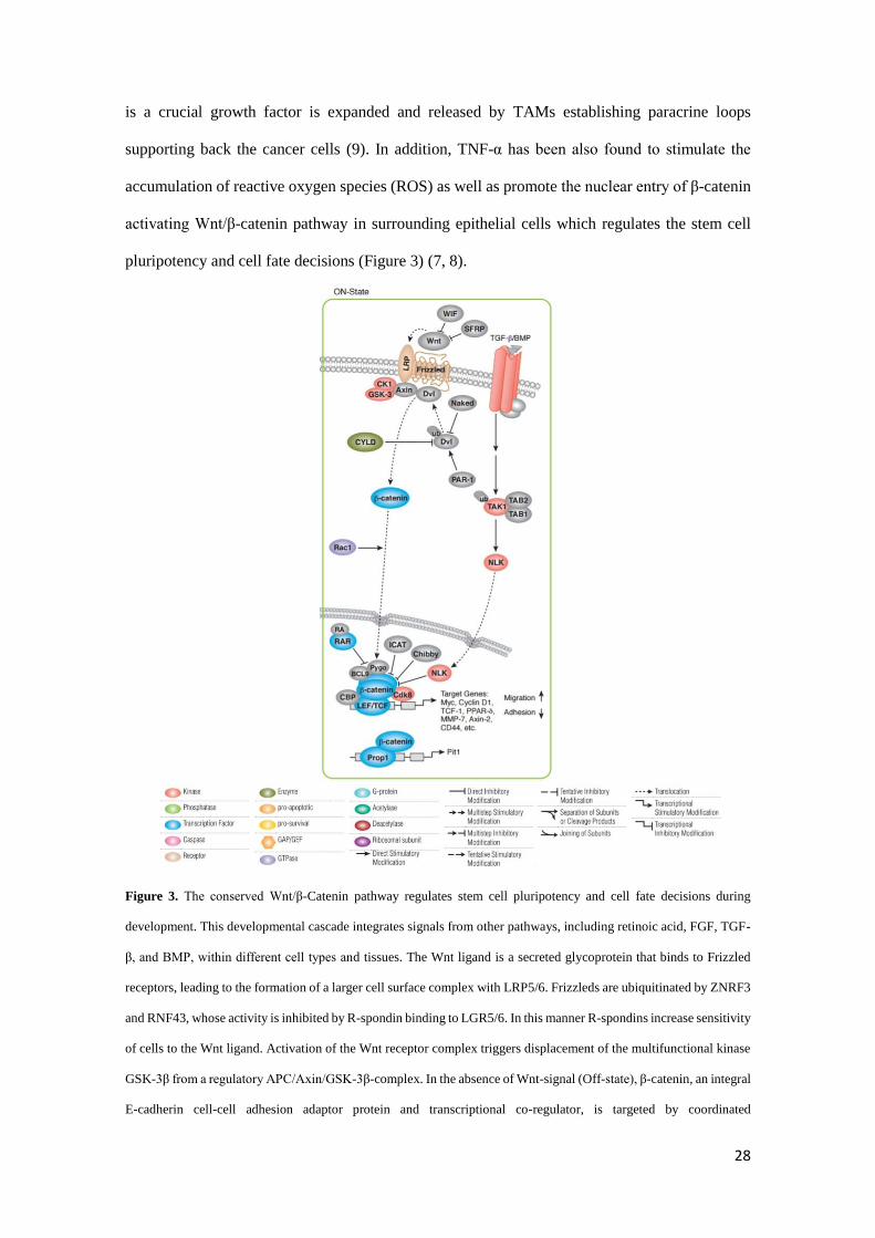
pluripotency and cell fate decisions (Figure 3) (7, 8).
Figure 3. The conserved Wnt/β-Catenin pathway regulates stem cell pluripotency and cell fate decisions during
development. This developmental cascade integrates signals from other pathways, including retinoic acid, FGF, TGF-
β, and BMP, within different cell types and tissues. The Wnt ligand is a secreted glycoprotein that binds to Frizzled
receptors, leading to the formation of a larger cell surface complex with LRP5/6. Frizzleds are ubiquitinated by ZNRF3
and RNF43, whose activity is inhibited by R-spondin binding to LGR5/6. In this manner R-spondins increase sensitivity
of cells to the Wnt ligand. Activation of the Wnt receptor complex triggers displacement of the multifunctional kinase
GSK-3β from a regulatory APC/Axin/GSK-3β-complex. In the absence of Wnt-signal (Off-state), β-catenin, an integral
E-cadherin cell-cell adhesion adaptor protein and transcriptional co-regulator, is targeted by coordinated
29
phosphorylation by CK1 and the APC/Axin/GSK-3β-complex leading to its ubiquitination and proteasomal
degradation through the β-TrCP/Skp pathway. In the presence of Wnt ligand (On-state), the co-receptor LRP5/6 is
brought in complex with Wnt-bound Frizzled. This leads to activation of Dishevelled (Dvl) by sequential
phosphorylation, poly-ubiquitination, and polymerization, which displaces GSK-3β from APC/Axin through an unclear
mechanism that may involve substrate trapping and/ or endosome sequestration. Stablized β-catenin is translocated to
the nucleus via Rac1 and other factors, where it binds to LEF/TCF transcription factors, displacing co-repressors and
recruiting additional co-activators to Wnt target genes. Additionally, β-catenin cooperates with several other
transcription factors to regulate specific targets. Importantly, researchers have found β-catenin point mutations in
human tumors that prevent GSK-3β phosphorylation and thus lead to its aberrant accumulation. E-cadherin, APC, R-
spondin and Axin mutations have also been documented in tumor samples, underscoring the deregulation of this
pathway in cancer. Wnt signaling has also been shown to promote nuclear accumulation of other transcriptional
regulator implicated in cancer, such as TAZ and Snail1. Furthermore, GSK-3β is involved in glycogen metabolism and
other signaling pathways, which has made its inhibition relevant to diabetes and neurodegenerative disorders. (Cell
Signaling Technology. Created January 2003-revised September 2016. www.cellsignal.com).
Inflammation and Tumorigenesis
Inflammation has an important role in the tumour initiation. Activated inflammatory cells
serve as source of ROS, RNI, growth factors and indeed cytokines that as it has been
aforementioned may confer stem-like properties upon the tumour progenitors or even induce the
expansion of stem cells (13).
The contribution of myeloid cells to the tumour play a crucial role in promoting tumour
progression, angiogenesis, cell invasion and metastasis. Specifically, mast cells are commonly
seen in various tumours and have been implicated in the regulation of tumour initiation and
development. They are likely the most productive chemical factory in the body and influence
other cells through both soluble mediators and cell-to-cell interaction, thereby have the capacity
to promote tumour proliferation and invasion both directly by stimulating tumour cells and
indirectly by modulating the TME (14). High number of mast cells may be found at the invasive
border of tumours. Since they are potent inducers of fibrosis and stimulate fibroblast and
myofibroblast proliferation it may lead to the tissue reorganization and the consequent reactive
30
tumour stroma. In addition, their high source of proteases confer them the ability to degrade the
extracellular matrix therefore facilitating the tumour growth and dissemination (15, 16). It has
also been seen that the outcome of cancer may come determined by the intricate interaction from
mast cells with the regulatory T cells (Treg). TNFα produced by mast cells upregulates OX40 in
Treg and activates the PI3K pathway, while MC IL6 works through pg130 to stimulate the JAK–
STAT3 pathway, allowing expression of IL17. Proinflammatory properties of Treg may help to
propagate tumor growth and dissemination and since mast cells expand in polyps and tumours of
the gastrointestinal tract, it is tempting to suggest that mast cells turn the tide in favour of cancer
progression by recruiting and then altering the functions of Treg to promote further cancer-
associated inflammation. (14). Thus, mast cells appear to have an indispensable role in the
development of solid tumours by promoting a gradual gain of pro-inflammatory properties with
the participation of Treg cells therefore favouring uncontrolled escalation of cancer inflammation,
tumour immune tolerance, and aggressive tumour growth.

31
References
1 Friedmann-Morvinski, D. et al. Dedifferentiation and reprogramming: origins of cancer
stem cells. EMBO Rep. 2014; 15(3): 244–253.
2 Diaz-Cano, SJ. Tumor Heterogeneity: Mechanisms and Bases for a Reliable Application
of Molecular Marker Design. Int J Mol Sci. 2012; 13(2): 1951–2011.
3 Sommer, CA. et al. The evolving field of induced pluripotency: recent progress and future
challenges. J Cell Physiol. 2013; 228:267–275.
4 Medema, JP. Cancer stem cells: The challenges ahead. Nature Cell Biology 2013; 15,
338–344.
5 Samuel Aparicio, B.M. et al. The Implications of Clonal Genome Evolution for Cancer
Medicine. N Engl J Med 2013; 368:842-851.
6 Blanpain, C. et al. Self-renewal, multipotency, and the existence of two cell populations
within an epithelial stem cell niche. Cell. 2004; 118(5):635-48.
7 Chen, F. et al. New horizons in tumor microenvironment biology: challenges and
opportunities. BMC Medicine 2015; 13:45.
8 Oskarsson, T. et al. Metastatic Stem Cells: Sources, Niches, and Vital Pathways. Cell
Stem Cell. 2014; 14(3): 306–321.
9 Junttila MR, de Sauvage FJ. Influence of tumour micro-environment heterogeneity on
therapeutic response. Nature. 2013; 501:346–54.
10 Joyce JA, Pollard JW. Microenvironmental regulation of metastasis. Nat Rev Cancer.
2009;9:239–252.
32
11 Feig, C. et al. Targeting CXCL12 from FAP-expressing carcinoma-associated fibroblasts
synergizes with anti-PD-L1 immunotherapy in pancreatic cancer. Proc Natl Acad Sci U S A.
2013; 110:20212–7.
12 Mani, SA. et al. The epithelial-mesenchymal transition generates cells with properties of
stem cells. Cell. 2008 16;133(4):704-15.
13 Grivennikov, SI. et al Immunity, inflammation, and cancer. Cell. 2010; 140(6): 883–899.
14 Khazaie, K. et al. The significant role of mast cells in cancer. Cancer Metastasis Rev.
2011 30, 45–60.
15 Hebda, P. A. et al. Mast cell and myofibroblast in wound healing. Dermatologic Clinics
1993, 11, 685–696.
16 Gailit, J. et al. The differentiation and function of myofibroblasts is regulated by mast cell
mediators. Journal of Investigative Dermatology 2001, 117, 1113–1119.
33
Chapter 3
A new PDAC mouse model originated from
iPSCs-converted pancreatic Cancer Stem Cells (CSCcm)
34
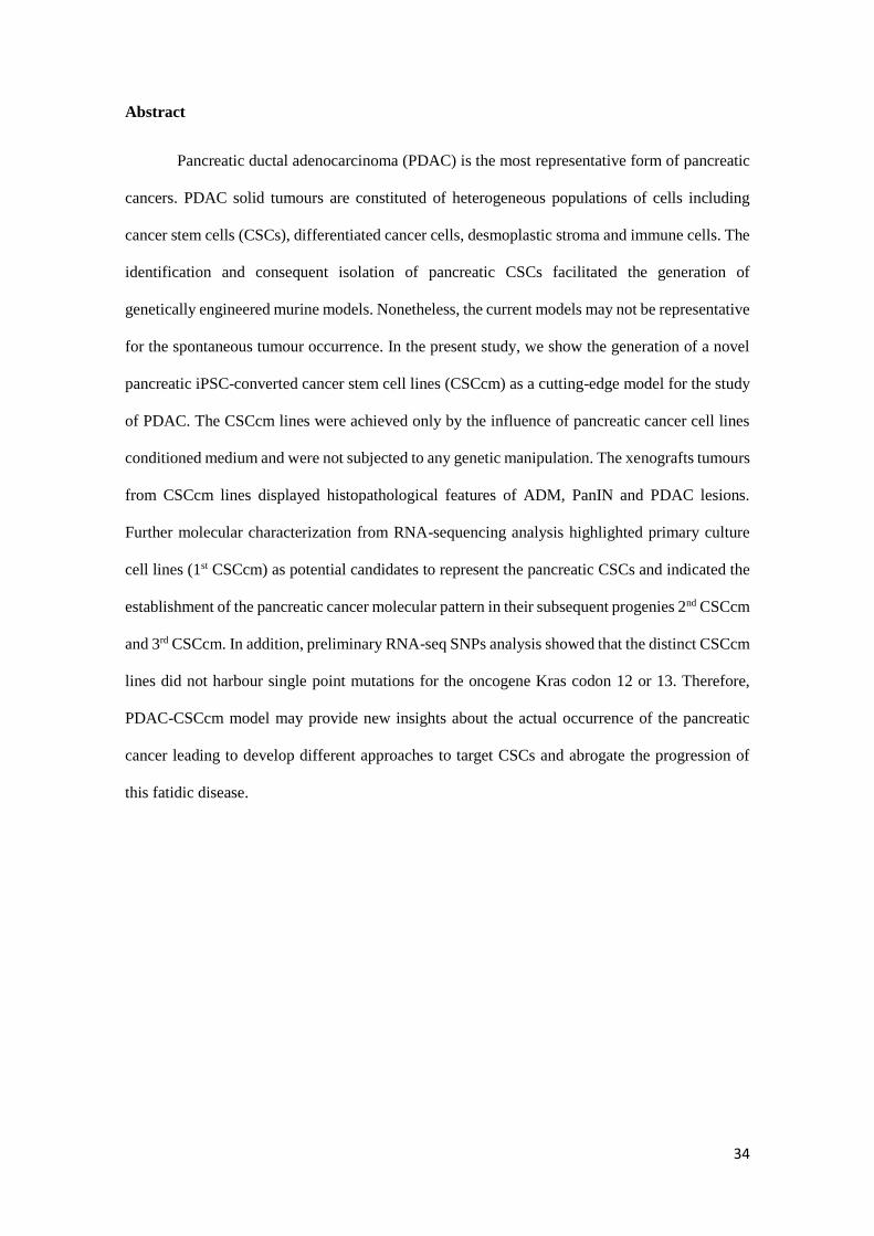
Abstract
Pancreatic ductal adenocarcinoma (PDAC) is the most representative form of pancreatic
cancers. PDAC solid tumours are constituted of heterogeneous populations of cells including
cancer stem cells (CSCs), differentiated cancer cells, desmoplastic stroma and immune cells. The
identification and consequent isolation of pancreatic CSCs facilitated the generation of
genetically engineered murine models. Nonetheless, the current models may not be representative
for the spontaneous tumour occurrence. In the present study, we show the generation of a novel
pancreatic iPSC-converted cancer stem cell lines (CSCcm) as a cutting-edge model for the study
of PDAC. The CSCcm lines were achieved only by the influence of pancreatic cancer cell lines
conditioned medium and were not subjected to any genetic manipulation. The xenografts tumours
from CSCcm lines displayed histopathological features of ADM, PanIN and PDAC lesions.
Further molecular characterization from RNA-sequencing analysis highlighted primary culture
cell lines (1st CSCcm) as potential candidates to represent the pancreatic CSCs and indicated the
establishment of the pancreatic cancer molecular pattern in their subsequent progenies 2nd CSCcm
and 3rd CSCcm. In addition, preliminary RNA-seq SNPs analysis showed that the distinct CSCcm
lines did not harbour single point mutations for the oncogene Kras codon 12 or 13. Therefore,
PDAC-CSCcm model may provide new insights about the actual occurrence of the pancreatic
cancer leading to develop different approaches to target CSCs and abrogate the progression of
this fatidic disease.
35
Introduction
PDAC is one of the most highly desmoplastic tumours which unfortunately due to its
aggressiveness and rapid dissemination together with the strong resistance to the radiation therapy
and chemotherapy contributes to the dismal prognosis. Over the past decades the strategies to find
new diagnostic approaches at early stages along with the effective treatments has not improved
significantly (1, 2). Therefore, this highlights the urgent need to find novel models to study the
origin as well as the progression of the disease.
PDAC solid tumours are comprised of a wide range of heterogeneous populations of cells
including cancer stem cells (CSCs), the actual differentiated cancer cells together with
desmoplastic stroma and immune cells which represent a high proportion of the tumour mass (3).
CSCs are considered as cells that possess stem cell properties and produce diverse lineages of
cancer cells. Hence, CSCs have been associated with the tumour initiation and progression, and
have been reported to be involved in tumour metastasis (4).
The isolation of pancreatic CSCs succeeded in providing new insights regarding the
chemoresistance and the high metastatic ability in PDAC. Since CSCs and non-CSCs share an
identical genetic background it is difficult to find appropriate in vitro and in vivo systems that
allows to select reproducibly and exclusively enriched CSCs populations. Furthermore, these new
approaches include particular markers that are also found in differentiated adult cells what makes
questionable the identification of the CSCs (7). Recent advances have been developed in targeting
CSCs and their identification and isolation consequently facilitate the generation of new murine
models (5, 6). However, the current models are genetically engineered and therefore may not be
suitable for a better understanding of the spontaneous tumour occurrence.
As have been seen in regenerative medicine field, iPSCs when exposed to appropriate
environments are able to directly differentiate into progenitor cells that lead to the latter matured
form of cells. Hence, the signals found in the niche simultaneously regulate the differentiation as
well as support the tissue homeostasis preserving the self-renewal potential from a minor but

36
required stem cells number (8). Based on this we previously hypothesized that CSCs might be
considered as progenitor cells that are destined to differentiate into cancer cells and that
consequently if the cell fate comes determined by the events and factors present in the niche, the
tumour microenvironment should exert the same effects when healthy cells are exposed to it.
Chen L and Kasai T et al. demonstrated the impact of the so-called cancerous niche when by
exposing Nanog iPSCs to a Lewis Lung carcinoma conditioned medium (LLCcm) a malignant
tumour was obtained exhibiting angiogenesis in vivo, capacity of self-renewal and expressed
markers associated to stem cell properties and undifferentiated state such as Nanog, Rex1, Eras,
Esg1 and Cripto. In contrast, when control Nanog iPSCs were implanted into Balb/c nude mice
formed typical teratomas displaying contained differentiated tissues without metastasis. Thus a
new model of CSC-like cells generated exclusively under the influence of the microenvironment
was proposed (9).
In the present study, we show the generation of a novel pancreatic iPSC-converted cancer
stem cell lines (CSCcm) together with the subsequent characterization of the tumours obtained as
a result from the transplantation of the CSCcm lines in vivo demonstrating that CSCcm is a
promising cutting-edge model for the study of PDAC occurrence and progression.
37
Results
iPSCs-converted CSCs (CSCcm) display CSCs features
As described in the protocol established by Chen L and Kasai T et al., to generate a model
of pancreatic CSCs, iPSCs were exposed to different conditioned medium (CM) from pancreatic
carcinoma cell lines PK-8CM and KLM-1CM, a process named as conversion (Figure 1A) (9).
Similarly to the established protocol, iPSCs were maintained in feeder-less conditions and the
Leukemia inhibitory factor (LIF) which is essential for their viability was removed from the
medium and thereby restricting them to the effect exclusively of the conditioned medium.
Whereas the iPSCs cultured only with iPSCs media without LIF expired after 7 to 10 days (Figure
1B), the viability and proliferation of the cells maintained in presence of conditioned medium was
not affected. Nanog iPSCs cells were generated by the retrovirus-mediated introduction of the
four factors Sox2, Oct3/4, Klf4 and c-Myc into the Nanog-GFP-IRES-Puror of mouse embryonic
fibroblast (42), therefore iPSCs could be monitored throughout the conversion by the GFP
expression as a validation of the stemness. The population of converted cells displayed a diverse
pattern of differentiation harbour cells expressing strong to moderate GFP and other differentiated
cells which the expression was null. Once the process was completed the cells were termed as
CSCcm PK8 and CSCcm KLM-1 and their stem-like properties were tested through sphere
suspension assay (Figure 1B).
A

38
Figure 1. A) Representative scheme of the conversion procedure. B) Viability of iPSCs maintained in control medium
without LIF was no longer than 10 days. iPSCs underwent differentiated with no remaining GFP positive cells and
eventually expired (upper panel). Stemness tracking during conversion by the presence of GFP protein. Self-renewing
potential was validated by sphere formation assay previously to the subcutaneous implantation (lower panel). Original
magnification 4x and 20x.
Since the CSCcm population was heterogeneous, in order to ensure a high frequency of
cells displaying CSC-like properties, 104, 105 and 106 cells from the two new pancreatic CSCcm
lines were subcutaneously transplanted into immunocompromised Balb/c nude mice. After 30
days, CSCcm engrafted and generated tumours in 9 out of 9 mice for each cell line indicating
experimental reproducibility and demonstrating their tumorigenic potential. Chen and Kasai et al.
B
iPSCs day 0
Converted iPSCs PK8CM (CSCcm PK8)
Converted iPSCs KLM1
(CSCcm KLM-1) CSCcm spheroids
iPSCs day 0 iPSCs day 4 iPSCs day 7 iPSCs day 10
C
39
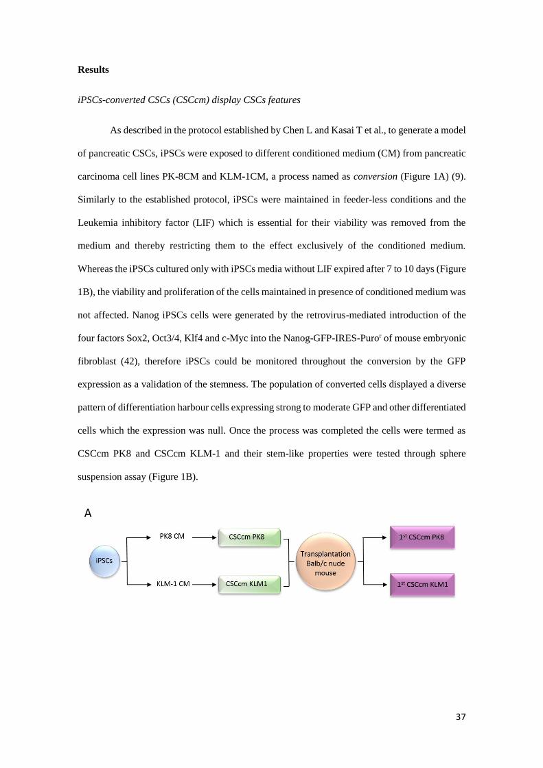
previously shown that iPSCs cultured under control medium generated benign teratoma.
Consistently, the xenograft tumours histology showed specific characteristics that resembled the
actual pancreatic ductal adenocarcinoma phenotype. Primary tumours were rich in stroma and
among the epithelial-like structures pancreatic intraepithelial neoplastic lesions (PanIN) lesions
were found together with moderate to poorly differentiated ductal structures (Figure 2A).
Figure 2. A) Histopathological features of 1st CSCcm primary tumours were evaluated by H&E staining. Specific
PanIN lesions are indicated with arrowheads. Original magnification 10x and 20x. B) Lineage tracing by GFP protein
showed that it was predominantly expressed in undifferentiated cells, however was also partially found in ductal-like
structures. Original Magnification 10x and 20x.
Primary Tumour 1st CSCcm PK8 Primary Tumour 1st CSCcm KLM1
A
GFP
Primary Tumour 1st CSCcm PK8 Primary Tumour 1st CSCcm KLM1 B
40
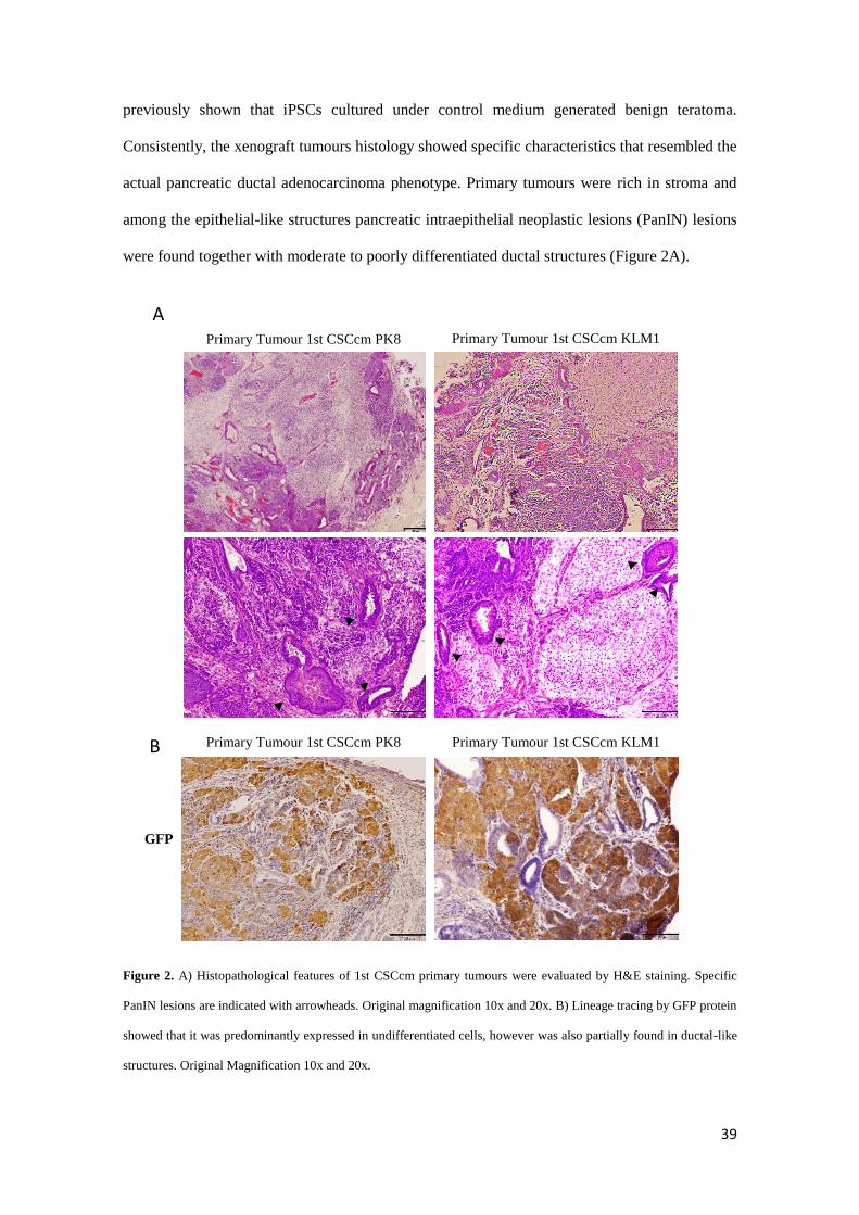
To further validate the PDAC-like structures arose from CSCcm we explored the
expression of GFP protein in primary tumour. GFP expressing cells were found all over the tissue
samples and although it was predominantly located by undifferentiated cells few epithelial cells
from ductal-like structures remained positive for its expression (Figure 2B). When the expression
of specific CSC markers CD133, CD24a and EpCAM was evaluated, their up-regulation was
already observed in CSCcm and was similarly detected or even enhanced in primary cultures (1st
CSCcm) (Figure 3A). CD133 is preferentially used in the identification of CSC in pancreatic solid
tumours and its expression is commonly found within the ductal structures. Likewise the primary
CSCcm tumour tissues showed a strong signal within the ductal-like structures (Figure 3B)
confirming their malignant phenotype.
A
41
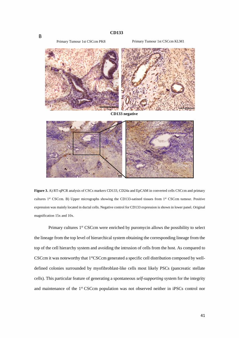
Primary Tumour 1st CSCcm PK8 Primary Tumour 1st CSCcm KLM1
CD133
CD133 negative
Figure 3. A) RT-qPCR analysis of CSCs markers CD133, CD24a and EpCAM in converted cells CSCcm and primary
cultures 1st CSCcm. B) Upper micrographs showing the CD133-satined tissues from 1st CSCcm tumour. Positive
expression was mainly located in ductal cells. Negative control for CD133 expression is shown in lower panel. Original
magnification 15x and 10x.
Primary cultures 1st CSCcm were enriched by puromycin allows the possibility to select
the lineage from the top level of hierarchical system obtaining the corresponding lineage from the
top of the cell hierarchy system and avoiding the intrusion of cells from the host. As compared to
CSCcm it was noteworthy that 1stCSCcm generated a specific cell distribution composed by well-
defined colonies surrounded by myofibroblast-like cells most likely PSCs (pancreatic stellate
cells). This particular feature of generating a spontaneous self-supporting system for the integrity
and maintenance of the 1st CSCcm population was not observed neither in iPSCs control nor
B
42
Primary Cultures 1st CSCcm
1st CSCcm 2
nd CSCcm 3
rd CSCcm
PK8C
M
KLM1C
M
CSCcm
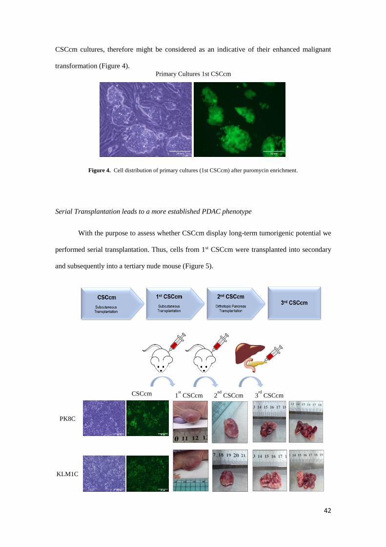
CSCcm cultures, therefore might be considered as an indicative of their enhanced malignant
transformation (Figure 4).
Figure 4. Cell distribution of primary cultures (1st CSCcm) after puromycin enrichment.
Serial Transplantation leads to a more established PDAC phenotype
With the purpose to assess whether CSCcm display long-term tumorigenic potential we
performed serial transplantation. Thus, cells from 1st CSCcm were transplanted into secondary
and subsequently into a tertiary nude mouse (Figure 5).
43
Figure 5. Serial transplantation scheme and images from sequential excised tumours obtained from subcutaneously
transplanted CSCcm and 1st CSCcm primary cultures. Primary tumours and their corresponding metastatic liver nodes
generated from orthotopically implanted 2rd CSCcm cells are also shown. Experiments were equally performed for both
PK8CM and KLM1CM lines.
Similarly to the primary culture the cells from secondary culture (2ndCSCcm) as well as
tertiary cultures (3rdCSCcm) were enriched by puromycin. Thereby 1stCSCcm PK8 and 1stCSCcm
KLM-1 were subcutaneously transplanted giving rise tumours within a short period of time of
25days. Next, 2ndCSCcm PK8 and 2ndCSCcm KLM-1 were subsequently transplanted, however
this time cells were orthotopically transplanted into the pancreas generating tumours within 15-
20days. Orthotopic tumours were remarkably bigger and specific liver metastasis was found
(Table 1).
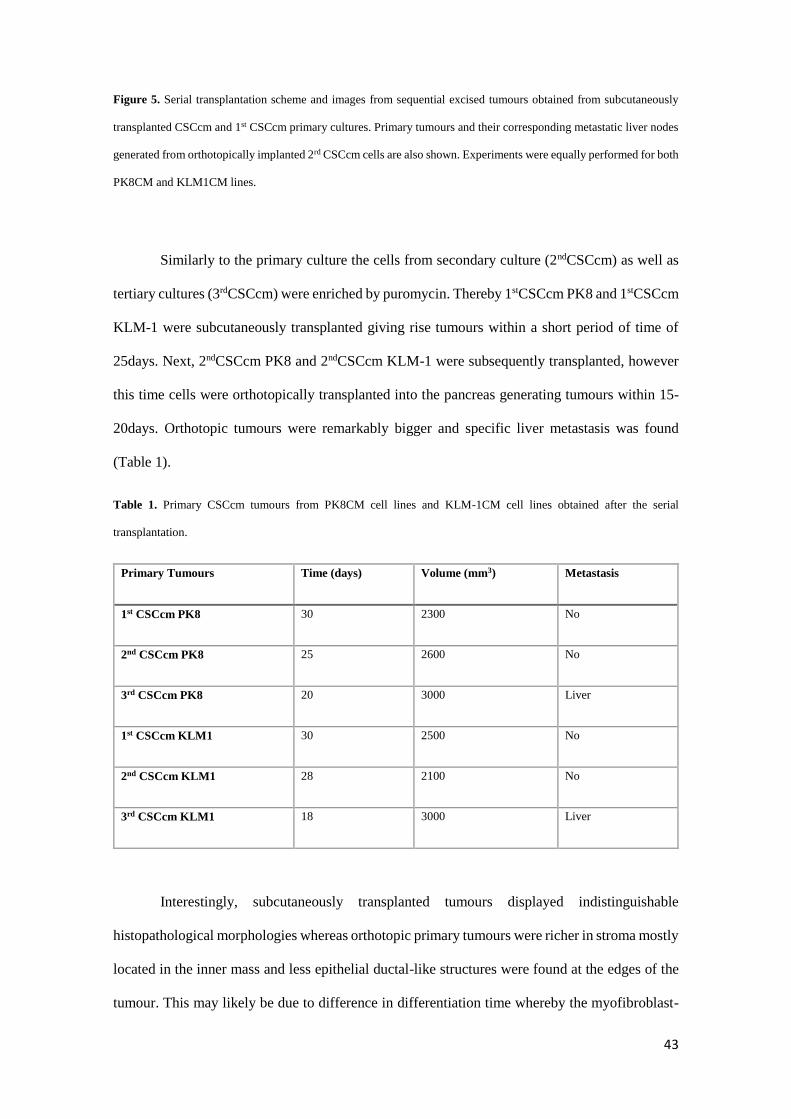
Table 1. Primary CSCcm tumours from PK8CM cell lines and KLM-1CM cell lines obtained after the serial
transplantation.
Primary Tumours Time (days) Volume (mm3) Metastasis
1st CSCcm PK8 30 2300 No
2nd CSCcm PK8 25 2600 No
3rd CSCcm PK8 20 3000 Liver
1st CSCcm KLM1 30 2500 No
2nd CSCcm KLM1 28 2100 No
3rd CSCcm KLM1 18 3000 Liver
Interestingly, subcutaneously transplanted tumours displayed indistinguishable
histopathological morphologies whereas orthotopic primary tumours were richer in stroma mostly
located in the inner mass and less epithelial ductal-like structures were found at the edges of the
tumour. This may likely be due to difference in differentiation time whereby the myofibroblast-

44
Primary tumour 1
st CSCcm
PK8 CM KLM1 CM
Primary tumour 2
nd CSCcm
Primary tumour 3
rd CSCcm
like phenotype is acquired earlier the epithelial-like phenotype requires a longer period of time
(Figure 6).
Figure 6. Micrographs from serial primary tumours showing the histopathological adenocarcinoma-like morphology
by H&E staining. Original magnification 4x.
When the liver metastatic nodes were examined a remarkable difference in the
histopathological features was observed. Unlike primary tumours, liver metastatic nodes
displayed a teratocarcinoma phenotype containing very few structures corresponding to PDAC.
To determine that CSCcm had the ability to metastasise we searched evidences of GFP protein.
45
Primary
Tumour
3rd
CSCcm
PK8
CM
KLM1
CM
Liver Metastatic
Nodes 3rd
CSCcm
GFP Liver Metastatic
Nodes 3rd
CSCcm
92µm
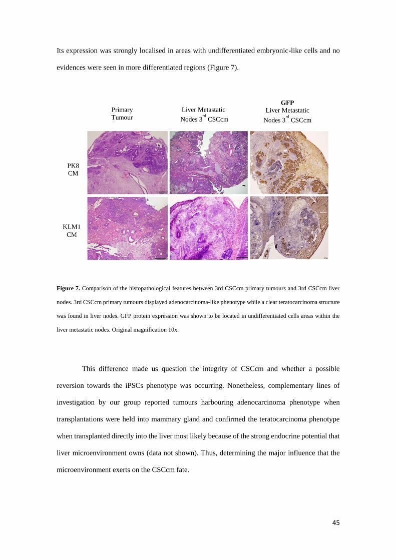
Its expression was strongly localised in areas with undifferentiated embryonic-like cells and no
evidences were seen in more differentiated regions (Figure 7).
Figure 7. Comparison of the histopathological features between 3rd CSCcm primary tumours and 3rd CSCcm liver
nodes. 3rd CSCcm primary tumours displayed adenocarcinoma-like phenotype while a clear teratocarcinoma structure
was found in liver nodes. GFP protein expression was shown to be located in undifferentiated cells areas within the
liver metastatic nodes. Original magnification 10x.
This difference made us question the integrity of CSCcm and whether a possible
reversion towards the iPSCs phenotype was occurring. Nonetheless, complementary lines of
investigation by our group reported tumours harbouring adenocarcinoma phenotype when
transplantations were held into mammary gland and confirmed the teratocarcinoma phenotype
when transplanted directly into the liver most likely because of the strong endocrine potential that
liver microenvironment owns (data not shown). Thus, determining the major influence that the
microenvironment exerts on the CSCcm fate.

46
Given that the reprogramming of iPSCs was performed with the proto-oncogenes Sox2,
Oct3/4, Klf4 and c-Myc, and that the implication specifically of c-Myc and Klf4 have been linked
to PDAC we explored the expression of the transgenes in order to determine that there was no
residual activity (Figure 8A). On the other hand the transcript levels of endogenous c-Myc, which
is generally overexpressed in PDAC, were notably increased (Figure 8B). Hence, strongly
supported that the malignant transformation arose by means of the intrinsically activated
mechanisms of the cells.
Figure 8. A) Agarose gel images from RT-qPCR products for the detection of Klf4 and cMyc transgenes. GAPDH was
taken as a housekeeping control gene. B) Transcript levels for the mRNA of endogenous c-Myc were analysed by RT-
qPCR in a panel of serial transplantation samples.
B
A
47
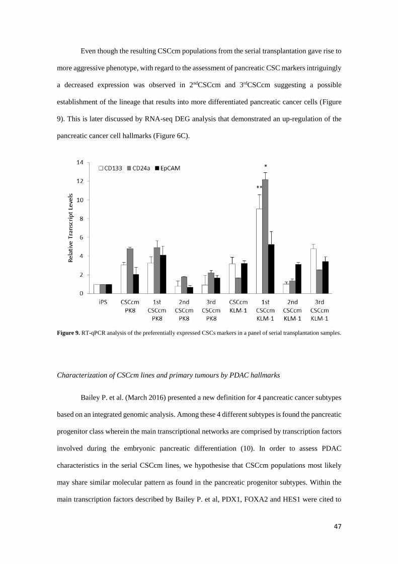
Even though the resulting CSCcm populations from the serial transplantation gave rise to
more aggressive phenotype, with regard to the assessment of pancreatic CSC markers intriguingly
a decreased expression was observed in 2ndCSCcm and 3rdCSCcm suggesting a possible
establishment of the lineage that results into more differentiated pancreatic cancer cells (Figure
9). This is later discussed by RNA-seq DEG analysis that demonstrated an up-regulation of the
pancreatic cancer cell hallmarks (Figure 6C).
Figure 9. RT-qPCR analysis of the preferentially expressed CSCs markers in a panel of serial transplantation samples.
Characterization of CSCcm lines and primary tumours by PDAC hallmarks
Bailey P. et al. (March 2016) presented a new definition for 4 pancreatic cancer subtypes
based on an integrated genomic analysis. Among these 4 different subtypes is found the pancreatic
progenitor class wherein the main transcriptional networks are comprised by transcription factors
involved during the embryonic pancreatic differentiation (10). In order to assess PDAC
characteristics in the serial CSCcm lines, we hypothesise that CSCcm populations most likely
may share similar molecular pattern as found in the pancreatic progenitor subtypes. Within the
main transcription factors described by Bailey P. et al, PDX1, FOXA2 and HES1 were cited to
48
be essentially expressed in the progenitor subtype. Thus, their transcript levels were examined
along with the KRAS which is well-known to be involved in PDAC. Despite the variation found
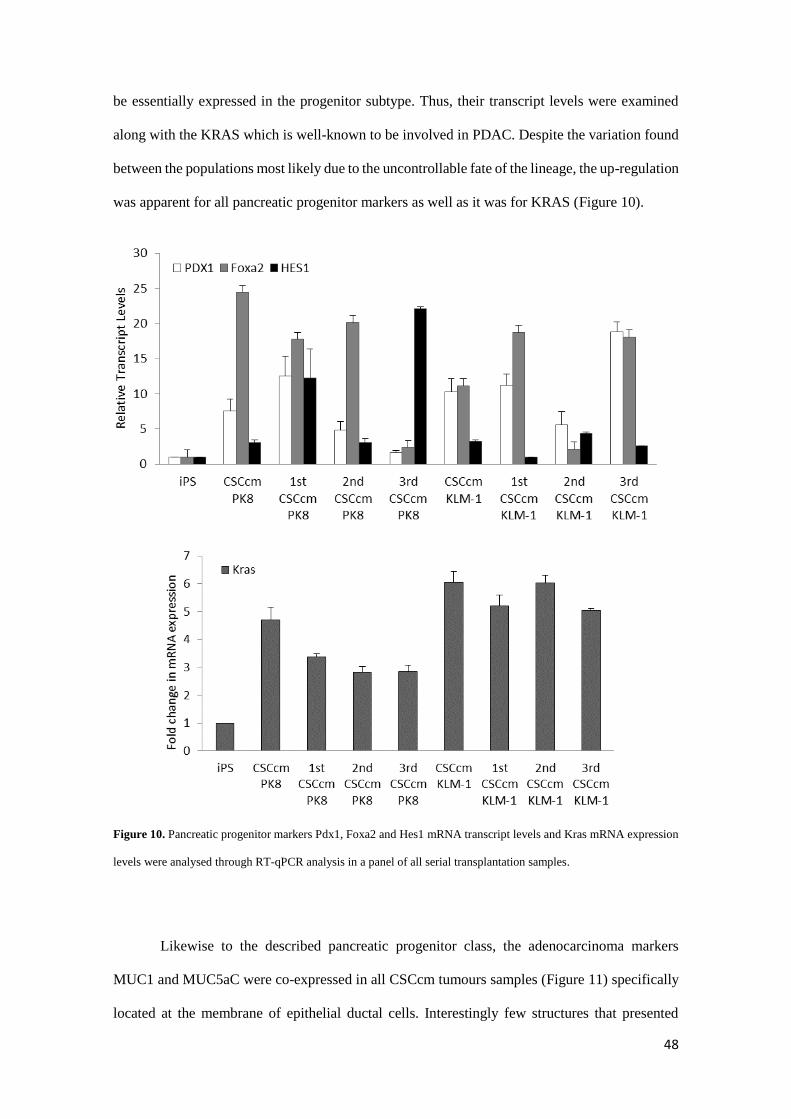
between the populations most likely due to the uncontrollable fate of the lineage, the up-regulation
was apparent for all pancreatic progenitor markers as well as it was for KRAS (Figure 10).
Figure 10. Pancreatic progenitor markers Pdx1, Foxa2 and Hes1 mRNA transcript levels and Kras mRNA expression
levels were analysed through RT-qPCR analysis in a panel of all serial transplantation samples.
Likewise to the described pancreatic progenitor class, the adenocarcinoma markers
MUC1 and MUC5aC were co-expressed in all CSCcm tumours samples (Figure 11) specifically
located at the membrane of epithelial ductal cells. Interestingly few structures that presented

49
Pri
mar
y T
um
or
2n
d C
SC
cm P
K8
MUC1
Pri
mar
y T
um
or
2n
d C
SC
cm K
LM
1
MUC5aC
Pri
mar
y T
um
or
2n
d C
SC
cm P
K8
Pri
mar
y T
um
or
2n
d C
SC
cm K
LM
1
ductal ectasia displayed strong expression in the infiltrating immune cells. Therefore these
evidences infer in the achievement of CSCcm lines to recapitulate the PDAC phenotype.
Figure 11. The adenocarcinoma phenotype was evaluated through the expression of the apomucinous of MUC1 and
MUC5aC expression was predominantly located in the membrane of ductal structures. Infiltrating ductal immune cells
also displayed high expression was also observed in mucinous ductal ectasia (MDE). Original magnification 4x.
50
Primary Tumor
3rd CSCcm PK8 Primary Tumor
3rd CSCcm KLM1
A possible model for lineage tracing ADM events
The acinoductal metaplasia (ADM) describes the process whereby the islet neogenesis is
accompanied by the transdifferentiation of the normal exocrine tissue into ductal complexes. This
pancreatic metaplasia may turn into a premalignant state by means of progressive changes in the
ductal epithelium that gives rise to PanIN lesions and may eventually lead to the progression of
PDAC (11, 12). Thus, proposed linear progression models for PDAC where ductal cells evolve
into hyperplastic and later into dysplastic epithelium resulting in an invasive carcinoma had to be
reconsidered since there are no formal evidences of these sequential events. Instead, in vitro and
genetically engineered mouse models of PDAC have shown that tumours can arise from acinar
cells (7).
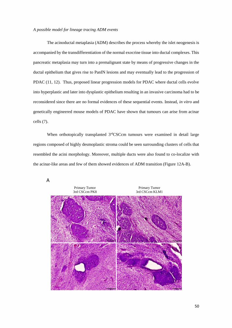
When orthotopically transplanted 3rdCSCcm tumours were examined in detail large
regions composed of highly desmoplastic stroma could be seen surrounding clusters of cells that
resembled the acini morphology. Moreover, multiple ducts were also found to co-localize with
the acinar-like areas and few of them showed evidences of ADM transition (Figure 12A-B).
A
51
ADM transition
GFP Ptf1a
3rd
CS
Ccm
PK
8
3rd
CS
Ccm
KL
M1
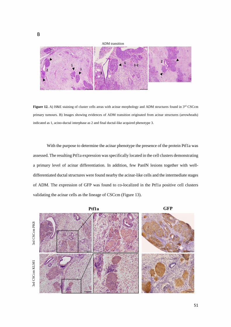
Figure 12. A) H&E staining of cluster cells areas with acinar morphology and ADM structures found in 3rd CSCcm
primary tumours. B) Images showing evidences of ADM transition originated from acinar structures (arrowheads)
indicated as 1, acino-ductal interphase as 2 and final ductal-like acquired phenotype 3.
With the purpose to determine the acinar phenotype the presence of the protein Ptf1a was
assessed. The resulting Ptf1a expression was specifically located in the cell clusters demonstrating
a primary level of acinar differentiation. In addition, few PanIN lesions together with well-
differentiated ductal structures were found nearby the acinar-like cells and the intermediate stages
of ADM. The expression of GFP was found to co-localized in the Ptf1a positive cell clusters
validating the acinar cells as the lineage of CSCcm (Figure 13).
B

52
Figure 13. Cell clusters and few ductal cells were positive for the acinar marker Ptf1a. GFP was equally predominantly
located in cell clusters and few cells from ductal epithelial cells. Micrographs original magnification 10x and 20x
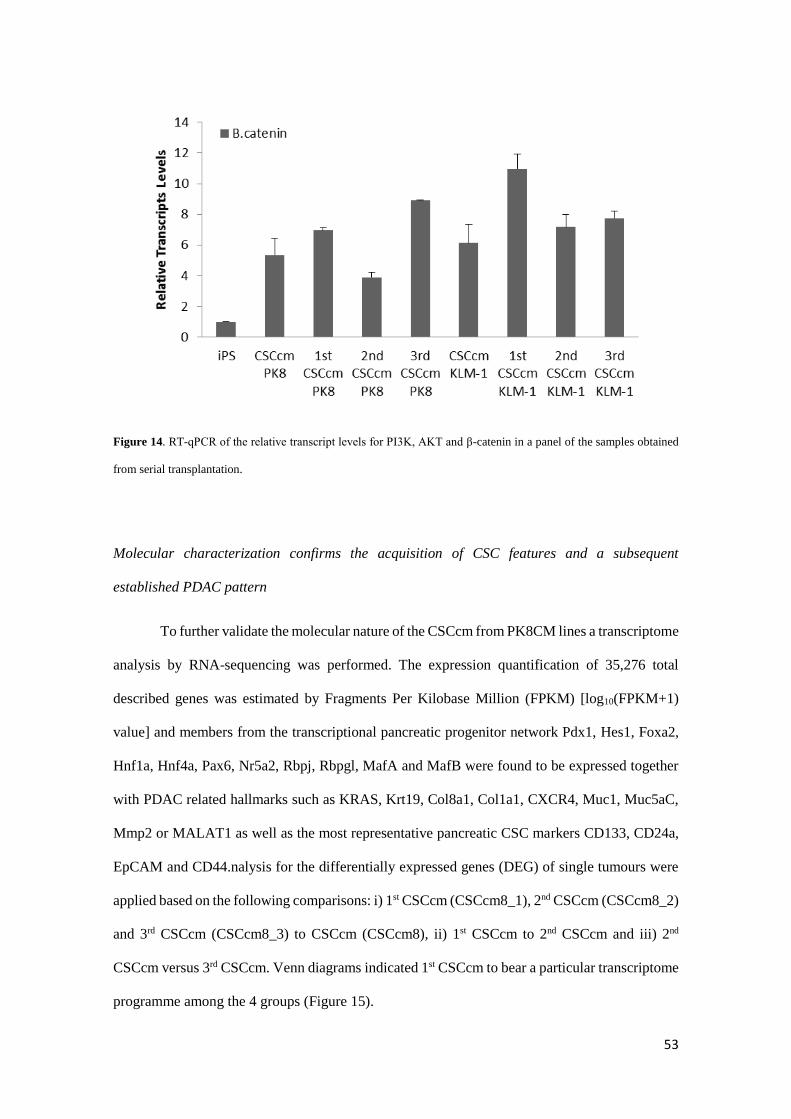
Supporting these evidences we detected the overexpression of Akt which through the
PI3K/Akt pathway have been reported to be involved in the acinar dedifferentiation along with
the accumulation of the β-catenin in the cytoplasm that has also been associated (Figure 14) (13-
16). Since it is important to elucidate whether the process of dedifferentiation plays an important
role in the appearance of early events in pancreatic cancer our CSCcm model may be suitable for
lineage tracing how acinar cells undergo into dedifferentiation losing its mature features and
discern whether eventually develop a malignant transformation.
53
Figure 14. RT-qPCR of the relative transcript levels for PI3K, AKT and β-catenin in a panel of the samples obtained
from serial transplantation.
Molecular characterization confirms the acquisition of CSC features and a subsequent
established PDAC pattern
To further validate the molecular nature of the CSCcm from PK8CM lines a transcriptome
analysis by RNA-sequencing was performed. The expression quantification of 35,276 total
described genes was estimated by Fragments Per Kilobase Million (FPKM) [log10(FPKM+1)
value] and members from the transcriptional pancreatic progenitor network Pdx1, Hes1, Foxa2,
Hnf1a, Hnf4a, Pax6, Nr5a2, Rbpj, Rbpgl, MafA and MafB were found to be expressed together
with PDAC related hallmarks such as KRAS, Krt19, Col8a1, Col1a1, CXCR4, Muc1, Muc5aC,
Mmp2 or MALAT1 as well as the most representative pancreatic CSC markers CD133, CD24a,
EpCAM and CD44.nalysis for the differentially expressed genes (DEG) of single tumours were
applied based on the following comparisons: i) 1st CSCcm (CSCcm8_1), 2nd CSCcm (CSCcm8_2)
and 3rd CSCcm (CSCcm8_3) to CSCcm (CSCcm8), ii) 1st CSCcm to 2nd CSCcm and iii) 2nd
CSCcm versus 3rd CSCcm. Venn diagrams indicated 1st CSCcm to bear a particular transcriptome
programme among the 4 groups (Figure 15).

54
Figure 15. Molecular characterization. A) Venn diagram shows the differentially expressed genes (DEG) of the 5
possible combinations among the CSCcm PK8 lines (left), and the combination of 1st CSCcm, 2nd CSCcm and 3rd
CSCcm versus the CSCcm (right).
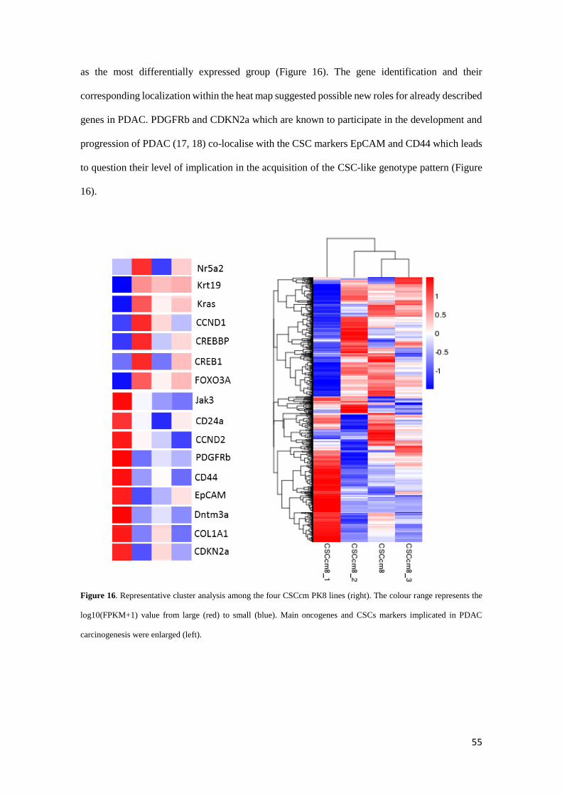
Thus, to gain insights of the similar expression patterns DEG data were arranged in a
cluster analysis using the log10(FPKM+1) value which revealed CSCcm and 3rd CSCcm to be
closer in expression whereas the 2nd CSCcm slightly differed and finally corroborated 1st CSCcm
55
as the most differentially expressed group (Figure 16). The gene identification and their
corresponding localization within the heat map suggested possible new roles for already described
genes in PDAC. PDGFRb and CDKN2a which are known to participate in the development and
progression of PDAC (17, 18) co-localise with the CSC markers EpCAM and CD44 which leads
to question their level of implication in the acquisition of the CSC-like genotype pattern (Figure
16).
Figure 16. Representative cluster analysis among the four CSCcm PK8 lines (right). The colour range represents the
log10(FPKM+1) value from large (red) to small (blue). Main oncogenes and CSCs markers implicated in PDAC
carcinogenesis were enlarged (left).

56
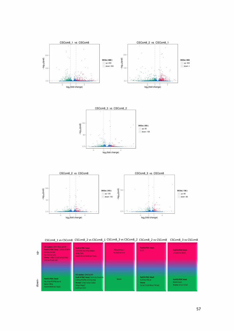
In order to elucidate potential candidates involved in the malignant transformation the
distribution of differentially expressed genes (DESeq) was analysed for each of the
aforementioned comparisons and represented in volcano plots |(log2 (FoldChange)|> 1&qvalue<
0.005) (Figure 17). The transcript levels of pancreatic CSC markers CD24a, EpCAM and CD44
were remarkably enhanced in 1st CSCcm whilst decreased in the subsequent generations. Instead,
the popular hallmarks of PDAC KRAS, Krt19 and Myc were notably activated in 2nd CSCcm and
persisted in 3rd CSCcm. Suggesting that the fate of the 1st CSCcm lineage may probably be
established. The activation of Nr5a2 which its heterozygosity has been recently proposed to likely
contribute to the occurrence of the PDAC (38), was sustained in 2nd CSCcm whereas in 1st CSCcm
was dysregulated. Interestingly, the expression of GATA6 which have been found to be
spontaneously lost in mouse model of Kras(G12V)-driven PDAC was not found in any of the
groups (39). On the other hand, the original converted cell line CSCcm only differed from 2nd
CSCcm in the increased expression of Krt19 and the activation of PI3K in 3rd CSCcm. Since
tumours from all CSCcm lines were rich in desmoplastic stroma we sought for corresponding
PSCs markers. Col8a1, Col1a1 and Col1a2 including TIMPs and Mmp2 which have been linked
to tumour progression and invasion (19-21) were observed in 1st CSCcm. In addition, family
members from CXC/CC chemokines Ccl2, Cxcl1 and Cxcl5 that are reported to favour the
occurrence, maintenance and progression of the tumour (22) were also expressed in a correlated
manner with PSCs markers. In overall, DESeq analysis consistently supported the gain of CSCs
features and the established PDAC molecular pattern in the subsequent generations of CSCcm.
Nonetheless a full transcriptome comparison between other RNA-seq datasets from other tumour
types would be required.
57
up
- d
ow
n-
CSCcm8_1 vs CSCcm8 CSCcm8_2 vs CSCcm8_1 CSCcm8_3 vs CSCcm8_2 CSCcm8_2 vs CSCcm8 CSCcm8_3 vs CSCcm8
Dntm3a Dnmt3b

58
Figure 17. Volcano plots for the differentially expressed genes screening (DESeq) distributed in significantly up-
regulated genes (red), donw-regulated (green) and not differentially expressed (blue). Threshold set as:
|log2(FoldChange)|>1 and qvalue < 0.005. Representative scheme of the up- and down-regulated genes linked to PDAC
occurrence and progression.
“Mutation or not mutation-that is the question”
There is a wide range of genetic abnormalities in PDAC most of them derived from
somatic single nucleotide variants (SNVs). In particular, the activation of the point mutation in
Kras codon 12 has an 85-90% of prevalence in PDAC cases. Several mouse models have
demonstrated that endogenous expression of oncogenic KrasG12D induces phenotypic changes
at molecular and cellular levels that eventually recapitulate PDAC (7, 23-25).
The variants obtained from the screening of repeated reads showed no evidences of single
point mutation in Kras codon 12 nor 13 for any of the groups. In fact, the resulting variants were
found to be located in the 3’-UTR region of the oncogene and were present in all CSCcm lines.
To discern whether the Kras variants had any biological relevance in the malignant transformation
of the CSCcm, and due to iPSCs have a high SNPs variability, we decided to screen the mouse
strains sources from where the original fibroblast was obtained previously to the iPSCs
reprogramming (26). Through the database platforms http://www.informatics.jax.org and
http://www.sanger.ac.uk, DBA mouse strains which are supposed to compose the 50% of the
fibroblast genome were found to harbour the exact Kras SNPs as CSCcm lines. Likewise, the
presence of INDELs were not localised within the oncogene (Table 2). Thereby, there were no
evidences of correlation between the SNPs found in Kras and the malignant transformation.
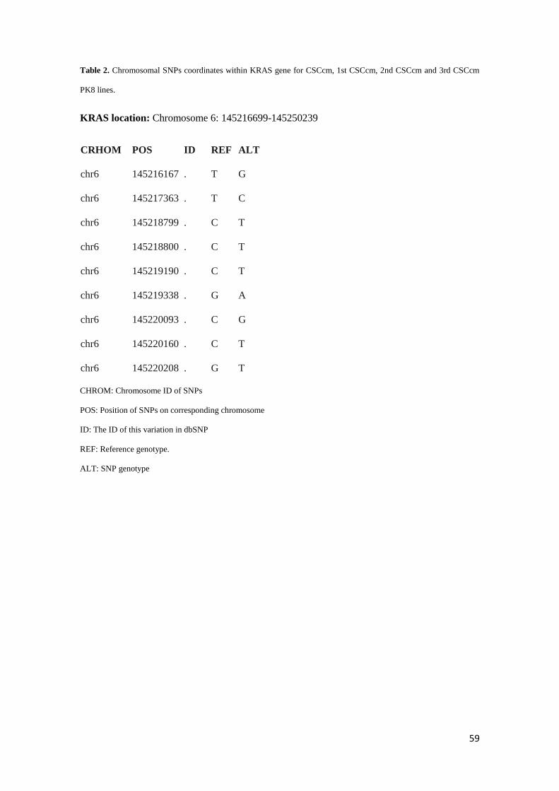
59
Table 2. Chromosomal SNPs coordinates within KRAS gene for CSCcm, 1st CSCcm, 2nd CSCcm and 3rd CSCcm
PK8 lines.
KRAS location: Chromosome 6: 145216699-145250239
CRHOM POS ID REF ALT
chr6 145216167 . T G
chr6 145217363 . T C
chr6 145218799 . C T
chr6 145218800 . C T
chr6 145219190 . C T
chr6 145219338 . G A
chr6 145220093 . C G
chr6 145220160 . C T
chr6 145220208 . G T
CHROM: Chromosome ID of SNPs
POS: Position of SNPs on corresponding chromosome
ID: The ID of this variation in dbSNP
REF: Reference genotype.
ALT: SNP genotype
60
Discussion
PDAC also known as pancreatic ductal adenocarcinoma is the most representative form
of pancreatic cancer. PDAC is characterised by heterogeneous population of tumour cells
structured in CSCs, differentiated cancer cells, tumour-associated PSCs and immune cells (1,4,6).
Similarly to iPSCs, CSCs are considered as cells bearing stem cell properties that give rise to a
diverse lineage of cancer cells. Driven by this convergence between CSCs and iPSCs, we opened
a new avenue to generate new cell lines endowed of CSCs properties and enable to recapitulate
the PDAC tumour phenotype.
Based on the observations that the xenograft tumours generated from CSCcm lines
recapitulate the ductal adenocarcinoma phenotype, and that puromycin-enriched primary cultures
ensured to start over from the top of the hierarchy where CSCs are residing, we can postulate that
CSCcm indeed give rise heterogeneous progenies composed by more differentiated cancer cells
and PSCs in a hierarchical manner. Nevertheless, it is important to note that our results have
pointed out 1st CSCcm lines as the potential candidates to represent the pancreatic CSCs. Thus,
even though the conditioned medium from cancer cell lines provides an appropriate
microenvironment able to initiate a malignant transformation, it is not until CSCcm get in touch
with an in vivo system that the CSCs features become robust at the level of transcriptome.
Supporting this, clear experimental evidences were seen when enriched primary cultures from 1st
CSCcm generated a particular cell distribution wherein specific myofibroblast-like cells arose
favouring the maintenance and stability of the CSCcm colonies. Therefore, the intervention of the
organisms is still essential. Certainly, this reliance on in vivo systems is an unresolved matter that
must eventually be overcome. Recent studies in the tumour microenvironment (TME) have
provided new insights about how the cancerous niche plays a significant role in disease
progression sustaining cell proliferation, activating invasion and metastasis (27). It has been
appreciated for some time the influence of the TME, but the precise function of each constituent
remains unknown. We expect that future analysis on the components of the conditioned medium
61
from cancer cell lines will help to elucidate new mechanisms to optimize the conversion of
CSCcm and end up being independent of the murine organisms.
Important early steps in pancreatic tumour initiation and progression are the genome
reprogramming and dedifferentiation. Thus, the acino-ductal metaplasia has a relevant
implication in the development of the pancreatic cancer. We observed that CSCcm tumours also
generated ADM. In order to confirm the acinar phenotype the expression protein Ptf1a was sought
in tumour samples and strong expression was found in the cell cluster structures. The expression
of Hes1 together with Ptf1a are attributed to the characterization of centroacinar cells (CACs)
which have been ascribed to bear stem-like features (7, 28). Since the RNA-seq expression
quantification analysis indicate Hes1 as one of the most highly expressed genes, prompted us to
question whether Ptf1a positive cells found in CSCcm tumours are either acinar or CACs. Further
experiments will be required in order to clarify the real identity of the cells responsible for the
ADM transition.
PDGFRb and CDKN2a were found to be highly expressed in 1st CSCcm and strongly
decreased in the rest of the groups. The overexpression of PDGFRb has been linked to a poor
disease-free survival promoting metastasis in a cell-autonomous manner in mutant p53 mice and
glioma stem cells have been reported to preferentially express PDGFRb which its activation
promotes self-renewal and its depletion completely abrogated their tumorigenicity (17, 29).
Intriguingly, CDKN2a acts as a tumour suppressor and is usually aberrant in 95% of pancreatic
cancer. This alteration may be due to either an abnormal methylation or several reported germline
mutations which leads to its inactivation (18, 35). When SNPs in CDKN2a were screened in
CSCcm lines there were no evidences of single point mutations, but a single INDEL C<CAA
located in the 3’-UTR region in CDKN2a was present only in 1st CSCcm group (Supplementary
Table II). Nevertheless the same INDEL was found in the murine genome but neither relevant
clinical nor functional information has been described so far. Thus, the sudden enhanced
expression of PDGFRb may be related to the CSCs phenotype of 1st CSCcm. On the other hand,
from the results obtained for CDKN2a we could not discern properly its implication, although its

62
sudden downregulation in 2nd CSCcm and maintained in 3rd CSCcm may be linked to the
establishment of the PDAC phenotype.
As it has been previously aforementioned, the frequency of single point mutations in Kras
occur at 85-90% in pancreatic cancer (25, 30, 31). The activation of the oncogenic Kras together
with its downstream effector Myc have been implicated in self-renewal and tumour plasticity
events such as dedifferentiation (24). In our study we demonstrated that the tumorigenicity of
CSCcm and consequent recapitulation of PDAC phenotype is not given by variants in the
oncogene Kras since no formal evidences were found in SNPs analysis. However, this premature
idea should be developed in extended eQTLs analysis which will provide information at the level
of genotype-gene relation in addition to karyotype and genomic profiling analysis that would
enable clear discrimination. MYC is generally overexpressed in PDAC. In accordance to this
DESeq analysis showed increased expression of Myc in the 2nd CSCcm generation, however it is
important to note that it was remarkably downregulated in 1st CSCcm. Recent findings on
genome-wide DNA methylation profiling in pancreatic CSCs determine CpG sites annotated to
Myc to be more methylated (5). This may be correlated with the overexpression of Dnmt3a and
Dntm3b observed in 1st CSCcm and the subsequent downregulation in 2nd CSCcm. The
predominant modification of DNMTs is the catalysis of the DNA methylation at 5-position
cytosine. Dntm3a and Dnmt3b function as de novo methyltransferases and are highly expressed
in embryonic cells and down-regulated in adult tissues. They have also been involved in self-
renewal and maintenance of colon cancer stem cells, and pancreatic cancer patients with higher
levels have been significantly attributed to have an overall lower survival (41). Thereby, the
activation of Kras and Myc are required but not sufficient to originate a PDAC tumour and their
turnover between 1st CSCcm and more differentiated 2nd CSCcm may be tightly related to
epigenetic alterations.
In the context of inflammation, the combination of Kras and the activation of NFkB have
been reported to induce the conversion of non-stem into stem cell properties of intestinal epithelial
cells (IEC) through the stabilization of β-catenin (32). Despite there are not strong evidences in
63
pancreatic cancer models that implicate NFkB in PDAC progression (25), it was noteworthy that
in 2nd CSCcm the levels of NFkB arose as Kras and Myc did. Experimental evidences are required
to evaluate whether the acquisition of stem-like features may arise in a stochastic way since our
model is based on iPSCs, however this fact may involve NFkB pathway in the prevalence of the
CSCcm in pancreatic cancer cells progeny. On the other hand, inflammatory cells constitute an
important part of the stromal tissue in pancreatic cancer and the existence of a consistent feedback
between PSCs and CXC/CC chemokines family members makes to consider them as potential
candidates to be effectors in the occurrence and progression of the tumour (19-22). Hence, these
facts were corroborated with our DESeq data which indeed showed that the PSCs markers Col81a,
Col1a1, Col1a2 were correlated with the expression of Ccl2, Cxcl1 and Cxcl5 chemokines
interestingly with the stimulation of the CSCs markers overexpression. In addition, IL-33 is
known to activate mast cells and stimulate pro-inflammatory cytokine production and has been
found to be expressed in the nuclei of activated PSCs (36, 37). Intrapancreatic mast cells also
express the cytokine stem cell factor (SCF) which plays a constitutively important role in
proliferation and survival of pluripotent progenitor cells together with its receptor proto-oncogene
tyrosine-kinase KIT (cKIT) (33, 34). Although pancreatic cancer cells express cKIT, its role in
CSCs still needs to be elucidated. Interestingly, in the expression quantification analysis it appears
to be overexpressed and DESeq results highlighted a differential expression of cKIT in 1st CSCcm
indicating that it could be a potential target to abrogate the acquisition of CSCs properties. Thus,
the aforementioned results notably remark the involvement of the inflammatory system as the
main effector for the acquisition of the CSCs properties.
Pancreatic CSCcm is a feasible model for pancreatic CSCs which recapitulates PDAC
phenotype. It is important to note that the conversion process has not been achieved under any
genetic manipulation and the present study has demonstrated a preliminary analysis where the
expected single point mutations were not found. Therefore, our model may provide new insights
about the actual occurrence of the pancreatic cancer leading to develop different approaches for

64
the early detection and find new effectors in order to target CSCs and abrogate the progression of
this fatidic disease.

65
Material & Methods
Cell culture
Human pancreatic carcinoma cell lines PK-8 and KLM-1 (RIKEN cell Bank, Japan) were cultured
in RPMI 1640 Sigma containing 10% FBS and 100 U/mL Penicillin. Undifferentiated mouse
iPSC lines (RIKEN Cell Bank, Japan) were maintained on feeder layers of mouse embryonic
fibroblasts (MEF) mitomycin C treated (ReproCell, Japan) in iPSCs medium (DMEM D5796
Sigma, 15% FBS, 2mM L-Glutamine, 0,1mM NEAA, 50 U/mL Penicillin and 50 U/mL
Streptomycin, 0,1mM 2-mercaptoethanol) supplemented with 1000 U/mL LIF, placed at 37ºC
incubator, 5% CO2 and 100% humidity. The conditioned medium (CM) was prepared following
the previously protocols published by our group, Chen et al. 2012 (9), the medium was collected
from confluent dishes of PK-8 and KLM-1 cell lines populations centrifuged at 1000rpm for 5min
and filtered using 0,22µm diameter pore filter (Millipore, Ireland).
The conversion of the iPSCs into pancreatic cancer stem cells by CM (CSCcm) were generated
as described below. For initiation of the PK-8 CSCcm and KLM-1 CSCcm lines established
undifferentiated feeder-less iPSCs were seeded at 5x10⁵ cells/mL and maintained with iPSCs
medium, without LIF, combined with the CM obtained from PK-8 and KLM-1 cell lines. The
medium was changed every 24h and cells were sub-cultured every 3 days.
For 1st CSCcm, 2nd CSCcm and 3rd CSCcm primary cultures of PK8CM and KLM-1CM mouse
allografts were cut into small pieces in 2mL HBSS and transferred into a 15mL tube containing
2mL of dissociation buffer (0,25% trypsin, collagenase IV, KSR, CaCl2) and were incubated at
37ºC for 40min. The cellular suspension was transferred into a new 15mL tube and centrifuged
at 1000rpm for 10min, iPSCs medium was added to terminate the digestion and again centrifuged
at 1000rpm for 5min. The cell pellet was placed into appropriate volume of iPSCs medium
without LIF and cells were seeded into a dish at a density of 5x10⁵ cells/mL. After 24h cultures
were enriched with 1 mg/mL puromycin and medium was replaced by the combination of CM
66
plus iPSCs medium. Medium was changed every 24h and cells were passaged every 3 days and
cells were maintained no longer than the 5th passage.
Sphere formation assay
4×10⁴ cells (1×10⁴ cells/ml) were seeded on 6cm ultra-low attachment dishes (Corning
incorporated, NY) with the iPSCs medium without FBS, supplied with Insulin-Transferrin-
Selenium-X (ITS-X, Life Technologies, CA). After 3 days primary spheres were dissociated at
single cell with dissociation buffer (0,25% trypsin, collagenase IV, KSR, CaCl2) and 1x10⁴ were
transferred into a new ultra-low attachment 6cm dish for 3 days more. Spheroids with diameters
above 100μm were judged as self-renewing spheroids.
Animal experiments
Nude mice (Balb/c -nu/nu, female, 4- to 6- week-old) were purchased from Charlesriver, Japan.
For subcutaneous transplantation 104, 105 and 106 cells were suspended in 100µl of HBSS buffer.
The tumour size and weight were recorded every 4 days. Pancreatic orthotopic transplantation
was performed as previously described by Bruns et al. 1999 (40) with the only variation in the
number of implanted cells (105/20µl and 106/20µl HBSS). The mice were housed under specific
pathogen-free conditions in accordance with the plan of animal experiments reviewed and
approved by the ethics committee for animal experiments of Okayama University under the IDs
OKU-2008211, OKU-2009144, OKU-2010179 and OKU-2011-305.
Histologic analysis
Tumours were fixed in 4% PFA (WAKO, Japan) for 24h at 4ºC, washed in 20% sucrose solution,
embedded in paraffin-wax and sectioned for histologic examination at 5µm. Sections were stained
with hematoxylin and eosin (Hematoxylin solution, Sigma-Aldrich, MO; 0.5% Eosin Y, Sigma
Aldrich, MO).
67
Immunohistochemistry (IHC)
Before staining, paraffin was removed from sections using an oven at 60ºC for 30min, rehydrated
and underwent antigen retrieval by treatment of 10mM citrate buffer (pH6.5) in a water bath at
100ºC for 30min. After hydrogen peroxide blocking (3% H2O2), slides were blocked with PBS +
1.5% normal serum, incubated with primary antibodies overnight at 4ºC, rinsed with PBS,
incubated with secondary antibody. Detection was accomplished using Ellite anti-rabbit and anti-
mouse ABC staining Vectastain kit (Vector, MI) and 3,30-diaminobenzidine tetrahydrochloride
(DAB, Vector, MI). Counterstain was achieved with hematoxylin. Incubation of primary
antibodies was carried out in 2.5% normal serum for rabbit polyclonal CD133 (1:100, #NB120-
16518, Novus Biologicals, USA), rabbit monoclonal GFP (1:200, #2956, Cell Signaling, MA),
rabbit monoclonal MUC1 (1:100, #ab15481, Abcam, UK), mouse monoclonal MUC5aC (1:200,
#NCL-MUC-5AC, Novocastra, Leica Biosystems Newcastle, UK), PTF1a mouse monoclonal
(1:100 ,#sc-393011, Santa Cruz Biotechnology INC., Europe). Slides served as negative controls
were incubated with PBS buffer.
RNA preparation and RT-qPCR
Total RNA was isolated by RNeasy Mini Kit (QIAGEN, Germany) and treated with DNase
Amplification Grade (Invitrogen, CA). cDNA synthesis was performed using SuperScript III First
strand kit (Invitrogen, CA). RT-qPCR was performed with Cycler 480 SYBR Green I Master mix
(Roche, Switzerland). Primers used are listed below:
Name Primer sequence (5‘ > 3‘)
Fw CD24a TTCTGGCACTGCTCCTACC
Rv CD24a GCGTTACTTGGATTTGGGGAA
Fw CD133 CCTTGTGGTTCTTACGTTTGTTG
Rv CD133 CGTTGACGACATTCTCAAGCTG
Fw B-Catenin TCCCATCCACGCAGTTTGAC
68
Rv B-Catenin TCCTCATCGTTTAGCAGTTTTGT
Fw PI3K TGGGACCTTTTTGGTACGAGA
Rv PI3K AGCTAAAGACTCATTCCGGTAGT
Fw Akt GGCCCCTGACCAGACCTTA
Rv Akt GATAGCCCGCATCCACTCTTC
Fw Pdx1 ATTCTTGAGGGCACGAGAGC
Rv Pdx1 GGTCCGTATTGGAACGCTCA
Fw Foxa2 TGGTCACTGGGGACAAGGGAA
Rv Foxa2 GCAACAACAGCAATAGAGAAC
Fw Hes1 CGGCATTCCAAGCTAGAGAAGG
Rv Hes1 GGTAGGTCATGGCGTTGATCTG
Fw Kras CAAGAGCGCCTTGACGATACA
Rv Kras CCAAGAGACAGGTTTCTCCATC
Fw EpCAM CTGGCGTCTAAATGCTTGGC
Rv EpCAM CCTTGTCGGTTCTTCGGACTC
Fw Tg Klf4 GCGAACTCACACAGGCGAGAAACC
Rv Tg Klf4 TTATCGTCGACCACTGTGCTGCTG
Fw Tg c-Myc CAGAGGAGGAACGAGCTGAAGCGC
Rv Tg c-Myc TTATCGTCGACCACTGTGCTGCTG

69
RNA-seq library construction and sequencing
Isolation of total RNA was performed using QIAGEN RNeasy kit. RNA samples were prepared
for sequencing using Illumina TruSeq RNA Sample Preparation Kit and were sequenced in an
Illumina HiSeq 2500. Sample preparation, RNA-sequencing and Bioinformatic analysis were
carried out by Fligen, INC. (Novogene, Nagoya Japan).
70
References
1 Zhan HX, Xu JW, Wu D, Zhang TP, Hu SY. Pancreatic cancer stem cells: New insight
into a stubborn disease. Cancer Lett 2014; 357:429-37
2 Shakya R, Gonda T, Quante M, Salas M, Kim S, Brooks J, Hirsch S, Davies J, Cullo A,
Olive K, Wang TC, Szabolcs M, Tycko B, and Ludwig T. Hypomethylating therapy in an
aggressive stroma-rich model of pancreatic carcinoma. Cancer Res 2012; 73: 885–896.
3 Sainz B Jr, Martín B, Tatari M, Heeschen C, Guerra S. ISG15 is a critical
microenvironmental factor for pancreatic cancer stem cells. Cancer Res 2014; 74:7309-20
4 Hermann PC, Huber SL, Herrler T, Aicher A, Ellwart JW, Guba M, Bruns CJ, Heeschen
C. Distinct populations of cancer stem cells determine tumor growth and metastatic activity in
human pancreatic cancer. Cell Stem Cell 2007; 1:313-23
5 Zagorac S, Alcala S, Fernandez Bayon G, Bou Kheir T, Schoenhals M, González-Neira
A, Fernandez Fraga M, Aicher A, Heeschen C, Sainz B Jr. DNMT1 Inhibition Reprograms
Pancreatic Cancer Stem Cells via Upregulation of the miR-17-92 Cluster. Cancer Res 2016;
76:4546-58
6 Sancho P, Alcala S, Usachov V, Hermann PC, Sainz B Jr.The ever-changing landscape
of pancreatic cancer stem cells. Pancreatology 2014; 16:489-96
7 Rooman I, Real FX: Pancreatic ductal adenocarcinoma and acinar cells: a matter of
differentiation and development? Gut 2012; 61:3 449-458
8 T Kasai, L Chen, AZ Mizutani, T Kudoh, H Murakami, L Fu and M Seno. Cancer stem
cells converted from pluripotent stem cells and the cancerous niche. J Stem Cells Regen Med
2014; 10:2-7
9 Chen L, Kasai T, Li Y, Sugii Y, Jin G, Okada M, Vaidyanath A, Mizutani A, Satoh A,
Kudoh T, Hendrix MJ, Salomon DS, Fu L, Seno M. A model of cancer stem cells derived from
mouse induced pluripotent stem cells. PLoS One 2012; 7:e33544
71
10 Bailey P, Chang DK, Nones K, Johns AL, Patch AM, Gingras MC, Miller DK, Christ
AN, Bruxner TJ, Quinn MC, Nourse C, Murtaugh LC, Harliwong I, Idrisoglu S, Manning S,
Nourbakhsh E, Wani S, Fink L, Holmes O, Chin V, Anderson MJ, Kazakoff S, Leonard C, Newell
F, Waddell N, Wood S, Xu Q, Wilson PJ, Cloonan N, Kassahn KS, Taylor D, Quek K, Robertson
A, Pantano L, Mincarelli L, Sanchez LN, Evers L, Wu J, Pinese M, Cowley MJ, Jones MD, Colvin
EK, Nagrial AM, Humphrey ES, Chantrill LA, Mawson A, Humphris J, Chou A, Pajic M, Scarlett
CJ, Pinho AV, Giry-Laterriere M, Rooman I, Samra JS, Kench JG, Lovell JA, Merrett ND, Toon
CW, Epari K, Nguyen NQ, Barbour A, Zeps N, Moran-Jones K, Jamieson NB, Graham JS, Duthie
F, Oien K, Hair J, Grützmann R, Maitra A, Iacobuzio-Donahue CA, Wolfgang CL, Morgan RA,
Lawlor RT, Corbo V, Bassi C, Rusev B, Capelli P, Salvia R, Tortora G, Mukhopadhyay D,
Petersen GM; Australian Pancreatic Cancer Genome Initiative, Munzy DM, Fisher WE, Karim
SA, Eshleman JR, Hruban RH, Pilarsky C, Morton JP, Sansom OJ, Scarpa A, Musgrove EA,
Bailey UM, Hofmann O, Sutherland RL, Wheeler DA, Gill AJ, Gibbs RA, Pearson JV, Waddell
N, Biankin AV, Grimmond SM. Genomic analyses identify molecular subtypes of pancreatic
cancer. Nature 2016; 531:47-52
11 Schmid RM: Acinar-to-ductal metaplasia in pancreatic cancer development. J Clin Invest
2002. 109: 1403–1404.
12 Means AL, Meszoely IM, Suzuki K, Miyamoto Y, Rustgi AK, Coffey RJ Jr, Wright CV,
Stoffers DA, Leach SD. Pancreatic epithelial plasticity mediated by acinar cell
transdifferentiation and generation of nestin-positive intermediates. Development. 2005 132:
3767-3776
13 Elghazi L, Weiss AJ, Barker DJ, Callaghan J, Staloch L, Sandgren EP, Gannon M, Adsay
VN and Bernal-Mizrachi E. Regulation of pancreas plasticity and malignant transformation by
Akt signaling. Gastroenterology 2009; 136:1091e103.

72
14 Jensen JN, Cameron E, Garay MV, Starkey TW, Gianani R, Jensen J. Recapitulation of
elements of embryonic development in adult mouse pancreatic regeneration. Gastroenterology
2005; 128:728e41.
15 Minami K, Okano H, Okumachi A, Seino S. Role of Cadherin mediated Cell-Cell
Adhesion in Pancreatic Exocrine-to-Endocrine Transdifferentiation. J Biol Chem 2008;
283:13753e61
16 Morris JP 4th, Cano DA, Sekine S, Wang SC, Hebrok M. Beta-catenin blocks Kras
dependent reprogramming of acini into pancreatic cancer precursor lesions in mice. J Clin Invest
2010; 120:508e20.
17 Weissmueller S, Manchado E, Saborowski M, Morris JP 4th, Wagenblast E, Davis CA,
Moon SH, Pfister NT, Tschaharganeh DF, Kitzing T, Aust D, Markert EK, Wu J, Grimmond SM,
Pilarsky C, Prives C, Biankin AV, Lowe SW. Mutant p53 drives pancreatic cancer metastasis
through cell-autonomous PDGF receptor β signaling. Cell 2014; 157(2):382-94
18 Rachakonda PS, Bauer AS, Xie H, Campa D, Rizzato C, Canzian F, Beghelli S, Greenhalf
W, Costello E, Schanne M, Heller A, Scarpa A, Neoptolemos JP, Werner J, Büchler M, Hoheisel
JD, Hemminki K, Giese N, Kumar R. Somatic mutations in exocrine pancreatic tumors:
association with patient survival. PLoS One 2013; 8(4):e60870
19 Sato N, Maehara N, Goggins M. Gene expression profiling of tumor-stromal interactions
between pancreatic cancer cells and stromal fibroblasts. Cancer Res. 2004; 64(19):6950–6956
20 Li X, Ma Q, Xu Q, Duan W, Lei J, Wu E. Targeting the Cancer-Stroma Interaction: A
Potential Approach for Pancreatic Cancer Treatment. Curr Pharm Des. 2012; 18(17): 2404–2415.
21 Seton-Rogers S: Pancreatic cancer: Fibroblast co-conspirators. Nature Reviews Cancer
2011; 758-759
73
22 Wang S, Wu Y, Hou Y, Guan X, Castelvetere MP, Oblak JJ, Banerjee S, Filtz TM, Sarkar
FH, Chen X, Jena BP, Li C. CXCR2 Macromolecular Complex in Pancreatic Cancer: A Potential
Therapeutic Target in Tumor Growth. Transl Oncol 2013; 6(2): 216–225.
23 Ischenko I, Petrenko O, Hayman MJ. Analysis of the tumor-initiating and metastatic
capacity of PDX1-positive cells from the adult pancreas. Proc Natl Acad Sci USA 2014;
111(9):3466-71.
24 Ischenko I, Zhi J, Moll UM, Nemajerova A, Petrenko O. Direct reprogramming by
oncogenic Ras and Myc. Proc Natl Acad Sci USA 2013; 110(10):3937-42
25 Guerra C, Schuhmacher AJ, Cañamero M, Grippo PJ, Verdaguer L, Pérez-Gallego L,
Dubus P, Sandgren EP, Barbacid M. Chronic pancreatitis is essential for induction of pancreatic
ductal adenocarcinoma by K-Ras oncogenes in adult mice. Cancer Cell 2007; 11(3):291-302
26 Fujishiro SH, Nakano K, Mizukami Y, Azami T, Arai Y, Matsunari H, Ishino R,
Nishimura T, Watanabe M, Abe T, Furukawa Y, Umeyama K, Yamanaka S, Ema M, Nagashima
H, Hanazono Y. Generation of naive-like porcine-induced pluripotent stem cells capable of
contributing to embryonic and fetal development. Stem Cells Dev 2013; 22(3):473-82
27 Chen F, Zhuang X, Lin L, Yu P, Wang Y, Shi Y, Hu G, Sun Y. New horizons in tumor
microenvironment biology: challenges and opportunities. BMC Med. 2015; 13:45
28 Rovira M, Scott SG, Liss AS, Jensen J, Thayer SP, Leach SD. Isolation and
characterization of centroacinar/terminal ductal progenitor cells in adult mouse pancreas. Proc
Natl Acad Sci U S A 2010; 107:75e80
29 Jiang Y, Boije M, Westermark B, Uhrbom L. PDGF-B can sustain self-renewal and
tumorigenicity of experimental glioma-derived cancer-initiating cells by preventing
oligodendrocyte differentiation. Neoplasia. 2011, 13: 492-503

74
30 Ben-Porath I, Thomson MW, Carey VJ, Ge R, Bell GW, Regev A, Weinberg RA. An
embryonic stem cell-like gene expression signature in poorly differentiated aggressive human
tumors. Nat Genet 2008; 40: 499 – 507
31 Gidekel Friedlander SY, Chu GC, Snyder EL, Girnius N, Dibelius G, Crowley D, Vasile
E, DePinho RA, Jacks T. Context-dependent transformation of adult pancreatic cells by
oncogenic K-Ras. Cancer Cell 2009; 16: 379 – 389
32 Schwitalla S1, Fingerle AA, Cammareri P, Nebelsiek T, Göktuna SI, Ziegler PK, Canli
O, Heijmans J, Huels DJ, Moreaux G, Rupec RA, Gerhard M, Schmid R, Barker N, Clevers H,
Lang R, Neumann J, Kirchner T, Taketo MM, van den Brink GR, Sansom OJ, Arkan MC, Greten
FR. Intestinal tumorigenesis initiated by dedifferentiation and acquisition of stem-cell-like
properties. Cell 2013; 152: 25 – 38
33 Esposito I1, Kleeff J, Bischoff SC, Fischer L, Collecchi P, Iorio M, Bevilacqua G, Büchler
MW, Friess H. The stem cell factor-c-kit system and mast cells in human pancreatic cancer. Lab
Invest 2002; 82(11):1481-92.
34 Yasuda A, Sawai H, Takahashi H, Ochi N, Matsuo Y, Funahashi H, Sato M, Okada Y,
Takeyama H, Manabe T. The stem cell factor/c-kit receptor pathway enhances proliferation and
invasion of pancreatic cancer cells. Mol Cancer. 2006; 5: 46.
35 Zhao R, Choi BY, Lee MH, Bode AM, and Donga Z. Implications of Genetic and
Epigenetic Alterations of CDKN2A (p16INK4a) in Cancer. EBioMedicine 2016; 8: 30–39.
36 Xu D, Jiang HR, Kewin P, Li Y, Mu R, Fraser AR, Pitman N, Kurowska-Stolarska M,
McKenzie AN, McInnes IB, Liew FY. IL-33 exacerbates antigen-induced arthritis by activating
mast cells. Proc. Natl. Acad. Sci. U.S.A. 2008; 105, 10913–10918.
37 Masamune A, Watanabe T, Kikuta K, Satoh K, Kanno A, Shimosegawa T. Nuclear
expression of interleukin-33 in pancreatic stellate cells. Am. J. Physiol. Gastrointest. Liver
Physiol 2010; 299, G821–G832.
75
38 Flandez M, Cendrowski J, Cañamero M, Salas A, del Pozo N, Schoonjans K, Real FX.
Nr5a2 heterozygosity sensitises to, and cooperates with, inflammation in KRas(G12V)-driven
pancreatic tumourigenesis. Gut. 2014; 63(4):647-55
39 Martinelli P, Madriles F, Cañamero M, Pau EC, Pozo ND, Guerra C, Real FX. The acinar
regulator Gata6 suppresses KrasG12V-driven pancreatic tumorigenesis in mice. Gut. 2016;
65(3):476-86
40 Bruns CJ, Harbison MT, Kuniyasu H, Eue I, Fidler IJ. In Vivo Selection and
Characterization of Metastatic Variants from Human Pancreatic Adenocarcinoma by Using
Orthotopic Implantation in Nude Mice. Neoplasia. 1999; 1(1): 50–62.
41 Subramaniam D, Thombre R, Dhar A, Anant S. DNA methyltransferases: a novel target
for prevention and therapy. Front Oncol. 2014; 4:80.
42 Okita K, Ichisaka T, Yamanaka S. Generation of germline-competent induced pluripotent
stem cells. Nature. 2007; 448(7151):313-7
76
Acknowledgements
My sincere gratitude to Professor Masaharu Seno for trusting me and give me such a great
opportunity to be part of this amazing project. From him I have learnt that with passion,
determination and hard work dreams may come true. I will always remember your words when
you said that before start the PhD I seemed like a drifting boat, and you decided to take me. Thank
you Sensei for this wonderful trip and for helping me to get back on track. I will never forget it.
Next, I want to thank with very much appreciation to my mentor Doctor Tomonari Kasai, from
whom I learnt everything I know. Thanks to him I realized that being a researcher could be
possible for me. He has trusted my skills even when he had no reasons, he has listened up to each
of my improbable stories and he has always given me his best answers and solutions at any time.
Thank you Sensei for sharing these great moments full of laughs, exciting discussions and
motivational support, without you would not have been possible. Moltes gracies. 毛虫寿司.
Special thanks to Professor Ohtsuki and Professor Tokumitsu for kindly reviewing my thesis.
To Mami Asakura San, always willing to help and always with the warmest smile. Thank you
very much for all the support during all these years.
To Kaoru Furuse San for her kindly assistance and help.
Thank you to Dr. Hiroshi Murakami and Dr. Akifumi Mizutani for their assistance and motivation.
To Dr. Junko Masuda for being a professional inspiration example.
To the assistant professors Arun and Seno San thank you for their assistance.
Dear April we are proud of you, trust yourself more. Oo San the golden boy. You are meant to be
a professor, and with no doubts you will be the best. Fizah, keep on working hard and these efforts
will have reward. To all of you I wish all the best for your career because you really deserve it.

77
Laura my lovely Mary. For all the cupcakes with Hello flavour you made us. Cheers to those
nights watching videos of cells budding while drinking beer and eating cakes in a 4th floor hidden
café. Will see you in Hawaii amore.
To Marta, the twinkle-twinkle star who jumped into my heart since the very beginning when she
received me after 30 hours of the most endless trip in my return to Japan. And since then and with
a 100% of success tried to put a smile on my face every single moment we have spent together.
Moltes gracies pel teu suport constant i per tot reina.
My dear Maram, what to say. I can’t be more thankful to your God for having sent you here. I
will always remember after the 3rd day we met when you said “Now you complain but you are
going to miss me”. Well, guess what? You just came as a hurricane into my peaceful japanese
life, but you came to stay habiby. Much love.
Neha. I have no words left for you. You are the best fiction couple a girl like me could ever had.
From strangers to soulmates, how did it happened? Thank you for our late night scientific
discussions text messages, for the trips, for the foods and the drinks Oh Mary!, for the million
times we shared lots of laughter and for those very few we shared some tears. You never let me
feel alone. Thank you one more time for your unconditional love and support. Love you so sister.
My family. The best I have in my life. They have always understood me, supporting my decisions
and never interpose their feelings over mine. They always believed in me. Your unconditional
love is everything to me. Love you very much.
La meva família. El millor que tinc a la vida. Sempre m’han entès, donant-me suport en les meves
decisions i mai interposant els seus sentiments als meus. Ells sempre han cregut en mi. El vostre
amor incondicional és tot per a mi. Us estimo molt.
Mi familia. Lo mejor que tengo en mi vida. Siempre me han entendido, apoyando mis decisiones
y nunca interponiendo sus sentimientos a los míos. Ellos siempre creyeron en mí. Vuestro amor
incondicional lo es todo para mí. Os quiero mucho.

78
Coming to Okayama, the best decision I have ever made so far.
With so much affection.
Anna.

79