Embed Size (px)
Citation preview
TitleStudies on the regulation of preadipocyte differentiation byparacrine factors secreted from muscle cells( Dissertation_全文)
Author(s) Hirai, Shizuka
Citation Kyoto University (京都大学)
Issue Date 2006-03-23
URL https://doi.org/10.14989/doctor.k12364
Right
Type Thesis or Dissertation
Textversion author
Kyoto University
Studies on the regulation of preadipocyte differentiation
by paracrine factors secreted from muscle cells
SHIZUKA HIRAI
2006
924
Studies on the regulation of preadipocyte differentiation
by paracrine factors secreted from muscle cells
SHIZUKA HIRAI
2006
CONTENTS
Chapter 1: Pagel
Introduction
Chapter 2: Page6
Review of Literature
Chapter 3: Page 18
Effects of the conditioned medium from C2C12 myocyte on the differentiation
of 3T3-L1 preadiopocyte
Chapter 4:
Effects of myostatin on the differentiation of bovine preadipocyte
Chapter 5:
The role of activin A on the preadipocyte differentiation
Page28
Page50
Section 1. Effects of activin A on the differentiation of bovine preadipocyte
Section 2. The mechanism of the inhibitory effect of activin A on the
differentiation of 3T3-L1 preadipocyte
Summary
References
Acknowledgement
Page91
Page95
Page 119
Chapter 1
Introduction
Beef marbling is characterized by adipose tissue deposition within skeletal
muscle in cattle and is one of the important meat quality traits that influences
juiciness and flavor of beef, which contributes directly to the value of beef on
especially Japanese market. It has been accepted that high-energy grain diets
achieve higher marbling than pasture diets (Price and Berg, 1981). The lipid
content of beef depends on breed, sex, diet, and length of fattening (Cianzio et aI.,
1985; Gills and Eskin, 1973; Price and Berg, 1981). Japanese black cattle
(Wagyu) are well known for its ability to produce high marbling (Lunt et aI., 1993,
Zenbayashi, 1994). The average marbling score according to the Japanese
Carcass Grading Standards of the M.longissimus thoracis (LT) is 6.0. This value
corresponds to approximately 17% intramuscular fat in LT (Cameron et aI.,
1994).
Amount of adipose tissue in an anima.l depend primarily on the number and of
size of the constituent adipocytes (Waters, 1909). The increase in adipocyte
number is considered to result from the proliferation and differentiation of
preadipocytes, adipocyte precursor cells. Although the proliferation and
differentiation of preadipocytes were completed in the perirenal and
subcutaneous adipose tissues by the first year of age in cattle (Garbutt et aI.,
1979; Hood and Allen, 1973), intramuscular adipose tissue evidently was still
actively growing by proliferation and differentiation in steers at 14 months of age
- 1 -
"
(Hood and Allen, 1973). Cianzio et aI. (1985) also reported that the development
of beef marbling was closely associated with an increase in adipocyte number
within muscle during 13 to 19 months of age.
Many researchers reported the positive relationship between body fat
accumulation and beef marbling in some foreign breeds of beef cattle (Riley et aI.,
2003; Wertz et aI., 2001). On the other hand, some researchers showed no
positive relationship between back fat depth and beef marbling in Japanese
black cattle (Mukai et aI., 1995; Yang et aI., 1985). These results suggest that the
systemic fat metabolism does not necessarily reflect the intramuscular fat
deposition in this breed, therefore, locally produced factors may contribute to the
development of adipocytes within muscle in Japanese black cattle. As
intramuscular preadipocytes are surrounded by mature skeletal muscle
myofibers, it is predicted that paracrine factors secreted from muscle fibres may
regulate the differentiation of preadipocyte via intercellular interactions. The
possibility of a direct regulatory interaction between myofibers and adipocytes
was suggested in an early study by Jarett et aI. (1985), who observed that
skeletal muscle in insulin-treated rats secreted a paracrine agent (or mediator)
which stimulated pyruvate dehydrogenase and glycogen synthase activity in
adipocytes.
Muscle fibers secrete various factors, such as fibroblast growth factor (FGF) ,
transforming growth factor-B (TGF-B), tumor necrosis factor-a (TNF-u),
insulin-like growth factors (IGFs) , hepatocyte growth factor (HGF), and
interleukin"6 (IL-6) (Charge et aI., 2004). These cytokines are known to regulate
the proliferation and differentiation of preadipocytes, as well as of myoblasts
- 2 -
(Charge et aI., 2004). Although IGF-1 promotes both proliferation and
differentiation of preadipocytes, other growth factors and cytokines are generally
considered as inhibitors of adipocyte differentiation (Choy et aI., 2000, Ohsumi et
aI., 1994, Torti et aI., 1989).
In most cell culture models, TGF-B is a potent inhibitor of adipocyte
differentiation (Choy et aI., 2000; Petruschke et aI., 1994; Vassaux et aI., 1994).
TGF-B is expressed in cultured preadipocytes and in adipose tissue In VIVO,
however, transgenic overexpression of TGF-B in adipose tissue inhibits
preadipocyte differentiation (Clouthier et aI., 1997).
Activin is a multifunctional growth and differentiation factor that belongs to
the TGF-B superfamily. Activin was discovered for its ability to regulate
follicle-stimulating hormone (FSH) production by pituitary cells (Ling et aI.,
1986; Vale et aI., 1986). Activin is a dimeric protein synthesized as a homo- or
heterodimer of the district B subunits (BA or BB), which combine to form activin A
(BA-BJ, activin B (BB-BB), or activin AB (BA-BB). Activin exerts its biological effects
by interacting with four types of transmembrane receptors (type lA, IB, IIA, lIB)
with protein serinelthreonin kinase activity (Attisano et aI., 1996). Activin BA
subunit (Meunier et aI., 1998; Tuuri et aI., 1994) and activin receptors (Feijen et
aI., 1994) are expressed in many tissues throughout the body, and activin A is
reported to act on many cell types including myoblast (Link and Nishi, 1997;
Shiozuka et aI., 1997). However, its action on preadipocyte differentiation is not
still investigated.
Myostatin (growth/differentiation factor-8 (GDF-8» is also a member ofTGF-B
superfamily that is essential for proper regulation of skeletal muscle mass (Lee
- 3 -
and McPherron, 1999). Mice carrying a targeted deletion of the gene encoding
myostatin have a dramatic and widespread increase in muscle mass, the result of
both hyperplasia and hypertrophy of muscle fibers, suggesting that myostatin
normally acts as a negative regulator of muscle growth (McPherron et aI., 1997).
Mutation of myostatin gene also results in increasing skeletal muscle in certain
breeds of cattle (Belgian Blue and Piedmontese), which is known as double
muscling (Kambadur et aI., 1997). On the other hand, the reduction of fat
accumulation is observed in knock-out mice (Lin et aI., 2002; McPherron and Lee,
2002) and in the double-muscled cattle (Kobolak and Gocza, 2002). Myostatin is
first expressed in somites, in the myotome layer that gives rises to skeletal
muscle (McPherron et aI., 1997), and is highly expressed in skeletal muscle at
later developmental stages and in adults. The expression of myostatin has been
also detected in both fetal and adult heart, and adipose tissue, but the expression
of myostatin is substantially lower in adipose tissue than in skeletal muscle
(McPherron et aI., 1997; Sharma et aI., 1999). Myostatin exerts its biological
function by binding to the activin receptors (Lee and McPherron 2001; Massague
and Chen, 2000), and is reported to inhibit the differentiation of mouse 3T3-L1
preadipocyte (Kim et aI., 2001). However, the action of myostatin on bovine
preadipocyte differentiation is still unknown. Furthermore, it is not clarified how
myostatin regulates postnatal fat accumulation.
Follistain is a monomeric glycoprotein first isolated from ovarian follicular
fluid on the basis of its ability to suppress FSH secretion by pituitary cells
(Robertson et aI., 1987; Deno et aI., 1987) Follistatin captures activin A and
neutralizes its activity (Schneyer et aI., 2003). Recently, follistatin is also
- 4 -
reported to capture myostatin, which prevents myostatin binding to activin
receptor (Lee and McPherron, 2001). The studies on gene deletion (Matzuk et aI.,
1995) and overexpression (Lee and McPherron, 2001) offollistatin demonstrated
that follistatin neutralized the inhibitory effect of its sensitive ligands on muscle
development, and myostatin is considered to be an obvious candidate for the
sensitive ligand. Follistatin is highly expressed in ovary, however, it is also
widely distributed in non-gonadal tissues including muscle and adipose tissue
(Schneider et aI., 2000), predicting that the follistatin attenuate activin A and
myostatin action on adipogenesis through forming the inactive complex in
muscular and adipose tissue.
The purpose of this study was to clarify the regulation of preadipocyte
differentiation by paracrine factors secreted from muscle cells. Especially, the
author targeted the effect of the TGF-B superfamily members, activin and
myostatin, and investigated their effect on the differentiation of bovine
preadipocytes in stromal vascular cells derived from adipose tissue. The author
also examined the mechanism of the inhibitory action of activin A on the
differentiation of mouse 3T3-L1 preadipocyte.
- 5 -
Chapter 2
Review of Literature
Beef Marbling
Beef marbling is characterized by adipose tissue deposition within skeletal
muscle in cattle and is one of the important meat quality traits that influences
juiciness and flavor of beef, which contributes directly to the value of beef on
especially Japanese market. It has been accepted that high-energy grain diets
achieve higher marbling than pasture diets (Price and Berg, 1981). The lipid
content of beef depends on strains, sex, diet, and length of fattening. Japanese
black cattle (Wagyu) is well known for its ability to produce high marbling (Lunt
et al., 1993, Zenbayashi, 1994). The average slaughter age of Japanese Black
cattle is 29.6 months, and 422 kg, respectively, in Japan. The average marbling
score according to the Japanese Carcass Grading Standards of the M.
longissimus thoracis (LT) is 6.0. This value corresponds to approximately 17%
intramuscular fat in LT (Cameron et al., 1994).
Amount of adipose tissue in an animal depend primarily on the number and of
size of the constituent adipocytes (Waters, 1909). It has been established that an
increase in adipocyte number spontaneously occurs in mature animals if they are
given high-energy diets (Faust et al., 1978; Miller et al., 1984). The increase in
adipocyte number is considered to result from the proliferation and
differentiation of preadipocytes, adipocyte precursor cells. Although the
proliferation and differentiation of preadipocytes were completed in the perirenal
- 6 -
and subcutaneous adipose tissues by the first year of age in cattle (Garbutt et al.,
1979; Hood and Allen, 1973), intramuscular adipose tissue evidently was still
actively growing by proliferation and differentiation in steers at 14 months of age
(Hood and Allen, 1973). Cianzio et al. (1985) also reported that the development
of beef marbling was closely associated with an increase in adipocyte number
within muscle during 13 to 19 months of age.
Many researches reported the positive relationship between body fat
accumulation and beef marbling in some foreign breeds of beef cattle (Riley et al.,
2003; Wertz et al., 2001). On the other hand, some researchers showed no
positive relationship between back fat depth and beef marbling in Japanese
Black cattle (Mukai et al., 1995; Yang et al., 1985). These results suggest that the
systemic fat metabolism does not necessarily reflect the intramuscular fat
deposition in this breed. Therefore, locally produced factors may contribute to the
development of adipocytes within muscle in Japanese black cattle.
Development of myocyte and adipocyte
During mammalian development, the embryonic mesoderm gIves rise to
several highly specialized cell types, including skeletal myocytes, adipocytes, and
chondrocytes (Cornelius et al., 1994; Taylor and Jones, 1979; Watt, 1991). The
development of distinct cell types from multipotent mesodermal precursors can
be viewed as a two'step process (Sager and Kovac, 1982; Weintraub et al., 1991).
In the first step, termed commitment or determination, the developmental
potential of the cell becomes limited to one particular lineage, be it adipose,
muscle, or cartilage. In the second step, terminal differentiation, the cell
. 7 .
develops along its determined lineage to become a functional cell.
The C3H 10T1I2 cell line was established in 1973 from 14- to 17-day-old C3H
mouse embryos (Reznikoff et aI., 1973). These cells display fibroblastic
morphology in cell culture and are functionally similar to mesenchymal stem
cells. Treatment of C3H 10T1I2 cells with 5'-azacytidine, an inhibitor of
mammalian DNA methylation, leaded to generate 25% of myocytes, 7% of
adipocytes and 1% of chondrocytes (Konieczny and Emerson, 1984; Taylor and
Jones, 1979). This in vitro model for development of three different cell types
revealed that determination of the muscle and adipocyte lineages is controlled at
the transcription level by a small number of tissue-specific transcription factors
(Konieczny and Emerson, 1984). In muscle, the basic helix-loop-helix (bHLH)
proteins MyoD and Myf-5 play an important role in lineage determination, while
the related factors myogenin and MRF4 function to execute the differentiation
program (Buckingham et aI., 1992; Lassar and Munsterberg, 1994). In
adipocytes, differentiation is controlled by two major transcription factors,
CCAAT enhancer binding proteins (C/EBPs) and peroxIsome
proliferator-activated receptor (PPAR) y, however, little is known about the
regulation of the commitment into preadipocytes. Recently, Tang et ai. (2004)
reported that bone morphogenic protein (BMP) 4 causes the mesenchymal cells
to undergo lineage commitment into preadipocytes. Further study is needed to
clarify the regulatory genes that trigger the commitment process.
Although preadipocytes first appear in embryonic life, major expansion of the
white adipocyte population is delayed until shortly after birth (Cook and Kozac,
1982; Slavin, 1979). The primary role of adipocytes is to store triglycerol during
- 8 -
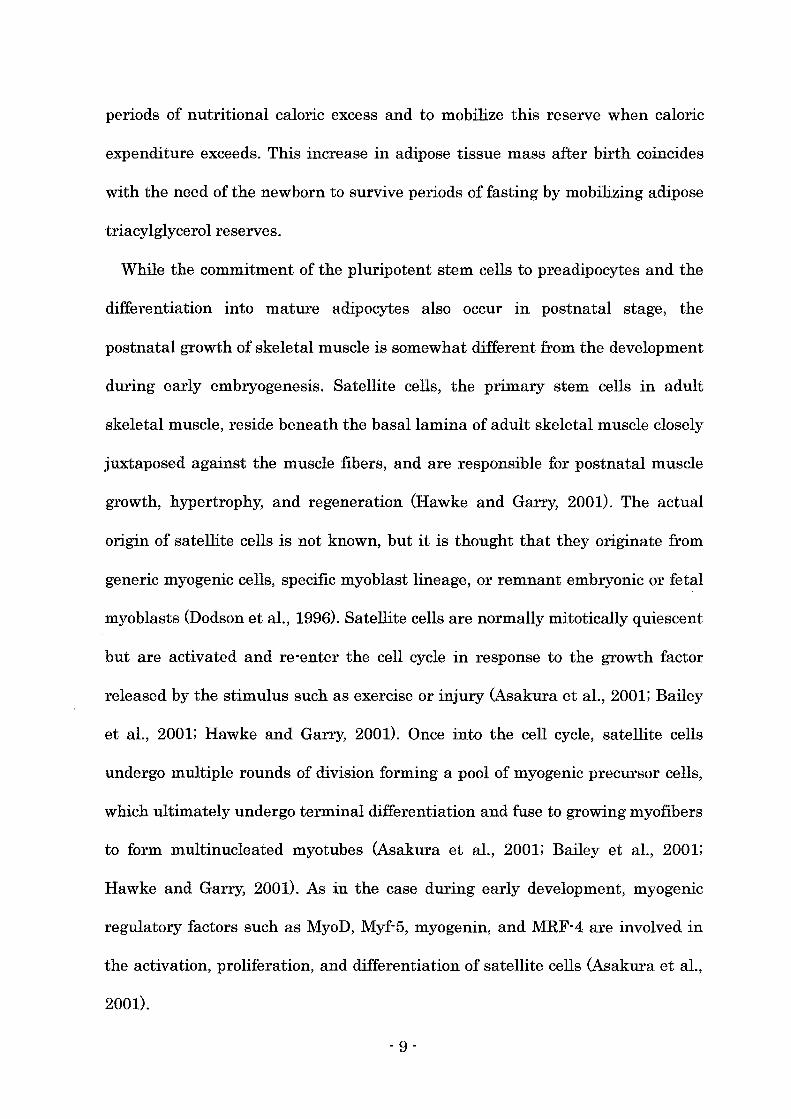
periods of nutritional caloric excess and to mobilize this reserve when caloric
expenditure exceeds. This increase in adipose tissue mass after birth coincides
with the need of the newborn to survive periods of fasting by mobilizing adipose
triacylglycerol reserves.
While the commitment of the pluripotent stem cells to preadipocytes and the
differentiation into mature adipocytes also occur in postnatal stage, the
postnatal growth of skeletal muscle is somewhat different from the development
during early embryogenesis. Satellite cells, the primary stem cells in adult
skeletal muscle, reside beneath the basal lamina of adult skeletal muscle closely
juxtaposed against the muscle fibers, and are responsible for postnatal muscle
growth, hypertrophy, and regeneration (Hawke and Garry, 2001). The actual
origin of satellite cells is not known, but it is thought that they originate from
generic myogenic cells, specific myoblast lineage, or remnant embryonic or fetal
myoblasts (Dodson et aI., 1996). Satellite cells are normally mitotically quiescent
but are activated and re-enter the cell cycle in response to the growth factor
released by the stimulus such as exercise or injury (Asakura et aI., 2001; Bailey
et aI., 2001; Hawke and Garry, 2001). Once into the cell cycle, satellite cells
undergo multiple rounds of division forming a pool of myogenic precursor cells,
which ultimately undergo terminal differentiation and fuse to growing myofibers
to form multinucleated myotubes (Asakura et aI., 2001; Bailey et aI., 2001;
Hawke and Garry, 2001). As in the case during early development, myogenic
regulatory factors such as MyoD, Myf-5, myogenin, and MRF-4 are involved in
the activation, proliferation, and differentiation of satellite cells (Asakura et aI.,
2001).
- 9 .
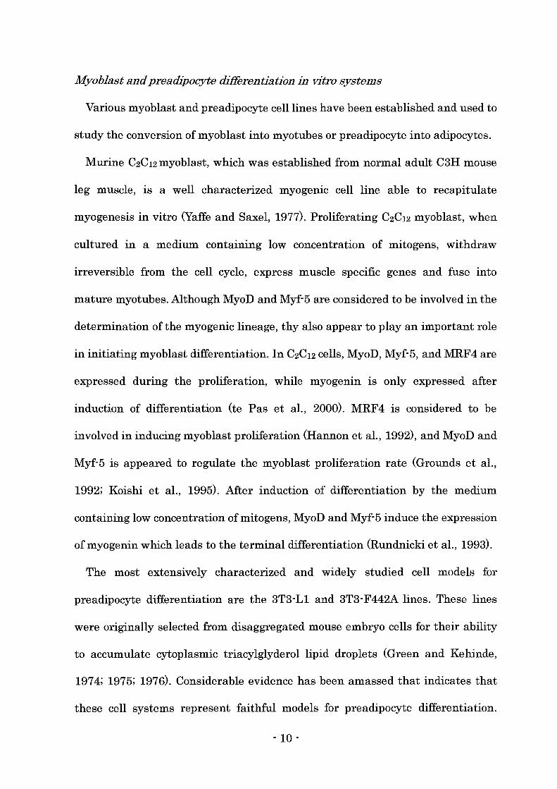
Myoblast and preadipocyte differentiation in vitro systems
Various myoblast and preadipocyte cell lines have been established and used to
study the conversion of myoblast into myotubes or preadipocyte into adipocytes.
Murine C2C12 myoblast, which was established from normal adult C3H mouse
leg muscle, is a well characterized myogenic cell line able to recapitulate
myogenesis in vitro (Yaffe and Saxel, 1977). Proliferating C2C12 myoblast, when
cultured in a medium containing low concentration of mitogens, withdraw
irreversible from the cell cycle, express muscle specific genes and fuse into
mature myotubes. Although MyoD and Myf-5 are considered to be involved in the
determination of the myogenic lineage, thy also appear to play an important role
in initiating myoblast differentiation. In C2C12 cells, MyoD, Myf-5, and MRF4 are
expressed during the proliferation, while myogenin is only expressed after
induction of differentiation (te Pas et aI., 2000). MRF4 is considered to be
involved in inducing myoblast proliferation (Hannon et aI., 1992), and MyoD and
Myf-5 is appeared to regulate the myoblast proliferation rate (Grounds et aI.,
1992; Koishi et aI., 1995). After induction of differentiation by the medium
containing low concentration ofmitogens, MyoD and Myf-5 induce the expression
of myogenin which leads to the terminal differentiation (Rundnicki et aI., 1993).
The most extensively characterized and widely studied cell models for
preadipocyte differentiation are the 3T3-Ll and 3T3-F442A lines. These lines
were originally selected from disaggregated mouse embryo cells for their ability
to accumulate cytoplasmic triacylglyderol lipid droplets (Green and Kehinde,
1974; 1975; 1976). Considerable evidence has been amassed that indicates that
these cell systems represent faithful models for preadipocyte differentiation.
- 10 -

Protocols have been developed that can induce 3T3 preadipocytes to rapidly and
synchronously progress through the differentiation program at high frequency
~90%). The agents most widely used (often in combination) to differentiate
3T3-L1 preadipocytes and other preadipocyte cell lines include dexamethasone (a
synthetic glucocorticoid agonist), high level of insulin {which act through the
insulin"like growth factor1 (IGF-1) receptor, methyl-isobutylxanthine (MIX; a
cAMP phosphodiesterase inhibitor), and fetal bovine serum (Student et al., 1980).
During the growth phase, 3T3-L1 and 3T3-F442A pre adipocyte s are
morphologically similar to the fibroblastic preadipocytes cells in the stroma of
adipose tissue. When induced to differentiate, 3T3 preadipocytes undergo several
rounds of mitotic clonal expansion (Tang and Lane, 1999). The necessity of this
mitotic event for the following adipogenesis is controversial (Qiu et aL, 2001,
Tang et al., 2003), however, the cells lose their fibroblastic character thereafter,
assume a rounded' up appearance, and acquire the morphological and
biochemical characteristics of adipocytes. PPARy and several members of the
C/EBP family of transcription factors participate in a signaling cascade
(Cornelius et al., 1994; Gregoire et al., 1998; MacDougald and Lane, 1995) that
culminates in the transcriptional activation of genes that produce the adipocyte
phenotype. C/EBPB is expressed immediately (within 2-4 h) after induction of
differentiation. At this point in the differentiation program, however, C/EBPB is
unable to bind DNA (Tang and Lane, 1999) and thus cannot function as a
transcriptional activator. Only after a long lag period (10-12 h) does C/EBPB
acquire DNA-binding activity (Tang and Lane, 1999), as the cells synchronously
reenter the cell cycle and begin mitotic clonal expansion. Coincident with the
- 11 -
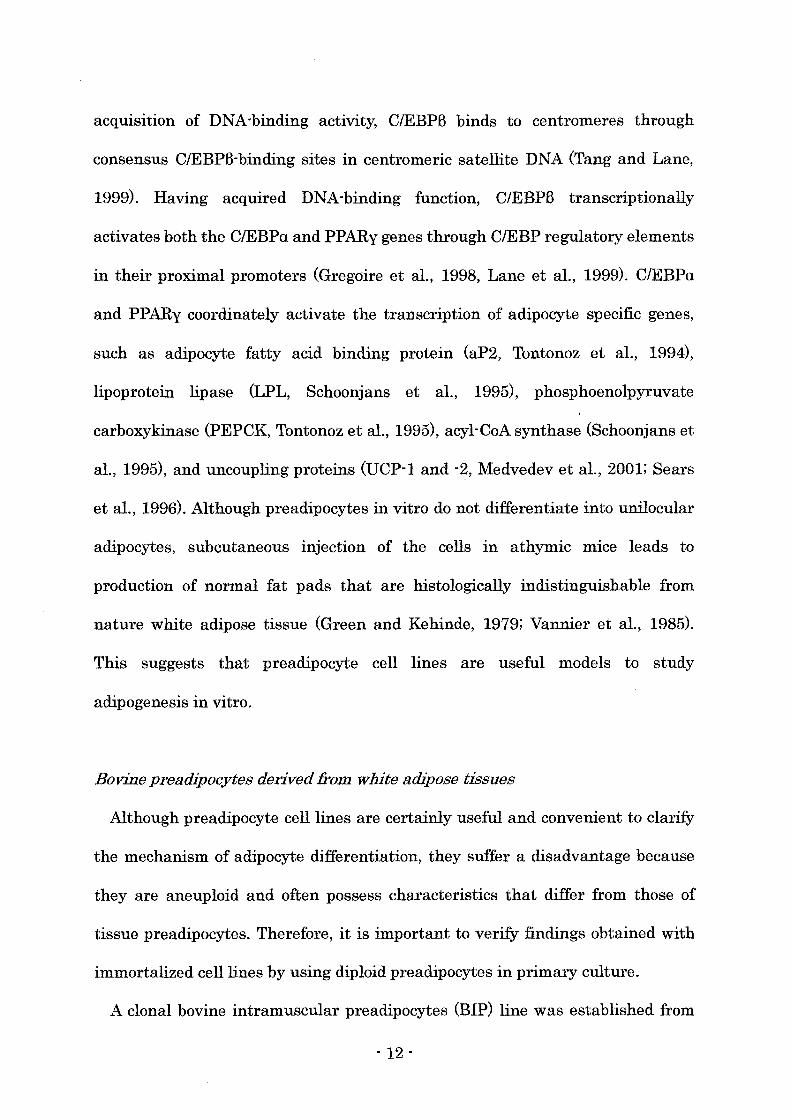
acquisition of DNA-binding activity, C/EBPB binds to centromeres through
consensus C/EBPB-binding sites in centromeric satellite DNA (Tang and Lane,
1999). Having acquired DNA-binding function, C/EBPB transcriptionally
activates both the C/EBPu and PPARy genes through C/EBP regulatory elements
in their proximal promoters (Gregoire et aI., 1998, Lane et aI., 1999). C/EBPu
and PPARy coordinately activate the transcription of adipocyte specific genes,
such as adipocyte fatty acid binding protein (aP2, Tontonoz et aI., 1994),
lipoprotein lipase (LPL, Schoonjans et aI., 1995), phosphoenolpyruvate
carboxykinase (PEPCK, Tontonoz et aI., 1995), acyl-CoA synthase (Schoonjans et
aI., 1995), and uncoupling proteins (DCP-1 and -2, Medvedev et aI., 2001; Sears
et aI., 1996). Although preadipocytes in vitro do not differentiate into unilocular
adipocytes, subcutaneous injection of the cells in athymic mice leads to
production of normal fat pads that are histologically indistinguishable from
nature white adipose tissue (Green and Kehinde, 1979; Vannier et aI., 1985).
This suggests that preadipocyte cell lines are useful models to study
adipogenesis in vitro.
Bovine preadipocytes derived from white adipose tissues
Although preadipocyte cell lines are certainly useful and convenient to clarify
the mechanism of adipocyte differentiation, they suffer a disadvantage because
they are aneuploid and often possess characteristics that differ from those of
tissue preadipocytes. Therefore, it is important to verify findings obtained with
immortalized cell lines by using diploid preadipocytes in primary culture.
A clonal bovine intramuscular preadipocytes (BIP) line was established from
- 12 -
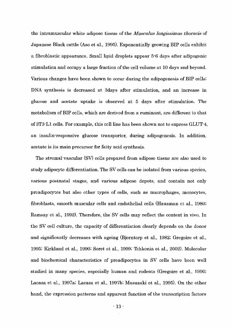
the intramuscular white adipose tissue of the Musculus longissimus thoracis of
Japanese Black cattle (Aso et aI., 1995). Exponentially growing BIP cells exhibit
a fibroblastic appearance. Small lipid droplets appear 5-6 days after adipogenic
stimulation and occupy a large fraction of the cell volume at 10 days and beyond.
Various changes have been shown to occur during the adipogenesis of BIP cells;
DNA synthesis is decreased at 3days after stimulation, and an increase in
glucose and acetate uptake is observed at 5 days after stimulation. The
metabolism ofBIP cells, which are derived from a ruminant, are different to that
of 3T3-Ll cells. For example, this cell line has been shown not to express GLUT-4,
an insulin-responsive glucose transporter, during adipogenesis. In addition,
acetate is its main precursor for fatty acid synthesis.
The stromal-vascular (SV) cells prepared from adipose tissue are also used to
study adipocyte differentiation. The SV cells can be isolated from various species,
various postnatal stages, and various adipose depots, and contain not only
preadipocytes but also other types of cells, such as macrophages, monocytes,
fibroblasts, smooth muscular cells and endothelial cells (Hausman et aI., 1980;
Ramsay et aI., 1992). Therefore, the SV cells may reflect the context in vivo. In
the SV cell culture, the capacity of differentiation clearly depends on the donor
and significantly decreases with ageing (Bjorntorp et aI., 1982; Gregoire et aI.,
1995; Kirkland et aI., 1990; Soret et aI., 1999; Tchkonia et aI., 2002). Molecular
and biochemical characteristics of preadipocytes in SV cells have been well
studied in many species, especially human and rodents (Gregoire et aI., 1990;
Lacasa et aI., 1997a; Lacasa et aI., 1997b; Masuzaki et aI., 1995). On the other
hand, the expression patterns and apparent function of the transcription factors
- 13 -

critical for preadiopocyte differentiation remains to be elucidated III bovine
preadipocytes.
Myostatin
Myostatin (growth/differentiation factor-8 (GDF-8» is a TGF-B family member
that is essential for proper regulation of skeletal muscle mass (Lee and
McPherron, 1999). Myostatin is expressed almost exclusively in cells of the
skeletal muscle lineage, from embryonic myotome to striated muscle in adults.
Mice carrying a targeted deletion of the gene encoding myostatin (Mstn) have a
dramatic and widespread increase in muscle mass, the result of both hyperplasia
and hypertrophy of muscle fibers, suggesting that myostatin normally acts as a
negative regulator of muscle growth (McPherron et at, 1997). Mutation of
myostatin gene also results in increasing skeletal muscle in certain breeds of
cattle (Belgian Blue and Piedmontese), which is known to double muscling
(Kambadur et at, 1997). The skeletal muscle of double muscled Belgian Blue
cattle is -20% greater than in normal- muscled cattle (Shahin and Berg, 1985).
Myostatin is synthesized as a preprotein activated by two proteplytic cleavages.
Removal of the signal sequence is followed by cleavage at a tetrabasic processing
site, resulting in a 26kDa NH2-terminal propeptide and a 12.5 kDa
COOH-terminal peptide, a dimmer of which is the biologically active portion of
the protein. The myostatin sequence has been highly conserved through
evolution (McPherron and Lee, 1997). Remarkably, the human, rat, murine,
porcine, turkey, and chicken myostatin sequences are identical in the biologically
active C-terminal portion of the molecule following the proteolytic processing site.
- 14 -
The mature peptide is reported to bind to the activin type II receptors, which
leads to the intracellular signal transduction (Lee and McPherron, 2001;
Massague and Chen, 2000; Rebbapragada et aI., 2003).
Activin
Activin is a multifunctional growth and differentiation factor that belongs to
the transforming growth factorB (TGF-B) superfamily. Activin is a dime ric
protein hormone synthesized as a homo- or heterodimer of the district B subunits
(BA or BB), which combine to form activinA (BA-B~, activin B (BB-BB), or activinAB
(BA-BB). Activin was discovered for its ability to regulate follicle-stimulating
hormone (FSH) production by pituitary cells. Recently, the expressions of activin
BA were found in many tissues throughout the body (Meunier et aI., 1998; Tuuri
et aI., 1994), and activin is reported to act on many cell types, regulating
hormone production in placental cell cultures (Petraglia et aI., 1989), including
differentiation in erythroblasts (Eto et aI., 1987) and osteoblasts (Ogawa et aI.,
1992), inhibiting proliferation of gonadal cell lines (Gonz3.lez-Manch6n and Vale,
1989), endothelial cells (McCarthy and Bicknell, 1993), lung epithelial cells
(Carcamo et aI., 1994), and hepatocytes (Yasuda et aI., 1993), and inhibiting the
differentiation of myoblasts (Link and Nishi, 1997; Shiozuka et aI., 1997). Activin
BA is highly expressed in adipose tissue (Vejda et aI., 2002), however, the effect of
activin on the adipocyte differentiation has been still unknown.
Activin exerts its biological effects by interacting with four types of
transmembrane receptors (type lA, lB, IIA, lIB) with protein serine/threonine
kinase activity (Attisano et aI., 1996). Activin binds directory to the type II
- 15 -
receptors, leading to recruitment and phosphorylation of the type I receptors by
the kinase domain of type II receptors. Once phosphorylated, type I receptors
exhibit kinase activity on Smad proteins, intracellular signal mediators. Smad2
and Smad3 are specific to the activin signaling and are phosphorylated by
activated activin receptors on serine residues. Phosphorylation of Smad2 and
Smad3 allows complex formation with Smad4, a common effector shared by
different TGFB family pathways (Heldin et al., 1997). Once formed the Smad2 /
Smad4 or Smad3/ Smad4 complex translocates into the nucleus to activate
transcription of specific target genes (Attisano and Wrana, 2000; Massague and
Wotton, 2000).
The expression sites of activin receptor mRNAs is reported to coincide with or
adjacent to the sites of activin B subunits expression during the period of
organogenesis (Feijen et al., 1994). In postnatal adipogenesis, Rebbapragada et
al. (2003) reported that myostatin blocks preadipocyte differentiation by binding
to activin receptor IIB, however, no one has yet identified the expression of
activin receptors or smads in adipose tissue or adipocytes.
Follista tin
Follistatin was purified from follicular fluids as a binding protein of activin
(Nakamura et al., 1990). Follistatin has been shown to reverse the effects of
activin on pituitary FSH release (Robertson et al., 1987; Veno et al., 1987).
Several follistatin proteins (FS-315, FS-303, and FS-288), resulting from
alternative splicing (Shimasaki et al., 1988) and proteolytic cleavage (Inouye et
al., 1991) have been purified. Alternative splicing generates two different
- 16 -
mRNAs, which encode FS-315 and its carboxy-terminally truncated homologue
FS-288. Proteolytic cleavage converts FS-315 into FS-303, which is the major
protein in follistatin preparations obtained from follicular fluid (Inouye et aI.,
1991). The affinities of the different follistatin proteins for activin A as
determined by polyethylene glycol precipitation are essentially similar, whereas
the affinity of FS-288 for heparan sulfate side chains of proteoglycans is much
higher than that of FS-315 and FS-303 (Sugino et aI., 1993). This can explain the
higher potency of FS-288 in inhibiting FSH release (Inouye et aI., 1991).
Furthermore, FS-288 seems to have a higher affinity for an immobilized activin
affinity column than FS-315 (Sumitomo et aI., 1995), which can also explain the
higher potency of FS-288.
Recently, myostatin is also reported to be captured by follistatin, which
prevents myostatin binding to activin receptor IIB (Lee and McPherron, 2001).
The studies on gene deletion (Matzuk et aI., 1995) and overexpression (Lee and
McPherron, 2001) of follistatin demonstrated that follistatin neutralizes the
inhibitory effect of its sensitive ligands on muscle development, and myostatin is
considered to be an obvious candidate for the sensitive ligand.
Follistatin mRNA and/or protein have been shown to be produced in many of
the same tissues that produce activin (Shimasaki et aI., 1988; Michel et aI., 1990;
DePaolo et aI., 1991) including muscle and adipose tissue. These results suggest
that follistatin can interact with activin and/or myostatin in several tissues.
- 17 -
Chapter 3
Effects of the conditioned medium from C2C12 myocyte
on the differentiation of 3T3-Ll preadipocyte
Introduction
Beef marbling is characterized by adipose tissue deposition within skeletal
muscle in cattle and is one of the important meat quality traits that influences
juiciness and flavor of meat and contributes directly to the value of beef on
especially Japanese markets. Japanese black cattle (Wagyu) is well known for its
ability to produce high marbling (Lunt et al., 1993, Zenbayashi, 1994), and
systemic fat metabolism does not necessarily reflect the fat deposition in the
muscle in this breed (Mukai et al., 1995; Yang et al., 1985). Hood and Allen
(1973) and Cianzio et al. (1985) reported that the development of beef marbling
was closely associated with an increase in adipocyte number within muscle,
suggesting that the proliferation and differentiation of adipose precursor cells
could occur within muscle during the formation of beef marbling. As
intramuscular preadipocytes are surrounded by mature skeletal muscle
myofibers, it is predicted that paracrine factors secreted from muscle fibers may
regulate the differentiation of preadipocyte via intercellular interactions.
Muscle fibers secrete various factors, such as fibroblast growth factor (FGF),
transforming growth factor-B (TGF-B), tumor necrosis factor-u (TNF-u),
insulin-like growth factors (IGFs), hepatocyte growth factor (HGF) , and
- 18 -
interleukin-6 (IL-6) (Charge et al., 2004). These cytokines are known to regulate
the proliferation and differentiation of preadipocytes, as well as of myoblasts
(Charge et al., 2004).
Myostatin, which is the member of TGF-B superfamily and is essential for
proper regulation of skeletal muscle mass, is also known to be expressed mainly
in skeletal muscle (Lee and McPherron, 1999). Furthermore, myostatin is
reported to inhibit the differentiation of mouse preadipocytes (Kim et al., 2001).
Follistain is a secretory protein and it binds to activin, another member of
TGF-B superfamily, and results in the prevention of activin from binding to the
own receptors (Schneyer et al., 2003). Follistatin is also reported to capture
myostatin, which prevents myostatin binding to activin receptors (Lee and
McPherron, 2001).
To evaluate the effect of the paracrlne factors from muscle cells on the
differentiation of preadipocytes, the author used C2C12 myoblast and 3T3-L1
preadipocyte cell lines, which are both well characterized and widely studied cell
models for myoblast or preadipocyte differentiation. The conditioned medium
from C2C12 myocytes which contains paracrine factors secreted from C2C12 cells
was added to the medium of 3T3-L1 cell culture. Furthermore, the author
hypothesized that the conditioned medium may contains myostatin, and
investigated the effect of the co-treatment of the conditioned medium and
follistatin on the differentiation of preadipocyte.
- 19 -
Materials and methods
C2C12 cell culture andpreparation of the conditioned medium
C2C12 myoblasts, obtained from RIKEN Cell Bank (Tsukuba, Japan), were
grown in Dulbecco's modified Eagle's medium (DMEM) (Nissui, Tokyo, Japan)
with 10% fetal bovine serum (FBS) (Trace Biosciences, Melbourne, Australia),
100 Ulml penicillin, and 100 }lg/ml streptomycin (each from Wako Chemicals,
Osaka, Japan) at 37°C under a humidified 5% C02 atmosphere. Two days after
C2C12 cells reached confluence (day 0), myogenesis was induced by changing the
medium to serum free DMEM, and the medium was changed every second day.
Conditioned medium (CM) was prepared from the medium collected on day 10
when most of the myoblasts were fused into myotubes. Part of the CM was
ultrafiltrated in microconcentrators with 3,000 MW cut-off (Centriprep YM-3,
Millipore, MA, USA.) by centrifuging at 3,000 rpm at 4°C until 90% of the
medium was filtrated. The nonpercolated medium was collected as the high
molecular weight fraction (HCM) and the filtrate was as the low molecular
weight fraction (LCM), and stored at -20°C.
3T3-Ll cell culture
3T3-L1 preadipocytes (Dainihon-seiyaku, Osaka, Japan), were subcultured in
DMEM with 5% FBS, 100 Ulml penicillin, and 100 }lg/ml streptomycin at 37°C
under a humidified 5% C02 atmosphere. Adipogenesis was induced by the
adip 0 genic agents (0.5 mM 3-isobutyl-1-methylxanthine, 0.25 pM
dexamethasone (each from Sigma, MO, USA), and 10 }lg/ml insulin (Wako
- 20 -
Chemicals» in DMEM containing 5% FBS for 2 days after 3T3-L1 cells reached
confluence (from day 0 to day 2). Then, the medium was replaced with DMEM
containing 5% FBS and 5 pg/ml insulin, and was changed every second day.
Thirty percent of the medium was replaced with each conditioned medium
throughout the differentiation period (from day 0 to day 8), in the early phase of
differentiation (from day 0 to day 2) or in the late phase of differentiation (from
day 2 to day 8). Recombinant human follistatin (Genzyme Techne, MN, USA) was
dissolved in phosphate-buffered saline (PBS) with 0.1% bovine serum albumin
(Sigma) and added to the medium containing 30% HCM at the concentrations of
100,300,500 ng/ml in the early phase of differentiation (from day 0 to day 2).
Analysis of glycerol-3-phosphate dehydrogenase activity
Cells were carefully washed twice with ice-cold PBS on 8 day of differentiation
period, and lysed in 25 mM Tris/1 mM EDTA, pH 7.5 for measurement of
glycerol-3-phosphate dehydrogenase (GPDH) specific activity. GPDH activity
was determined according to the procedure of Wise and Green (1979). Protein
concentration was measured with the method of Lowry et al. (1951), and one unit
of enzyme activity was defined as the amount of protein required for the use of 1
nmol NADH per min per mg protein.
Statistics
Data were expressed as means ± SE, and were statistically analyzed using
student's t-test. Statistical significance was set at P<0.05.
- 21 -
Results
Effect of the conditioned medium from C2C12 myocyte on the differentiation of
3T3-Ll preadipocyte
To investigate the effect of the paracrine factors secreted from muscle cells on
the differentiation of preadipocytes, 3T3-L1 cells were treated with the
conditioned medium (CM, HCM, LCM) collected from almost fully differentiated
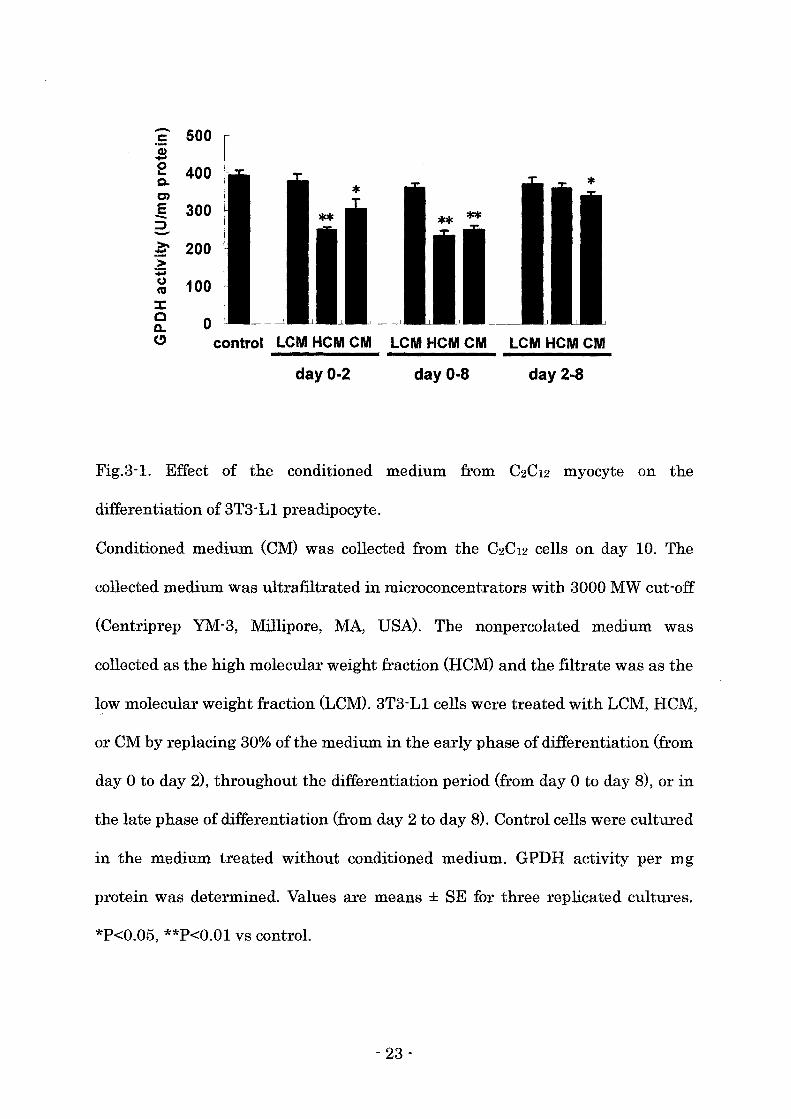
C2C12 cells (C2C12 myocytes). LCM treatment did not affect GPDH activity
regardless of the treatment period (Fig. 3-1). CM or HCM treatment throughout
the differentiation period or in the early phase of differentiation significantly
reduced the GPDH activity (Fig. 3-1). GPDH activity was also decreased by the
CM treatment in the late phase, however, HCM treatment in the late phase did
not affect GPDH activity (Fig. 3-1).
Effect of the co-treatment of high molecular weight fraction of the conditioned
medium and follistatin on the differentiation of 3T3-Ll preadipocyte
To examine the possibility that myostatin and/or activin in the conditioned
medium affect the differentiation of 3T3-L1 preadipocytes, the cells were treated
with HCM and follistatin in the early phase of differentiation. HCM suppressed
GPDH activity when the cells were treated in the early phase of differentiation
(Fig. 3-2). The simultaneous treatment with follistatin reversed the suppressive
effect ofHCM in a dose dependent manner (Fig. 3-2).
- 22 -
:s 500 .! e 400 Q.
tn .s 300 :::J -~ 200
100
o control LCM HCM CM
day 0-2
LCM HCMCM
day 0-8
LCM HCMCM
day 2-8
Fig.3-1. Effect of the conditioned medium from C2C12 myocyte on the
differentiation of 3T3-L1 preadipocyte.
Conditioned medium (CM) was collected from the C2C12 cells on day 10. The
collected medium was ultrafiltrated in microconcentrators with 3000 MW cut-off
(Centriprep YM-3, Millipore, MA, USA). The nonpercolated medium was
collected as the high molecular weight fraction (HCM) and the filtrate was as the
low molecular weight fraction (LCM). 3T3-L1 cells were treated with LCM, HCM,
or CM by replacing 30% of the medium in the early phase of differentiation (from
day 0 to day 2), throughout the differentiation period (from day 0 to day 8), or in
the late phase of differentiation (from day 2 to day 8). Control cells were cultured
in the medium treated without conditioned medium. GPDH activity per mg
protein was determined. Values are means ± SE for three replicated cultures.
*P<0.05, **P<O.Ol VB control.
- 23 .

1000
800
-~c 'S: 'i 600 ,- -u e co Q.
5 E 400 a..(!):::l
200
o control o 100 300 500
follistatin ( ng/ml )
Fig. 3-2. Effect of the high molecular weight fraction of the conditioned medium
and follistatin on the differentiation of 3T3-Ll preadipocyte.
3T3-Ll cells were treated with high molecular weight fraction of the conditioned
medium (HCM) by replacing 30% of the medium, and follistatin in various
concentrations in the early phase of differentiation (from day 0 to day 2). Control
cells were cultured with neither HCM nor follistatin. Cells were harvested at the
end of culture (day 8) and GPDH activity was determined as described in the Fig.
3-1. Values are means ± SE for three replicated cultures. +P<0.05 vs control
culture treated neither HCM nor follistatin; *P<0.05 vs culture treated with
HCM alone.
- 24 -
Discussion
In the present study, the treatment with CM or HCM throughout the
differentiation period significantly reduced GPDH activity, a terminal
differentiation marker. The treatment with CM or HCM also decreased GPDH
activity when the cells were treated in the early phase of differentiation.
However, the treatment with HCM in the late phase did not affect GPDH activity
and only CM slightly decreased the activity. These results suggest that the high
molecular weight factors secreted from C2C12 myocytes mostly suppressed the
preadipocyte differentiation in the early phase that is the step of commitment to
differentiate in 3T3-L1 cells (Gregoire et aI., 1998).
The 3T3-L1 cell line is the most well-characterized and reliable model for
studying preadipocyte differentiation. A study using 3T3-L1 preadipocyte
showed that CCAAT/enhancer binding protein (CIEBP) B was expressed
immediately (within 2-4 hours) by induction of differentiation, reached maximal
level within 4 hours, and begins to disappear 2 days after initiation of
differentiation (Lane et aI., 1999). CIEBPB transcriptionally activates peroxisome
proliferator-activated receptor (PPAR) y expression during the induction of
differentiation, which reaches maximal level on day 3 (Chawla et aI., 1994). The
expression of C/EBPa is also activated by C/EBPB but the level of C/EBPa is faint
during the induction and reaches maximal level by 5 days after initiation of
differentiation (Christy et aI., 1991). Thereafter, PPARy and C/EBPa upregulate
each other to maintain their expression (Mandrup and Lane, 1997) despite the
reduction of C/EBPB level. PPARy and C/EBPa coordinately activate the
- 25 -
expression of adipocyte-specific genes (Gregoire et al., 1998).
Myostatin, which is essential for proper regulation of skeletal muscle mass, is
known to be expressed mainly in skeletal muscle. Artaza et al. (2002) reported
that the expression of myostatin mRNA was found in C2C12 myotubes not in
C2C12 myoblasts, and that myostatin was also secreted into the medium of C2C12
myotube culture. Furthermore, myostatin is reported to inhibit the
differentiation of 3T3-L1 preadipocytes via affecting the expression of PPARy
and C/EBPu, but not C/EBPB (Kim et aI., 2001).
In the present study, HCM treatment inhibited the preadipocyte
differentiation in the early phase when the expressions of PPARy and C/EBPu
are induced in 3T3-L1 preadipocytes. Therefore, it is predicted that HCM
inhibited the differentiation of 3T3-L1 preadipocyte by suppressing the
expression of PPARy and C/EBPu, although the mechanism was not investigated
in this study.
Based on these reports and results in the present study, myostatin was
considered to be a candidate for the effective factor in the HCM that inhibited the
differentiation of 3T3-Ll preadipocyte. To examine this possibility, the
differentiating 3T3-Ll cells were co-treated with the HCM and follistatin, which
binds to myostatin and activin and inhibits their action. Follistatin reversed the
inhibitory effect ofHCM on the differentiation of 3T3-L1 preadipocytes in a dose
dependent manner, which suggested that myostatin and/or activin in the HCM
possibly inhibited the preadipocyte differentiation.
The expression of activin is widely distributed in gonadal and non-gonadal
tissues including muscle (Tuuri et aI., 1994) and fat (Schneider et aI., 2000; Vejda
- 26 -
et aI., 2002). Furthermore, C2C12 myoblasts and myocytes in the present study
also expressed activin mRNA (data not shown). Activin is reported to act on
many cell types, including placental cells (Petraglia et aI., 1989), erythroblasts
(Eto et aI., 1987), osteoblasts (Ogawa et aI., 1992), endothelial cells (McCarthy
and Bicknell, 1993), and myoblasts (Link and Nishi, 1997; Shiozuka et aI., 1997).
However, effect of activin on the differentiation of preadipocytes has been still
unknown. Moreover, the expression of activin receptors, which mediate the
action of activin (Mathews, 1994; Piek et aI., 1999) and myostatin (Lee and
McPherron, 2001; Massague and Chen, 2000), has not been investigated in
adipose tissue. Further study is needed to determine the effect of activin on the
differentiation of preadipocytes.
In summary, these results presented here showed that conditioned medium
from C2C12 myocytes inhibited the preadipocytes differentiation in the early
phase, and this inhibitory effect was possibly caused by myostatin and/or activin
secreted from C2C12 myocytes.
- 27 -
Chapter 4
Effects of myostatin on the differentiation of bovine preadipocyte
Introduction
CCAAT/enhancer binding proteins (C/EBPs) and peroXIsome
proliferator-activated receptor (PPAR) y have shown to be critical transcription
factors for preadipocyte differentiation (Gregoire et aI., 1998; Rosen et aI., 2000).
Studies using 3T3-L1 preadipocytes indicate that C/EBPB is highly expressed
after the treatment with adipogenic agents (Lane et aI., 1999). C/EBPB activates
the expression of PPARy and C/EBPa mRNAs that coordinately activate the
transcription of adipocyte-specific genes (Rosen et aI., 2000). On the other hand,
these 3 transcription factors were highly expressed before inducing the
differentiation of preadipocytes m the prImary cultures of porcine
stromal-vascular (SV) cells derived from adipose tissue (Ding et aI., 1999).
In Chapter 3, the author revealed that the high molecular weight fraction of
the conditioned medium from C2C12 cells suppressed the differentiation of
3T3-L1 preadipocytes, and this inhibitory effect of the conditioned medium was
reversed by follistatin which is known to bind to myostatin (Lee and McPherron,
2001) and/or activin (Schneyer et aI., 2003). These results suggest that myostatin
and/or activin in the conditioned medium possibly affected the preadipocyte
differentiation.
Myostatin (growth differentiation factor-8, GDF-8) IS a member of the
- 28 -
transforming growth factor-B (TGF-B) superfamily, and a key critical regulator of
skeletal muscle development (McPherron et aI., 1997). In cattle, defective
mutation of myostatin gene increased skeletal muscle, which is known as
double-muscling (McPherron and Lee, 1997). The expression of my os tat in mRNA
is found primarily in skeletal muscle, but it is also detected in the adipose tissue
(McPherron et aI., 1997). Myostatin can bind the activin type II receptors, which
leads to the intracellular signal transduction (Lee and McPherron, 2001;
Massague and Chen, 2000). Myostatin was reported to suppress the induction of
PPARyand C/EBPa in 3T3-L1 cells after the initiation of differentiation, which
interfered with preadipocyte differentiation (Kim et aI., 2001). However, the
effect of myostatin on the differentiation of bovine preadipocytes has been
unknown. Furthermore, the expression patterns of the critical transcription
factors of bovine preadipocyte differentiation have not well studied.
The purpose in this chapter was to investigate the expression of these
transcription factor mRNAs during preadipocyte differentiation in stromal
vascular (SV) cells derived from bovine adipose tissue, and to examine the effect
of myostatin on the expression of these transcription factor mRNAs and on the
terminal differentiation of bovine preadipocytes. Furthermore, the author
investigated whether follistatin interferes with myostatin action during bovine
preadipocyte differentiation when the cells were treated with myostatin and
follistatin.
- 29 -
Materials and methods
Preparation of stromal-vascular cells from bovine adipose tissue
Perirenal adipose tissues were collected from 28-32 months old Japanese black
steers at a local slaughter house and transported to the laboratory in sterile
Hanks balanced salt solution (HBSS) containing 100 D/ml penicillin, 100 lIg/ml
streptomycin (each from Wako Chemicals), and 250 ng/ml amphotericin B
(Invitrogen, CA, DSA). The adipose tissues were digested in HBSS added with 1
mg/ml Type I collagenase (Sigma) for 1 h at 37°C with shaking at 170 cycles/min.
The cell suspension was filtrated through a 250 lIm nylon mesh filter to remove
undigested tissue fragments and debris. The filtrate was then centrifuged at
1,500 rpm for 5 min. Floating adipocytes and digestion medium were removed by
decantation. The pellet consists of SV cells containing preadipocytes. The SV
cells were washed twice with the growth medium, i.e., DMEM (Nissui)
containing 5% FBS (Trace Biosciences), 100 JiM ascorbic acid phosphate
magnesium, 100 D/ml penicillin, 100 Jig/ml streptomycin (Wako Chemicals). The
SV cells were then resuspended with the growth medium containing 10%
dimethyl sulfoxide and stored in liquid nitrogen.
Cell culture
The SV cells were seeded on 12-well (22 mm diameter) culture plates (Corning,
NY, DSA) at a density of 1 xl04 cells/cm2 and incubated in the growth medium at
37°C under a humidified 5% C02 atmosphere. After reaching confluence,
preadipocyte differentiation was induced by the adipogenic agents consisting of
- 30 -
0.5 mM 3-isobutyl-1-methylxanthine, 0.25 11M dexamethasone (each from Sigma),
2.5 11g/ml insulin (Wako Chemicals) and 5 11M troglitasone (Sankyo, Tokyo,
Japan) for 2 days (from day 0 to day 2). The medium was replaced with DMEM
containing 5% FBS, 2.5 11g/ml insulin, and 5 11M troglitasone, and was changed
every second day. Recombinant human myostatin (PeproTech EC, London, UK)
and recombinant human follistatin (Genzyme Techne) was dissolved in
phosphate-buffered saline (PBS) with 0.1% bovine serum albumin (Sigma) and
added to the medium in the concentrations as shown in each figure during the
early phase of differentiation (from day 0 to day 2) or throughout the
differentiation period (from day 0 to day 8).
Glycerol-3-phosphate dehydrogenase activity
Cells were carefully washed twice with ice-cold PBS on day 8, and lysed in 25
mM Tris/ 1 mM EDTA, pH 7.5 by sonication for measurement of GPDH specific
activity. GPDH activity was determined according to the method shown in
Chapter 3.
Oil Red a staining
Cultures were washed twice with PBS on day 0 and day 8, fixed with 10%
formalin in PBS, stained with 0.5% Oil Red 0 and photographed.
RNA isolation
Total RNA was extracted from the SV cells using Trizol Reagent (Invitrogen)
according to the manufacturer's protocol. The extracted RNA was dissolved in
- 31 -
diethyl pyrocarbonate-treated water and total RNA concentration was
determined spectrophotometrically at 260 nm.
Detection of activin receptor mRNAs in bovine preadipocyte
The expression of activin receptor mRNAs encoding bovine type I (ActRI) and
type II (ActRIIA and ActRIIB) receptors on day 0 were analyzed by reverse
transcription-polymerase chain reaction (RT-PCR). Single-strand cDNA was
synthesized using a TaKaRa RNA PCR Kit (AMV) Ver.3.0 (TaKaRa, Shiga,
Japan) according to the manufacture's protocol. The PCR were conducted using
Platinum PCR Super Mix (Invitrogen). The primer sets for each receptor were
shown in Table 1. The amplification parameters consisted of denaturation at
94°C for 30 s, annealing at the temperature (Table 4) for 30 s, and extension at
72°C for 1 min, for 35 cycles. The sub cloning and sequencing confirmed the PCR
products to be the expected fragments of bovine activin receptors. The products
were separated on 2% agarose gel and visualized with ethidium bromide. To
avoid false-positive results from contamination by genomic DNA, samples with
or without treatment of reverse transcriptase were prepared, and as a negative
control, RT-PCR without RNA samples was also carried out.
Northern blot analysis for the expression of aP2 mRNA
The author analyzed the expression of aP2 mRNA on day 0 and day 8. cDNA
fragment of bovine adipocyte fatty acid-binding protein (aP2) gene was amplified
using specific primers (Table 4) and inserted into the pCR II TOPO plasmid
vector (Invitrogen). The template was linearized with Not I (TOYOBO, Tokyo,
- 32 -
Japan) and then used in DIG RNA labeling Kit (Roche Diagnostics, Mannheim,
Germany) to synthesize the digoxigenin (DIG) -dUTP-Iabeled RNA antisense
probe. RNA sample (5 p.g) was separated on 1% agarose gels containing 6.7%
formaldehyde and transferred to a Hybond N+ membrane (Amersham
Biosciences, NJ, USA). The membrane was hybridized with DIG-labeled RNA
probe. The band corresponding to aP2 mRNA was detected using DIG
Luminescant Detection Kit for Nucleic Acids (Roche Diagnostics).
Semi-quantitative RT-PCR for the determination ofCIEBP and PPARy mRNAs
The expression of C/EBP and PPARy mRNAs were assessed by
semi-quantitative RT-PCR. Single-strand cDNA was synthesized using a
TaKaRa RNA PCR Kit (AMV) Ver.3.0 according to the manufacture's protocol.
The thermal amplifications of C/EBP fragments were conducted using TaKaRa
LA Taq with GC Buffer. PPARyand 18S rRNA fragments were amplified by
using Platinum PCR Super Mix (Invitrogen). The primer sets were shown in
Table 1. PCR conditions were optimized for detection within a linear range. The
amplification parameters consisted of denaturation at 94°C for 30 s, annealing
at the temperature (Table 4) for 30 s, and extension at 72°C for 1 min, for the
appropriate number of cycles; 35 cycles for the detection of C/EBPB, 27 cycles for
PPARy, 30 cycles for C/EBPa, and 18 cycles for 18S rRNA. The subcloning and
sequencing confirmed the PCR products to be the expected fragments of bovine
C/EBP, PPARy, and 18S rRNA. The level of 18S rRNA was adopted as the
internal standards for the determination of targeted mRNA levels.
- 33 -
Statistics
Data were expressed as means ± SE, and were statistically analyzed using
student's t-test. Statistical significance was set at P<O.05.
- 34 -
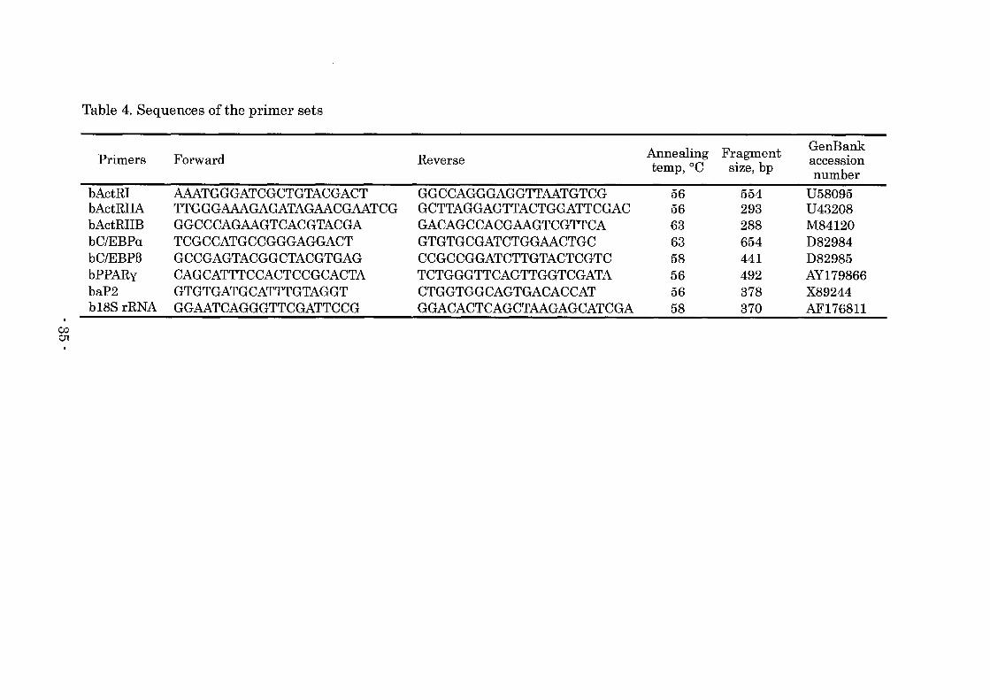
Table 4. Sequences of the primer sets
Annealing Fragment GenBank Primers Forward Reverse acceSSIOn temp,OC size, bp number
bActRI AAATGGGATCGCTGTACGACT GGCCAGGGAGGTTAATGTCG 56 554 U58095 bActRIIA TTGGGAAAGAGATAGAACGAATCG GCTTAGGAGTTACTGGATTCGAC 56 293 U43208 bActRIIB GGCCCAGAAGTCACGTACGA GACAGCCACGAAGTCGTTCA 63 288 M84120 bC/EBPa TCGCCATGCCGGGAGGACT GTGTGCGATCTGGAACTGC 63 654 D82984 bC/EBPB GCCGAGTACGGCTACGTGAG CCGCCGGATCTTGTACTCGTC 58 441 D82985 bPPARy CAGCATTTCCACTCCGCACTA TCTGGGTTCAGTTGGTCGATA 56 492 AY179866 baP2 GTGTGATGCATTTGTAGGT CTGGTGGCAGTGACACCAT 56 378 X89244 b18S rRNA GGAATCAGGGTTCGATTCCG GGACACTCAGCTAAGAGCATCGA 58 370 AF176811
00 01
Results
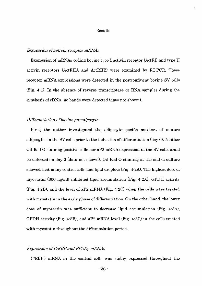
Expression of activin receptor mRNAs
Expression of mRNAs coding bovine type I activin receptor (ActRI) and type II
activin receptors (ActRIIA and ActRIIB) were examined by RT-PCR. These
receptor mRNA expressions were detected in the postconfiuent bovine SV cells
(Fig. 4-1). In the absence of reverse transcriptase or RNA samples during the
synthesis of eDNA, no bands were detected (data not shown).
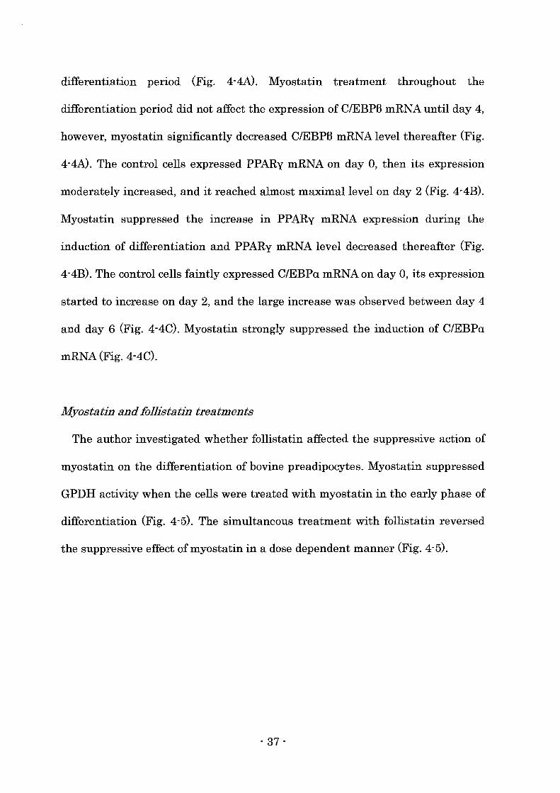
Differentiation of bovine preadipocyte
First, the author investigated the adipocyte-specific markers of mature
adipocytes in the SV cells prior to the induction of differentiation (day 0). Neither
Oil Red 0 staining-positive cells nor aP2 mRNA expression in the SV cells could
be detected on day 0 (data not shown). Oil Red 0 staining at the end of culture
showed that many control cells had lipid droplets (Fig. 4-2A). The highest dose of
myostatin (300 ng/ml) inhibited lipid accumulation (Fig. 4-2A), GPDH activity
(Fig. 4-2B), and the level of aP2 mRNA (Fig. 4-2C) when the cells were treated
with myostatin in the early phase of differentiation. On the other hand, the lower
dose of myostatin was sufficient to decrease lipid accumulation (Fig. 4-3A),
GPDH activity (Fig. 4-3B), and aP2 mRNA level (Fig. 4-3C) in the cells treated
with myostatin throughout the differentiation period.
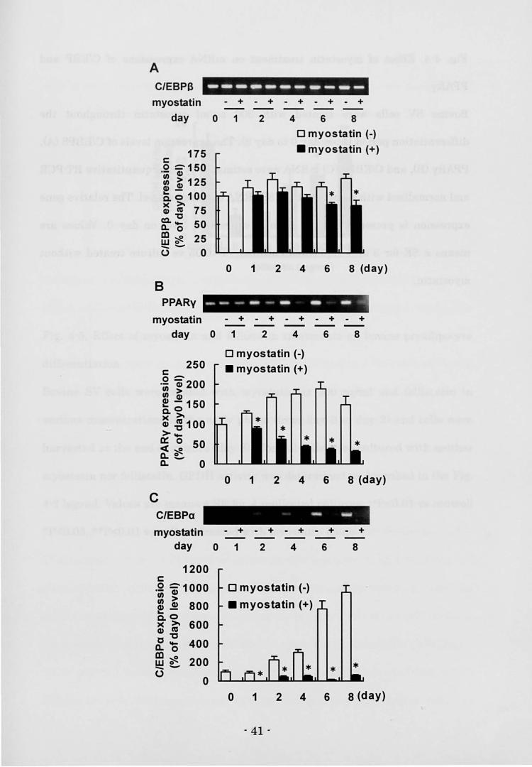
Expression of CIEBP and PPARy mRNAs
C/EBPB mRNA in the control cells was stably expressed throughout the
- 36 -
differentiation period (Fig. 4-4N. Myostatin treatment throughout the
differentiation period did not affect the expression of C/EBPB mRNA until day 4,
however, myostatin significantly decreased C/EBPB mRNA level thereafter (Fig.
4-4A). The control cells expressed PPARy mRNA on day 0, then its expression
moderately increased, and it reached almost maximal level on day 2 (Fig. 4-4B).
Myostatin suppressed the increase in PPARy mRNA expression during the
induction of differentiation and PPARy mRNA level decreased thereafter (Fig.
4-4B). The control cells faintly expressed C/EBPa mRNA on day 0, its expression
started to increase on day 2, and the large increase was observed between day 4
and day 6 (Fig. 4-4C). Myostatin strongly suppressed the induction of C/EBPa
mRNA(Fig.4-4C).
Myostatin and follistatin treatments
The author investigated whether follistatin affected the suppressive action of
myostatin on the differentiation of bovine preadipocytes. Myostatin suppressed
GPDH activity when the cells were treated with myostatin in the early phase of
differentiation (Fig. 4-5). The simultaneous treatment with follistatin reversed
the suppressive effect of my os tat in in a dose dependent manner (Fig. 4-5).
- 37 -
IIA liB
Fig. 4-1. Expression of activin receptor mRNAs in bovine stromal-vascular (SV)
cells.
Post confluent (day 0) bovine SV cells were harvested and RT-peR was
performed using primer sets for bActRI (1), bActRlIA (IrA), or bActRlIB (lIB).
The products were separated on 2% agarose gel and visualized with ethidium
bromide.
- 38 -
A
B
100
~~80 > Q) .- -t) e 60 ns Co
::I: C) 40 C E 0.-C) :::l 20 -
0
C
ap21 285 185
0 100 300
myostatin ( ng/ml )
0 100 300 myostatin ( ng/ml )
--- ........ ---o 100 300
myostatin ( ng/ml )
Fig. 4-2. Effect of myostatin treatment in the early phase on bovine preadipocyte
differentiation.
Bovine SV cells were treated with myostatin for 2 days after confluence (from
day 0 to day 2) and cells were harvested at the end of culture (day 8).
Intracellulal'lipid was stained with Oil Red 0 (N. GPDH activity per mg protein
was determined (B). The expression of aP2 mRNA was measured by Northern
blot analysis. Ethidium bromide staining demonstrates the RNA loading (C).
Values al'e means ± SE for 4 replicated cultures. **P<O.Ol vs culture treated
without myostatin.
- 39 -
A
B
100
>.- 80 ... c: .; 'Q) ;; 0 60 (,,) ~ ftI Q.
:I: C) 40 C E a.. - 20 (!)2.
o
c
aP2
285 185
o 100 300
myostatin ( ng/ml )
o 100 300 myostatin ( ng/ml )
o 100 300 myostatin ( ng/ml )
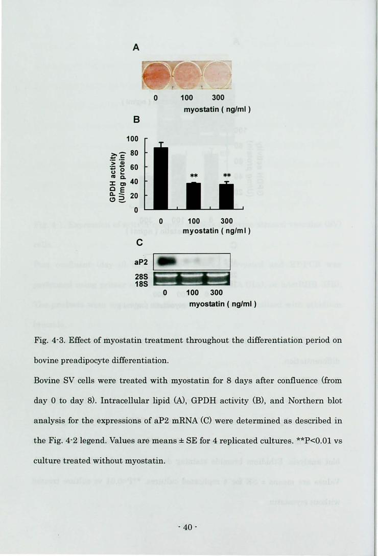
Fig. 4-3. Effect of myostatin treatment throughout the differentiation period on
bovine preadipocyte di.fferentiation.
Bovine SV cells were treated with myostatin for 8 days after confluence (from
day 0 to day 8). Intracellular lipid (N, GPDH activity (B), and Northern blot
analysis for the expressions of aP2 mRNA (C) were determined as described in
the Fig. 4-2 legend. Values are means ± SE for 4 replicated cultures. **P<O.Ol vs
culture treated without myostatin.
- 40 -
A C/EBPB
myostatin
day
--- .. -------
175 c: .2 -;150 ~ ~ 125 <P-o. 0 100
~ ~ 75 ~'O 50 ffi ~ 25 - -U 0
B
- +
o 1
o
- +
2
1
- + - + - +
4 6 8
o myostatin (-)
• myostatin (+)
246 8 (day)
PPARy ------- - - , myostatin - + - + - + - + - +
day 0 1 2 4 6 8
250 c: o=-
'c;; ~ 200 I/) <p
~ ~ 150 c.>. ~ ~ 100 > .....
a:::: 0 50 <C~ Q.~ 0. 0
C C/EBPa
myostatin day
1200 c: .2 ;-1000 I/) > ~ ~ 800 ... c.O ~ i;' 600 0'0
400 0.'0 al~ 200 w 0 --U
0
0
o myostatin (-)
• myostatin (+)
0 1 2 4 6 8 (day)
- + - + - + - + - +
1 2 4 6 8
o myostatin (-)
• myostatin (+)
0 1 2 4 6 8 (day)
- 41 -
Fig. 4-4. Effect of myostatin treatment on mRNA expressions of e/EBP and
PPARy.
Bovine SV cells were treated with 300 ng/ml myostatin throughout the
differentiation period (from day 0 to day 8). The expression levels of elEBP8 (A),
PPARy (B), and C/EBPa (e) mRNA were estimated by semi-quantitative RT-peR
and normalized with respect to the 18S rRNA expression leveL The relative gene
expression is presented as the ratio of expression level on day O. Values are
means ± SE for 3 or 4 replicated cultures. *P<0.05 vs culture treated without
myostatin.
·42 -

60
_50 a-c :~ :s 40 1:) 2 cu 0.30 ::I:m
~ .e 20 e>2,.
10
o
**
control 0 100 300 500 1000
follistatin ( ng/ml )
Fig. 4-5. Effect of myostatin and follistatin treatments on bovine preadipocyte
differentiation.
Bovine SV cells were treated with myostatin at 300 ng/ml and follistatin in
various concentrations in the early phase (from day 0 to day 2) and cells were
harvested at the end of culture (day 8). Control cells were cultured with neither
myostatin nor follistatin. GPDH activity was determined as described in the Fig.
4-2 legend. Values are means ± SE for 4 replicated cultures. ++P<O.Ol vs control;
*P<0.05, **P<O.Ol vs culture treated with myostatin alone.
- 43 -
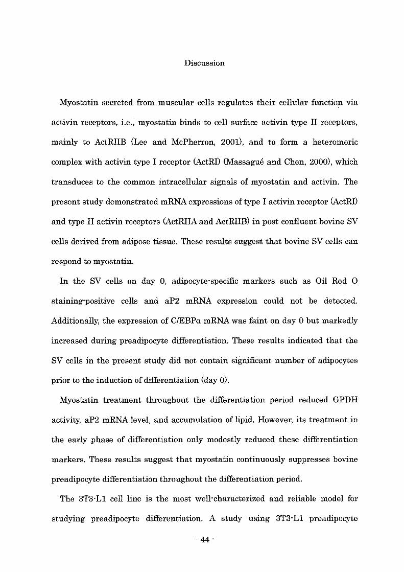
Discussion
Myostatin secreted from muscular cells regulates their cellular function via
activin receptors, i.e., myostatin binds to cell surface activin type II receptors,
mainly to ActRIIB (Lee and McPherron, 2001), and to form a heteromeric
complex with activin type I receptor (ActRI) (Massague and Chen, 2000), which
transduces to the common intracellular signals of myostatin and activin. The
present study demonstrated mRNA expressions of type I activin receptor (ActRI)
and type II activin receptors (ActRIIA and ActRIIB) in post confluent bovine SV
cells derived from adipose tissue. These results suggest that bovine SV cells can
respond to myostatin.
In the SV cells on day 0, adipocyte"specific markers such as Oil Red 0
staining"positive cells and aP2 mRNA expression could not be detected.
Additionally, the expression of C/EBPa mRNA was faint on day 0 but markedly
increased during preadipocyte differentiation. These results indicated that the
SV cells in the present study did not contain significant number of adipocytes
prior to the induction of differentiation (day 0).
Myostatin treatment throughout the differentiation period reduced GPDH
activity, aP2 mRNA level, and accumulation of lipid. However, its treatment in
the early phase of differentiation only modestly reduced these differentiation
markers. These results suggest that myostatin continuously suppresses bovine
preadipocyte differentiation throughout the differentiation period.
The 3T3-L1 cell line is the most well-characterized and reliable model for
studying preadipocyte differentiation. A study using 3T3" L1 preadipocyte
" 44-
showed that C/EBPB was expressed immediately (within 2-4 hours) by induction
of differentiation, reached maximal level within 4 hours, and begins to disappear
2 days after initiation of differentiation (Lane et aI., 1999). C/EBPB
transcriptionally activates PPARy expression during the induction of
differentiation, which reaches maximal level on day 3 (Chawla et aI., 1994). The
expression of C/EBPa is also activated by C/EBPB but the level of C/EBPa is faint
during the induction and reaches maximal level by 5 days after initiation of
differentiation (Christy et aI., 1991). Thereafter, PPARyand C/EBPa upregulate
each other to maintain their expression (Mandrup and Lane, 1997) despite the
reduction of C/EBPB level. PPARy and C/EBPa coordinately activate the
expression of adipocyte-specific genes (Gregoire et aI., 1998).
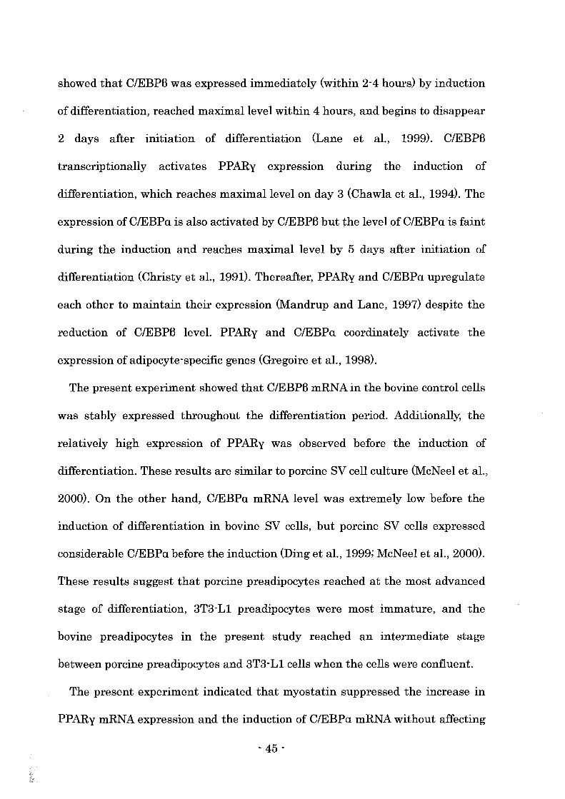
The present experiment showed that C/EBPB mRNA in the bovine control cells
was stably expressed throughout the differentiation period. Additionally, the
relatively high expression of PPARy was observed before the induction of
differentiation. These results are similar to porcine SV cell culture (McNeel et aI.,
2000). On the other hand, C/EBPa mRNA level was extremely low before the
induction of differentiation in bovine SV cells, but porcine SV cells expressed
considerable C/EBPa before the induction (Ding et aI., 1999; McNeel et aI., 2000).
These results suggest that porcine preadipocytes reached at the most advanced
stage of differentiation, 3T3-L1 preadipocytes were most immature, and the
bovine preadipocytes in the present study reached an intermediate stage
between porcine preadipocytes and 3T3-L1 cells when the cells were confluent.
The present experiment indicated that myostatin suppressed the increase in
PPARy mRNA expression and the induction of C/EBPa mRNA without affecting
- 45 -

the expression of C/EBPB mRNA in the early phase of differentiation.
Additionally, the expression of C/EBPB mRNA was not affected by myostatin
until day 4 but PPARy mRNA level was decreased and C/EBPa mRNA level was
continuously low in the bovine preadipocytes treated with myostatin. These
results suggest that myostatin suppressed bovine preadipocyte differentiation
via inhibiting the transcriptional cascade downstream of C/EBPB mRNA
expression. Kim et ai. (2001) reported that myostatin reduced PPARy and
C/EBPa levels but did not affect C/EBPB expression in 3T3-L1 preadipocytes.
Activin A is reported to repress the transactivation functions of C/EBPB in
hepatocyte culture mediated by Smad3 interaction with the DNA binding domain
of C/EBPB (Zauberman et aI., 2001). As mentioned above, myostatin is reported
to regulate cellular function via activin receptors and the intracellular signals of
activin (Rebbapragada et aI., 2003). Therefore, myostatin possibly prevents
bovine preadipocyte differentiation via Smad3-mediated impairment of the
transactivation function of C/EBPB. In the present study, the differentiation was
more severely suppressed by the treatment with myostatin throughout the
differentiation period than by the treatment in the early phase of differentiation.
The expression of C/EBPB begins to decline 2 days after the initiation of
differentiation in 3T3-L1 cells (Lane et aI., 1999), but bovine preadipocyte
expressed C/EBPB mRNA throughout the differentiation period. Additionally,
myostatin reduced C/EBPB mRNA level of bovine preadipocyte in the late phase
of differentiation. The severe inhibition of differentiation induced by the
treatment throughout the differentiation period probably result from the
continuous suppression of C/EBPB function and/or the reduction of C/EBPB
- 46 -
mRNAexpression in the late phase.
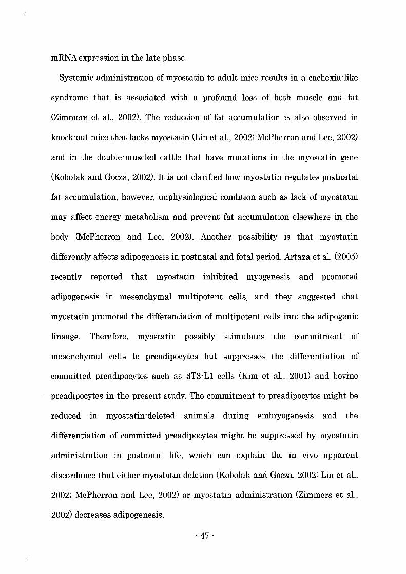
Systemic administration of myostatin to adult mice results in a cachexia -like
syndrome that is associated with a profound loss of both muscle and fat
(Zimmers et aI., 2002). The reduction of fat accumulation is also observed in
knock-out mice that lacks myostatin (Lin et aI., 2002; McPherron and Lee, 2002)
and in the double-muscled cattle that have mutations in the myostatin gene
(Kobolak and Gocza, 2002). It is not clarified how myostatin regulates postnatal
fat accumulation, however, unphysiological condition such as lack of myostatin
may affect energy metabolism and prevent fat accumulation elsewhere in the
body (McPherron and Lee, 2002). Another possibility is that myostatin
differently affects adipogenesis in postnatal and fetal period. Artaza et ai. (2005)
recently reported that myostatin inhibited myogenesls and promoted
adipogenesis in mesenchymal multipotent cells, and they suggested that
myostatin promoted the differentiation of multipotent cells into the adipogenic
lineage. Therefore, myostatin possibly stimulates the commitment of
mesenchymal cells to preadipocytes but suppresses the differentiation of
committed preadipocytes such as 3T3-L1 cells (Kim et aI., 2001) and bovine
preadipocytes in the present study. The commitment to preadipocytes might be
reduced In myostatin-deleted animals during embryogenesis and the
differentiation of committed preadipocytes might be suppressed by myostatin
administration in postnatal life, which can explain the in vivo apparent
discordance that either myostatin deletion (Kobolak and Gocza, 2002; Lin et aI.,
2002; McPherron and Lee, 2002) or myostatin administration (Zimmers et aI.,
2002) decreases adipogenesis.
- 47 -
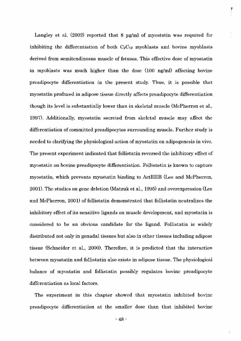
Langley et ai. (2002) reported that 8 pg/ml of myostatin was required for
inhibiting the differentiation of both C2C12 myoblasts and bovine myoblasts
derived from semitendinosus muscle of fetuses. This effective dose of myostatin
in myoblasts was much higher than the dose (100 ng/m!) affecting bovine
preadipocyte differentiation in the present study. Thus, it is possible that
myostatin produced in adipose tissue directly affects preadipocyte differentiation
though its level is substantially lower than in skeletal muscle (McPherron et aI.,
1997). Additionally, myostatin secreted from skeletal muscle may affect the
differentiation of committed preadipocytes surrounding muscle. Further study is
needed to clarifying the physiological action of myostatin on adipogenesis in vivo.
The present experiment indicated that follistatin reversed the inhibitory effect of
myostatin on bovine preadipocyte differentiation. Follistatin is known to capture
myostatin, which prevents myostatin binding to ActRIIB (Lee and McPherron,
2001). The studies on gene deletion (Matzuk et aI., 1995) and overexpression (Lee
and McPherron, 2001) of follistatin demonstrated that follistatin neutralizes the
inhibitory effect of its sensitive ligands on muscle development, and myostatin is
considered to be an obvious candidate for the ligand. Follistatin is widely
distributed not only in gonadal tissues but also in other tissues including adipose
tissue (Schneider et aI., 2000). Therefore, it is predicted that the interaction
between myostatin and follistatin also exists in adipose tissue. The physiological
balance of myostatin and follistatin possibly regulates bovine preadipocyte
differentiation as local factors.
The experiment in this chapter showed that myostatin inhibited bovine
preadipocyte differentiation at the smaller dose than that inhibited bovine
- 48 -

myoblast differentiation (Langley et aI., 2002). The inhibitory effect of my os tat in
was mediated by the reduction of PPARy and C/EBPa mRNA levels. Additionally,
follistatin affected bovine preadipocyte differentiation, which suggested that
follistatin modulated the action of myostatin on preadipocyte differentiation.
- 49 -
Chapter 5
The role of activin A on the preadipocyte differentiation
Section 1
Effects of activin A on the differentiation of bovine preadipocyte
Introduction
Activin A, a member of the transforming growth factor-B (TGF-B) superfamily,
is homodimer of activin BA subunit. Recently, the expressions of activin BA
(Meunier et aI., 1988; Tuuri et aI., 1994) and activin receptor mRNAs (Mathews,
1994) were found in many tissues throughout the body. Especially, activin BA was
highly expressed in adipose tissue (Vejda et aI., 2002).
CCAAT/enhancer binding proteins (C/EBPs) and peroxisome proliferator
activated receptor (PPAR) y have shown to be critical transcription factors for
preadipocyte differentiation (Gregoire et aI., 1998; Rosen et aI., 2000). Studies
using 3T3-L1 preadipocytes indicate that C/EBPB is highly expressed after the
treatment with adipogenic agents (Lane et aI., 1999). C/EBPB activates the
expression of PPARy and C/EBPa mRNAs that coordinately activate the
transcription of adipocyte-specific genes (Rosen et aI., 2000).
In Chapter 4, the author investigated the expression patterns of the critical
transcription factors in stromal vascular (SV) cells from bovine adipose tissue.
- 50 -
SV cells did not express adipogenic differentiation markers such as lipid
accumulation and adipocyte fatty acid-binding protein (aP2) mRNA before the
induction of differentiation. On the other hand, C/EBPB mRNA was stably
expressed throughout the differentiation period, and the expression of PPARy
was relatively high before the induction of differentiation. C/EBPa mRNA was
only faintly expressed before the induction of differentiation and increased
thereafter. These results suggested that bovine SV cells derived from adipose
tissue contained the preadipocytes reaching at more advanced stage of
differentiation than 3T3-L1 preadipocytes did.
Kim et al. (2001) reported that myostatin, which is also a member of TGF-B
superfamily, inhibited the differentiation of 3T3-L1 preadipocytes by reducing
the expression of PPARy and C/EBPa but not of C/EBPB. Despite the different
expression patterns of the critical transcription factors in bovine preadipocytes,
myostatin also inhibited bovine preadipocyte differentiation via affecting the
transcriptional cascade downstream of C/EBPB as demonstrated in Chapter 4.
The action of myostatin is reported to be mediated by activin receptors (Lee and
McPherron, 2001; Massague and Chen, 2000) and the author demonstrated the
mRNA expressions of activin receptors in post confluent bovine SV cells derived
from adipose tissue in Chapter 4. On the other hand, the effect of activin A on the
differentiation of bovine preadipocytes has been still unknown.
In this chapter, the author investigated the effect of activin A on the
differentiation of bovine preadipocytes. Activin A reduced PPARy mRNA
expression and interfered with the increase of C/EBPa mRNA in bovine
preadipocytes, which suppressed the induction of terminal differentiation
- 51 -
markers.
Follistatin is a secretory protein and it binds to activin and myostatin, which
results in the prevention of their binding to the activin receptor (Lee and
McPherron, 2001; Schneyer et aI., 2003). The inhibitory action of myostatin on
the differentiation of bovine preadipocyte was suppressed by follistatin as shown
in Chapter 4. The present experiment indicated that follistatin also interfered
with the suppressive action of activin A on bovine preadipocyte differentiation.
Furthermore, the higher doses of follistatin stimulated the differentiation even
in the presence of activin A compared with the control culture treated with
neither activin A nor follistatin. The author demonstrated the mRNA expressions
of activin A and myostatin in the SV cells. Therefore, endogenous activin A
and/or myostatin possibly inhibited the differentiation of bovine preadipocytes.
Materials and methods
Culture of stromal-vascular cells from bovine adipose tissue
SV cells were prepared from bovine perirenal adipose tissue and cultured
according to the method shown in Chapter 4. Recombinant human activin A
(Wako Chemicals) and recombinant human follistatin (Genzyme Techne) was
dissolved in phosphate-buffered saline (PBS) with 0.1% bovine serum albumin
(Sigma) and added to the medium in the concentrations as shown in each figure
during the early phase of differentiation (from day 0 to day 2) or throughout the
differentiation period (from day 0 to day 8).
- 52 -
Glycerol-3phosphate dehydrogenase activity
Cells were carefully washed twice with ice-cold PBS on day 8, and lysed in 25
mM Trisl 1 mM EDTA, pH 7.5 by sonication for measurement of GPDH specific
activity. GPDH activity was determined according to the method shown in
Chapter 3.
011 Red 0 staining
Cultures were washed twice with PBS on day 8, fixed with 10% formalin in
PBS, stained with 0.5% Oil Red 0 and photographed.
RNA isolation
Total RNA was extracted from the SV cells according to the method shown in
Chapter 4.
Northern blot analysis for the expression of adipocyte fatty acid-binding protein
mRNA
The expression of aP2 mRNA in the SV cells on day 8 was analyzed by
Northern blotting according to the method shown in Chapter 4.
Semi-quantitative RTPCR for the determination ofCIEBP and PPARy mRNAs
The expression of C/EBP and PPARy mRNAs were assessed by
semi -quantitative RT-PCR according to the method shown in Chapter 4.
- 53 -
Detection of activin BA, myostatin, and foiJistatin mRNAs
The expression of activin BA, myostatin, and follistatin mRNAs in bovine
adipose tissue and in SV cells on day 0 were analyzed by RT-PCR according to
the method shown in Chapter 4. The PCR were conducted using Platinum PCR
Super Mix (Invitrogen). The primer sets for each receptor were shown in Table
5-1. The amplification parameters consisted of denaturation at 94°C for 30 s,
annealing at the temperature (Table 5-1) for 30 s, and extension at 72°C for 1
min, for 45 cycles. The sub cloning and sequencing confirmed the PCR products to
be the expected fragments of bovine activin BA, myostatin, and follistatin. The
products were separated on 2% agarose gel and visualized with ethidium
bromide. To avoid false-positive results from contamination by genomic DNA,
samples with or without treatment of reverse transcriptase were prepared, and
as a negative control, RT-PCR without RNA samples was also carried out.
Statistics
Data were expressed as means ± SE, and were statistically analyzed using
student's t-test.
- 54 -
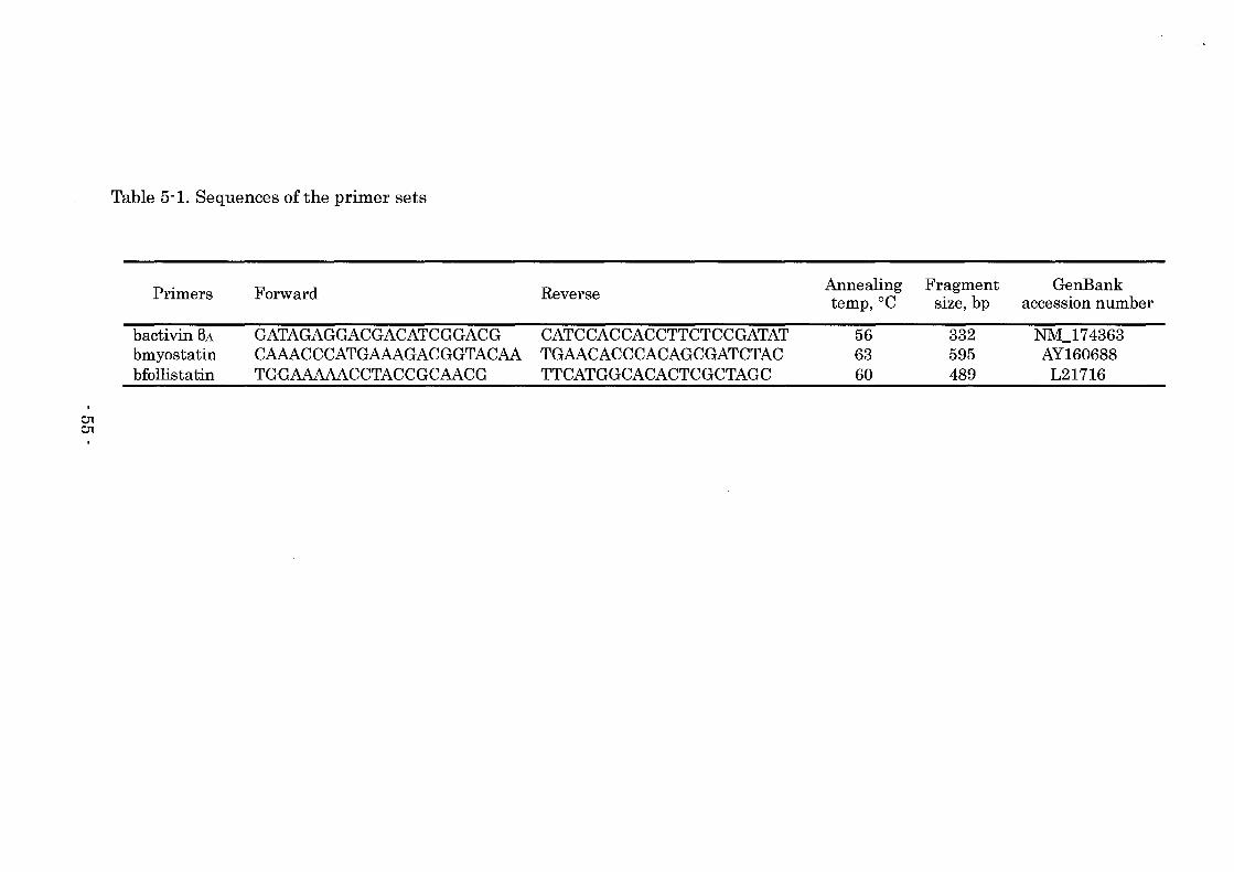
01 01
Table 5-1. Sequences of the primer sets
Primers
bactivin BA bmyostatin bfollistatin
Forward
GATAGAGGACGACATCGGACG CAAACCCATGAAAGACGGTACAA TGGAAAAACCTACCGCAACG
Reverse
CATCCACCACCTTCTCCGATAT TGAACACCCACAGCGATCTAC TTCATGGCACACTCGCTAGC
Annealing Fragment GenBank temp,OC size, bp accession number
56 332 NM_174363 63 595 AY160688 60 489 L21716
Results
Differentiation of bovine preadipocytes
Oil Red 0 staining showed that many control cells had lipid droplets at the end
of culture (Fig. 5-1-1A). However, lipid accumulation was inhibited by the
treatment with 1.25 ng/ml activin A in the early phase of differentiation, and was
progressively reduced at higher doses (Fig. 5-1-1A). Activin A treatment
dose-dependently reduced GPDH activity (P<O.Ol) (Fig. 5-1-1B) and the level of
aP2 mRNA (Fig. 5-1-1C). The treatment with activin A throughout the
differentiation period also reduced lipid accumulation (Fig. 5-1-2A), GPDH
activity (P<O.Ol) (Fig. 5-1-2B), and aP2 mRNAlevel (Fig. 5-1-2C).
Expression of CIEBP and PPARy mRNAs
C/EBPB mRNA expression in the control cells slightly increased after induction
of differentiation, and was stable after day 2 (Fig. 5-1-3A). Activin A treatment
throughout the differentiation period did not affect the expression of C/EBPB
mRNA (Fig. 5-1-3A). The control cells expressed PPARy mRNA on day 0, then its
expression moderately increased, and it reached almost maximal level on day 6
(Fig. 5-1-3B). Activin A suppressed the increase in PPARy mRNA expression
(P<0.05) during the induction of differentiation, and PPARy mRNA level was
stably low thereafter (Fig. 5-1-3B). The control cells faintly expressed C/EBPa
mRNA on day 0 and its expression gradually increased after the induction of
differentiation (Fig. 5-1-3C). Activin A suppressed the increase in CIEBPa mRNA
level (P<0.05) (Fig. 5-1-3C).
- 56 -
Activin A and follistatin treatments
The author investigated whether follistatin affected the suppressive action of
activin A on the differentiation of bovine preadipocytes. Activin A suppressed
GPDH activity (P<O.Ol) when the cells were treated with activin A in the early
phase of differentiation (Fig. 5-1-4). The simultaneous treatment with 50 ng/ml
follistatin reversed the suppressive effect of activin A (P<O.Ol) (Fig. 5-1-4).
Furthermore, the higher dose (300 ng/ml) of follistatin increased GPDH activity
(P<0.05) compared with the control culture treated with neither activin A nor
follistatin (Fig. 5-1-4). The treatment with follistatin (500 ng/ml) alone increased
GPDH activity (P=0.055) compared with the control culture (Fig. 5-1-4).
Expressions of activin RA, myostatin, and follistatin mRNAs
Expressions of bovine activin BA, myostatin, and follistatin mRNAs were
examined by RT-PCR in bovine adipose tissue and in postconfiuent bovine SV
cells. We detected all these mRNA expressions in both adipose tissue and in the
SV cells (Fig. 5-1-5). No bands were detected in the absence of reverse
transcriptase or RNA samples during the synthesis of cDNA (data not shown)
- 57 -
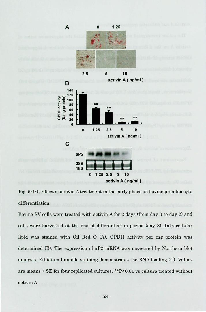
A
B 140
_120 ~c: .- .- 100 >~ ~ e 80 III Q.
60 :I: Cl C E 40 Q,-e>2. 20
0
C
2.5
0
288 188
o 1.25
5 10
activin A ( ng/ml )
1.25 2.5 5 10
activin A ( ng/ml )
o 1.25 2.5 5 10 activin A ( ng/ml )
Fig. 5-1-1. Effect of activin A treatment in the early phase on bovine preadipocyte
differentiation.
Bovine SV cells were treated with activin A for 2 days (from day 0 to day 2) and
cells were harvested at the end of differentiation period (day 8). Intracellular
lipid was stained with Oil Red 0 (N. GPDH activity per mg protein was
determined (B). The expression of aP2 mRNA was measured by Northern blot
analysis. Ethidium bromide staining demonstrates the RNA loading (C). Values
are means ± SE for four replicated cultures. **P<O.Ol vs cultul'e treated without
activin A.
- 58 -
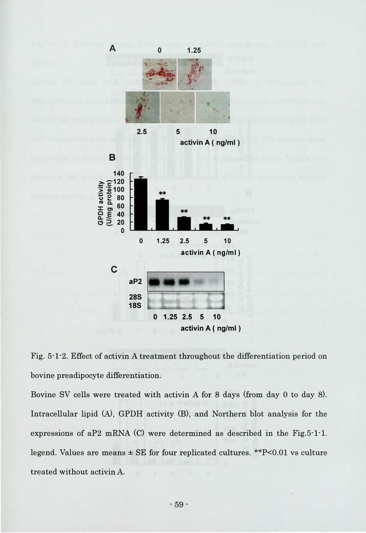
A 0 1.25
B
140 ~c120 .:; .a; 100 ~ ~ 80 CV Q. 60 :I:C) C E 40 a..-e>2. 20
o
C
2.5 5 10
0
aP2
28S 18S
activin A ( ng/ml )
1.25 2.5 5 10
activin A ( ng/ml )
o 1.25 2.5 5 10
activin A ( ng/ml )
Fig. 5-1-2. Effect of activin A treatment throughout the differentiation period on
bovine preadipocyte differentiation.
Bovine SV cells were treated with activin A for 8 days (from day 0 to day 8).
Intracellulal' lipid W , GPDH activity (B) , and Northern blot analysis for the
expressions of aP2 mRNA (C) were determined as described in the Fig.5-1-1.
legend. Values are means ± SE for four replicated cultures. **P<O.Ol vs culture
treated without activin A.
- 59 -
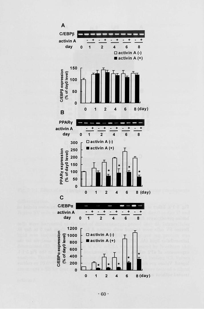
A
C/EBPP
activin A day
c:::: 150 0= ·iii ~ ~ ~ 100 "'0 0.>0-)( III Q)-o ~'O 50 ffi~ (3-
o
B
PPARy
------------ + - +
o 1 2
o 1 2
- + - + - +
4 6 8
o activin A (-) • activin A (+)
4 6 8 (day)
- - - - - - - - -activin A - + - + - + - + - +
day 0 1 2 4 6 8
300
g = 250 .- GI :: ~ 200 GI-
~~ 150 GI I'll
>~ 100 a::: 0
~ C 50 Q.
o
c C/EBPa
Dactivin A H • activin A (+)
o 1 2 4 6 8 (day)
- - - - - . activin A - + - + - + - + - +
day 0 1 2 4 6 8
1200 c: .Q ~1000 :: > ! ~ 800 Q,O
~ 1;' 600 c "0
Q. '0 400 m~ ~ ~ 200 ()
o
o activin A (-)
• activin A (+)
o 1 2
- 60 -
4 6 8 (day)
Fig. 5-1-3. Effect of activin A treatment on mRNA expressions of C/EBP and
PPARy.
Bovine SV cells were treated with 10 ng/ml activin A throughout the
differentiation period (from day 0 to day 8). The expression levels of C/EBP8 (A),
PPARy (B), and C/EBPa (C) mRNAwere estimated by semi-quantitative RT-PCR
and normalized with respect to the 18S rRNA expression level. The relative gene
expression is presented as the ratio of the day 0 expression level. Values are
means ± SE for three or four replicated cultures. *P<0.05 vs culture treated
without activin A.
- 61 -
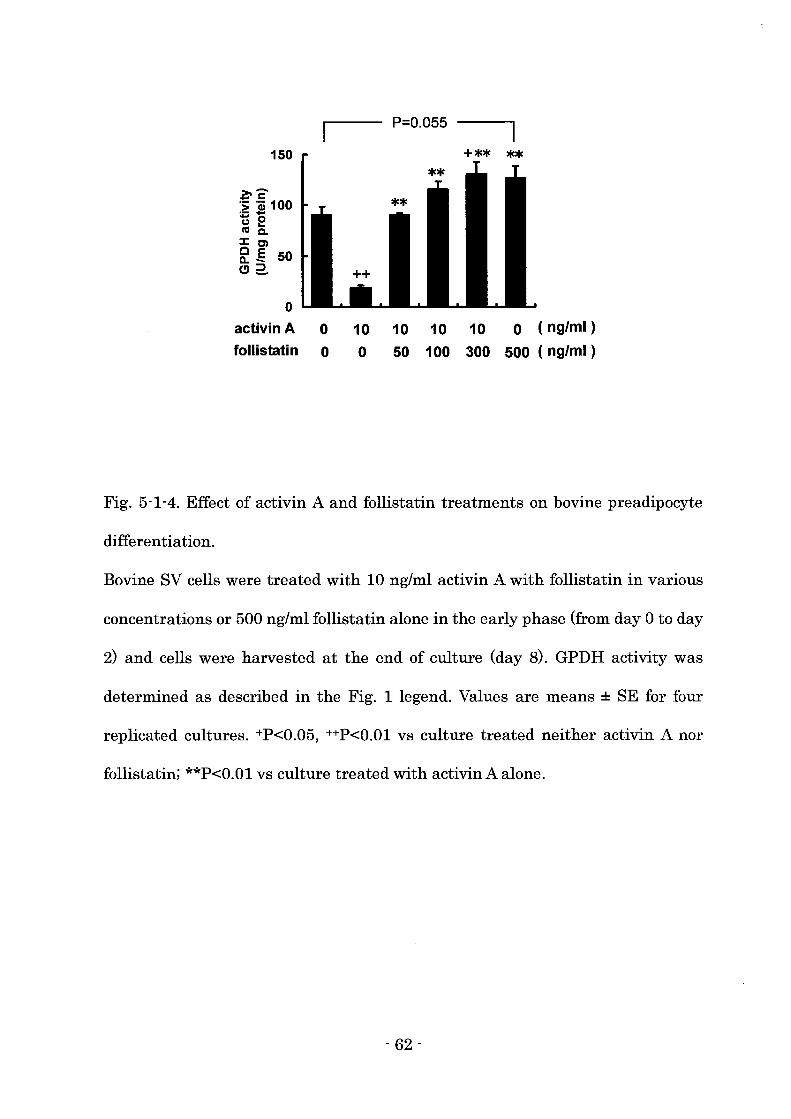
150
a-c .; .Qj 100 .- -t) 2 co Co Ie) f[.§ 50 e>2..
o activin A 0
follistatin 0
P=O.055
10 10 10 10 0 (ng/ml)
o 50 100 300 500 (ng/ml)
Fig. 5-1-4. Effect of activin A and follistatin treatments on bovine preadipocyte
differentiation.
Bovine SV cells were treated with 10 ng/ml activin A with follistatin in various
concentrations or 500 ng/ml follistatin alone in the early phase (from day 0 to day
2) and cells were harvested at the end of culture (day 8). GPDH activity was
determined as described in the Fig. 1 legend. Values are means ± SE for four
replicated cultures. +P<0.05, ++P<O.OI vs culture treated neither activin A nor
follistatin; **P<O.OI vs culture treated with activin A alone.
- 62 -
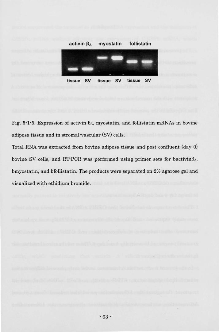
activin J3A myostatin follistatin
tissue SV tissue SV tissue SV
Fig. 5-1-5. Expression of activin BA, myostatin, and follistatin mRNAs in bovine
adipose tissue and in stromal-vascular (SV) celis.
Total RNA was extracted from bovine adipose tissue and post confluent (day 0)
bovine SV celis, and RT-peR was performed using primer sets for bactivinBA,
bmyostatin, and bfollistatin. The products were separated on 2% agarose gel and
visualized with ethidium bromide.
- 63 -
Discussion
The present study showed that activin A inhibited the differentiation of bovine
preadipocytes and this inhibitory effect of activin A was reversed by
simultaneous treatment with follistatin. Furthermore, the higher doses of
follistatin stimulated the differentiation even in the presence of activin A
compared with the control culture treated with neither activin A nor follistatin.
The SV cells in the present study expressed activin A and myostatin mRNAs.
These results suggest that follistatin also interferes with endogenous activin A
and/or myostatin that inhibit the differentiation of preadipocytes in the SV cells
derived from bovine adipose tissue.
The bovine control cells almost stably expressed C/EBPB mRNA, the
expression of PPARy was relatively high on day 0 and moderately increased
thereafter, and C/EBPa mRNA level was faint on day 0 and drastically increased
between day 4 and day 6.adipose tissue.
The present experiment showed that C/EBPB mRNA in the bovine control cells
was stably expressed until day 6, the expression of PPARy was moderately
increased after induction of differentiation, and C/EBPa mRNA level was
drastically increased between day 4 and day 6. These results were almost similar
to the results in Chapter 4.
In the present study, activin A treatment in the early phase of differentiation
reduced lipid accumulation, GPDH activity, and aP2 mRNA level, and its
treatment throughout the differentiation period also reduced these terminal
differentiation markers. Activin A treatment throughout the differentiation
- 64 -
period suppressed the increase in PPARy mRNA expression and the induction of
C/EBPa mRNA without affecting the expression of C/EBPB mRNA, which
suggests that activin A suppressed bovine preadipocyte differentiation via
inhibiting the transcriptional cascade downstream of C/EBPB mRNA expression.
Activin A was reported to suppress the function of C/EBPB in human hepatocyte
culture, which was mediated by Smad3 protein that interacted with the DNA
binding domain of C/EBPB and repressed the transactivation function of C/EBPB
(Zauberman et aI., 2001). TGF-B also inhibited the function of C/EBPB mediated
by Smad3 protein, which decreased gene expression of PPARy, and thus TGF-B
inhibited adipogenesis (Choy and Derynck, 2003). Therefore, activin A possibly
prevents bovine preadipocyte differentiation via Smad3-mediated impairment of
the transactivation function of C/EBPB.
During human pregnancy, serum concentration of activin A is reported to
increase progressively (Petraglia et aI., 1995). On the other hand, activin A is
normally present in relatively low amounts in the circulation (below 0.1 ng/mI) in
adult sheep (Jenkin et aI., 2001; Jones et aI., 2004), and the activin A level did
not change throughout the gestation (McFarlane O'Shea et aI., 1998). Vejda et aI.
(2002) reported that activin BA was highly expressed in adipose tissue in rat, and
the author also found the mRNA expression of activin BA in adipose tissue in
cattle, which predicting that activin A affects adipogenesis through
autocrine/paracrine manner. Serum concentration of activin A in cattle has not
been studied, however, locally produced activin A may be highly involved in the
preadipocyte differentiation than serum activin A in ruminants.
The present experiment indicated that follistatin reversed the inhibitory effect
- 65 -
of activin A on bovine preadipocyte differentiation. Follistatin is known to
capture activin A, which neutralizes its activity (Schneyer et al., 2003).
Follistatin is likely to attenuate activin A action on preadipocyte differentiation
through forming the inactive complex.
In the present study, the high dose (300 ng/mn of follistatin increased GPDH
activity in the presence of activin A compared with the control culture treated
with neither activin A nor follistatin. Furthermore, the treatment with 500 ng/ml
follistatin alone stimulated the differentiation compared with the control culture.
The present study indicated that the bovine SV cells expressed both activin BA
and myostatin mRNAs. Follistatin also captures myostatin and inhibits its
action (Lee and McPherron, 2001). Endogenous activin A and/or myostatin
probably inhibited preadipocyte differentiation in the SV cell culture, and thus
exogenous follistatin promoted adipogenesis. The expression of follistatin, as well
as aGtivin A, is widely distributed in gonadal and non-gonadal tissues including
adipose tissue (Schneider et al., 2000). The author demonstrated the expression
of follistatin in bovine adipose tissue. Myostatin mRNA is found primarily in
skeletal muscle (McPherron et al., 1997), however, it was also detected in bovine
adipose tissue. Furthermore, the author reported in Chapter 4 that myostatin
inhibited the differentiation of bovine preadipocytes, and this inhibitory effect of
myostatin was interfered with the simultaneous treatment of follistatin. The
interaction between activin A and follistatin, or myostatin and follistatin may
exist in the adipose tissue. The physiological balance of these autocrine/paracrine
factors possibly regulates preadipocyte differentiation in adipose tissue.
In summary, the results presented in this section showed that activin A
- 66 -
inhibited bovine preadipocyte differentiation, and this inhibitory effect of activin
A was mediated by the reduction of PPARy and C/EBPa mRNA levels.
Additionally, follistatin affected bovine preadipocyte differentiation, which
suggests that follistatin modulates the action of activin A and/or myostatin on
preadipocyte differentiation.
- 67 -
Section 2
The mechanism of the inhibitory effect of activin A
on the differentiation of 3T3-Llpreadipocyte
Introduction
Activin A, a member of the transforming growth factor-B (TGF-B) superfamily,
is homodimer of activin BA subunit. Recently, the expressions of activin BA
(Meunier et al., 1988; Tuuri et al., 1994) and activin receptor mRNAs (Mathews,
1994) were found in many tissues throughout the body. Especially, activin BA was
highly expressed in adipose tissue (Vejda et al., 2002).
3T3-L1 preadipocyte is one of the most well-characterized and reliable models
for studying adipogenesis. Upon reaching confluence, the treatment with
adipogenic agents induces mitotic clonal expansion (MacDougald and Lane,
1995; Richon et al., 1997). CCAAT/enhancer binding protein (C/EBP) B is highly
expressed after the treatment with adipogenic agents (Lane et al., 1999) and
C/EBPB activates the expression of peroxisome proliferator-activated receptor
(PPAR) y and C/EBPa mRNAs that coordinately activate the transcription of
adipocyte-specific genes (Rosen et al., 2000).
In Section 1 in Chapter 5, the author demonstrated that activin A inhibited the
differentiation of bovine preadipocyte. Additionally, the author reported that
myostatin, a member of TGF-B superfamily also inhibited the differentiation of
bovine preadipocyte in Chapter 4. The inhibitory effects of activin A and
- 68 -
myostatin on the differentiation of bovine preadipocyte were both mediated by
the suppression of PPAR y and C/EBPa mRNA expression. However, further
mechanism that activin A and myostatin inhibit preadipocyte differentiation has
been still unclear.
Zauberman et al. (2001) reported that activin A inhibited haptoglobin
expression via suppressing C/EBP8 function in human hepatoma cells. Thus,
activin A may inhibit adipocyte differentiation via suppressing C/EBP8 function.
In this chapter, the author investigated the mechanism of the inhibitory effect
of activin A on the differentiation of 3T3-Ll preadipocyte. Activin A inhibited the
early phase of differentiation via suppressing PPARy and C/EBPa mRNA
expression. Activin A interfered with the DNA binding activity of C/EBP8, which
was considered to suppress the differentiation.
Materials and methods
Cell culture
3T3-Ll preadipocyte were cultured according to the method shown in Chapter
3. Recombinant human activin A (Wako Chemicals) was dissolved in PBS with
0.1 % bovine serum albumin (Sigma) and added to the medium in various
concentrations as shown in each figure throughout the differentiation period
(from day 0 to day 8), during the early phase of differentiation (from day 0 to day
2) or during the late phase of differentiation (from day 2 to day 8).
- 69 -
Quantifica tion of mitotic clonal expansion
Cells were treated with activin A during the early phase of differentiation,
trypsinized in trypsin / EDTA and resuspended in DMEM. Then the number of
cells was counted on Thoma-type hemocytometer.
Glycerol-3phosphate dehydrogenase activity
Cells were carefully washed twice with ice-cold PBS on day 8, and lysed in 25
mM Tris/1 mM EDTA, pH 7.5 for measurement of GPDH specific activity. GPDH
activity was determined according to the method shown in Chapter 3.
Oil Red a staining
Cultures were washed twice with PBS on day 8, fixed with 10% formalin in
PBS, stained with 0.5% Oil Red 0 and photographed.
RNA isolation
Total RNA was extracted from 3T3-L1 cells according to the method shown in
Chapter 4.
Detection of activin receptor mRNAs in 3T3-Ll preadipocyte
The expression of activin receptor mRNAs encoding type I (ActRI and ActRIB)
and type II (ActRII and ActRIIB) receptors on day 0 were examined by RT-PCR.
Single-strand cDNA was synthesized using a SuperScript First-Strand Synthesis
System for RT-PCR (Invitrogen) according to the manufacture's protocol. The
PCR were conducted using Platinum PCR Super Mix (Invitrogen). The primer
- 70 -
sets for each receptor were shown in Table 5-2. Thirty-five cycles were selected
for detection. The products were separated on 2% agarose gel and visualized with
ethidium bromide.
Northern blot analysis for the expression of aP2 mRNA
RNA sample (4 pg) was separated in 1% agarose gels containing 6.7%
formaldehyde and transferred to a Hybond N+ membrane (Amersham
Biosciences, NJ, USA). cDNA fragment of mouse adipocyte fatty acid-binding
protein (aP2) gene was amplified using specific primers (Table 5-2.) and cDNA
probe was labeled with digoxigenin-dUTP by using random priming (Roche
Diagnostics). The band corresponding to aP2 mRNA was detected using DIG
Luminescant Detection Kit for Nucleic Acids (Roche Diagnostics).
Semi-quantitative RT-PCR for the determination ofCIEBP and PPARy mRNAs
The expression of C/EBP and PPARy mRNAs were assessed by
semi-quantitative RT-PCR. The thermal amplifications of C/EBP, PPARy, and
18S rRNA fragments were conducted using Platinum PCR Super Mix
(Invitrogen). The primer sets were shown in Table 5-2. PCR conditions were
optimized for detection within a linear range. Twenty-eight cycles were selected
for experimental detection of C/EBPB, 26 cycles for PPARy, 30 cycles for C/EBPu,
and 24 cycles for 18S rRNA. The level of 18S rRNA was adopted as the internal
standards for the determination of targeted mRNA levels.
- 71 -
Western immunoblot analysis
3T3-L1 cells were harvested and lysed in 25 mM Tris 1 1 mM EDTA, pH 7.5 by
sonication. Protein concentration of cell lysate was determined and 20 p.g of
protein was subjected to 12.5% SDS-PAGE and transferred to Immobilon-P
membrane (Millipore). After blocking with 3% skim milk in PBS, membrane was
incubated with primary antibody, polyclonal anti - C/EBPB (Santa Cruz
Biotechnology, CA, USA; 1/500) for 1 h, followed by incubation with secondary
antibody conjugated to horseradish peroxidase, anti -rabbit IgG (Amersham
Biosciences, 1/2000) for 30 min. Visualization of the second antibody binding was
performed by chemiluminescence with the ECL plus Western blotting detection
system (Amersham Biosciences).
Electrophoretic mobllity shift assay (EMSA)
3T3-L1 cells treated with 20 ng/ml activin A in 60-mm dishes (Corning) were
carefully rinsed twice with ice-cold 10 mM HEPES buffer, pH 7.5 on day 2. Then
the cells were incubated with 10 mM HEPES buffer for 15 min on ice and scraped.
The harvested cells were lysed by the dounce homogenizer with 15 strokes. After
centrifugation at 10,000 rpm for 3 min at 4 oC, the supernatant was discarded.
After a crude nuclear pellet was washed twice with ice-cold 25 mM HEPES 1 3
mMgCb 1 1 mM DTT, pH 7.5., the pellet was resuspended in ice-cold 25 mM
HEPES 1 0.4 mM KCII 1mM DTT, pH 7.5. Then the pellet was incubated on ice
for 30 min with occasional vortexing. Mter the incubation, glycerol was added to
become 10% of final concentration and the reaction buffer containing nuclear
extract was centrifuged at 12,000 rpm for 1 h at 4 °C. The supernatant was
- 72 -
stored as nuclear extract at -80 ac. Protein concentration was measured with the
method of Lowry et al. (1951). Five pg of nuclear extract protein was subjected to
electrophoretic mobility shift assay (EMSA) using EMSA kit for C/EBP
(Panomics, CA, USA), according to the manufacture's protocol. The sequence of
C/EBP probe was 5'-TGCAGATTGCGCAATCTGCA-3', which contained the
binding site of C/EBPB. Polyclonal anti- C/EBPB (Santa Cruz Biotechnology) was
used for the determination of the complex of C/EBPB and C/EBP probe.
Statistics
Data were expressed as means ± SE, and were statistically analyzed using
student's t-test. Statistical significance was set at P<0.05.
- 73 -
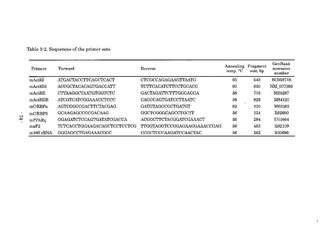
Table 5-2. Sequences of the primer sets
Annealing Fragment GenBank Primers Forward Reverse acceSSIOn temp,OC size, bp number
mActRI ATGACTACCTTCAGCTCACT CTCGCCAGAGAAGTTAATG 60 542 BC058718 mActRIB ACCGCTACACAGTGACCATT TCTTCACATCTTCCTGCACG 60 630 NM_007395 mActRII CTTAAGGCTAATGTGGTCTC GACTAGATTCTTTGGGAGGA 58 703 M65287 mActRIIB ATCGTCATCGGAAACCTCCC CAGCCAGTGATCCTTAATC 59 929 M84120 mCIEBPa AGTCGGCCGACTTCTACGAG GATGTAGGCGCTGATGT 62 200 M62362
-:) mCIEBPB GCAAGAGCCGCGACAAG GGCTCGGGCAGCCTGCTT 56 154 X62600
~ mPPARy GGAGATCTCCAGTGATATCGACCA ACGGCTTCTACGGATCGAAACT 56 294 U01664 maP2 TCTCACCTGGAAGACAGCTCCTCCTCG TTGGTAGGTCCGGAGAAGGAAACCGAG 56 463 K02109 m18S rRNA GGGAGCCTGAGAAACGGC CCGCTCCCAAGATCCAACTAC 56 262 X00686
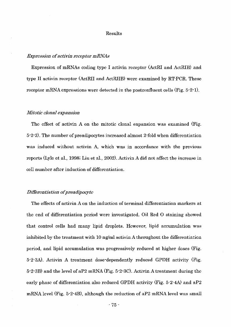
Results
Expression of activin receptor mRNAs
Expression of mRNAs coding type I activin receptor (ActRI and ActRIB) and
type II activin receptor (ActRII and ActRIIB) were examined by RT-PCR. These
receptor mRNAexpressions were detected in the postconfluent cells (Fig. 5-2-1).
Mitotic clonal expansion
The effect of activin A on the mitotic clonal expansion was examined (Fig.
5-2-2). The number ofpreadipocytes increased almost 2-fold when differentiation
was induced without activin A, which was in accordance with the previous
reports (Lyle et aI., 1998; Liu et aI., 2002). Activin A did not affect the increase in
cell number after induction of differentiation.
Differentiation ofpreadipocyte
The effects of activin A on the induction of terminal differentiation markers at
the end of differentiation period were investigated. Oil Red 0 staining showed
that control cells had many lipid droplets. However, lipid accumulation was
inhibited by the treatment with 10 ng/ml activin A throughout the differentiation
period, and lipid accumulation was progressively reduced at higher doses (Fig.
5-2-3A). Activin A treatment dose-dependently reduced GPDH activity (Fig.
5-2-3B) and the level of aP2 mRNA (Fig. 5-2-3C). Activin A treatment during the
early phase of differentiation also reduced GPDH activity (Fig. 5-2-4A) and aP2
mRNA level (Fig. 5-2-4B), although the reduction of aP2 mRNA level was small
- 75 -

compared with the treatment throughout the differentiation period. Activin A
treatment during the late phase of differentiation modestly decreased GPDH
activity (Fig. 5-2-5A) and aP2 mRNA level (Fig. 5-2-5B).
Expressions of CIEBP and PPARy mRNA
The expression of C/EBPB mRNA drastically increased 12 h after the induction
of differentiation in the control cells, reached maximal level after 24 h, and then
gradually declined (Fig. 5-2-6A). Activin A treatment did not affect the
expression of C/EBPB mRNA. The expression of PPARy mRNA gradually
increased from 12 h after the induction of differentiation and reached maximal
level after 6 days in the control cells but the treatment with activin A suppressed
the expression of PPARy mRNA (Fig. 5-2-6B). C/EBPa mRNA expression
gradually increased from 24 h after the induction of differentiation in the control
cells but the treatment with activin A suppressed C/EBPa mRNA expression (Fig.
5-2-6C). However, the expression ofPPARy and C/EBPa mRNAs partly recovered
after activin A was withdrawn.
Expression of CIEBPB protein
The protein expression of C/EBPB was also examined by Western immunoblot
analysis. There are two active isoforms of C/EBPB protein of which molecular
weights were 35 and 38 kDa. These isoforms increased within 12 h after the
induction of differentiation, and then gradually declined in the control cells (Fig.
5-2-7). Activin A treatment did not affect the expression of C/EBPB protein, which
was in accordance with its mRNA level.
- 76 -
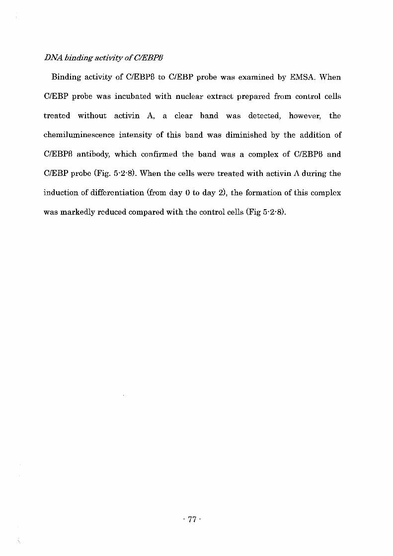
DNA binding activity of CIEBPB
Binding activity of C/EBPB to C/EBP probe was examined by EMSA. When
C/EBP probe was incubated with nuclear extract prepared from control cells
treated without activin A, a clear band was detected, however, the
chemiluminescence intensity of this band was diminished by the addition of
C/EBPB antibody, which confirmed the band was a complex of C/EBPB and
C/EBP probe (Fig. 5-2-8). When the cells were treated with activin A during the
induction of differentiation (from day 0 to day 2), the formation of this complex
was markedly reduced compared with the control cells (Fig 5-2-8).
- 77 -
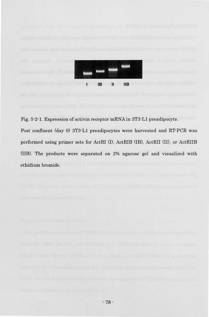
I .... --IB II
IiiiiiIt
liB
Fig. 5-2-1. Expression of activin receptor mRNA in 3T3-Ll preadipocyte.
Post confluent (day 0) 3T3-Ll preadipocytes were harvested and RT-peR was
performed using primer sets for ActRI (I), ActRIB (IB) , ActRII (II), or ActRIIB
(IIB). The products were separated on 2% agarose gel and visualized with
ethidium bromide.
- 78 -
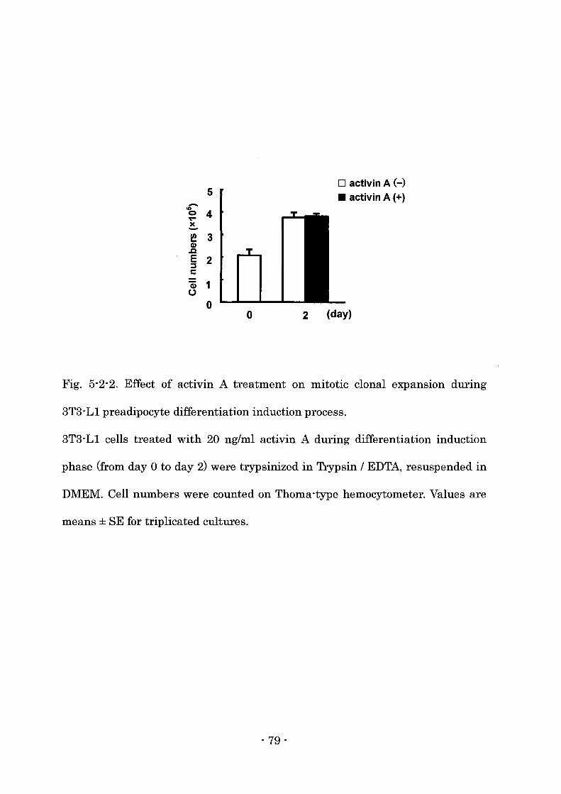
D activin A (-) 5 • activin A (+) -.., 0 4 or-
x -~ 3 CI) .c E 2 ::::J C
CD 1 0
0 0 2 (day)
Fig. 5-2-2. Effect of activin A treatment on mitotic clonal expanSIOn during
3T3-Ll preadipocyte differentiation induction process.
3T3-Ll cells treated with 20 ng/ml activin A during differentiation induction
phase (from day 0 to day 2) were trypsinized in Trypsin / EDTA, resuspended in
DMEM. Cell numbers were counted on Thoma -type hemocytometer. Values are
means ± SE for triplicated cultures.
- 79 -
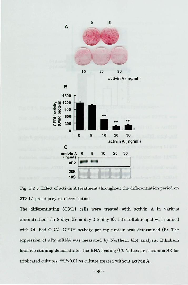
o 5 A
10 20 30
activin A ( ng/ml )
B
1500
~:? 1200 .:; Ci) .- - 900 -0 u ... cue.. :LC) 600 C E 0.- 300 (!)2.
0 0 5 10 20 30
activin A ( ng/ml )
C activin A 0 5 10 20 30
(ng/ml)
aP2
285 185
Fig. 5-2-3. Effect of activin A treatment throughout the differentiation period on
3T3-Ll preadipocyte differentiation.
The differentiating 3T3-Ll cells were treated with activin A in val'lOUS
concentrations for 8 days (from day 0 to day 8) . Intracellular lipid was stained
with Oil Red 0 (N. GPDH activity per mg protein was determined (B). The
expression of aP2 mRNA was measured by Northern blot analysis. Ethidium
bromide staining demonstrates the RNA loading (C). Values are means ± SE for
triplicated cultures. **P<O.Ol vs cultill"e t reated without activin A.
- 80 -
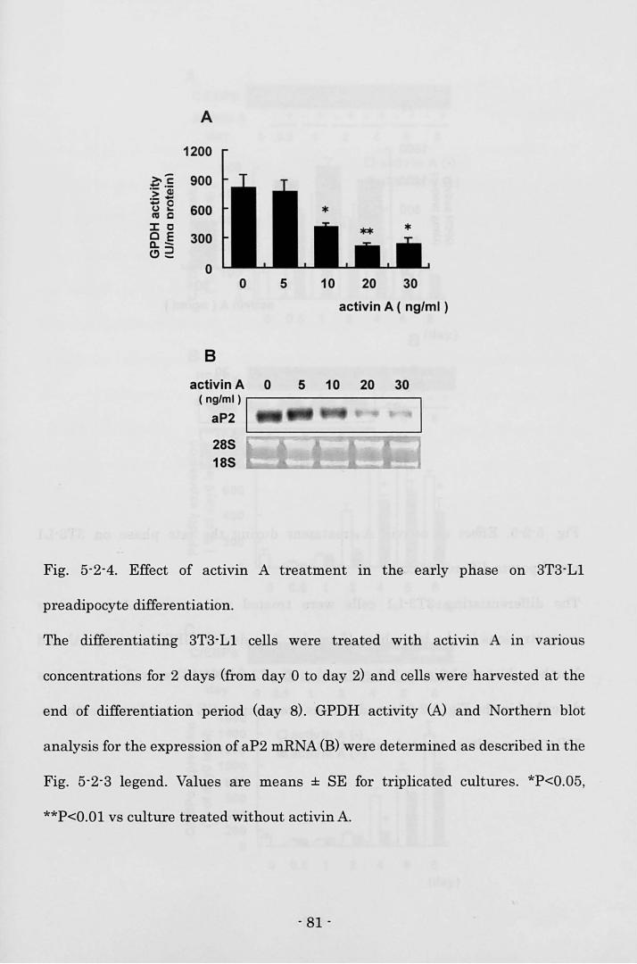
A
1200
~~ 900 .::; QJ
.- -- 0 600 u ... "' c
::I: C o E 300 a..-e>2
0 0 5 10 20 30
activin A ( ng/ml )
B
activin A 0 5 10 20 30 ( ng/ml ) I
aP2 . 285 ~~~~~~~~~
185
Fig. 5-2-4. Effect of activin A treatment In the early phase on 3T3-L1
preadipocyte differentiation.
The differentiating 3T3-L1 cells were treated with activin A in varIOUS
concentrations for 2 days (from day 0 to day 2) and cells were harvested at the
end of differentiation period (day 8). GPDH activity Wand Northern blot
analysis for the expression of aP2 mRNA (B) were determined as described in the
Fig. 5-2-3 legend. Values are means ± SE for triplicated cultures. *P<O.05,
**P<O.Ol vs culture treated without activin A.
- 81 -
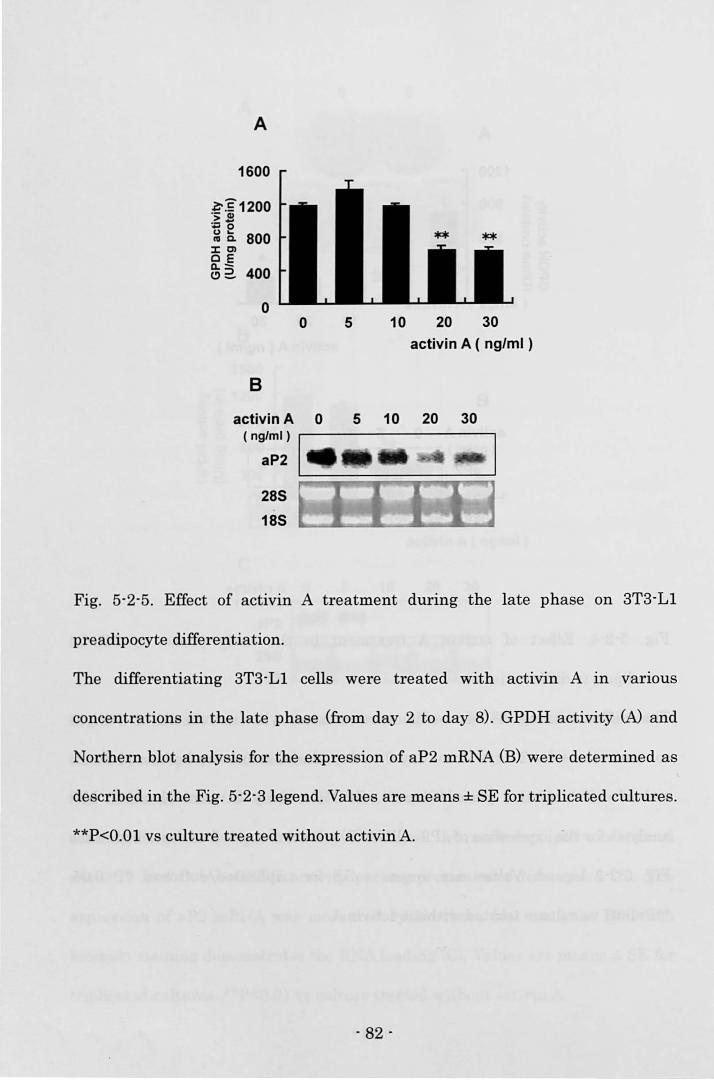
A
1600
l;-E1200 .:;; <II
~e IV Co 800 J:CI OE 11.-(!) 2. 400
o
8
o 5 10 20 30
activin A ( ng/ml )
5 10 20 30 activin A 0 (ng/ml) I
aP2 . ~;;:=::::::;:;;;;;;:;;~;;;=,
285
185
Fig. 5-2-5. Effect of activin A treatment during the late phase on STS-Ll
preadipocyte differentiation.
The differentiating STS-Ll cells were treated with activin A in varIOUS
concentrations in the late phase (from day 2 to day 8). GPDH activity (A) and
Northern blot analysis for the expression of aP2 mRNA (B) were determined as
described in the Fig. 5-2-Slegend. Values are means ± SE for triplicated cultUl'es.
**P<O.Ol vs culture treated without activin A.
- 82 -
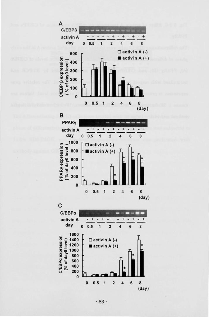
A C/EBP~
activin A day
500 c:: 0-·iii a; 400 en > ~ ~ 300 0.0 )( >. !.g 200 Cl.O CD ~ 100 w 0
C3 - 0
B PPARy
activin A day
c:: _1000 0-.: ~ 800 cu~ Q. c;. 600 : "' >~ 400 0:: 0
~ ~ 200 Cl. -
o
C C/EBPa
activin A day
c:: 1600 .Q :; 1400 : ~ 1200 cu-e. 0 1000 :.g 800 0_ 600 Cl. 0
ffi~ 400 -- 200 0
0
.~ p -. ""- - •• ..,...- -.,"", ---------_ .. --- + - + - + - + - + - +
o 0.5 1 2 4 6 8
o activin A (-)
• activin A (+)
o 0.5 1 2 4 6 8 (day)
- -- -- -_ ... - + - + - + - + - + - +
o 0.5 1 2 4 6 8
0
o activin A (-)
• activin A (+) * * *
o 0.5 1 2 4 6 8 (day)
... -. --.. -- + - + - + - + - + - + -----------0.5 1 2 4 6 8
o activin A (-) • activin A (+) *
0 0.5 1 2 4 6 8 (day)
- 83 -
Fig. 5-2-6. Effect of activin A treatment on mRNA expression of C/EBPs and
PPARy.
The differentiating 3T3-Ll cells were treated with 20 ng/ml activin A in the early
phase of differentiation (from day 0 to day 2). The expression levels of C/EBPB
(A), PPARy (B), and C/EBPa (C) mRNA were estimated by RT-PCR and
normalized with respect to the 188 rRNA expression level. The relative gene
expression is presented as the ratio of the day 0 expression level. Values are
means ± SE for three or four replicated cultures. *P<0.05 vs culture treated
without activin A.
- 84 -
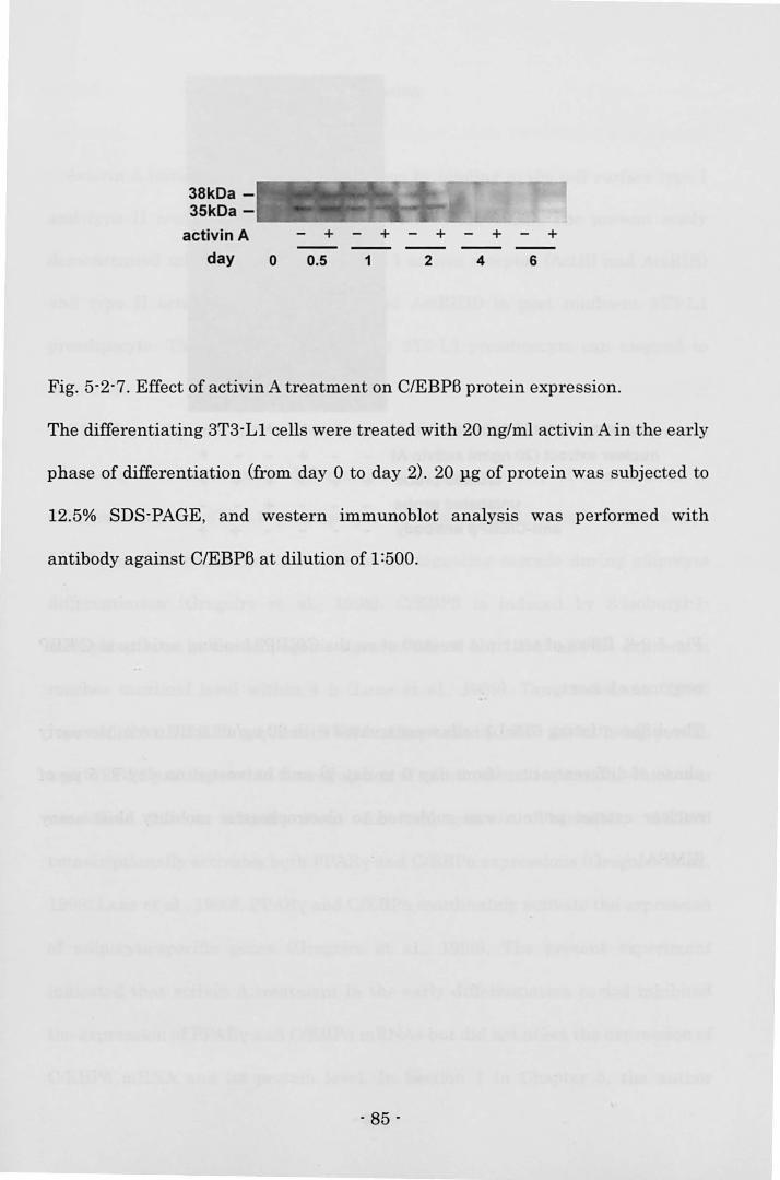
38kDa -35kDa - ...... _----'''---
activin A - + - + - + - + - +
day o 0.5 1 2 4 6
Fig. 5-2-7. Effect of activin A treatment on C/EBPB protein expression.
The differentiating 3T3-L1 cells were treated with 20 ng/ml activin A in the early
phase of differentiation (from day 0 to day 2). 20 pg of protein was subjected to
12.5% SDS-PAGE, and western immunoblot analysis was performed with
antibody against C/EBPB at dilution of 1:500.
- 85 -
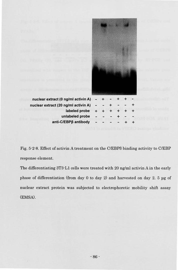
nuclear extract (0 ng/ml activin A) - + + + nuclear extract (20 ng/ml activin A) + +
labeled probe + + + + + + unlabeled probe +
anti-C/EBPI3 antibody + +
Fig. 5-2-8. Effect of activin A treatment on the CIEBPB binding activity to CIEBP
response element.
The differentiating 3T3-Ll cells were treated with 20 ng/ml activin A in the early
phase of differentiation (from day 0 to day 2) and harvested on day 2. 5 llg of
nuclear extract protein was subjected to electrophoretic mobility shift assay
(EMSA).
- 86 -
Discussion
Activin A initiates its biological functions by binding to the cell surface type I
and type II receptors (Mathews, 1994; Piek et aI., 1999). The present study
demonstrated mRNA expressions of type I activin receptor (ActRI and ActRIB)
and type II activin receptor (ActRII and ActRIIB) in post confluent 3T3-L1
preadipocyte. These results suggest that 3T3-L1 preadipocyte can respond to
activinA.
GPDH activity and aP2 mRNA level were decreased in the cultures treated
with activin A in the early differentiation period, the step of commitment to
differentiation in 3T3-L1 preadipocyte (Gregoire et aI., 1998). C/EBPs and
PPARyare key transcription factors in the signaling cascade during adipocyte
differentiation (Gregoire et aI., 1998). C/EBPB is induced by 3-isobutyl-1-
methylxanthine in the adipogenic agents (Yeh et aI., 1995) and its expression
reaches maximal level within 4 h (Lane et aI., 1999). Tang and Lane (1999)
reported that C/EBPB acquired DNA binding activity after 10-12 h lag period,
and C/EBPB participated in the regulation of mitotic clonal expansion, which was
considered as a critical event for adipogenesis. Thereafter, C/EBPB
transcriptionally activates both PPARy and C/EBPa expressions (Gregoire et aI.,
1998; Lane et aI., 1999). PPARyand C/EBPa coordinately activate the expression
of adipocyte-specific genes (Gregoire et aI., 1998). The present experiment
indicated that activin A treatment in the early differentiation period inhibited
the expression ofPPARy and C/EBPa mRNAs but did not affect the expression of
C/EBPB mRNA and its protein level. In Section 1 in Chapter 5, the author
- 87 -
showed that activin A inhibited bovine preadipocyte differentiation via affecting
downstream of C/EBPB mRNA expression and suggested that activin A possibly
prevented bovine preadipocyte differentiation via Smad3-mediated impairment
of the transactivation function of C/EBPB. In the present study, the effect of
activin A on the 3T3-L1 preadipocyte differentiation was not affected by the
different expression patterns of the critical transcription factors between 3T3-L1
cells and bovine SV cells, that was similar to the effect of myostatin on the
differentiation of 3T3-L1 preadipocytes (Kim et aI., 2001) and bovine
preadipocytes in the study in Chapter 3.
In the present study, activin A treatment in the early phase of differentiation
reduced the formation of C/EBPB and C/EBP probe complex, which suggested
that activin A inhibited adipogenesis via affecting the DNA binding ability of
C/EBPB. TGF-B inhibited the function of C/EBPB, which decreased gene
expression of PPARy and thus TGF-B inhibited adipogenesis (Choy and Derynck,
2003). The inhibitory actions ofTGF-B were mediated by Smad3 protein, a signal
effecter of TGF-B, that interacted with the DNA binding domain of C/EBPB and
repressed the transactivation function of C/EBPB, although the DNA binding
ability of C/EBPB was not affected by Smad3 (Choy and Derynck, 2003). On the
other hand, activin A was reported to suppress haptoglobin promoter activation
mediated by C/EBPB in human hepatoma cells, which was involved in the
repression of DNA binding activity of C/EBPB by the activation of Smad3 protein
(Zauberman et aI., 2001). Therefore, in the present study, activin A possibly
prevented adipogenesis in the early phase via Smad3-mediated impairment of
the DNA binding ability of C/EBPB, and thus of the transactivation function of
- 88 -
C/EBPB.
Activin A did not affect mitotic clonal expansion in the present experiment.
Ignotz and Massague (1985) showed that TGF-B did not affect mitotic clonal
expansion but inhibited adipogenesis when the cells were exposed to TGF-B in
the early phase of differentiation. Thus, the activation of Smad3 by activin A or
TGF-B possibly suppresses adipocyte differentiation without affecting mitotic
clonal expansion. CIEBPB was reported to localize to centromere through
consensus C/EBP-binding sites in centromeric satellite DNA at the onset of
mitotic clonal expansion (Tang and Lane, 1999) though the mechanism of
C/EBPB action on mitotic clonal expansion has not been clear. The mechanism of
C/EBPB action on mitotic clonal expansion might be different from that on the
expression ofPPARy and C/EBPa mRNAs. Otherwise, as mentioned later, activin
A might suppress the expression of PPARy and C/EBPa mRNAs and
adipogenesis via other mechanisms than the impairment of C/EBPB function.
The reduction of GPDH activity and aP2 mRNA level was also observed in the
cultures treated with activin A during the late phase though the treatment
during the late phase was less effective than the treatment during the other
phases. Moreover, the expression of PPARy and C/EBPa mRNAs partly recovered
after activin A was withdrawn in the late differentiation period and the
expression of aP2 mRNA was lower in the culture treated with activin A
throughout the differentiation period than in the culture treated during the early
differentiation period. Additionally, the suppressive effects on the expression of
PPARy and C/EBPu mRNAs were also observed in the cells treated during the
late differentiation period (data not shown). Thus, activin A probably inhibits
- 89 -
adipogenesis via suppressing the expression ofPPARy and C/EBPa mRNAs even
in the late period. The level of CIEBPB is very low in the late phase of
differentiation, which suggests that activin A interferes with the expression of
PPARy and C/EBPa mRNAs through other mechanisms than the impairment of
CIEBPB function in the late period. Therefore, it can postulate that activin A also
suppresses the expression of these transcription factors in the early
differentiation period via the same mechanism observed in the late
differentiation period. However, there have not been any reports showing that
expression of PPARy and C/EBPa mRNAs are mediated by other factors than
C/EBPB in the early phase of differentiation. Further study is necessary for
clarifying this other mechanisms of activin A action on the adipogenesis.
Vejda et al. (2002) reported that activin A was highly expressed in adipose
tissue, predicting that activin A affects adipogenesis in adipose tissue through
autocrine/paracrine manner. On the other hand, serum concentration of activin A
ranged between 4.8 ng/ml and 25.4 ng/ml in women with normal pregnant
(Petraglia et al., 1995). These concentrations corresponded with doses that
affected adipogenesis in the present experiment. Therefore, serum activin A may
affect adipogenesis as an endocrine factor.
In summary, the results presented here showed that activin A inhibited
adipogenesis and this inhibitory effect was mediated by the suppression of the
DNA binding ability of C/EBPB, and thus by the reduction of PPARy and C/EBPa
mRNA levels.
. 90 .
Summary
Beef marbling is characterized by adipose tissue deposition within skeletal
muscle in cattle. Japanese black cattle (Wagyu) is well known for its ability to
produce high marbling, and fat deposition in the muscle is not necessarily
reflected by systemic fat metabolism in this breed. As intramuscular
preadipocytes are surrounded by mature skeletal muscle myofibers, it is
predicted that paracrine factors secreted from muscle fibers may regulate the
differentiation of preadipocytes via intercellular interactions.
In the present study, the conditioned medium from C2C12 myocytes mostly
suppressed the 3T3-L1 preadipocyte differentiation in the early phase that is the
step of commitment to differentiate (Chapter 3). During the induction of
differentiation, C/EBPB is expressed immediately (within 2-4 hours), and
C/EBPB transcriptionally activats PPARy and C/EBPa expression Thereafter,
PPARy and C/EBPa upregulate .each other to maintain their expression despite
the reduction of C/EBPB leveL PPARy and C/EBPa coordinately activate the
expression of adipocyte-specific genes. Myostatin, which is essential for proper
regulation of skeletal muscle mass, is known to be expressed mainly in skeletal
muscle. Furthermore, myostatin is reported to be expressed in C2C12 myotubes
and to be secreted into the culture medium. Additionally, myostatin is reported to
inhibit the differentiation of 3T3-L1 preadipocytes via affecting the expression of
PPARy and C/EBPa. Based on these reports and results in Chapter 3, myostatin
was thought to be a candidate for the paracrine factor from C2C12 myotubes that
inhibited the differentiation of 3T3-L1 preadipocytes. To examine this possibility,
- 91 -
the differentiating 3T3-Ll cells were co-treated with the conditioned medium
and follistatin that binds to myostatin and activin and inhibit their action.
Follistatin reversed the inhibitory effect of the conditioned medium on the
3T3-Ll preadipocyte differentiation in a dose dependent manner, which
suggested that myostatin and/or activin in conditioned medium possibly
inhibited the preadipocyte differentiation (Chapter 3).
To investigate the effect of myostatin on the differentiation of bovine
preadipocytes, the author examined the expression patterns of the critical
transcription factors mRNAs during the differentiation of preadipocytes in
stromal vascular (SV) cells derived from bovine adipose tissue (Chapter 4).
C/EBPB mRNA in the bovine SV cells was stably expressed throughout the
differentiation period. Additionally, the relatively high expression of PPARy was
observed before the induction of differentiation. On the other hand, C/EBPa
mRNA level was extremely low before the induction of differentiation. These
results suggested that bovine SV cells derived from adipose tissue contained the
preadipocytes reaching at more advanced stage of differentiation than 3T3-Ll
preadipocytes did. Despite the different expression patterns of these
transcription factors, myostatin suppressed the increase in PPARy mRNA
expression and the induction of C/EBPa mRNA without affecting the expression
of C/EBPB mRNA in the early phase of differentiation, which was similar to the
effect of myostatin on 3T3-Ll preadipocyte differentiation. Furthermore, the
author revealed that follistatin interfered with myostatin action during bovine
preadipocyte differentiation when the cells were co-treated with myostatin and
follistatin.
- 92 -
Activin, a multifunctional growth and differentiation factor that belongs to the
TGF-B superfamily, is homodimer of activin BA subunit. Recently, the expressions
of activin BA and activin receptor mRNAs were found in many tissues throughout
the body including muscle and fat. Activin A was reported to act on many cell
types, however, its action on preadipocyte differentiation was not still
investigated. In Chapter 5, the author investigated the effect of activin A on the
differentiation of bovine and STS-LI preadipocytes. Activin A inhibited the
differentiation of bovine and STS-LI preadipocytes by reducing PPARy and
C/EBPa mRNA expression but not affecting C/EBPB expression, which was
similar to the effect of myostatin on the preadipocyte differentiation. In Section 2
in Chapter 5, the author investigated the further mechanism that activin A
inhibited the differentiation of STS-LI preadipocyte. Activin A inhibited the DNA
binding activity of C/EBPB, which was considered to suppress the preadipocyte
differentiation. Activin A possibly prevented adipogenesis in the early phase via
SmadS-mediated impairment of the DNA binding ability of C/EBPB, and thus of
the transactivation function of C/EBPB.
In Section 1 in Chapter 5, the author revealed that follistatin reversed the
inhibitory effect of activin A on bovine preadipocyte differentiation. Additionally,
the treatment with follistatin alone increased GPDH activity, which suggested
that bovine SV cells in the present study may produce substantial level of activin
A and/or myostatin to inhibit preadipocyte differentiation, and thus exogenous
follistatin promoted adipogenesis.
The expression of activin A and follistatin is widely distributed in gonadal and
non-gonadal tissues including muscular and adipose tissues. Myostatin is found
- 9S -
primarily in skeletal muscle, and is also detected in adipose tissue. Therefore,
myostatin and/or activin A secreted from muscular and/or adipose tissues
possibly inhibit the differentiation ofpreadipocytes within muscle, and follistatin
secreted from these tissues may suppress the inhibitory effect of myostatin and
activin A. The physiological balance of these paracrine/autocrine factors possibly
regulates preadipocyte differentiation in muscle.
- 94 -
References
Artaza, J.N., Bhasin, S., Magee, T.R., Reisz-Porszasz, S., Shen, R., Groome, N.P.,
Fareez, M.M., and Gonzalez-Cadavid, N.F., 2005. Myostatin inhibits myogenesis
and promotes adipogenesis in C3H lOT (112) mesenchymal multipotent cells.
Endocrinology, 146:3547-3557.
Artaza, J.N., Bhasin, S., Mallidis, C., Taylor, W., Ma, K., and Gonzalez-Cadavid,
N.F., 2002. Endogenous expression and localization of myostatin and its relation
to myosin heavy chain distribution in C2C12 skeletal muscle cells. J. Cell. Physiol.,
190:170-179.
Asakura, A., Komaki, M., and Rudnicki, M., 2001. Muscle satellite cells are
multipotential stem cells that exhibit myogenic, osteogenic, and adipogenic
differentiation. Differentiation, 68:245-253.
Aso, H., Abe, H., Nakajima, 1., Ozutsumi, K., Yamaguchi, T., Takamori, Y.,
Kodama, A., Hoshino, F.B., and Takano, S., 1995. A preadipocyte clonal line from
bovine intramuscular adipose tissue: nonexpression of GLUT-4 protein during
adipocyte differentiation. Biochem. Biophys. Res. Commun., 213:369-375.
Attisano, L. and Wrana, J.L., 2000. Smads as transcriptional co-modulators.
Curro Opin. Cell. BioI., 12:235-243.
- 95 -
Attisano, L., Wrana, J.L., Montalvo, E., and Massague, J., 1996. Activation of
signalling by the activin receptor complex. Mol. Cell. BioI., 16:1066-1073.
Bailey, P., Holowacz, T., and Lassar, A.B., 2001. The origin of skeletal muscle
stem cells in the embryo and the adult. Curro Opin. Cell. BioI., 13:679-689.
Bjorntorp, P., Karlsson, M., and Pettersson, P., 1982. Expansion of adipose tissue
storage capacity at different ages in rats. Metabolism, 31:366-373.
Buckingham, M., 1992. Making muscle in mammals. Trends. Genet., 8:144-148.
Cameron, P.J., Zembayashi, M., Lunt, D.K., Mitsuhashi, T., Mitsumoto, M.,
Ozawa, S., and Smith, S.B., 1994. Relationship between Japanese Beef Marbling
Standard and intramuscular lipid in the M longissimus thoracis of Japanese
Black and American Wagyu cattle. Meat Sci., 38:361-364.
Carcamo, J., Weis, F.M., Ventura, F., Wieser, R., Wrana, J.L., Attisano, L., and
Massague, J., 1994. Type I receptors specify growth-inhibitory and
transcriptional responses to transforming growth factor beta and activin. Mol.
Cell. BioI., 14:3810-3821.
Charge, S.B. and Rudnicki, M.A., 2004. Cellular and molecular regulation of
muscle regeneration. Physiol. Rev., 84:209-238.
- 96 -
Chawla, A., Schwarz, E.J., Dimaculangan, D.D., and Lazar, M.A., 1994.
Peroxisome proliferator-activated receptor (PPAR) y: adipose-predominant
expression and induction early in adipocyte differentiation. Endocrinology,
135:798-800.
Choy, L., Skillington, J., and Derynck, R., 2000. Roles of autocrine TGF-B
receptor and Smad signaling in adipocyte differentiation. J. Cell. BioI., 149:667-
682.
Choy, L. and Derynck, R., 2003. Transforming growth factor-B inhibits adipocyte
differentiation by Smad3 interacting with CCAAT/enhancer-binding protein
(C/EBP) and repressing C/EBP transactivation function. J. BioI. Chem.,
278:9609-9619.
Christy, R.J., Kaestner, K.H., Geiman, D.E., and Lane, M.D., 1991.
CCAAT/enhancer binding protein gene promoter: Binding of nuclear factors
during differentiation of 3T3-L1 preadipocytes. Proc. NatI. Acad. Sci. U.S.A.,
88:2593-2597.
Cianzio, D.S., Topel, D.G., Whitehurst, G.B., Beitz, D.C., and Self, H.L., 1985.
Adipose tissue growth and cellularity: changes in bovine adipocyte size and
number. J. Anim. Sci., 60:970-976.
Clouthier, D.E., Comerford, S.A., and Hammer, R.E., 1997. Hepatic fibrosis,
- 97 -
glomerulosclerosis, and a lipodystrophy-like syndrome In PEPCK-TGF-B1
transgenic mice. J. Clin. Invest., 100:2697-2713.
Cook, J.R. and Kozak, L.P., 1982. Sn-glycerol-3-phosphate dehydrogenase gene
expression during mouse adipocyte development in vivo. Dev. BioI., 92:440-448.
Cornelius, P., MacDougald, O.A., and Lane, M.D., 1994. Regulation of adipocyte
development. Annu. Rev. Nutr., 14:99-129.
DePaolo, L.V., Bicsak, T.A., Erickson, G.F., Shimasaki, S., and Ling, N., 1991.
Follistatin and activin: a potential intrinsic regulatory system within diverse
tissues. Proc. Soc. Exp. BioI. Med., 198:500-512.
Ding, S.T., McNeel, R.L., and Mersmann, H.J., 1999. Expression of porcine
adipocyte transcripts: tissue distribution and differentiation in vitro and in vivo.
Compo Biochem. PhysioI. B, 123:307-318.
Dodson, M.V., McFarland, D.C., Grant, A.L., Doumit, M.E., and Velleman, S.G.,
1996. Extrinsic regulation of domestic animal-derived satellite cells. Domest.
Anim. Endocrinol., 13:107-126.
Eto, Y., Tsuji, T., Takezawa, M., Takano, S., Yokogawa, Y., and Shibai, H., 1987.
Purification and characterization of erythroid differentiation factor (EDF)
isolated from human leukemia cell line THP-l. Biochem. Biophys. Res. Commun.,
- 98 -
142:1095-1103.
Faust, I.M., Johnson, P.R., Stern, J.S., and Hirsch, J., 1978. Diet-induced
adipocyte number increase in adult rats: a new model of obesity. Am. J. Physiol.,
235:E279-E286.
Feijen, A., Goumans, M.J., and van den Eijnden-van Raaij, A.J., 1994. Expres
sion of activin subunits, activin receptors and follistatin in postimplantation
mouse embryos suggests specific developmental functions for different activins.
Development., 120:3621-3637.
Garbutt, G.J., Anthony, W.B., Walker, D.F., and McGuire, J.A., 1979. Perirectal
adipose tissue development of postweaned rapidly growing bull calves. J. Anim.
Sci., 48:525-530.
Gonzitlez-Manch6n, C. and Vale, W., 1989. Activin-A, inhibin and transforming
growth factor-B modulate growth of two gonadal cell lines. Endocrinology,
125:1666-1672.
Green, H. and Kehinde, 0., 1974. Sublines of mouse 3T3 cells that accumulate
lipid. Cell, 1:113-116.
Green, H. and Kehinde, 0., 1975. An established preadipose cell line and its
differentiation in culture. II. Factors affecting the adipose conversion. Cell,
- 99 -
Green, H. and Kehinde, 0., 1976. Spontaneous heritable changes leading to
increased adipose conversion in 3T3 cells. Cell, 7:105-113.
Green, H. and Kehinde, 0., Formation of normally differentiated subcutaneous
fat pads by an established pre adipose cell line. 1979. J. Cell. PhysioI.,
101:169-171.
Gregoire, F. Todoroff, G., Hauser, N., and Remacle, C., 1990. The
stroma -vascular fraction of rat inguinal and epididymal adipose tissue and the
adipoconversion of fat cell precursors in primary culture. BioI. Cell., 69:215-222.
Gregoire, F.M., Johnson, P.R., and Greenwood, M.R., 1995. Comparison of the
adipoconversion of preadipocytes derived from lean and obese Zucker rats in
serum-free cultures. Int. J. Obes. Relat. Metab. Disord., 19:664-670.
Gregoire, F.M., Smas, C.M., and SuI, H.S., 1998. Understanding adipocyte
differentiation. PhysioI. Rev., 78:783-809.
Grounds, M.D., Garrett, K.L., Lai, M.C., Wright, W.E., and Beilharz, M.W., 1992.
Identification of skeletal muscle precursor cells in vivo by use of MyoD1 and
myogenin probes. Cell Tissue Res., 267:99-104.
- 100 -
Hannon, K., Smith, C.K. 2nd, Bales, K.R., and Santerre, R.F., 1992. Temporal
and quantitative analysis of myogenic regulatory and growth factor gene
expression in the developing mouse embryo. Dev. BioI., 151:137-144.
Hausman, G.J., Campion, D.R., and Martin, R.J., 1980. Search for the adipocyte
precursor cell and factors that promote its differentiation. J. Lipid. Res.,
21:657-670.
Hawke, T.J. and Garry, D.J., 2001. Myogenic satellite cells: physiology to
molecular biology. J. AppI. PhysioI., 91:534-551.
Heldin, C.H., Miyazono, K., and ten Dijke, P., 1997. TGF-~ signalling from cell
membrane to nucleus through SMAD proteins. Nature, 390:465-471.
Hood, R.L. and Allen, C.E., 1973. Cellularity of bovine adipose tissue. J. Lipid.
Res., 14:605-610.
Ignotz, R.A. and Massague, J., 1985. Type B transforming growth factor controls
the adipogenic differentiation of 3T3 fibroblasts. Proc. NatI. Acad. Sci. U.S.A.,
82:8530-8534.
Inouye, S., Guo, Y., DePaolo, L., Shimonaka, M., Ling, N., and Shimasaki, S.,
1991. Recombinant expression of human follistatin with 315 and 288 amino
acids: chemical and biological comparison with native porcine follistatin.
- 101 -
Endocrinology, 129:815-822.
Jarett, L., Wong, E.H., Smith, J.A., and Macaulay, S.L., 1985. Stimulation of
pyruvate dehydrogenase activity in intact rat adipocytes by insulin mediator
from rat skeletal muscle. Endocrinology, 116:1011-1016.
Jenkin, G., Ward, J., Loose, J., Schneider-Kolsky, M., Young, R., Canny, B.,
O'Connor, A., de Kretser, D., and Wallace, E., 2001. Physiological and regulatory
roles of activin A in late pregnancy. Mol. Cell. Endocrinol., 180:131-138.
Jones, K.L., De Kretser, D.M., and Phillips, D.J., 2004. Effect of heparin
administration to sheep on the release profiles of circulating activin A and
follistatin. J. Endocrinol., 181:307-314.
Kambadur, R., Sharma, M., Smith, T.P., and Bass, J.J., 1997. Mutations in
myostatin (GDF 8) in double-muscled Belgian Blue and Piedmontese cattle.
Genome Res., 7:910-916.
Kim, H.S., Liang, L., Dean, R.G., Hausman, D.B., Hartzell, D.L., and Baile, C.A.,
2001. Inhibition of preadipocyte differentiation by myostatin treatment in
3T3-L1 cultures. Biochem. Biophys. Res. Commun., 281:902-906.
Kirkland, J.L., Hollenberg, C.H., and Gillon, W.S., 1990. Age, anatomic site, and
the replication and differentiation of adipocyte precursors. Am. J. Physiol.,
- 102 -
258:C206-C210.
Kobolak, J. and Gocza, E., 2002. The role of the myostatin protein in meat
quality - a review. Arch. Anim. Breed., 45:159-170.
Koishi, K., Zhang, M., McLennan, 1.S., and Harris, A.J., 1995. MyoD protein
accumulates in satellite cells and is neurally regulated in regenerating myotubes
and skeletal muscle fibers. Dev. Dyn., 202:244-254.
Konieczny, S.F. and Emerson, C.P. Jr., 1984. 5-Azacytidine induction of stable
mesodermal stem cell lineages from 10T1I2 cells: evidence for regulatory genes
controlling determination. Cell, 38:791-800.
Lacasa, D., Garcia, E., Agli, B., and Giudicelli, Y., 1997a. Control of rat pre
adipocyte adipose conversion by ovarian status: regional specificity and possible
involvement of the mitogen-activated protein kinase-dependent and c-fos
signaling pathways. Endocrinology, 138:2729-2734.
Lacasa, D., Garcia, E., Henriot, D., Agli, B., and Giudicelli, Y., 1997b. Site-related
specificities of the control by androgenic status of adipogenesis and mitogen
activated protein kinase cascade/c-fos signaling pathways in rat preadipocytes.
Endocrinology, 138:3181-3186.
Lane, M.D., Tang, Q.Q., and Jiang, M.S., 1999. Role of the CCAAT enhancer
- 103 -
binding proteins (C/EBPs) in adipocyte differentiation. Biochem. Biophys. Res.
Commun., 266:677-683.
Langley, B., Thomas, M., Bishop, A., Sharma, M., Gilmour, S., and Kambadur, R.,
2002. Myostatin inhibits myoblast differentiation by down-regulating MyoD
expression. J. BioI. Chem., 277:49831-49840.
Lassar, A. and Munsterberg, A., 1994. Wiring diagrams: regulatory circuits and
the control of skeletal myogenesis. Curro Opin. Cell. BioI., 6:432-442.
Lee, S.J. and McPherron, A.C., 1999. Myostatin and the control of skeletal
muscle mass. Curro Opin. Genet. Dev., 9:604-607.
Lee, S.J. and McPherron, A.C., 2001. Regulation of myostatin activity and
muscle growth. Proc. Natl. Acad. Sci. V.S.A., 98:9306-9311.
Lin, J., Arnold, B., Della-Fera, M.A., Azain, M.J., Hartzell, D.L., and Baile, C.A.,
2002. Myostatin knockout in mice increases myogenesis and decreases
adipogenesis. Biochem. Biophys. Res. Commun, 291:701-706.
Ling, N., Ying, S.Y., Veno, N., Shimasaki, S., Esch, F., Hotta, M., and Guillemin,
R., 1986. Pituitary FSH is released by a heterodimer of the beta-subunits from
the two forms of in hi bin. Nature, 321:779-782.
- 104-
Link, B.A., and Nishi, R., 1997. Opposing effects of activinA and follistatin on
developing skeletal muscle cells. Exp. Cell Res., 233:350-362.
Liu, K., Guan, Y., MacNicol, M.C., MacNicol, A.M., and McGehee, R.E. Jr, 2002. Early
expression of p107 is associated with 3T3-L1 adipocyte differentiation. Mol. Cell.
Endocrinol., 194:51-61.
Lowry, O.H., Rosebrough, N.J., Farr, A.L., and Randall, R.J., 1951. Protein
measurement with the Folin phenol reagent. J. BioI. Chern., 193:265-275.
Lunt, D.K., RIley, R.R., and Smith S.B., 1993. Growth and carcass characteristics of
Angus and American Wagyu steers. Meat Sci., 34:327-334.
Lyle, R.E., Richon, V.M., and McGehee, R.E. Jr, 1998. TNFa disrupts mitotic
clonal expansion and regulation of retinoblastoma proteins p130 and p107
during 3T3-L1 adipocyte differentiation. Biochem. Biophys. Res. Commun.,
247:373-378.
MacDougald, O.A. and Lane, M.D., 1995. Transcriptional regulation of gene
expression during adipocyte differentiation. Annu. Rev. Biochem., 64: 345-373.
Mandrup, S. and Lane, M.D., 1997. Regulating adipogenesis. J. BioI. Chern.,
272:5367-5370.
- 105 -
Masuzaki, H., Ogawa, Y., Isse, N., Satoh, N., Okazaki, T., Shigemoto, M., Mori,
K., Tamura, N., Hosoda, K., Yoshimasa, Y., 1995. Human obese gene expression.
Adipocyte-specific expression and regional differences in the adipose tissue.
Diabetes, 44:855-858.
Massague, J. and Chen, Y.G., 2000. Controlling TGF-8 signaling. Genes Dev.,
14:627-644.
Massague, J. and Wotton, D., 2000. Transcriptional control by the TGF-WSmad
signaling system. EMBO J., 19:1745-1754.
Mathews, L.S., 1994. Activin receptors and cellular signaling by the receptor
serine kinase family. Endocr. Rev., 15:310-325.
Matzuk, M.M., Lu, N., Vogel, H., Sellheyer, K., Roop, D.R., and Bradley, A., 1995.
Multiple defects and perinatal death in mice deficient in follistatin. Nature,
374:360-363.
McCarthy, S.A. and Bicknell, R., 1993. Inhibition of vascular endothelial cell
growth by activin-A. J. BioI. Chem., 268:23066-23071.
McFarlane O'Shea, T., O'Conno, A.E., Jenkin, G., and de Kretser, D.M., 1998. A
longitudinal study of serum activin A and follistatin level across gestation and
parturition in sheep. Proc. Aus. Physiol. Pharmacol. Soc., 27:p74 (Abstract).
- 106 -
McNeel R.L., Ding, S.T., Smith. E.O., and Mersmann, H.J., 2000. Expression of
porcine adipocyte transcripts during differentiation in vitro and in vivo. Compo
Biochem. PhysioI. B, 126:291-302.
McPherron, A.C., Lawler, A.M., and Lee, S.J., 1997. Regulation of skeletal muscle
mass in mice by a new TGF-B superfamily member. Nature, 387:83-90.
McPherron, A.C. and Lee, S.J., 1997. Double muscling in cattle due to mutations
in the myostatin gene. Proc. NatI. Acad. Sci. U.S.A., 94:12457-12461.
McPherron, A.C. and Lee, S.J., 2002. Suppression of body fat accumulation in
myostatin-deficient mice. J. Clin. Invest., 109:595-601.
Medvedev, A.V., Snedden, S.K., Raimbault, S., Ricquier, D., and Collins, S., 2001.
Transcriptional regulation of the mouse uncoupling protein-2 gene. Double E-box
motif is required for peroxisome proliferator-activated receptor-y-dependent
activation. J. BioI. Chem., 276:10817-10823.
Meunier, H., Rivier, C., Evans, R.M., and Vale, W., 1988. Gonadal and
extragonadal expression of inhibin a, BA, and BB subunits in various tissues
predicts diverse functions. Proc. NatI. Acad. Sci. U.S.A., 85:247-251.
Michel, U., Albiston, A., and Findlay, J.K., 1990. Rat follistatin: gonadal and
extragonadal expression and evidence for alternative splicing. Biochem. Biophys.
- 107 -
Res. Commun., 173:401-407.
Miller, W.H. Jr, Faust, LM., and Hirsch, J., 1984. Demonstration of de novo
production of adipocytes in adult rats by biochemical and radioautographic
techniques. J. Lipid. Res., 25:336-347.
Mukai, F., Oyama, K., and Kohno, S., 1995. Genetic relationships between
performance test traits and field carcass traits in Japanese Black cattle. Livest.
Prod. Sci., 44:199-205.
Nakamura, T., Takio, K., Eto, Y., Shibai, H., Titani, K., and Sugino, H., 1990.
Activin-binding protein from rat ovary is follistatin. Science, 247:836-838.
Ogawa, Y., Schmidt, D.K., Nathan, RM., Armstrong, RM., Miller, K.L., Sawamura,
S.J., Ziman, J.M., Erickson, K.L., de Leon, E.R, Rosen, D.M., Seyedin, S.M., Glaser,
C.B., Chang, R -J., Corrigan, A.Z., and Vale, W., 1992. Bovine bone activin enhances
bone morphogenetic protein-induced ectopic bone formation. J. BioI. Chem.,
267:14233-14237.
Ohsumi, J., Sakakibara, S., Yamaguchi, J., Miyadai, K., Yoshioka, S., Fujiwara, T.,
Horikoshi, H., and Serizawa, N., 1994. Troglitazone prevents the inhibitory effects of
inflammatory cytokines on insulin-induced adipocyte differentiation in 3T3-L1 cells.
Endocrinology, 135:2279-2282.
- 108 -
Petraglia, F., Vaughan, J., and Vale, W., 1989. Inhibin and activin modulate the
release of gonadotropin-releasing hormone, human chorionic gonadotropin, and
progesterone from cultured human placental cells. Proc. NatI. Acad. Sci. U.S.A.,
86:5114-5117.
Petraglia, F., Vita, D.D., Gallinelli, A., Aguzzoli, L., Genazzani, A.R., Romero, R.,
and Woodruff, T.K., 1995. Abnormal concentration of maternal serum activin-A
in gestational diseases. J. Clin. Endocr. Metab., 80:558-561.
Petruschke, T., Rohrig, K., and Hauner, H., 1994. Transforming growth factor B
(TGF-B) inhibits the differentiation of human adipocyte precursor cells in
primary culture. Int. J. Obes. Relat. Metab. Disord., 18:532-536.
Piek, E., Heldin, C.H., and Ten Dijke, P., 1999. Specificity, diversity, and
regulation in TGF-B superfamily signaling. FASEB J., 13:2105-2124.
Price, M.A. and Berg, R.T., 1981. On the consequences and economics of feeding
grain ad libitum to culled beef cows. Can. J. Anim. Sci., 61:105-112.
Qiu, Z., Wei, Y., Chen, N., Jiang, M., Wu, J., and Liao, K., 2001. DNA synthesis and
mitotic clonal expansion is not a required step for 3T3-L1 preadipocyte
differentiation into adipocytes. J. BioI. Chern., 276:11988-11995.
Ramsay, T.G., Rao, S.v., and Wolverton, C.K., 1992. In vitro systems for the
- 109 -
analysis of the development of adipose tissue in domestic animals. J. Nutr.,
122:806-817.
Rebbapragada, A., Benchabane, H., Wrana, J.L., Celeste, A.J., and Attisano, L.,
2003. Myostatin signals through a transforming growth factor B-like signaling
pathway to block adipogenesis. Mol. Cell. BioI., 23:7230-7242.
Reznikoff, C.A., Brankow, D.W., and Heidelberger, C., 1973. Establishment and
characterization of a cloned line of C3H mouse embryo cells sensitive to
postconfluence inhibition of division. Cancer Res., 33:3231-3238.
Richon, V.M., Lyle, R.E., and McGehee, R.E. Jr, 1997. Regulation and expression
of retinoblastoma proteins p 107 and p 130 during 3T3-Ll adipocyte
differentiation. J. BioI. Chem., 272:10117-10124.
Riley, D.G., Chase, C.C. Jr, Hammond, A.C., West, R.L., Johnson, D.D., Olson,
T.A., and Coleman, S.W., 2003. Estimated genetic parameters for palatability
traits of steaks from Brahman cattle. J. Anim. Sci., 81:54-60.
Robertson, D.M., Klein, R., de Vos, F.L., McLachlan ,R.I., Wettenhall, R.E., Hearn,
M.T., Burger, H.G., and de Kretser, D.M., 1987. The isolation of polypeptides with
FSH suppressing activity from bovine follicular fluid which are structurally different
to inhibin. Biochem. Biophys. Res. Commun., 149:744-749.
- 110 -
Rosen, E.D., Walkey, C.J., Puigserver, P., and Spiegelman, B.M., 2000.
Transcriptional regulation of adipogenesis. Genes Dev., 14:1293-1307.
Rudnicki, M.A., Schnegelsberg, P.N., Stead, R.H., Braun, T., Arnold, H.H., and
Jaenisch, R., 1993. MyoD or Myf-5 is required for the formation of skeletal
muscle. Cell, 75:1351-1359.
Sager, R. and Kovac, P., 1982. Pre-adipocyte determination either by insulin or
by 5-azacytidine. Proc. NatI. Acad. Sci. U.S.A., 79:480-484.
Schneider, 0., Nau, R., and Michel, U., 2000. Comparative analysis offollistatin-,
activin beta A- and activin beta B- mRNA steady-state levels in diverse porcine
tissues by multiplex Sl nuclease analysis. Eur. J. EndocrinoI., 142:537-544.
Schneyer, A., Schoen, A., Quigg, A., and Sidis, Y., 2003. Differential binding and
neutralization of activins A and B by follistatin and follistatin like-3 (FSTL-31
FSRP/FLRG). Endocrinology, 144:1671-1674.
Schoonjans, K., Watanabe, M., Suzuki, H., Mahfoudi, A., Krey, G., Wahli, W.,
Grimaldi, P., Staels, B., Yamamoto, T., and Auwerx, J., 1995. Induction of the
acyl-coenzyme A synthetase gene by fibrates and fatty acids is mediated by a
peroxisome proliferator response element in the C promoter. J. BioI. Chern.,
270:19269-19276.
- 111 -
Sears, I.B., MacGinnitie, M.A., Kovacs, L.G., and Graves, R.A., 1996.
Differentiation-dependent expression of the brown adipocyte uncoupling protein
gene: regulation by peroxisome proliferator-activated receptor y. Mol. Cell. BioI.,
6:3410-3419.
Shahin, K.A. and Berg, R.T., 1985. Growth patterns of muscle, fat and bone, and
carcass composition of double muscled and normal cattle. Canad. J. Anim. Sci.,
65:279-293.
Sharma, M., Kambadur, R., Matthews, K.G., Somers, W.G., Devlin, G.P.,
Conaglen, J.V., Fowke, P.J., and Bass, J.J., 1999. Myostatin, a transforming
growth factor-B superfamily member, is expressed in heart muscle and is
upregulated in cardiomyocytes after infarct. J. Cell. Physiol., 180:1-9.
Shimasaki, S., Koga, M., Esch, F., Cooksey, K., Mercado, M., Koba, A., Veno, N.,
Ying, S.Y., Ling, N., and Guillemin, R., 1988. Primary structure of the human
follistatin precursor and its genomic organization. Proc. Natl. Acad. Sci. V.S.A.,
85:4218-4222.
Shiozuka, M., Momoi, T., and Kimura, I., 1997. Activin A inhibits differentiation
of chick myogenic cells in vitro. Zool. Sci., 14:327-330.
Slavin, B.G., 1979. Fine structural studies on white adipocyte differentiation.
Anat. Rec., 195:63-72.
- 112 -
Soret, B., Lee, H.J., Finley, E., Lee, S.C., and Vernon, R.G., 1999. Regulation of
differentiation of sheep subcutaneous and abdominal preadipocytes in culture. J.
EndocrinoI., 161:517-524.
Student, A.K., Hsu, RY., and Lane, M.D., 1980. Induction of fatty acid
synthetase synthesis in differentiating 3T3-L1 preadipocytes. J. BioI. Chem.,
255:4745-4750.
Sugino, K., Kurosawa, N., Nakamura, T., Takio, K., Shimasaki, S., Ling, N.,
Titani, K., and Sugino, H., 1993. Molecular heterogeneity of follistatin, an
activin-binding protein. Higher affinity of the carboxyl-terminal truncated forms
for heparan sulfate proteoglycans on the ovarian granulosa cell. J. BioI. Chem.,
268:15579-15587.
Sumitomo, S., Inouye, S., Liu, X.J., Ling, N., and Shimasaki, S., 1995. The
heparin binding site of follistatin is involved in its interaction with activin.
Biochem. Biophys. Res. Commun., 208:1-9.
Tang, Q.Q. and Lane, M.D., 1999. Activation and centromeric localization of
CCAAT/enhancer-binding proteins during the mitotic clonal expansion of
adipocyte differentiation. Genes Dev., 13:2231-2241.
Tang, Q.Q., Otto, T.C., and Lane, M.D., 2003. Mitotic clonal expansion: a
synchronous process required for adipogenesis. Proc. NatI. Acad. Sci. U.S.A.,
- 113 -
100:44-49.
Tang, Q.Q., Otto, T.C., and Lane, M.D., 2004. Commitment of C3H10T1I2
pluripotent stem cells to the adipocyte lineage. Proc. Natl. Acad. Sci. U.S.A.,
101:9607-9611.
Taylor, S.M. and Jones, P.A., 1979. Multiple new phenotypes induced in 10T1/2
and 3T3 cells treated with 5-azacytidine. Cell, 17:771-779.
Tchkonia, T., Giorgadze, N., Pirtskhalava, T., Tchoukalova, Y., Karagiannides, I.,
Forse, RA., DePonte, M., Stevenson, M., Guo, W., Han, J., Waloga, G., Lash, T.L.,
Jensen, M.D., and Kirkland, J.L., 2002. Fat depot origin affects adipogenesis in
primary cultured and cloned human preadipocytes. Am. J. Physiol. Regul. Integr.
Compo Physiol., 282:R1286-R1296.
te Pas, M.F., de Jong, P.R, and Verburg, F.J., 2000. Glucocorticoid inhibition of
C2C12 proliferation rate and differentiation capacity in relation to mRNA levels
of the MRF gene family. Mol. BioI. Rep., 27:87-98.
Tontonoz, P., Hu, E., Devine, J., Beale, E.G., and Spiegelman, B.M., 1995.
PPARy2 regulates adipose expression of the phosphoenolpyruvate carboxykinase
gene. Mol. Cell. BioI., 15:351-357.
Tontonoz, P., Hu, E., Graves, R.A., Budavari, A.I., and Spiegelman, B.M., 1994.
- 114 -
mPPAR y2: tissue-specific regulator of an adipocyte enhancer. Genes Dev.,
8:1224-1234.
Torti, F.M., Torti, S.v., Larrick, J.W., and Ringold, G.M., 1989. Modulation of
adipocyte differentiation by tumor necrosis factor and transforming growth
factor B. J. Cell. Biol., 108:1105-1113.
Tuuri, T., Eramaa, M., Hilden, K., and Ritvos, 0.,1994. The tissue distribution of
activin BA- and BB-subunit and follistatin messenger ribonucleic acids suggests
multiple sites of action for the activin-follistatin system during human
development. J. Clin. Endocr. Metab., 78:1521-1524.
Ueno, N., Ling, N., Ying, S.Y., Esch, F., Shimasaki, S., and Guillemin, R., 1987.
Isolation and partial characterization of follistatin: a single-chain Mr 35,000
monomeric protein that inhibits the release of follicle-stimulating hormone. Proc.
Natl. Acad. Sci. U.S.A., 84:8282-8286.
Vale, W., Rivier, J., Vaughan, J., McClintock, R., Corrigan, A., Woo, W., Karr, D.,
and Spiess, J., 1986. Purification and characterization of an FSH releasing
protein from porcine ovarian follicular fluid. Nature, 321:776-779.
Vannier, C., Gaillard, D., Grimaldi, P., Amri, E.Z., Djian, P., Cermolacce, C.,
Forest, C., Etienne, J., Negrel, R., and Ailhaud, G., 1985. Adipose conversion of
ob17 cells and hormone-related events. Int. J. Obes., 9 (Suppl1):41-53.
- 115 -
Vassaux, G., Negrel, R., Ailhaud, G., and Gaillard, D., 1994. Proliferation and
differentiation of rat adipose precursor cells in chemically defined medium:
differential action of anti-adipogenic agents. J. Cell. Physiol., 161:249-256.
Vejda, S., Cranfield, M., Peter, B.~ Mellor, S.L., Groome, N., Schulte-Hermann, R.,
and Rossmanith, W.J., 2002. Expression and dimerization of the rat activin
subunits Be and BE: evidence for the formation of novel activin dimers. J. Mol.
Endocrinol., 28:137-148.
Waters, H.J, 1909. The influence of nutrition upon the animal form. Proc. 30th
Annu. Meet. Soc. Promotion Agr. Sci. p70. Soc. Promotion. Agr. Sci.
Pennsylvania.
Watt, F.M., 1991. Cell culture models of differentiation. FASEB J., 5:287-294.
Weintraub, H., Davis, R., Tapscott, S., Thayer, M., Krause, M., Benezra, R.,
Blackwell, T.K., Turner, D., Rupp, R., and Hollenberg, S., 1991. The myoD gene
family: nodal point during specification of the muscle cell lineage. Science,
251:761-766.
Wertz, E., Berge, L.L., Walker, P.M., Faulkner, D.B., McKeith, F.K., and
Rodriguez-Zas, S., 2001. Early weaning and postweaning nutritional
management affect feedlot performance of angus x simmental heifers and the
relationship of 12th rib fat and marbling score to feed efficiency. J. Anim. Sci.,
- 116 -
79:1660-1669.
Wise, L.S. and Green, H., 1979. Participation of one isozyme of cytosolic
glycerophosphate dehydrogenase in the adipose conversion of 3T3 cells. J. BioI.
Chem., 254:273-275.
Yaffe, D. and Saxel, 0., 1977. Serial passaging and differentiation of myogenic
cells isolated from dystrophic mouse muscle. Nature, 270:725-727.
Yang, M., Mukai, F., Sasaki, Y., 1985. Estimation of heritabilities for growth and
carcass traits and their genetic and phenotypic correlations in Japanese black
cattle. Jpn. J. Zootech. Sci., 56:193-198.
Yasuda, H., Mine, T., Shibata, H., Eto, Y., Hasegawa, Y., Takeuchi, T., Asano, S.,
and Kojima, I., 1993. Activin A: an autocrine inhibitor of initiation of DNA
synthesis in rat hepatocytes. J. Clin. Invest., 92:1491-1496.
Yeh, W.C., Cao, Z., Classon, M., and McKnight, S.L., 1995. Cascade regulation of
terminal adipocyte differentiation by three members of the C/EBP family of
leucine zipper proteins. Genes Dev., 9:168-181.
Zauberman, A., Lapter, S., and Zipori, D., 2001. Smad proteins suppress
CCAAT/enhancer-binding protein (C/EBP) B- and STAT3-mediated
transcriptional activation of the haptoglobin promoter. J. BioI. Chem.,
- 117 -
276:24719-24725.
Zenbayashi, M., 1994. Effects of nutritional planes and breeds on
intramuscular-lipid deposition in M longissimus dorsi of steers. Meat Sci.,
38:367-374.
Zimmers, T.A., Davies, M.V., Koniaris, L.G., Haynes, P., Esquela, A.F.,
Tomkinson, K.N., McPherron, A.C., Wolfman, N.M., and Lee, S.J., 2002.
Induction of cachexia in mice by systemically administered myostatin. Science,
296:1486-1488.
- 118 -
Acknowledgement
I would like to express my gratitude to professor, Dr. Hideo Yano, Graduate
School of Agriculture, Kyoto University, for his invaluable guidance and advices
and encouragements during my graduate study in laboratory of nutritional
sCIence.
I would like to express my gratitude to associate professor, Dr. Tohru Matsui,
Graduate School of Agriculture, Kyoto University, for his many valuable
discussions, helpful suggestions, and encouragement throughout my graduate
study and English revision to my publication paper.
I would like to express my gratitude to assistant professor, Dr. Hiroyuki
Kawachi, Graduate School of Agriculture, Kyoto University, for his valuable
discussions, helpful suggestions, technical advices, and invaluable
encouragement throughout my graduate study and English revision to my
publication paper.
I would like to appreciate to professor Dr. Teruo Kawada, Graduate School of
Agriculture, Kyoto University, for his valuable discussions, technical advices,
and support in the analysis of preadipocyte differentiation.
I would like to appreciate to assistant professor, Dr. Masayuki Funaba, Faculty
of Veterinarian, Azabu University, for his many valuable discussions, helpful
suggestions, technical advices, and valuable encouragement throughout my
graduate study.
I would like to appreciate to research associate, Dr. Nobuyuki Takahashi,
National Institute for Physiological Science, National Institutes of Natural
. 119 -
Sciences, for his helpful suggestions, technical advices in analysis of
preadipocyte differentiation, and encouragement throughout my graduate study.
I am grateful to the members of laboratory of animal nutritional science, Kyoto
University, especially the graduate members; Naoko H. Moriya for her helpful
suggestions, technical advices in preadipocyte culture, and encouragement,
Yasuhiko Ikari, Mai Yamanaka, and Haruka Matsumoto for their encouragement
and support in conducting my graduate study.
I am grateful to the members of laboratory of molecular function of food, Kyoto
University, for their support in the analysis of preadipocyte differentiation.
Finally, I wish to thank my family for their encouragement and support
throughout my graduate study.
- 120 -