Embed Size (px)
Citation preview
ARCHIVES OF BIOCHEMISTRY AND BIOPHYSICS
Vol. 348, No. 1, December 1, pp. 183–189, 1997Article No. BB970397
Subunit Interactions of Escherichia coli F1 –ATPase:Mutants of the g Subunits Defective in Interaction with thee Subunit Isolated by the Yeast Two-Hybrid System
Ken Sawada, Hikaru Watanabe, Chie Moritani-Otsuka, and Hiroshi Kanazawa1
Department of Biotechnology, Faculty of Engineering, Okayama University, Tsushima-naka, Okayama 700, Japan
Received July 14, 1997, and in revised form September 9, 1997
The proton-translocating ATPase has a central rolein energy transduction in living cells (1–6). This en-Previously, we established a method to detect sub-
unit interactions of F1-ATPase by the yeast two-hy- zyme catalyzes ATP hydrolysis and proton pumpingbrid system (Moritani, C., et al. Biochim. Biophys. across energy-transducing membranes such as mito-Acta 1274, 67–72, 1996). Here, we isolated mutants chondrial, bacterial, or chloroplast membranes. As theof the g subunits defective in interaction with the e reverse reaction, the enzyme catalyzes synthesis ofsubunit by this new procedure to study the molecu- ATP from ADP and inorganic phosphate coupled withlar basis of coupling mechanisms of the F1F0-ATPase. chemiosmotic gradient of H/ formed by respiration orBased on the intensities of the reporter gene expres- the photosynthetic electron transport system. The en-sion in this system, five mutants of the g subunit with zyme is composed of a membrane peripheral portion F1different levels of g–e interactions were isolated and and membrane integral portion F0. F1 has five indepen-their single base substitutions were determined. Mu- dent subunits, a, b, g, d, and e. F0 has three subunits, a,tants with a substitution of Pro-55 for Leu, Thr-102 b, and c for Escherichia coli. For many different speciesfor Met, Val-141 for Asp, or Gln-235 for Leu exhibited including E. coli, primary sequences of the subunits ofdecreased reporter gene expression, suggesting de- this enzyme were reported based on DNA sequencingcreased levels of interaction, while Asp-85 for Gly of the genes for subunits (1–6). Recently, the three-mutation caused a higher level of expression, sug- dimensional structures of the a, b and a portion of thegesting increased interaction. Among these point g subunits from bovine and the e subunit from E. colimutations, G85D, M102T, or D141V mutations were were described with X-ray crystallographic analyses (7)introduced into the g subunit gene in the plasmid and NMR (8), respectively.carrying whole unc operon. Transformants carrying Based on kinetic analysis of ATP hydrolysis or syn-a deletion mutant of the whole unc operon with these
thesis (9, 10) together with X-ray crystallographic anal-expression plasmids were analyzed. Mutationsysis of the abg structure (7), the alternating catalyticM102T and D141V with decreased g–e interactionsite model was proposed (9). Moreover, rotation of thecaused increases of membrane-bound F1-ATPase ac-ab core around the g subunit within the F1 was sug-tivity and proton pumping activity, while G85D withgested in the catalytic mechanisms based on differentincreased g–e interaction exhibited lower levels ofexperiments (11, 12). Several lines of supportive evi-F1-ATPase activity in the membranes. Molecular as-dence have been presented (11, 12), and further directsembly of the F1 subunits on the mutant membranesvisualization of the rotation of the g subunit within thedetected by Western blotting exhibited no defect forabg complex reconstituted from thermophilic bacteriaall three mutants. These results suggested that thePS3 subunits was reported very recently (13). To un-correlation between the ATPase activity and g–e in-derstand the molecular basis of this rotation, subunit–teraction is reciprocal and this interaction may reg-subunit interactions should be clarified at the level ofulate the ATPase activity. The topological and func-amino acid residues, especially for minor subunits in-tional importance of Gly-85, Met-102, and Asp-141 to-cluding g, d, and e which form a connecting portiongether with Leu-55 and Leu-235 in g–e interaction isbetween the catalytic core with a and b, and the F0discussed. q 1997 Academic Press
portion (14). Although the tertiary structures of theabg portion and e subunits have been reported (7, 8),precise final images for the connecting portion are not1 To whom correspondence should be addressed. Fax: 086-253-
7399. E-mail: [email protected]. available.
1830003-9861/97 $25.00Copyright q 1997 by Academic PressAll rights of reproduction in any form reserved.
AID ABB 0397 / 6b44$$$201 10-28-97 11:26:18 arcal

184 SAWADA ET AL.
Yeast two-hybrid assay of subunit interaction. The chimeric plas-For analysis of subunit interactions, genetic andmids of pGAD424 and pGBT9 derivatives constructed as describedchemical cross-linking experiments have been reportedabove or those reported previously (19) for F1 subunits were intro-(2, 7, 15–17). Although many mutants with defects in duced into yeast SFY526 carrying the b-galactosidase reporter gene.
molecular assembly of the enzyme have been described b-Galactosidase expression was measured as described previously(19). The optical density at 420 nm of o-nitrophenol released from(2, 15), it is difficult to clarify the subunit with whichthe substrate o-nitrophenyl b-galactoside was normalized by the cellthe mutated subunit interacts to affect the structuraldensity of yeast measured photometrically at 600 nm and was ex-alteration caused by the mutation within the complex.pressed in Miller units (19).
Chemical cross-linkages in pairs of subunits are usefulIsolation of g subunit mutant defective in interaction with the ebut available cross-linkage sites are limited and not subunit by two-hybrid system. To obtain mutated DNA segments
suitable for analysis of residues involved in subunit of the g subunit, we used DNA (2 ng) of the g subunit plasmidinteraction without prior information regarding possi- pGBT9-g as template for PCR with Tth and Pfu DNA polymerase
(29) by the procedure of PCR-mediated random mutation describedble sites of interaction in the subunits. Previously, wepreviously (30): primer nucleotides TN222 for the amino-terminaldescribed two different new procedures to address theend (5*-AGTGCGACATCATCATCGG), and pGBT9-RV for the car-molecular basis of subunit interactions in F1–ATPase boxyl-terminal end (5*-AAGAGTTACTCAAGAACAAG), of the g sub-
(18, 19): in vitro binding assay with F1 subunits fused to unit were synthesized for PCR. Mutations were introduced at limitedglutathione S-transferase and affinity chromatography substrate concentrations as described previously (30). The amplified
products were used for the template in second PCR to fix random(18) and an in vivo genetic assay with the yeast two-mutations within the region with Pfu polymerase and primers,hybrid system (19). These approaches are differentTN222 and pGBT9-RV. The amplified DNA was digested with thefrom previous genetic and cross-linkage type experi- restriction endonucleases EcoRI and BamHI, and the resulting 1077-
ments and provide new clues to solve subunit interac- base-pair fragment was introduced into the plasmid pGBT9-g.tions at the amino acid level in the F1F0 complex. As a pGBT9-g carrying random mutations was introduced into yeast
SFY526 (Trp0, Leu0) with pGAD424 carrying the e subunit gene.result of these approaches, the most stable interactionColonies showing Trp/ and Leu/ phenotype on minimal agar plateswithin the F1 subunits was observed between g and e(SD plate) (19) were replicated on nylon filter and the expression of(18, 19). Although several extensive analyses have beenb-galactosidase reporter gene was detected as follows. Cells on the
performed for the g–e interaction (20–24), residues of nylon filters were frozen and thawed and then subjected to b-galac-the g subunit involved in g–e interaction have not been tosidase assay with X-gal as the substrate. Mutants with defective
g–e interaction were white colored in the assay.fully solved at present. We applied the yeast two-hybridsystem to obtain mutants with phenotypically altered Preparation of mutant E. coli with defective g–e interaction. The
DNA fragments containing the mutation sites of the mutant g sub-interaction between g and e in two-hybrid system.unit genes, g Gly-85 to Asp, g Met-102 to Thr, and g Asp-141 toHere, we successfully isolated such mutants and intro-Val cloned into pGBT9-g were recovered from the plasmids afterduced these mutations back into the whole unc operondigestion with MluI and StuI and then replaced with the correspond-
and then analyzed their effects on the ATPase in vivo. ing region in pKM02 carrying the unc operon. The resultant chimericThe biochemical properties of these mutants revealed plasmids were introduced into a deletion mutant of the whole unc
operon, DK8 (24).structural and functional importance of Gly-85, Met-102, and Asp-141 for g and e interaction. Sequencing. The nucleotide sequences of mutant g subunit genes
were determined using isolated plasmids as the templates for dide-oxynucleotide chain termination reaction (31) with [a-35S]dCTP(37
MATERIALS AND METHODS TBq/mmol).E. coli and yeast strains and culture media. E. coli DK8 (DuncB- Preparation of membranes and ATPase assay. E. coli cells were
C, ilv : :Tn10) (25), JM109, and yeast SFY526 (MATa, ura 3-52, his grown in Tanaka medium supplemented with glycerol. Cells har-3-200, ade 2-101, lys 2-801, trp 1-901, leu 2-3, 112, canr, gal4-542, vested during the late-logarithmic phase of growth were passedgal80-538, URA3::GAL1-lacZ), HF7c (MATa, ura 3-52, his 3-200, through a French pressure cell and the membranes were isolated asade 2-101, lys 2-801, trp 1-901, leu 2-3, 112, gal4-542, gal80-538, described previously (32). The ATPase activity and protein concen-LYS2:: :GAL1-HIS3, URA 3 : : (GAL4 17-mers)3-CYC1-lacZ) were tration were assayed using the reported procedures.used (19, 25). E. coli cells were grown in LB medium or in minimal
Immunological detection of proteins. Aliquots of 2.0 mg (for a, b,Tanaka medium (26) at 377C with vigorous shaking. Yeast cells wereor g subunits), 10 mg (for d subunit), or 15 mg (for e subunit) of thegrown in YPD medium or minimal SD medium at 307C (19).membrane proteins from the wild-type and various mutants were
Construction of plasmids. The coding sequences for the g and d subjected to polyacrylamide gel electrophoresis (12.5% acrylamide)subunit genes were obtained from previously reported expression with SDS (32), and separated proteins were blotted onto nylon mem-vectors derived from the pET plasmid (18, 27) after digestion with branes (Millipore GVHP). The a, b, d, and e subunits were detectedNcoI, end-filling with Klenow enzyme (28), and then cleaving with with specific monoclonal antibodies (19). The g subunit was detectedBamHI. The coding sequences thus generated were inserted into the
by polyclonal antibody raised against this subunit (33). Immunoreac-unique SmaI and BamHI sites in pGBT9 carrying the DNA-bindingtive materials were visualized using an ABC Vectastain kit as de-domain or pGAD424 carrying the activation domain, respectively, ofscribed previously (18).the yeast GAL4 gene (24).
Reagents and enzymes. Restriction endonucleases, T4 DNA li-The coding sequence (28) of the e subunit gene was obtained fromgase, Tth and Pfu DNA polymerase (30), and T7 DNA polymerasethe previously reported expression vector (18) derived from pGEX-were purchased from Bethesda Research Labs, Toyobo Co., New En-2T (18) after digestion with BamHI and inserted into pUC18. Thisgland Biolabs, and Takara Co. Oligonucleotides were synthesized bysequence in pUC18 was again recovered after digestion with EcoRIDNAgency (Malvern, PA). Other materials were of the highest gradeand PstI sites and inserted into the unique EcoRI and PstI sites in
pGBT9 or pGAD424. commercially available.
AID ABB 0397 / 6b44$$$202 10-28-97 11:26:18 arcal
185H/-ATPase WITH DEFECTIVE g–e INTERACTION
TABLE I gene. The expression levels of reporter b-galactosidasereflect intensities of g–e interaction to a certain extentIdentified Mutations in the g Subunitas described previously (19). As shown in Table II,L55P, M102T, D141V, and L235Q mutations exhibitedOriginal residue Mutationlower b-galactosidase activity than that for the wild-
Leu-55 (CTT) r Pro (CCT) type g–e, suggesting that g–e interaction decreased forGly-85 (GGT) r Asp (GAT) these mutations. The G85D mutation which exhibitedMet-102 (ATG) r Thr (ACG)
stronger blue staining of colonies on selection agarAsp-141 (GAT) r Val (GTT)plates with X-gal showed a twofold increase in b-galac-Leu-235 (CTG) r Gln (CAG)tosidase activity. This result suggested that the g–einteraction is tighter than that between wild-type sub-units.
RESULTS Altered g–e interactions in the mutants were con-firmed by introducing the plasmids into HF7c with theIsolation of Mutants Defective in g–e Interaction inHIS0 genotype, which is essential for histidine biosyn-the Yeast Two-Hybrid Systemthesis. 3-Aminotriazol is an analogue of histidine, andAs we have shown previously, fusion genes encoding yeast cells without sufficient histidine biosynthesis dothe g and e subunits with DNA binding domain or acti- not grow in the presence of this reagent. As shown invation domain of yeast Gal4 showed expression of b- Table III, the wild-type g and e subunits gave extensivegalactosidase reporter gene (19). We introduced ran- growth of HF7c, while the control without these plas-dom mutations into the g subunit gene here within mids did not. L55P, M102T, D141V, and L235Q muta-the g-GAL4 DNA binding domain (pGBT9) fusion and tions resulted in lower level of cell growth, while G85Dconstructed a bank of plasmids carrying randomly mu- mutation caused an increase in cell growth, correspond-tated g gene. This plasmid bank was introduced into ing with the results of reporter b-galactosidase geneyeast SFY526 (Trp0, Leu0) bearing fusion plasmids expression.with the activation domain of Gal4 and e subunit gene
(pGAD424). One thousand transformants with Trp/Interaction of the Mutant g Subunits with the dand Leu/ phenotype thus obtained were replicated on
Subunitminimal agar plates (SD plate) and cells on one of thereplicas were transfered onto nylon filters. After cells Because interactions were detected between the d
and g subunits (19), we tested this interaction for theon the filter had been permeabilized by freeze–thawcycling, expression of b-galactosidase reporter gene mutants g G85D, M102T, and D141V and the wild-
type d with the two-hybrid system. As shown in Tablewas assayed with X-gal as the substrate. Although thewild-type g and e fusion plasmids were positive for IV, G85D mutation caused a significant decrease in the
interaction with d, while M102T did not.reporter expression as demonstrated by the blue colorof the substrate X-gal, the g mutant defective in bind-ing was negative for b-galactosidase expression. Of1000 replicated colonies, 21 and 1 colonies exhibited
TABLE IIwhite and strong blue staining, respectively. After two
Interaction of the Mutant g and Wild-Type e Subunitssteps of colony purification and confirmation of pheno-Detected by the Two-Hybrid Systemtypes by reintroduction of purified plasmids, 5 indepen-
dent plasmid clones were obtained. The plasmids fromb-Galactosidase activitythese mutants with a mutated g subunit gene were Mutation (Miller units)
recovered from yeast cells and mutation sites were de-termined by DNA sequencing (Table I) with purified Wild-type 7.7 { 0.7
L55P 4.2 { 0.2plasmids after their amplification in E. coli. Accord-G85D 13.8 { 0.8ingly, single amino acid substitutions of Pro-55 for Leu,M102T 2.6 { 0.1Thr-102 for Met, Val-141 for Asp, and Gln-235 for Leu D141V 2.3 { 0.1
were found for these mutants. Clones with L55P, L235Q 4.5 { 0.3Control 0.0M102T, D141V, or L235Q mutation gave the white phe-
notype in b-galactosidase assay, and a clone with G85DNote. The mutant g subunits were fused with the DNA-bindingmutation gave a more intense blue color than the wild
domain of GAL4. The wild-type or mutant g fusion plasmid and thetype. e fusion plasmid were cotransfected into SFY526. A combination ofg fusion protein and activation domain of GAL4 served as a control.Every mutant g fusion with GAL4 binding domain and activationInteraction of Mutant g and Wild-Type e Subunitsdomain of GAL4 also did not show significant reporter gene expres-
The plasmids bearing mutated g subunit genes were sion. b-Galactosidase activity was determined as described underMaterials and Methods.reintroduced into yeast SFY526 with the LacZ reporter
AID ABB 0397 / 6b44$$$203 10-28-97 11:26:18 arcal
186 SAWADA ET AL.
TABLE VTABLE III
Oxidative Phosphorylation-Dependent Growth of G85D,Expression of HIS3 Reporter Gene in the YeastTwo-Hybrid System with the Mutant g M102T, and D141V Mutants
and Wild-Type e SubunitsGrowth at 377C in
Mutation liquid succinate mediumCell growth:Amount of 3-AT (mg/ml)
% of wild typeWild-type (pKM02) 100Mutation 0 6.6 9.9 13.2 26.4G85D 52M102T 73Wild-type // // // / 0D141V 87gL55P / / / 0 0pKM02DuncG 0gG85D // // // // /
gM102T { 0 0 0 0Note. Expression plasmids carrying g mutation, G85D, M102T,gD141V { 0 0 0 0
and D141V were constructed by replacing the MluI–StuI fragmentgL235Q / / / 0 0by the corresponding fragment of pKM02 encoding the ATPase op-Control 0 0 0 0 0eron. E. coli DK8 carrying the constructed plasmids was cultured inTanaka medium supplemented with succinate. Turbidity of culturesNote. The mutant g subunits were fused with the DNA-bindingwas monitored spectrophotometrically at 650 nm. OD650 at saturateddomain of GAL4. The wild-type or mutant g fusion plasmid and egrowth phase of the wild-type was taken as 100%.fusion plasmid were cotransfected into HF7c. Transformants were
selected by plating onto SD medium lacking Trp, Leu, and His butcontaining various amounts of 3-AT (3 aminotriazol). Transformantscapable of growth on this plate are shown as plus (/). A combinationof g fusion protein and activation domain alone of GAL4 served as letion mutant of the whole unc operon (25). The effectsa control. of g mutations on F1F0–ATPase activities were ana-
lyzed in vivo with cells transformed with these plas-mids. Growth of the transformants in synthetic liquid
Effects of gG85D, gM102T, and gD141V Mutations medium with succinate as the sole carbon source wason Oxidative Phosphorylation-Dependent Cell measured as a marker of ATP synthesis with F1F0–Growth ATPase. All of the mutations (G85D, M102T, and
D141V) caused decreases in oxidative phosphorylation-To confirm the altered interaction between the mu-dependent growth, showing decreases in ATP synthesistant g and e and the effects of defective interaction onactivities (Table V). The decrease was moderate forthe whole F1F0 ATPase complex in vivo, we reintro-these cases but these results supported the altered in-duced G85D, M102T, and D141V mutations into theteraction between the g and e subunits.whole unc operon. DNA fragments carrying these mu-
tations in the plasmids for the two-hybrid system wereEffects of gG85D, gM102T, and gD141V Mutationsreplaced with the corresponding DNA fragments be-
on the ATPase and Proton Pump Activitiestween MluI and StuI sites in the expression plasmidpKM02 carrying the entire unc operon. The resulting The F1–ATPase activity on the membranes of M102Texpression plasmids were reintroduced into DK8, a de- and D141V showed an approximately twofold relative
to that of the wild-type, while the activity of the G85Dmutant was decreased (Table VI). Although changes in
TABLE IV
Effects of g Mutations on the Subunit Interactionswith the Wild-Type d Subunit TABLE VI
ATPase Activity in the Mutant Membranesb-Galactosidase activityMutation (Miller unit)
ATPase activityWild-type 2.4 { 0.2
Mutation Units/mg protein % of wild-typeG85D 1.9 { 0.4M102T 2.4 { 0.1D141V 1.4 { 0.1 Wild-type (pKM02) 2.3 { 0.5 100
gG85D 1.5 { 0.3 65Control 0.4 { 0.1gM102T 4.5 { 0.5 196gD141V 5.8 { 0.4 252Note. The wild-type or mutant g fusion plasmid and d fusion plas-pKM02DBuncG 0.1 { 0.0 4mid were cotransfected into SFY526. A combination of g fusion pro-
tein and activation domain alone of GAL4 served as a control. b-Galactosidase activity was determined as described under Materials Note. Membrane vesicles were prepared as described previously.
ATPase activity was assayed in the presence of Mg2/ as reportedand Methods. The mutant g subunits were fused with the DNA-binding domain of GAL4. previously (32).
AID ABB 0397 / 6b44$$$203 10-28-97 11:26:18 arcal

187H/-ATPase WITH DEFECTIVE g–e INTERACTION
ATP synthesis was decreased by the mutations. Theseresults supported the notion that binding properties ofthe mutant g and the wild-type e are changed and theprimary defects caused decreases in ATP synthesis ac-tivity. Based on these observations we concluded thatresidues Leu-55, Met-102, Asp-141, Leu-235, and Gly-85 of the g subunit contribute to the interaction withthe e subunit.
In the atomic structure of the abg portion of bovineF1 (7), three a helical domains (E. coli residues 1–45,82–99, and 223–286) are present in the g subunit andother residues are missing in the structure. Gly-85 andLeu-235 are present in these helical domains (residues82–99 and 223–286) but the other residues Leu-55,Met-102, and Asp-141 are not located within these heli-cal domains. However, since Leu-55 and Met-102 areclose to the end of one helical domain (residues 82–99), they could lie in close proximity to each other. Asp-
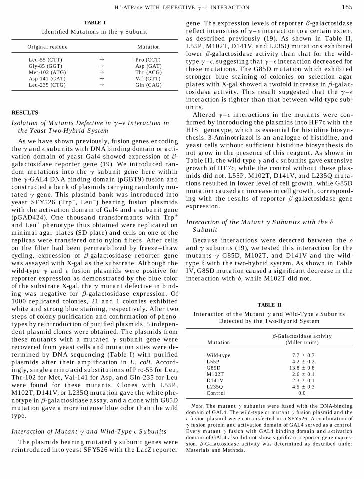
FIG. 1. Molecular assembly of a, b, g, d, and e subunits on mem-141, however, is not predicted in the topological imagebrane vesicles of mutants. For detection of the a, b, g, d, and eof the g subunit. Since Cys-87 was cross-linked to thesubunits, proteins, 2.0, 2.0, 2.0, 10, and 15 mg of the membranes,
respectively, prepared from cells carrying the wild-type (DK8 plus DELSEED loop of the b subunit (21), Gly-85 and possi-pKM02) or mutant plasmids, pKM02 g G85D, pKM02 g M102T and bly Met-102 may be also close to this loop. Since Glu-pKM02 g D141V, were separated by SDS–PAGE. The proteins blot- 235 is located close to the end of the long C-terminalted onto nylon filters were reacted with monoclonal antibodies to the
a helix (residues 223–286), this residue also seems toa, b, d, and e subunits, and polyclonal antibody to the g subunit.Immunoreactive materials were visualized using an ABC Vectastain be close to the DELSEED. Thus, a domain of the gkit as described previously (28). subunit including these residues (Gly-85, Met-102, and
Glu-225) except Asp-141 may interact directly or indi-rectly with the e subunit and may also affect interac-tions with the a and b subunits. Asp-141 might lie inthe F1–ATPase activities on the mutant membranes
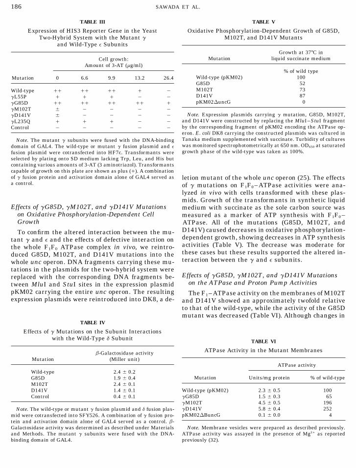
were observed, amounts of the F1 subunits detected this domain. Capaldi and co-workers extensively stud-immunologically in the membrane fraction were essen- ied the topological arrangement of the residues in thetially not altered (Fig. 1) among the mutants, sug- g subunit and g–e interaction by chemical labeling andgesting that marked disassembly did not occur for any trypsin digestion (21–24). They concluded that resi-of the mutants. The ATP-dependent proton channel ac- dues around Arg-70 and possibly Thr-106 and/or resi-tivity of M102T mutant membranes was higher than dues 200–212 are involved in g–e interaction, sug-the wild-type, while it was similar to the wild-type for gesting that two different domains are involved in g–D141V (Fig. 2). For G85D mutant, the activity de- e interaction. They also showed an atomic structure ofcreased, which corresponded well with the F1–ATPase E. coli e based on NMR analysis. e has a 10-strandedactivity. b sandwich structure in the N-terminal 84 residues
and the rest of the residues in the C-terminal regionDISCUSSION include two a helices. They also showed that Cys intro-
duced at Tyr-205 in g bound to Cys at residue 38 or 43We found that L55P, M102T, D141V, and L235Q mu-of e (24). These results suggested that one of the g–etations and G85D mutation in the g subunit causedinteraction sites lies between the N-terminal b sand-lower and higher reporter gene expression, respec-wich region and the bottom b-stranded region of the gtively, than the wild-type control in the yeast two-hy-subunit. The other site lies between the middle of thebrid system using the e subunit as the partner. Weg subunit and the two a helical regions of the e subunit.reported previously that reporter gene expression forResidues identified here as the mutation sites (Gly-85,the F1 subunit pairs reflects the intensities of protein–Met-102, and Leu-235) seem to be located in the latterprotein interaction observed by biochemical assayssite. The e subunit also interacts with the c subunit of(19). The results of this study suggested that interac-F0 via the bottom of the b sandwhich (21, 34). There-tions between the mutant g subunits and the wild-fore, some portions of the residues missing in thetype e subunit were altered. When G85D, M102T, andatomic structure of the g subunit shown for bovine F1D141V mutations were introduced back into the whole(7) are close to the c subunit of F0.unc operon and the properties of the ATPase were ana-
Here, the effects of primary defects in g–e interac-lyzed, decreased oxidative phosphorylation-dependentcell growth was observed for all cases, suggesting that tion on the F1F0–ATPase were successfully analyzed.
AID ABB 0397 / 6b44$$$203 10-28-97 11:26:18 arcal

188 SAWADA ET AL.
FIG. 2. Proton pumping activity depending on ATP or lactate in the membrane vesicles of mutants. E. coli DK8 transformed with pKM02carrying the entire ATPase operon, pKM02 DuncG (plasmid without the g subunit gene), pKM02 g G85D, pKM02 g M102T, or pKM02 gD141V was cultured in minimal medium supplemented with glycerol as the sole carbon source. Cell membranes were prepared, and ATPor lactate-driven proton pump activity was assayed using quinacrine for pH detection as described previously (26).
G85D, M102T, and D141V caused decreases in oxida- M102T mutation caused an increase in the protonpump activity, but no such alteration was seen for thetive phosphorylation-dependent cell growth, indicating
that these mutations caused decreases in ATP synthe- D141V mutation. The reason of this difference is notclear at present. Although the location of Asp-141 can-sis. Since the molecular assembly of the F1 portion in
these mutants was normal, local conformational not be predicted, these two residues may be involvedin different mechanisms because of their mutually dif-changes around the mutation sites rather than marked
structural changes in the whole F1F0 assembly may ferent locations in the topology. This assumption is con-sistent with the altered d–g interaction observed fortake place. However, the F1–ATPase activity on the
membranes was affected. G85D mutation caused de- D141V mutation, but not for M102T mutation. Theseresidues may be located at different domains in termscrease in the F1–ATPase activity. Since Gly-85 may be
close to the DELSEED loop of the b subunit as well as of binding to the d subunit; Asp-141 is close to d butMet-102 is not. These observations suggested that g–the e subunit, G85D mutation may cause tighter bind-
ing of g to e and may also affect the b subunit, leading d interaction has an important regulatory function inthe proton pump activity driven by ATP hydrolysis.to a decrease in ATPase activity. M102T and D141V
mutations showed decreases in g–e binding and in- Decreased d–g interaction was also observed for G85Dmutation, in which the proton pump activity was de-creases in F1–ATPase activity on the membranes.
Thus, the intensity of g–e interaction and the ATPase creased.The mutations described here (M102T and D141V)activity showed reciprocal changes. Since e is well es-
tablished as an F1–ATPase inhibitor (1–6), it is reason- exhibited novel phenotypes in which the loose interac-tion of g and e did not cause decreases in ATP-depen-able to observe increased ATPase activity on the mem-
branes caused by decreasing g–e interaction. Although dent proton pumping. Many mutants of the g or e sub-units have been reported (1, 2, 6) and some have defectsno previous evidences of the e subunit as an integral
inhibitor in the F0F1 complex were presented, the pres- in F1 assembly. For these mutations, the primary de-ent results of M102T phenotype may raise possibility fects in terms of subunit interaction were not detected.of e as a modulator of the catalysis. Further, our results The approach taken in this study is different from con-strongly suggested that the inhibitory action of the e ventional mutant screening. Phenotypes similar tosubunit is achieved through the g subunit. Rotation of those of M102T and D141V mutants have not beenthe g subunit within the ab core complex has been reported to our knowledge. Further detailed analysesvisualized (13) but it is not certain whether the e sub- of these new mutants will facilitate understanding ofunit is involved in this rotation. Comigration of the g the subunit–subunit interactions involved in the cou-and e subunits during ATP hydrolysis was reported pling of ATP hydrolysis and proton pumping.(24). Therefore, the g–e interaction may regulate ormodulate rotation of the g. Thus, the decreased g–e ACKNOWLEDGMENTSinteraction might cause an increase in the rotation of The present study was supported partly by Grants-in-Aid for scien-
tific research from the Japanese ministry of Education, Science, Cul-the g subunit.
AID ABB 0397 / 6b44$$$203 10-28-97 11:26:18 arcal

189H/-ATPase WITH DEFECTIVE g–e INTERACTION
ture and Sports, Ciba-Geigy Science Foundation, and Okayama 17. Hermolin, J., Gallant, J., and Fillingame, R. H. (1983) J. Biol.Chem. 58, 14550–14555.Foundation for Science and Technology.
18. Shin, Y., Sawada, K., Nagakura, T., Miyanaga, M., Moritani,C., Noumi, T., Tsuchiya, T., and Kanazawa, H. (1996) Biochim.
REFERENCES Biophys. Acta 1273, 62–70.19. Moritani, C., Sawada, K., Takemoto, K., Shin, Y., Nemoto, S.,1. Futai, M., and Kanazawa, H. (1983) Microbiol. Rev. 47, 285–
Noumi, T., and Kanazawa, H. (1996) Biochim. Biophys. Acta312.258, 14599–14609.2. Futai, M., Noumi, T., and Maeda, M. (1989) Annu. Rev. Biochem.
20. Dunn, S. D. (1982) J. Biol. Chem. 257, 7354–7359.58, 111–136.21. Aggeler, R., Haughton, M. A., and Capaldi, R. A. (1995) J. Biol.3. Walker, J. E., Saraste, M., and Gay, N. J. (1984) Biochem. Bio-
Chem. 270, 9185–9191.phys. Acta 768, 164–200.22. Tang, C., and Capaldi, R. (1996) J. Biol. Chem. 271, 3018–3024.
4. Fillingame, R. H. (1990) Bacteria 12, 345–391.23. Watts, S., Tang, C., and Capaldi, R. A. (1996) J. Biol. Chem. 271,
5. Pedersen, P. L., and Amzel, L. M. (1993) J. Biol. Chem. 268, 28341–28347.9937–9940. 24. Capaldi, R. A., Aggeler, R., Wilkens, S., and Gruber, G. (1996)
6. Weber, J., and Senior, A. (1997) Biochim. Biophys. Acta 1319, J. Bioenrg. Biomembr. 28, 397–401.19–58. 25. Klionsky, D. J., Brusilow, W. S., and Simoni, R. D. (1984) J. Bac-
7. Abrahams, J. P., Leslie, A. G. W., Lutter, R., and Walker, J. teriol. 160, 2055–1060.(1994) Nature 370, 621–628. 26. Fields, S., and Song, O. (1989) Nature 340, 245–246.
8. Wilkens, S., Dahlquist, F. W., McIntosh, L. P., Logan, E. W., 27. Kanazawa, H., Miki, T., Tamura, F., Yura, T., and Futai, M.Donaldson, and Capaldi, R. A. (1995) Nature Struct. Biol. 2, 961– (1979) Proc. Natl. Acad. Sci. USA 76, 1126–1130.966. 28. Studier, F. W., Rosenberg, A. H., Dunn, J. J., and Dubendorff,
9. Boyer, P. D. (1993) Biochim. Biophys. Acta 1140, 215–250. J. W. (1990) in Methods in Enzymology (Goeddel, D. V., Ed.),Vol. 185, pp. 60–89, Academic Press, San Diego.10. Penefsky, H. S., and Cross, R. L. (1991) Adv. Enzymol. 64, 173–
29. Maniatis, T., Fritsch, E. F., and Sambrook, J. (1982) Molecular214.Cloning: A Laboratory Manual, Cold Spring Harbor Laboratory,11. Duncan, T. M., Bulygin, V. V., Zhou, Y., Hutcheon, M. L., andCold Spring Harbor, NY.Cross, R. L. (1995) Proc. Natl. Acad. Sci. USA 92, 10964–10968.
30. Miki, J., Matsuda, T., Kariya, H., Ohmori, H., Tsuchiya, T., Fu-12. Sabbert, D., Engelbrecht, S., and Junge, W. (1996) Nature 381,tai, M., and Kanazawa, H. (1992) Arch. Biochem. Biophys. 294,623–625.373–381.
13. Noji, H., Yasuda, R., Yoshida, M., and Kinoshita, K., Jr. (1997) 31. Miki, J., Kusuki, H., Tsugumi, S., and Kanazawa, H. (1994) J.Nature 386, 299–302. Biol. Chem. 269, 4227–4232.
14. Capaldi, R. A., Aggeler, R., Turina, P., and Wilkens, S. (1994) 32. Sanger, F., Coulson, A. R., Barrell, B. G., Smith, A. J. H., andTrends Biochem. Sci. 19, 284–288. Roe, B. A. (1980) J. Mol. Biol. 143, 161–178.
15. Noumi, T., Oka, N., Kanazawa, H., and Futai, M. (1986) J. Biol. 33. Kanazawa, H., Tamura, F., Mabuchi, K., Miki, T., and Futai, M.Chem. 261, 7070–7076. (1980) Proc. Natl. Acad. Sci. USA 77, 7005–7009.
34. Zang, Y., and Fillingame, R. H. (1995) J. Biol. Chem. 270,16. Aris, J. P., and Simoni, R. D. (1983) J. Biol. Chem. 258, 14599–14609. 24609–24614.
AID ABB 0397 / 6b44$$$204 10-28-97 11:26:18 arcal


![Exercises for Chapter 3 3.1 Solution for Chapter 3 3.1 [Fermions and bosons; the ultimate elementary problem] There is a system with only three states with energies 0, ϵ and ϵ (ϵ](https://img.pdfslide.tips/doc/110x75/5acb65997f8b9a63398baa06/exercises-for-chapter-3-31-for-chapter-3-31-fermions-and-bosons-the-ultimate.jpg)