Embed Size (px)
Citation preview
Vol.:(0123456789)1 3
International Cancer Conference Journal (2018) 7:71–75 https://doi.org/10.1007/s13691-018-0323-4
CASE REPORT
Successful radiotherapy for endometrial serous carcinoma with local repeated recurrence
Shinya Kusumoto1 · Hisaya Fujiwara1 · Maiko Sagawa1 · Takahiro Nobuzane1 · Toshihiro Nishida2 · Yukio Akagi3 · Yutaka Hirokawa3 · Yasuhiro Katsube1
Received: 25 January 2018 / Accepted: 26 March 2018 / Published online: 5 April 2018 © The Author(s) 2018
AbstractThe incidence of endometrial serous carcinoma (ESC) has been increasing, and ESC is resistant to treatment. We report a patient with ESC who responded to radiotherapy for multiple recurrences. The first recurrence was detected in the vaginal wall and left internal iliac lymph node 5 months after the initial treatment. Concurrent chemoradiotherapy (CCRT) was administered. Radiation was delivered using the intensity modulated radiation therapy technique. The second recurrent tumor was detected in the right internal iliac lymph node after 4 months, and CCRT was conducted. After 4 months, the third recurrence was detected in the right common iliac node, and CCRT was performed. After 8 months, the fourth recurrence was detected in the horizontal portion of the duodenum, and radiotherapy was administered. After 9 months, the fifth recur-rence was detected in the vaginal wall. Interstitial brachytherapy was conducted. Grade 2 gastrointestinal injury, nausea and radiodermatitis were observed. During the subsequent 13-month follow-up, there has been no recurrence. Although ESC is resistant to treatment, radiotherapy could be effective in some cases. Even when multiple recurrences occur, radiotherapy may be considered a treatment option if the irradiation level is permissible.
Keywords Endometrial serous carcinoma · Multiple recurrence · Intensity modulated radiation therapy · Complete remission
Introduction
In Japan, the incidence of endometrial serous carcinoma (ESC) has been increasing [1]. ESC is a histological sub-type of endometrial carcinoma with a poor prognosis [2]. The recurrence of ESC frequently occurs and its treatment is very difficult. In this study, we report a 68-year-old woman with ESC who experienced repeated recurrence 5 times and responded to radiotherapy for each recurrence.
Case report
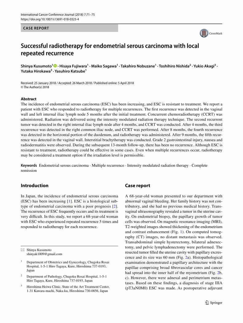
A 68-year-old woman presented to our department with abnormal vaginal bleeding. Her family history was not con-tributory, and she had no previous medical history. Trans-vaginal ultrasonography revealed a tumor in the uterine cav-ity. On endometrial biopsy, the papillary growth of tumor cells was observed. On magnetic resonance imaging (MRI), T2-weighted images showed thickening of the endometrium and contrast enhancement (Fig. 1). On computed tomog-raphy (CT) images, no distant metastasis was observed. Transabdominal simple hysterectomy, bilateral adnexec-tomy, and pelvic lymphadenectomy were performed. The resected tumor filled the uterine cavity with papillary excres-cence and its size was 60 mm (Fig. 2a). Histopathological examination demonstrated a papillary architecture with the papillae comprising broad fibrovascular cores and cancer had spread into the inner half of the myometrium (Fig. 2b, c). However, there were adnexal and perimetrium metas-tases. Based on these findings, a diagnosis of stage IIIA (pT3aN0M0) ESC was made. As postoperative adjuvant
* Shinya Kusumoto [email protected]
1 Department of Obstetrics and Gynecology, Chugoku Rosai Hospital, 1-5-1 Hiro Tagaya, Kure, Hiroshima 737-0193, Japan
2 Department of Pathology, Chugoku Rosai Hospital, 1-5-1 Hiro Tagaya, Kure, Hiroshima 737-0193, Japan
3 Hiroshima Heiwa Clinic, State of the Art Treatment Center, 1-31 Kawara-machi, Naka-ku, Hiroshima 730-0856, Japan
72 International Cancer Conference Journal (2018) 7:71–75
1 3
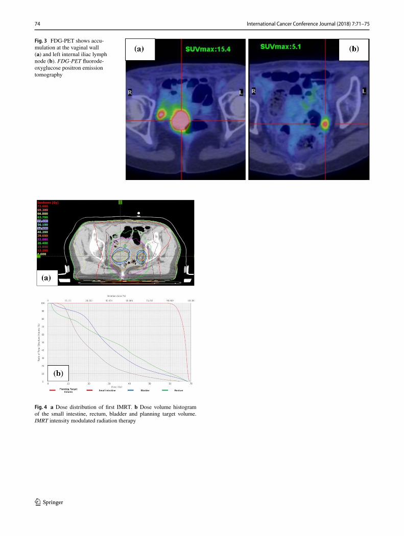
therapy, combination chemotherapy of paclitaxel and car-boplatin (TC) was administered. Before the second cycle, the regimen was changed to docetaxel and cisplatin (DP) because of skin eruptions induced by paclitaxel or carbopl-atin. Four cycles of DP were administered. After 5 months, CT revealed tumors in the vaginal wall and left internal iliac lymph node. As fluorodeoxyglucose positron emis-sion tomography (FDG-PET) showed accumulation with maximum standardized uptake values (SUVmax) of 15.4 in the vaginal wall and 5.1 in the left internal iliac lymph node, the first recurrence of ESC was diagnosed (Fig. 3a, b). Concurrent chemoradiotherapy (CCRT) was performed. Chemotherapy comprised nedaplatin and docetaxel (neda-platin 20 mg/body plus docetaxel 20 mg/body, on day two, every week for three cycles). Concurrent radiotherapy of 66 Gy (22 fractions of 3 Gy, 5 days/week) was delivered over 5 weeks using intensity modulated radiation therapy (IMRT) (Fig. 4a, b). The planning target volume (PTV) was the clinical target volume (CTV) + a 5-mm margin. Tumor regression was observed and the uptake in the recurrent
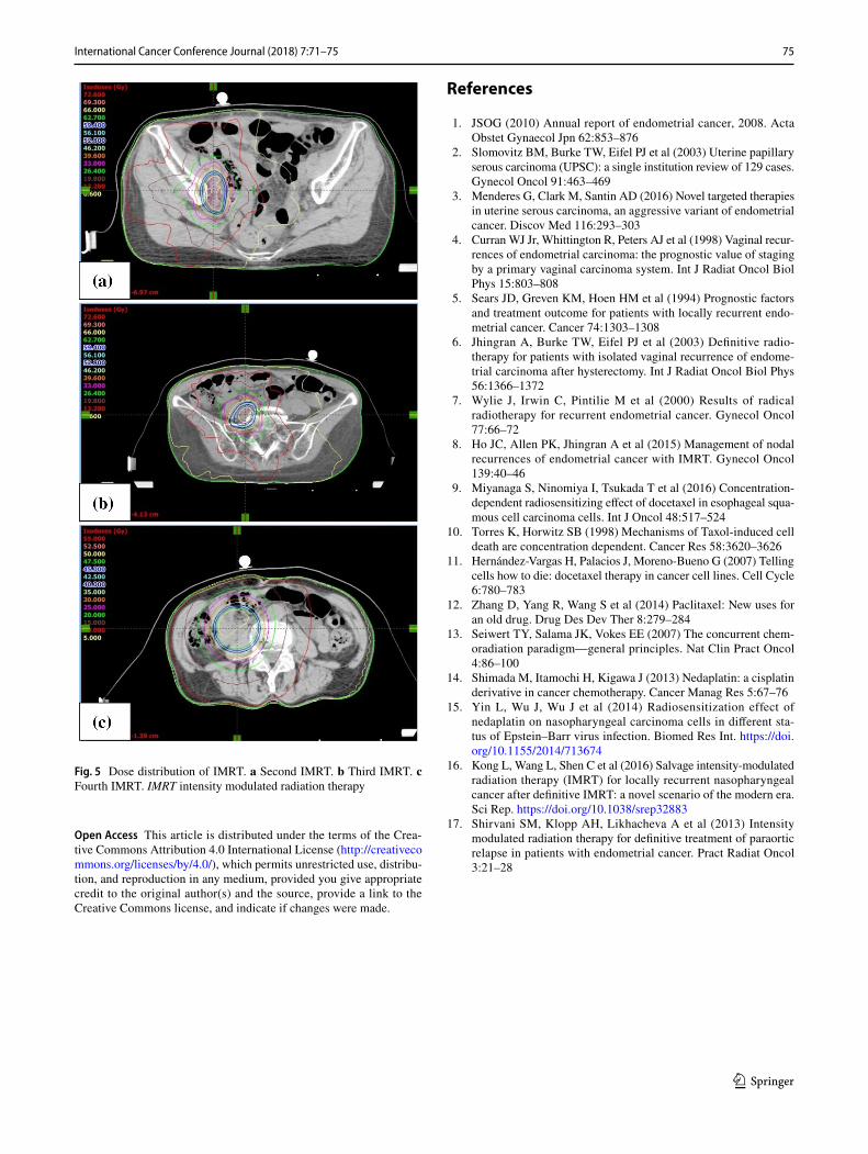
site decreased considerably on the FDG-PET scan. After 4 months, the second recurrence was detected in the right internal iliac lymph node using FDG-PET with an SUVmax of 13.8. CCRT was performed again (Fig. 5a). The PTV was also the same. Tumor regression was observed and the uptake in the recurrent site decreased considerably on the FDG-PET scan. After 4 months, the third recur-rence was detected in the right common iliac node using FDG-PET with an SUVmax of 9.0. CCRT was performed once more (Fig. 5b). The PTV again was the same. Tumor regression was observed and the uptake in the recurrent site decreased considerably on the FDG-PET. After 8 months, the fourth recurrence was detected in the horizontal portion of the duodenum using FDG-PET with an SUVmax of 8.6. IMRT (50 Gy in 25 fractions) was performed (Fig. 5c). The PTV was the same. The tumor regression was observed and the uptake in the recurrent site decreased considerably on the FDG-PET scan. After 9 months, small tumor induration was palpable on vaginal and rectal examinations. The fifth recur-rence was detected in the vaginal wall, via vaginal tumor biopsy. Histological examination revealed papillary tumor cells, which were identical to those of the primary uterine lesion, with necrosis, and FDG-PET showed accumulation with an SUVmax of 4.2 in this site. Interstitial brachytherapy (48 Gy in 8 fractions) was performed. Tumor regression was observed and the uptake in the recurrent site decreased con-siderably on the FDG-PET. Grade 2 gastrointestinal fistula, nausea and radiodermatitis (CTCAE; Common Toxicity Cri-teria for Adverse Events, version 4.03) were observed during the treatment. During the subsequent 13-month follow-up, there has been no recurrence.
Discussion
In this study, we report a patient with ESC with local repeated recurrence who responded to five sessions of radi-otherapy to the vaginal wall-, the internal-, common-, and iliac nodes, and the horizontal portion of the duodenum. ESC is chemotherapy-resistant and has a poor prognosis [3]. Management of recurrent ESC is difficult. Radiation is the treatment of choice for women who experience recurrence at the vaginal cuff [4–6]. The histologic type is an impor-tant prognostic factor for recurrent endometrial carcinoma when radiotherapy is performed [4–7]. The present case suggested that definitive radiotherapy for locally repeated recurrent ESC was effective in situations different from the above-mentioned case of recurrence in the vaginal wall. To
Fig. 1 Sagittal T2-weighted MRI findings. The thickened and uneven endometrium lining was observed. MRI magnetic resonance imaging
73International Cancer Conference Journal (2018) 7:71–75
1 3
our knowledge, the efficacy of radiotherapy for repeated recurrent ESC has not been reported previously. A recently published study on IMRT for nodal recurrences of endo-metrial carcinoma demonstrated that patients who received CCRT had significantly longer median survival as com-pared to patients treated with radiotherapy without concur-rent chemotherapy (61.9 versus 28.7 months, p = 0.034) [8]. Based on this information, CCRT for endometrial carcinoma with high-grade malignant potential, such as ESC, could be an effective therapy if adverse effects to adjacent organs, particularly the bowels and bladder, are acceptable. In the present case, there were three reasons for using this chemo-therapy. First, it was used to enhance radiosensitization, as antitumor platinum (e.g., cisplatin and nedaplatin), pacli-taxel, and docetaxel therapies have been reported as effective radiosensitizers [9–15]. Second, the chemotherapy was used for an antitumor effect, and third, nedaplatin and docetaxel were used, because they are easy to prepare and are avail-able at outpatient clinics. Unlike conventional radiotherapy, IMRT is a method of highly conformal radiation that permits the delivery of high doses of radiation to the tumor, while minimizing doses to surrounding healthy tissues. Therefore, recently, reirradiation with IMRT has been performed. How-ever, response time, tolerance dose, and toxic adverse effects of IMRT for recurrence are unclear. According to a report on salvage IMRT for locally recurrent nasopharyngeal can-cer after definitive IMRT, the median interval between the completion of initial radiotherapy and the start of reirradia-tion was 27.9 (range 11.7–79.0) months; grade 3–5 toxici-ties occurred in 50 of the 77 patients and treatment-induced severe adverse effects were the most important contributor to mortality [16]. In another study about IMRT for paraaortic recurrence of endometrial cancer, late grade 3 or 4 gastroin-testinal toxic effects occurred in 2 of 14 patients who were treated with pelvic radiotherapy as the initial treatment [17]. However, there are some points that must be kept in mind when referring to these studies. The method of setting PTV varies depending on the facility. Unlike in the present case, in the studies cited above, patients were treated with IMRT using 45–50 Gy with a 0.7–1-cm margin [17]. In the present case, adverse effects of grade 3 or greater were not observed. Thus, toxic adverse effects of IMRT may differ depending on the technique. In summary, we encountered a patient with ESC who responded to five sessions of radiotherapy. Radio-therapy (mainly IMRT) could be an effective treatment for locally repeated recurrent ESC; however, its safety should be individually considered.
Fig. 2 a The size of the resected tumor was 60 mm. b The cancer had spread into the inner half of the myometrium (triangles), and perime-trium metastases were observed (arrows). c The histologic type was endometrial serous carcinoma
▸
74 International Cancer Conference Journal (2018) 7:71–75
1 3
Fig. 3 FDG-PET shows accu-mulation at the vaginal wall (a) and left internal iliac lymph node (b). FDG-PET fluorode-oxyglucose positron emission tomography
Fig. 4 a Dose distribution of first IMRT. b Dose volume histogram of the small intestine, rectum, bladder and planning target volume. IMRT intensity modulated radiation therapy
75International Cancer Conference Journal (2018) 7:71–75
1 3
Open Access This article is distributed under the terms of the Crea-tive Commons Attribution 4.0 International License (http://creat iveco mmons .org/licen ses/by/4.0/), which permits unrestricted use, distribu-tion, and reproduction in any medium, provided you give appropriate credit to the original author(s) and the source, provide a link to the Creative Commons license, and indicate if changes were made.
References
1. JSOG (2010) Annual report of endometrial cancer, 2008. Acta Obstet Gynaecol Jpn 62:853–876
2. Slomovitz BM, Burke TW, Eifel PJ et al (2003) Uterine papillary serous carcinoma (UPSC): a single institution review of 129 cases. Gynecol Oncol 91:463–469
3. Menderes G, Clark M, Santin AD (2016) Novel targeted therapies in uterine serous carcinoma, an aggressive variant of endometrial cancer. Discov Med 116:293–303
4. Curran WJ Jr, Whittington R, Peters AJ et al (1998) Vaginal recur-rences of endometrial carcinoma: the prognostic value of staging by a primary vaginal carcinoma system. Int J Radiat Oncol Biol Phys 15:803–808
5. Sears JD, Greven KM, Hoen HM et al (1994) Prognostic factors and treatment outcome for patients with locally recurrent endo-metrial cancer. Cancer 74:1303–1308
6. Jhingran A, Burke TW, Eifel PJ et al (2003) Definitive radio-therapy for patients with isolated vaginal recurrence of endome-trial carcinoma after hysterectomy. Int J Radiat Oncol Biol Phys 56:1366–1372
7. Wylie J, Irwin C, Pintilie M et al (2000) Results of radical radiotherapy for recurrent endometrial cancer. Gynecol Oncol 77:66–72
8. Ho JC, Allen PK, Jhingran A et al (2015) Management of nodal recurrences of endometrial cancer with IMRT. Gynecol Oncol 139:40–46
9. Miyanaga S, Ninomiya I, Tsukada T et al (2016) Concentration-dependent radiosensitizing effect of docetaxel in esophageal squa-mous cell carcinoma cells. Int J Oncol 48:517–524
10. Torres K, Horwitz SB (1998) Mechanisms of Taxol-induced cell death are concentration dependent. Cancer Res 58:3620–3626
11. Hernández-Vargas H, Palacios J, Moreno-Bueno G (2007) Telling cells how to die: docetaxel therapy in cancer cell lines. Cell Cycle 6:780–783
12. Zhang D, Yang R, Wang S et al (2014) Paclitaxel: New uses for an old drug. Drug Des Dev Ther 8:279–284
13. Seiwert TY, Salama JK, Vokes EE (2007) The concurrent chem-oradiation paradigm—general principles. Nat Clin Pract Oncol 4:86–100
14. Shimada M, Itamochi H, Kigawa J (2013) Nedaplatin: a cisplatin derivative in cancer chemotherapy. Cancer Manag Res 5:67–76
15. Yin L, Wu J, Wu J et al (2014) Radiosensitization effect of nedaplatin on nasopharyngeal carcinoma cells in different sta-tus of Epstein–Barr virus infection. Biomed Res Int. https ://doi.org/10.1155/2014/71367 4
16. Kong L, Wang L, Shen C et al (2016) Salvage intensity-modulated radiation therapy (IMRT) for locally recurrent nasopharyngeal cancer after definitive IMRT: a novel scenario of the modern era. Sci Rep. https ://doi.org/10.1038/srep3 2883
17. Shirvani SM, Klopp AH, Likhacheva A et al (2013) Intensity modulated radiation therapy for definitive treatment of paraortic relapse in patients with endometrial cancer. Pract Radiat Oncol 3:21–28
Fig. 5 Dose distribution of IMRT. a Second IMRT. b Third IMRT. c Fourth IMRT. IMRT intensity modulated radiation therapy