Embed Size (px)
Citation preview
SSUULLFFOOLLOOBBUUSS SSOOLLFFAATTAARRIICCUUSS AASS
SSOOUURRCCEE OOFF GGLLYYCCOOSSYYLL
HHYYDDRROOLLAASSEESS WWIITTHH
BBIIOOTTEECCHHNNOOLLOOGGIICCAALL PPOOTTEENNTTIIAALL
Michele Girfoglio
Dottorato in Scienze Biotecnologiche – XXI ciclo Indirizzo Biotecnologie Industriali Università di Napoli Federico II

Dottorato in Scienze Biotecnologiche – XXI ciclo Indirizzo Biotecnologie Industriali Università di Napoli Federico II
SSUULLFFOOLLOOBBUUSS SSOOLLFFAATTAARRIICCUUSS AASS
SSOOUURRCCEE OOFF GGLLYYCCOOSSYYLL
HHYYDDRROOLLAASSEESS WWIITTHH
BBIIOOTTEECCHHNNOOLLOOGGIICCAALL PPOOTTEENNTTIIAALL
Michele Girfoglio
Dottorando: Michele Girfoglio Relatore: Prof. Mosè Rossi Coordinatore: Prof. Giovanni Sannia

A tutte le persone a me care, che mi hanno sempre sostenuto ed hanno rappresentato un fondamentale supporto durante il percorso di cui questa tesi rappresenta il compimento. A Elia P.


INDEX ABBREVIATIONS RIASSUNTO SUMMARY INTRODUCTION
Archaea Sulfolobus Genetic elements Plasmid-fusellovirus genetic systems S. solfataricus-E. coli shuttle vectors Thermophilic enzymes as biocatalysts in industrial applications Cellulose Concepts, methodology and objectives
MATERIAL AND METHODS Strains, enzymes and reagents used in this study E. coli transformation techniques Proteins analyses Cloning of the lacS gene into pMSSV and characterization of the vector sso1354 gene expression in S. solfataricus sso1354 gene expression in E. coli sso1354 gene expression in K. lactis Enzyme assays and analyses
RESULTS 1. CHARACTERIZATION OF pMSSV SHUTTLE VECTOR: CLONING AND EXPRESSION OF THE lacs GENE
Cloning of the expression cassette tf55αlacS in the pMSSV vector lacS expression in the β-galactosidase deficient mutant GθW β-galactosidase assays on pMSSVlacS transfectants
2. XENOLOGOUS AND HETEROLOGOUS EXPRESSION OF sso1354 GENE Sequence analysis Cloning of the expression cassette araS1354 in the pMSSV vector and expression in S. solfataricus Expression of the sso1354 gene in E. coli Expression and characterization of the sso1354 gene in Kluyveromyces lactis
DISCUSSION
pag. 1
pag. 3
pag. 10
pag. 12
pag. 12
pag. 13
pag. 14
pag. 20
pag. 22
pag. 24
pag. 24
pag. 28
pag. 30
pag. 30
pag. 30
pag. 31
pag. 32
pag. 35
pag. 37
pag. 38
pag. 38
pag. 40
pag. 40
pag. 40
pag. 41
pag. 44
pag. 45
pag. 45
pag. 49
pag. 53
pag. 54
pag. 57


1
ABBREVIATIONS
bp Base pairs
BSA Bovine serum albuine
CAPS N-cyclohexyl-3-aminopropanesulfonic acid
CBD Cellulose binding domain
CPs Conjugative plasmids
Da dalton
DNA Deoxyribonucleic acid
dNTP Deoxyribonucleotide triphosphate
DSM Deutsche Sammlung von Mikroorganismen
EDTA Ethylenediaminetetraacetic acid
h Hours
HGT Horizontal gene transfer
hph Hygromycin B phosphotransferase
IPTG Isopropyl β-D-1-thiogalactopyranoside
IS Insertion sequence
LB Luria Bertani medium
min Minutes
MITEs Miniature inverted transposable elements
OD Optical density
ORF Open reading frame
PCR Polimerase chain reaction
PMSF Phenyl methane sulphonyl fluoride
PVDF Polyvinylidene difluoride
RNA Ribonucleic acid

2
SBP Sugar binding protein
SDS Sodium Dodecyl Sulphate
SDS-PAGE Sodium Dodecyl Sulphate - PolyAcrylamide Gel Electrophoresis
TBE 45 mM Tris-borate, 1mM EDTA pH 8.0
TBP TATA-binding protein
TLC Thin Layer Chromatography
Tris tris(hydroxymethyl)aminomethane
TYS tryptone yeast sucrose (medium)
UTR untraslated region
X-gal 5-bromo-4-chloro-3-indolyl-b-D-galactopyranoside
YPG Yeast extract Peptone Galactose
3
Riassunto Introduzione Uno dei principali ambiti di interesse delle Biotecnologie Industriali riguarda gli enzimi sintetizzati dagli organismi estremofili, in particolar modo ipertermofili; l’attenzione con cui il mondo scientifico guarda a tali proteine è motivata dalle doti di stabilità e resistenza alle alte temperature che mostrano caratteristiche peculiari che le distinguono dalle controparti da organismi mesofili. In tale contesto un ruolo di primo piano è rivestito dagli enzimi che prendono parte al processo degradativo di carboidrati, i quali possiedono un ampio spettro di campi applicativi che vanno dall’industria di produzione della carta a quella alimentare, da quella tessile a quella farmaceutica; in particolare, grande risalto negli ultimi anni sta assumendo l’applicazione che vede tali enzimi impiegati nel processo di produzione del bioetanolo, carburante il cui crescente utilizzo sta assumendo ritmi esponenziali. La produzione e l’impiego di tale carburante risulterebbe particolarmente vantaggiosa se questo venisse ottenuto utilizzando come materia prima scarti industriali ad alto contenuto cellulosico, evitando di impiegare a tal scopo cereali appositamente ed intensivamente coltivati; si potrebbe in tal modo superare uno dei maggiori problemi legati alla produzione di biocarburanti, che riguarda l’aumento dei prezzi delle materie prime alimentari ed il conseguente aggravamento delle condizioni di vita nelle aree in via di sviluppo. A tale riguardo, diversi microrganismi ipertermofili eterotrofi sono stati studiati rispetto alla loro capacità di crescere su carboidrati complessi come unica fonte di carbonio e di energia. Tale caratteristica può essere particolarmente vantaggiosa per questi organismi, in quanto spesso il loro habitat risiede in nicchie ecologiche dove le fonti di carbonio, di origine vegetale, sono scarse. La maggior parte degli Archaea mostra un eccezionale adattamento alla vita in condizioni estreme, tra cui l’alta temperatura e i valori di pH estremi; alla luce di ciò, essi rappresentano una fonte di biocatalizzatori particolarmente adatti ad essere impiegati per applicazioni industriali nell’idrolisi di matrici complesse di zuccheri. Fino a pochi decenni fa gli Archaea non erano riconosciuti dal punto di vista della classificazione filogenetica degli esseri viventi. Guardando un “albero della vita” infatti, avremmo potuto notare esclusivamente due domini, quello dei Procarioti (identificati con i Batteri) e quello degli Eucarioti. Una fondamentale innovazione in questo contesto è scaturita dagli studi di Carl Woese che, alla fine degli anni ’70, dopo aver condotto approfondite analisi comparative di sequenze di rRNA 16S procariotiche allo scopo di delineare un più chiaro quadro delle correlazioni intergenere nel mondo batterico, giunse alla conclusione che il mondo dei viventi fosse da suddividere in tre domini. Nacque così la denominazione di “ArchaeoBatteri”, creata per definire il nuovo dominio rispetto agli EuBatteri, poi modificata in Archaea per affermarne definitivamente la separazione dal dominio dei Batteri. Facendo quindi cadere l’idea dell’unicità procariotica, Woese distinse i Procarioti in due gruppi, gli Archaea appunto, ed i Bacteria. I suoi studi avevano messo in evidenza che questi microrganismi si differenziano talmente dai Batteri da meritare appieno una classificazione filogenetica indipendente. Gli Archaea risultano attualmente ulteriormente suddivisi in tre regni: Crenarchaeota (ipertermofili e termoacidofili); Euryarchaeota (ipertermofili, metanogeni ed alofili); Korarchaeota (poco conosciuti a livello biochimico e fisiologico).
4
In questo contesto S. solfataricus, isolato originariamente dalle pozze solfatariche di Pisciarelli a Napoli, e che cresce ad un pH acido (in un intervallo tra 2.0 e 5.0) e ad alte temperature (75-90°C), rappresenta un organismo modello per lo studio dei Crenarchaeota, in quanto risulta essere facilmente propagabile in coltura liquida così come in mezzo solido. Inoltre, risulta rilevante segnalare che il genoma di S. solfataricus P2 è stato uno dei primi genomi crenarchaeali ad essere completamente sequenziato, e ciò ha aperto nuove prospettive di indagine riguardo tali organismi, dando un forte impulso allo studio delle loro caratteristiche. Le specie appartenenti al genere Sulfolobus, come pure S. solfataricus, sono provviste di un’ampia gamma di diversi elementi genetici, comprendenti virus, plasmidi, sequenze d’inserzione, che costituiscono un potente mezzo di indagine filogenetica per gli organismi nei quali si ritrovano. Inoltre, tali elementi, in particolar modo virus e plasmidi, vengono sempre con maggiore interesse studiati nell’ottica di poterli adoperare come base nella messa a punto di sistemi stabili di clonaggio/espressione, i quali, per gli organismi archaeali, risultano essere ancora ad uno stato incipiente di sviluppo. Inoltre, S. solfataricus è una interessante fonte di attività enzimatiche di tipo cellulasico; tali enzimi potrebbero essere utilizzati in maniera vantaggiosa per la produzione di bioetanolo, grazie alla loro peculiare combinazione di termostabilità e resistenza a pH acidi. Infatti, nel processo di produzione del bioetanolo i materiali cellulosici vengono idrolizzati impiegando acidi ad alte temperature, ed in tale contesto operativo la conduzione di idrolisi enzimatica con enzimi resistenti ad alte temperature e bassi pH porta ad una semplificazione del processo produttivo, con un conseguente abbassamento dei costi. Il sequenziamento del genoma di S. solfataricus ha mostrato l’esistenza di un completo gruppo di ventidue glicosil idrolasi capaci di processare carboidrati complessi quali l’amido, la cellulosa e le emicellulose, compresi mannani, xiloglucani e xilano. In particolare, sono state identificate tre sequenze codificanti per putative β-endoglucanasi (SSO2534, SSO1354 ed SSO1949), sulle quali si è concentrata l’attenzione nello svolgimento del presente progetto; inoltre in S. solfataricus è stata rilevata un’attività β -endoglucanasica extracellulare, sia liberamente rilasciata che adesa alle cellule. Per quanto riguarda SSO2534, il prodotto proteico è stato ampiamente caratterizzato, anche se i tentativi di espressione in E. coli non hanno avuto successo; il gene sso1949 invece è stato clonato ed espresso in E. coli , ma a livelli molto bassi. Alla luce di ciò, al fine di ottenere la (sovra)espressione di sequenze codificanti per attività enzimatiche provenienti da S. solfataricus, risulta utile intraprendere la strada della sovra-espressione di geni nello stesso organismo di origine o in uno fisiologicamente più correlato, considerati i diversi fattori limitanti che si possono presentare esprimendo tali sequenze in maniera eterologa, in ospiti filogeneticamente troppo distanti. Si comprende quindi l’importanza di disporre di efficaci sistemi-vettore per l’espressione xenologa-eterologa in S. solfataricus, anche se il numero dei sistemi ad oggi sviluppati e che sono risultati efficaci risulta essere ancora scarso. Nel laboratorio dove si svolge tale progetto di Dottorato è stato progettato e messo a punto un efficiente vettore navetta E. coli-S. solfataricus, definito pMSSV, costruito sulla base del plasmide archaeale pSSVx da S. shibatae REY 15/4, al quale sono state aggiunte sequenze derivanti dal plasmide pUC19.
5
Il pSSVx è un elemento genetico isolato, insieme al fusellovirus SSV2, dal ceppo REY 15/4 di S. islandicus. Il genoma è formato da 5705 bp e mostra regioni di alta similarità di sequenza con i plasmidi criptici pRN1, pRN2 e pDL10; per questo viene considerato appartenente alla famiglia dei pRNs. All’interno del genoma del pSSVx si trovano nove ORFs delle quali tutte, tranne una, sono orientate nella stessa direzione. Quattro di queste, insieme a due elementi di sequenza, mostrano similarità con gli altri plasmidi della famiglia. In particolare due delle quattro ORFs sono quelle che codificano per la proteina Rep (ORF892) e per la CopG (ORF60), mentre le due sequenze sono costituite dalle origini di replicazione a singolo e doppio filamento (sso e dso). In aggiunta a ciò, è presente una regione che non mostra similarità con i pRNs; questa contiene due ORFs adiacenti, 154 e 288, per le quali è possibile trovare regioni codificanti omologhe nei fusellovirus SSV1 (a153 e b251) e SSV2 (153 e 233); queste sequenze si strutturano tutte con simile arrangiamento (è presente una regione di sovrapposizione tra le due ORFs). La similarità risulta essere maggiore per la ORF 154, e le similarità mostrate dai virus tra loro sono maggiori di quelle tra virus e plasmide. L’individuazione del pSSVx si è avuta durante una procedura di screening volta a identificare elementi genetici extracromosomali, specialmente del tipo SSV1. Dopo aver effettuato analisi di restrizione, volte ad accertare la presenza di DNA episomale, attraverso indagini di microscopia elettronica sui supernatanti cellulari sono state trovate particelle virali, dalla caratteristica forma a “limone”, di due distinte dimensioni: le più grandi mostravano grandezza uguale alle particelle di SSV1, ovvero 80nm-55nm, mentre le più piccole si attestavano su dimensioni di 60nm-40nm. Il genoma di SSV2, una volta clonato, produceva soltanto le particelle di dimensioni maggiori, all’interno delle quali si trovava impacchettato il DNA virale. Dal confronto tra i pattern di restrizione del DNA del solo SSV2 e di quello derivante dalla miscela di particelle, e da analisi di cross-ibridazione, si poteva evincere che nella miscela era presente un ulteriore elemento genetico della grandezza di circa 5.7 kb. Ciò forniva evidenza del fatto che le particelle SSVx più piccole potessero contenere tale elemento, chiamato appunto pSSVx. Risulta interessante notare che il pSSVx risulta propagare efficacemente in mancanza di pressione selettiva solo se co-trasfettato con SSV2; tale plasmide non è capace infatti di impacchettarsi e dar luogo ad un processo infettivo autonomamente, come testimoniato dalla constatazione che risulta essere privo delle regioni codificanti per le proteine strutturali del virus. In quest’ottica, le due ORFs che il pSSVx mostra nella regione non conservata, e che hanno omologia con le sequenze degli SSV1 e SSV2, sembrano codificare per proteine che sono implicate nel processo di assemblaggio delle particelle virali, e con buona probabilità sono state acquisite dallo stesso genoma di SSV2. Risultano essere essenziali per l’impacchettamento e la propagazione del plasmide, in quanto gli elementi pRN1 e pRN2, che ne sono privi, non mostrano la capacità di propagarsi sfruttando come helper SSV1 o SSV2. Alla luce delle affermazioni fin qui riportate, risulta chiara la comprensione dell’appellativo dato a tale elemento, che viene sovente definito come un “ibrido” tra un plasmide ed un virus. Il vettore pMSSV ricalca quindi le caratteristiche fondamentali del plasmide pSSVx da cui prende origine; si mantiene in alto numero di copie in entrambi gli ospiti, e si è dimostrato efficace nel condurre all’espressione del gene lacS di S. solfataricus, codificante per una β-glicosidasi, sotto il controllo del promotore del gene tf55α, codificante per una subunità del complesso-termosoma di S. shibatae e quindi inducibile da shock termico.
6
Risultati e discussione Allo scopo di delucidarne le potenzialità applicative, è stata effettuata una approfondita caratterizzazione del vettore pMSSV. A tale scopo, la cassetta d’espressione costituita dalla sequenza codificante per una β-glicosidasi da S. solfataricus (lacS) sotto il controllo del promotore inducibile del gene tf55α è stata clonata all’interno del vettore, e con tale elemento sono state elettroporate cellule del ceppo GθW/SSV2 di S. solfataricus. L’utilizzo del gene lacS per il clonaggio è stato adottato in quanto comporta il vantaggio di permettere un facile monitoraggio dell’espressione; è possibile infatti effettuare la rilevazione dell’attività su centrifugati cellulari utilizzando il substrato cromogenico X-gal, ed avendo trasferito il costrutto in cellule del ceppo GθW (che presenta a livello del genoma un’estesa delezione che interessa il locus del gene lacS, e per tale motivo risulta essere difettivo dell’attività β-galattosidasica), la presenza di attività può essere senza incertezza attribuita all’espressione della sequenza contenuta nel vettore. Il vettore messo a punto è risultato capace di portare con successo all’espressione del gene inseritovi, riuscendo a complementare in questo caso la mutazione presente a livello del locus del gene lacS. Rispetto ai sistemi genetici E. coli-S. solfataricus finora sviluppati presenta diverse caratteristiche vantaggiose, tra le quali spiccano un’elevata efficienza di trasformazione, dovuta alla modalità propagativa di natura virale utilizzata dal vettore, e la stabilità nella propagazione, senza integrazione nel cromosoma dell’ospite e con un elevato numero di copie per cellula (fino a circa 130 in fase stazionaria, con accumulo in dipendenza dalla fase di crescita). Il pMSSV rappresenta quindi un potente strumento genetico, con forti potenzialità applicative tanto nello studio di base quanto nella produzione biotecnologica di proteine omologhe/eterologhe. Il gene codificante per la putativa endoglucanasi extracellulare SSO1354, l’unico per il quale non sono presenti in letteratura studi di caratterizzazione sul prodotto proteico, è stato quindi impiegato in tentativi di espressione xenologa/eterologa. Inizialmente, la sequenza proteica corrispondente ad SSO1354 è stata sottoposta ad analisi bioinformatiche; da un allineamento con le sequenze presenti nelle banche dati, utilizzando il programma FASTA 3, si è potuto constatare che la più significativa similarità si evince ripetto alla sequenza della sopracitata SSO1949, con cui SSO1354 mostra una percentuale di identità dell’85%. Inoltre, SSO1354 mostra un allineamento significativo con altre endoglucanasi, la maggior parte delle quali risultano appartenere a Batteri termofili del genere Thermotoga (in particolare di specie maritima e neapolitana); escludendo SSO1949, l’allineamento con altre endoglucanasi è possibile però solo per il dominio catalitico, mentre per quello N-terminale non è possibile trovare riscontri positivi. Particolarmente interessante risulta inoltre il raffronto della sequenza di SSO1354 con quelle delle sugar binding proteins (SBP) di S. solfataricus AraS, GlcS e TreS; queste proteine extracellulari, che legano rispettivamente l’arabinosio, il glucosio e il trealosio, sono delle subunità dei trasportatori ATP binding cassette (ABC), i quali sono responsabili per l’assimilazione degli zuccheri da parte di S. solfataricus. Dall’allineamento delle sequenze proteiche, che è particolarmente significativo all’estremità N-terminale, è stato possibile notare come SSO1354 abbia un’organizzazione in domini simile a quella delle SBPs; una regione N-terminale
7
idrofobica è seguita da una regione ricca in serine e treonine, con il dominio di catalitisi/legame posizionato all’estremità C-terminale. Diversamente da SSO1354, le SBPs possiedono un ulteriore dominio idrofobico all’estremità C-terminale. In AraS, GlcS e TreS la regione idrofobica serve per ancorare la proteina alla membrana, mentre quella ricca in Ser/Thr assume la funzione di linker flessibile tra il dominio di ancoraggio e quello di legame agli zuccheri. Considerata la significatività dell’allineamento nella regione N-terminale, è possibile ipotizzare che in SSO1354 tali domini assolvano alle stesse funzioni; SSO1354 potrebbe essere quindi con buona probabilità una proteina legata alla membrana. In quest’ottica, la co-localizzazione di una cellulasi e delle proteine coinvolte nell’assimilazione degli zuccheri potrebbe rappresentare un vantaggio in una strategia volta ad ottimizzare l’internalizzazione ed il metabolismo degli zuccheri, soprattutto per organismi che popolano habitat dove i carboidrati complessi rappresentano la maggiore, seppur scarsa, fonte di nutrimento. Per quanto riguarda l’espressione del gene sso1354, questa è stata tentata in prima istanza per via xenologa, considerando le difficoltà riportate in letteratura nell’espressione delle cellulasi da S. solfataricus in un ospite mesofilo tradizionale come E. coli. La sequenza di SSO1354 è stata quindi utilizzata per la costruzione di una cassetta di espressione, nella quale tale gene è stato posto sotto il controllo del promotore del gene araS, forte ed inducibile da arabinosio. La cassetta di espressione araS1354 è stata inserita nel vettore pMSSV, con il quale poi sono state trasformate cellule di S. solfataricus Gθ-white. Tale ceppo è stato in questo caso scelto in quanto presenta il vantaggio di minori tempi di duplicazione rispetto al ceppo wild-type. I risultati dei tentativi di espressione hanno permesso di riscontrare la produzione della proteina ricombinante; questa però, inaspettatamente, è stata trovata nel mezzo di coltura in frazione significativamente maggioritaria. Comunque, il frazionamento delle proteine su gradiente di cesio ha consentito di localizzare la proteina esclusivamente in bande corrispondenti a complessi macromolecolari lipidi-proteine, tipicamente derivati dalla autolisi e quindi rottura delle membrane cellulari. Quindi il rilascio dell’enzima dalle cellule, che erano state tenute in crescita in fase stazionaria per tempo prolungato, è spiegabile con una lisi cellulare piuttosto che con un tipico “sorting” molecolare nel mezzo di coltura; la mancanza di attività cellulasica negli estratti di membrana può essere invece dovuta ai trattamenti di solubilizzazione di quest’ultima, che possono aver portato ad una inattivazione dell’enzima. La proteina è risultata attiva ad alta temperatura e pH basso (pH 2, 80°C); inoltre, un peso molecolare apparente di 45kDa su SDS-PAGE, contro un peso teorico di 37.2 kDa, indicava una probabile glicosilazione della proteina, dato coerente con quanto riscontrato dalle analisi bioinformatiche, che identificavano 11 siti ipotetici di glicosilazione. L’ottimizzazione della produzione xenologa di SSO1354 in S. solfataricus non ha raggiunto i risultati sperati; il costrutto pMSSVaraS1354 si è rivelato instabile quando le cellule venivano cresciute in terreno minimo utilizzando l’arabinosio come fonte di carbonio, condizione necessaria per ottenere una completa risposta induttiva del promotore. Sono a tutt’oggi in corso esperimenti volti alla stabilizzazione genetica del vettore con risultati molto promettenti. La nuova strategia è basata sull’inserimento di un marcatore genetico di selezione nel vettore e l’uso di un diverso ospite, ovvero il ceppo parentale S. islandicus, curato, ovvero privo, di entrambi gli elementi SSV2 e pSSVX, o del solo pSSVX. Nell’ottica di diversificare l’approccio all’espressione del gene sso1354, è stata successivamente intrapresa la strada dell’espressione in ospite mesofilo, inizialmente E. coli. I tentativi iniziali di esprimere la proteina nella sua intera

8
lunghezza sono risultati fallimentari; la proteina quindi è stata successivamente clonata nel vettore di espressione pET28c a partire dal 28° codone, in modo da eliminare la regione idrofobica N-terminale probabilmente responsabile dell’ancoraggio alla membrana. Il vettore è stato utilizzato per trasformare due ceppi differenti di E. coli: BL21(DE3) ed il ceppo denominato arctic express; quest’ultimo possiede la peculiarità di permettere la crescita a basse temperature, riducendo i rischi che la resa dell’espressione possa essere compromessa dall’aggregazione dalla proteina ricombinante, dalla formazione di corpi di inclusione e/o dalla degradazione da parte delle proteasi indotte in condizioni di overespressione. In tal modo, è stata ottenuta la produzione della SSO1354 in conformazione cataliticamente attiva in entrambi i ceppi, con risultati paragonabili. La cellulasi espressa in E. coli ha mostrato, così come rilevato per quella espressa in S. solfataricus, una preferenza per basso pH (2.0) ed alta temperatura (80°C). Tale proteina è risultata però instabile e contraddistinta da una bassa attività specifica. Il lievito Kluyveromyces lactis è stato quindi scelto come ospite per effettuare ulteriori tentativi di espressione eterologa del gene sso1354. Tale microrganismo è attualmente impiegato nel campo delle biotecnologie per la produzione di diverse proteine eterologhe in scala industriale, e possiede infatti numerose caratteristiche che lo rendono particolarmente interessante per essere utilizzato a tale scopo: la manipolazione genetica è facile, possiede un apparato secretore versatile ed efficiente ed inoltre le proteine prodotte da K. lactis posseggono la certificazione GRAS (Generally Regarded As Safe) dell’FDA (Food and Drug Administration). Il gene sso1354 è stato quindi adeguatamente clonato in un vettore integrativo (pKLAC1), e la sequenza è stata inserita in modo che venisse espressa come proteina di fusione con il peptide leader di secrezione del fattore α-MF (α-Mating Factor) di K. lactis. Tale peptide è necessario affinché il prodotto proteico venga correttamente secreto e venga rimosso dalle proteasi dell’ospite, garantendo la secrezione di una proteina nativa. Con il vettore così costruito sono state trasformate cellule di K. lactis GG799, un ceppo che non ha autotrofie, cresce rapidamente e raggiunge alte densità cellulari. I risultati dell’espressione mostrano la produzione di proteina in forma attiva, ma che presenta una migrazione su gel SDS anomala, probabilmente attribuibile ad una iper-glicosilazione del prodotto proteico; tale dato sperimentale è coerente con dati riportati in letteratura circa i pattern di glicosilazione di proteine espresse in maniera eterologa in lievito. Inoltre, mentre la temperatura ottimale di attività (80°C) risulta essere in linea con i dati precedentemente acquisiti, il pH ottimale (pH 5) si discosta da quello atteso; inoltre, la proteina mostra una termostabilità di molto incrementata (emivita di 3h a 90°C) rispetto sia alla stessa SSO1354 prodotta in E. coli che a SSO1949. Entrambi gli effetti, sul pH e sulla termostabilità, potrebbero essere dovuti alla iperglicosilazione della proteina; in particolare, in letteratura è documentato l’effetto della glicosilazione nell’incrementare la resistenza delle proteine alle condizioni estreme; ciò potrebbe rappresentare infatti un modo che gli organismi (iper)termofili hanno evoluto per fornire al proprio corredo di proteine extracellulari un grado di extra stabilità. La cellulasi espressa in lievito si è dimostrata capace di mantenere l’attività catalitica persino quando è stato utilizzato come mezzo di reazione un composto facente parte di una classe di solventi di nuova generazione, denominati ionic liquids. Tali sostanze, non acquose ma polari, sono completamente composte di ioni, e si trovano allo stato liquido a temperatura ambiente; posseggono inoltre determinate caratteristiche che hanno attirato l’interesse sempre maggiore del mondo scientifico
9
e produttivo: hanno una tensione di vapore prossima allo zero, sono termostabili ed hanno proprietà chimico-fisiche grandemente modificabili attraverso la sostituzione appropriata del catione e/o dell’ anione, quali la polarità, l’idrofobicità e la miscibilità in altri solventi. Tali caratteristiche, abbinate ad un basso impatto ambientale, fanno degli ionic liquids una promettente classe di sostanze, che risultano estremamente vantaggiose da utilizzare in particolar modo per reazioni biocatalitiche. Determinati ionic liquids si sono dimostrati capaci di solubilizzare efficacemente la cellulosa, difficile da rendere solubile in solventi acquosi; in questo contesto, si comprende l’importanza nell’ottenere degli enzimi ad attività cellulasica che siano attivi in tali solventi. Gli ionic liquids potrebbero infatti essere utilizzati con successo in approcci volti ad impiegare materiali di scarto ad alto contenuto in cellulosa come materia prima nella produzione di biocarburanti in modo ecosostenibile. Ad oggi, la cellulasi SSO1354 è l’unico esempio di glicosil idrolasi in grado di esplicare la sua attività catalitica in questo tipo di solventi. I risultati circa la caratterizzazione di SSO1354 confermano l’ipotesi del grande potenziale che tale enzima possiede dal punto di vista biotecnologico: risulta essere un candidato ideale da impiegare nei processi di degradazione della cellulosa, in particolar modo allo scopo di produrre biocarburanti. Inoltre, dal confronto delle proprietà della cellulasi espressa nei vari ospiti è possibile trarre interessanti conclusioni circa l’impatto della glicosilazione sulle proprietà chimico-fisiche e di attività dell’enzima; in particolare, risulta interessante dal punto di vista biotecnologico notare come la forma iperglicosilata della proteina mostri un forte incremento nella termoresistenza. Inoltre, il presente studio ha permesso di acquisire nuovi elementi circa la relazione tra struttura e funzione di SSO1354: sulla base dei dati derivanti dal confronto di sequenze e di quelli presenti in letteratura circa la caratterizzazione dell’attività cellulasica di S. solfataricus in vivo, e a differenza di quanto precedentemente ipotizzato per la cellulasi SSO1949, è stato possibile affermare che SSO1354 è un enzima legato alla membrana, e non rilasciato nel mezzo extracellulare. Tale ipotesi apre nuovi ed interessanti scenari di studio circa la strategia secondo la quale un ipertermofilo come S. solfataricus coordina ed organizza le attività coinvolte nell’utilizzo dei nutrienti.

10
Summary The present PhD work was focused on the expression and characterization of the Sulfolobus solfataricus sso1354 gene encoding a putative cellulase, which has not been characterized yet. In this study, the results about the characterization of the S. solfataricus expression vector pMSSV are also reported. This plasmid was also able to propagate in Escherichia coli cells and was used to express the cellulase in a xenologous fashion. This element had been previously constructed on the base of the pSSVx genetic element from Sulfolobus islandicus REY15/4, which is a hybrid between a plasmid and a fusellovirus, able to be maintained in nonintegrative form and to spread when the helper SSV2 virus is present in the cells. An expression cassette carrying lacS gene encoding a β-glycosidase from S. solfataricus under the control of the Sulfolobus chaperonin (thermosome tf55) heat shock promoter was inserted into pMSSV vector. Using the lacS gene as a genetic marker the vector could be characterized, in order to elucidate the potentiality of the genetic element as a gene transfer/expression vehicle. The results obtained pointed out some interesting features of this genetic system, namely it showed to be a powerful genetic tool, with a wide potential both in basic research and in the applicative field of biotechnology. In fact, the vector was able to spread efficiently through infected S. solfataricus cells as a virus, overcoming the usual low transformation efficiency of Sulfolobus cells. Moreover, the vector stably transformed S. solfataricus and propagated at high copy number with no rearrangement, recombination or integration into the host chromosome. The pMSSV vector was also shown to be an efficient cloning vehicle for the expression of passenger genes in S. solfataricus; in fact the genetic element was also able to drive the expression of the lacS sequence, leading to the production of a functional β-glycosidase enzyme. The pMSSV vector was used to attempt the xenologous production of the recombinant SSO1354 enzyme from S. solfataricus. The sso1354 gene was cloned under the control of the arabinose inducible araS promoter, and expressed into S. solfataricus cells. Moreover, the cellulase was expressed also in the mesophilic hosts E. coli and Kluyveromyces lactis, with the aim to optimize the yield of the active recombinant enzyme. The comparison of the SSO1354 protein expressed in the different hosts have pointed out some interesting differences: when expressed in E. Coli, the cellulase showed to be unstable, whereas the recombinant counterpart produced by K. lactis was hyperglycosilated and was endowed with high thermostability (half life of 3h at 90°). These data confirm the theory about the glycosylation as an important stabilizing effector for proteins. Moreover, the SSO1354 from yeast showed a pH optimum (5.0) higher than SSO1354 from E. coli and the closely related S. solfataricus SSO1949 cellulase, indicating that the hyperglycosilation also affects the pH dependence of the enzyme. The characterization of the SSO1354 cellulase revealed also its significant potential in biotechnological applications. Particularly, this is the first cellulase to date able to work in ionic liquids. These “molten salts” represent a new class of solvents which have a great potential as media for several biocatalytical reactions and are involved in promising strategies for the eco-sustainable production of bioethanol. Moreover, new structure-function relationship was assigned to cellulases from S. solfataricus using SSO1354 sequence as a model. The alignment of this cellulase
11
sequence with those of S. solfataricus sugar binding proteins (SBP) and the characterization in vivo of the overall cellulase activity in S. solfataricus allowed to determine the cell localization of the SSO1354 protein as an extracellular and cell-surface associated cellulase. In fact, it was shown to be not free released in the culture media but anchored at the membrane most probably by its hydrophobic N-terminal region. This finding opens new and intriguing perspectives on the strategies adopted by a hyperthermophilic Archaeon, such as S. solfataricus, to coordinate and organize the activities involved in the sugar uptake and metabolism.
12
Introduction Archaea The Archaea domain has been distinguished in the phylogenetic tree only on late 1970s. Until then, all living organisms were divided between the prokaryotic bacteria and the four eukaryotic kingdoms. A revolutionary finding came from the studies of Dr Carl Woese on the relationship among prokaryotes; Woese and his colleagues identified this new group of prokaryotic microorganisms, by identifying their unusual small ribosomal RNA sequences. They differ significantly from the typical bacterial 16S rRNA sequences and hence required to be clustered together into an independent group, fairly distant also from eukaryotes. Therefore Woese proposed that life can be divided into three domains: Eukaryota, Eubacteria, and the new Archaebacteria. He later decided that the term Archaebacteria was a misnomer, and shortened it to Archaea to remark the separation with Eubacteria (Fig 1).
Further works about Archaea have revealed that these microorganisms are nearly ubiquitous on earth, and that some species live in extreme environments, with high temperature, pression and saline concentration, extreme pH values and strict anaerobiosis, such as hot springs and hydrothermal submarine vents, in alkaline or acidic waters. This peculiarity have captured the attention of scientific world, and the number of studies on archaeal organisms is constantly increasing. An important goal of scientist is to shed light on the molecular mechanisms of adaptation and resistance to extreme conditions. Moreover, enzymes from Archaea have several biotechnological applications, due to the ability of working under otherwise prohibitive physical and chemical conditions. Several studies have clarified that Archaea are biochemically and genetically closer to Eukarya than to Bacteria; the genomic organization is similar to bacterial one, whereas some components of replication, transcription and translation complexes
Fig 1. The universal philogenetic tree of life. It can be noted the subdivision in three different kingdoms: Bacteria, Eukarya and Archaea.
13
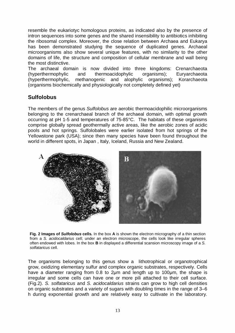
resemble the eukariotyc homologous proteins, as indicated also by the presence of intron sequences into some genes and the shared insensibility to antibiotics inhibiting the ribosomal complex. Moreover, the close relation between Archaea and Eukarya has been demonstrated studying the sequence of duplicated genes. Archaeal microorganisms also show several unique features, with no similarity to the other domains of life, the structure and composition of cellular membrane and wall being the most distinctive. The archaeal domain is now divided into three kingdoms: Crenarchaeota (hyperthermophylic and thermoacidophylic organisms); Euryarchaeota (hyperthermophylic, methanogenic and alophylic organisms); Korarchaeota (organisms biochemically and physiologically not completely defined yet) Sulfolobus The members of the genus Sulfolobus are aerobic thermoacidophilic microorganisms belonging to the crenarchaeal branch of the archaeal domain, with optimal growth occurring at pH 1-5 and temperatures of 75-85°C. The habitats of these organisms comprise globally spread geothermally active areas, like the aerobic zones of acidic pools and hot springs. Sulfolobales were earlier isolated from hot springs of the Yellowstone park (USA); since then many species have been found throughout the world in different spots, in Japan , Italy, Iceland, Russia and New Zealand.
The organisms belonging to this genus show a lithotrophical or organotrophical grow, oxidizing elementary sulfur and complex organic substrates, respectively. Cells have a diameter ranging from 0.8 to 2μm and length up to 100μm, the shape is irregular and some cells can have one or more pili attached to their cell surface. (Fig.2). S. solfataricus and S. acidocaldarius strains can grow to high cell densities on organic substrates and a variety of sugars with doubling times in the range of 3–6 h during exponential growth and are relatively easy to cultivate in the laboratory.
Fig. 2 Images of Sulfolobus cells. In the box A is shown the electron micrography of a thin section from a S. acidocaldarius cell; under an electron microscope, the cells look like irregular spheres often endowed with lobes. In the box B in displayed a differential scansion microscopy image of a S. solfataricus cell.
14
Moreover, the complete genome sequence is available for both strains (S. solfataricus P2 genome was one of first crenarchaeal genome to be completely sequenced). Consequently, S. solfataricus and S. acidocaldarius have developed into important model organisms for the study of Crenarchaeota. Genetic elements The crenarchaeote Sulfolobus spp. is a host for a large spectrum of genetic elements. Some viruses, plasmids and insertion sequences have been found, and the elements often show a large extent of novelty with respect to morphology and genome content. These genetic elements has been extensively studied over the last few years, with the aim to develop shuttle vectors for Sulfolobus strains. Genetic methods for studying these organisms are not as advanced as for the halophilic and mathanogenic Euryarchaeota, and shuttle vectors are also important tools to (over)express homologous or heterologous proteins of interest. The proteins can be expressed and characterized in native host, be it for reasons of post-translational modifications or the determination of the localization of the protein or because the respective protein is difficult to express in functional form in mesophilic expression systems. Shuttle vectors are not only crucial for basic research applications but have also potential for exploitation in biotechnological applications, e.g., for the (over)production of heat stable enzymes. Transposable elements Transposable elements (TEs) are DNA sequences capable of moving into genome, and cause dramatic changes in genes and genomes via molecular events that do not depend on the proximity or similarity of the DNA sequences affected. These events include inactivation of functional genes by insertion, activation of cryptic genes by positioning of a promoter 5' to the coding region, deletion or inversion of DNA adjacent to the TE, and stable incorporation of DNA transferred from outside the lineage (Kleckner N. et al., 1975; Ciampi M.S. et al., 1982; Prentki P. et al., 1986). In addition to these TE-promoted changes, homologous recombination between dispersed copies of a TE can rearrange large genomic segments (Haack K.R. and Roth J.R., 1995). These properties make TEs, which occur in nearly all organisms, a major source of genomic plasticity. The mobility of TEs is due to the presence of repeats at the ends of sequence, and the elements need a transposase, normally encoded by the element itself, to effect their mobility. Insertion sequences (ISs) are the smallest TEs capable of independent transposition. A typical IS consists of a transposase-encoding gene flanked by inverted repeats (IRs), which provide the recognition and cleavage sites for the transposase (Mahillon J. and Chandler M., 1998). In situ, an IS is usually bounded by short direct repeats (DRs) that represent target-site duplications (TSDs) resulting from transposition. TEs were found in several bacterial, eukaryotic and archaeal genomes (Jurka J. 1998; Mahillon J. and Chandler M. 1998). In bacteria, TEs often inactivates genes by insertion, otherwise a TE can confer to the microorganism new useful properties, e.g. carrying a sequence for an antibiotic resistance or a degradative methabolic pathway. The presence of TEs is also relevant in hyperthermophilic archaeal genomes. Each of the sequenced S. solfataricus and S. tokodaii genomes contains large numbers of putatively mobile elements, both IS elements (insertion sequence elements) and MITEs (miniature inverted-repeat transposable elements). There are 344 IS elements
15
in the 3.0 Mb genome of S. solfataricus P2 and 95 in the 2.7 Mb genome of S.tokodaii (Brügger K. et al. 2004). In the former they constitute more than 10% of the genome and the elements are mainly clustered in two broad areas (replicore 1 and replicore 2) separated by the putative oriC and terC (replication origin and termination) regions (Brügger K. et al. 2002); these sequences, fundamental for replication, should act as barriers for the mobility of transposable elements. Furthermore, experimental data suggest that transposition of IS elements occurs frequently. The ISC 1058, 1217, 1359 and 1439 have shown to be active and able to spread and interrupt a functional gene (Martusewitsch E. et al. 2000) by spontaneous excision-insertion or copying-insertion events; it has been proposed that they are probably mobilized by integrase and excisionase activities, often encoded by the same transposase open reading frame (ORF) (Brügger K. et al. 2002, Redder P. et al. 2001, Rezsöhazy R. et al. 1993) located on the IS elements themselves. Moreover the presence of repeats (identical copies) of some elements suggests a recent duplication. The large number of TEs gives a remarkable plasticity to genome, and this could be a key process in the adaptation strategy of hyperthermophlic organisms to extreme habitats. In this regard, lacS- mutants of S. solfataricus defective in β-glycosidase activity were isolated (Bartolucci S. et al. 2003), in which mutations were caused by an insertion of transposable elements into lacS gene. One of the mutants, named GθW, showed a stable phenotype with no reversion; analysis of its chromosome revealed the total absence of the β-glycosidase gene (lacS), with an extended deletion of 13 kb including the whole lacS sequence, xylS (α-xylosidase) and lacTr (lactose transporter) genes. This chromosomal rearrangement was a nonconservative transposition event driven by the mobile insertion sequence element ISC1058, belonging to IS5 family (Mahillon, J. and Chandler M. 1998) and found in 14 complete copies in S. solfataricus P2 genome. The isolation of stable complementable mutations from S. solfataricus, as lacS deletion found in GθW, opens new possibilities for the development of genetic tools. In this work, we have used this strain in complementation experiments in the aim of characterize the shuttle vector pMSSV (see above). Moreover, GθW mutant is faster growing in rich medium than the wild-type, with a doubling time of 4 versus 6 h; this is an interesting feature in the perspective of use the mutant in biotechnological application, e.g. expression of homologous/heterologous proteins. Virus Viruses are the largest reservoir of genetic material on the planet; in particular microbial viruses are extremely abundant on our planet; they have important and diverse roles in ecosystems and biogeochemical processes. Archaea and their viruses are poorly understood when compared with the Eukarya and Bacteria domains of life, so over the last few years efforts focused on the characterization and comparison of archaeal viruses are constantly increasing. All archaeal viruses isolated up to now have linear or circular, double-stranded (ds) DNA genomes. In common with their hosts, they show adaptation to extreme environments. Thus, viruses of extreme halophiles are stable only in solutions of high salt concentration (3–5 M) and are inactivated in solutions of low ionic strength (Witte A. et al. 1997). Similarly, viruses of hyperthermophiles are stable at extremely high temperatures (Prangishvili D. et al. 1999 ; Schleper C. et al. 1992). From a morphological approach, DNA viruses of the Archaea have highly diverse and often exceptionally complex morphotypes. Many have been isolated from

16
geothermally heated hot environments, contradicting the widespread notion of limited biodiversity in extreme environments. Archaeal viruses exhibit a range of virion morphotypes, most of which have not been observed before for any dsDNA virus (Prangishvili D. et al. 2006a,); these viruses, particulary from hyperthermophilic Crenarchaeota, show exceptional forms, including fusiform, droplet and bottle shapes, as well as linear and spherical virions, and also more complex virions combining features of these different forms (Haring M. et al. 2005; Rice G. et al. 2001) (Fig.3). Moreover, genome-sequence analyses have demonstrated that most of the archaeal viruses are unrelated to other known viruses and suggest that they might have different, and possibly multiple, evolutionary origins (Prangishvili D. et al. 2006b). Most of the viruses isolated thus far infect hyperthermophilic hosts belonging to the Sulfolobales family. The viruses found in Sulfolobus are arranged to four families: Fuselloviridae (SSV1, SSV2, SSV RH, SSV K1 and the recently isolated SSV4), Rudiviridae (SIRV1 and SIRV2), Lipothrixviridae (SIFV) and Guttaviridae (SNDV); furthermore an icosahedral virus from the Yellowstone National Park has been recently discovered, which could not be assigned to a virus family (Rice G. et al. 2004; Lipps G. 2006). However, the relatively largest amount of data provides information available about the Fuselloviridae family; the viruses belonging to this group show an inusual lemon
Fig. 3 Scansion electron microscopy images of Sulfolobus virus Viral particles of SIRV2, belonging to Rudiviridae family, are shown in the box A; in B is shown the SIFV virus (Lipothrixviridae family); both viruses were isolated from S. islandicus. Box C: Acidianus bottle-shaped virus (ABV).
17
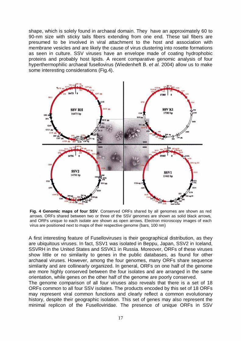
shape, which is solely found in archaeal domain. They have an approximately 60 to 90-nm size with sticky tails fibers extending from one end. These tail fibers are presumed to be involved in viral attachment to the host and association with membrane vesicles and are likely the cause of virus clustering into rosette formations as seen in culture. SSV viruses have an envelope made of coating hydrophobic proteins and probably host lipids. A recent comparative genomic analysis of four hyperthermophilic archaeal fusellovirus (Wiedenheft B. et al. 2004) allow us to make some interesting considerations (Fig.4). A first interesting feature of Fuselloviruses is their geographical distribution, as they are ubiquitous viruses. In fact, SSV1 was isolated in Beppu, Japan, SSV2 in Iceland, SSVRH in the United States and SSVK1 in Russia. Moreover, ORFs of these viruses show little or no similarity to genes in the public databases, as found for other archaeal viruses. However, among the four genomes, many ORFs share sequence similarity and are collinearly organized. In general, ORFs on one half of the genome are more highly conserved between the four isolates and are arranged in the same orientation, while genes on the other half of the genome are poorly conserved. The genome comparison of all four viruses also reveals that there is a set of 18 ORFs common to all four SSV isolates. The products encoded by this set of 18 ORFs may represent viral common functions and clearly reflect a common evolutionary history, despite their geographic isolation. This set of genes may also represent the minimal replicon of the Fuselloviridae. The presence of unique ORFs in SSV
Fig. 4 Genomic maps of four SSV. Conserved ORFs shared by all genomes are shown as red arrows. ORFs shared between two or three of the SSV genomes are shown as solid black arrows, and ORFs unique to each isolate are shown as open arrows. Electron microscopy images of each virus are positioned next to maps of their respective genome (bars, 100 nm)
18
genomes are likely the consequence of their individual evolutionary history, geographic isolation, factors required for replication in their specific hosts, or adaptation to unique features of their respective thermal environments. SSV1 is the first high-temperature virus to be characterized in detail and is the best-studied virus of the genus Sulfolobus. This Fusellovirus was isolated from the strain B12 of Sulfolobus shibatae in Beppu, Japan, (Grogan D. et al. 1990; Martin A. et al. 1984; Palm P. et al. 1991; Yeats S. 1982) but, like SSV viruses of different sources, it is also capable to infect S. solfataricus cells (Schleper, C. et al. 1992). In both hosts, virus production is UV inducible (Martin A. et al. 1984), and viral genome is packaged into viral particles in a positively supercoiled form, while the virus episomal DNA can exist in positively supercoiled, negatively supercoiled, or relaxed double-stranded DNA form inside the host cells (Nadal, M. et al. 1986). Infection and production of virions causes only a significant growth retardation of the host cells which can be visualized as turbid plaques around propagation foci on plated lawns of indicator host cells (Schleper, C. et al. 1992). SSV1 can integrate into the host arginyl tRNA gene using a tyrosine recombinase family integrase, in a similar way of other SSVs (SSV2 integrates into the glicyl tRNA gene, SSV RH and K1 can integrate into different tRNA genes; in addition SSVK1 can integrate into a non-tRNA spot of S. solfataricus genome, and this is the first example in SSV viruses). A 7.4-kbp segment inserted into an S. solfataricus arginyl tRNA gene shares extensive sequence similarity with a portion of the SSV1 genome and is likely a remnant of viral integration. The SSV1 genome (15465 bp) has 34 open reading frames (ORFs), which are tightly arranged in the genome (Palm P. et al. 1991); nine transcripts cover all 34 SSV1 ORFs (Reiter, W.D. et al. 1987), and this suggests that the viral genes are translated via a polycistronic strategy. However, only four genes of SSV1 could be assigned a function up to now; one of this ORFs encodes a type I tyrosine recombinase, which catalyses the site-specific integration (Muskhelishvili, G.P. et al. 1993), while the other three ORFs encode structural proteins, VP1, VP2, and VP3, which were assigned by sequencing of proteins from purified virus particles (Reiter W.D. et al. 1987). VP2 have no homologous proteins in other SSV viruses, unlike VP1 and VP3, even if the protein is known to play a key role in the virus infection mechanism, being involved in the packaging of genome into the viral particles. The fusellovirus SSV2, isolated from S. islandicus REY 15/4, shares with SSV1 similar morphology, replication and DNA size (Stedman K.M. et al. 2003). The overall genome architecture is conserved but the low similarity in some regions of the sequences should be responsible for the higher copy number and the lack of a strong ultraviolet induction of episomal SSV2 DNA and particle production, as well as the different integration of the SSV2 genome which occurs into the host chromosome at the site of a glycyl tRNA (Wiedenheft B. et al. 2004). Recently a new fusellovirus, named SSV4, has been isolated (Peng X. 2008); this virus shows a similar genomic composition and organization of others SSVs, and integrates into a Glu t-RNA gene. Plasmids Two types of plasmids have been isolated for the genus Sulfolobus: cryptic plasmids, with genome sizes of 5–14 kb and conjugative plasmids, with genomes larger than 25 kb (Lipps G. 2006). The first conjugative plasmid discovered was the plasmid pNOB8 which was isolated from a Japanese strain of Sulfolobus (Schleper et al. 1995). The plasmid can propagate in liquid cultures through cells, that form aggregates of 2 to 30 individuals with intercellular cytoplasmic bridges connecting two or more cells. Conjugation is quite efficient and the plasmid is able to spread
19
through the whole culture. The conjugative plasmids are actually classified in three subfamilies: pNOB8, pING (Stedman K.M. et al. 2000) and pSOG2/4 (Erauso G. et al. 2006). These plasmids are smaller than PNOB8 (they are from 25kb to 35kb in length, while pNOB8 is 42kb); the genomes show each other a partial sequence homology, but also regions which are completely different. The plasmids have similar characteristics as the plasmid pNOB8 including high copy number and growth retardation and, excepting pSOG2/4, have shown to be stable upon conjugative transfer, undergoing high genetic variation only upon prolonged growth of the host cells. Comparisons of the genome sequences of the CPs sequenced allow to delineate the essential genes of this plasmid family (Greve B. et al. 2004). The analyses recognized three conserved and functionally distinct domains. The first one is a cluster of genes covering up to half of each genome (~13.5 kb) and contains six conserved ORFs implicated in conjugation; the second is a putative replication origin; finally there is a region including an operon with six to nine short genes some of which are involved in the initiation of plasmid replication, an integrase and the DNA binding protein, PlrA. The other family of Sulfolobus plasmids comprises plasmids known as cryptic, because these elements give no distinguishable features to the hosts. The first cryptic plasmid completely sequenced was the plasmid pRN1, isolated from the S. islandicus strain REN1H1 (Keeling P.J. et al. 1996); this strain also harbours the plasmid pRN2. Detailed studies about replication of these plasmids have pointed out that both pRN1 and pRN2 can replicate and propagate independently, being not essential for the host cells (Purschke W.G. and Shäfer G. 2001). pRN plasmids give the name to a plasmid family composed of elements which share regions of high sequence similarity (Peng X. et al. 2000); pHEN7, a 7.8-Kb plasmid from S. islandicus HEN7H2 (Zillig W. et al.,1998), pDL10 (7.6 Kb) from the chemolithoautotrophic crenarchaeon Acidianus ambivalens, and pSSVx (5.7 kb) from the S. islandicus strain REY15/4 (Arnold H.P. et al., 1999), pIT3 (5.0 kb) from S. solfataricus (Prato S. et al. 2006) belong to this family (Fig.5). In the conserved regions, these plasmids share three ORFs. One of these conserved genes (named orf56 in pRN1) encodes for a sequence-specific double-stranded DNA-binding protein (CopG homologue), which could be involved in the plasmid copy number control. The orf80 genes of the plasmids are the most conserved genes of the plasmid family pRN, and encode for a DNA binding protein which have an unknown physiological function; the orf80 homologues, which are highly conserved also within the conjugative plasmids of Sulfolobus, are partly annotated as plrA, meaning plasmid regulatory gene A (Greve B. et al. 2004). The other conserved sequence is a large orf (2676 bp in pSSVx to 2970 bp in pRN2) which occupy about a third to a half of the plasmids. The proteins encoded by the Orf (904) from pRN1 (Lipps G. et al. 2003) and the Rep245 from pIT3 (Prato S. et al. 2008) have been expressed in E.coli and characterized, and showed to be multifunctional proteins with helicase, primase and DNA polymerase activity. This protein could play a crucial role in plasmid replication; has been suggested that Orf904 alone or in concert with another protein, probably Orf80, recognizes the origin of replication. Subsequently, the helicase domain of Orf904 will melt the replication origin using chemical energy from the hydrolysis of ATP and the prim/pol domain will synthesize a primer (Lipps G. 2006). In this contest Orf 80 could have a structural role, e.g. marking the replication origin, while the CopG homologous protein could limit the intracellular concentration of the replication protein and so down-regulate plasmidal replication initiation.

20
Plasmid-fusellovirus genetic systems pSSVx plasmid was isolated together with SSV2 from S. islandicus strain REY15/4 type (Arnold H.P. et al., 1999). This element has been discovered in a screening procedure to detect extrachromosomal genetic elements of Sulfolobus, especially viruses of the SSV1 type. Arnold and collegues discovered the presence of spindle-shaped, short-tailed virus particles of two distinct sizes analyzing the supernatant of S. islandicus REY15/4 culture by electron microscopy (Fig.6). Both particles resembled those of the Sulfolobus virus SSV1 in shape (Martin A. et al., 1984); the larger particles had the same size as SSV1, 80nm x 55 nm, whereas the smaller particles measured only 60 x 40 nm. Further analysis allow Arnold to assert that the larger particles contained a fusellovirus genome, named SSV2, while the smaller one contained a 5.7kb plasmidic DNA, pSSVx. By sequence similarity, the pSSVx plasmid has been assigned to the pRN family of Sulfolobales plasmids (as previously described), but it shows an unique feature among pRNs, the ability to generate virus particles and spread, also in the cell cultures of S. solfataricus, using the packaging mechanism of SSV2. This genetic element lacks the genes that encode the major structural proteins of the virus, therefore is unable to package and spread without a virus helper. Therefore, pSSVx can be rewarded as a hybrid between a plasmid and a virus. Differently from the members of the pRN family that are incapable to spread
Fig. 6 Maps of the genomes of the plasmids pSSVx, pRN2, pRN1 and pDL10 forming the pRN family. ORFs were named by the number of codons. ‘c’ (complementary) indicates opposite orientation. ORFs in the `minimal replicon' (conserved region) are shown as filled arrows; sequence elements in this region as filled boxes. Unassigned ORFs in the variable region are shown as empty arrows, and SSV homologues are cross-hatched. Homologous ORFs are indicated by the same fill pattern. Consensus motifs located between conserved and variable regions are indicated.

21
SSV2
pSSVx
even in the presence of helper virus, pSSVx contains two open reading frames, named 154 and 288, showing high sequence similarity to a tandem of ORFs in both SSV1 and SSV2 genomes. It has been suggested that the proteins encoded by these ORFs can specifically recognize pSSVx DNA but associate with viral helper components necessary for capsid formation and packaging. The relationship of pSSVx to its helper fusellovirus SSV2 resembles that of the parasitic “defective” bacteriophage P4 to its compulsory helper, bacteriophage P2, especially in the packaging of these elements into helper-virus-like but smaller particles assembled by structural components of the helper. However, pSSVx appears to use the structural genes of SSV2 only for spreading, whereas bacteriophage P4 modifies the lifestyle of its helper. A trascriptional analysis of the pSSVx genetic element was developed by Contursi et al. (2007): a combination of Northern blot, primer extension and RT-PCR experiments, revealed the presence of nine major transcripts whose expression was differentially and temporally regulated over the growth cycle of S. islandicus. Recently two novel fusellovirus-plasmid systems have been discovered. An integrative non-conjugative extrachromosomal genetic element, denoted as pSSVi, has been isolated from a S. solfataricus P2 strain and characterized (Wang Y. et al. 2007). This genetic element is a double-stranded DNA of 5740 bp in size and contains eight ORFs. It resembles members of the pRN plasmid family in the genome organization but shows only weak similarity to the latter in the most conserved regions. pSSVi has a copG gene similar to that of a pRN plasmid, encodes a large replication protein which, unlike a typical pRN RepA, contains no polymerase/primase domain, and lacks the plrA gene. Interestingly, pSSVi encodes an SSV-type integrase which probably catalyzes the integration of its genome into a specific site (an Arg-tRNA gene) in the S. solfataricus P2 genome. Like pSSVx, pSSVi can be packaged into a spindle-like viral particle and spread with the help of SSV1 or SSV2. pSSVi genome is stably integrated into the host chromosome and it is excised in presence of SSV2 virus. In addition, both SSV1 and SSV2 appeared to
Fig. 6 Electron microscopy images of the S. islandicus REY 15/4 colture supernatants. The image shows mixtures of large SSV2 and small pSSVx particles, mostly in rosettes, from cultures of strain REY 15/4.
22
replicate more efficiently in the presence of pSSVi. Given the versatile genetic abilities, pSSVi appears to be well suited for a role in horizontal gene transfer. Moreover, a new fusellovirus, SSV4, and a pRN-like plasmid, pXZ1, were co-isolated from the ARN3/6 strain of S. islandicus (Peng X. 2008). In contrast to the previously characterized virus–plasmid hybrids pSSVx and pSSVi, which can coexist intracellulary with a fusellovirus, pXZ1 is not packaged into viral particles and shows no viral infectivity. The virus and plasmid have a genome of respectively 15135 and 6970 bp, with 33 and 7 ORFs. Three ORFs of pXZ1 encode an atypical RepA, a PlrA and a CopG protein. A fourth ORF exhibits a high nucleotide sequence identity to the SSV4 integrase gene, which suggests that it has been transferred to the plasmid from SSV4. A single point mutation within an otherwise identical 500 bp region of the integrase gene occurs in the viral attachment site (attP), which corresponds to the anticodon region of the targeted tRNA gene in the host chromosome. This point mutation confers on pXZ1 the ability to integrate into the tRNAGlu[CUC] gene, which differs from the integration site of SSV4, tRNAGlu[UUC]. Comparing the features of pSSVi/pSSVx and pXZ1 systems, it could be assumed that packaging or integration are different strategies adopted by plasmid to survive in Sulfolobus cells growing under extreme conditions. When coexisting with a fusellovirus either they exploit the viral integrase, as in case of pXZ1, to integrate into a different chromosomal site or they exploit the viral packaging system and spread as a virus satellite, as found for pSSVx and pSSVi. S. solfataricus-E. coli shuttle vectors Over the last few years, great efforts have been made to develop shuttle vectors for hyperthermophilic Archaea; shuttle vectors are a powerful tool for genetic analysis and manipulation in vivo, and can also be successfully used in biotechnological applications, such as the overproduction of heat stable proteins which are difficult to produce in mesophilic hosts. Despite the importance of these systems, there are not many vectors constructed for these organisms, unlike the various systems set-up for the bacterial domain or for other Archaea (halophiles or methanogens) (Allers T. and Mevarech M. 2005), demonstrated to be hosts for very advanced vector development. This scarcity of available vectors can be a reflection of the difficulties in establishing efficient systems, due to low transformation efficiencies, inefficient selection and/or instability of the vectors in the host. The common criterion to develop shuttle vectors has been based on the suitable modification of extrachromosomal elements of Sulfolobus. The highly efficient self-spreading capabilities of fusellovirus SSV1 has been exploited to construct several elements. The vector pEXSs is based on a part of the genome of the SSV1 cloned into pGEM5Zf(−) and contains a heterologous selectable marker gene coding for a thermostabilized version of the hygromycin phosphotransferase (Hph) from E. coli (Cannio R. et al. 2001). This vector was used to express a thermostable alcohol dehydrogenase from Bacillus stearothermophilus (Contursi P. et al. 2003), and to complement a lacS deletion mutant (S. solfataricus GθW) by expressing the genes lacS and lacTr coding for a β-glycosidase and a lactose transporter (Bartolucci S. et al. 2003). Stedman et al. (1999) has constructed a series of Sulfolobus–E. coli shuttle vectors based on the complete genome of the virus SSV1. The vector pBluescript has been inserted at different sites within the virus genome and constructs have been identified that have been not impaired in replication or infectivity. To one of

23
these constructs the pyrEF genes from S. solfataricus P2 coding for orotatphosphoribosyl transferase and orotidine-5′-monophosphate decarboxylase has been added as selectable marker. The expression of these marker genes allows for the complementation of uracil auxotroph recipients to uracil prototrophy. Additionally the lacS gene under control of the heat shock tf55α-promoter has been cloned into the vector as a phenotypic marker. The resulting shuttle vector pMJ03 (Jonuscheit M. et al. 2003) replicates to high copy numbers in the primary transformation mixture as an episome. After plating and isolation of single transformants this vector has been found to be integrated as a single copy into the chromosomal arginyl-tRNA gene of the recipient S. solfataricus PH1-16 (Martusewitsch E. et al. 2000) as previously observed for the wild type virus (Schleper C. et al. 1992). The pMJ03 vector has been improved by Albers et al. (2006) by the development of preassembly constructs and an inducible promoter. Recently a series of Sulfolobus–Escherichia coli shuttle vectors based on the small multicopy plasmid pRN1 from S.islandicus have been constructed (Berkner S. et al. 2007). The shuttle vectors do not integrate into the genome and do not rearrange; selection in suitable uracil auxotrophs is provided through inclusion of pyrEF genes in the plasmid. The plasmids allow functional overexpression of genes, as has been demonstrated for the β-glycosidase (lacS) gene of S. solfataricus. In this PhD work a S. solfataricus – E. coli shuttle vector, named pMSSV, based on the hybrid genetic element pSSVx was used; it was obtained by a suitable site-specific insertion into pSSVx of the sequences from an E. coli plasmid essential for replication and selection of transformants (Fig. 7). This vector showed the ability to efficiently propagate in both hosts and to replicate at high copy number in a fashion similar to the wild type pSSVx.
Fig. 7 Map of vector pMSSV. The sequences necessary for replication and ampicillin selection in E. coli (indicated as a solid bar) was inserted into pSSVx plasmid, to generate a new E. coli- S. solfataricus shuttle vector, named pMSSV.

24
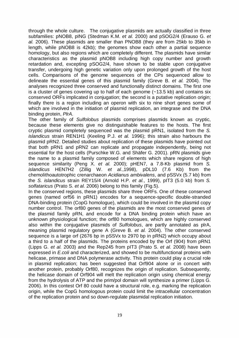
Thermophilic enzymes as biocatalysts in industrial applications The field of industrial enzyme applications has grown tremendously over the last years; enzymes are actually used in several industrial sectors, such as food, textile, detergent and pharmaceutical industry. Enzymes allow to improve the process execution, making the processes economically and environmentally more sustainable than the traditional chemical manufacturing approaches. Moreover, the utilization of enzymes provides the market with better quality products when compared to those obtained by traditional methodologies. Particularly, enzymes from (hyper)thermophilic organisms show a resistance to extreme physical-chemical conditions, prohibitive for their counterparts from mesophilic organisms. These unique features make enzymes from hyperthermophiles very advantageous in most of the industrial processes that requires high temperature regimens, and for which the employment of the thermolabile mesophilic enzymes is limited or even completely unsuccessful. The main advantages of performing processes at high temperatures are the reduced risk of microbial contamination, lower viscosity, improved transfer rates, and improved solubility of substrates/products. Many studies are still currently devoted to a defined understanding of the molecular basis conferring stability to thermozymes. There are no major conformational differences with mesophilic enzymes, and a small number of extra salt bridges, hydrophobic interactions, hydrogen bounds or a decreased length of surface loops seem to contribute to the extra degree of stabilization (Bruins M. et al. 2001). Recently, the increasing numbers of known three-dimensional structures and some studies on mutant proteins have clarified the importance of ion pairs, when organized in large networks. It is believed that these networks at the surface of proteins are a major stabilizing factor for thermostability (Sadeghi M et al. 2006; Ge M. et al. 2008). Stabilizing interactions between domains and subunits also contribute significantly to the intrinsic stability of proteins. Enzymes from (hyper)thermophilic organisms are known to exist in conformational states of higher-order association when compared to their mesophilic analogs, suggesting that the formation of oligomers is one way of increasing thermostability (Jaenicke R. 1998). Although most enzymes from (hyper)thermophilic organisms are intrinsically very stable, some intracellular enzymes obtain their thermostability from intracellular environmental factors. The presence of salts (hypersolutes), high-protein concentrations, coenzymes, substrates, activators, or general stabilizers such as thermamine, sorbitol, or cyclic polyphosphates can also further stabilize the enzyme (Bruins M. et al. 2001). Cellulose Structure and composition Cellulose is the most widespread polysaccharide in nature and it forms the primary structural component of plants. Cellulose is a linear polymer made of repeated units of the D-glucopyranose linked by β-(1→4) bounds. Cellulose occurs naturally in pure form in cotton fibres, or it can be found in association with other biopolymers, like lignin and hemicelluloses (Fig.8).
25
Cellulose was discovered and isolated as the common material of plant cell walls by Anselme Payen in 1838, and it was further characterized by Cross and Bevan in the early 1900s. They removed the related plant materials that occur in combination with cellulose by dissolving them in a concentrated sodium hydroxide solution. They designated the undissolved residue as α-cellulose. The soluble materials (designated as β-cellulose and γ-cellulose) were later shown not to be celluloses, but rather, relatively simple sugars and other carbohydrates. The α-cellulose of Cross and Bevan is what is usually meant when the term “cellulose” is used now. In the cellulose chain, glucopyranoside units are joined by acetal linkages between the C-1 of one pyranose ring and the C-4 of the next ring. The stereochemistry of these acetal linkages is very important for typical structural conformation of cellulose; the C-1 oxygen is in the opposite side of C-6 carbon, (β configuration). This β configuration, with all functional groups in equatorial positions, causes the molecular chain of cellulose to extend in a more-or-less straight line, making it a good fiber-forming polymer.
Fig. 8 Schematic representation of cellulose polymer in the plant cell wall. Glucose monomers are linked by β 1,4 glycosidic bonds; this causes the molecular chain of cellulose to extend in a more-or-less straight line, making it a good fiber-forming polymer. The single chains are organized in microfibrils, linked by interchain hydrogen bounds.
26
Because of the equatorial positions of the hydroxyls on the cellulose chain, they protrude laterally along the extended molecule. This positioning makes them readily available for hydrogen bonding. These hydrogen bonds cause the chains to group together in highly ordered (crystal-like) structures. Since the chains are usually longer than the crystalline regions, they are thought to pass through several different crystalline regions, with areas of disorder in between (the “fringed-micelle” model). The inter-chain hydrogen bonds in the crystalline regions are strong, giving the resultant fibers good strength and insolubility in most solvents. They also prevent cellulose from melting. In the less ordered regions, the chains are further apart and more available for hydrogen bonding to other molecules, such as water. Most cellulose structures can absorb large quantities of water (i.e., it is very hygroscopic). Thus, cellulose swells, but does not dissolve, in water. Enzymatic degradation and biotechnological applications There are three main enzymatic activities involved in cellulose degradation, which can be discerned as follows:
- Endoglucanases, or endo-1,4-β glucanases (EC 3.2.1.4), which randomly hydrolyze the 1,4-β bonds within cellulose chains acting on amorphous regions and producing oligosaccharides of various length.
- Exoglucanases, which include 1,4-β-D-glucan-4-glucohydrolases, also named cellodextrinases (EC 3.2.1.74), and 1,4- β -D-glucan cellobiohydrolases (EC 3.2.1.91); these enzymes act on reducing and nonreducing ends of cellulose chains, releasing respectively glucose (glucohydrolases) or cellobiose (cellobiohydrolases), and can also hydrolyze microcrystalline cellulose (Teeri T. T. 1998).
- β-glucosidases or β-D-glucoside glucohydrolases (EC 3.2.1.21), which catalyse the hydrolysis of soluble cellodextrins (oligosaccharides made of three to seven glucose molecules) and cellobiose to glucose.
These enzymes catalyze the hydrolysis of β -1,4-glycosidic bounds via general acid catalysis that requires two critical residues: a proton donor and a nucleophile/base. This hydrolysis occurs via two major mechanisms giving rise to either an overall retention, or an inversion, of anomeric configuration (Davies G. and Henrissat B.1995; Lynd L.R. et al. 2002) In both the retaining and the inverting mechanisms, the position of the proton donor is identical, in other words it is within hydrogen-bonding distance of the glycosidic oxygen. In retaining enzymes, the nucleophilic catalytic base is in close vicinity of the sugar anomeric carbon. This base, however, is more distant in inverting enzymes which must accommodate a water molecule between the base and the sugar. The enzymes involved in cellulose hydrolysis have large biotechnological potentialities; in fact cellulases are widely utilized in industrial processes, e.g. in paper, textile and detergent industries. In the paper production, the step by step process used to separate cellulose from lignin and other wood components is known as pulping. It is a time and energy consuming process, involving the mechanical processing of wood or the treatment of wood with harsh chemicals. In biopulping, cellulase and xylanase enzymes are used to pre-treat wood and break down the lignin fibres. Removing lignin prior to further wood pulping saves time and energy, and decreases the quantities of chemicals used and water wasted. In textile industry, enzymes are used to treat and modify fibres, particularly during textile processing and in caring for textiles afterwards (desizing, biopolishing and stonewashing processes). Cellulases are also added to detergents to improve the cleaning and
27
softening power. In general, detergents containing enzymes are more environmental friendly than the traditional ones, allowing lower washing temperatures and shorter periods of agitation to be employed. Cellulose is the earth’s most widespread natural organic substance, making it an important biorenewable resource; in particular, cellulose can be the feedstock for the generation of renewable energy. In fact, the glucose obtained from cellulose hydrolysis can be used to produce ethanol by fermentation. The enzyme based approach is advantageous over chemical treatments also in this field. The main improvements are its higher conversion efficiency, the absence of substrate loss following chemical modifications and the use of more moderate and non-corrosive physical-chemical operating conditions, such as lower reaction temperatures, “neutral” pH and the use of biodegradable and non-toxic reagents. The production of bioethanol is particularly beneficial if it is obtained from cellulosic industrial or agricultural wastes (Sjöde A. et al. 2007; Chen Y. et al. 2007; Georgieva T.I. and Ahring B.K. 2007). This new promising approach could overcome the limits of traditional bioethanol production, which is performed using as feedstock agricultural biomasses like corn and sugarcane; these biomasses are specifically and intensively cultivated to produce bioethanol, diverting food resources and driving up food prices across the world. Moreover, intensive exploitation of cellulose as a biorenewable feedstock has to date been prevented by the lack of a suitable solvent that can be used in the chemical processes. Rogers and co-worker have found that solutions of cellulose can now be produced for the first time at useful concentration using a new class of non-aqueous but polar solvents, named ionic liquids or “molten salts” (Swatloski R.P. et al. 2002); this new technology opens up substantial potential for cellulose processing and manufacturing of new cellulose-based materials. Ionic liquids are substances that are completely composed of ions and are liquid at or close to room temperature. Interest in these compounds, often heralded as the green, high-tech media of the future, is still increasing rapidly and stems from their near-zero vapor pressure, their thermal stability, and their widely tunable properties as regards polarity, hydrophobicity, and solvent miscibility behavior through appropriate modification of the cation and the anion (van Rantwijk F. and Sheldon R.A. 2007). Owing to their special properties and possible advantages, ILs may provide an ideal solvent for engineering media for biocatalytic reactions (Kragl U. et al. 2002); an increasingly number of studies are now focused to identify enzymes that are active in ionic liquids and to optimize these new biocatalysis reaction systems (Lee S.H. et al. 2008; Gorke J.T. et al. 2007) endo-1,4-β glucanases The endo-1,4-β glucanases cover 15 of the 113 families in which are actually divided the glycoside hydrolases (GH) on the base of amino acid sequence similarities; since the fold of proteins is better conserved than their sequences, some of the families can be further grouped in superfamilies or 'clans'. These enzymes, as well as the most of other glycoside hydrolases, show a modular structure; generally the proteins have a catalytic domain, which is reasonably large and represent more than 70% of the total protein, and a cellulose binding domain (CBD); these two domains are linked by a short amino acid sequence of varying length (6-59 residues), rich in proline and hydroxyamino acids which are often highly glycosylated (Bhat M.K. and Bhat S. 1997). Enzymes belonging to family 12 differ from this description, lacking the cellulose binding domain.

28
In the S. solfataricus P2 genome there are three ORFs coding for putative extracellular endoglucanases: SSO1354, SSO1949 and SSO2534. These enzymes belong to family 12 of glycoside hydrolases, clan C; SSO2534 (Limauro D. et al. 2001) and SSO1949 have been so far characterized and the sso1949 gene expressed in E. coli (Huang Y. et al. 2005). β-glucosidases As previously described, β-glucosidases are involved in the hydrolysis of cellulose; consequently these are biotechnological relevant enzymes, finding application in several industrial processes. Recently, glycosidases are also used as base for the creation of new enzymatic activities, named glycosynthases, which are mutated versions of glycosidases (Perugino P. et al. 2004; Watts A.G. and Withers S.G. 2004). These enzymes efficiently synthesize oligosaccharides but do not hydrolyse them, and therefore can be used to create new biomolecules which have potential application as therapeutics. Considering their potentiality and application fields, in the last years β-glucosidases have been extensively studied. In particular, the S. solfataricus β-glucosidase (lacS) has undergone to extensive studies; a first crystallographic structure has been obtained (Aguilar C.F. et al. 1997), which has subsequently been integrated by further structural studies. This archeal protein, which is highly thermostable and thermophilic, has also been studied to elucidate the mechanisms allowing proteins to resist to high temperature. LacS belongs to GH family 1; it shows a tetrameric structure with a 240kD total molecular weight and the structure is a (beta/alpha) 8 barrel. The active site occurs at the centre of the top face of the barrel. The lacS protein have a large ion-pair network located at the tetrameric interface of the molecule; circular dicroism, spectroscopy and mutagenic analysis have demonstrated that ion pair electrostatic interactions are fundamental for enzyme resistance and stability to high temperatures (D’Auria S. et al. 1998; Cobucci-Ponzano B. et al. 2002; Ausili A. et al. 2004), giving support to the current theories on the mechanism of ion pair stabilization in proteins from hyperthermophiles. Moreover, lacS has been expressed in Saccharomyces cerevisiae (Moracci M. et al. 1992), in E. coli (Moracci M. et al. 1995) and in mammal cells (Cannio R. et al. 1994); in these experiments the enzyme has been shown to retain the characteristics of thermostability and thermophilicity. The lacS gene is also widely used as marker in archaeal expression vectors, due to the simple method for the revelation of the protein gene expression product: a chromogenic artificial substrate, named X-gal (5-bromo-4-chloro-3-indolyl-b-D-galactopyranoside) is used, which produce a blue coloration after β-glucosidase action. The activity is quantified by a spectrofotometric assay at 405nm using a p-nitrofenil-β-D-glucopyranoside. To efficiently use lacS as marker in vectors, defective cells in β-glucosidase activity can be employed in the transformation and expression experiments, in order to be sure that an eventually revealed activity is due to expression of the marker gene carried from the vector.

29
Concepts, methodology and objectives The archaeal world strongly attracts the interest of scientists for several reasons; the attention is focused both to elucidate some physiological-molecular aspects that are still obscure and to give increasingly larger feasibility to biotechnological application of enzymes from (hyper)thermophilic organisms. The present PhD work is fitted in this dual context; on the one hand findings are reported about the characterization and tuning study of the S. solfataricus expression vector pMSSV, also able to propagate in E. coli cells (previously constructed in the same laboratory where this work has been performed); on the other hand a cellulase S. solfataricus gene was expressed both in xenologous and heterologous fashions, using in the former case the same pMSSV vector. The development of an effective expression vector for archaeal organisms is a key step to study their characteristics in depth. The genome sequencing progress provides a large quantity of DNA sequences and the function of the putative encoded proteins has been assumed by similarity analysis for some of them. In this contest an effective transformation-expression system can be very useful, allowing the gene product to be studied in vivo by homologus expression. Moreover, an expression vector could be a powerful tool to study the expression of heterologous proteins in archaeal hosts. The endolglucanases from hyperthermophilic organisms, as already explained, are biotechnogically relevant enzymes; in particular in S. solfataricus sequence among three cellulose sequences SSO1354 is the only one which remained still uncharacterized and unexpressed; a characterization of the product of sso1354 gene could point out the applicative potentiality of this endoglucanase, allowing to make a comparison with the other characterized S. solfataricus endoglucanases. Therefore this work was developed through different steps:
- Cloning of lacS gene in the pSSVx-derived E. coli-S. solfataricus shuttle vector pMSSV, and characterization of this element.
- Cloning of sso1354 gene in the pMSSV vector and expression of the endoglucanase in S. solfataricus
- Heterologous expression of sso1354 gene in mesophilic hosts and characterization of the enzyme
30
Materials and Methods Strains, enzymes and reagents used in this study The reagents used for preparation of buffers and growth media of Sulfolobus solfataricus were supplied by Sigma Aldrich; the yeast extract and casaminoacids were supplied from Becton-Dickinson (BD). The reagents for polyacrylamide gels electrophoresis were supplied by Biorad. The restriction enzymes, the “modification enzymes” (alkaline phosphatase, T4 DNA ligase, T4 DNA polymerase) and the molecular weight markers for nucleic acids were supplied by New England Biolabs (NEB). The Phusion DNA polymerase was supplied by Finnzymes. The synthesis of the oligonucleotides was committed at PRIMM; the radioactive material was supplied by Perkin Elmer. tThe pET 28c vector was supplied by Novagen. Escherichia coli mos-blue strain (endA1 hsdR17(rk12-mk12
+) supE44 thi-1 recA1 gyrA96 relA1 lac[F′ proA+B+ laqIq Z∆M15: Tn10 (TetR)], used for cloning, were supplied by Amersham Pharmacia biotech; E. coli BL21-Codon Plus (DE3) RIL cells (F– ompT hsdS(rB–mB–) dcm+ Tetr gal λ (DE3) endA The [argU ileY leuW Camr ), and E. coli arctic-express (DE3) cells (E. coli B F– ompT hsdS(rB
– mB–) dcm+
Kluyveromyces lactis GG799 competent cells and the pKLAC1 plasmid were supplied by NEB. Acetamide and YCB medium, used for selection of yeast transormants, were supplied by Becton-Dickinson (BD).
Tet’ gal λ(DE3) endA Hte [cpn10 cpn60 Gent’], used for overexpression, were supplied by Stratagene.
S. islandicus REY15/4 and the mutant derivative strains reported below were kindly provided by Dr. P. Contursi and Dr. Q. She at the University of Copenhagen. Pyr-
mutants of two strains of S. islandicus, cured for pSSVx (REY31A) or for both SSV2 and pSSVx (REY 15A), was used as hosts for transformations with the modified pMSSV containing the pyrEF genes. S. islandicus REY31A is a pSSVx-cured strain isolated from REY15/4. S. solfataricus P2 (DSM1617) was purchased from the German Collections of Microorganisms and Cell Cultures. The strain S. solfataricus GθW, a spontaneous derivative mutant of Gθ strain lacking the β-galactodidase activity, was previously isolated in our laboratory (Bartolucci S. et al., 2003).
E. coli transformation techniques Transformation of E. coli mos-blue cells by electroporation 1µl of ligase solutions was added to aliquots of 40 µl of electrocompetent cells (previously preparated using the procedure described as “Transformation of E. coli by electroporation” in Sambrook J. and Russel D.W. 2001). The cells were incubated with the DNA for 1minute in ice, and then transferred into precooled electroporation couvettes (0.2 cm). The electroporator Gene PulserTM
(Bio-Rad) was used, setting the following parameters: 25 μF, 2,5 kV, 200 Ohm. The transformed cells were immediately diluted with 1ml of LB, then transferred into a new 1.5 ml tube and incubated for 1h at 37° degrees under agitation.
31
Transformation of E. coli BL 21-Codon Plus (DE3) and arctic-express (DE3) cells Aliquots of 200 μl of competent cells (previously preparated using the procedure described as “Preparation and transformation of competent E. coli using calcium chloride” in Sambrook J. and Russel D.W. 2001) were mixed with 50 ng of plasmidic DNA and stored on ice for ~20 min. The cells mixtures were transferred at 42 °C for 90 sec, on ice for two min and then diluted to 1 ml with LB medium. An incubation for ~1 h at 37°C under shaking was performed before plating the cells onto selective solid medium supplemented with 50 μg/ml kanamicin and 33 μg/ml chloramphenicol. Proteins analyses Determination of the protein concentration The concentration of the proteins in solution was determined according to the Bradford’ s method (Bradford, 1976). The Coomassie Brilliant (Bio-Rad) reagent was added to the samples and the absorbance at 595 nm was monitored. A solution of 1 μg/μl of bovine serum albumin (BSA) was used as standard. Electrophoretic analysis of proteins (SDS-PAGE) The electrophoresis on polyacrylamide gel in the presence of SDS (SDS-PAGE) was performed according Laemmli’s protocol (Laemmli, 1970). The samples were denaturized at 100°C for 20 min in 2% SDS, 5% β-mercaptoethanol, 0.001% bromophenol blue and 10 % glycerol. The samples were then loaded on a 10% polyacrylamide gel and electrophoresed in 0.025 M Tris-HCl, 0.2M glycine pH 8.3 and 0.1% SDS. The electrophoresis was performed at 25 mA for ~1.5 hours; the proteins were then revealed by Coomassie Briliant-Blue; the gel was submerged in the staining solution (0.25% Coomassie Brilliant-Blue G250, 50% methilic alcohol and 10% acetic acid) for ~2 min with gentle agitation. The gel was washed in a solution containing 5% methilic alcohol and 7.5% acetic acid to remove the excess of Coomassie and then stored in 10% acetic acid. Alternatively the proteins were transferred by elettro-blotting from gel to a polivinildienefluoride (PVDF) membrane to sequence the N-terminal extremity of recombinant proteins (see below). Protein Electroblotting After electrophoresis on polyacrylamide gel in the presence of SDS, the proteins were transferred from gel to a polivinildienefluoride (PVDF) membrane by the following electro-blotting protocol. The PVDF membrane was submerged in methilic alcohol for 10 sec before using, to eliminate the hydrophobic properties, washed and equilibrated in transferring buffer (CAPS) for 5 min. The electro-blotting was performed for 2h (4°C) at 50 Volts using a Mini Trans Blot cell (BIORAD). Next, the membrane was washed with H2O and submerged for few seconds in methilic alcohol. The proteins were then revealed by Coomassie Briliant-Blue: the membrane was submerged in the staining solution (0.25% Coomassie Brilliant-Blue G250, 50% methilic alcohol and 10% acetic acid) for 5 min. The membrane was washed in 50% methilic alcohol to remove the excess of Coomassie and then washed in H2O. The
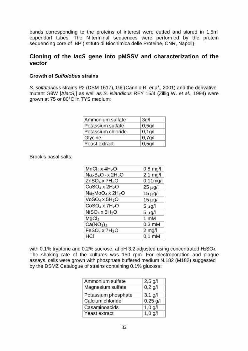
32
bands corresponding to the proteins of interest were cutted and stored in 1.5ml eppendorf tubes. The N-terminal sequences were performed by the protein sequencing core of IBP (Istituto di Biochimica delle Proteine, CNR, Napoli). Cloning of the lacS gene into pMSSV and characterization of the vector Growth of Sulfolobus strains S. solfataricus strains P2 (DSM 1617), Gθ (Cannio R. et al., 2001) and the derivative mutant GθW [ΔlacS,] as well as S. islandicus REY 15/4 (Zillig W. et al., 1994) were grown at 75 or 80°C in TYS medium: Brock’s basal salts: with 0.1% tryptone and 0.2% sucrose, at pH 3.2 adjusted using concentrated H2SO4. The shaking rate of the cultures was 150 rpm. For electroporation and plaque assays, cells were grown with phosphate buffered medium N.182 (M182) suggested by the DSMZ Catalogue of strains containing 0.1% glucose:
Ammonium sulfate 3g/l Potassium sulfate 0,5g/l Potassium chloride 0,1g/l Glycine 0,7g/l Yeast extract 0,5g/l
MnCl2 x 4H2 0,8 mg/l O Na2B4O7 x 2H2 2,1 mg/l O ZnSO4 x 7H2 0,11mg/l O CuSO4 x 2H2 25 µg/l O Na2MoO4 x 2H2 15 µg/l O VoSO4 x 5H2 15 µg/l O CoSO4 x 7H2 5 µg/l O NiSO4 x 6H2 5 µg/l O MgCl 1 mM 2 Ca(NO3) 0,3 mM 2 FeSO4 x 7H2 2 mg/l O HCl 0,1 mM
Ammonium sulfate 2,5 g/l Magnesium sulfate 0,2 g/l Potassium phosphate 3,1 g/l Calcium chloride 0,25 g/l Casaminoacids 1,0 g/l Yeast extract 1,0 g/l

33
Basal salts: The pH of this medium was adjusted at 3,8 with H2SO4
10%. The optical density of liquid cultures was monitored at 600 nm. For solid media, gellan gum (Gelrite, Sigma) was added to a final concentration of 0.8% (0.35% for overlays), and MgCl2 and CaCl2 were added to 10.0 and 3.0 mM, respectively.
Viral DNA isolation and pMSSVlacS plasmid construction Extrachromosomal SSV2 and pSSVx DNAs from both S. islandicus REY 15/4 and transfectants of S. solfataricus strains were performed with Qiaprep Spin Miniprep kit (Qiagen) following the standard procedure suggested by the manufacturer for E. coli cells. An expression cassette of the lacS gene (Cubellis M.V. et al. 1990) was PCR amplified by the vector pMJ03 (Jonuscheit M. et al. 2003) and inserted between the XhoI and PstI sites of pMSSV generating the expression vector pMSSVlacS. Excision of the E. coli minimal plasmid was obtained by digestion of pMSSVlacS (1.0 mg) with SacI and purification of the pSSVx/lacS moiety from agarose gel. The DNA was re-circularized by ligation in a final volume of 5.0 µl, diluted in water to 20 ng/ml final concentration, and 2 µl were used for electroporation. Transformation procedure and analyses of Sulfolobus transformants. S. solfataricus cells were grown up to midlogarithmic phase (0.3–0.45 OD600), harvested by centrifugation and repeatedly washed in 20 mM sucrose as described previously by Schleper et al. (1992). Aliquots of 1010 cells/ml (50 ml) were mixed with 1 ml DNA (10–100 ng/ml), incubated for 1 min on ice and transferred to chilled plastic cuvettes with an electrode gap of 0.1 cm (BioRad). High voltage electroporation (25 µF) was performed with a BioRad Gene Pulser XcellTM
MnCl
at a field strength of 1.5 kV/cm and 400 Ω resistance; two successive shock pulses were applied to competent cells producing pulse length of ~10.0 and 9.8 ms, respectively. Immediately after electroporation cell mixtures were diluted with 1 ml of medium M182 containing 0.1% glucose, transferred to glass vials and incubated for 3 h at 75 or 80°C. After suitable scale-up, 5–15 ml aliquots of the cultures (the volumes varying in order to withdraw the same number of cells per aliquot) were harvested at
2 x 4H2 1,8 mg/l O Na2B4O7 x10H2 4,5 mg/l O
ZnSO4 x 7H2 0,22 mg/l O CuCl2 x 2H2 0,05 mg/l O Na2MoO4 x 2H2 0,03 mg/l O VoSO4 x 2H2 0,03 mg/l O CoSO4 x 7H2 0,01 mg/l O

34
increasing cell density for DNA extractions. For monitoring propagation of the extrachromosomal elements, extrachromosomal DNA mini preparations and plaque assays were performed. Transformants were stored at ~80°C in 15% glycerol stocks. For Southern blot analysis, ~2 μg of total cellular DNAs, extracted according to Arnold et al. (1999), and 5 ng of pMSSVlacS plasmid purified from E. coli, were cut with HindIII and BglII, and electrophoresed in a 0.8% agarose gel; DNA digests were blotted and hybridized according to standard procedures (Sambrook J. and Russel D.W. 2001). The probe was prepared by cutting out and purifying a HindIII restriction fragment from the pMSSVlacS vector encompassing the lacS gene and a portion (up to -302) of the tf55α 5’ flanking region. This restriction DNA fragment was randomly labeled using the random prime DNA labeling kit (Boehringer Mannheim). β-galactosidase complementation and isolation of mixed and single transformants pMSSVlacS transformed cells were tested for β-galactosidase activity; 1 ml aliquots of cultures were centrifuged and cell pellets overlaid with a X-Gal solution (2 mg/ml in phosphate buffered medium) and incubated for 15 min at 75°C for blue color development. In situ assays were performed on the same cultures seeded on plates and grown as circular colonized areas, as previously described (Bartolucci S. et al. 2003). Supernatants of cultures grown up to 0.5, 1.0 and 1.3 OD600
For infection in liquid culture, 400 ml supernatants from pMSSVlacS transformants were added to a 20 ml culture of cells transfected only with SSV2 and grown up to 0.5 OD
were checked for plaque formation as already described and analyzed by X-Gal staining of plaques formed on continuous lawns of the S. solfataricus GθW strain.
600. After incubation under shaking for 48 h, cells were diluted 1:50, grown up to 0.36 and 1.0 OD600Single transformants were selected either on rich (M182, glucose 0.1%) or on minimal (lactose 0.25% as the only nutrient) solid media, by plating or by streaking of electropored cells propagated until they showed positive staining with X-Gal and diluted after they had reached different cell densities (0.3–1.0 OD
and tested for β-galactosidase activity as already described.
600
After in situ X-Gal test on plates, cell spots were transferred to M182 medium containing glucose, propagated up to early stationary phase (1.0 OD
). Single clones formed after 2 weeks of incubation at 75°C were stained with X-Gal (incubation at 75°C for 3–4 h for color development on lactose plates), picked and resuspended in 100 ml lactose minimal medium and seeded as spots onto fresh rich (M182) medium plates.
600
) and analyzed for extrachromosomal DNA content as described above.
β-galactosidase enzyme activity β-galactosidase activity of transformants was visualized and quantified as follows. Crude extracts were prepared by a freeze thaw technique, suspension of the cells in 10 mM Tris–HCl buffer, pH 8.0, placing at 80°C for 10 min, then at 50°C for 5 min. This procedure was repeated four times, and the extract was spun for 30 min at ~10000x g. The supernatant was either assayed immediately or stored at -80°C before testing. Protein concentrations of the crude mcell extracts were determined by the Bradford assay (Bio-Rad). Detection of the β-galacosidase in protein extracts was performed on 10% acrylamide SDS–PAGE gels after electrophoretic separation and extensive
35
washing in 10 mM Tris–HCl buffer, pH 8.0. The specific enzyme band was mnvisualized incubating the gel 30 min at 75°C in the same buffer containing 2 mg/ml X-gal. The β-galactosidase assay procedure followed essentially the protocol of Pisani et al. (1990). A sample of 10 μl extract was transferred to a preheated (75°C) quartz cuvette containing 990 μl assay buffer (2.8 mM ONPG in 50 mM sodium phosphate buffer, pH 6.5). The ONPG hydrolysis reaction was followed spectrophotometrically at 75°C by measuring the increase in absorbance at 405 nm in a Beckman spectrophotometer with heatable cuvettes. One unit was defined as the amount of enzyme catalyzing the hydrolysis of 1 mmol of ONPG min-1 at 75°C with a molar absorption coefficient of 3100 M-1 cm-1
at 405 nm for ONP.
SSV plaque assay The virus titre of a culture was determined by means of plaque assay using S. solfataricus P2 as an indicator strain according to Schleper et al. (1992). The plates were incubated for 2–3 days at 80°C and examined for the appearance of the turbid halos which the inhibition of host growth by SSV2 virus generates. sso1354 gene expression in S. solfataricus Plasmid constructions mos-blue cells were used for all clonings in E. coli. The sequences of sso1354 gene and araS promoter were amplified by PCR using the primers 1354-Bsph (TAAAGTAGGATCATGAATAAATTATATATTG), 1354-His (CGTCGACTTAATGGTG ATGATGGTGATGCTCGAGGAGAGTTTCAGA); araS-Fw(TAAAACCCCATCTCGAG AAATAATGAGGAG), araS-Rv (CTACGCCTAGCCATGGTCTCGGGTACTTTTATG). The primers were designed to insert a BsphI restriction site (bold letters) at the start of SSO1354 coding sequence and a sequence coding for a 6x(His) tag at 3’ end; upstream and downstream of the His tag sequence a XhoI and SalI restriction sites (bold letters) were inserted, respectively. araS primers were designed to insert a XhoI and NcoI sites (bold letters) at the 5’ and 3’ ends. The S. solfataricus P2 genomic DNA was used as the template for amplifications. After amplifications, the fragments sso1354 and araS were purified (with the Stratagene DNA purification kit) and digested with the enzyme mixtures BsphI/SalI and NcoI/XhoI, respectively. The restricted fragments were ligated with the pGEM T-Easy vector, previously digested with Xho I and Sal I. After isolation and partial sequence of the correct construct, the expression cassette araS1354 was excised from the pGEM-araS1354 with ApaI and NsiI restriction enzymes, and ligated with the 5’ dephosphorylated pMSSV shuttle vector previously made compatible by digestion with ApaI and PstI. A modified version of pMSSV, containing the pyrEF genes was also constructed. As a first step, an expression cassette containing the glcS promoter and the SSO1354 coding sequence was constructed. The glcS sequence was amplified using the primers glcS-Fw (CCCAATAACTACTCGAGTTACTGACAACTC) and glcS-Rv (CTTCCTTTT CATGACAATTTATGGTAACC). The sso1354 gene was amplified using the pGEM araS-1354 vector as the template in order to give a 5’ blunt extremity; the primers were 1354-Fw (AATAAATTATATATTGTGCTTCCGG) and sp6 (ATTTAGGTGACACTATAG). The glcS amplification primers were designed to insert a XhoI site (bold letters) at the 5’ end and to amplify the promoter sequence plus the

36
first three codons with the 3’ extremity blunt. S. solfataricus P2 genome was used as the template for amplification of the glcS sequence. After amplifications, the fragments were purified (with the Stratagene DNA purification kit) and restricted with the following enzymes: sso1354, NsiI; glcS, XhoI; The vector pGEM T-Easy was digested with XhoI and NsiI. These three fragments were ligated and the correct vector pGEM T-Easy glcS-1354 obtained. The expression cassette was excised from this plasmid with SalI enzyme and ligated with pyrEF amplified sequence also digested with SalI. The pyrEF sequence had been amplified from the S. solfataricus P2 genome with the primers pyr-Fw TTTTGCTATCGAA GTCGACTCCTCGGATGCAAAT and pyr-Rv CACACTTCTACCCTTGTCGACTAAA TCGTTTTCG so that the SalI restriction site (bold letters) was inserted at both ends. The desired pGEM T-Easy vector containing the glcS-1354 cassette and the pyrEF genes was selected. The ApaI/NsiI fragment comprising the expression cassette and the pyrEF genes was ligated with the pMSSV shuttle vector made compatible with ApaI and PstI and dephosphorylated. Expression and purification of sso1354 gene S. solfataricus GθW cells were transformed by electroporation with 50ng of pMSSVaraS1354 vector (following the procedure described above). Transformed cells were scaled up in 182 medium up to 50ml. The cells were pelleted and resuspended in an equal volume of Brock’s basal salts medium supplemented with 0.25% arabinose. After a scale-up to 500ml, the cells were grown up to 1.2 OD600
and harvested by centrifugation. The proteins of the supernatant were precipitated with ammonium sulphate (90% saturation); the pellet was resuspended in Tris-HCl buffer (25mM, pH 7.5), and the protein sample extensively dialyzed against the same buffer. After dialysis, the sample was loaded onto a resource Q anion exchange column (Amersham). After eluition (the SSO1354 protein was eluted at 500 mM NaCl), the recombinant enzyme carrying the 6x(His) tag was purified by nickel affinity chromatography (HIS-Select Spin Columns, Sigma).The active fractions (around 250 mM imidazole concentration) were pooled and dialyzed against Tris-HCl buffer pH 7.5.
CsCl gradient analysis The supernatant from 500-ml coltures of S. solfataricus GθW cells transformed with pMSSVaraS1354 vector was filtered through a 0.22-µm Seritop filter (Millipore). The filtrate was concentrated by ultrafiltration through a membrane (Amicon, Millipore) with a cut-off of 30 kDa, until the retained volume was ~15 ml. Further purification on a CsCl gradient (0.45 g/ml) by centrifugation for 48 h (SW60-Ti rotor, 250,000x g, 4°C, Beckman Optima LE-80K ultracentrifuge) was performed, and the opaque bands obtained were isolated and dialyzed extensively against 5 mM Tris-HCl buffer (pH 7) at 4°C. Membrane preparation Wet cells from 500 ml Brock’s basal medium supplemented with 0.25% arabinose, harvested in the stationary phase, were suspended in 10 ml 50 mM Tris-HCl pH 7.0, and ground in a mortar with sand for 1 h. After centrifugation at 2,000x g for 10 min in order to remove sand and unbroken cells, the supernatant was ultracentrifuged at

37
55,000x g for 30 min. The clear crude extract was stored at 4°C, while the pellet, containing membrane fragments, was suspended in 25 ml 50 mM Tris-HCl pH 7.0 and ultra-centrifuged again. The clear pellet was resuspended in 10 ml of the same buffer containing 0.5% Triton X-100 and incubated overnight at 70°C. After incubation, the suspension was ultracentrifuged as described above. The pellet was discarded and the supernatant was extensively dialyzed against 25 mM Tris-HCl pH 7.0. sso1354 gene expression in E. coli Plasmid constructions The whole SSO1354 coding sequence plus a sequence coding for a C-terminal 6x(His) tag was amplified by PCR with the primers 1354-Bsph and 1354-His (see above in “Expression of sso1354 gene in S. solfataricus” pharagraph). S. solfataricus P2 genome was used as the template for amplification. After amplification, the fragment was purified (with the Stratagene DNA purification kit), digested with the BsphI and SalI enzymes and cloned into pET-28c (previously made compatible with the NcoI and SalI enzymes). The DNA sequence to obtain a N-terminally truncated SSO1354 version, namely a protein lacking the hydrophobic region (amino acids 1-27) was cloned as follows: a PCR fragment obtained with the forward primer 1354-NheI GATGCTAGCCAGTCTCT
CAGCGTTAAACCCGTAACTA and the reverse primer 1354-HindIII GGTCTTAGAAGCTTATATTGTTTAGAGGAGAG was cut with NheI and HindIII and cloned into the vector pET-28c (the restriction sites are indicated as bold letters and the three nucleotides corresponding to the codon 28 of SSO1354 coding sequence are underlined).
Expression and purification of sso1354 gene The expression plasmids were used to transform E. coli BL21 (codon plus) cells and E. coli arctic-express cells. For expression, cells were grown overnight in 10 ml of LB medium with 50 μg/ml kanamycin and 50 μg/ml chloramphenicol at 37°C. After scale up to 1-litre colture, the growth was continued up to an A600
of about 0.6-0.8 and IPTG was added to a final concentration of 0.4 mM. The BL21 cells were incubated for further 16 h at 22°C, while the arctic-express cells were grown for 40h at 10°C. Cells were harvested by centrifugation, resuspended in 50 ml of buffer [100 mM Tris/HCl, pH 7.5, 300 mM NaCl, 2 mM 2-mercaptoethanol, 0.7 mM PMSF, 5% (v/v) glycerol], and disrupted by sonication for 5 cycles of 1min each (B. Braun sonicator). The recombinant cellulase versions carrying the 6x(His) tag were then purified by Nickel affinity chromatography. Both intact and N-terminus truncated SSO1354 enzymes were eluted at 250 mM imidazole concentration from a HIS-Select spin column (Sigma) and dialysed against Tris-HCl buffer pH 7.5.

38
sso1354 gene expression in K. lactis Plasmid constructions The SSO1354 coding sequence starting from the 28° codon was amplified by PCR with the primers 1354-Fw-Yeast (GATCTCGAGAAAAGACAG
TCTCTCAGCGTT AAACCCGTAACTACA) and 1354-Rv-Yeast (GGTCTTAGATCTTATATTGT TTAGAGGAGAG); these primers were designed to amplify the coding sequence from the 28° codon (underlined), and to insert XhoI and BglII restriction sites (bold letters) respectively upstream and downstream of the SSO1354 sequence. The coding sequence for the recognition site of the yeast Kex endoprotease (in italics letters, needed for the correct secretion of the recombinant proteins) was also added, immediately upstream to the SSO1354 coding sequence. After amplification, the fragment was purified (with the Stratagene DNA purification kit) and digested with the XhoI and BglII enzymes. The adapted sso1354 fragment was ligated with the pKLAC vector, previously digested with the same enzymes.
Expression and purification of sso1354 gene Cells of K. lactis GG799 strain were transformed with pKLAC-1354 vector following the protocol provided by the supplier (New England Biolabs) and also available at the http://www.neb.com/nebecomm/products/productE1000.asp web site. One out of 12 transformed clones was selected after 4-day growth in 10ml YPG medium and cellulase assay of the supernatants. A 10-ml colure of this clone was scaled-up to 1 liter and cells grown up to about 45 OD600
. The cells were centrifuged and the supernatant was 20-fold concentrated times using a stirred ultrafiltration system (Amicon, Millipore) with a YM 10 membrane. The proteins in the concentrated supernatant were precipitated with ammonium sulphate (90% saturation); the recombinant SSO1354 enzyme do not precipitate in ammonium sulphate, so the supernatant of the precipitation was further processed, namely extensively dialyzed against pH 7.5 Tris-HCl buffer (25mM), aliquoted and stored at -20°C.
Enzyme assays and analyses Activity gel The protein preparations was separated by SDS/PAGE in gels containing 0.1% (w/v) CMC (carboxymethylcellulose; Sigma). The protein was then renatured by several washes, once with a mixture (1:4, v/v) of propan-2-ol and 20 mM potassium phosphate buffer (in the pH range 1.8-2.5) or, alternatively, 20 mM phosphate citrate buffer (in the a pH range 3.0-5.0) for 30 min and, subsequently, with the same buffers without propanol for 30 min. Finally, the gel was incubated in 10 mM potassium phosphate or phosphate citrate buffer at 75 ◦C for 1 h with one buffer exchange. All gels were equilibrated with 50 mM potassium phosphate (pH 7) for 30 min, stained with 0.1% (w/v) Congo Red (Sigma,) for 30 min and destained with 1 M NaCl.
39
Enzyme assays For enzyme activity, two assay methodologies were used. In the first assay, the soluble chromogenic substrates (AZO-polymers, polysaccharides dyed with Remazolbrilliant Blue R, Megazime). This assay was used to follow the enzyme activity during purification, using as substrate the AZO-CM-Cellulose and to determine the enzyme specificity towards pachyman and oat-spelt xylan polymers. The activity was measured by adding 170 µl of the specific 1% AZO-polysaccharide solution in 50mM phosphate-citrate buffer to 170µl of enzyme solutions and incubating the reaction mix at 80° C for 5–30 min at pH 5.0; pH was measured and adjusted in the final buffer–substrate mix. The reaction was stopped by addition of 850 µl 96% ethanol to the mixture, followed by incubation at room temperature for 10 min and centrifugation at 1,000xg for 10 min. The absorbance of the supernatant was measured at 590 nm. One unit of enzymatic activity (RBB-unit) was defined as the amount of enzyme required to increase the absorbance at 590 nm of 1 Δ/min under standard conditions. Alternatively, enzyme activity was measured by determining the amount of reducing sugars released from the polysaccharide substrates. This assay was used also to evaluate the enzyme specificity toward debranched arabinan, curdlan and lichenan and to determine the pH and temperature optima. The standard reaction mixture consisted of 50 µl 1% polysaccharidic substrate in 50 mM phosphate citrate buffer, pH 5.0 and 50 µl of enzyme solution (the pH was always measured and adjusted in each final buffer–substrate mix); for the pH dependence, two buffers were used: potassium phosphate buffer in the the pH range 1.8-2.5 and phosphate citrate buffer in the pH range 3.0-7.5. After 10-60 min incubation at 80°C, the reactions were stopped on ice and the amount of reducing sugars released was measured at 520 nm by the Somogyi-Nelson method (Nelson 1944). One unit of enzymatic activity (SNunit) was defined as the micromoles of sugars released per minute per milliliter. Reactions in ionic liquids and analysis of degradation products by TLC Enzymatic reactions were performed in 2 hydrophilic ionic liquids, 1-ethyl-3-methyl-imidazolium-acetate and 1-ethyl-3-methyl-imidazolium-hydrogen sulfate. Solutions of 1 % α-cellulose were used for the assays. The assays were performed at 80°C for various incubation times adding a premixed solution of 30 µl of pure ionic liquid and 20 µl of enzymatic solution to 50 µl of ionic liquid-cellulose. Both solution were prewarmed at the assay temperature before mixing in order to reduce viscosity. The assay was blocked by chilling and cellulose precipitated by adding 100 µl of H2O. After removal of unsoluble cellulose by centrifugation, the hydrolysis products were separated from ionic liquids and protein by three water saturated phenol/chloroform (1:1) and one chloroform extractions. After extraction, a solution of ~150 µl was obtained. Aliquots (60 μl) were spotted on a silica 60 TLC plate (Macherey-Nagel), which was developed in ethyl acetate, H2
O, acetic acid, isopropanol, formic acid, (25:15:10:5:1, by vol.) for ~2 h. Reducing sugars were stained with the α-naphtol reagent after a 5-min incubation at 150°C.

40
Results 1. Characterization of pMSSV shuttle vector: cloning and expression of the lacS gene Cloning of the expression cassette tf55αlacS in the pMSSV vector Beta-D-galactosidase activity of S. solfataricus is displayed by the lacS gene product (Pisani F.M. et al. 1990; Grogan D. et al. 1991) and is responsible for the typical blue stain of the cells when exposed to the chromogenic substrate, X-gal; the lack of gene function in defective mutant strains is hence responsible for colorless colonies (Schleper C. et al. 1994). An expression cassette carrying the lacS coding sequence fused to a 448 bp fragment containing the promoter region of tf55α plus upstream sequences and the first five codons of its ORF was inserted in the polycloning site of the pMSSV vector by a directional cloning (tf55α is an heat shock inducible promoter, in fact tf55α gene encode for the α subunit of the tf55 thermosome, a protein involved in the heat shock response) (Fig.9). Both the cassette and the vector were already available; the vector, as previously described, had been constructed on the base of pSSVx, at which had been added
Fig. 9 Map of pMSSVlacS. An expression cassette containing the lacS gene fused to the thermosome tf55α subunit promoter was inserted into the shuttle vector pMSSV producing the expression vector pMSSVlacS.

41
the sequences essential for replication in E. coli from the plasmid pUC19, while the cassette had been provided by Dr C. Schleper and had been already demonstrated to efficiently complement a beta-galactosidase defect in Sulfolobus when carried by a viral SSV1- derived shuttle vector (Jonuscheit M. et al. 2003; Albers S.V. et al. 2006). The pMSSVlacS plasmid was introduced in E. coli mos-blue cells by electroporation; all the clones analyzed were recombinant, with no rearrangements, and the cells showed a regular growth. pMSSVlacS was subsequently transferred into S. solfataricus GθW cells, already transfected stably with the helper virus SSV2, by electroporation; the GθW strain was chosen because it is a stable beta-galactosidase mutant with an extended deletion in the lacS genetic locus (Bartolucci S. et al. 2003). lacS expression in the β-galactosidase deficient mutant GθW Transformed GθW/SSV2 cells were propagated, and then extrachromosomal DNAs were prepared and analyzed by agarose electrophoresis (Figure 10). The presence and growth-dependent accumulation of the vector in GθW/SSV2 revealed successful transformation and DNA replication; moreover the lacS gene is harmless and does not induce recombination of the vector. Identical results were obtained when SSV2 lysogens of Sulfolobus GθW were transformed with the plasmid pSSVxlacS lacking the E. coli sequences and obtained by cleavage with SacI and re-ligation (Fig. 10).
Fig. 10 Transformation of S. solfataricus with the vectors pMSSV and pMSSVlacS. Competent cells of S.solfataricus GθW infected with the SSV2 virus were electroporated with the vector carrying the β-glycosidase gene pMSSVlacS and also with pSSVxlacS, a pMSSVlacS-derived plasmid obtained by excision of the E.coli-specific sequences and religation. pMSSV and wild-type pSSVx were also transferred into the GθW/SSV2 lysogen for comparison of vector transfer and propagation efficiency in the presence and absence of the lacS gene. Extrachromosomal DNAs extracted from transformed cells after propagation of the cultures were checked by agarose electrophoresis for the presence of pMSSVlacS (lanes 1, 3, 5 and 7) and pMSSV (lanes 2, 4, 6 and 8) at the different growth stages indicated as cell densities (OD600). Similarly, recovery of the pSSVxlacS plasmid (SacI cut, lanes 9 and 10) was monitored in comparison to the parental pSSVx (SmaI cut, lanes 11 and 12). Mobility of the virus SSV2 is highlighted. M1 and M2: molecular weight markers (Kb)
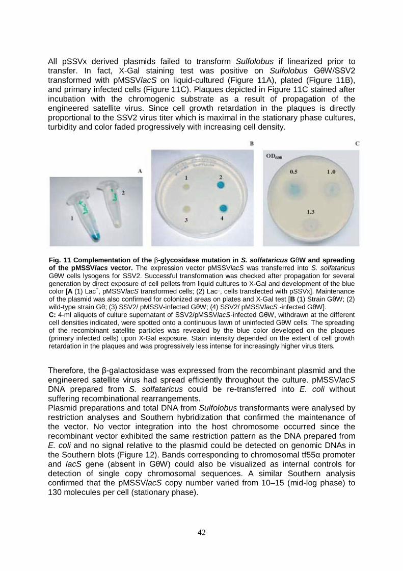
42
All pSSVx derived plasmids failed to transform Sulfolobus if linearized prior to transfer. In fact, X-Gal staining test was positive on Sulfolobus GθW/SSV2 transformed with pMSSVlacS on liquid-cultured (Figure 11A), plated (Figure 11B), and primary infected cells (Figure 11C). Plaques depicted in Figure 11C stained after incubation with the chromogenic substrate as a result of propagation of the engineered satellite virus. Since cell growth retardation in the plaques is directly proportional to the SSV2 virus titer which is maximal in the stationary phase cultures, turbidity and color faded progressively with increasing cell density.
Therefore, the β-galactosidase was expressed from the recombinant plasmid and the engineered satellite virus had spread efficiently throughout the culture. pMSSVlacS DNA prepared from S. solfataricus could be re-transferred into E. coli without suffering recombinational rearrangements. Plasmid preparations and total DNA from Sulfolobus transformants were analysed by restriction analyses and Southern hybridization that confirmed the maintenance of the vector. No vector integration into the host chromosome occurred since the recombinant vector exhibited the same restriction pattern as the DNA prepared from E. coli and no signal relative to the plasmid could be detected on genomic DNAs in the Southern blots (Figure 12). Bands corresponding to chromosomal tf55α promoter and lacS gene (absent in GθW) could also be visualized as internal controls for detection of single copy chromosomal sequences. A similar Southern analysis confirmed that the pMSSVlacS copy number varied from 10–15 (mid-log phase) to 130 molecules per cell (stationary phase).
Fig. 11 Complementation of the β-glycosidase mutation in S. solfataricus GθW and spreading of the pMSSVlacs vector. The expression vector pMSSVlacS was transferred into S. solfataricus GθW cells lysogens for SSV2. Successful transformation was checked after propagation for several generation by direct exposure of cell pellets from liquid cultures to X-Gal and development of the blue color [A (1) Lac+, pMSSVlacS transformed cells; (2) Lac_, cells transfected with pSSVx]. Maintenance of the plasmid was also confirmed for colonized areas on plates and X-Gal test [B (1) Strain GθW; (2) wild-type strain Gθ; (3) SSV2/ pMSSV-infected GθW; (4) SSV2/ pMSSVlacS -infected GθW]. C: 4-ml aliquots of culture supernatant of SSV2/pMSSVlacS-infected GθW, withdrawn at the different cell densities indicated, were spotted onto a continuous lawn of uninfected GθW cells. The spreading of the recombinant satellite particles was revealed by the blue color developed on the plaques (primary infected cells) upon X-Gal exposure. Stain intensity depended on the extent of cell growth retardation in the plaques and was progressively less intense for increasingly higher virus titers.
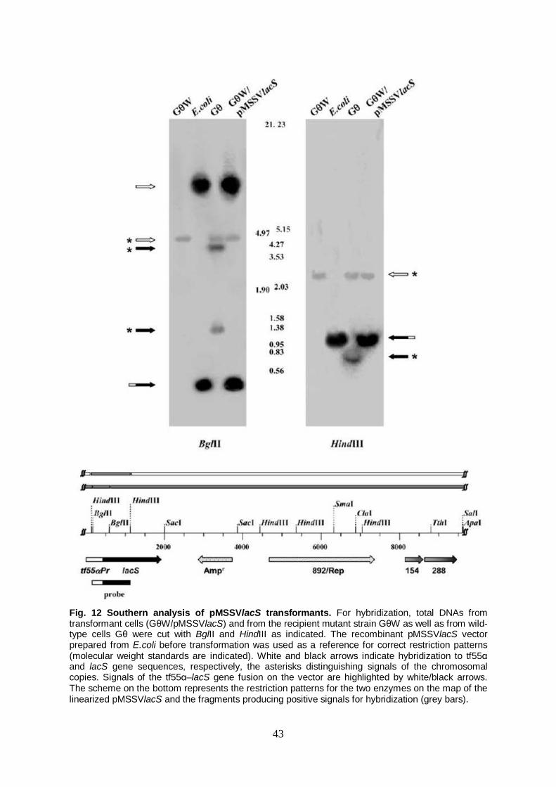
43
Fig. 12 Southern analysis of pMSSVlacS transformants. For hybridization, total DNAs from transformant cells (GθW/pMSSVlacS) and from the recipient mutant strain GθW as well as from wild-type cells Gθ were cut with BglII and HindIII as indicated. The recombinant pMSSVlacS vector prepared from E.coli before transformation was used as a reference for correct restriction patterns (molecular weight standards are indicated). White and black arrows indicate hybridization to tf55α and lacS gene sequences, respectively, the asterisks distinguishing signals of the chromosomal copies. Signals of the tf55α–lacS gene fusion on the vector are highlighted by white/black arrows. The scheme on the bottom represents the restriction patterns for the two enzymes on the map of the linearized pMSSVlacS and the fragments producing positive signals for hybridization (grey bars).

44
β-galactosidase assays on pMSSVlacS transfectants Detection of β-galactosidase activity on denaturing gels by enzyme staining revealed that the lacS gene product was indistinguishable when expressed as heterologous in E. coli and xenologous in S. solfataricus, namely the Sulfolobus expression system did not interfere with the correct polypeptide syntesis (data not shown). β-galactosidase activity was measured using a spectrophotometric assay with the specific substrate ONPG in crude cell extracts of both transfected mutant GθW strain and wild type Gθ. The mutant recipient strain GθW has been shown to exhibit no detectable activity (Bartolucci S. et al. 2003), whereas the specific activities of pMSSVlacS transformants in primary transformation mixtures rose from undetectable levels to 1.2 U/mg protein; this value was ~2-fold higher than that found in wild-type cells expressing lacS under the control of its own promoter. The activity remained stable in diluted and propagated cells cultured as described above, when assays were performed at the same growth phase. Under identical growth conditions, the value of endogenous β-galactosidase activity in the P2 wild-type strain (i.e. the natural source of both the lacS gene and tf55a promoter sequences inserted in the pMSSVlacS), reached 0.1 U/mg as the highest value. Therefore, using this value as reference the expression level in GθW/pMSSVlacS is ~12-fold higher. For a single culture, activity increased up to late logarithmical growth phase and then maintained approximately the same value up to late stationary phase (Figure 13). This result confirmed that the recombinant satellite viral DNA was replicated inside the cells and that virus particle formed and spread throughout the culture; the increasing activity should therefore depend only on the copy number of the plasmid and hence on the number of the lacS gene copies per cell. Heat shock of the stably transfected cells, shifting the culture temperature from 75° to 88°C, induced an increase of the specific activity up to 2.5-fold (3.0 U/mg) after 3 h and remained constant in cells exposed to thermal stress for 24 h.
Figure 13. β-galactosidase activity and plasmid copy number in cultures of pMSSVlacS transformants. The β-galactosidase activity was measured in crude extracts of pMSSVlacS transformants harvested at different optical densities from a single culture. Specific activity (indicated by solid bars) is plotted together with the growth curve and the relative plasmid copy number. All values are the average of independent experiments performed on three different cultures and standard deviation is indicated for optical density and activity as well as for plasmid copy number. The dashed red line indicates a typical growth curve of the empty strain GθW. UD, undectectable; N.D., not determined.

45
2. Xenologous and heterologous expression of endoglucanse sso1354 gene Sequence analysis Three putative extracellular endoglucanase sequences have been found in the complete genome sequence of S. solfataricus P2 (She Q., et al. 2001): sso1354, sso1949 and sso2534. This work have focused on the still uncharacterized SSO1354 gene/protein. The first approach was to perform a deep sequence analysis using bioinformatic tools. The proteic sequence translated from sso1354 gene was alligned with sequences avaiable in databanks using FASTA 3 program on EBI (European Bioinformatics Institute), website (http://www.ebi.ac.uk/services/index.html). The highest score was produced in the comparison with SSO1949, which shows 85% identity with SSO1354 (Fig.14) . Interestingly, these sequences show also some similarities in the flanking genomic regions; in particular, both sequences have an ORF immediately downstream (SSO1353 and SSO1948) encoding a putative glycosyl hydrolase. The corresponding proteic sequences of SSO1353 and SSO1948 show an high identity
1 60 SSO1354 M--NKLYIVL -PVIVIIAIG VMGGIIYLHQ QSLSVKP--V TTTEFSTTTS TSTTTNAITT SSO1949 MIMNKLYIII VPIIVIIVVG VIGGAIYLHH QSPNVKTSSI TVTTNETTTL MSITTNTVPT SSO2534 M--NKLIPIF --VVVIIVLG II-------- ---------V SIEFGKFHQN ASLTRSTERF 61 120 SSO1354 TVTQTVTSIT SYNQLIYVTS SASSPTPVYL NNSTIPSFYL EVNMWNAKNY NGNYTMVFNP SSO1949 TVTPTTSSIP ---QLIYVTS SASSPTPVYL NNSTVPSFYL EVNMWNAKTW NGNYTMVFNP SSO2534 TLFPAHNRPF ---SVLGNYS SNSADALAIL NSSTNATLMV SPFLWNIGYA LGNVNMTIN- 121 180 SSO1354 LARTLSVSFN LTQVKPLEWT --NGYPEIYV GRKPW----- -DTAYAGNIF PMRIGNMTPF SSO1949 LTRTLSVSFN LTQVNPLQWT --NGYPEIYV GRKPW----- -DTSYAGNIF PMRIGNMTPF SSO2534 -INYLHVAIN LSQISKISSN VVDGYPGLMY GQELWWPFMY RTTQLQFLSL PMIVLRLPNF 181 240 SSO1354 MVSFYINLTK LDPSINFDIA SDAWIVRPQI AFSPGTAPGN GDIEIMVWLF -SQNLQ--PA SSO1949 MVSFYINLTK LDPSINFDIA SDAWIVRPQI AFSPGTAPGN GDIEIMVWLF -SQNLQ--PA SSO2534 YSILNYSVYL INGSID-DFS YDIWLSQNPN I----TSLQY GDFEIMIWMY WNENLSHTPY 241 300 SSO1354 GEQVGKVVVP IYINHTLVNA TFQVWEM-KS VPWGGWEYIA FRPD------ GWKVTNGYVS SSO1949 GQQVGEVVIP IYINHTLVNA TFQVWKM-KN VPWGGWEYIA FRPD------ GWKVTNGYVA SSO2534 FIYVGNMSIP TLINGKIENL SWEVYVLPRT GSANGWTGVY FLSPLKEPKA EFGVPIGYIL 301 356 SSO1354 YEPNLFIKAL SNFTSYNITN YYLTDWEFGT EWGTMTSNGT AYFSWTVSNF SETLL SSO1949 YEPNLFIKAL NNFASYNITN YYLTDWEFGT EWGTMTSNGT AYFSWTISNF YETLL SSO2534 KNMGSYIEK- AGVNIYNVNT YYLDAIQVGM EFSD--NQGT AIMGYYLYSW QIWLLS Fig. 14 Alignment of SSO1354 protein sequence with the other two endo-1,4-β glucanase from S. solfataricus . Aligned are the protein sequences of the three putative extracellular endoglucanases found in the S. solfataricus P2 genome. High consensus and low consensus residues are indicated respectively in red and blue. SSO1354 shows 85% identity with SSO1949 and 26% identity with SSO2534 (celS).
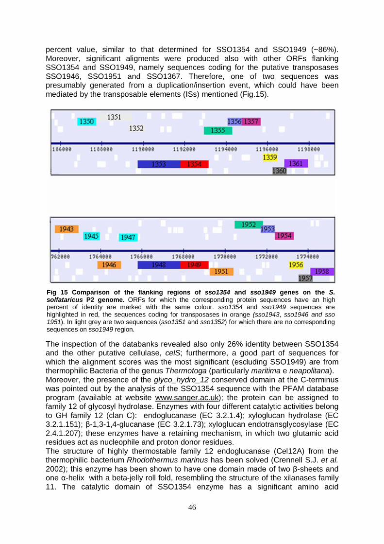
46
percent value, similar to that determined for SSO1354 and SSO1949 (~86%). Moreover, significant aligments were produced also with other ORFs flanking SSO1354 and SSO1949, namely sequences coding for the putative transposases SSO1946, SSO1951 and SSO1367. Therefore, one of two sequences was presumably generated from a duplication/insertion event, which could have been mediated by the transposable elements (ISs) mentioned (Fig.15).
The inspection of the databanks revealed also only 26% identity between SSO1354 and the other putative cellulase, celS; furthermore, a good part of sequences for which the alignment scores was the most significant (escluding SSO1949) are from thermophilic Bacteria of the genus Thermotoga (particularly maritima e neapolitana). Moreover, the presence of the glyco_hydro_12 conserved domain at the C-terminus was pointed out by the analysis of the SSO1354 sequence with the PFAM database program (available at website www.sanger.ac.uk); the protein can be assigned to family 12 of glycosyl hydrolase. Enzymes with four different catalytic activities belong to GH family 12 (clan C): endoglucanase (EC 3.2.1.4); xyloglucan hydrolase (EC 3.2.1.151); β-1,3-1,4-glucanase (EC 3.2.1.73); xyloglucan endotransglycosylase (EC 2.4.1.207); these enzymes have a retaining mechanism, in which two glutamic acid residues act as nucleophile and proton donor residues. The structure of highly thermostable family 12 endoglucanase (Cel12A) from the thermophilic bacterium Rhodothermus marinus has been solved (Crennell S.J. et al. 2002); this enzyme has been shown to have one domain made of two β-sheets and one α-helix with a beta-jelly roll fold, resembling the structure of the xilanases family 11. The catalytic domain of SSO1354 enzyme has a significant amino acid
Fig 15 Comparison of the flanking regions of sso1354 and sso1949 genes on the S. solfataricus P2 genome. ORFs for which the corresponding protein sequences have an high percent of identity are marked with the same colour. sso1354 and sso1949 sequences are highlighted in red, the sequences coding for transposases in orange (sso1943, sso1946 and sso 1951). In light grey are two sequences (sso1351 and sso1352) for which there are no corresponding sequences on sso1949 region.
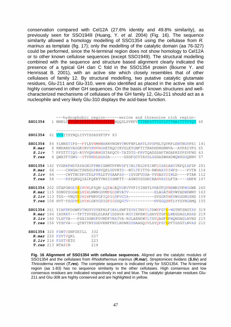
47
conservation compared with Cel12A (27.6% identity and 49.8% similarity), as previously seen for SSO1949 (Huang, Y. et al. 2004) (Fig. 16). The sequence similarity allowed a homology modelling of SSO1354 using the cellulase from R. marinus as template (fig. 17); only the modelling of the catalytic domain (aa 76-327) could be performed, since the N-terminal region does not show homology to Cel12A or to other known cellulose sequences (except SSO1949). The structural modelling combined with the sequence and structure based alignment clearly indicated the presence of a typical GH clan C fold in the SSO1354 protein (Bourne Y. and Henrissat B. 2001), with an active site which closely resembles that of other cellulases of family 12. By structural modelling, two putative catalytic glutamate residues, Glu-211 and Glu-310, were also identified as placed in the active site and highly conserved in other GH sequences. On the basis of known structures and well-characterized mechanisms of cellulases of the GH family 12, Glu-211 should act as a nucleophile and very likely Glu-310 displays the acid-base function.
---hydrophobic region------serine and threonine rich region-SSO1354 1 MNKLYIVLPVIVIIAIGVMGGIIYLHQQSLSVKPVTTTEFSTTTSTSTTTNAITTTVTQT 60 ----------------------- SSO1354 61 VTSITSYNQLIYVTSSASSPTPV 83 SSO1354 84 YLNNSTIPS--FYLEVNMWNAKNYNGNYTMVFNPLARTLSVSFNLTQVKPLEWTNGYPEI 141 R.mar 8 RWDARDVAGGRYRVINNVWGAETAQCIEVGLETGNFTITRADHDNGNNVA--AYPAIYFG 65 S.liv 7 PFGTTTIQG-RYVVQNNRWGSTAPQCV-TATDTG-FRVTQADGSAPTNGAPKSYPSVFNG 63 T.res 6 QWATFTGNG--YTVSNNLWGASA------GSGFGCVTAVSLSGGASWHADWQWSGGQNNV 57 SSO1354 142 YVGRKPWDTAYAGNIFPMRIGNMTPFMVSFYINLTKLDPSINFDIASDAWIVRPQIAFSP 201 R.mar 66 ----CHWGACTSNSGLPRRVQELSDVRTS--WTLTPITTG-RWNAAYDIWFS----PVTN 114 S.liv 64 ----CHYTNCSPGTDLPVRLDTVSAAPSS--ISYGFVDGA-VYNASYDIWLD----PTAR 112 T.res 58 ----KSYQNSQIAIPQKRTVNSISSMPTT--ASWSYSGSNIRANVAYDLFTA----ANPN 107 SSO1354 202 GTAPGNGDIEIMVWLFSQN-LQPAGEQVGKVVVPIYINHTLVNATFQVWEMKSVPWGGWE 260 R.mar 115 SGNGYSGGAELMIWLNWNGGVMPGGSRVATV-----------ELAGATWEVWYADWDWNY 163 S.liv 113 TDG--VNQTEIMIWFNRVGPIQPIGSPVGTA-----------SVGGRTWEVWSGGNGSND 159 T.res 108 HVT-YSGDYELMIWLGKYGDIGPIGSSQGTV-----------NVGGQSWTLYYGYNGAMQ 155 SSO1354 261 YIAFRPDGWKVTNGYVSYEPNLFIKALSNFTSYNITNYYLTDWEFGTE-WGTMTSNGTAY 319 R.mar 164 IAYRRT---TPTTSVSELDLKAFIDDAVA-RGYIRPEWYLHAVETGFELWEGGAGLRSAD 219 S.liv 160 VLSFVA---PSAISGWSFDVMDFVRATVA-RGLAENDWYLTSVQAGFEPWQNGAGLAVNS 215 T.res 156 VYSFVA---QTNTTNYSGDVKNFFNYLRDNKGYNAAGQYVLSYQFGTEPFTGSGTLNVAS 212 SSO1354 320 FSWTVSNFSETLL 332 R.mar 220 FSVTVQKL 227 S.liv 216 FSSTVETG 223 T.res 213 WTASIN 218
Fig. 16 Alignment of SSO1354 with cellulase sequences. Aligned are the catalytic modules of SSO1354 and the cellulases from Rhodothermus marinus (R.mar), Streptomices lividans (S.liv) and Thricoderma reesei (T.res). The complete sequence is indicated only for SSO1354. The N-terminal region (aa 1-83) has no sequence similarity to the other cellulases. High consensus and low consensus residues are indicated respectively in red and blue. The catalytic glutamate residues Glu-211 and Glu-308 are highly conserved and are highlighted in yellow.

48
Due to the striking difference with other cellulases in the corresponding region, the N-terminal sequence of SSO1354 was analyzed in depth, in order to complete a confident structural-functional prediction to the whole protein sequence. We found that SSO1354 can be successfully aligned to the sugar binding proteins (SBP) from S. solfataricus AraS, GlcS and TreS; these proteins, that respectively bind arabinose, glucose and trehalose, are the subunits of ATP binding cassette (ABC) transporters responsible for sugar uptake in S. solfataricus (Elferink M.G. et al. 2001) (Fig,18).
1 60 SSO1354 MNK-----------LYIVLPVIVIIAIGVMGGIIYLHQQSLSVKPVTTTEFST------- treS MRR--------GLSTTTIIGIVVAIVIIVIGAVAAVTLLSHKPSQVVST----------- araS MSRRRLY---KAISRTAIIIIVVVIIIAAIAGGLAAYYSSSKPPATSTSLTSTSSSLSVT glcS MKRKYPYSLAKGLTSTQIAVIVAVIVIVIIIGVVAGFVLTKGPSTTAVTTTVTS------ 61 120 SSO1354 TTSTSTTTNAITTTVTQTVTSITSYNQLIYVTSSASSPTPVYLNNSTIPSFYLEVNM--- treS --TSPSTSQSATSTSPSQVITITYFDDLSPSEANITQKIIIPQFEATHPNIKINYVD--- araS TSSTTSTLSSITTTTSTASSYVVDFINPWGAEDPVGLKWIGGNFSIYYPGYSVQFTSLPG glcS TFTTTTTIPS--TTTSTPSNTVVFYTWWGGGDGGEALSQIIPAVK-QYAGLQMQTYSIPG
Fig.18 Aligment of N-terminal amino acid sequences of SSO1354 and SBPs. High consensus and low consensus residues are indicated respectively in red and blue; putative transmembrane segments are highlighted in yellow, and the long hydrophobic stretch rich in hydroxylated amino acids is indicated in light blue.
Fig. 17 Structural model of SSO1354 cellulase. The structure of catalityc domain of SSO1354 (aa 76-327) was modeled with the EsyPred3D program using the cellulase from R. marinus (PDB no. 1H0B) as template. The β-strands are coloured in light blue, α helices in red. The catalytic glutamate residues are depicted in purple. The active-site cleft is best viewed from the side (right picture)
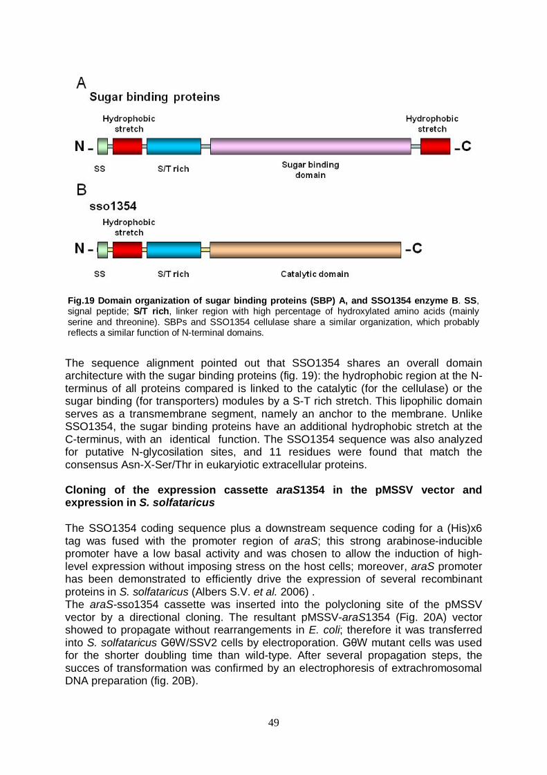
49
The sequence alignment pointed out that SSO1354 shares an overall domain architecture with the sugar binding proteins (fig. 19): the hydrophobic region at the N-terminus of all proteins compared is linked to the catalytic (for the cellulase) or the sugar binding (for transporters) modules by a S-T rich stretch. This lipophilic domain serves as a transmembrane segment, namely an anchor to the membrane. Unlike SSO1354, the sugar binding proteins have an additional hydrophobic stretch at the C-terminus, with an identical function. The SSO1354 sequence was also analyzed for putative N-glycosilation sites, and 11 residues were found that match the consensus Asn-X-Ser/Thr in eukaryiotic extracellular proteins. Cloning of the expression cassette araS1354 in the pMSSV vector and expression in S. solfataricus The SSO1354 coding sequence plus a downstream sequence coding for a (His)x6 tag was fused with the promoter region of araS; this strong arabinose-inducible promoter have a low basal activity and was chosen to allow the induction of high-level expression without imposing stress on the host cells; moreover, araS promoter has been demonstrated to efficiently drive the expression of several recombinant proteins in S. solfataricus (Albers S.V. et al. 2006) . The araS-sso1354 cassette was inserted into the polycloning site of the pMSSV vector by a directional cloning. The resultant pMSSV-araS1354 (Fig. 20A) vector showed to propagate without rearrangements in E. coli; therefore it was transferred into S. solfataricus GθW/SSV2 cells by electroporation. GθW mutant cells was used for the shorter doubling time than wild-type. After several propagation steps, the succes of transformation was confirmed by an electrophoresis of extrachromosomal DNA preparation (fig. 20B).
Fig.19 Domain organization of sugar binding proteins (SBP) A, and SSO1354 enzyme B. SS, signal peptide; S/T rich, linker region with high percentage of hydroxylated amino acids (mainly serine and threonine). SBPs and SSO1354 cellulase share a similar organization, which probably reflects a similar function of N-terminal domains.
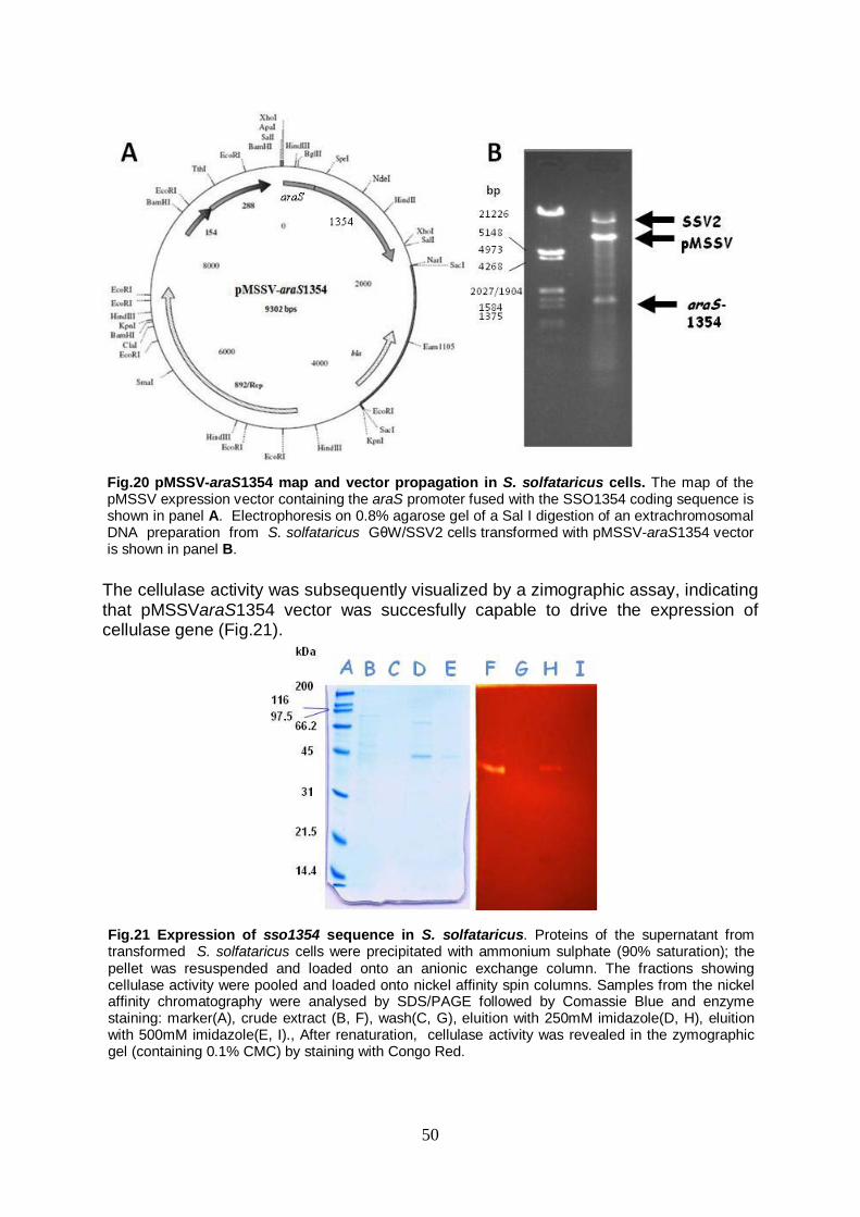
50
The cellulase activity was subsequently visualized by a zimographic assay, indicating that pMSSVaraS1354 vector was succesfully capable to drive the expression of cellulase gene (Fig.21).
Fig.21 Expression of sso1354 sequence in S. solfataricus. Proteins of the supernatant from transformed S. solfataricus cells were precipitated with ammonium sulphate (90% saturation); the pellet was resuspended and loaded onto an anionic exchange column. The fractions showing cellulase activity were pooled and loaded onto nickel affinity spin columns. Samples from the nickel affinity chromatography were analysed by SDS/PAGE followed by Comassie Blue and enzyme staining: marker(A), crude extract (B, F), wash(C, G), eluition with 250mM imidazole(D, H), eluition with 500mM imidazole(E, I)., After renaturation, cellulase activity was revealed in the zymographic gel (containing 0.1% CMC) by staining with Congo Red.
Fig.20 pMSSV-araS1354 map and vector propagation in S. solfataricus cells. The map of the pMSSV expression vector containing the araS promoter fused with the SSO1354 coding sequence is shown in panel A. Electrophoresis on 0.8% agarose gel of a Sal I digestion of an extrachromosomal DNA preparation from S. solfataricus GθW/SSV2 cells transformed with pMSSV-araS1354 vector is shown in panel B.
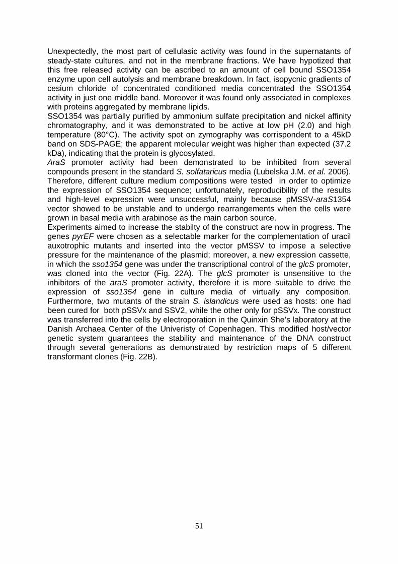
51
Unexpectedly, the most part of cellulasic activity was found in the supernatants of steady-state cultures, and not in the membrane fractions. We have hypotized that this free released activity can be ascribed to an amount of cell bound SSO1354 enzyme upon cell autolysis and membrane breakdown. In fact, isopycnic gradients of cesium chloride of concentrated conditioned media concentrated the SSO1354 activity in just one middle band. Moreover it was found only associated in complexes with proteins aggregated by membrane lipids. SSO1354 was partially purified by ammonium sulfate precipitation and nickel affinity chromatography, and it was demonstrated to be active at low pH (2.0) and high temperature (80°C). The activity spot on zymography was corrispondent to a 45kD band on SDS-PAGE; the apparent molecular weight was higher than expected (37.2 kDa), indicating that the protein is glycosylated. AraS promoter activity had been demonstrated to be inhibited from several compounds present in the standard S. solfataricus media (Lubelska J.M. et al. 2006). Therefore, different culture medium compositions were tested in order to optimize the expression of SSO1354 sequence; unfortunately, reproducibility of the results and high-level expression were unsuccessful, mainly because pMSSV-araS1354 vector showed to be unstable and to undergo rearrangements when the cells were grown in basal media with arabinose as the main carbon source. Experiments aimed to increase the stabilty of the construct are now in progress. The genes pyrEF were chosen as a selectable marker for the complementation of uracil auxotrophic mutants and inserted into the vector pMSSV to impose a selective pressure for the maintenance of the plasmid; moreover, a new expression cassette, in which the sso1354 gene was under the transcriptional control of the glcS promoter, was cloned into the vector (Fig. 22A). The glcS promoter is unsensitive to the inhibitors of the araS promoter activity, therefore it is more suitable to drive the expression of sso1354 gene in culture media of virtually any composition. Furthermore, two mutants of the strain S. islandicus were used as hosts: one had been cured for both pSSVx and SSV2, while the other only for pSSVx. The construct was transferred into the cells by electroporation in the Quinxin She’s laboratory at the Danish Archaea Center of the Univeristy of Copenhagen. This modified host/vector genetic system guarantees the stability and maintenance of the DNA construct through several generations as demonstrated by restriction maps of 5 different transformant clones (Fig. 22B).

52
Fig. 22 Map of pMSSV-1354pyr and propagation of the construct. A: The expression cassette glcS1354 fused with the pyrEF genes was cloned into the vector pMSSV, producing the expression vector pMSSV1354pyr B: S. islandicus pyr - cells, cured for both pSSVx and SSV2, were electroporated with the pMSSV1354pyr vector. Five S. islandicus transformed clones were isolated. Extrachromosomal DNAs extracted from E. coli mos blue cells (lanes 1 a,b,c) and S. islandicus isolated clones (lanes 2-6 a,b,c), were checked by agarose electrophoresis for the presence of pMSSV1354pyr (lanes 1-6a: BamHI cut, lanes 1-6b: EcoRI cut, lanes 1-6c: HindIII cut). M: molecular weight markers (bp).
A
B
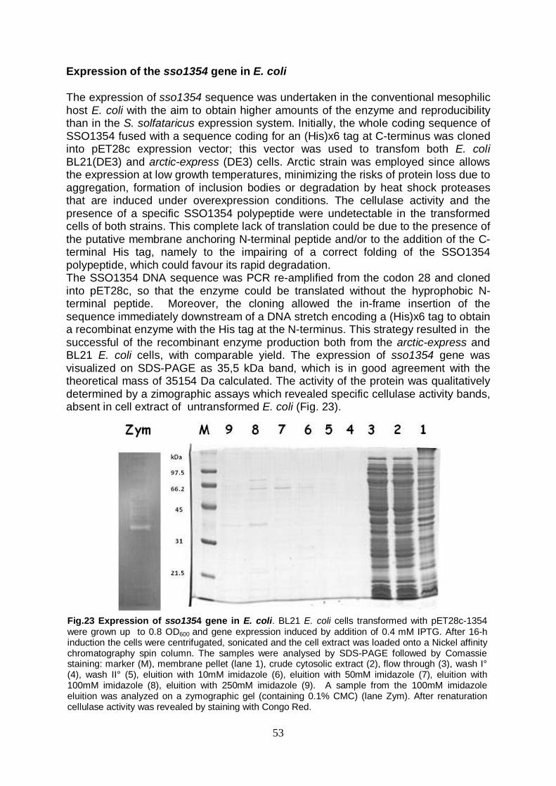
53
Expression of the sso1354 gene in E. coli The expression of sso1354 sequence was undertaken in the conventional mesophilic host E. coli with the aim to obtain higher amounts of the enzyme and reproducibility than in the S. solfataricus expression system. Initially, the whole coding sequence of SSO1354 fused with a sequence coding for an (His)x6 tag at C-terminus was cloned into pET28c expression vector; this vector was used to transfom both E. coli BL21(DE3) and arctic-express (DE3) cells. Arctic strain was employed since allows the expression at low growth temperatures, minimizing the risks of protein loss due to aggregation, formation of inclusion bodies or degradation by heat shock proteases that are induced under overexpression conditions. The cellulase activity and the presence of a specific SSO1354 polypeptide were undetectable in the transformed cells of both strains. This complete lack of translation could be due to the presence of the putative membrane anchoring N-terminal peptide and/or to the addition of the C-terminal His tag, namely to the impairing of a correct folding of the SSO1354 polypeptide, which could favour its rapid degradation. The SSO1354 DNA sequence was PCR re-amplified from the codon 28 and cloned into pET28c, so that the enzyme could be translated without the hyprophobic N-terminal peptide. Moreover, the cloning allowed the in-frame insertion of the sequence immediately downstream of a DNA stretch encoding a (His)x6 tag to obtain a recombinat enzyme with the His tag at the N-terminus. This strategy resulted in the successful of the recombinant enzyme production both from the arctic-express and BL21 E. coli cells, with comparable yield. The expression of sso1354 gene was visualized on SDS-PAGE as 35,5 kDa band, which is in good agreement with the theoretical mass of 35154 Da calculated. The activity of the protein was qualitatively determined by a zimographic assays which revealed specific cellulase activity bands, absent in cell extract of untransformed E. coli (Fig. 23).
Fig.23 Expression of sso1354 gene in E. coli. BL21 E. coli cells transformed with pET28c-1354 were grown up to 0.8 OD600 and gene expression induced by addition of 0.4 mM IPTG. After 16-h induction the cells were centrifugated, sonicated and the cell extract was loaded onto a Nickel affinity chromatography spin column. The samples were analysed by SDS-PAGE followed by Comassie staining: marker (M), membrane pellet (lane 1), crude cytosolic extract (2), flow through (3), wash I° (4), wash II° (5), eluition with 10mM imidazole (6), eluition with 50mM imidazole (7), eluition with 100mM imidazole (8), eluition with 250mM imidazole (9). A sample from the 100mM imidazole eluition was analyzed on a zymographic gel (containing 0.1% CMC) (lane Zym). After renaturation cellulase activity was revealed by staining with Congo Red.
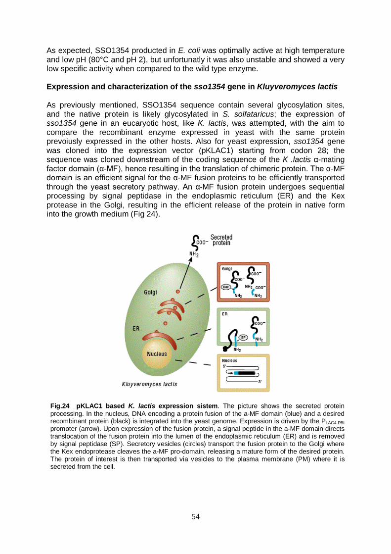
54
As expected, SSO1354 producted in E. coli was optimally active at high temperature and low pH (80°C and pH 2), but unfortunatly it was also unstable and showed a very low specific activity when compared to the wild type enzyme. Expression and characterization of the sso1354 gene in Kluyveromyces lactis As previously mentioned, SSO1354 sequence contain several glycosylation sites, and the native protein is likely glycosylated in S. solfataricus; the expression of sso1354 gene in an eucaryotic host, like K. lactis, was attempted, with the aim to compare the recombinant enzyme expressed in yeast with the same protein prevoiusly expressed in the other hosts. Also for yeast expression, sso1354 gene was cloned into the expression vector (pKLAC1) starting from codon 28; the sequence was cloned downstream of the coding sequence of the K .lactis α-mating factor domain (α-MF), hence resulting in the translation of chimeric protein. The α-MF domain is an efficient signal for the α-MF fusion proteins to be efficiently transported through the yeast secretory pathway. An α-MF fusion protein undergoes sequential processing by signal peptidase in the endoplasmic reticulum (ER) and the Kex protease in the Golgi, resulting in the efficient release of the protein in native form into the growth medium (Fig 24).
Fig.24 pKLAC1 based K. lactis expression sistem. The picture shows the secreted protein processing. In the nucleus, DNA encoding a protein fusion of the a-MF domain (blue) and a desired recombinant protein (black) is integrated into the yeast genome. Expression is driven by the PLAC4-PBI promoter (arrow). Upon expression of the fusion protein, a signal peptide in the a-MF domain directs translocation of the fusion protein into the lumen of the endoplasmic reticulum (ER) and is removed by signal peptidase (SP). Secretory vesicles (circles) transport the fusion protein to the Golgi where the Kex endoprotease cleaves the a-MF pro-domain, releasing a mature form of the desired protein. The protein of interest is then transported via vesicles to the plasma membrane (PM) where it is secreted from the cell.
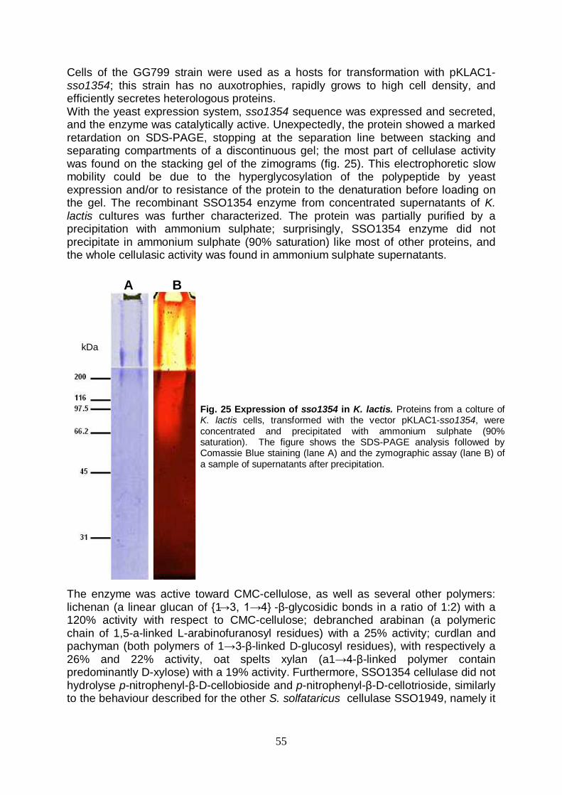
55
Cells of the GG799 strain were used as a hosts for transformation with pKLAC1-sso1354; this strain has no auxotrophies, rapidly grows to high cell density, and efficiently secretes heterologous proteins. With the yeast expression system, sso1354 sequence was expressed and secreted, and the enzyme was catalytically active. Unexpectedly, the protein showed a marked retardation on SDS-PAGE, stopping at the separation line between stacking and separating compartments of a discontinuous gel; the most part of cellulase activity was found on the stacking gel of the zimograms (fig. 25). This electrophoretic slow mobility could be due to the hyperglycosylation of the polypeptide by yeast expression and/or to resistance of the protein to the denaturation before loading on the gel. The recombinant SSO1354 enzyme from concentrated supernatants of K. lactis cultures was further characterized. The protein was partially purified by a precipitation with ammonium sulphate; surprisingly, SSO1354 enzyme did not precipitate in ammonium sulphate (90% saturation) like most of other proteins, and the whole cellulasic activity was found in ammonium sulphate supernatants.
The enzyme was active toward CMC-cellulose, as well as several other polymers: lichenan (a linear glucan of {1→3, 1→4} -β-glycosidic bonds in a ratio of 1:2) with a 120% activity with respect to CMC-cellulose; debranched arabinan (a polymeric chain of 1,5-a-linked L-arabinofuranosyl residues) with a 25% activity; curdlan and pachyman (both polymers of 1→3-β-linked D-glucosyl residues), with respectively a 26% and 22% activity, oat spelts xylan (a1→4-β-linked polymer contain predominantly D-xylose) with a 19% activity. Furthermore, SSO1354 cellulase did not hydrolyse p-nitrophenyl-β-D-cellobioside and p-nitrophenyl-β-D-cellotrioside, similarly to the behaviour described for the other S. solfataricus cellulase SSO1949, namely it
Fig. 25 Expression of sso1354 in K. lactis. Proteins from a colture of K. lactis cells, transformed with the vector pKLAC1-sso1354, were concentrated and precipitated with ammonium sulphate (90% saturation). The figure shows the SDS-PAGE analysis followed by Comassie Blue staining (lane A) and the zymographic assay (lane B) of a sample of supernatants after precipitation.
A B
kDa

56
has no exo-cellulase activity, confirming that SSO1354 is mainly an endo-1,4-β glucanase. pH and temperature dependance of recombinant SSO1354 activity, investigated in the range of 1.8-7.5 and 37-90°C, respectively, are shown in fig. 26. The temperature optimum was 80°C; at this temperature the highest activity was found at pH 5.0. Moreover, the recombinant cellulase showed a strong thermostability; when incubed at 90°C, the enzyme had an half life of 180 min. SSO1354 was also tested to verify whether the enzyme retains the cellulasic activity in ionic liquids; first, the highest water concentration tolerated for solubility of α-cellulose in solutions of ionic liquids was determined as 20%. Qualitative activity detection by thin layer chromatography analysis of the idrolyzed products, revealed that the enzyme retained activity in 1-ethyl-3-methyl-imidazolium-acetate, producing cellobiose and cellotriose as main products of hydrolysis in a time dependent accumulation over 2.0-16.0 hour incubations (Fig. 27).
Fig.26 pH and temperature optimum of SSO1354 enzyme. A: The pH optimum was assayed at 80°C. The reactions were performed in potassium phosphate buffer (25mM) in the range of 1.8-2.8 (●); citrate phosphate buffer (25mM) was used in the range of 3.3-7.5 (♦). B: The cellulolytic activity of SSO1354 enzyme was assayed at various temperatures in 25mM citrate phosphate buffer (pH 5)
Fig.27 Degradation of cellulose in ionic liquids. A 1% α-cellulose solution in 1-ethyl-3-methyl-imidazolium-acetate was incubated with SSO1354 enzyme. Samples were withdrawn at several time points, and the ionic liquid was removed by three phenol/chloroforme and one chloroforme extractions. Reactions with (lanes 2-4) and without (lane 1) enzyme were spotted on silica plates and developed with a mixture of ethyl acetate, H2O, acetic acid, isopropanol, formic acid, (25:15:10:5:1, by vol.) Sugars were visualized with α-naphtol after incubation of 5min at 120°C. Lane M: mix of cello-oligomers (3µl of a solution with a concentration of 5mM of each oligomer); 1: reaction mix incubated for 16h without enzyme; 2: 2h incubation; 3: 4h incubation; 4: 16h incubation. (60 µl of samples are loaded on lanes 1-4)
57
Discussion The present PhD work was developed following two strongly interconnected experimental lines: in the one hand, a deep characterization of a shuttle vector E.coli – S. solfataricus has been performed, in order to elucidate the potentialities of this genetic element. In the other hand, the same vector has been used to express sso1354 gene, which encodes for a thermostable enzyme with cellulasic activity from S. solfataricus, in a xenologous fashion; sso1354 has been also expressed in two heterologous hosts, with the aim to optimize the expression yield and to compare the recombinant protein obtained from different sources. Stable and efficient transformation/expression genetic systems for hyperthermophilic Archaea represent key tools both for basic and biotechnological researches. These systems can be used to efficiently characterize promoters and coding sequences; moreover, archaeal expression systems can be also employed to (over)express proteins with commercial potential, like the enzymes from (hyper)thermophiles, in the same organisms from which the sequences are derived, avoiding the common problems in expressing a protein from a thermophilic organism into a mesophilic hosts (i.e. incorrect folding and/or incompatible post-translational modifications). A wide variety of systems have been developed for protein production in members of the Bacteria and Eukarya, using numerous combinations of vector and promoter systems. The development of genetic systems is not so advanced for archaeal microorganisms, particularly for that belonging to chrenarchaeal kingdom (in fact the most of the few existing transformation tools for the production of recombinant proteins have been developed for halophiles and methanogenes Euryarchaea). The crenarchaeote S. solfataricus has developed into an important model organism for molecular and biochemical studies of hyperthermophilic Archaea, and it is highly desirable to develop genetic tools for in vivo studies and for high-level production of proteins in this organism; S. solfataricus could be employed as “cell factory” in plants for the production of biotechnological relevant proteins. In the present work, the expression shuttle vector pMSSV for S. solfataricus was tested, using an expression cassette containing the marker gene lacS, which encode for a S. solfataricus β-glycosidase. This system presents many advantages compared with others already mentioned. After DNA transfer, the vector propagates efficiently throughout the culture as a virus; the high propagation efficiency of the pMSSV vector, in SSV2 lysogens of the strain GθW, was found to be comparable to the wild-type pSSVx. Therefore, the pMSSV vector is proper to overcome the usual low transformation efficiency of Sulfolobus cells (10-3-10-4
Interestingly, β-galactosidase activity in complemented mutant cells followed an increasing trend which correlated to the plasmid accumulation up to stationary phase and was not dependent on the number of the generations. lacS gene expression was
cells/μg of plasmidic DNA used). Moreover, this genetic system have shown another important feature: the vector is stably maintained at high-copy number in episomal form inside host cells (up to 130 copies per chromosome, the number depending on the cell growth phase), with no integration into the chromosome and hence no reduction in the number of episomal molecules. This feature is particularly advantageuos in the expression of passenger genes; the yield of expression would not be affected by the decrease of the copy number as consequence of the integration of the vector into the host chromosome. In fact, the analysis have shown that pMSSV vector is succesfully capable to drive the expression of the marker gene lacS.

58
inducible by thermal stress, namely it was confirmed to be transcriptionally regulated by the chaperonin gene promoter. However, heat shock did not produce the same β-galactosidase activity increase (~10-fold) described by Jonuscheit et al. (2003), although maximum gene expression resulted at comparable levels in the two host/vector systems (3 versus 5 U/mg). Therefore, the characterization of pMSSV have pointed out some interesting features of this system, which make it a powerful genetic tool, with a strong application potential in basic researches as well as in biotechnology. Therefore, the vector/host system developed was used for the expression of the cellulase gene from S. solfataricus. As previously mentioned, the cellulases, particularly from (hyper)thermophilic organisms, have a wide range of industrial application, and consequently they are biotechnological relevant enzymes. The gene which encodes for the putative extracellular endoglucanase SSO1354 was expressed in xenologous and heterologous fashion; SSO1354 is the only one of three S. solfataricus putative cellulases for which a characterization study have been not performed yet. Some interesting remarks can be made on the base of the sequence analysis of SSO1354; particularly interesting is the comparison with the sugar binding proteins (SBP) of S. solfataricus AraS, GlcS e TreS, that bind arabinose, glucose and trehalose, respectively. The sequence similarity is significant, particuarly in a “module” in the N-terminus, which instead does not produce any alignment with N-terminal regions in other known cellulase sequences (except SSO1949), namely the SSO1354 protein and the SBPs share a similar domain architecture. Therefore, SSO1354 N-terminal domains was assumed to have the same function as in SBPs. AraS, GlcS and TreS, such as various S. solfataricus solute binding proteins, have an unusual leader peptide that resembles bacterial type IV prepilin signal sequences and is cleaved by the S. solfataricus homolog of bacterial type IV prepilin peptidases, named PibD (Albers S.V. et al. 2003); SSO1354 putative signal sequence matchs only in part the relatively conserved consensus sequence which have been found to be recognized from PibD. However, this enzyme has been demonstrated to be equipped with a relatively broad substrate specificity, and to tolerate some mutations in the core of four amino acids around the cleavage site. SSO1354 (and probably also the other two cellulases from S. solfataricus that also show a similar organization at N-terminus) could be a cell-bound cellulase, with the catalytic domain linked by the S-T rich stretch to the transmebrane region; this architercture could give the needed flexibility to the enzyme. In fact the S-T rich stretch has been previously demonstrated to be fundamental for catalytic activity in the closely related SSO1949 enzyme; the enzyme expressed without this domain has been shown to have no endoglucanasic activity. Our hypotesys of cell bound cellulases is confirmed by the data avaiable in literature: in fact the most part of cellulasic activity in S. solfataricus cultures has been found in membrane extracts (Cannio R. et al. 2004); however our hypotesys is in contrast with that previously assumed for SSO1949 (Huang Y. et al. 2005); Huang and collegues have been assigned to the N-terminal hydrophobic region the role of signal peptide by a bioinformatic prediction with SignalP, and consequently have assumed that SSO1949 is a medium-released cellulase. This hypothesis was indeed not supported by any data about characterization and localization in vivo of cellulase activity. The co-localization of the endoglucanases and the sugar binding proteins could be an advantageous strategy for the optimization of nutrient assimilation, particularly for

59
cells living in habitats where complex carbohydrate are a major, but scarce, energy source. The SSO1354 cellulase was successfully producted both in homologous and heterologous hosts. The first strategy was to express the protein in S. solfataricus, considering the documented difficulty to express these cellulase genes from (hyper)thermophilic organisms in phylogenetical distant conventional hosts, like E.coli. In fact, the other two S. solfataricus cellulase genes sso1949 (Huang Y. et al. 2005) and sso2534 (data not shown) have been shown to be considerably difficult to handle in conventional mesophilic hosts. The cellulase gene was expressed under the control of a strong and inducible promoter, araS, using the vector pMSSV. SSO1354 was obtained in catalytical active form and, like the genome encoded natural product, it was glycosylated and active at extreme conditions. The membrane bound extracellular localization, predicted by the sequence analysis and suggested by literature data, was confirmed; SSO1354 activity was found mostly in culture surnatants, but the protein was demonstrated to be associated with lipids. Therefore, the cellulase was likely released in the culture medium as a consequence of cell lysis since the highest overall activity was obtained in cultures grown up to late stationary phase. The main problem encountered with the expression in S. solfataricus was the stability of the construct pMSSV-araS1354 when the cells were grown in specific media for the induction of the araS promoter activity. This bottleneck was overcome by imposing a selective pressure (a genetic selection marker was added to the vector) and using a different host strain; these experiments, which are now in progress, are giving very promising results. In fact, this strategy resulted in the improving of the genetic system based on the pMSSV vector, by providing it with increased genetic stability. Moreover, the vector with the selection genes was demonstrated to be able to replicate indipendently as a plasmid without the virus helper SSV2 under the selective pressure. This feature showed by the modified pMSSV is a fundamental requirement to next isolate single transformant clones and to use this vector for mutagenesis and isolation of single mutated gene variants. SSO1354 was also producted in both a bacterial and eukaryotic mesophilic hosts, with the aim to diversify the strategies of expression and to improve the yield of recombinant protein. In E. coli, the exclusion of the N-terminal hydrophobic sequence was necessary to reach a succesful expression of the cellulase, suggesting that this sequence could interfere with translation or provoke a misfolding of the protein. SSO1354 producted in E. coli conserved the preference for low pH and high temperature of the wild type enzyme obtained from S. solfataricus but unfortunately showed also a marked instability and low specific activity. Therefore, due to the more strict similarity in the secretion/post translational modification apparatus, K. lactis has been chosen as a more suitable host to express sso1354 gene; K. lactis have a large potential for the biotechnological production of recombinant proteins, as demonstrated by a large number of sequence which have been successfully expressed in this host (van Ooyen A.J. et al 2006). Moreover, K.lactis has a number of advantages over other yeast expression systems; these include easy genetic manipulation, GRAS certification (generally regarded as safe) and an efficient and versatile secretory apparatus. The cellulase expressed in yeast was found to be hyperglycosylated, according with the data present in literature about expression patterns of heterologous proteins in yeast. The protein was characterized, and the comparison of the main features with those found for SSO1354 producted in the other hosts pointed out some interesting differences: the temperature optimum (80°C) was in agreement with the precedent findings, whilst the
60
pH optimum (5.0) diverged from that expected, even if the enzyme retained the activity also at lower pH; moreover, the enzyme showed a strong increase of thermostability (half-life of 3h at 90°C) when compared to SSO1354 producted in E.coli and SSO1949 (which has been characterized after the production in E. coli). The effect on pH optimum was probably due to the different hyperglycosilation of the protein in the yeast host when compared to the wild type enzyme produced by S. solfataricus. Hyperglycosylation was also responsible for the higher thermostability of the recombinant enzyme expressed in Sulfolobus and yeast cells with respect to the non glycosilated polypeptide produced in E. coli. In particular, the effect of glycosylation in increasing the resistance of proteins to extreme conditions is well documented (Koseki T. et al. 2006; Kim Y.O. et al. 2006). This could be a post-translational modification mechanism selected by evolution to provide an extra stability even to the proteins from hyperthermophiles. The cellulase expressed in yeast was also demonstrated to retain the catalytic activity in ionic liquids. This result is particularly relevant; in fact these new class of solvents, as previously discussed (see “Introduction”), have a great potential in eco-sustainable production of bioethanol: it can be used as reaction media for suspension and subsequent increased susceptibily to hydrolytic attack of cellulosic materials. To our knowledge, this is the first example of a glycosyl hydrolase capable to work in ionic liquids and in reaction environments at very low water content. The findings obtainend in this work shed light the great biotechnological relevance of the SSO1354 cellulase, which is an ideal candidate to be employed in several industrial processes, and particularly in the biofuel production. Furthermore, new elements on structure-function relationship was acquired: from sequence comparison and literature background about the characterization of S. solfataricus cellulasic activity in vivo, was assumed that SSO1354, as well as SSO2534 and SSO1949, is a cell-bound enzyme, and not free released in extracellular medium. This finding opens new and intriguing perspectives on the strategy adopted by a hyperthermophilic archaeon as S. solfataricus to coordinate and organize the activities involved in the nutrient metabolism.
61
Bibliography Aguilar CF, Sanderson I, Moracci M, Ciaramella M, Nucci R, Rossi M, Pearl LH (1997) “Crystal structure of the beta-glycosidase from the hyperthermophilic archeon Sulfolobus solfataricus: resilience as a key factor in thermostability” .J Mol Biol. 271, 789-802. Albers SV, Szabó Z, Driessen AJ. (2003) “Archaeal homolog of bacterial type IV prepilin signal peptidases with broad substrate specificity”. J Bacteriol. 185, 3918-25. Allers T, Mevarech M (2005) “Archaeal genetics—the third way”. Nat Rev Genet 6, 58–73 Arnold HP, Zillig W, Ziese U, Holz I, Crosby M, Utterback T, Weidmann JF, Kristjanson JK, Klenk HP, Nelson KE, Fraser CM (2000) “A novel lipothrixvirus, SIFV, of the extremely thermophilic crenarchaeon Sulfolobus”. Virology 267, 252–266 Arnold HP, She Q, Phan H, Stedman K, Prangishvili D, Holz I, Kristjansson JK, Garrett R, Zillig W. (1999) “The genetic element pSSVx of the extremely thermophilic crenarchaeon Sulfolobus is a hybrid between a plasmid and a virus”.Mol Microbiol. 34, 217-26. Ausili A, Di Lauro B, Cobucci-Ponzano B, Bertoli E, Scirè A, Rossi M, Tanfani F, Moracci M. (2004) “Two-dimensional IR correlation spectroscopy of mutants of the beta-glycosidase from the hyperthermophilic archaeon Sulfolobus solfataricus identifies the mechanism of quaternary structure stabilization and unravels the sequence of thermal unfolding events”. Biochem J. 2004 384 69-78. Bartolucci S, Rossi M, Cannio R. (2003) “Characterization and functional complementation of a nonlethal deletion in the chromosome of a beta-glycosidase mutant of Sulfolobus solfataricus”.J Bacteriol. 185, 3948-57. Bettstetter M, Peng X, Garrett RA, Prangishvili D (2003) “AFV1, a novel virus infecting hyperthermophilic archaea of the genus acidianus”. Virology 315, 68–79 Blake Wiedenheft, Kenneth Stedman, Francisco Roberto, Deborah Willits, Anne-Kathrin Gleske, Luisa Zoeller, Jamie Snyder, Trevor Douglas, and Mark Young (2004) “Comparative Genomic Analysis of Hyperthermophilic Archaeal Fuselloviridae Viruses” Journal of Virology, February 78, 1954-1961 Bourne Y, Henrissat B. (2001) “Glycoside hydrolases and glycosyltransferases: families and functional modules”. Curr Opin Struct Biol. 11, 593-600. Cannio R, Di Prizito N, Rossi M, Morana A. (2004) “A xylan-degrading strain of Sulfolobus solfataricus: isolation and characterization of the xylanase activity”. Extremophiles. Apr; 8,117-24.

62
Cannio R, Contursi P, Rossi M, Bartolucci S. (2001) “Thermoadaptation of a mesophilic hygromycin B phosphotransferase by directed evolution in hyperthermophilic Archaea: selection of a stable genetic marker for DNA transfer into Sulfolobus solfataricus”.Extremophiles. 5, 153-9. Crennell SJ, Hreggvidsson GO, Nordberg Karlsson E. (2002) “The structure of Rhodothermus marinus Cel12A, a highly thermostable family 12 endoglucanase, at 1.8 A resolution”. J Mol Biol. 320, 883-97 Cubellis,M.V., Rozzo,C., Monteucchi,P. and Rossi,M. (1990) “Isolation and sequencing of a new beta-glycosidase-encoding archaebacterial gene” Gene, 94, 89–94. D'Auria S, Moracci M, Febbraio F, Tanfani F, Nucci R, Rossi M. (1998) “Structure-function studies on beta-glycosidase from Sulfolobus solfataricus. Molecular bases of thermostability”.Biochimie. 80, 949-57. Davies G, Henrissat B. (1995) “Structures and mechanisms of glycosyl hydrolases”. Structure 3, 853-9. Elferink MG, Albers SV, Konings WN, Driessen AJ. (2001) “Sugar transport in Sulfolobus solfataricus is mediated by two families of binding protein-dependent ABC transporters”. Mol Microbiol. 39, 1494-503. Erauso G, Stedman KM, van de Werken HJ, Zillig W, van der Oost J. (2006) „Two novel conjugative plasmids from a single strain of Sulfolobus”. Microbiology. 152, 1951-68. Ge M, Xia XY, Pan XM. (2008) “Salt bridges in the hyperthermophilic protein Ssh10b are resilient to temperature increases”. J Biol Chem. 8 Georgieva TI, Ahring BK. (2007) “Potential of agroindustrial waste from olive oil industry for fuel ethanol production”.Biotechnol J. 2, 1547-55. Gorke JT, Okrasa K, Louwagie A, Kazlauskas RJ, Srienc F. (2007) “Enzymatic synthesis of poly(hydroxyalkanoates) in ionic liquids”.J Biotechnol. 132, 306-13. Greve B, Jensen S, Brügger K, Zillig W, Garrett RA. (2004) “Genomic comparison of archaeal conjugative plasmids from Sulfolobus”. Archaea. 1, 231-9. Grogan, D., P. Palm, and W. Zillig. (1990). “Isolate B12, which harbours a virus-like element, represents a new species of the archaebacterial genus Sulfolobus, Sulfolobus shibatae”, Arch. Microbiol. 154, 594-599. Haack KR, Roth JR. (1995) “Recombination between chromosomal IS200 elements supports frequent duplication formation in Salmonella typhimurium”. Genetics.141, 1245-52. Haring, M., Rachel, R., Peng, X., Garrett, R.A., Prangishvili, D. (2005a). “Viral diversity in hot springs of Pozzuoli, Italy, and characterization of a unique

63
archaeal virus, Acidianus bottle-shaped virus, from a new family, the Ampullaviridae” J. Virol. 79, 9904-9911. Häring M, Rachel R, Peng X, Garrett RA, Prangishvili D. (2005b) ”Viral diversity in hot springs of Pozzuoli, Italy, and characterization of a unique archaeal virus, Acidianus bottle-shaped virus, from a new family, the Ampullaviridae”.J Virol. 79, 9904-11. Huang Y, Krauss G, Cottaz S, Driguez H, Lipps G. (2005) “A highly acid-stable and thermostable endo-beta-glucanase from the thermoacidophilic archaeon Sulfolobus solfataricus.” Biochem J. 385, 581-8 Jaenicke R. (1998) “What ultrastable globular proteins teach us about protein stabilization”.Biochemistry (Mosc). 63, 312-21. Jonuscheit M, Martusewitsch E, Stedman KM, Schleper C. (2003) “A reporter gene system for the hyperthermophilic archaeon Sulfolobus solfataricus based on a selectable and integrative shuttle vector”.Mol Microbiol. 48, 1241-52. Keeling PJ, Klenk HP, Singh RK, Feeley O, Schleper C, Zillig W, Doolittle WF, Sensen CW. (1996) “Complete nucleotide sequence of the Sulfolobus islandicus multicopy plasmid pRN1”. Plasmid. 35, 141-4. Kim YO, Kim HW, Lee JH, Kim KK, Lee SJ. (2006) “Molecular cloning of the phytase gene from Citrobacter braakii and its expression in Saccharomyces cerevisiae”. Biotechnol Lett. 28. 33-8. Kleckner N, Chan RK, Tye BK, Botstein D. (1975) “Mutagenesis by insertion of a drug-resistance element carrying an inverted repetition”.J Mol Biol. 97 75 Koseki T, Takahashi K, Handa T, Yamane Y, Fushinobu S, Hashizume K. (2006) “N-linked oligosaccharides of Aspergillus awamori feruloyl esterase are important for thermostability and catalysis”. Biosci Biotechnol Biochem. 70, 2476-80. Kragl U, Eckstein M, Kaftzik N.( 2002) “Enzyme catalysis in ionic liquids”.Curr Opin Biotechnol. 13, 565-71. Lee SH, Ha SH, Hiep NM, Chang WJ, Koo YM. (2008) ”Lipase-catalyzed synthesis of glucose fatty acid ester using ionic liquids mixtures”.J Biotechnol. 133, 486-9. Limauro D, Cannio R, Fiorentino G, Rossi M, Bartolucci S. (2001) “Identification and molecular characterization of an endoglucanase gene, celS, from the extremely thermophilic archaeon Sulfolobus solfataricus.” Extremophiles. 5, 213-9. Lipps G (2006) “Plasmids and viruses of the thermoacidophilic crenarchaeote Sulfolobus”. Extremophiles.10,17-28
.
64
Lipps G, Röther S, Hart C, Krauss G. (2003) ”A novel type of replicative enzyme harbouring ATPase, primase and DNA polymerase activity”.EMBO J. 22, 2516-25. Lubelska JM, Jonuscheit M, Schleper C, Albers SV, Driessen AJ. (2006) “Regulation of expression of the arabinose and glucose transporter genes in the thermophilic archaeon Sulfolobus solfataricus”. Extremophiles. 10, 383-91. Lynd LR, Weimer PJ, van Zyl WH, Pretorius IS. (2002) “Microbial cellulose utilization: fundamentals and biotechnology”.Microbiol Mol Biol Rev. 66, 506-77 Mahillon J, Chandler M. “Insertion sequences”. (1998) Microbiol Mol Biol Rev. 62, 725-74. Martin, A., S. Yeats, D. Janekovic, W.-D. Reiter, W. Aicher, and W. Zillig. (1984). “SAV1, a temperate U. V. inducible DNA virus like particle from the archaebacterium Sulfolobus acidocaldarius isolate B12”. EMBO J. 3, 2165-2168. Martusewitsch E, Sensen CW, Schleper C. (2000) “ High spontaneous mutation rate in the hyperthermophilic archaeon Sulfolobus solfataricus is mediated by transposable elements”. J Bacteriol. 182, 2574-81 Moracci M, Nucci R, Febbraio F, Vaccaro C, Vespa N, La Cara F, Rossi M. (1995) “Expression and extensive characterization of a beta-glycosidase from the extreme thermoacidophilic archaeon Sulfolobus solfataricus in Escherichia coli: authenticity of the recombinant enzyme”. Enzyme Microb Technol. 17, 992-7. Moracci M, La Volpe A, Pulitzer JF, Rossi M, Ciaramella M. (1992) “Expression of the thermostable beta-galactosidase gene from the archaebacterium Sulfolobus solfataricus in Saccharomyces cerevisiae and characterization of a new inducible promoter for heterologous expression”. J Bacteriol. 174, 873-82. Muskhelishvili, G., P. Palm, and W. Zillig. (1993). “SSV1-encoded site-specific recombination system in Sulfolobus shibatae”. Mol. Gen. Genet. 237, 334-342. Nadal, M., G. Mirambeau, P. Forterre, W.-D. Reiter, and M. Duguet. (1986). “Positively supercoiled DNA in a virus-like particle of an archaebacterium”. Nature 321, 256-258. Nelson N. (1944) “A photometric adaptation of the Somogyi method for the determination of glucose”. J Biol Chem 153, 375–380. Palm, P., C. Schleper, B. Grampp, S. Yeats, P. McWilliam, W.-D. Reiter, and W. Zillig. (1991). “Complete nucleotide sequence of the virus SSV1 of the archaebacterium Sulfolobus shibatae”. Virology 185, 242-250. Peng X. (2008) “Evidence for the horizontal transfer of an integrase gene from a fusellovirus to a pRN-like plasmid within a single strain of Sulfolobus and the implications for plasmid survival”.Microbiology. 154, 383-91.
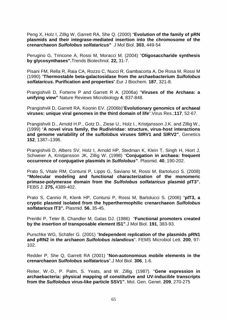
65
Peng X, Holz I, Zillig W, Garrett RA, She Q. (2000) “Evolution of the family of pRN plasmids and their integrase-mediated insertion into the chromosome of the crenarchaeon Sulfolobus solfataricus” J Mol Biol. 303, 449-54 Perugino G, Trincone A, Rossi M, Moracci M. (2004) “Oligosaccharide synthesis by glycosynthases”.Trends Biotechnol. 22, 31-7. Pisani FM, Rella R, Raia CA, Rozzo C, Nucci R, Gambacorta A, De Rosa M, Rossi M (1990) ”Thermostable beta-galactosidase from the archaebacterium Sulfolobus solfataricus. Purification and properties”.Eur J Biochem. 187, 321-8. Prangishvili D, Forterre P and Garrett R A. (2006a) “Viruses of the Archaea: a unifying view” Nature Reviews Microbiology 4, 837-848. Prangishvili D, Garrett RA, Koonin EV. (2006b)”Evolutionary genomics of archaeal viruses: unique viral genomes in the third domain of life”.Virus Res.;117, 52-67. Prangishvili D., Arnold H.P., Gotz D., Ziese U., Holz I., Kristjansson J.K. and Zillig W., (1999) “A novel virus family, the Rudiviridae: structure, virus-host interactions and genome variability of the sulfolobus viruses SIRV1 and SIRV2”, Genetics 152
, 1387–1396.
Prangishvili D, Albers SV, Holz I, Arnold HP, Stedman K, Klein T, Singh H, Hiort J, Schweier A, Kristjansson JK, Zillig W. (1998) “Conjugation in archaea: frequent occurrence of conjugative plasmids in Sulfolobus”. Plasmid. 40, 190-202. Prato S, Vitale RM, Contursi P, Lipps G, Saviano M, Rossi M, Bartolucci S. (2008) “Molecular modeling and functional characterization of the monomeric primase-polymerase domain from the Sulfolobus solfataricus plasmid pIT3”. FEBS J. 275, 4389-402. Prato S, Cannio R, Klenk HP, Contursi P, Rossi M, Bartolucci S. (2006) “pIT3, a cryptic plasmid isolated from the hyperthermophilic crenarchaeon Sulfolobus solfataricus IT3”. Plasmid. 56, 35-45. Prentki P, Teter B, Chandler M, Galas DJ. (1986) “Functional promoters created by the insertion of transposable element IS1”.J Mol Biol. 191, 383-93. Purschke WG, Schäfer G. (2001) ”Independent replication of the plasmids pRN1 and pRN2 in the archaeon Sulfolobus islandicus”. FEMS Microbiol Lett. 200, 97-102. Redder P, She Q, Garrett RA (2001) ”Non-autonomous mobile elements in the crenarchaeon Sulfolobus solfataricus”.J Mol Biol. 306, 1-6. Reiter, W.-D., P. Palm, S. Yeats, and W. Zillig. (1987). “Gene expression in archaebacteria: physical mapping of constitutive and UV-inducible transcripts from the Sulfolobus virus-like particle SSV1”. Mol. Gen. Genet. 209, 270-275
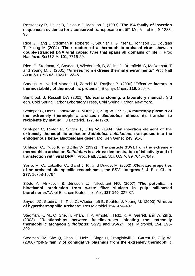
66
Rezsöhazy R, Hallet B, Delcour J, Mahillon J. (1993) ”The IS4 family of insertion sequences: evidence for a conserved transposase motif”. Mol Microbiol. 9, 1283-95. Rice G, Tang L, Stedman K, Roberto F, Spuhler J, Gillitzer E, Johnson JE, Douglas T, Young M (2004) “The structure of a thermophilic archaeal virus shows a double-stranded DNA viral capsid type that spans all domains of life”. Proc Natl Acad Sci U S A. 101, 7716-20. Rice, G, Stedman, K, Snyder, J, Wiedenheft, B, Willits, D, Brumfield, S, McDermott, T and Young M. J. (2001) “Viruses from extreme thermal environments” Proc Natl Acad Sci USA 98, 13341-13345. Sadeghi M, Naderi-Manesh H, Zarrabi M, Ranjbar B. (2006) “Effective factors in thermostability of thermophilic proteins”. Biophys Chem. 119, 256-70. Sambrook J, Russell DW (2001) “Molecular cloning, a laboratory manual”. 3rd edn. Cold Spring Harbor Laboratory Press, Cold Spring Harbor, New York. Schleper C, Holz I, Janekovic D, Murphy J, Zillig W (1995) „A multicopy plasmid of the extremely thermophilic archaeon Sulfolobus effects its transfer to recipients by mating”. J Bacteriol. 177, 4417-26. Schleper C, Röder R, Singer T, Zillig W. (1994) “An insertion element of the extremely thermophilic archaeon Sulfolobus solfataricus transposes into the endogenous beta-galactosidase gene”. Mol Gen Genet.;243, 91-6. Schleper C., Kubo K. and Zillig W. (1992) “The particle SSV1 from the extremely thermophilic archaeon Sulfolobus is a virus: demonstration of infectivity and of transfection with viral DNA”, Proc. Natl. Acad. Sci. U.S.A. 89
7645–7649.
Serre, M. C., Letzelter C., Garel J. R., and Duguet M. (2002) „Cleavage properties of an archaeal site-specific recombinase, the SSV1 integrase”. J. Biol. Chem. 277, 16758-16767 Sjöde A, Alriksson B, Jönsson LJ, Nilvebrant NO. (2007) “The potential in bioethanol production from waste fiber sludges in pulp mill-based biorefineries”.Appl Biochem Biotechnol. Apr; 137-140, 327-37. Snyder JC, Stedman K, Rice G, Wiedenheft B, Spuhler J, Young MJ (2003) “Viruses of hyperthermophilic Archaea”. Res Microbiol 154, 474–482. Stedman, K. M., Q. She, H. Phan, H. P. Arnold, I. Holz, R. A. Garrett, and W. Zillig. (2003). “Relationships between fuselloviruses infecting the extremely thermophilic archaeon Sulfolobus: SSV1 and SSV2”. Res. Microbiol. 154, 295-302. Stedman KM, She Q, Phan H, Holz I, Singh H, Prangishvili D, Garrett R, Zillig W. (2000) “pING family of conjugative plasmids from the extremely thermophilic
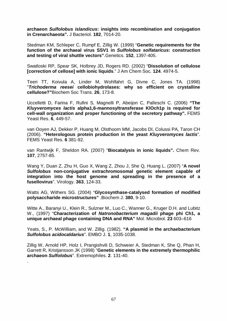
67
archaeon Sulfolobus islandicus: insights into recombination and conjugation in Crenarchaeota”. J Bacteriol. 182, 7014-20. Stedman KM, Schleper C, Rumpf E, Zillig W. (1999) “Genetic requirements for the function of the archaeal virus SSV1 in Sulfolobus solfataricus: construction and testing of viral shuttle vectors”.Genetics. 152, 1397-405. Swatloski RP, Spear SK, Holbrey JD, Rogers RD. (2002) “Dissolution of cellulose [correction of cellose] with ionic liquids.” J Am Chem Soc. 124. 4974-5. Teeri TT, Koivula A, Linder M, Wohlfahrt G, Divne C, Jones TA. (1998) ”Trichoderma reesei cellobiohydrolases: why so efficient on crystalline cellulose?”Biochem Soc Trans. 26, 173-8. Uccelletti D, Farina F, Rufini S, Magnelli P, Abeijon C, Palleschi C. (2006) “The Kluyveromyces lactis alpha1,6-mannosyltransferase KlOch1p is required for cell-wall organization and proper functioning of the secretory pathway”. FEMS Yeast Res. 6, 449-57. van Ooyen AJ, Dekker P, Huang M, Olsthoorn MM, Jacobs DI, Colussi PA, Taron CH (2006). “Heterologous protein production in the yeast Kluyveromyces lactis”. FEMS Yeast Res. 6 381-92. van Rantwijk F, Sheldon RA. (2007) “Biocatalysis in ionic liquids”. Chem Rev. 107, 2757-85. Wang Y, Duan Z, Zhu H, Guo X, Wang Z, Zhou J, She Q, Huang L. (2007) “A novel Sulfolobus non-conjugative extrachromosomal genetic element capable of integration into the host genome and spreading in the presence of a fusellovirus”. Virology. 363, 124-33. Watts AG, Withers SG. (2004) “Glycosynthase-catalysed formation of modified polysaccharide microstructures” .Biochem J. 380, 9-10. Witte A., Baranyi U., Klein R., Sulzner M., Luo C., Wanner G., Kruger D.H. and Lubitz W., (1997) “Characterization of Natronobacterium magadii phage phi Ch1, a unique archaeal phage containing DNA and RNA” Mol. Microbiol. 23
603–616
Yeats, S., P. McWilliam, and W. Zillig. (1982). “A plasmid in the archaebacterium Sulfolobus acidocaldarius”. EMBO J. 1, 1035-1038. Zillig W, Arnold HP, Holz I, Prangishvili D, Schweier A, Stedman K, She Q, Phan H, Garrett R, Kristjansson JK (1998) ”Genetic elements in the extremely thermophilic archaeon Sulfolobus”. Extremophiles. 2. 131-40. .
68
Pubblications: Aucelli T., Contursi P., Girfoglio M., Rossi M., Cannio R. "A spreadable, non-integrative and high copy number shuttle vector for Sulfolobus solfataricus based on the genetic element pSSVx from Sulfolobus islandicus." Nucleic Acids Res. (2006) Comunications: Girfoglio M., Rossi M., Cannio R. “Acetate kinase from moderately thermophilic and hyperthermophilic sources: structure-function relationship” International Conference on Extremophiles, 17-21 Sept. 2006 Brest, France
A spreadable, non-integrative and high copy numbershuttle vector forSulfolobus solfataricus based on thegenetic element pSSVx from Sulfolobus islandicusTiziana Aucelli1, Patrizia Contursi1, Michele Girfoglio, Mose Rossi and Raffaele Cannio*
Istituto di Biochimica delle Proteine, Via P. Castellino 111, 80131 CNR, Napoli, Italy and 1Dipartimento di BiologiaStrutturale e Funzionale, Universita degli Studi di Napoli Federico II, Via Cinthia, 80126 Napoli, Italy
Received July 5, 2006; Revised and Accepted August 5, 2006
ABSTRACT
The pSSVx genetic element from Sulfolobusislandicus REY15/4 is a hybrid between a plasmidand a fusellovirus, able to be maintained in non-integrative form and to spread when the helperSSV2 virus is present in the cells. In this work,the satellite virus was engineered to obtain anEscherichia coli–Sulfolobus solfataricus shuttlevector for gene transfer and expression inS.solfataricus by fusing site-specifically the pSSVxchromosome with an E.coli plasmid replicon andthe ampicillin resistance gene. The pSSVx-basedvector was proven functional like the parental virus,namely it was able to spread efficiently throughinfected S.solfataricus cells. Moreover, the hybridplasmid stably transformed S.solfataricus andpropagated with no rearrangement, recombinationor integration into the host chromosome. Thehigh copy number of the artificial genetic elementwas found comparable with that calculated for thewild-type pSSVx in the new host cells, with no needof genetic markers for vector maintenance in thecells and for transfomant enrichment.
The newly constructed vector was also shown tobe an efficient cloning vehicle for the expressionof passenger genes in S.solfataricus. In fact, aderivative plasmid carrying an expression cassetteof the lacS gene encoding the b-glycosidase fromS.solfataricus under the control of the Sulfolobuschaperonine (thermosome tf55) heat shock pro-moter was also able to drive the expression of afunctional enzyme. Complementation of the b-galac-tosidase deficiency in a deletion mutant strain ofS.solfataricus demonstrated that lacS gene wasan efficient marker for selection of single transfor-mants on solid minimal lactose medium.
INTRODUCTION
Host/virus interaction modes have provided windows to studymicrobial diversity (1) as well as genetic processes at themolecular level, in particular for prokaryotes, and hencehave helped in clarifying the physiological mechanisms, thedependence on the specific biochemical environment andevolution of their host cells (2,3).
Very few viruses have been identified from Archaea (4) ascompared with Bacteria and Eukarya and detailed descrip-tion has been reported for those from hyperthermophilicarchaea (5,6) with representatives that replicate in thegenus Sulfolobus being the majority within the kingdomCrenarchaeota (6–8). To date, the Fuselloviridae are themost widespread on earth in the Sulfolobus genus withviruses sharing similar morphology as well as DNA genomesize and organization (9–12).
Sulfolobus spindle-shaped virus 1 (SSV1) is the best stud-ied member of this family and demonstrated to be temperateboth in Sulfolobus shibatae and in non-natural but relatedSulfolobus hosts, such as Sulfolobus solfataricus (13,14);infection, integration of DNA into the host chromosomeand production of virions cause apparently no phenotypechange but a significant growth retardation of the host cellswhich can be visualized as turbid plaques around propagationfoci on plated lawns of indicator host cells (14–18).
More recently, another fusellovirus, SSV2 from Sulfolobusislandicus strain 15/4 was isolated, characterized and itscomplete genomic sequence determined. SSV2 shares withSSV1 similar morphology, replication and DNA size (19).The overall genome architecture is conserved but the lowsimilarity in the sequences should be responsible for thehigher copy number and the lack of a strong ultraviolet induc-tion of episomal SSV2 DNA and particle production, as wellas for the different integration of the SSV2 genome whichoccurs into the host chromosome at the site of a glycyltRNA instead of arginyl-tRNA (12).
S.islandicus REY15/4 harbors also a small plasmid, pSSVx,assigned to the pRN family (20,21) of Sulfolobales plasmids;pSSVx is also capable of spreading in the cell cultures of
*To whom correspondence should be addressed at Istituto di Biochimica delle Proteine, Consiglio Nazionale delle Ricerche, Via Pietro Castellino 111, 80131,Naples, Italy. Tel: +39 081 613 2285; Fax: +39 081 613 2248; Email: [email protected]
� 2006 The Author(s).This is an Open Access article distributed under the terms of the Creative Commons Attribution Non-Commercial License (http://creativecommons.org/licenses/by-nc/2.0/uk/) which permits unrestricted non-commercial use, distribution, and reproduction in any medium, provided the original work is properly cited.
Published online 13 September 2006 Nucleic Acids Research, 2006, Vol. 34, No. 17 e114doi:10.1093/nar/gkl615

S.solfataricus but only in the presence of either SSV2 orSSV1, necessary as helpers (9). In fact, pSSVx containstwo open reading frames showing high-sequence similarityto a tandem of ORFs in both SSV1 and SSV2 genomes; theproteins encoded by these ORFs are probably necessary forspecific recognition of the pSSVx DNA but need viral helpercomponents for capsid formation and packaging (9,19).
In general, the choice of S.solfataricus as a model forfundamental understanding of the genetics of extremelythermophilic archaea is due to growth conditions operativelynon-prohibitive (22) and capability of maintaining andpropagating either natural or genetically modified extrachro-mosomal DNAs (23,24) from other sources. The completegenome of S.solfataricus has also been determined (25), thebiochemical characterization of many gene products obtained(26) and the development of post-genomics tools such asproteomics and metabolic pathway reconstruction recentlyattempted (27,28). Some progress has been made to developstable transformation (29–32), specific gene disruption meth-ods (33) as well as overexpression of foreign and homologousgenes (34); nevertheless none of the systems described sofar has been proven efficient for reproducibility and stabilityof gene cloning and protein expression levels in Sulfolobus,probably due, in most cases, to low transformation efficien-cies, inefficient selection and/or instability of the vectors inthe host as well as changes in the ratio episomal versusintegrative forms occurring in the cell during replication ofvirus-derived constructs (35).
In this study, a genetic system for Sulfolobus wasdeveloped that is based on the satellite virus pSSVx fromS. islandicus 15/4. The different recombinant Escherichiacoli–Sulfolobus solfataricus shuttle vectors constructedretained the wild-type capability to replicate at high copy-number and to spread in cell cultures in the presence of itshelper virus SSV2. Sulfolobus transformants were demon-strated to be stable and propagate the pSSVx derivedplasmids in a reproducible and constant fashion withoutany rearrangement, recombination or integration into thechromosome.
Moreover, stable complementation of a b-galactosidasemutant of S.solfataricus previously isolated and characterizedin our laboratory (32) and reproducible gene expressionlevels were also obtained by introducing the b-galactosidasegene (lacS) as a reporter under the control of a strong andheat-inducible promoter into the shuttle vector.
MATERIALS AND METHODS
Growth of Sulfolobus strains and isolation ofSSV2-infected S.solfataricus GuW and P2
S.solfataricus strains P2 (DSM 1617), Gq (23) and the deriva-tive mutant GqW [DlacS, (32)] as well as S.islandicus REY15/4 (22) were grown at 75 or 80�C in glycine bufferedBrock’s medium (36) with 0.1% tryptone, 0.05% yeastextract and 0.2% sucrose at pH 3.2. For electroporation andplaque assays, cells were grown with phosphate bufferedmedium N.182 (M182) suggested by the DSMZ Catalogueof strains containing 0.1% glucose. For isolation of indepen-dent clones, medium M182 contained only 0.25% lactose andno other nutrient. The optical density of liquid cultures was
monitored at 600 nm. For solid media, gellan gum (Gelrite,Sigma) was added to a final concentration of 0.8% (0.35%for overlays), and MgCl2 and CaCl2 were added to 10.0and 3.0 mM, respectively.
The S.solfataricus strains GqW and P2 transfected with theSSV2 and pSSVx were extracted from zones of growthinhibition (plaques) formed on indicator lawns around spotsof the S.islandicus REY 15/4 culture supernatants (2–6 mlfor each spot), as described by Arnold et al. (9). Clonescured selectively for pSSVx were obtained by isolation ofsingle colonies formed on plates by the cells extracted fromplaques and revitalized in liquid cultures.
Viral DNA isolation and plasmid constructions
Extrachromosomal SSV2 and pSSVx DNAs from bothS.islandicus REY 15/4 and transfectants of S.solfataricusstrains were performed with Qiaprep Spin Miniprep kit(Qiagen) following the standard procedure suggested by themanufacturer for E.coli cells.
Plasmid pSSVrt was constructed by cloning the pSSVxDNA linearized at the position 2812 with AflIII and modifiedwith Klenow DNA polymerase into the SmaI site of thepUC19 E.coli vector. Linearization was obtained by AflIIIpartial digestion in the presence of ethidium bromide usinga protocol already described for the SSV1 virus DNAlinearized with Sau3AI (37). Clones with insertion at thespecific position were selected by restriction analysis of theresulting plasmid collection. A size reduced derivative ofpSSVrt was also constructed; the polycloning sequence(between the AatII and EcoRI sites) of the pUC28 vectorwas inserted into a 1812 bp AatII/AflIII DNA fragmentfrom pUC18 after suitable modification of incompatibleends. The pSSVx sequence was excised from the pSSVrtplasmid with SacI and PstI and inserted into the same sitesof the minimal plasmid obtained to produce the pMSSV vec-tor. An expression cassette of the lacS gene (38) was PCRamplified by the vector pMJ03 (35) and inserted betweenthe XhoI and PstI sites of pMSSV generating the expressionvector pMSSVlacS. Excision of the E.coli minimal plasmidwas obtained by digestion of pMSSVlacS (1.0 mg) withSacI and purification of the pSSVx/lacS moiety from agarosegel. The DNA was re-circularized by ligation in a finalvolume of 5.0 ml, diluted in water to 20 ng/ml final concentra-tion, and 2 ml were used for electroporation.
Transformation procedure and analyses ofSulfolobus transformants
S.solfataricus cells of SSV2 lysogenes were grown up to mid-logarithmic phase (0.3–0.45 OD600), harvested by centrifuga-tion and repeatedly washed in 20 mM sucrose as describedpreviously by Schleper et al. (14). Aliquots of 1010 cells/ml(50 ml) were mixed with 1 ml DNA (10–100 ng/ml), incubatedfor 1 min on ice and transferred to chilled plastic cuvetteswith an electrode gap of 0.1 cm (BioRad). High voltageelectroporation (25 mF) was performed with a BioRad GenePulser Xcell� at a field strength of 1.5 kV/cm and 400 Wresistance; two successive shock pulses were applied to com-petent cells producing pulse length of �10.0 and 9.8 ms,respectively. Immediately after electroporation cell mixtureswere diluted with 1 ml of medium M182 containing 0.1%
e114 Nucleic Acids Research, 2006, Vol. 34, No. 17 PAGE 2 OF 12
glucose, transferred to glass vials and incubated for 3 h at 75or 80�C. After suitable scale-up, 5–15 ml aliquots ofthe cultures (the volumes varying in order to withdraw thesame number of cells per aliquot) were harvested at increas-ing cell density for DNA extractions. For monitoring propa-gation of the recombinant satellite virus, extrachromosomalDNA mini preparations and plaque assays were performed.Transformants were stored at �80�C in 15% glycerol stocks.
For Southern blot analysis, �2 mg of total cellular DNAs,extracted according to Arnold et al. (9), and 5 ng ofpMSSVlacS plasmid purified from E.coli, were cut withHindIII and BglII, and electrophoresed in a 0.8% agarosegel; DNA digests were blotted and hybridized according tostandard procedures (39). The probe was prepared by cuttingout and purifying a HindIII restriction fragment from thepMSSVlacS vector encompassing the lacS gene and a portion(up to �302) of the tf55a 50 flanking region. This restrictionDNA fragment was randomly labeled using the random primeDNA labeling kit (Boehringer Mannheim).
b-galactosidase complementation and isolation ofmixed and single transformants
pMSSVlacS transformed cells were tested for b-galactosidaseactivity; 1 ml aliquots of cultures were centrifuged and cellpellets overlaid with a X-Gal solution (2 mg/ml in phosphatebuffered medium) and incubated for 15 min at 75�C forblue color development. In situ assays were performed onthe same cultures seeded on plates and grown as circularcolonized areas, as previously described (32). Supernatantsof cultures grown up to 0.5, 1.0 and 1.3 OD600 were checkedfor plaque formation as already described and analyzed byX-Gal staining of plaques formed on continuous lawns ofthe S.solfataricus GqW strain. For infection in liquid culture,400 ml supernatants from pMSSVlacS transformants wereadded to a 20 ml culture of cells transfected only withSSV2 and grown up to 0.5 OD600. After incubation undershaking for 48 h, cells were diluted 1:50, grown up to 0.36and 1.0 OD600 and tested for b-galactosidase activity asalready described.
Single transformants were selected either on rich (M182,glucose 0.1%) or on minimal (lactose 0.25% as the only nutri-ent) solid media, by plating or by streaking of electroporedcells propagated until they showed positive staining withX-Gal and diluted after they had reached different cell densi-ties (0.3–1.0 OD600). Single clones formed after 2 weeks ofincubation at 75�C were stained with X-Gal (incubation at75�C for 3–4 h for color development on lactose plates),picked and resuspended in 100 ml lactose minimal mediumand seeded as spots onto fresh rich (M182) medium plates.After in situ X-Gal test on plates, cell spots were transferredto M182 medium containing glucose, propagated up to earlystationary phase (1.0 OD600) and analyzed for extrachromo-somal DNA content as described above.
b-galactosidase enzyme activity
b-galactosidase activity of transformants was visualizedand quantified as follows. Crude extracts were prepared bya freeze thaw technique, suspension of the cells in 10 mMTris–HCl buffer, pH 8.0, placing at 80�C for 10 min, thenat 50�C for 5 min. This procedure was repeated four times,
and the extract was spun for 30 min at �10 000g. Thesupernatant was either assayed immediately or stored at�80�C before testing. Protein concentrations of the crudecell extracts were determined by the Bradford assay (Bio-Rad). Detection of the b-galacosidase in protein extractswas performed on 10% acrylamide SDS–PAGE gels afterelectrophoretic separation and extensive washing in 10 mMTris–HCl buffer, pH 8.0. The specific enzyme band was visu-alized incubating the gel 30 min at 75�C in the same buffercontaining 2 mg/ml X-gal.
The b-galactosidase assay procedure followed essentiallythe protocol of Pisani et al. (40). A sample of 10 ml extra-ct was transferred to a preheated (75�C) quartz cuvettecontaining 990 ml assay buffer (2.8 mM ONPG in 50 mMsodium phosphate buffer, pH 6.5). The ONPG hydrolysisreaction was followed spectrophotometrically at 75�C bymeasuring the increase in absorbance at 405 nm in a Beck-man spectrophotometer with heatable cuvettes. One unitwas defined as the amount of enzyme catalyzing the hydrol-ysis of 1 mmol of ONPG min�1 at 75�C with a molar absorp-tion coefficient of 3100 M�1 cm�1 at 405 nm for ONP.
RESULTS
Transfection and isolation of Sulfolobus solfataricusSSV2 transfectants
The strains P2 and GqW, a b-glycosidase defective mutantisolated earlier (32), were tested for susceptibility to infec-tion by SSV2 and pSSVx viruses produced by S.islandicusREY15/4. Supernatants of REY15/4 cultures generatedgrowth inhibition halos on continuous lawns of bothS.solfataricus strains (Figure 1A), similar to those observedfor the strain P1 (9); like for SSV1 virus (37), plaques didnot form on Sulfolobus acidocaldarius cells, namely no infec-tion occurred. Cells extracted from areas of these plaquesand revitalized in liquid cultures contained extrachromosomalDNA indistinguishable from that isolated from REY15/4,indicating the presence and active replication of both virusand satellite elements (Figure 1B). From this culture, clonesinfected only by the SSV2 virus, namely cured for thepSSVx, were isolated as single colonies on plates aftersuitable dilutions, as described for the P1 strain by Arnoldet al. (9). The absence of the satellite virus was confirmedby restriction analysis of extrachromosomal DNA(Figure 1C) and Southern hybridization (data not shown).
Shuttle vector construction
A sequence analysis on the pSSVx DNA was performedin order to locate regions that could be manipulated withoutaffecting DNA replication and particle proliferation/spreading, and thus representing candidate targets for site-specific insertion of foreign DNA sequences. A 200 bp seg-ment was identified which contains the tail-to-tail intergenicregion between the ORFs C68 and 288. The segment showsarchaeal transcription termination signals (41) and tendencyto form hairpin loops as for rho-independent terminationmechanisms (42,43). Moreover, it has an AflIII site (cut atthe position 2812 on the pSSVx genome), useful for cloning,since it is situated in the 30 direction beyond the ORF288
PAGE 3 OF 12 Nucleic Acids Research, 2006, Vol. 34, No. 17 e114

transcription termination region, and far upstream of the ORFc68 stop codon. Since the AflIII site is present in five copieson the pSSVx sequence (positions 677, 814, 982, 2812 and4994), singly cut pSSVx genomes were generated by diges-tion with the restriction enzyme under conditions that allowedsingle cleavage of the DNA molecules presumably at everyspecific site (37). After suitable modification of termini,these DNAs were inserted into E.coli pUC19 plasmid vectorand specific insertion at the position 2812 produced the fusionplasmid pSSVrt (Figure 2).
pSSVrt shuttle vector transfection and spreading
The plasmid DNA pSSVrt selected from E.coli was trans-ferred by electroporation into Sulfolobus, using differentstrains, namely SSV2 lysogens of the strains P2, MT4 andGqW, which is a stable b-galactosidase mutant with anextended deletion in the lacS genetic locus (32). To test trans-formation efficiency, S.solfataricus was transformed withvarying amounts of the plasmid pSSVrt (10–100 ng) andthen checked for the presence and amount of the vector atdifferent cell densities and after several generations. Threehours after electroporation no extrachromosomal DNAcould be detected, confirming the low transformation effici-ency already determined both by Schleper et al. (14) andCannio et al. (23). Nevertheless the plasmid pSSVrt spreadefficiently throughout a culture after transformation; in factafter the first scale-up from 3 to 50 ml of the culture, theamount of the plasmid and of the helper virus SSV2 increasedand could be detected by ethidium bromide fluorescence onagarose gels (Figure 3). Identical results were obtained withall the different DNA concentrations used for electroporationbut a lower amount (�5-fold) of the hybrid plasmid couldbe detected in the P2 and MT4 (data not shown) strainswhen compared with the GqW strain (Figure 3).
As expected, the same experiments performed on wild-typestrains not infected with SSV2 produced no transformationby the shuttle vector, confirming the need of the helpervirus for the propagation of the engineered plasmid.
10.008.006.005.004.003.002.001.501.00
0.50 0.80
0.60
1.000.90
1.521.20
0.50
0.70
A
CB
1 2 3 4 5 6 7 1 2 3 4 5 6 7 8
SSV2
pSSVx
Figure 1. Transfection of S.solfataricus GqW with conditioned medium of S.islandicus REY15/4. The spontaneous mutant GqW lacking the b-glycosidaseactivity was first tested for infection with both SSV2 and pSSVx by formation of growth inhibition areas in cell lawns on plate. Virus plaques of SSV2 andpSSVx were obtained by spotting aliquots of REY 15/4 supernatants (2 ml for the three plaques in the first row, 4 ml for those in the second row and 6 ml for thosein the third row) onto a continuous indicator lawn of the GqW cells (A). (B) Extrachromosomal DNAs (uncut in lanes 1 and 5), extracted from both the naturalsource S.islandicus (lanes 1–3) and the infected S.solfataricus (lanes 5–7), propagated after extraction from the plaques and revitalization, was cut with EcoRI(lanes 2 and 6) and BamHI (lanes 3 and 7). Episomal DNAs were confirmed to be identical and to contain both SSV2 and pSSVx DNAs, as demonstrated by thetwo distinct EcoRI restriction patterns. Lane 4: DNA molecular weight standards (kb). (C) The cells entrapped inside the plaques have been extracted from theplates, revitalized in liquid medium and then streaked onto solid medium to obtain single colonies. The comparative analysis with extrachromosomal DNAs fromnon-transformed recipient cells (lanes 1 and 5) and from the cells transfected with both viral and satellite DNAs (lanes 3 and 7) revealed the absence of thepSSVx plasmid in one colony isolated on plate and analyzed (lanes 2 and 6). Lanes 1–3: uncut DNAs; lanes 5–7: EcoRI digests; lanes 4 and 8: DNA molecularweight standards (kb).
pSSVrt8395 bps
2000
4000
6000
8000
NdeIEcoRISacIKpnI
SpeI
HindIIINdeI
EcoRI
EcoRIHindIII
XbaIAflII
AflIII
XbaIAvaISmaI
EcoRI
ClaINdeI
BamHIKpnI
HindIIIEcoRI
EcoRI
AflIII
AflIIIAflIII
BamHIEcoRI
Tth111I
EcoRI
BamHIXbaISalIPstI
SphIHindIII
AflIII
AatII
892/Rep
154
288
bla
Figure 2. Plasmid map of the pSSVx/pUC19 shuttle vector. The pSSVrtshuttle vector has been obtained by cloning the pUC19 bacterial sequence(heavy bar) into an AflIII restriction site (all AflIII sites are underlined)downstream of the ORF288 of the pSSVx plasmid and by propagation inE.coli. Main ORFs allow easy location of archaeal (replicase ORF892/Repas well as ORF154 and ORF288 putative DNA packaging components ofpSSVx) and bacterial (bla, ampicillin resistance gene) sequences; bold faceletters indicate unique restriction sites.
e114 Nucleic Acids Research, 2006, Vol. 34, No. 17 PAGE 4 OF 12
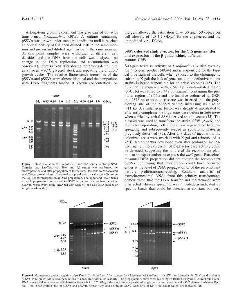
A long-term growth experiment was also carried out withtransformed S.solfataricus GqW. A culture containingpSSVrt was grown under standard conditions until it reachedan optical density of 0.8, then diluted 1:10 in the same med-ium and grown and diluted again twice in the same manner.At this point samples were withdrawn at different celldensities and the DNA from the cells was analyzed; nochange in the DNA replication and accumulation wasobserved (Figure 4) even after storing the propagated cultureas a frozen �80�C glycerol stock and repeating the dilution/growth cycles. The relative fluorescence intensities of thepSSVrt and pSSVx were almost identical and the comparisonwith DNA fragments loaded at known concentrations on
the gels allowed the estimation of �130 and 150 copies percell (density of 1.0–1.2 OD600) for the engineered and theunmodified viral DNAs.
pSSVx-derived shuttle vectors for the lacS gene transferand expression in the b-galactosidase deficientmutant GuW
b-D-galactosidase activity of S.solfataricus is displayed bythe lacS gene product (40,44) and is responsible for the typi-cal blue stain of the cells when exposed to the chromogenicsubstrate, X-gal; the lack of gene function in defective mutantstrains is hence responsible for colorless colonies (45). ThelacS coding sequence with a 648 bp 30-untranslated region(30-UTR) was fused to a 448 bp fragment containing the pro-moter region of tf55a and the first five codons of its ORF;this 2578 bp expression cassette was inserted into the poly-cloning site of the pSSVrt vector, increasing its size to�11 kb. A similar gene fusion was already demonstrated toefficiently complement a b-galactosidase defect in Sulfolobuswhen carried by a viral SSV1-derived shuttle vector (35). Theplasmid was used to transform the strain GqW (DlacS) andafter electroporation, cell culture was regenerated to allowspreading and subsequently seeded as spots onto plates aspreviously described (32). After 2–3 days of incubation, thecolonized areas were overlaid with X-gal and reincubated at75�C. No color was developed even after prolonged incuba-tion, namely no expression of b-galactosidase activity couldbe detected, suggesting the failure of the recombinant plas-mid to transport and/or to express the lacS gene. Extrachro-mosomal DNA preparation did not contain the recombinantpSSVx confirming that interference could have occurredeither at the level of DNA propagation or of the recombinantparticle proliferation/spreading. Southern analysis ofextrachromosomal DNAs from this primary transformantsdemonstrated that the DNA transfer and maintenance wereunaffected whereas spreading was impeded, as indicated byspecific bands that could be detected at constant but very
KpnI
OD600
21. 23
5.154.974.273.53
2.031.901.58
12.229.16
6.115.10
3.05
2.041.64
1.02
pSSVrt
SSV2
pSSVx
SmaI
pSSVx
pSSVrt
SSV2
Figure 4.Maintenance and propagation of pSSVrt in S.solfataricus. After storage, SSV2 lysogens of S.solfataricus GqW transformed with pSSVrt and wild-typepSSVx were grown for several generations to check transformation stability. The propagated cultures were tested by restriction analysis of extrachromosomalDNAs (extracted at increasing cell densities from �0.5 to 1.2 OD600); the SmaI enzyme produced single cuts in both satellite and SSV2 elements, whereas KpnIhad 1 and 2 recognition sites in pSSVx and pSSVrt, respectively, and no site on SSV2. Standards of DNA molecular weight are indicated (kb).
OD6000.65 0.84 1.21.0 0.60 0.80 1.0M1 M2
21. 23
5.154.97 4.273.53
2.031.901.581.38
0.950.83
12.2210.18
11.209.16
8.14 7.136.11 5.104.073.05
2.041.64
1.02
P2GθθW
Figure 3. Transformation of S.solfataricus with the shuttle vector pSSVrt.Transfer into S.solfataricus GqW and P2 strains was performed byelectroporation and after propagation of the cultures, the cells were harvestedat different growth phases (indicated as optical density values at 600 nm onthe top) for extrachromosomal DNA preparation. The upper and lower bandsin each preparation correspond to SSV2 virus and recombinant satellitepSSVrt, respectively, both linearized with SalI. M1 and M2: DNA molecularweight markers (kb).
PAGE 5 OF 12 Nucleic Acids Research, 2006, Vol. 34, No. 17 e114
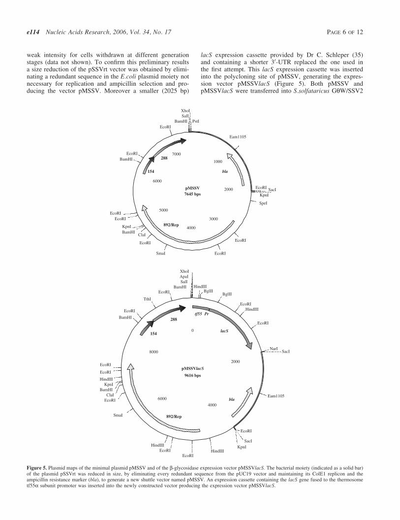
weak intensity for cells withdrawn at different generationstages (data not shown). To confirm this preliminary resultsa size reduction of the pSSVrt vector was obtained by elimi-nating a redundant sequence in the E.coli plasmid moiety notnecessary for replication and ampicillin selection and pro-ducing the vector pMSSV. Moreover a smaller (2025 bp)
lacS expression cassette provided by Dr C. Schleper (35)and containing a shorter 30-UTR replaced the one used inthe first attempt. This lacS expression cassette was insertedinto the polycloning site of pMSSV, generating the expres-sion vector pMSSVlacS (Figure 5). Both pMSSV andpMSSVlacS were transferred into S.solfataricus GqW/SSV2
pMSSV7645 bps
1000
2000
3000
4000
5000
6000
7000
KpnI
SpeI
EcoRI
EcoRISmaI
EcoRI
ClaIBamHIKpnI
EcoRIEcoRI
BamHIEcoRI
EcoRI
Eam1105
EcoRI SacI
BamHISalIXhoI
892/Rep
bla154
288
pMSSVlacS
9616 bps
HindIII
EcoRIHindIII
EcoRI
NarI
Eam1105
EcoRI
KpnIHindIII
EcoRIEcoRI
HindIII
SmaI
EcoRIClaI
BamHIKpnI
HindIII
EcoRI
EcoRI
BamHI
EcoRI
TthI
EcoRIBamHI
SalIApaIXhoI
lacS
bla
154
288
2000
4000
6000
8000
0
892/Rep
tf55 Pr
SacI
SacI
BglIIBglII
PstI
Figure 5. Plasmid maps of the minimal plasmid pMSSV and of the b-glycosidase expression vector pMSSVlacS. The bacterial moiety (indicated as a solid bar)of the plasmid pSSVrt was reduced in size, by eliminating every redundant sequence from the pUC19 vector and maintaining its ColE1 replicon and theampicillin resistance marker (bla), to generate a new shuttle vector named pMSSV. An expression cassette containing the lacS gene fused to the thermosometf55a subunit promoter was inserted into the newly constructed vector producing the expression vector pMSSVlacS.
e114 Nucleic Acids Research, 2006, Vol. 34, No. 17 PAGE 6 OF 12

cells by electroporation, whereas pMSSVlacS was alsotransferred into cells of the P2/SSV2 strain. After culturepropagation, extrachromosomal DNAs were prepared andanalyzed by agarose electrophoresis (Figure 6). The presenceand growth-dependent accumulation of both vectors in GqW/SSV2 and of pMSSVlacS in P2/SSV2 revealed successfultransformation and DNA replication, confirming that the plas-mid size was critical for particle formation and spreading;moreover the lacS gene is harmless and does not inducerecombination of the vector also in the P2 strain whichalready contains a wild-type chromosomal copy of the gene.Identical results were obtained when SSV2 lysogens of
Sulfolobus GqW were transformed with the plasmidpSSVxlacS lacking the E.coli sequences and obtained bycleavage with SacI and re-ligation (Figure 6). All pSSVxderived plasmid failed to transform Sulfolobus if linearizedprior to transfer. In fact, X-Gal staining test was positive onSulfolobus GqW/SSV2 transformed with pMSSVlacS (orpSSVxlacS, data not shown) on liquid-cultured (Figure 7A),plated (Figure 7B), and primary infected cells (Figure 7C).Plaques depicted in Figure 7C stained after incubation withthe chromogenic substrate as a result of propagation of theengineered satellite virus. Since cell growth retardation inthe plaques is directly proportional to the SSV2 virus titer
Figure 6. Transformation of S.solfataricus with the vectors pMSSV and pMSSVlacS. Competent cells of S.solfataricus P2 and GqW infected with the SSV2 viruswere electroporated with the vector carrying the b-glycosidase gene pMSSVlacS. Strain GqW was also transformed with pSSVxlacS, a pMSSVlacS-derivedplasmid obtained by excision of the E.coli-specific sequences and re-ligation. pMSSV and wild-type pSSVx were also transferred into the GqW/SSV2 lysogenfor comparison of vector transfer and propagation efficiency in the presence and absence of the lacS gene. Extrachromosomal DNAs extracted from transformedcells after propagation of the cultures were checked by agarose electrophoresis for the presence of pMSSVlacS (all lanes in P2 and lanes 1, 3, 5 and 7 in GqW)and pMSSV (lanes 2, 4, 6 and 8 in GqW) at the different growth stages indicated as cell densities (OD600). Similarly, recovery of the pSSVxlacS plasmid(SacI cut, lanes 9 and 10) was monitored in comparison to the parental pSSVx (SmaI cut, lanes 11 and 12). Mobility of the virus SSV2 is highlighted. M1 and M2:molecular weight markers (kb).
A
1
2
C
OD600
0.5 1 .0
1.3
1
3
B
2
4
Figure 7. Complementation of the b-glycosidase mutation in S.solfataricus GqW and spreading of the pMSSVlacs vector. The expression vector pMSSVlacSwas transferred into S.solfataricus GqW cells lysogens for SSV2. Successful transformation was checked after propagation for several generation by directexposure of cell pellets from liquid cultures to X-Gal and development of the blue color [A (1) Lac+, pMSSVlacS transformed cells; (2) Lac�, cells transfectedwith pSSVx]. Maintenance of the plasmid was also confirmed for colonized areas on plates and X-Gal test [B (1) Strain GqW; (2) wild-type strain Gq; (3) SSV2/pMSSV-infected GqW; (4) SSV2/ pMSSVlacS -infected GqW]. C. 4-ml aliquots of culture supernatant of SSV2/pMSSVlacS-infected GqW, withdrawn at thedifferent cell densities indicated, were spotted onto a continuous lawn of uninfected GqW cells. The spreading of the recombinant satellite particles was revealedby the blue color developed on the plaques (primary infected cells) upon X-Gal exposure. Stain intensity depended on the extent of cell growth retardation in theplaques and was progressively less intense for increasingly higher virus titers.
PAGE 7 OF 12 Nucleic Acids Research, 2006, Vol. 34, No. 17 e114
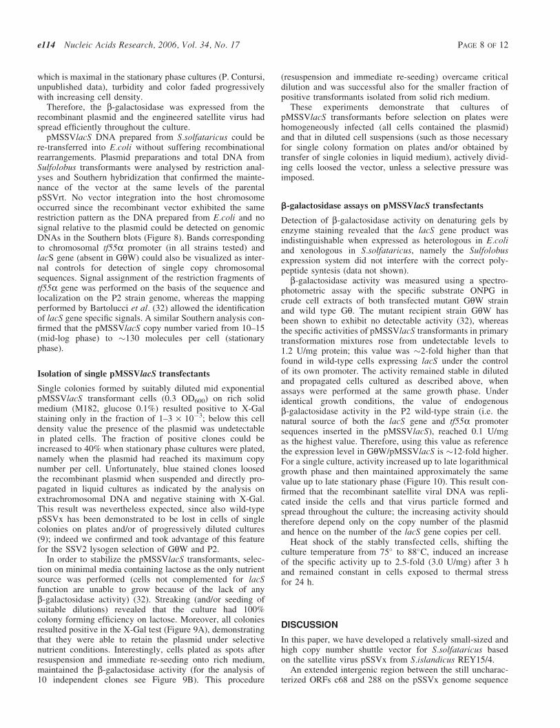
which is maximal in the stationary phase cultures (P. Contursi,unpublished data), turbidity and color faded progressivelywith increasing cell density.
Therefore, the b-galactosidase was expressed from therecombinant plasmid and the engineered satellite virus hadspread efficiently throughout the culture.
pMSSVlacS DNA prepared from S.solfataricus could bere-transferred into E.coli without suffering recombinationalrearrangements. Plasmid preparations and total DNA fromSulfolobus transformants were analysed by restriction anal-yses and Southern hybridization that confirmed the mainte-nance of the vector at the same levels of the parentalpSSVrt. No vector integration into the host chromosomeoccurred since the recombinant vector exhibited the samerestriction pattern as the DNA prepared from E.coli and nosignal relative to the plasmid could be detected on genomicDNAs in the Southern blots (Figure 8). Bands correspondingto chromosomal tf55a promoter (in all strains tested) andlacS gene (absent in GqW) could also be visualized as inter-nal controls for detection of single copy chromosomalsequences. Signal assignment of the restriction fragments oftf55a gene was performed on the basis of the sequence andlocalization on the P2 strain genome, whereas the mappingperformed by Bartolucci et al. (32) allowed the identificationof lacS gene specific signals. A similar Southern analysis con-firmed that the pMSSVlacS copy number varied from 10–15(mid-log phase) to �130 molecules per cell (stationaryphase).
Isolation of single pMSSVlacS transfectants
Single colonies formed by suitably diluted mid exponentialpMSSVlacS transformant cells (0.3 OD600) on rich solidmedium (M182, glucose 0.1%) resulted positive to X-Galstaining only in the fraction of 1–3 · 10�3; below this celldensity value the presence of the plasmid was undetectablein plated cells. The fraction of positive clones could beincreased to 40% when stationary phase cultures were plated,namely when the plasmid had reached its maximum copynumber per cell. Unfortunately, blue stained clones loosedthe recombinant plasmid when suspended and directly pro-pagated in liquid cultures as indicated by the analysis onextrachromosomal DNA and negative staining with X-Gal.This result was nevertheless expected, since also wild-typepSSVx has been demonstrated to be lost in cells of singlecolonies on plates and/or of progressively diluted cultures(9); indeed we confirmed and took advantage of this featurefor the SSV2 lysogen selection of GqW and P2.
In order to stabilize the pMSSVlacS transformants, selec-tion on minimal media containing lactose as the only nutrientsource was performed (cells not complemented for lacSfunction are unable to grow because of the lack of anyb-galactosidase activity) (32). Streaking (and/or seeding ofsuitable dilutions) revealed that the culture had 100%colony forming efficiency on lactose. Moreover, all coloniesresulted positive in the X-Gal test (Figure 9A), demonstratingthat they were able to retain the plasmid under selectivenutrient conditions. Interestingly, cells plated as spots afterresuspension and immediate re-seeding onto rich medium,maintained the b-galactosidase activity (for the analysis of10 independent clones see Figure 9B). This procedure
(resuspension and immediate re-seeding) overcame criticaldilution and was successful also for the smaller fraction ofpositive transformants isolated from solid rich medium.
These experiments demonstrate that cultures ofpMSSVlacS transformants before selection on plates werehomogeneously infected (all cells contained the plasmid)and that in diluted cell suspensions (such as those necessaryfor single colony formation on plates and/or obtained bytransfer of single colonies in liquid medium), actively divid-ing cells loosed the vector, unless a selective pressure wasimposed.
b-galactosidase assays on pMSSVlacS transfectants
Detection of b-galactosidase activity on denaturing gels byenzyme staining revealed that the lacS gene product wasindistinguishable when expressed as heterologous in E.coliand xenologous in S.solfataricus, namely the Sulfolobusexpression system did not interfere with the correct poly-peptide syntesis (data not shown).
b-galactosidase activity was measured using a spectro-photometric assay with the specific substrate ONPG incrude cell extracts of both transfected mutant GqW strainand wild type Gq. The mutant recipient strain GqW hasbeen shown to exhibit no detectable activity (32), whereasthe specific activities of pMSSVlacS transformants in primarytransformation mixtures rose from undetectable levels to1.2 U/mg protein; this value was �2-fold higher than thatfound in wild-type cells expressing lacS under the controlof its own promoter. The activity remained stable in dilutedand propagated cells cultured as described above, whenassays were performed at the same growth phase. Underidentical growth conditions, the value of endogenousb-galactosidase activity in the P2 wild-type strain (i.e. thenatural source of both the lacS gene and tf55a promotersequences inserted in the pMSSVlacS), reached 0.1 U/mgas the highest value. Therefore, using this value as referencethe expression level in GqW/pMSSVlacS is �12-fold higher.For a single culture, activity increased up to late logarithmicalgrowth phase and then maintained approximately the samevalue up to late stationary phase (Figure 10). This result con-firmed that the recombinant satellite viral DNA was repli-cated inside the cells and that virus particle formed andspread throughout the culture; the increasing activity shouldtherefore depend only on the copy number of the plasmidand hence on the number of the lacS gene copies per cell.
Heat shock of the stably transfected cells, shifting theculture temperature from 75� to 88�C, induced an increaseof the specific activity up to 2.5-fold (3.0 U/mg) after 3 hand remained constant in cells exposed to thermal stressfor 24 h.
DISCUSSION
In this paper, we have developed a relatively small-sized andhigh copy number shuttle vector for S.solfataricus basedon the satellite virus pSSVx from S.islandicus REY15/4.
An extended intergenic region between the still uncharac-terized ORFs c68 and 288 on the pSSVx genome sequence
e114 Nucleic Acids Research, 2006, Vol. 34, No. 17 PAGE 8 OF 12
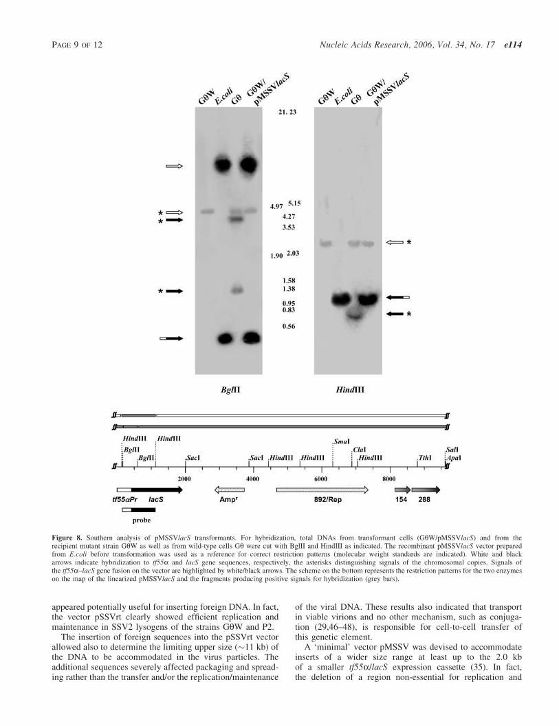
appeared potentially useful for inserting foreign DNA. In fact,the vector pSSVrt clearly showed efficient replication andmaintenance in SSV2 lysogens of the strains GqW and P2.
The insertion of foreign sequences into the pSSVrt vectorallowed also to determine the limiting upper size (�11 kb) ofthe DNA to be accommodated in the virus particles. Theadditional sequences severely affected packaging and spread-ing rather than the transfer and/or the replication/maintenance
of the viral DNA. These results also indicated that transportin viable virions and no other mechanism, such as conjuga-tion (29,46–48), is responsible for cell-to-cell transfer ofthis genetic element.
A ‘minimal’ vector pMSSV was devised to accommodateinserts of a wider size range at least up to the 2.0 kbof a smaller tf55a/lacS expression cassette (35). In fact,the deletion of a region non-essential for replication and
Figure 8. Southern analysis of pMSSVlacS transformants. For hybridization, total DNAs from transformant cells (GqW/pMSSVlacS) and from therecipient mutant strain GqW as well as from wild-type cells Gq were cut with BglII and HindIII as indicated. The recombinant pMSSVlacS vector preparedfrom E.coli before transformation was used as a reference for correct restriction patterns (molecular weight standards are indicated). White and blackarrows indicate hybridization to tf55a and lacS gene sequences, respectively, the asterisks distinguishing signals of the chromosomal copies. Signals ofthe tf55a–lacS gene fusion on the vector are highlighted by white/black arrows. The scheme on the bottom represents the restriction patterns for the two enzymeson the map of the linearized pMSSVlacS and the fragments producing positive signals for hybridization (grey bars).
PAGE 9 OF 12 Nucleic Acids Research, 2006, Vol. 34, No. 17 e114
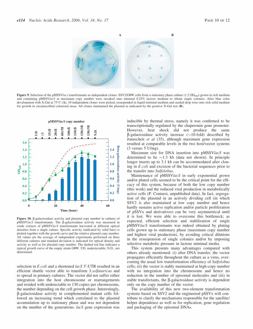
selection in E.coli and a shortened lacS 30-UTR resulted in anefficient shuttle vector able to transform S.solfataricus andto spread in primary cultures. The vector did not suffer eitherintegration into the host chromosome or rearrangementsand resided with undetectable to 130 copies per chromosome,the number depending on the cell growth phase. Interestingly,b-galactosidase activity in complemented mutant cells fol-lowed an increasing trend which correlated to the plasmidaccumulation up to stationary phase and was not dependenton the number of the generations. lacS gene expression was
inducible by thermal stress, namely it was confirmed to betranscriptionally regulated by the chaperonin gene promoter.However, heat shock did not produce the sameb-galactosidase activity increase (�10-fold) described byJonuscheit et al (35), although maximum gene expressionresulted at comparable levels in the two host/vector systems(3 versus 5 U/mg).
Maximum size for DNA insertion into pMSSVlacS wasdetermined to be �1.3 kb (data not shown). In principlelonger inserts up to 3.1 kb can be accommodated after clon-ing in E.coli and excision of the bacterial sequences prior tothe transfer into Sulfolobus.
Maintenance of pMSSVlacS in early exponential grownand/or plated cells seemed to be the critical point for the effi-cacy of this system, because of both the low copy number(this work) and the reduced viral production in metabolicallyactive cells (P. Contursi, unpublished data). In fact, segrega-tion of the plasmid in an actively dividing cell (in whichSSV2 is also maintained at low copy number and hencehardly sustains active replication and/or particle proliferationof pSSVx and derivatives) can be very asymmetrical untilit is lost. We were able to overcome this bottleneck; asexpected, efficient selection and stabilization of singlepMSSVlacS transformants was indeed obtained by platingcells grown up to stationary phase (maximum copy numberand highest viral production), by avoiding critical dilutionsin the resuspension of single colonies and/or by imposingselective metabolic pressure in lactose minimal media.
This system presents many advantages compared withothers already mentioned: (i) after DNA transfer, the vectorpropagates efficiently throughout the culture as a virus, over-coming the usual low transformation efficiency of Sulfolobuscells; (ii) the vector is stably maintained at high-copy numberwith no integration into the chromosome and hence noreduction in the number of episomal molecules and (iii) instable transfectants, the b-galactosidase activity is dependentonly on the copy number of the vector.
The availability of this new two-element transformationsystems based on SSV2 and the engineered pSSVx will con-tribute to clarify the mechanisms responsible for the satellite/helper dependence as well as for replication, gene regulationand packaging of the episomal DNAs.
A B
Figure 9. Selection of the pMSSVlacs transformants as independent clones. SSV2/GqW cells from a stationary phase culture (1.2 OD600) grown in rich mediumand containing pMSSVlacS at maximum copy number were streaked onto minimal 0.25% lactose medium to obtain single colonies. After blue colordevelopment with X-Gal at 75�C (A), 10 independent clones were picked, resuspended in liquid minimal medium and seeded drop wise onto rich solid mediumfor growth in circumscribed colonized areas. All clones maintained the plasmid as indicated by the positive X-Gal test (B).
6040200
1.6
1.4
1.2
1.0
0.8
0.6
0.4
0.2
0.0
OD
600
Beta-galactosidase activity (U
/mg)
1.2
1.0
0.8
0.6
0.4
0.2
0.0
pMSSVlacS copy number
130±
10
130±
10
N.D
.
110±
5
100±
5
60±1
0
20±5
15±5
UD
≤1
1.4
Time (hour)
Figure 10. b-galactosidase activity and plasmid copy number in cultures ofpMSSVlacS transformants. The b-galactosidase activity was measured incrude extracts of pMSSVlacS transformants harvested at different opticaldensities from a single culture. Specific activity (indicated by solid bars) isplotted together with the growth curve and the relative plasmid copy number.All values are the average of independent experiments performed on threedifferent cultures and standard deviation is indicated for optical density andactivity as well as for plasmid copy number. The dashed red line indicates atypical growth curve of the empty strain GqW. UD, undectectable; N.D., notdetermined.
e114 Nucleic Acids Research, 2006, Vol. 34, No. 17 PAGE 10 OF 12
Further work will explore the use of this vector for theexpression of both homologous and heterologous genes inS.solfataricus as well as for testing Sulfolobus regulatorysequences.
ACKNOWLEDGEMENTS
The authors are grateful to Prof. Quinxin She for providingus with the strain REY 15/4 of S.islandicus. This workwas funded by the Ministero dell’Universita e della RicercaScientifica (Decreto Direttoriale Prot. N. 1105/2002).Funding to pay the Open Access publication charges forthis article was provided by ‘MIUR-Decreto Direttorialeprot. n. 1105/2002’ Project.
Conflict of interest statement. None declared.
REFERENCES
1. Weinbauer,M.G. and Rassoulzadegan,F. (2004) Are viruses drivingmicrobial diversification and diversity? Environ. Microbiol.,6, 1–11.
2. Forterre,P. (2001) New viruses for the new millennium. TrendsMicrobiol, 9, 114.
3. Wommack,K.E. and Colwell,R.R. (2000) Virioplankton: viruses inaquatic ecosystems. Microbiol. Mol. Biol. Rev., 64, 69–114.
4. Zillig,W., Prangishvilli,D., Schleper,C., Elferink,M., Albers,S.,Janekovic,D. and Gotz,D. (1996) Viruses, plasmids and other geneticelements of thermofilic and hypertermofilic Archaea. FEMS Microbiol.Rev., 18, 225–236.
5. Prangishvili,D. (2003) Evolutionary insights from studies on viruses ofhyperthermophilic Archaea. Res. Microbiol., 154, 289–294.
6. Snyder,J.C., Stedman,K., Rice,G., Wiedenheft,B., Spuhler,J. andYoung,M.J. (2003) Viruses of hyperthermophilic Archaea. Res.Microbiol., 154, 474–482.
7. Arnold,H.P., Ziese,U. and Zillig,W. (2000) SNDV, a novel virus of theextremely thermophilic and acidophilic archaeon Sulfolobus. Virology,272, 409–416.
8. Zillig,W., Arnold,H.P., Holz,I., Prangishvili,D., Schweier,A.,Stedman,K., She,Q., Phan,H., Garrett,R. and Kristjansson,J.K. (1998)Genetic elements in the extremely thermophilic archaeon Sulfolobus.Extremophiles, 2, 131–140.
9. Arnold,H.P., She,Q., Phan,H., Stedman,K., Prangishvilli,D., Holz,I.,Kristjansson,J.K., Garret,R. and Zillig,W. (1999) The genetic elementpSSVx of the extremely thermophilic crenarchaeon Sulfolobus is ahybrid between a plasmid and a virus. Mol. Microbiol., 34,217–226.
10. Rice,G., Stedman,K., Snyder,J., Wiedenheft,B., Willits,D.,Brumfield,S., McDermott,T. and Young,M.J. (2001) Viruses fromextreme thermal environments. Proc. Natl Acad. Sci. USA, 98,13341–1345.
11. Rice,G., Tang,L., Stedman,K., Roberto,F., Spuhler,J., Gillitzer,E.,Johnson,J.E., Douglas,T. and Young,M. (2004) The structure of athermophilic archaeal virus shows a double-stranded DNA viral capsidtype that spans all domains of life. Proc. Natl Acad. Sci. USA, 101,7716–7720.
12. Wiedenheft,B., Stedman,K., Roberto,F., Willits,D., Gleske,A.K.,Zoeller,L., Snyder,J., Douglas,T. and Young,M. (2004) Comparativegenomic analysis of hyperthermophilic archaeal Fuselloviridae viruses.J. Virol., 78, 1954–1961.
13. Martin,A., Yeats,S., Janekovic,D., Reiter,W.D., Aicher,W. andZillig,W. (1984) SAV1, a temperature u.v.-inducible DNA virus-likeparticle from the archaebacterium Sulfolobus acidocaldarius.EMBO J., 3, 2165–2168.
14. Schleper,C., Kubo,K. and Zillig,W. (1992) The particle SSV1 from theextremely thermophilic archaeon Sulfolobus is a virus: demonstrationof infectivity and of transfection with viral DNA. Proc. Natl Acad. Sci.USA, 89, 7645–7649.
15. Palm,P., Schleper,C., Grampp,B., Yeats,S., McWilliam,P., Reiter,W.D.and Zillig,W. (1991) Complete nucleotide sequence of the
virus SSV1 of the archaebacterium Sulfolobus shibatae. Virology,185, 245–250.
16. She,Q., Peng,X., Zillig,W. and Garret,R.A. (2001) Gene capture inarchaeal chromosomes. Nature, 409, 478.
17. Reiter,W.D., Zillig,W. and Palm,P. (1988) Archaebacterial viruses.Adv. Virus Res., 34, 143–188.
18. She,Q., Brugger,K. and Chen,L. (2002) Archaeal integrative geneticelements and their impact on genome evolution. Res Microbiol.,153, 325–332.
19. Stedman,K., She,Q., Phan,H., Arnold,H.P., Holtz,I., Garret,R. andZillig,W. (2003) Relationships between fuselloviruses infecting theextremely thermofilic archaeon Sulfolobus: SSV1 and SSV2.Res. Microbiol., 154, 295–302.
20. Kletzin,A., Lieke,A., Urich,T., Charlebois,R.L. and Sensen,C.W.(1999) Molecular analysis of pDL10 from Acidianus ambivalens fromextremely thermophilic and acidophilic Archaea. Genetics, 152,1307–1314.
21. Peng,X., Holz,I., Zillig,W., Garrett,R.A. and She,Q. (2000) Evolutionof the family of pRN plasmids and their integrase-mediated insertioninto the chromosome of the crenarchaeon Sulfolobus solfataricus.J. Mol. Biol., 303, 449–454.
22. Zillig,W., Kletzin,A., Schlepr,C., Holz,I., Janekovic,D., Nain,J.,Lanzerdorfer,M. and Kristjanson,J.K. (1994) Screening forSulfolobales, their plasmids and their viruses in Icelandic solfataras.Syst. Appl. Microbiol., 16, 609–628.
23. Cannio,R., Contursi,P., Rossi,M. and Bartolucci,S. (2001)Thermoadaptation of a mesophilic hygromicin B phosphotransferase bydirected evolution in hyperthermophilic Archaea: selection of a stablegenetic marker for DNA transfer into Sulfolobus solfataricus.Extremophiles, 5, 153–159.
24. Contursi,P., Cannio,R., Prato,S., Fiorentino,G., Rossi,M. andBartolucci,S. (2003) Development of a genetic system forhyperthermophilic Archaea: expression of a moderate thermophilicbacterial alcohol dehydrogenase gene in Sulfolobus solfataricus.FEMS Microbiol. Lett., 218, 115–120.
25. She,Q., Singh,R.K., Confalonieri,F., Zivanovic,Y., Allard,G.,Awayez,M.J., Chan-Weiher,C.C.Y., Clausen,I.G., Curtis,B.A.,De Moors,A. et al. (2001) The complete genome of thecrenarchaeon Sulfolobus solfataricus P2. Proc. Natl Acad. Sci. USA,98, 7835–7840.
26. Pisani,F.M. and Rossi,M. (1994) Evidence that an archaeal alpha-likeDNA polymerase has a modular organization of its associatedcatalytic activities. J. Biol. Chem., 269, 7887–7892.
27. Chong,P.K. and Wright,P.C. (2005) Identification andcharacterization of the Sulfolobus solfataricus P2 proteome.J. Proteome Res., 4, 1789–1798.
28. Snijders,A.P., de Koning,B. and Wright,P.C. (2005) Perturbation andinterpretation of nitrogen isotope distribution patterns in proteomics.J. Proteome Res., 4, 2185–2191.
29. Elferink,M., Schleper,C. and Zillig,W. (1996) Transformation of theextremely thermoacidophilic archaeon Sulfolobus solfataricus via aself-spreading vector. FEMS Microbiol. Lett., 137, 31–35.
30. Aravalli,R.N. and Garret,R. (1997) Shuttle vectors forhyperthermophilic archaea. Extremophiles, 1, 183–191.
31. Cannio,R., Contursi,P., Rossi,M. and Bartolucci,S. (1998) Anautonomously replicating vector for Sulfolobus solfataricus.J. Bacteriol., 180, 3237–3240.
32. Bartolucci,S., Rossi,M. and Cannio,R. (2003) Characterization andFunctional complementation of a nonlethal deletion in the cromosomeof a b-glycosidase mutant of Sulfolobus solfataricus. J. Bacteriol.,185, 3948–3957.
33. Worthington,P., Hoang,V., Perez-Pomares,F. and Blum,P. (2003)Targeted disruption of the alpha-amylase gene in thehyperthermophilic archaeon Sulfolobus solfataricus J. Bacteriol.,185, 482–488.
34. Albers,S.V., Jonuscheit,M., Dinkelaker,S., Urich,T., Kletzin,A.,Tampe,R., Driessen,AJ. and Schleper,C. (2006) Production ofrecombinant and tagged proteins in the hyperthermophilicarchaeon Sulfolobus solfataricus. Appl. Environ. Microbiol.,72, 102–111.
35. Jonuscheit,M., Martusewitsch,E., Stedman,K. and Schleper,C. (2003)A reporter gene system for the hypertermophilic archaeon Sulfolobussolfataricus based on a selectable and integrative shuttle vector.Mol. Microbiol., 48, 1241–1252.
PAGE 11 OF 12 Nucleic Acids Research, 2006, Vol. 34, No. 17 e114

36. Grogan,D.W. (1989) Phenotypic characterization of the archaebacterialgenus Sulfolobus: comparison of five wild-type strains. J. Bacteriol.,171, 6710–6719.
37. Stedman,K., Schleper,C., Rumpf,E. and Zillig,W. (1999) Geneticrequirements for the function of the archaeal virus SSV1 in Sulfolobussolfataricus: construction and testing of viral shuttle vectors.Genetics, 152, 1397–1405.
38. Cubellis,M.V., Rozzo,C., Monteucchi,P. and Rossi,M. (1990) Isolationand sequencing of a new beta-glycosidase-encoding archaebacterialgene. Gene, 94, 89–94.
39. Sambrook,J. and Russel,D.W. (2001)Molecular Cloning: A LaboratoryManual. 3rd edn. Cold Spring Harbor Laboratory Press, Cold SpringHarbor, NY.
40. Pisani,F.M., Rella,R., Raia,C.A., Rozzo,C., Nucci,R., Gambacorta,A.,De Rosa,M. and Rossi,M. (1990) Thermostable beta-galactosidase fromthe archaebacterium Sulfolobus solfataricus. Purification andproperties. Eur. J. Biochem., 187, 321–328.
41. Reiter,W.D., Palm,P. and Zillig,W. (1988) Transcription termination inthe archaebacterium Sulfolobus: signal structures and linkage totranscription initiation. Nucleic Acids Res., 16, 2445–2459.
42. Ermolaeva,M.D., Khalak,H.G., White,O., Smith,H.O. andSalzberg,S.L. (2000) Prediction of transcription terminators in bacterialgenomes. J. Mol. Biol., 301, 27–33.
43. de Hoon,M.J.L., Makita,Y., Nakai,K. and Miyano,S. (2005) Predictionof transcriptional terminators in Bacillus subtilis and related species.PLoS Comput. Biol., 1, e25.
44. Grogan,D.W. (1991) Evidence that beta-galactosidase ofSulfolobus solfataricus is only one of several activities of athermostable beta-D-glycosidase. Appl. Environ. Microbiol., 57,1644–1649.
45. Schleper,C., Roder,R., Singer,T. and Zillig,W. (1994) An insertionelement of the extremely thermophilic archaeon Sulfolobus solfataricustransposes into the endogenous b-galactosidase gene. Mol. Gen. Genet.,243, 91–96.
46. Prangishvilli,D., Albers,S.V., Holz,I., Arnold,H.P., Stedman,K.,Klein,T., Singh,H., Hiort,J., Schwiver,J., Kristjansson,J.K. et al. (1998)Conjugation in Archaea: frequent occurrence of conjugative plasmidsin Sulfolobus. Plasmid, 40, 190–202.
47. She,Q., Phan,H., Garret,R., Albers,S., Stedman,K. and Zillig,W. (1998)Genetic profile of pNOB8 from Sulfolobus: the first conjugativeplasmid from an archaeon. Extremophiles, 2, 417–425.
48. Stedman,K., She,Q., Phan,H., Holz,H., Singh,H., Prangishvilli,D.,Garret,R. and Zillig,W. (2000) pING family of conjugative plasmidsfrom the extremely thermophilic archaeon Sulfolobus islandicus:insights into recombination and conjugation in Crenarchaeota.J. Bacteriol., 182, 7014–7020.
e114 Nucleic Acids Research, 2006, Vol. 34, No. 17 PAGE 12 OF 12